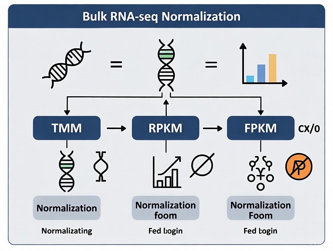
Bulk RNA-seq Normalization Methods Decoded: A Practical Guide to TMM, RPKM, FPKM, and Beyond for Reliable Gene Expression Analysis
This article provides a comprehensive guide for researchers and drug development professionals on navigating the critical step of normalization in bulk RNA-seq data analysis.
Bulk RNA-seq Normalization Methods Decoded: A Practical Guide to TMM, RPKM, FPKM, and Beyond for Reliable Gene Expression Analysis
Abstract
This article provides a comprehensive guide for researchers and drug development professionals on navigating the critical step of normalization in bulk RNA-seq data analysis. We cover the foundational principles explaining why normalization is non-negotiable, detail the methodologies and calculations behind popular methods like TMM, RPKM, FPKM, TPM, and DESeq2, and offer troubleshooting for common pitfalls. By synthesizing recent benchmarking studies and validation protocols, this guide empowers scientists to select the optimal normalization strategy to ensure their downstream analyses, from differential expression to metabolic modeling, are biologically accurate and technically sound.
Why Normalize? Uncovering the Critical Sources of Technical Bias in RNA-seq Data
RNA sequencing (RNA-seq) has become the cornerstone of modern transcriptomics, enabling genome-wide quantification of RNA abundance. However, the raw count data generated by sequencing platforms are not directly comparable due to multiple technical variables. Normalization is the critical mathematical adjustment of this raw data to account for technical biases, ensuring that observed differences reflect true biological variation rather than experimental artifacts. Without appropriate normalization, even well-designed studies risk generating misleading results, incorrect biological interpretations, and wasted resources. This application note details why normalization is indispensable and provides practical guidance for selecting and implementing appropriate normalization strategies in bulk RNA-seq analysis.
Why Normalization is Non-Negotiable: The Problem with Raw Counts
Raw RNA-seq counts are inherently biased by multiple technical factors that must be corrected before biological interpretation:
- Sequencing depth: Samples with more total reads will have higher counts, regardless of actual expression levels.
- Gene length: Longer genes accumulate more reads than shorter genes at identical expression levels.
- Library composition: Highly expressed genes in one sample can consume sequencing resources, skewing the representation of other genes.
- Technical variability: Batch effects, RNA quality, and library preparation protocols introduce non-biological variance.
The fundamental goal of normalization is to eliminate these technical confounders while preserving genuine biological signals, enabling accurate within-sample and between-sample comparisons [1] [2].
A Landscape of Normalization Methods
RNA-seq normalization methods can be categorized by their approach and application context. The table below summarizes the most common methods and their characteristics:
Table 1: Common RNA-Seq Normalization Methods and Their Applications
| Method | Category | Corrects For | Best Use Cases | Key Considerations |
|---|---|---|---|---|
| CPM (Counts Per Million) | Within-sample | Sequencing depth | Data visualization; not for DE | Does not correct for gene length or composition [2] |
| FPKM/RPKM | Within-sample | Sequencing depth + gene length | Gene expression within a single sample | Not ideal for cross-sample comparison due to variable totals between samples [3] [4] |
| TPM (Transcripts Per Million) | Within-sample | Sequencing depth + gene length | Within-sample comparison; some cross-sample applications | Sum of TPMs constant across samples; preferred over FPKM for proportion-based analysis [3] [2] |
| TMM (Trimmed Mean of M-values) | Between-sample | Sequencing depth + RNA composition | Differential expression analysis | Assumes most genes not differentially expressed; robust to skewed expression [5] [2] |
| RLE (Relative Log Expression) | Between-sample | Sequencing depth | Differential expression analysis | Used in DESeq2; similar assumptions to TMM [5] |
| Quantile | Between-sample | Distribution shape | Making distributions identical across samples | Assumes same expression distribution across samples; may remove biological signal in unbalanced data [6] [7] |
These methods are typically applied at different stages of analysis, from within-sample adjustments to cross-dataset integration:
Experimental Evidence: How Normalization Impacts Biological Interpretation
Case Study 1: Normalization Effects on Metabolic Model Reconstruction
A comprehensive 2024 benchmark study evaluated how normalization choices affect the reconstruction of condition-specific genome-scale metabolic models (GEMs) using iMAT and INIT algorithms. The research compared five normalization methods (TPM, FPKM, TMM, GeTMM, and RLE) with Alzheimer's disease and lung adenocarcinoma datasets [5].
Table 2: Performance of Normalization Methods in Metabolic Model Reconstruction
| Normalization Method | Model Variability | Disease Gene Accuracy (AD) | Disease Gene Accuracy (LUAD) | Effect of Covariate Adjustment |
|---|---|---|---|---|
| TPM/FPKM (within-sample) | High variability in active reactions | Lower accuracy | Lower accuracy | Moderate improvement |
| RLE/TMM/GeTMM (between-sample) | Low variability in active reactions | ~0.80 accuracy | ~0.67 accuracy | Significant improvement |
| Key Finding: Between-sample methods reduced false positives at the expense of missing some true positives when mapping to GEMs. |
The study demonstrated that between-sample normalization methods (RLE, TMM, GeTMM) produced models with significantly lower variability and higher accuracy in capturing disease-associated genes compared to within-sample methods (TPM, FPKM). Furthermore, adjusting for covariates like age and gender improved accuracy across all methods [5].
Case Study 2: Reproducibility in Patient-Derived Xenograft Models
A 2021 study compared quantification measures using replicate samples from 20 patient-derived xenograft (PDX) models. The research evaluated which method minimized technical variation between replicates while preserving biological signals [4].
The findings revealed that normalized count data (using between-sample methods like TMM or RLE) outperformed TPM and FPKM in several key metrics:
- Superior clustering of replicate samples from the same PDX model
- Lowest median coefficient of variation across replicates
- Highest intraclass correlation coefficient values
This evidence demonstrates that methods designed for between-sample comparison provide more reliable results for differential expression analysis [4].
Decision Framework: Selecting the Right Normalization Strategy
Protocol: Choosing an Appropriate Normalization Method
Define your research question and comparison type
- For within-sample gene expression comparison (e.g., identifying the most highly expressed genes in a sample): Use TPM or FPKM
- For between-sample comparison (e.g., differential expression between conditions): Use between-sample methods (TMM, RLE)
Assess your dataset characteristics
- Check for global shifts in transcript distribution using PCA before normalization
- Evaluate whether basic assumptions of normalization methods are met:
- TMM/RLE: Most genes not differentially expressed
- Quantile: Similar expression distribution across samples
Address covariates and batch effects
- Identify known technical batches (sequencing date, library preparation)
- Consider biological covariates (age, sex, cell type composition)
- Apply covariate adjustment after initial normalization when needed [5]
Validate your normalization choice
- Examine reduction in technical variation between replicates
- Verify preservation of expected biological signals
- Assess whether results align with orthogonal validation methods (e.g., qPCR)
The Scientist's Toolkit: Essential Research Reagent Solutions
Table 3: Key Reagents and Tools for RNA-Seq Normalization
| Reagent/Tool | Function | Considerations |
|---|---|---|
| RNA Stabilization Reagents (e.g., PAXgene) | Preserve RNA integrity during sample collection | Critical for maintaining RIN >7 for polyA-selection protocols [8] |
| rRNA Depletion Kits | Remove abundant ribosomal RNA | Increases sequencing depth for non-rRNA targets; assess off-target effects on genes of interest [8] |
| Stranded Library Prep Kits | Preserve transcript orientation | Essential for identifying overlapping transcripts and accurate isoform quantification [8] |
| Spike-in Controls | Monitor technical variation | External RNA controls added before library prep for normalization quality assessment [7] |
| Quality Control Tools (FastQC, MultiQC) | Assess RNA and library quality | Identify adapter contamination, sequencing biases, and RNA degradation [1] |
Advanced Considerations and Future Directions
Normalization Challenges with Unbalanced Data
Traditional normalization methods assume symmetric distribution of differentially expressed genes and that most genes are not differentially expressed. These assumptions fail in specific biological contexts:
- Different tissues or developmental stages with varying total RNA content
- Cancer vs. normal cells with global shifts in transcription
- Small targeted arrays (e.g., miRNA panels) with limited invariant genes
For these unbalanced scenarios, specialized methods have been developed that use data-driven invariant gene sets or external controls as reference [7].
The Emerging Impact of Transcriptome Size Variation
Recent research has highlighted that transcriptome size—the total number of RNA molecules per cell—varies significantly across cell types and conditions. A 2025 study revealed that conventional single-cell RNA-seq normalization methods that force equal transcriptome sizes across cells can distort biological signals and impair deconvolution of bulk RNA-seq data [9]. While this study focused on single-cell applications, it underscores the importance of considering transcriptome size variations in bulk analyses, particularly when comparing tissues with fundamentally different RNA content.
Normalization is not merely a computational convenience but an essential prerequisite for biologically meaningful RNA-seq analysis. The choice of normalization method directly impacts which genes are identified as differentially expressed, how samples cluster in dimensional space, and ultimately what biological conclusions are drawn from the data. Between-sample normalization methods like TMM and RLE generally provide more robust performance for differential expression analysis, while within-sample methods like TPM are suitable for assessing relative transcript abundance within individual samples. As transcriptomic technologies evolve and applications diversify, understanding the principles and practical implications of normalization will remain fundamental to extracting accurate biological insights from RNA-seq data.
The fine detail provided by sequencing-based transcriptome surveys suggests that RNA-seq is likely to become the platform of choice for interrogating steady-state RNA. However, contrary to early expectations that RNA-seq would not require sophisticated normalization, the reality is that normalization continues to be an essential step in the analysis to discover biologically important changes in expression [10]. Technical variability in RNA-seq data arises from multiple sources throughout the experimental process, and this variability can substantially obscure biological signals if not properly addressed [11].
The fundamental challenge stems from the fact that RNA-seq is a digital sampling process that captures only a tiny fraction (approximately 0.0013%) of the available molecules in a typical library [12]. This low sampling fraction means that technical variability can lead to substantial disagreements between replicates, particularly for features with low coverage [12]. Furthermore, the counting efficiency of different transcripts varies due to sequence-specific biases, with GC-content having a particularly strong sample-specific effect on gene expression measurements [11].
This Application Note examines three primary sources of technical variability—sequencing depth, gene length, and RNA composition—and provides detailed protocols for normalization methods that address these challenges in the context of bulk RNA-seq experiments.
Sequencing Depth and Sampling Limitations
Sequencing depth refers to the number of reads generated per sample, which directly impacts the power to detect transcripts, especially those expressed at low levels. In practice, technical variability is too high to ignore, with exon detection between technical replicates being highly variable when coverage is less than 5 reads per nucleotide [12].
The relationship between sequencing depth and transcript recovery follows a pattern of diminishing returns. For de novo assembly, the amount of exonic sequence assembled typically plateaus using datasets of approximately 2 to 8 Gbp, while genomic sequence may continue to increase with additional sequencing, primarily due to the recovery of single-exon transcripts not documented in genome annotations [13].
Table 1: Impact of Sequencing Depth on Key Analysis Outcomes
| Sequencing Depth | Exon Detection Consistency | TCRαβ Reconstruction Success | Transcript Assembly Completeness |
|---|---|---|---|
| < 0.25 million PE reads | Highly variable | < 1% of cells | Limited to highly expressed genes |
| 0.25-1 million PE reads | Improved for moderate-high expression | >80% success rate | Recovery of most annotated exons |
| 1-5 million PE reads | Good for most exons | >95% success rate | Plateau of annotated exonic regions |
| >5 million PE reads | High consistency | ~100% success rate | Increasing single-exon, unannotated transcripts |
The sampling limitations of RNA-seq technology mean that even with millions of reads, we are surveying only a tiny fraction of the RNA molecules present in a sample. This sampling artifact can force differential expression analysis to be skewed toward one experimental condition if not properly adjusted [10].
Gene Length Bias
The number of reads mapping to a gene is not only dependent on its expression level but also on its transcript length. Longer genes generate more fragments than shorter genes with similar expression levels, creating a fundamental bias in raw count data [10] [14]. This bias prevents direct comparison of expression levels between genes within the same sample without appropriate normalization.
Gene length bias is particularly problematic when trying to identify changes in the expression of shorter transcripts, such as non-coding RNAs or specific isoforms, as the length effect can mask true biological differences. Normalization methods like RPKM, FPKM, and TPM were specifically designed to account for this bias by normalizing counts by transcript length [3] [14].
RNA Composition Effects
One of the most counterintuitive yet critical sources of technical variability arises from RNA composition differences between samples. The proportion of reads attributed to a given gene depends not only on that gene's expression level but also on the expression properties of the entire sample [10].
A hypothetical scenario illustrates this effect: if sample A contains twice as many expressed genes as sample B, but both are sequenced to the same depth, a gene expressed in both samples will have approximately half the number of reads in sample A because the reads are distributed across more genes [10]. This composition effect means that library size scaling alone is insufficient for normalization when comparing samples with different RNA repertoires.
In real biological contexts, composition effects occur when one condition has a set of highly expressed genes that "use up" sequencing capacity, thereby proportionally reducing the counts for all other genes in that sample. This can lead to false positives in differential expression analysis if not properly corrected [10].
Figure 1: Sources of Technical Variability in RNA-seq Experiments. Multiple technical factors introduce biases that can obscure biological signals if not properly addressed during normalization.
Normalization Methods: Theoretical Foundations
RPKM/FPKM and TPM
RPKM (Reads Per Kilobase per Million mapped reads) and its paired-end counterpart FPKM (Fragments Per Kilobase per Million mapped fragments) were among the first normalization methods developed for RNA-seq data [3] [14]. These approaches normalize for both sequencing depth and gene length, allowing comparison of expression levels within a sample.
TPM (Transcripts Per Kilobase Million) represents an evolution of RPKM/FPKM with a different order of operations [3]. While RPKM/FPKM first normalizes for sequencing depth then gene length, TPM reverses this order: it first normalizes for gene length, then for sequencing depth. This seemingly minor difference has profound implications—TPM values sum to the same constant (1 million) across samples, making them directly comparable in terms of the proportion of transcripts each gene represents [3] [14].
Table 2: Comparison of RNA-seq Normalization Methods
| Normalization Method | Formula | Recommended Use | Limitations |
|---|---|---|---|
| RPKM/FPKM | Reads/(Gene length × Total reads) × 10^9 | Within-sample gene comparisons | Not comparable across samples with different RNA composition |
| TPM | (Reads/Gene length) / Σ(Reads/Gene length) × 10^6 | Within- and between-sample comparisons | Still affected by RNA composition differences |
| TMM | Uses weighted trimmed mean of log expression ratios | Differential expression between samples | Assumes most genes are not differentially expressed |
The calculation of these metrics follows specific workflows:
Figure 2: RPKM/FPKM vs. TPM Calculation Workflows. Though mathematically similar, the different order of operations in TPM ensures that values sum to the same constant across samples, facilitating comparison.
TMM Normalization
The Trimmed Mean of M-values (TMM) method addresses a critical limitation of RPKM/FPKM and TPM: their susceptibility to RNA composition effects [10]. TMM operates on the assumption that most genes are not differentially expressed between samples, and uses this assumption to calculate scaling factors that account for composition biases.
The TMM algorithm selects a reference sample and calculates scaling factors for all other samples based on a weighted trimmed mean of the log expression ratios (M-values) [10] [15]. This approach robustly summarizes observed M values by trimming both the M values and the absolute expression levels (A values) before taking the weighted average, with precision weights accounting for the fact that log fold changes from genes with larger read counts have lower variance on the logarithm scale [10].
The resulting normalization factors can be incorporated into statistical models for differential expression analysis by adjusting the effective library sizes, thus accounting for the sampling properties of RNA-seq data [10].
Experimental Evidence of Technical Biases
Liver vs. Kidney Case Study
A publicly available dataset comparing technical replicates of liver and kidney RNA sources demonstrates the necessity of sophisticated normalization [10]. When standard normalization (accounting for total number of reads) was applied, the distribution of log ratios between liver and kidney samples was significantly offset toward higher expression in kidney, with housekeeping genes also showing a significant shift away from zero [10].
The explanation for this bias was straightforward: a prominent set of genes with higher expression in liver consumed substantial sequencing "real estate," leaving less capacity for sequencing the remaining genes in the liver sample. This proportionally distorted the expression values toward being kidney-specific [10]. Application of TMM normalization to this pair of samples corrected this composition bias, demonstrating the practical importance of appropriate normalization methods.
Protocol-Dependent Composition Effects
Different RNA extraction and library preparation protocols can dramatically alter the RNA repertoire that ultimately gets sequenced. A comparison of poly(A)+ selection and rRNA depletion protocols demonstrated strikingly different transcriptomic profiles from the same biological sample [16].
With poly(A)+ selection, the most abundant category was protein-coding genes, while rRNA depletion resulted in small RNAs being the most abundant category [16]. Consequently, TPM values from the different protocols were not directly comparable—in blood samples, the top three genes represented only 4.2% of transcripts in poly(A)+ selection but 75% of transcripts in rRNA depletion [16]. This massive shift in transcript distribution means that expression levels of many genes are artificially deflated in rRNA depletion samples relative to poly(A)+ selected samples.
Protocols for Effective Normalization
Best Practices for Differential Expression Analysis
A recommended workflow for bulk RNA-seq analysis involves multiple steps to ensure robust results:
Step 1: Read Alignment and Quantification Use a splice-aware aligner like STAR to align reads to the genome, facilitating generation of comprehensive QC metrics. Then use alignment-based quantification with tools like Salmon to handle uncertainty in read assignment and generate count matrices [17].
Step 2: Normalization Method Selection
- For differential expression analysis, incorporate TMM normalization within statistical frameworks like edgeR [15].
- For within-sample gene comparisons, use TPM values [14].
- Avoid using RPKM/FPKM for cross-sample comparisons, especially when RNA composition may differ [16].
Step 3: Accounting for Additional Biases Implement additional normalization steps to address sequence-specific biases like GC-content effects. Conditional quantile normalization has been shown to improve precision by 42% by removing systematic bias introduced by deterministic features such as GC-content [11].
Experimental Design Considerations
Sequencing Depth Guidelines Based on empirical studies, aim for at least 20-30 million reads per sample for standard differential expression analysis in bulk RNA-seq. For detection of low-abundance transcripts or complex applications like de novo assembly, 50-100 million reads may be necessary [13].
Replication Strategy Include both technical and biological replicates in experimental designs. Technical replicates help quantify and account for technical variability, while biological replicates are essential for drawing conclusions about biological conditions [12].
Protocol Consistency Maintain consistent library preparation protocols throughout an experiment. When comparing across datasets, be aware that different protocols (e.g., poly(A)+ selection vs. rRNA depletion) yield fundamentally different RNA repertoires that complicate direct comparison of normalized expression values [16].
Figure 3: RNA-seq Experimental Design and Analysis Workflow. Proper planning at each step minimizes technical variability and ensures biologically meaningful results.
Research Reagent Solutions
Table 3: Essential Reagents and Tools for RNA-seq Normalization Studies
| Reagent/Tool | Function | Application Notes |
|---|---|---|
| STAR Aligner | Spliced alignment of RNA-seq reads to genome | Provides comprehensive QC metrics; enables subsequent alignment-based quantification [17] |
| Salmon | Alignment-based or pseudoalignment quantification | Handles read assignment uncertainty; generates count matrices for downstream analysis [17] |
| EdgeR | Differential expression analysis package | Implements TMM normalization within statistical framework for DE testing [15] |
| Trim Galore! | Adapter and quality trimming | Essential preprocessing step; removes technical sequences that interfere with alignment [13] |
| Poly(A) Selection Beads | mRNA enrichment protocol | Isolates mature mRNAs; creates specific RNA repertoire biased toward protein-coding genes [16] |
| rRNA Depletion Kits | Removal of ribosomal RNA | Retains broader RNA classes including immature transcripts; alters TPM distribution [16] |
| Universal Human Reference RNA (UHRR) | Standard reference material | Enables assessment of technical variability and normalization accuracy across experiments [11] |
Technical variability in RNA-seq data arising from sequencing depth, gene length, and RNA composition effects presents significant challenges that must be addressed through appropriate normalization methods. While RPKM/FPKM and TPM provide solutions for within-sample comparisons and length biases, they remain susceptible to composition effects that can skew differential expression analysis between samples with different RNA repertoires.
The TMM normalization method offers a robust solution for composition biases by leveraging the assumption that most genes are not differentially expressed, while additional considerations like GC-content normalization can further improve precision. Researchers should select normalization approaches based on their specific experimental context and comparison goals, always mindful that even "normalized" values like TPM may not be directly comparable across different library preparation protocols.
By understanding these sources of technical variability and implementing appropriate normalization strategies, researchers can ensure that their RNA-seq analyses reveal true biological signals rather than technical artifacts.
RNA sequencing (RNA-seq) has become the most popular method for transcriptome profiling, replacing gene expression microarrays in many applications [4]. The analysis of RNA-seq data, however, presents unique challenges due to technical variations introduced during library preparation, sequencing, and data processing. Normalization serves as a critical computational procedure to remove these non-biological variations, ensuring that differences in gene expression measurements reflect true biological signals rather than technical artifacts [2].
The complexity of RNA-seq normalization necessitates a structured approach across multiple levels of analysis. This framework organizes normalization into three distinct stages: within-sample correction (accounting for gene length and sequencing depth), between-sample normalization (enabling comparative analysis across specimens), and cross-dataset harmonization (addressing batch effects when integrating multiple studies) [2]. Understanding this hierarchical structure is essential for researchers, scientists, and drug development professionals working with transcriptomic data, as improper normalization can lead to false conclusions in downstream analyses such as differential expression testing and biomarker discovery [18].
Within the context of bulk RNA-seq analysis, this framework provides a systematic approach to selecting appropriate normalization methods—including TMM, RPKM, FPKM, and others—based on the specific analytical goals and experimental design. The following sections detail each stage, providing practical guidance for implementation and highlighting applications in pharmaceutical and clinical research settings.
The Three Stages of RNA-seq Normalization
Stage 1: Within-Sample Normalization
Within-sample normalization addresses technical factors that affect gene expression measurements within individual samples, primarily sequencing depth and gene length [2]. Sequencing depth refers to the total number of reads generated per sample, which varies between experiments due to technical rather than biological reasons. Gene length bias arises because longer transcripts typically yield more sequencing fragments than shorter transcripts at the same expression level [2]. Without correction, these factors prevent accurate comparison of expression levels between different genes within the same sample.
The most common within-sample normalization methods include:
- CPM (Counts Per Million): Normalizes for sequencing depth only by scaling raw counts by the total number of reads multiplied by one million [2]. Suitable for comparing expression of the same gene across samples but not for comparing different genes within a sample.
- RPKM (Reads Per Kilobase Million) and FPKM (Fragments Per Kilobase Million): Adjust for both sequencing depth and gene length. RPKM is designed for single-end experiments, while FPKM is used for paired-end RNA-seq where two reads can correspond to a single fragment [4] [3] [2].
- TPM (Transcripts Per Million): Similar to RPKM/FPKM but employs a different order of operations—normalizing for gene length first, then sequencing depth [3]. This approach ensures that the sum of all TPM values in each sample is constant, facilitating comparison of the proportion of reads mapped to a gene across samples [3].
The calculation methods for these normalization approaches are as follows:
| Method | Formula | Application Context |
|---|---|---|
| CPM | ( \text{CPM} = \frac{\text{Reads mapped to gene}}{\text{Total reads}} \times 10^6 ) | Sequencing depth normalization only |
| RPKM/FPKM | ( \text{RPKM/FPKM} = \frac{\text{Reads/Fragments mapped to gene}}{\text{Gene length (kb)} \times \text{Total reads (million)}} ) | Within-sample gene expression comparison |
| TPM | ( \text{TPM}i = \frac{\frac{ri}{li} \times 10^6}{\sumj \frac{rj}{lj}} ) where (ri): reads mapped to gene i, (li): length of gene i | Within-sample comparison with consistent sample sums |
Table 1: Within-sample normalization methods and their applications
While these within-sample normalized values are suitable for comparing gene expression within a single sample, they are insufficient for cross-sample comparisons due to additional technical variations between samples [2]. Specifically, RPKM and FPKM have been shown to introduce biases in cross-sample comparisons because the sum of normalized reads can differ between samples, making it difficult to determine if the same proportion of reads mapped to a gene across samples [3].
Stage 2: Between-Sample Normalization
Between-sample normalization enables meaningful comparisons of gene expression across different biological specimens by accounting for technical variability introduced during sample processing and sequencing. This stage is critical for downstream analyses such as differential expression testing, where the goal is to identify biologically relevant changes rather than technical artifacts [18].
The fundamental challenge in between-sample normalization stems from the compositionality of RNA-seq data—changes in the expression of a few highly expressed genes can create the false appearance of differential expression for other genes because the total number of reads is fixed [18]. Between-sample normalization methods address this by assuming that most genes are not differentially expressed or that the expression of certain control genes remains constant across conditions.
Figure 1: Between-sample normalization methods and their underlying assumptions
Key between-sample normalization methods include:
- TMM (Trimmed Mean of M-values): Implemented in the edgeR package, TMM assumes most genes are not differentially expressed [2] [5]. It calculates scaling factors between samples by comparing each sample to a reference after trimming extreme log-fold changes and absolute expression levels [2].
- RLE (Relative Log Expression): Used by DESeq2, this method similarly assumes that most genes are non-DE and calculates size factors as the median of the ratios of each gene's count to its geometric mean across all samples [5].
- Quantile Normalization: Makes the distribution of gene expression values identical for each sample by replacing values with the average across samples for genes of the same rank [2].
Experimental evidence demonstrates that between-sample normalization methods like TMM and RLE outperform within-sample methods for cross-sample comparisons. A comparative study on patient-derived xenograft models revealed that normalized count data (using between-sample methods) showed lower median coefficient of variation and higher intraclass correlation values across replicate samples compared to TPM and FPKM [4]. Similarly, a benchmark study on metabolic network mapping showed that RLE, TMM, and GeTMM (a gene length-corrected version of TMM) produced more consistent results with lower variability compared to within-sample methods like TPM and FPKM [5].
Stage 3: Cross-Dataset Normalization
Cross-dataset normalization, often referred to as batch correction, addresses technical variations introduced when integrating RNA-seq data from multiple independent studies, sequencing platforms, or experimental batches. These batch effects can constitute the greatest source of variation in combined datasets, potentially masking true biological signals and leading to incorrect conclusions if not properly addressed [2].
The need for cross-dataset normalization has grown with the increasing practice of meta-analyses that combine data from public repositories to increase statistical power or validate findings across diverse populations. Batch effects arise from differences in sample preparation protocols, sequencing technologies, laboratory conditions, personnel, and reagent batches across experiments conducted at different times or facilities [2].
Figure 2: Cross-dataset normalization approaches for batch effect correction
Common cross-dataset normalization approaches include:
- Limma and ComBat: These methods remove batch effects when the sources of variation (e.g., sequencing date, facility) are known [2]. Both use empirical Bayes statistics to estimate prior probability distributions from the data, effectively "borrowing" information across genes in each batch to make robust adjustments even with small sample sizes.
- Surrogate Variable Analysis (SVA): Identifies and estimates unknown sources of variation using singular value decomposition or factor analysis after initially correcting for known batch effects [2].
- Training Distribution Matching (TDM): Specifically designed for cross-platform normalization, TDM transforms RNA-seq data to make it compatible with machine learning models trained on microarray data, enabling the creation of larger, more diverse training datasets [19].
A critical consideration in cross-dataset normalization is that between-sample normalization should typically be applied before batch correction to ensure gene expression values are on the same scale across samples [2]. Additionally, covariates such as age, gender, and post-mortem interval (for specific tissues) should be considered, as they can significantly impact results in disease studies [5].
Experimental Protocols and Applications
Protocol: Comparative Analysis of Normalization Methods
This protocol outlines a systematic approach for evaluating different normalization methods using replicate samples, based on experimental designs from published studies [4] [5].
Materials and Reagents
- RNA-seq datasets with biological replicates (minimum 3 replicates per condition)
- Computing environment with R/Bioconductor
- Normalization software: DESeq2 (for RLE), edgeR (for TMM), and standard tools for TPM/FPKM calculation
Procedure
- Data Acquisition: Obtain RNA-seq data from public repositories (e.g., NCI PDMR, TCGA) or generate new data. Ensure datasets include biological replicates for assessing technical variability.
- Quantification: Generate raw count data using alignment-based (e.g., STAR, HTSeq) or alignment-free (e.g., Salmon, kallisto) methods.
- Normalization Implementation:
- Apply within-sample methods (TPM, FPKM) using standard formulas
- Implement between-sample methods (TMM, RLE) using DESeq2 and edgeR packages
- Perform cross-dataset normalization using limma or ComBat when integrating multiple datasets
- Evaluation Metrics:
- Calculate coefficient of variation (CV) between replicates
- Compute intraclass correlation coefficient (ICC) for the same gene across replicates
- Perform hierarchical clustering to assess replicate concordance
- Evaluate false positive rates in differential expression analysis
Expected Results
Based on comparative studies, between-sample normalization methods (TMM, RLE) should demonstrate lower median CV and higher ICC values compared to within-sample methods (TPM, FPKM) [4]. Hierarchical clustering should group replicate samples from the same biological origin more accurately with between-sample normalization methods.
Protocol: Normalization for Metabolic Network Mapping
This specialized protocol describes RNA-seq normalization for constructing condition-specific genome-scale metabolic models (GEMs), particularly relevant for drug development applications [5].
Materials and Reagents
- RNA-seq data from disease and control samples (e.g., Alzheimer's disease, cancer)
- Human metabolic network reconstruction (e.g., Recon3D)
- iMAT or INIT algorithm for metabolic model construction
- Covariate information (age, gender, clinical variables)
Procedure
- Data Preprocessing: Obtain RNA-seq counts for metabolic genes in the reconstruction model.
- Normalization Application:
- Apply five different normalization methods (TPM, FPKM, TMM, GeTMM, RLE) to the count data
- Generate covariate-adjusted versions using linear models
- Model Construction: Map normalized expression values to the metabolic network using iMAT or INIT algorithms to generate personalized metabolic models.
- Validation:
- Compare the number of active reactions in generated models
- Assess variability across samples for each normalization method
- Evaluate accuracy in capturing disease-associated genes using known biomarkers
Expected Results
Between-sample normalization methods (RLE, TMM, GeTMM) should produce metabolic models with lower variability in active reactions compared to within-sample methods (TPM, FPKM) [5]. Covariate adjustment should further improve accuracy, particularly for diseases with strong age or gender associations.
Successful implementation of RNA-seq normalization requires both computational tools and methodological knowledge. The following table summarizes key resources for researchers conducting normalization analyses.
| Resource Type | Specific Tools/Methods | Application Context | Key Considerations |
|---|---|---|---|
| Within-Sample Methods | TPM, FPKM, RPKM | Single-sample analysis, gene expression comparison within sample | TPM provides consistent sample sums; FPKM/RPKM suitable for single-sample comparisons only [3] [2] |
| Between-Sample Methods | TMM (edgeR), RLE (DESeq2), Quantile | Differential expression, cross-sample comparisons | Assume most genes not differentially expressed; robust to composition effects [18] [2] [5] |
| Cross-Dataset Methods | ComBat, limma, SVA, TDM | Data integration, meta-analysis, machine learning | Require prior between-sample normalization; TDM enables cross-platform compatibility [2] [19] |
| Experimental Design | Biological replicates, spike-in controls | All normalization stages | Replicates essential for evaluating normalization performance; spike-ins assess technical variability [4] [18] |
| Implementation Platforms | R/Bioconductor (DESeq2, edgeR, limma), Python | Practical application | DESeq2 and edgeR provide integrated normalization and DE analysis; limma extends to batch correction [2] [5] |
Table 2: Essential resources for implementing RNA-seq normalization across the three stages
The three-stage framework for RNA-seq normalization provides a systematic approach to addressing technical variations at different levels of analysis. Within-sample normalization enables accurate gene expression comparisons within individual specimens, between-sample methods facilitate robust cross-sample analyses, and cross-dataset normalization allows integration of diverse studies while minimizing batch effects.
Current evidence indicates that between-sample normalization methods like TMM and RLE generally outperform within-sample methods for comparative analyses, demonstrating lower variability and better replication concordance [4] [5]. The selection of appropriate normalization methods, however, remains context-dependent, requiring consideration of experimental design, biological questions, and data characteristics. As RNA-seq applications continue to evolve in pharmaceutical and clinical research, adherence to this structured normalization framework will enhance the reliability and reproducibility of transcriptomic findings.
In bulk RNA-seq analysis, accurate gene expression quantification hinges on a clear understanding of the fundamental units of measurement: reads, fragments, and transcripts. These units form the basis of all downstream normalization and differential expression analyses. A read represents the raw sequence data output, a fragment corresponds to the original RNA molecule segment, and a transcript reflects the biological entity of interest. Confusion between these units can lead to improper normalization and incorrect biological interpretations [3] [16].
The relationship between these units is critical for selecting appropriate normalization methods such as TMM, RPKM, FPKM, and TPM. These methods account for technical variations including sequencing depth, gene length, and RNA population composition to enable valid comparisons within and between samples [10] [2]. This document provides a comprehensive framework for understanding these core quantification units and their practical applications in bulk RNA-seq normalization.
Defining the Fundamental Units
Reads
In sequencing, a read is the string of base calls (A, T, C, G) derived from a single DNA (or RNA-derived) fragment, representing the sequencer's attempt to "read" that fragment's nucleotides [20]. Reads serve as the fundamental raw data unit in RNA-seq, with their quality and length significantly impacting downstream analyses.
- Read Length: The number of nucleotides sequenced from a fragment, fixed by sequencing chemistry and instrument configuration [20]
- Configuration: Single-end sequencing reads one fragment end, while paired-end sequences both ends, providing better mapping resolution and structural variant detection [20]
- Impact on Quantification: Longer reads (100-250 bp) improve transcript isoform detection and mapping across exon junctions [20]
Fragments
A fragment refers to the original segment of RNA (or cDNA) molecule from which reads are generated [3]. This distinction becomes crucial in paired-end sequencing protocols.
- Relationship to Reads: In paired-end sequencing, two reads can correspond to a single fragment, though sometimes one read may represent a fragment if its pair fails to map [3]
- Quantification Impact: Counting fragments rather than reads prevents double-counting when both ends of a fragment successfully map to the same transcript
- Protocol Dependency: Fragment length distribution varies experimentally and must be considered in abundance estimation [21]
Transcripts
A transcript represents the biological RNA molecule of interest, serving as the ultimate target of quantification efforts. Transcript abundance reflects genuine biological expression levels rather than technical measurements [21].
- Biological Significance: Transcript concentration represents the actual number of RNA molecules per cell or unit volume
- Quantification Challenge: Inference from reads/fragments is indirect and requires normalization for technical factors
- Effective Length: The number of positions where a mapped read could start, calculated as transcript length minus read length plus one [21]
Table 1: Key Characteristics of RNA-seq Quantification Units
| Unit | Definition | Primary Use | Technical Dependencies |
|---|---|---|---|
| Read | String of base calls from sequencing | Raw data input | Sequencing depth, base quality |
| Fragment | Original RNA/cDNA segment | Paired-end quantification | Library preparation, fragmentation |
| Transcript | Biological RNA molecule | Expression analysis | Gene length, isoform complexity |
Mathematical Relationships and Normalization Methods
From Reads to Expression Values
The transformation of raw read counts to comparable expression values follows a logical progression with distinct normalization steps. The following workflow illustrates this transformation process from raw sequencing data to normalized expression values:
Normalization Methods Framework
RPKM and FPKM
RPKM (Reads Per Kilobase per Million mapped reads) and FPKM (Fragments Per Kilobase per Million mapped fragments) represent closely related normalization approaches that account for both sequencing depth and gene length [22] [16].
RPKM Calculation:
- Divide read counts by total reads (in millions) for sequencing depth normalization
- Divide result by transcript length in kilobases [3]
FPKM Calculation:
- Identical to RPKM but uses fragments instead of reads to accommodate paired-end data [3]
Limitations: Both RPKM and FPKM produce values with sample-specific sums, complicating direct comparison between samples as the same RPKM/FPKM value may represent different proportional abundances in different samples [3] [16].
TPM
TPM (Transcripts Per Million) addresses a key limitation of RPKM/FPKM by reversing the normalization order [3].
TPM Calculation:
- Divide read counts by transcript length in kilobases (yielding Reads Per Kilobase)
- Sum all RPK values and divide by 1,000,000 for a "per million" scaling factor
- Divide RPK values by this scaling factor [3]
Advantage: TPM values sum to the same constant (1,000,000) across samples, enabling direct comparison of a transcript's proportional abundance between samples [3].
TMM
The TMM (Trimmed Mean of M-values) method operates on raw counts and focuses specifically on between-sample normalization for differential expression analysis [10].
TMM Calculation:
- Select a reference sample and compute log fold-changes (M-values) and absolute expression levels (A-values) for all genes
- Trim extreme M-values and A-values (typically 30% and 5%, respectively)
- Calculate weighted mean of remaining M-values to obtain scaling factor [10]
Application: TMM factors are incorporated into statistical models for differential expression testing by adjusting effective library sizes, addressing composition biases where highly expressed genes in one condition reduce available sequencing "real estate" for other genes [10].
Table 2: Comparison of RNA-seq Normalization Methods
| Method | Normalization For | Primary Application | Key Advantage | Key Limitation |
|---|---|---|---|---|
| RPKM/FPKM | Gene length, Sequencing depth | Within-sample comparison | Intuitive interpretation | Sample-specific sums limit between-sample comparison |
| TPM | Gene length, Sequencing depth | Within- and between-sample comparison | Constant sum across samples | Still requires between-sample normalization for DE analysis |
| TMM | Library composition, Sequencing depth | Between-sample differential expression | Robust to differentially expressed genes | Requires raw counts, not applicable to already normalized data |
Experimental Protocols for Quantification
RNA-seq Library Preparation and Sequencing
Proper experimental design is crucial for accurate quantification. The following protocol outlines key steps:
Sample Preparation:
- RNA Isolation: Purify RNA using appropriate methods for sample type (blood, tissue, cell lines)
- RNA Selection: Employ either poly(A)+ selection (enriching mature mRNAs) or rRNA depletion (capturing both mature and immature transcripts) [16]
- Fragmentation: Fragment RNA to 200-500 bp fragments compatible with sequencing platforms
- cDNA Synthesis: Reverse transcribe to cDNA with adaptor ligation
Sequencing Considerations:
- Use paired-end sequencing (e.g., 2×100 bp or 2×150 bp) for improved mapping accuracy and isoform detection [17]
- Sequence all samples to comparable depth (e.g., 20-30 million reads per sample for standard differential expression studies)
- Include technical replicates to assess variability and biological replicates to capture population variation
Read Processing and Quantification Pipeline
The nf-core RNA-seq workflow provides a robust framework for processing raw sequencing data into quantification values [17]:
Input Requirements:
- Paired-end FASTQ files for all samples
- Reference genome FASTA file and annotation GTF file
- Sample sheet specifying sample IDs, file paths, and strandedness
Processing Steps:
- Quality Control: Assess read quality using FastQC
- Read Alignment: Map reads to reference genome using STAR, a splice-aware aligner [17]
- Transcriptome Projection: Convert genomic alignments to transcriptomic coordinates
- Quantification: Generate counts using Salmon in alignment-based mode to model uncertainty in read assignment [17]
Output:
- Gene-level and transcript-level count matrices
- Comprehensive quality control metrics
The Scientist's Toolkit: Essential Research Reagents and Materials
Table 3: Essential Research Reagents and Computational Tools for RNA-seq Quantification
| Category | Item | Function | Example Products/Tools |
|---|---|---|---|
| Wet Lab Reagents | Poly(A) Selection Beads | Enrichment of mRNA from total RNA | Oligo(dT) magnetic beads |
| rRNA Depletion Kits | Removal of ribosomal RNA | Ribozero, NEBNext rRNA Depletion | |
| Library Prep Kits | cDNA synthesis, adaptor ligation | Illumina TruSeq, NEBNext Ultra II | |
| Computational Tools | Read Alignment | Mapping reads to reference genome | STAR, HISAT2, Bowtie2 |
| Quantification | Estimating transcript abundances | Salmon, Kallisto, RSEM | |
| Differential Expression | Statistical analysis of expression changes | edgeR, DESeq2, limma-voom | |
| Reference Data | Genome Sequence | Reference for read alignment | ENSEMBL, UCSC genome databases |
| Gene Annotations | Transcript structure definitions | ENSEMBL GTF, RefSeq, GENCODE |
Critical Considerations for Accurate Quantification
Technical Biases and Challenges
Several technical factors can compromise quantification accuracy if not properly addressed:
- Library Composition Bias: When a few genes dominate transcript abundance, they reduce sequencing coverage for other genes, skewing proportional comparisons [10]
- Protocol Selection Effects: Poly(A)+ selection and rRNA depletion produce fundamentally different RNA populations, making direct TPM comparison invalid between protocols [16]
- Gene Length Effects: Longer genes naturally accumulate more reads independent of actual expression level [22] [2]
- Multimapping Reads: Reads mapping to multiple genomic locations create ambiguity in assignment, requiring probabilistic resolution [21]
Normalization Method Selection Guidelines
Choice of normalization strategy should align with specific research questions:
- Within-Sample Comparisons: TPM provides the most interpretable measure of relative transcript abundance [3]
- Differential Expression Analysis: TMM normalization of raw counts followed by statistical testing with methods like edgeR or DESeq2 offers robust performance [10]
- Cross-Study Comparisons: Batch correction methods (ComBat, limma) must be applied after within-dataset normalization to address technical variability [2]
Validation and Quality Assessment
- Spike-In Controls: Implement external RNA controls of known concentration to monitor technical performance
- Housekeeping Genes: Monitor expression of stable reference genes across samples as internal quality check
- QC Metrics: Assess sequencing depth, mapping rates, and coverage uniformity before proceeding to differential expression analysis
A Deep Dive into Normalization Methods: From RPKM/FPKM to TMM and DESeq2
In bulk RNA sequencing (RNA-Seq) analysis, the initial digital read counts obtained from sequencing instruments cannot be directly compared without proper normalization. Raw read counts are influenced by two significant technical biases: sequencing depth (the total number of reads per sample) and gene length (the transcript length in kilobases). Without correction, longer genes will naturally have more reads mapped to them than shorter genes expressed at the same biological abundance, and samples with deeper sequencing will show higher counts across all genes independent of true expression levels. Within-sample normalization methods specifically address these biases to enable meaningful comparison of gene expression levels within a single sample [2] [14].
The most commonly used within-sample normalization methods are RPKM (Reads Per Kilobase Million), FPKM (Fragments Per Kilobase Million), and TPM (Transcripts Per Kilobase Million). While these methods share similar goals and mathematical foundations, they differ importantly in their calculation approaches and appropriate applications. Understanding these methods is crucial for proper interpretation of RNA-Seq data, as misapplication can lead to incorrect biological conclusions [3] [16]. These normalization approaches rescale expression values to account for technical variability, thereby revealing the biological signals of interest.
Core Normalization Methods: Calculations and Comparisons
RPKM and FPKM Normalization
RPKM (Reads Per Kilobase Million) was developed for single-end RNA-seq experiments, where each read corresponds to a single fragment. The calculation follows three sequential steps [3] [23]:
- Calculate per million scaling factor: Divide the total reads in the sample by 1,000,000
- Normalize for sequencing depth: Divide read counts by the scaling factor to get Reads Per Million (RPM)
- Normalize for gene length: Divide RPM values by gene length in kilobases to obtain RPKM
FPKM (Fragments Per Kilobase Million) is virtually identical to RPKM but designed for paired-end RNA-seq experiments. In paired-end sequencing, two reads can correspond to a single fragment, and FPKM ensures that these fragments are not double-counted [3] [14]. The distinction between reads and fragments represents the only practical difference between RPKM and FPKM. For single-end sequencing, RPKM and FPKM yield identical results [14].
The RPKM/FPKM calculation can be represented by the following formula:
$$RPKM{i} ~or~FPKM{i} = \frac{q{i}}{l{i} * \sum{j} q{j}} * 10^{9}$$
Where $q{i}$ represents raw read or fragment counts for gene i, $l{i}$ is feature length, and $\sum{j} q{j}$ corresponds to the total number of mapped reads/fragments [4].
TPM Normalization
TPM (Transcripts Per Kilobase Million) represents a modification of the RPKM/FPKM approach with a changed order of operations. While TPM accounts for the same technical variables (sequencing depth and gene length), it reverses the normalization sequence [3] [23]:
- Normalize for gene length first: Divide read counts by the length of each gene in kilobases, yielding Reads Per Kilobase (RPK)
- Calculate per million scaling factor: Sum all RPK values in a sample and divide by 1,000,000
- Normalize for sequencing depth: Divide RPK values by this scaling factor to obtain TPM
The TPM calculation formula is:
$$TPM = \frac{{q{i}/l{i}}}{{\sum{j} (q{j}/l_{j})}} * 10^{6}$$
Where $q{i}$ represents reads mapped to transcript i, $l{i}$ is transcript length, and the denominator sums the length-normalized counts across all transcripts [16].
This reversed order of operations has important implications. In TPM, the sum of all normalized values per sample is constant (approximately 1 million), making comparative interpretation more straightforward [3] [23]. As noted in StatQuest, "When you use TPM, the sum of all TPMs in each sample are the same. This makes it easier to compare the proportion of reads that mapped to a gene in each sample" [3].
Table 1: Comparison of Within-Sample Normalization Methods
| Method | Full Name | Sequencing Type | Order of Operations | Sum of Normalized Values | Primary Use Case |
|---|---|---|---|---|---|
| RPKM | Reads Per Kilobase Million | Single-end | Depth then Length | Variable across samples | Within-sample gene comparison [14] |
| FPKM | Fragments Per Kilobase Million | Paired-end | Depth then Length | Variable across samples | Within-sample gene comparison [14] |
| TPM | Transcripts Per Kilobase Million | Both | Length then Depth | Constant (1 million) | Within-sample comparison; preferred for cross-sample analysis [3] [14] |
Comparative Analysis of Normalization Performance
Theoretical and Practical Differences
The different calculation approaches lead to distinct practical properties. Because TPM normalization ends with the per-million scaling, the sum of all TPM values across a sample is constant (1 million). This provides a natural interpretation: a TPM value represents the number of transcripts you would expect to see for a gene if you sequenced exactly one million full-length transcripts [2] [16]. This property makes TPM values more intuitive for understanding relative transcript abundance within and between samples.
In contrast, RPKM and FPKM values sum to different totals across samples due to their calculation method. This means that identical RPKM/FPKM values for a gene in two different samples do not necessarily indicate the same proportional abundance [3] [23]. As one analysis explains, "With RPKM or FPKM, the sum of normalized reads in each sample can be different. Thus, if the RPKM for gene A in Sample 1 is 3.33 and the RPKM in Sample 2 is 3.33, I would not know if the same proportion of reads in Sample 1 mapped to gene A as in Sample 2" [3].
Experimental Comparisons in Biological Research
Empirical studies have compared the performance of these normalization methods in real research scenarios. A 2021 study published in Translational Medicine compared TPM, FPKM, and normalized counts using replicate samples from 20 patient-derived xenograft models spanning 15 tumor types [4]. The researchers evaluated reproducibility using coefficient of variation, intraclass correlation coefficient, and cluster analysis.
This research found that normalized count data (using between-sample methods) tended to group replicate samples from the same model together more accurately than TPM and FPKM data. Furthermore, normalized count data showed lower median coefficient of variation and higher intraclass correlation across replicates compared to TPM and FPKM [4]. These findings highlight that within-sample normalization methods alone may be insufficient for cross-sample comparisons in differential expression analysis.
A 2024 benchmark study further investigated how normalization methods affect downstream analysis when building condition-specific metabolic models [24]. The study compared five normalization methods (TPM, FPKM, TMM, GeTMM, and RLE) using Alzheimer's disease and lung adenocarcinoma datasets. The research found that within-sample normalization methods (TPM and FPKM) produced metabolic models with high variability across samples in terms of active reactions, while between-sample methods (TMM, RLE, GeTMM) showed considerably lower variability [24].
Table 2: Experimental Performance Comparison of Normalization Methods
| Performance Metric | RPKM/FPKM | TPM | Between-Sample Methods (TMM, RLE) |
|---|---|---|---|
| Within-sample comparison | Suitable [14] | Suitable [14] | Not primary purpose |
| Cross-sample comparison | Not recommended [14] [25] | Limited suitability [25] [16] | Recommended [4] [25] |
| Sum of normalized values | Variable [3] | Constant (~1 million) [3] | Variable |
| Effect on downstream analysis | High variability in metabolic models [24] | High variability in metabolic models [24] | Lower variability in metabolic models [24] |
| Reproducibility across replicates | Lower compared to normalized counts [4] | Lower compared to normalized counts [4] | Higher in replicate analysis [4] |
Experimental Protocols and Implementation
Workflow for Calculation and Analysis
The following workflow diagram illustrates the procedural steps for implementing within-sample normalization methods:
Detailed Protocol for TPM Calculation
For researchers implementing these calculations directly, here is the step-by-step protocol for TPM normalization, which is currently considered the most robust among the within-sample methods [3]:
Step 1: Normalize for gene length
- Input: Raw read counts for each gene
- For each gene, divide the read count by the transcript length in kilobases
- This yields Reads Per Kilobase (RPK) values
- Formula: $RPKi = \frac{\text{read count}i}{\text{transcript length in kb}}$
Step 2: Calculate sample-specific scaling factor
- Sum all RPK values in the sample
- Divide this sum by 1,000,000 to create the "per million" scaling factor
- Formula: $\text{Scaling factor} = \frac{\sum RPK_i}{1,000,000}$
Step 3: Normalize for sequencing depth
- Divide each RPK value by the scaling factor calculated in Step 2
- This yields the final TPM values
- Formula: $TPMi = \frac{RPKi}{\text{Scaling factor}}$
Validation check: The sum of all TPM values in the sample should equal approximately 1,000,000 [3] [23].
For RPKM/FPKM calculation, the protocol differs in the order of operations: normalize for sequencing depth first (RPM calculation), then normalize for gene length.
Table 3: Essential Computational Tools for RNA-Seq Normalization
| Tool/Resource | Function | Implementation |
|---|---|---|
| RSEM | Transcript quantification and TPM calculation [4] | Standalone software package |
| Kallisto | Pseudoalignment and TPM calculation [16] | R package or standalone |
| Salmon | Transcript quantification and TPM calculation [16] | R package or standalone |
| SAMtools | Processing alignment files [4] | Command-line tool |
| HTSeq | Generate raw read counts from BAM files [4] | Python package |
| DESeq2 | Between-sample normalization and differential expression [4] [24] | R/Bioconductor package |
| edgeR | Between-sample normalization and differential expression [24] | R/Bioconductor package |
Applications and Limitations in Research Contexts
Appropriate Use Cases
Within-sample normalization methods serve specific purposes in RNA-Seq analysis. RPKM, FPKM, and TPM are all appropriate for comparing expression levels of different genes within the same sample [2] [14]. For example, these methods can answer questions such as "Is Gene A more highly expressed than Gene B in this specific sample?" after accounting for differences in gene length and sequencing depth.
TPM has become the preferred within-sample metric because it enables more straightforward comparisons across samples. As noted in the literature, "TPM is a better unit for RNA abundance since it respects the invariance property and is proportional to the average relative molar concentration, and thus adopted by the latest computational algorithms for transcript quantification" [16]. When visualizing gene expression patterns in heatmaps or conducting exploratory analysis across limited sample sets, TPM values often provide a reasonable starting point.
Critical Limitations and Misapplications
A critical limitation of all within-sample normalization methods is that they do not account for library composition effects, which can create significant biases in cross-sample comparisons [4] [16]. These composition effects occur when a few highly expressed genes consume a substantial portion of the sequencing depth, thereby distorting the apparent expression of other genes in the sample [18].
As explicitly stated in current literature, "RPKM, FPKM, and TPM represent the relative abundance of a transcript among a population of sequenced transcripts, and therefore depend on the composition of the RNA population in a sample" [16]. This means that differences in transcript distribution between samples – such as those caused by different RNA extraction methods, library preparation protocols, or biological conditions – can make TPM values incomparable despite their normalization [16].
For differential expression analysis between conditions, specialized between-sample normalization methods like TMM (edgeR) or RLE (DESeq2) are generally recommended over direct comparison of TPM values [4] [25]. These methods specifically address composition biases that within-sample normalization cannot correct.
Within-sample normalization methods RPKM, FPKM, and TPM play essential roles in RNA-Seq data analysis by accounting for technical biases from gene length and sequencing depth. While RPKM and FPKM served as early standards, TPM has emerged as the preferred method due to its constant sum across samples, which facilitates more intuitive interpretation. However, researchers must recognize that these within-sample methods have limitations, particularly for cross-sample comparisons, where between-sample normalization approaches generally provide more robust results for differential expression analysis. Proper selection of normalization strategies remains fundamental to deriving accurate biological insights from RNA-Seq data.
The evolution of RNA-sequencing (RNA-seq) data analysis has witnessed a significant shift in normalization methodologies, moving from traditional measures like RPKM and FPKM towards Transcripts Per Million (TPM). This application note delineates the critical procedural distinction in TPM's calculation order—normalizing for gene length before sequencing depth—and its profound impact on the accuracy of transcriptomic studies. Framed within a comprehensive evaluation of bulk RNA-seq normalization methods, including TMM, RPKM, and FPKM, we provide structured quantitative comparisons, detailed experimental protocols, and essential reagent solutions. This resource is designed to equip researchers, scientists, and drug development professionals with the knowledge to implement TPM effectively, thereby enhancing the reliability of inter-sample comparisons and downstream analyses in translational research.
Normalization is a critical first step in bulk RNA-seq data analysis, correcting for technical variations to enable valid biological conclusions. Key technical biases include sequencing depth (the total number of reads per sample) and gene length (longer genes generate more reads at identical expression levels) [2]. Without correction, these factors mask true biological differences and lead to false discoveries.
The field has developed a spectrum of normalization methods, often categorized by their application scope:
- Within-sample normalization: Adjusts for gene length and sequencing depth to compare expression levels of different genes within the same sample. RPKM, FPKM, and TPM fall into this category [2].
- Between-sample normalization: Adjusts for differences in library composition and size to enable comparisons of the same gene across different samples. Methods like TMM (Trimmed Mean of M-values) and RLE (Relative Log Expression) are designed for this purpose [2] [5].
Understanding this distinction is vital for selecting the appropriate tool. While RPKM/FPKM and TPM are often grouped together, a fundamental difference in their calculation order places TPM in a superior position for specific applications, a nuance explored in this note.
The TPM Paradigm: A Critical Difference in Order of Operations
The Mathematical Foundation
TPM, RPKM, and FPKM all account for sequencing depth and gene length, but the order of operations in TPM calculation confers a distinct advantage [3] [14].
RPKM/FPKM Calculation Workflow:
- Normalize for sequencing depth: Divide the read counts for a gene by the total number of reads (in millions) in the sample, yielding Reads/Fragments Per Million (RPM).
- Normalize for gene length: Divide the RPM values by the gene length in kilobases to yield RPKM or FPKM [3].
TPM Calculation Workflow:
- Normalize for gene length: Divide the read counts for each gene by its length in kilobases, giving Reads Per Kilobase (RPK).
- Sum all RPK values in a sample and divide this number by 1,000,000 to obtain a sample-specific scaling factor.
- Normalize for sequencing depth: Divide each RPK value by this scaling factor to yield TPM [3].
The following diagram visualizes this critical difference in workflow:
The Profound Impact: Proportionality and Sample Comparison
The reversed order of operations has a profound practical consequence: in TPM, the sum of all TPM values in each sample is always equal to one million [3] [14].
This property makes TPM a proportional measure. If a gene has a TPM value of 500 in two different samples, it represents the same proportion of the total transcribed mRNA in each sample (500 / 1,000,000 = 0.05%) [3]. This is not guaranteed with RPKM/FPKM, where the sum of normalized counts can vary between samples, making it difficult to determine if the same proportion of reads mapped to a gene across samples [3]. Consequently, TPM is generally considered more accurate for comparing the relative abundance of a specific transcript across different samples [14].
Comparative Quantitative Analysis of Normalization Methods
The following table synthesizes findings from key benchmarking studies to provide a clear, structured comparison of popular normalization methods, highlighting their performance in various analytical contexts.
Table 1: Benchmarking of RNA-seq Normalization Methods Across Different Analytical Applications
| Normalization Method | Category | Key Assumption/Principle | Performance in Differential Expression (DE) Analysis | Performance in Metabolic Model Reconstruction (iMAT) [5] | Recommendation for Use |
|---|---|---|---|---|---|
| TPM | Within-sample | Sum of normalized counts is constant per sample. | Less suitable for DE analysis alone; outperformed by between-sample methods [4]. | High variability in model size and content; high number of predicted affected reactions [5]. | Within-sample comparisons; cross-sample comparisons when total RNA content is similar [14] [26]. |
| FPKM/RPKM | Within-sample | Normalizes for depth then length. | Not recommended for DE analysis; similar or inferior performance to TPM [4]. | Performance similar to TPM; high variability and high number of predicted affected reactions [5]. | Not recommended for between-sample comparisons. Use for gene count comparisons within a single sample [14]. |
| TMM | Between-sample | Most genes are not differentially expressed. | Robust performance; used by edgeR for DE analysis [4] [5]. |
Low variability in model size; consistent and lower number of significantly affected reactions [5]. | Between-sample comparisons; DE analysis when library sizes and compositions differ. |
| RLE | Between-sample | Similar assumption to TMM; used by DESeq2. |
Robust performance; considered a best practice for DE analysis [5]. | Low variability in model size; consistent performance comparable to TMM [5]. | Between-sample comparisons; DE analysis, especially with DESeq2. |
| Normalized Counts (e.g., via DESeq2/edgeR) | Between-sample | Employs sophisticated scaling factors (e.g., RLE, TMM). | Superior performance: Highest intraclass correlation (ICC) and lowest coefficient of variation (CV) among replicates [4]. | Not explicitly tested, but underlying principles inform RLE/TMM. | Best choice for DE analysis and cross-sample comparisons [4]. |
Experimental Protocols and Workflows
Protocol: Best-Practice Bulk RNA-seq Analysis with STAR and Salmon
This protocol outlines a hybrid workflow that leverages the alignment quality control of STAR with the accurate, uncertainty-aware quantification of Salmon [17].
- Objective: To generate high-quality gene-level count matrices from raw RNA-seq FASTQ files, suitable for both within-sample (TPM) and between-sample (Normalized Counts) analyses.
- Experimental Principles: This workflow addresses two levels of uncertainty: 1) identifying the transcript of origin for each read, and 2) converting these assignments into a count matrix that models probabilistic assignments [17].
Workflow Diagram:
Step-by-Step Methodology:
Input Preparation:
- Obtain paired-end RNA-seq FASTQ files. The use of paired-end reads is strongly recommended for more robust expression estimates [17].
- Prepare a sample sheet in the nf-core format, specifying sample IDs, paths to FASTQ files (
fastq_1andfastq_2), and strandedness (recommended: "auto"). - Obtain the reference genome sequence (FASTA) and annotation (GTF/GFF) files for your species.
Spliced Alignment with STAR:
- Use the STAR aligner to perform splice-aware alignment of reads to the reference genome.
- This step generates BAM files, which are crucial for generating comprehensive quality control (QC) metrics for individual samples [17].
Alignment-Based Quantification with Salmon:
- Use Salmon in its alignment-based mode, using the BAM files generated by STAR as input.
- Salmon will internally project the genomic alignments onto the transcriptome and use its statistical model to estimate transcript abundances, effectively handling the uncertainty in read assignments [17].
- The primary output of this step is a set of sample-level abundance estimates in TPM.
Gene-Level Count Matrix Generation:
- Aggregate the sample-level transcript abundance estimates from all samples into a single gene-level count matrix.
- Tools like
tximportin R can be used to summarize transcript-level TPM and estimated counts to the gene level, creating the final matrices for downstream analysis.
Protocol: Differential Expression Analysis with Normalized Counts
While TPM is valuable for qualitative assessment, differential expression analysis requires between-sample normalized counts for optimal performance [4].
- Objective: To identify genes that are statistically significantly differentially expressed between two or more biological conditions.
- Experimental Principles: This protocol relies on count-based methods that explicitly model the mean-variance relationship in RNA-seq data, using robust between-sample normalization factors like TMM or RLE [4] [5].
Step-by-Step Methodology:
- Input: Start with the gene-level raw count matrix generated in Section 4.1.
- Normalization and Model Fitting:
- Use established Bioconductor packages such as
DESeq2oredgeR. - In
DESeq2, theDESeqDataSetFromMatrixfunction is used to create a data object. The internalDESeqfunction then performs its default normalization (RLE) and fits negative binomial generalized linear models to test for differential expression. - In
edgeR, thecalcNormFactorsfunction calculates scaling factors using the TMM method, which are then used in the subsequent statistical testing (e.g.,glmQLFTest).
- Use established Bioconductor packages such as
- Result Interpretation: Extract the list of significantly differentially expressed genes based on False Discovery Rate (FDR) adjusted p-values and log2 fold changes.
The Scientist's Toolkit: Essential Research Reagent Solutions
The following table details key reagents, tools, and software essential for implementing the protocols described in this application note.
Table 2: Essential Research Reagents and Computational Tools for Bulk RNA-seq Analysis
| Item Name | Specifications / Version | Function / Application |
|---|---|---|
| STAR Aligner | Version 2.7.x or higher | Spliced alignment of RNA-seq reads to a reference genome; generates BAM files for QC and downstream quantification [17]. |
| Salmon | Version 1.5.0 or higher | Fast and bias-aware quantification of transcript abundances; can operate in alignment-based mode for use with STAR BAM files [17]. |
| nf-core/rnaseq | A community-built, peer-reviewed pipeline | Automated, reproducible workflow that orchestrates the entire process from FASTQ to count matrix, including STAR and Salmon [17]. |
| DESeq2 R Package | Bioconductor release | Performs differential expression analysis from raw count data using its RLE normalization and negative binomial models [4] [5]. |
| edgeR R Package | Bioconductor release | Performs differential expression analysis from raw count data using the TMM normalization method [5]. |
| Reference Transcriptome | e.g., GENCODE, Ensembl | A comprehensive set of transcript sequences and annotations (FASTA and GTF); essential for alignment and quantification. |
| External RNA Controls (ERCC) | ERCC Spike-In Mixes | Artificial RNA spikes at known concentrations; used in specialized protocols to establish experimental ground truth for normalization assessment [27]. |
The "TPM Revolution" is rooted in a fundamental yet simple change in calculation order that transforms the metric into a proportional measure, offering a significant advantage over RPKM/FPKM for comparing transcript abundance across samples. This application note has detailed the mathematical underpinnings, quantitative performance, and practical protocols for implementing TPM and other normalization methods.
However, the choice of normalization method must be application-dependent. For within-sample analysis and qualitative cross-sample assessment, TPM is the recommended measure. For formal differential expression analysis between samples, normalized counts generated by methods like TMM (in edgeR) or RLE (in DESeq2) are superior and represent the current best practice [4] [5].
The field continues to evolve with the development of methods like GeTMM, which attempts to reconcile within- and between-sample approaches [5], and new evaluation metrics like cdev that use spike-in ground truths for more rigorous benchmarking [27]. Furthermore, the impact of transcriptome size variation, a key factor in single-cell RNA-seq, underscores the ongoing need for careful consideration of normalization choices in all transcriptomic studies [28]. By understanding the principles and applications outlined in this document, researchers can make informed decisions that enhance the validity and impact of their RNA-seq research.
In bulk RNA-seq analysis, between-sample normalization stands as a critical preprocessing step to ensure that observed differences in gene expression reflect true biological variation rather than technical artifacts. Technical variability arising from differences in sequencing depth, library preparation, and other experimental factors can significantly obscure biological signals if not properly accounted for [18]. The core challenge lies in distinguishing these technical effects from genuine biological differences, particularly when dealing with complex experimental designs involving multiple conditions, replicates, and batches.
The fundamental mathematical goal of between-sample normalization is to find a set of scaling factors that, when applied to raw count data, allow for meaningful cross-sample comparisons. The Trimmed Mean of M-values (TMM) from the edgeR package and the Relative Log Expression (RLE) method from DESeq2 have emerged as two of the most widely adopted and robust methods for this purpose [29] [24]. Both operate on a common core principle: the assumption that the majority of genes across samples are not differentially expressed [18] [2]. This foundational assumption allows these methods to estimate technical biases by focusing on the non-differentially expressed "housekeeping" genes within a dataset.
The Mathematical Foundations of TMM and RLE
Core Computational Principles
TMM (Trimmed Mean of M-values) normalization, implemented in the edgeR package, operates by selecting a reference sample and then calculating scaling factors for all other samples relative to this reference. The method computes M-values (log expression ratios) and A-values (average expression levels) for each gene between a test sample and the reference [29]. It then trims the data by a predetermined percentage (typically 30% from both ends) based on M-values and A-values to remove genes with extreme fold-changes and very low expression, which are likely to be differentially expressed or uninformative. The resulting scaling factor is the weighted mean of the remaining M-values, which is then used to adjust library sizes [2].
RLE (Relative Log Expression) normalization, implemented in DESeq2, employs a different but conceptually similar approach. The method calculates a pseudo-reference sample by taking the geometric mean of each gene across all samples. For each individual sample, the ratio of each gene's count to the pseudo-reference is computed, and the median of these ratios (or a more robust variant in later implementations) for that sample serves as the size factor [29] [24]. This approach effectively estimates the technical bias present in each sample relative to the global expression profile.
Comparative Properties of TMM and RLE
Table 1: Key Characteristics of TMM and RLE Normalization Methods
| Characteristic | TMM (edgeR) | RLE (DESeq2) |
|---|---|---|
| Core assumption | Most genes are not differentially expressed | Most genes are not differentially expressed |
| Reference point | Single reference sample | Geometric mean across all samples |
| Statistical approach | Weighted trimmed mean of log ratios | Median of ratios to pseudo-reference |
| Handling of library size | Factors not directly correlated with library size [29] | Factors show positive correlation with library size [29] |
| Robustness features | Trimming of extreme fold-changes; weighting by inverse of variance | Use of median (robust to outliers) |
| Typical application | Differential expression analysis with edgeR | Differential expression analysis with DESeq2 |
Experimental Protocols and Implementation
Practical Implementation in R
For researchers implementing these normalization methods, the following code examples demonstrate standard application in R:
TMM Normalization with edgeR:
RLE Normalization with DESeq2:
Protocol for Comparative Evaluation
To evaluate the performance of TMM and RLE normalization on a specific dataset, researchers can employ the following systematic protocol:
Data Preparation: Begin with a raw count matrix (genes × samples) and associated sample metadata specifying experimental conditions and batches.
Normalization Execution:
- Apply both TMM and RLE methods using the standard parameters in their respective packages.
- Generate normalized expression values for downstream analysis.
Performance Assessment:
- Evaluate the reduction in technical variability by examining the relationship between principal components and known batch effects [30].
- Assess the preservation of biological signal by measuring the separation of known biological groups in PCA plots.
- Test for residual composition biases by examining whether normalized expression values maintain expected linear relationships in mixture experiments [6].
Differential Expression Validation:
- Perform differential expression analysis using both normalized datasets.
- Compare the results to validated standards (e.g., qPCR validation for key genes) where available [6].
Addressing Composition Bias: A Key Challenge
Understanding Composition Effects
Composition bias represents a significant challenge in RNA-seq normalization that arises when differentially expressed genes between samples systematically alter the distribution of expression values [18]. This phenomenon occurs because RNA-seq measures relative, rather than absolute, abundance. When a small subset of genes is highly upregulated in one condition, they consume a larger proportion of the sequencing resources, making all other genes appear downregulated even when their absolute abundance remains unchanged [18].
Table 2: Impact of Composition Bias on Gene Expression Interpretation
| Scenario | Effect on Non-DE Genes | Consequence |
|---|---|---|
| Balanced DE | Up- and down-regulation cancel out | Minimal composition bias |
| Few highly upregulated genes | Apparent downregulation of non-DE genes | False positive DE calls |
| Global expression shift | Systematic distortion of all fold-changes | Biased biological interpretation |
How TMM and RLE Counter Composition Bias
Both TMM and RLE incorporate specific strategies to mitigate composition bias:
TMM's approach uses trimming to exclude genes that potentially contribute to composition effects (those with extreme fold-changes) from the calculation of scaling factors. By focusing on the majority of genes that fall within expected expression ratios, TMM obtains more stable estimates of technical bias [2].
RLE's method employs the geometric mean across all samples as a stable reference point that is less susceptible to distortion by individual highly-expressed genes. The use of the median rather than mean further protects against the influence of outlier genes that might dominate the signal [18].
The following diagram illustrates how composition bias arises and how normalization methods address it:
Performance Benchmarking and Comparative Analysis
Quantitative Performance Metrics
Multiple studies have evaluated the performance of TMM and RLE normalization across various biological contexts. In benchmark analyses, both methods generally outperform within-sample normalization approaches (like FPKM and TPM) for between-sample comparisons, particularly in differential expression analysis [24].
When applied to genome-scale metabolic modeling (GEM) using algorithms like iMAT and INIT, data normalized with RLE, TMM, and GeTMM (a gene-length corrected variant of TMM) produced models with lower variability in the number of active reactions compared to within-sample methods [24]. This consistency translates to more reliable biological interpretations in downstream analyses.
Experimental Factors Influencing Method Selection
Several experimental considerations should guide the choice between TMM and RLE:
- Library size dependence: RLE factors typically show stronger correlation with library size, while TMM factors are more independent of this parameter [29].
- Sample heterogeneity: In datasets with extreme heterogeneity, TMM's trimming approach may be more robust to outlier samples.
- Experimental design: For simple two-condition designs without replicates, both methods perform similarly, but as complexity increases, differences may emerge [31].
- Downstream analysis: The choice may be influenced by whether subsequent analysis will use edgeR (optimized for TMM) or DESeq2 (optimized for RLE).
Table 3: Key Research Reagents and Computational Tools for RNA-seq Normalization
| Resource | Type | Function/Purpose |
|---|---|---|
| edgeR package | Software | Implements TMM normalization and differential expression analysis [29] |
| DESeq2 package | Software | Implements RLE normalization and differential expression analysis [29] |
| External RNA Controls Consortium (ERCC) spike-ins | Biochemical | Synthetic RNA standards for absolute normalization and quality control [30] |
| SCONE framework | Software | Comprehensive platform for evaluating normalization performance in sequencing data [30] |
| Unique Molecular Identifiers (UMIs) | Molecular biology tool | Barcoding system to correct for PCR amplification biases [30] |
| Reference RNA samples | Biochemical | Standardized RNA samples for cross-laboratory comparison and quality control |
Advanced Applications and Integration into Analytical Workflows
Integration with Downstream Analyses
The impact of normalization choice extends throughout the analytical pipeline. In principal component analysis (PCA), different normalization methods can substantially influence both sample clustering and biological interpretation [32]. Similarly, in genome-scale metabolic modeling, the normalization approach affects the number of identified active reactions and subsequently enriched pathways [24].
For complex study designs involving multiple batches or conditions, TMM and RLE normalization should be viewed as foundational steps that may need supplementation with additional batch correction methods like ComBat or surrogate variable analysis (SVA) when significant technical confounding is present [2].
Emerging Methodologies and Future Directions
While TMM and RLE remain widely used, newer approaches continue to emerge. The deconvolution method implemented in the scran package addresses composition bias by pooling cells with similar expression profiles to improve size factor estimation [33]. Similarly, spike-in normalization provides an alternative strategy when absolute quantification is required, though it depends on the assumption that spike-in transcripts respond to technical biases similarly to endogenous genes [33].
The following workflow diagram illustrates how TMM and RLE normalization integrate into a comprehensive RNA-seq analysis pipeline:
As RNA-seq technologies continue to evolve and application domains expand, the principles underlying TMM and RLE normalization remain fundamentally important for ensuring biological validity in transcriptomic studies. Understanding their assumptions, strengths, and limitations empowers researchers to make informed decisions that enhance the reliability of their scientific conclusions.
Quantile normalization is a powerful between-sample normalization technique designed to eliminate technical variation in high-throughput genomic data, making the distribution of gene expression levels identical across all samples [2]. Originally developed for gene expression microarrays, it is now widely used in RNA-sequencing (RNA-seq) analysis and other -omics technologies to remove unwanted technical variability that can obscure true biological signals [34] [35]. The method operates on the fundamental assumption that the global differences in distribution shapes between samples are primarily due to technical artifacts rather than biological differences [2]. By forcing each sample to follow the same distribution, quantile normalization allows for more robust cross-sample comparisons, which is essential for reliable differential expression analysis and other downstream applications in transcriptomic research.
Despite its widespread use, proper application of quantile normalization requires careful consideration of experimental design and potential pitfalls. When applied blindly to entire datasets without accounting for significant biological differences between sample classes, quantile normalization can introduce artifacts, increase false-positive and false-negative rates, and potentially remove biologically meaningful variation [34]. Recent research has therefore developed more sophisticated implementation strategies that preserve biological signals while still removing technical noise, making quantile normalization an evolving methodology within the broader context of RNA-seq normalization techniques that include TMM, RPKM, FPKM, and others [34] [35].
Theoretical Foundation and Comparative Framework
The Role of Normalization in RNA-Seq Analysis
RNA-seq normalization is essential for accurate transcriptomic data analysis due to multiple sources of technical variability that can mask true biological effects. The primary technical factors requiring correction include sequencing depth (total number of reads per sample), gene length (longer genes generate more reads at identical expression levels), and RNA composition (varying proportions of different RNA molecules between samples) [2] [36] [37]. Without proper normalization, these technical artifacts can lead to incorrect biological conclusions in downstream analyses. Normalization methods specifically address these variables through mathematical adjustments that make expression measurements comparable within and between samples while limiting false positive or negative results in differential expression testing [2].
In RNA-seq data analysis, normalization occurs at three distinct stages, each serving different comparative purposes. Within-sample normalization enables comparison of gene expression levels within an individual sample by adjusting for transcript length and sequencing depth. Between-sample normalization (within a dataset) addresses technical variations such as sequencing depth differences across multiple samples analyzed together. Cross-dataset normalization corrects for batch effects when integrating data from multiple independent studies sequenced at different times, locations, or with varying methodologies [2]. Quantile normalization primarily functions as a between-sample normalization method, though specialized versions can address cross-dataset challenges [38].
Comparative Analysis of RNA-Seq Normalization Methods
Multiple normalization approaches exist for RNA-seq data, each with distinct theoretical foundations, advantages, and limitations. Understanding how quantile normalization compares to other common methods is essential for selecting the appropriate technique for specific research contexts.
Table 1: Comparison of Major RNA-Seq Normalization Methods
| Method | Theoretical Basis | Primary Applications | Advantages | Limitations |
|---|---|---|---|---|
| Quantile Normalization | Forces identical expression distributions across samples [2] | Between-sample normalization; Microarray-RNA-seq integration [38] | Effectively removes technical variation; Produces perfectly aligned distributions [34] | Assumes global distribution differences are technical; Can remove biological signals if misapplied [34] |
| TMM (Trimmed Mean of M-values) | Assumes most genes are not differentially expressed; Calculates scaling factors using trimmed mean of log-expression ratios [2] [37] | Differential expression analysis between conditions [2] | Robust to highly differentially expressed genes; Handles composition bias [37] | Requires pre-specified reference sample; Performance depends on trimming parameters [2] |
| RPKM/FPKM | Normalizes for sequencing depth and gene length [2] [3] | Within-sample gene expression comparisons [39] | Standardized units for single-sample analysis; Accounts for both length and depth [2] | Unsuitable for between-sample comparisons; Sensitive to outlier genes [4] [37] |
| TPM (Transcripts Per Million) | Normalizes for gene length first, then sequencing depth [2] [3] | Within-sample comparisons; Some cross-sample applications [39] | Sum of all TPMs consistent across samples; Better for proportional comparisons [3] | Still problematic for differential expression between samples [4] |
| DESeq2 (Median of Ratios) | Assumes most genes not DE; Estimates size factors from geometric means [37] | Differential expression analysis [37] | Robust to outliers; Handles large expression variations [37] | Requires sufficient sample size for dispersion estimation [37] |
Quantile normalization differs fundamentally from other methods in its distribution-altering approach. While TMM, DESeq2, and related methods calculate sample-specific scaling factors to adjust counts while preserving distribution shapes, quantile normalization completely transforms each sample's distribution to match a common reference distribution [2] [37]. This makes it particularly powerful for removing systematic technical biases but also more aggressive in altering the raw data structure, necessitating careful application and interpretation.
Methodological Approaches and Implementation
Core Algorithm of Quantile Normalization
The standard quantile normalization procedure follows a specific sequence of operations to transform the statistical distributions of gene expression across multiple samples. The algorithm assumes an expression matrix with genes as rows and samples as columns, aiming to make the distribution of expression values identical across all columns [2] [34].
Table 2: Step-by-Step Procedure for Standard Quantile Normalization
| Step | Operation | Mathematical Description | Purpose |
|---|---|---|---|
| 1 | Rank genes in each sample by expression value | For each sample, sort expression values from lowest to highest | Establish positional correspondence across distributions |
| 2 | Calculate average expression for each rank position | Compute mean expression value across all samples for each rank position: ( \bar{x}{\text{rank}} = \frac{1}{n} \sum{i=1}^{n} x_{i,\text{rank}} ) | Create reference distribution based on average quantiles |
| 3 | Replace original values with rank-specific averages | Substitute each original expression value with the corresponding average for its rank position | Force identical distributions across all samples |
| 4 | Restore original gene order | Reorder genes in each sample to their original sequence | Maintain gene identity for downstream analysis |
The following workflow diagram illustrates this process for a typical multi-sample RNA-seq dataset:
Advanced Implementation Strategies
Recent research has revealed limitations in the standard "all-sample" quantile normalization approach, particularly when significant biological differences exist between sample classes. To address these challenges, several advanced implementation strategies have been developed:
Class-Specific Quantile Normalization involves splitting the dataset by biological class or condition before applying quantile normalization independently to each subset [34]. The normalized splits are then recombined into a final dataset. This approach prevents the averaging out of true biological differences between classes while still removing technical variation within each class.
Discrete Quantile Normalization extends the class-specific approach by further splitting data by both biological class and technical batch factors [34]. Each subset (defined by class × batch) is normalized separately before recombination, addressing both class effects and batch effects simultaneously.
Smooth Quantile Normalization (qsmooth) is a generalized method that computes weights at each quantile based on the ratio of between-group to within-group variability [35]. These weights determine the degree of normalization applied, allowing the method to preserve global distribution differences between biological conditions while removing technical variation. The algorithm can be represented as:
Feature-Specific Quantile Normalization (FSQN) performs quantile normalization at the individual gene level rather than across the entire transcriptome, making it particularly valuable for cross-platform normalization between microarray and RNA-seq data [38]. Unlike standard quantile normalization that operates on the whole expression vector, FSQN normalizes the distribution of each gene separately across samples.
Experimental Protocols and Applications
Protocol for Class-Specific Quantile Normalization
Class-specific quantile normalization has demonstrated superior performance in maintaining true biological signals while effectively removing technical variation [34]. The following protocol provides a detailed methodology for implementing this approach:
Sample Requirements and Experimental Design
- RNA-seq datasets with known biological class labels (e.g., disease vs. control, multiple tissue types)
- Minimum of 3-5 biological replicates per class for reliable estimation of within-class variation
- Metadata documenting technical batch information (sequencing date, lane, library preparation batch)
Step-by-Step Procedure
- Data Preprocessing: Begin with raw count data that has undergone quality control checks. Remove lowly expressed genes (e.g., those with counts <10 in >90% of samples) to reduce noise.
Class-Based Data Splitting: Partition the dataset by biological class labels, creating separate expression matrices for each class while maintaining sample and gene identifiers for subsequent recombination.
Within-Class Normalization: Apply standard quantile normalization independently to each class-specific expression matrix following the core algorithm described in Section 3.1.
Data Recombination: Merge the normalized class-specific matrices into a complete dataset, preserving the original sample order and gene identifiers.
Quality Assessment: Validate normalization effectiveness through:
- Distribution diagnostics (boxplots, density plots) showing identical distributions within classes but potential differences between classes
- Principal Component Analysis (PCA) to confirm reduced technical variation while maintaining biological separation
- Evaluation of positive control genes with known class-specific expression patterns
Performance Metrics and Validation Benchmarking should include assessment of both technical correction and biological signal preservation [34]. Calculate the following metrics:
- Batch effect correction: Use gPCA delta statistic to quantify the proportion of variance due to batch effects
- Feature selection performance: Compute precision, recall, and F-scores using known differentially expressed genes
- Intraclass correlation: Measure consistency between biological replicates within the same class
Protocol for Cross-Platform Normalization with FSQN
Feature-Specific Quantile Normalization (FSQN) enables integration of microarray and RNA-seq datasets, facilitating meta-analyses that leverage publicly available data from multiple technological platforms [38].
Experimental Prerequisites
- Paired datasets with the same biological samples profiled on both platforms (for validation)
- Alternatively, independent datasets from each platform with similar biological conditions
- Common gene identifiers between platforms (e.g., official gene symbols)
Implementation Workflow
- Platform-Specific Preprocessing:
- Microarray data: Perform background correction, log2 transformation, and quality control
- RNA-seq data: Process raw counts with variance-stabilizing transformation or log2(CPM+1)
Gene Matching and Filtering:
- Identify common genes present in both platforms
- Filter genes with low expression or minimal variability
- Retain 10,000-15,000 most variable genes for optimal performance
Target Selection:
- Designate one platform as the target distribution (e.g., microarray as target when normalizing RNA-seq to microarray)
- Alternatively, create a combined reference distribution from both platforms
Feature-Specific Application:
- For each gene independently, apply quantile normalization across all samples
- Transform the distribution of each gene in the test dataset to match its distribution in the target dataset
Bidirectional Validation:
- Validate normalization in both directions (microarray→RNA-seq and RNA-seq→microarray)
- Assess preservation of biological signals using known class labels or molecular subtypes
Performance Evaluation in Classification Tasks Apply machine learning classifiers to evaluate cross-platform normalization effectiveness [38]:
- Train classifiers on source platform, test on normalized target platform
- Compare balanced accuracy between within-platform and cross-platform analyses
- Use nested cross-validation with feature selection to assess robustness
- Evaluate mean absolute scaled error (MASE) between normalized data and target reference distribution
Research Reagent Solutions and Computational Tools
Successful implementation of quantile normalization requires both appropriate computational tools and high-quality experimental reagents. The following table details essential resources for conducting robust RNA-seq normalization analyses.
Table 3: Essential Research Reagents and Computational Tools for RNA-Seq Normalization
| Category | Specific Tool/Reagent | Function/Purpose | Implementation Notes |
|---|---|---|---|
| Computational Packages | preprocessCore (R/Bioconductor) | Provides efficient implementation of standard quantile normalization | Core function: normalize.quantiles(); Handles large datasets efficiently |
| qsmooth (R/Bioconductor) | Implements smooth quantile normalization preserving inter-group differences | Key function: qsmooth(); Requires group labels as input |
|
| FSQN (R package) | Performs feature-specific quantile normalization for cross-platform applications | Enables microarray-RNAseq integration; Uses normalize.quantiles.use.target() |
|
| limma (R/Bioconductor) | Provides additional normalization methods and wrapper functions | Includes normalizeQuantiles() function; Integrates with differential expression pipeline |
|
| Experimental Controls | ERCC Spike-In Controls | Synthetic RNA standards for evaluating technical variation and normalization efficacy | Added prior to library preparation; Enable discrimination of technical vs. biological variation |
| UMIs (Unique Molecular Identifiers) | Molecular barcodes to correct for PCR amplification biases | Essential for single-cell RNA-seq; Increasingly used in bulk protocols | |
| Quality Assessment Tools | FASTQC | Pre-normalization quality control of raw sequence data | Identifies systematic biases requiring normalization |
| MultiQC | Aggregates quality metrics across multiple samples pre- and post-normalization | Visualizes normalization effectiveness across entire dataset | |
| Reference Materials | GTEx Consortium Data | Reference distributions for tissue-specific gene expression patterns | Enables assessment of biological signal preservation during normalization |
| TCGA Paired Datasets | Matched microarray and RNA-seq data for cross-platform method validation | Gold standard for evaluating cross-platform normalization performance |
Performance Evaluation and Comparative Analysis
Benchmarking Framework for Normalization Methods
Rigorous evaluation of normalization performance requires multiple assessment metrics applied to datasets with known characteristics. The following framework provides a comprehensive approach for comparing quantile normalization with alternative methods:
Evaluation Datasets and Experimental Design
- Use datasets with technical replicates to assess technical variation removal
- Include datasets with known biological classes to evaluate signal preservation
- Incorporate spike-in controls (e.g., ERCC standards) with known concentrations as objective standards
- Utilize datasets with global distribution differences between classes (e.g., different tissues, cancer vs. normal)
Key Performance Metrics
- Precision and Recall in Feature Selection: Calculate using known differentially expressed genes as gold standards [34]:
- Precision = TP / (TP + FP)
- Recall = TP / (TP + FN)
- F-score = 2 × (Precision × Recall) / (Precision + Recall)
- Batch Effect Correction: Quantify using gPCA delta statistic, which measures the proportion of variance due to batch effects [34]
- Intraclass Correlation Coefficient (ICC): Measure consistency between biological replicates within the same class [4]
- Coefficient of Variation (CV): Assess technical noise reduction by comparing CV across replicate samples [4]
Table 4: Comparative Performance of Quantile Normalization Strategies
| Normalization Method | Batch Effect Removal (gPCA delta) | Feature Selection (F-score) | Biological Signal Preservation | Optimal Use Case |
|---|---|---|---|---|
| Standard QN (All-sample) | High (effectively removes batch effects) | Moderate (may decrease with high CEP*) | Poor (may remove true biological differences) | Technically similar samples with minimal biological differences |
| Class-Specific QN | High (effective within classes) | High (maintains true differences) | High (preserves inter-class variation) | Datasets with strong biological class structure |
| qsmooth | Moderate to High (adaptive approach) | High (maintains true differences) | High (explicitly models biological groups) | Datasets with known biological groups and technical batches |
| FSQN | High for cross-platform effects | High for cross-platform applications | Moderate (platform-specific biases) | Integrating microarray and RNA-seq datasets |
| TMM | Moderate (requires additional batch correction) | High for differential expression | High (preserves biological variation) | Differential expression analysis with complex designs |
*CEP: Class-effect proportion (proportion of truly differential genes)
Practical Guidelines for Method Selection
Based on comprehensive benchmarking studies, the following evidence-based guidelines support appropriate selection and application of quantile normalization methods:
When to Use Standard Quantile Normalization
- Technical replicates or samples with minimal expected biological differences
- Single-class datasets where distribution differences are primarily technical
- Initial exploratory analysis when biological class structure is unknown
- Preprocessing for methods requiring identical distributions (e.g., certain classification algorithms)
When to Prefer Class-Specific Approaches
- Datasets with known biological classes and high class-effect proportion (>20% differentially expressed genes)
- Studies comparing fundamentally different biological conditions (e.g., different tissues, disease states)
- Preservation of true biological signals is prioritized over complete distribution alignment
- Integration of datasets with global distribution differences between classes
When to Choose Alternative Methods
- Differential expression analysis with simple two-group comparisons (TMM or DESeq2 often preferable)
- Datasets with unknown batch effects or hidden covariates (consider surrogate variable analysis)
- Single-cell RNA-seq data (require specialized methods like SCnorm)
- RNA composition differs dramatically between samples (TMM handles this better)
Implementation Recommendations
- Always compare normalization methods using a subset of known positive control genes
- Validate with spike-in controls when available
- Assess impact on both technical variance reduction and biological signal preservation
- Document and justify normalization choices in methodological reporting
- Provide access to both normalized and raw data for reproducibility
Quantile normalization remains a powerful tool for technical variation removal in RNA-seq analysis when applied appropriately to suitable experimental contexts. By understanding its theoretical foundations, methodological variations, and performance characteristics relative to alternative approaches, researchers can make informed decisions that maximize analytical robustness while preserving biologically meaningful signals.
A foundational step in the analysis of bulk RNA-sequencing (RNA-Seq) data is the choice of an appropriate normalization method. This decision is critical because raw gene counts are not directly comparable; they are confounded by technical variables such as sequencing depth (the total number of reads per sample) and gene length (the number of bases in a transcript) [4] [2]. Normalization adjusts for these factors to ensure that observed differences in gene expression reflect true biological variation rather than technical artifacts. Selecting an incorrect method can lead to the misidentification of differentially expressed genes, ultimately compromising the biological validity of the study's conclusions [16].
The challenge is compounded by the availability of multiple quantification measures (RPKM, FPKM, TPM) and normalization strategies (TMM, median-of-ratios) embedded within differential expression tools like DESeq2 and edgeR. Each approach is designed with specific use cases and underlying assumptions in mind. This application note provides a clear, decision-oriented framework to help researchers choose the correct method based on their analytical goal: whether it is comparing expression levels across different samples or performing a formal differential expression analysis between defined experimental conditions.
Understanding the Quantification Measures: RPKM, FPKM, TPM, and Normalized Counts
To make an informed choice, one must first understand the properties, calculation, and intended use of common expression measures. These measures can be broadly categorized into those suitable for within-sample comparisons and those designed for cross-sample comparative analysis.
Table 1: Comparison of Common RNA-Seq Expression Measures
| Measure | Full Name | Corrects for Sequencing Depth? | Corrects for Gene Length? | Primary Use Case | Key Characteristic |
|---|---|---|---|---|---|
| CPM | Counts Per Million | Yes | No | Not recommended for DE; simple scaling [1]. | Simple scaling by total reads; highly affected by library composition [1]. |
| RPKM/FPKM | Reads/Fragments Per Kilobase per Million | Yes | Yes | Within-sample gene expression comparison [4] [2]. | Sum can vary across samples, complicating cross-comparison [3]. |
| TPM | Transcripts Per Million | Yes | Yes | Within-sample comparison; cross-sample comparison of expression proportions [3] [16]. | Sum is constant across samples, allowing comparison of a gene's relative contribution [3]. |
| Normalized Counts (DESeq2/edgeR) | - | Yes | No, for most tools. | Input for cross-sample Differential Expression (DE) analysis [4] [1]. | Uses advanced factors (e.g., median-of-ratios, TMM) to correct for library composition [1]. |
Measures for Visualizing and Comparing Expression Levels
- RPKM and FPKM: These are functionally equivalent, with FPKM being the paired-end sequencing version of RPKM [4] [3]. They are calculated by normalizing counts first for sequencing depth and then for gene length. A key limitation is that the total sum of RPKM/FPKM values can differ from sample to sample. This means a value of 10 RPKM in Sample A does not necessarily represent the same proportional abundance as 10 RPKM in Sample B, making them unsuitable for cross-sample comparison of expression levels [3].
- TPM: This measure reverses the order of operations: it first normalizes for gene length (yielding Reads Per Kilobase, RPK), and then normalizes for sequencing depth [3]. This results in a constant sum of one million TPMs per sample. Consequently, a TPM value represents the relative proportion of transcripts within that sample. This allows for a more direct comparison of a specific gene's relative abundance across different samples, as a TPM of 3.33 in one sample indicates the same proportional abundance as 3.33 in another [3]. However, TPM (like RPKM/FPKM) does not account for complex composition biases where a few highly expressed genes can skew the distribution of counts for all other genes [4].
Measures for Differential Expression Analysis
- Normalized Counts from DESeq2 and edgeR: For identifying statistically significant differentially expressed genes between conditions, dedicated tools like DESeq2 and edgeR are the standard. They do not use RPKM/FPKM/TPM as input. Instead, they operate on a matrix of raw counts and apply their own sophisticated normalization techniques, such as the median-of-ratios method (DESeq2) or the Trimmed Mean of M-values (TMM) method (edgeR) [1] [40]. These methods are designed to be robust to the presence of differentially expressed genes, which can distort simpler scaling factors. They generate "normalized counts" that are used for subsequent statistical modeling based on the negative binomial distribution [41] [40]. Research has shown that these normalized counts demonstrate superior performance in cross-sample analyses, exhibiting lower technical variation between replicate samples compared to TPM and FPKM [4].
A Decision Framework for Method Selection
The core of selecting the right method lies in defining the analytical objective. The following workflow and protocol provide a concrete guide for researchers.
Protocol 1: Using TPM for Cross-Sample Expression Comparison
Objective: To visualize and compare the expression level of one or more genes across multiple individual samples (e.g., in a heatmap) or to understand the relative abundance of transcripts within and across samples.
Rationale: TPM is recommended because the constant sum across samples allows for a more biologically meaningful interpretation of relative expression levels [3] [16]. It answers the question: "What fraction of this sample's total transcript pool is made up of this specific gene?"
Workflow Steps:
- Transcript Quantification: Process your raw RNA-Seq reads (FASTQ files) using a lightweight alignment-free tool like Salmon [4] or Kallisto [1]. These tools directly output TPM values by default and are computationally efficient.
- Data Aggregation: Aggregate the TPM estimates from all samples into a single gene-by-sample matrix.
- Analysis and Visualization: Use the TPM matrix for downstream exploratory analysis. This includes:
- Generating heatmaps to visualize expression patterns across samples.
- Performing clustering or principal component analysis (PCA) to assess sample similarity.
- Directly comparing the TPM values for a gene of interest across different samples.
Caveats: Be aware that TPM values can be highly sensitive to the sample preparation protocol (e.g., poly-A selection vs. rRNA depletion) [16]. Comparing TPM values from studies using different protocols is not advised. Furthermore, TPM should not be used as input to statistical tools designed for count-based data (e.g., DESeq2, edgeR) [41].
Protocol 2: Using Normalized Counts from DESeq2/edgeR for Differential Expression
Objective: To identify genes that are statistically significantly differentially expressed between two or more predefined experimental conditions (e.g., treated vs. control).
Rationale: Tools like DESeq2 and edgeR use statistical models (negative binomial distribution) that account for both biological variability and technical noise. Their internal normalization methods (median-of-ratios, TMM) are robust to the presence of highly variable and differentially expressed genes, which can bias TPM and similar measures [4] [41] [1].
Workflow Steps:
- Read Quantification: Generate a raw count matrix. This can be done using transcript quantification tools like Salmon (run in alignment or mapping-based mode to generate count estimates) or by using alignment-based counters like featureCounts or HTSeq-count on BAM files [1].
- Data Input: Load the raw count matrix and sample metadata (describing the experimental conditions) into R, and create a data object specific to your chosen tool (
DESeqDataSetfor DESeq2 orDGEListfor edgeR). - Normalization and Analysis:
- DESeq2: The
DESeq()function will automatically perform median-of-ratios normalization and fit the model to test for differential expression [40]. - edgeR: The
calcNormFactors()function calculates TMM scaling factors. This is followed by dispersion estimation and the use of a generalized linear model (GLM) to test for differences [40].
- DESeq2: The
- Result Extraction: Extract the list of differentially expressed genes with their log2 fold changes and adjusted p-values.
Key Consideration: These methods require a matrix of integer counts. Do not use TPM, RPKM, or FPKM values as input, as their continuous nature violates the assumptions of the underlying statistical models [41].
The Scientist's Toolkit: Essential Research Reagents and Computational Tools
Table 2: Key Tools and Resources for RNA-Seq Normalization and Analysis
| Item Name | Type | Function/Brief Explanation |
|---|---|---|
| Salmon [1] | Software Tool | Fast, alignment-free transcript quantification tool that accurately estimates transcript abundance and outputs TPM values. |
| Kallisto [1] | Software Tool | Another pseudo-aligner for rapid transcript quantification, directly providing TPM estimates. |
| featureCounts [1] | Software Tool | Efficiently counts mapped sequencing reads/fragments that overlap with genomic features (e.g., genes), generating a raw count matrix. |
| DESeq2 [4] [40] | R/Bioconductor Package | A comprehensive package for differential expression analysis that implements the median-of-ratios normalization and a negative binomial model. |
| edgeR [41] [40] | R/Bioconductor Package | A bioconductor package for differential expression analysis, known for its TMM normalization and flexibility with complex experimental designs. |
| limma-voom [41] [40] | R/Bioconductor Package | A method that applies a precision weight (voom transformation) to log-CPM values, allowing the use of linear models for RNA-seq data. |
| Raw Count Matrix [1] | Data Object | A table of integer counts for each gene (rows) in each sample (columns); the essential input for DESeq2 and edgeR. |
| TPM Matrix [3] [16] | Data Object | A table of TPM values for each gene and sample; the recommended data structure for cross-sample visualization and profiling. |
The choice between TPM and normalized counts for RNA-seq analysis is not a matter of one being universally superior to the other, but rather of selecting the right tool for the specific task at hand. To ensure biologically valid and statistically robust results, adhere to this core principle: use TPM for visualizing and comparing expression levels across individual samples, and use the normalized counts generated by DESeq2 or edgeR for formal differential expression testing between experimental conditions. By applying this structured decision-making framework, researchers can navigate this critical step in the RNA-seq workflow with greater confidence and clarity.
Normalization is a critical computational step in bulk RNA-seq data analysis, serving to remove technical variations and ensure that observed differences in gene expression reflect true biological changes rather than experimental artifacts. Technical variations in RNA-seq data primarily arise from differences in sequencing depth (the total number of reads obtained per sample), gene length (longer genes naturally accumulate more reads), and RNA composition (where a few highly expressed genes can skew the count distribution) [37]. Without proper normalization, these factors can lead to both false positives and false negatives in downstream differential expression analysis [2].
The choice of normalization method is particularly crucial when integrating transcriptomic data with other analytical frameworks, such as Genome-Scale Metabolic Models (GEMs), where accurate transcriptome mapping directly impacts the predictive accuracy of resulting models [5]. Within the broader context of RNA-seq methodology, normalization methods are generally categorized into three distinct stages: within-sample normalization (enabling comparison of different genes within the same sample), between-sample normalization (enabling comparison of the same gene across different samples), and cross-dataset normalization (correcting for batch effects between different studies or sequencing runs) [2]. This protocol focuses primarily on between-sample normalization methods, specifically TMM (edgeR) and RLE (DESeq2), which are essential components of differential expression analysis workflows.
Core Normalization Methods: Principles and Algorithms
Theoretical Foundations
Between-sample normalization methods operate under the key assumption that the majority of genes across the samples being compared are not differentially expressed [42] [37]. This fundamental premise allows these methods to calculate scaling factors that effectively minimize technical variations while preserving biological signals. The mathematical models underlying these approaches are designed to handle the specific characteristics of RNA-seq count data, which typically follows a negative binomial distribution due to both technical and biological variability [43].
Table 1: Core RNA-Seq Normalization Methods and Their Characteristics
| Method | Package | Underlying Principle | Handles Composition Bias | Primary Output | Best Application Context |
|---|---|---|---|---|---|
| TMM | edgeR | Weighted trimmed mean of M-values (log-fold changes) relative to a reference sample | Yes | Effective library size & CPM | General DE analysis; datasets with different RNA compositions |
| RLE | DESeq2 | Median ratio of counts relative to geometric mean across all samples | Yes | Size factors & normalized counts | General DE analysis; experiments with complex designs |
| TPM | Standard | Within-sample normalization for sequencing depth and gene length | No | Length-normalized expression | Within-sample gene expression comparison |
| FPKM/RPKM | Standard | Similar to TPM but with different normalization order | No | Length-normalized expression | Within-sample gene expression comparison (largely superseded by TPM) |
| GeTMM | Independent | Combines gene-length correction with TMM normalization | Yes | Effective library size & CPM | Comparisons requiring both within and between-sample normalization |
The TMM Method in edgeR
The Trimmed Mean of M-values (TMM) normalization method, implemented in the edgeR package, operates by selecting one sample as a reference and then calculating scaling factors for all other samples relative to this reference [37]. The algorithm follows these specific steps:
- M-value Calculation: For each gene, compute the M-value (log2 fold-change) between the sample and reference.
- A-value Calculation: Compute the A-value (average log2 expression) for each gene.
- Trimming: Trim the data by a fixed percentage (default 30%) from both the upper and lower ends of the M-values and A-values to remove extreme genes that are likely differentially expressed or have very low counts.
- Weighted Mean: Calculate the weighted mean of the remaining M-values, with weights derived from inverse approximate variances.
- Scaling Factor: Use this weighted mean to determine the scaling factor for the sample [42].
The TMM method is particularly effective for datasets with different RNA compositions, such as when comparing different tissues or conditions where a subset of genes may be highly differentially expressed [37]. A key advantage of TMM is its robustness against outliers and highly expressed genes due to the trimming process.
The RLE Method in DESeq2
The Relative Log Expression (RLE) method, used by DESeq2, employs a different approach based on the geometric mean of counts across samples. The algorithm proceeds as follows:
- Geometric Mean: For each gene, calculate the geometric mean across all samples.
- Ratios: For each gene in each sample, compute the ratio of its count to the geometric mean.
- Median Ratio: For each sample, take the median of these ratios (excluding genes with zero counts in any sample).
- Size Factor: Use this median ratio as the size factor for the sample [44].
The RLE method assumes that most genes are not differentially expressed, similar to TMM, but uses a more robust median-based approach that performs well across diverse experimental designs [5]. This method is particularly effective for experiments with complex designs involving multiple factors.
Comparative Performance of Normalization Methods
Benchmarking Studies and Practical Implications
Recent benchmarking studies have systematically evaluated the performance of different normalization methods in specific biological contexts. When mapping RNA-seq data to Genome-Scale Metabolic Models (GEMs) using algorithms like iMAT and INIT, between-sample normalization methods (TMM, RLE, GeTMM) consistently outperformed within-sample methods (TPM, FPKM) in generating condition-specific metabolic models with lower variability in the number of active reactions [5].
Table 2: Performance Comparison of Normalization Methods in Disease Modeling Applications
| Normalization Method | Category | Model Variability (Active Reactions) | Accuracy for Alzheimer's Disease Genes | Accuracy for Lung Adenocarcinoma Genes | Effect of Covariate Adjustment |
|---|---|---|---|---|---|
| TMM | Between-sample | Low variability | ~0.80 | ~0.67 | Increase in accuracy |
| RLE | Between-sample | Low variability | ~0.80 | ~0.67 | Increase in accuracy |
| GeTMM | Between-sample | Low variability | ~0.80 | ~0.67 | Increase in accuracy |
| TPM | Within-sample | High variability | Lower than between-sample methods | Lower than between-sample methods | Increase in accuracy |
| FPKM | Within-sample | High variability | Lower than between-sample methods | Lower than between-sample methods | Increase in accuracy |
In these studies, between-sample normalization methods demonstrated superior accuracy in capturing disease-associated genes, with approximately 80% accuracy for Alzheimer's disease and 67% for lung adenocarcinoma [5]. Importantly, the performance of all normalization methods improved when covariate adjustment (for factors such as age, gender, and post-mortem interval) was applied to the normalized data, highlighting the importance of accounting for known technical and biological covariates in the experimental design [5].
Between-sample methods tend to reduce false positive predictions at the expense of potentially missing some true positive genes when mapped to metabolic networks. This conservative approach is generally preferable in downstream analyses where specificity is prioritized over sensitivity [5].
Experimental Protocols and Implementation
Complete RNA-Seq Analysis Workflow
The following diagram illustrates the standard RNA-seq analysis workflow with emphasis on normalization steps:
Protocol 1: TMM Normalization with edgeR
Materials and Reagents
Table 3: Research Reagent Solutions for edgeR TMM Normalization
| Item | Function | Implementation Details |
|---|---|---|
| edgeR package | Differential analysis of RNA-seq data | Available through Bioconductor; requires R (≥ 3.6.0) |
| Raw count matrix | Input data for analysis | Matrix with rows as genes and columns as samples; generated by tools like HTSeq, featureCounts, or Salmon |
| Experimental design metadata | Defines sample groups and conditions | Data frame specifying treatment groups, batches, and other covariates |
| High-performance computing environment | Handles computational demands | Personal computer for small datasets (<20 samples); HPC cluster for larger datasets |
Step-by-Step Procedure
Installation and Loading:
Data Input and DGEList Object Creation:
Filtering Lowly Expressed Genes:
TMM Normalization Calculation:
Normalized Expression Values Extraction:
It is crucial to understand that edgeR does not store "TMM-normalized counts" internally. Instead, TMM normalization produces effective library sizes (original library sizes multiplied by normalization factors), which are then used to compute normalized expression values like CPM (Counts Per Million) [45]. These CPM values should be used for between-sample comparisons and visualization, but not as input for differential expression testing in edgeR, which uses the original counts with the normalization factors incorporated into the statistical model [45].
Protocol 2: RLE Normalization with DESeq2
Materials and Reagents
Table 4: Research Reagent Solutions for DESeq2 RLE Normalization
| Item | Function | Implementation Details |
|---|---|---|
| DESeq2 package | Differential gene expression analysis | Available through Bioconductor; requires R (≥ 3.6.0) |
| Raw count matrix | Input data for analysis | Non-normalized integer counts; avoid preliminary normalization |
| Sample information data | Links samples to experimental design | Data frame with sample IDs, conditions, and covariates |
| BiocParallel package | Enables parallel processing | Optional for reducing computation time with multiple cores |
Step-by-Step Procedure
Installation and Loading:
DESeqDataSet Object Creation:
Parallel Processing Setup (Optional):
Differential Expression Analysis with Integrated RLE Normalization:
Extraction of Normalized Counts and Results:
In DESeq2, the RLE normalization is automatically performed during the DESeq() function call, and the normalized counts are stored within the DESeqDataSet object [44]. Unlike edgeR, DESeq2 does directly produce normalized counts through the counts(dds, normalized=TRUE) command, which applies the size factors (RLE normalization) to the original counts.
Protocol 3: Integration with Full RNA-Seq Pipelines
For comprehensive analysis, normalization methods are typically embedded within larger RNA-seq workflows. The nf-core/rnaseq pipeline provides a robust, containerized approach that incorporates best practices for alignment, quantification, and normalization [17].
Pipeline Setup:
Sample Sheet Preparation: The sample sheet should be in CSV format with columns: sample, fastq1, fastq2, and strandedness.
Downstream Analysis Integration: After pipeline execution, the resulting count matrix can be directly used as input for edgeR or DESeq2 normalization and differential expression analysis as described in Protocols 1 and 2.
Technical Considerations and Troubleshooting
Method Selection Guidelines
The choice between TMM and RLE normalization depends on several experimental factors. TMM (edgeR) is particularly robust in situations with asymmetric differential expression or when comparing tissues with substantially different RNA compositions [37]. The RLE method (DESeq2) often performs better with smaller sample sizes and exhibits greater stability when the assumption of few differentially expressed genes is violated [5].
For specialized applications such as metabolic model integration, between-sample methods (TMM, RLE, GeTMM) are strongly recommended over within-sample methods (TPM, FPKM) due to their superior performance in reducing variability and improving accuracy [5]. When working with datasets containing prominent covariates (e.g., age, gender, batch effects), applying covariate adjustment to the normalized data consistently improves accuracy across all normalization methods [5].
Common Pitfalls and Solutions
Library Size Disparities: Extreme differences in library sizes between samples (>10-fold) can challenge normalization methods. Consider additional filtering or use of the TMMwsp (weighted sibling pairs) variant in edgeR for such cases [42].
High Proportion of Zeros: Datasets with numerous zero counts (common in single-cell RNA-seq but occasionally in bulk) may benefit from the TMMwsp method in edgeR, which specifically addresses this issue [42].
Incorrect Design Specification: Ensure the experimental design formula correctly reflects the study structure. Incorrect design specification can lead to inappropriate normalization and invalid results [44] [46].
Batch Effects: When integrating datasets from different sources, apply batch correction methods (e.g., ComBat, limma's removeBatchEffect) after normalization but before differential expression analysis [37].
The normalization approaches detailed in this protocol provide a robust foundation for accurate differential expression analysis while ensuring reproducibility and biological validity in bulk RNA-seq studies.
Troubleshooting Normalization: Avoiding Pitfalls and Optimizing for Complex Experiments
In bulk RNA-sequencing (RNA-seq) analysis, normalization is a critical pre-processing step that enables meaningful comparison of gene expression levels across different samples. The primary goal is to account for technical variations, such as sequencing depth (the total number of reads per sample) and gene length, while preserving biological signals [3] [4]. Commonly used methods include TMM (Trimmed Mean of M-values), RPKM (Reads Per Kilobase Million), and FPKM (Fragments Per Kilobase Million), each relying on specific statistical assumptions about the data [4]. RPKM, designed for single-end RNA-seq, and its paired-end counterpart FPKM, normalize for both sequencing depth and gene length, allowing for within-sample gene expression comparisons [3]. TMM, in contrast, is primarily used for between-sample normalization in differential expression analysis. It operates under the assumption that the majority of genes are not differentially expressed (DE) between samples, using a trimmed mean of log expression ratios (M-values) to calculate a scaling factor [4].
A more recent measure, TPM (Transcripts Per Kilobase Million), reverses the order of operations compared to RPKM/FPKM. TPM first normalizes for gene length before normalizing for sequencing depth, which results in the sum of all TPM values in a sample being constant [3]. This property makes TPM more intuitive for comparing the proportional expression of genes across samples. The fundamental assumption underlying these methods, particularly TMM, is a stable transcriptome composition. This means that despite any true biological differences between samples, the overall distribution of gene expression levels and the total RNA output are assumed to be largely consistent. However, molecular biology is complex, and experimental conditions often violate these assumptions. Global shifts in expression can occur during major biological events such as cellular differentiation, oncogenic transformation, or in response to strong environmental stressors. Furthermore, the presence of extreme outliers—for example, a few genes with extraordinarily high expression—can disproportionately influence scaling factors, leading to normalized data that obscures, rather than reveals, true biological differences. This application note explores the scenarios where standard assumptions break down and provides detailed protocols for diagnosing and addressing these challenges.
Key Challenges and Limitations of Standard Methods
Theoretical Vulnerabilities
Standard normalization methods can fail dramatically when their core assumptions are violated. The TMM method is highly sensitive to the assumption that most genes are not differentially expressed. In situations involving global transcriptional shifts, such as comparisons between fundamentally different cell types (e.g., a quiescent versus a highly active immune cell), this assumption is no longer valid. In these cases, the trimming procedure may be based on a subset of genes that are unrepresentative of the true scaling factor, leading to systematic bias in downstream analyses [4]. Similarly, RPKM/FPKM and TPM are susceptible to distortions from variations in transcriptome size and composition. A key, often overlooked, biological fact is that different cell types can have vastly different total RNA contents, a factor referred to as "transcriptome size" [9]. Methods like RPKM, FPKM, and TPM inherently normalize away this information. When a cell type with a larger transcriptome size is compared to one with a smaller one, normalization methods that force all libraries to a common total (like TPM) can artificially deflate the expression values of the larger transcriptome and inflate those of the smaller one [9]. This "scaling effect" can lead to incorrect biological interpretations, especially in deconvolution analyses where bulk tissue RNA-seq is decomposed into its cellular constituents using single-cell RNA-seq (scRNA-seq) data as a reference.
Another significant challenge arises from the presence of extreme outliers. A small number of genes with extremely high abundance (e.g., hemoglobin genes in blood samples or insulin in pancreatic tissue) can consume a substantial fraction of the total sequencing library. In TMM normalization, these outliers can skew the calculation of the scaling factor. In RPKM/FPKM/TPM, they can dominate the "per million" scaling factor, causing the normalized counts for all other genes to be compressed, thereby reducing the power to detect true differential expression for low-to-moderately expressed genes [4]. The table below summarizes the core assumptions and their associated failure modes.
Table 1: Core Assumptions and Failure Modes of Common Normalization Methods
| Normalization Method | Core Assumptions | Common Failure Modes | Impact of Failure |
|---|---|---|---|
| TMM (Trimmed Mean of M-values) | - Majority of genes are not DE.- Expression distributions are similar across samples. | - Global shifts in expression.- Presence of extreme outlier genes. | - Inaccurate scaling factors.- Biased DE results, including false positives/negatives. |
| RPKM/FPKM | - Normalized values are suitable for cross-sample comparison.- Transcriptome size/composition is consistent. | - Differing transcriptome sizes across samples/cell types.- Dominant expression of a few genes. | - Incorrect proportional expression estimates.- Reduced power to detect DE for most genes. |
| TPM (Transcripts Per Kilobase Million) | - Sum of TPM is constant, enabling proportion-based comparison.- Transcriptome composition is stable. | - Biological variation in total RNA content (transcriptome size). | - Scaling effect misrepresents true abundance in heterogeneous samples. |
Empirical Evidence from Comparative Studies
Recent research provides compelling evidence for these limitations. A comprehensive 2021 study compared TPM, FPKM, and normalized counts using patient-derived xenograft (PDX) models, which are known for their inherent variability. The study found that normalized count data (using methods like DESeq2's median-of-ratios, which is related to TMM) demonstrated superior performance in reproducibility metrics compared to TPM and FPKM. Specifically, normalized counts had the lowest median coefficient of variation (CV) and the highest intraclass correlation coefficient (ICC) across replicate samples. Furthermore, hierarchical clustering more accurately grouped biological replicates from the same PDX model when using normalized counts [4]. This suggests that TPM and FPKM may introduce unwanted noise in cross-sample comparisons, especially in complex and variable systems.
The problem of transcriptome size variation is particularly acute in the context of deconvolution. A 2025 study highlighted that standard scRNA-seq normalization (e.g., Counts Per 10 Thousand, or CP10K) assumes constant transcriptome size across all cells. When this normalized data is used as a reference to deconvolute bulk RNA-seq data, it causes a "scaling effect" that can lead to severe inaccuracies, particularly in underestimating the proportion of rare cell types with small transcriptome sizes [9]. This work introduced a new normalization approach, Count based on Linearized Transcriptome Size (CLTS), which explicitly incorporates transcriptome size information, thereby mitigating this type of bias and improving deconvolution accuracy.
Experimental Protocols for Diagnosis and Mitigation
Protocol 1: Diagnosing Assumption Violations
Objective: To systematically assess whether an RNA-seq dataset contains global expression shifts or extreme outliers that would violate the assumptions of standard normalization methods.
Materials:
- Raw count matrix (genes as rows, samples as columns)
- R or Python statistical environment
- Required R packages:
edgeR,DESeq2,ggplot2; or Python libraries:scanpy,numpy,pandas,matplotlib,seaborn
Procedure:
- Data Input and Initialization: Load the raw count matrix into your analysis environment. Preserve the raw counts without any normalization for initial diagnostics.
- Library Size and Distribution Visualization:
- Calculate the total library size (sum of counts) for each sample.
- Generate a boxplot or violin plot of log2(counts+1) for all samples. This visualizes the overall distribution of expression values.
- Interpretation: Look for large variations in library sizes or stark differences in distribution shapes between sample groups, which may indicate a global shift.
- Extreme Outlier Detection:
- Calculate the percentage of total reads consumed by the top 10 most highly expressed genes in each sample.
- Create a bar plot showing these percentages per sample.
- Interpretation: If any gene accounts for more than 5-10% of the total library in multiple samples, or if the top 10 genes collectively account for a very large proportion (e.g., >30-50%), they are considered extreme outliers that may skew normalization.
- Analysis of Compositional Differences (PCA on Raw Data):
- Perform a Principal Component Analysis (PCA) on the variance-stabilized or log-transformed raw counts.
- Plot the first two principal components, colored by sample group.
- Interpretation: Strong separation of groups along the first principal component (PC1), which often correlates with library size, suggests major compositional differences that violate TMM's core assumption.
The following workflow diagram illustrates the diagnostic process.
Diagnostic Workflow for Normalization Assumptions
Protocol 2: Mitigation Strategies for Global Shifts and Outliers
Objective: To apply robust normalization strategies that are less sensitive to global shifts and extreme outliers.
Materials:
- Raw count matrix
- R or Python environment with required packages (
edgeR,DESeq2,svain R; orscanpyin Python)
Procedure:
- Strategy A: Using Robust Normalization Factors (TMM with Adjustments)
- Rationale: The
edgeRpackage's TMM method can be made more robust by adjusting the default trimming parameters. - Steps:
a. Calculate TMM normalization factors in R using the
calcNormFactorsfunction. b. To increase robustness against outliers, adjust thetrimoption. For example, settingtrim=0.2trims 20% of the log-fold-changes (M-values) from both the lower and upper ends before calculating the mean. c. Use these robust normalization factors in subsequent DE analysis withedgeR.
- Rationale: The
- Strategy B: Employing Transcript-Aware Methods (For Deconvolution Contexts)
- Rationale: When analyzing data from tissues with known cellular heterogeneity and varying transcriptome sizes, methods like CLTS are preferable.
- Steps (Conceptual, as per ReDeconv algorithm [9]): a. Estimate Transcriptome Size: For each sample (or cell, in scRNA-seq), determine the "measured" transcriptome size from the raw counts. b. Linearize: Apply the CLTS normalization, which corrects for technology-derived effects without removing biologically meaningful variation in transcriptome size. c. Deconvolute: Use the CLTS-normalized data as a reference for bulk deconvolution to achieve more accurate cellular proportion estimates.
- Strategy C: Strategic Filtering of Extreme Outliers
- Rationale: Genes that are hyper-variable and uninformative for the biological question can be removed prior to normalization.
- Steps: a. Before normalization, identify genes that are extreme outliers (e.g., those in the top 0.1% of mean expression or with variance vastly exceeding others). b. Create a filtered count matrix excluding these genes. c. Perform standard normalization (e.g., TMM) on the filtered matrix. d. Caution: This approach should be used judiciously and documented transparently, as it removes biological signal. It is best suited for genes known to be technical artifacts or biologically irrelevant to the study (e.g., ribosomal genes in a non-secretory study).
The decision process for selecting a mitigation strategy is summarized below.
Mitigation Strategy Decision Guide
The Scientist's Toolkit: Research Reagent Solutions
Table 2: Essential Computational Tools for Robust RNA-seq Normalization
| Tool / Resource Name | Type | Primary Function in Normalization | Key Feature for Handling Outliers/Shifts |
|---|---|---|---|
| edgeR [4] | R/Bioconductor Package | Differential expression analysis; implements TMM normalization. | calcNormFactors(trim=...) parameter allows for robustification against outliers by adjusting the trim level. |
| DESeq2 [4] | R/Bioconductor Package | Differential expression analysis; uses a median-of-ratios method. | The median-of-ratios is inherently robust to outliers. The core assumption of a non-DE majority gene set can be a limitation with global shifts. |
| ReDeconv [9] | Computational Algorithm / Toolkit | scRNA-seq normalization and bulk RNA-seq deconvolution. | Introduces CLTS normalization, which explicitly corrects for transcriptome size variation, a key source of global shift bias. |
| Salmon [17] | Quantification Tool | Fast, transcript-level quantification from RNA-seq data. | Provides accurate, bias-aware quantification of expression, which forms a robust foundation for any downstream normalization. |
| STAR [17] | Read Aligner | Spliced alignment of RNA-seq reads to a reference genome. | Produces high-quality alignments essential for accurate raw count generation, the input for normalization. |
| nf-core/rnaseq [17] | Nextflow Workflow | Automated, reproducible RNA-seq analysis pipeline. | Standardizes the data preparation process from raw reads to count matrix, ensuring consistency and quality before normalization. |
In bulk RNA sequencing (RNA-Seq) analysis, normalization is an indispensable step to correct for technical variations, thereby enabling accurate biological comparisons. A central challenge in this process is RNA composition bias, a phenomenon where the expression profile of a sample—specifically, the presence of a few highly abundant transcripts—can skew the normalization of all other genes [47]. This bias arises because RNA-Seq data represents relative, not absolute, measurement; the count of reads for a gene is proportional to its abundance relative to the total RNA population in the sample [47]. When a handful of genes are dramatically overexpressed in one condition, they consume a larger share of the sequencing library, making the remaining genes appear under-expressed compared to another condition, even if their true biological levels are unchanged [47]. This article delves into the mechanisms of composition bias, evaluates common normalization methods, and provides actionable protocols to mitigate its effects.
The Mechanism and Impact of Composition Bias
Understanding the Core Problem
RNA composition bias fundamentally stems from the finite nature of sequencing reads. In any RNA-Seq experiment, the total number of sequenced reads (the library size) is fixed. Each transcript within a sample competes for a proportion of these reads. If one or several genes are exceptionally highly expressed in a particular sample, they account for a disproportionately large fraction of the total reads. Consequently, the reads available for all other genes are reduced, making them appear down-regulated in that sample even in the absence of any true biological change [47].
This problem is not merely theoretical. Consider a scenario comparing two sample groups, A and B. In Group A, all genes are expressed at a moderate level. In Group B, a single gene (Gene X) is massively upregulated due to the experimental condition. During library preparation and sequencing, the overabundance of transcripts from Gene X in Group B will "drown out" the signal from other genes. A normalization method that only accounts for total library size (like CPM) would incorrectly interpret the reduced counts for other genes in Group B as biological down-regulation.
A Concrete Numerical Example
The following example, inspired by discussions on Biostars and in the edgeR manual, illustrates how composition bias leads to misleading conclusions [47].
Table 1: Hypothetical Read Counts Demonstrating Composition Bias
| Gene | Group 1 | Group 2 |
|---|---|---|
| Gene1 | 500 | 0 |
| Gene2 | 500 | 0 |
| Gene3 | 500 | 1000 |
| Gene4 | 500 | 1000 |
| Total Library Size | 2000 | 2000 |
In this example, both groups have the same total library size (2000 reads). Genes 1 and 2 are expressed only in Group 1, while Genes 3 and 4 have twice the expression in Group 2. A simple library size normalization would force the totals to be equal and could not correct for the fact that in Group 2, the high expression of Genes 3 and 4 is what's depleting reads from Genes 1 and 2. A robust normalization method must recognize that Genes 3 and 4 are not differentially expressed between the groups relative to each other and use this stable subset to calculate a scaling factor.
The diagram below visualizes this concept of how highly expressed genes can distort the representation of other genes in the library.
Comparative Analysis of Normalization Methods
Different normalization methods handle composition bias with varying degrees of success. The following table summarizes the performance of key methods based on comparative studies.
Table 2: Normalization Method Performance Against Composition Bias
| Method | Underlying Principle | Handles Composition Bias? | Key Strengths | Key Limitations / Assumptions |
|---|---|---|---|---|
| CPM | Scales counts by total library size [37] | No | Simple and intuitive [37] | Fails when a few genes dominate library size [47] |
| RPKM/FPKM | Normalizes for gene length and library size [4] [3] | No | Allows within-sample gene comparison [2] | Unsuitable for cross-sample comparison; sensitive to expression profile [4] |
| TPM | Normalizes for gene length first, then sequencing depth [3] [2] | No | Sum is constant across samples, improving comparability [3] [2] | Still biased by RNA composition; not for differential expression [4] [37] |
| TMM | Uses a robust, trimmed mean of log-expression ratios (M-values) against a reference sample [5] [2] [37] | Yes | Robust against highly expressed genes and different RNA compositions [5] [37] | Assumes most genes are not differentially expressed [2] [37] |
| RLE (DESeq2) | Calculates median ratio of counts to a pseudo-reference sample [5] [37] | Yes | Robust to outliers; handles large variation in expression levels [37] | Assumes most genes are not DE and sufficient replicates are available [37] |
Quantitative benchmarks from real RNA-seq studies consistently show the superiority of between-sample methods like TMM and RLE. A 2021 comparative study on patient-derived xenograft (PDX) models revealed that data normalized with methods like TMM exhibited the lowest median coefficient of variation (CV) across replicate samples and the highest intraclass correlation (ICC) values, indicating superior reproducibility [4]. Furthermore, a 2024 benchmark study on metabolic network mapping demonstrated that TMM, RLE, and GeTMM (a gene-length-corrected TMM) produced models with considerably lower variability and more accurately captured disease-associated genes compared to TPM and FPKM [5].
Protocols for Mitigating Composition Bias
Protocol 1: Normalization with TMM using edgeR
The TMM method is designed to be robust against the presence of highly differentially expressed genes, making it a strong choice against composition bias [37].
Workflow Overview:
Step-by-Step Procedure:
- Input Data Preparation: Begin with a matrix of raw gene-level read counts for all samples. Raw counts are essential as normalized measures like TPM or FPKM should not be used as input for differential expression analysis [4] [26].
- Reference Sample Selection: One sample is selected as a reference. This is often the sample whose upper quartile of counts is closest to the mean upper quartile across all samples.
- Calculate M-values and A-values: For each gene in a test sample, compute the M-value (log2 fold-change relative to the reference) and the A-value (average log2 expression level).
- Trimming: To ensure robustness, genes with extreme M-values (default: top and bottom 30%) and extreme A-values (default: top and bottom 5%) are excluded from the calculation. This step is crucial as it removes genes that are highly differentially expressed or have very low counts, which could bias the scaling factor.
- Compute Scaling Factor: The scaling factor for the test sample is calculated as the weighted mean of the remaining M-values. The weights account for the uncertainty in the fold-change estimates.
- Normalization: The scaling factors are applied to adjust the library sizes of each sample. These adjusted library sizes are then used in subsequent statistical models for differential expression.
Protocol 2: Normalization with RLE using DESeq2
The RLE method in DESeq2 operates under a similar assumption as TMM but uses a different calculation strategy [5].
Workflow Overview:
Step-by-Step Procedure:
- Input Data Preparation: As with TMM, start with a matrix of raw gene-level read counts.
- Pseudo-Reference Sample Creation: A pseudo-reference sample is defined. For each gene, its expression value in the pseudo-reference is the geometric mean of its counts across all actual samples.
- Ratio Calculation: For each gene in every sample, the ratio of its count to the count in the pseudo-reference sample is calculated.
- Size Factor Calculation: The size factor for a given sample is the median of all gene ratios for that sample. Using the median makes this estimate robust to outliers, including highly upregulated genes.
- Normalization: The raw counts for each sample are divided by its calculated size factor to yield normalized expression values.
Protocol 3: Spike-in Normalization
For experiments where the assumption of a non-DE majority is violated (e.g., global transcriptional shifts), spike-in RNAs provide an external standard for normalization [48].
Workflow Overview:
Step-by-Step Procedure:
- Spike-in Addition: A known, constant volume of synthetic spike-in RNA molecules (e.g., from the ERCC set) is added to each cell lysate or RNA sample prior to library preparation [48].
- Library Preparation and Sequencing: Proceed with standard RNA-Seq library construction and sequencing. The spike-in transcripts are reverse-transcribed, amplified, and sequenced alongside the endogenous RNAs.
- Read Mapping and Counting: Map sequencing reads to a combined reference genome that includes both the target organism and the spike-in sequences. Generate separate raw count tables for endogenous genes and spike-in transcripts.
- Scaling Factor Calculation: For each sample, a scaling factor is derived to make the coverage of the spike-in transcripts consistent across all samples. This typically involves normalizing the total spike-in counts for each sample to a common value.
- Normalization: Apply the calculated scaling factors to the counts of the endogenous genes.
Table 3: Key Research Reagents and Computational Tools
| Item | Function / Application |
|---|---|
| ERCC Spike-In Mix | A commercially available set of synthetic RNA transcripts at known concentrations used to monitor technical performance and normalize samples with global transcriptomic changes [48]. |
| UMIs (Unique Molecular Identifiers) | Short random nucleotide sequences used to tag individual RNA molecules before PCR amplification, allowing for the accurate counting of original transcripts and correction of amplification bias [48]. |
| rRNA Depletion Kits | Kits that selectively remove ribosomal RNA, increasing the sequencing depth of mRNA and other non-ribosomal RNAs, which can improve the detection of lowly expressed genes [49] [50]. |
| edgeR | A Bioconductor software package for differential expression analysis that implements the TMM normalization method, robust against composition bias [5] [37]. |
| DESeq2 | A Bioconductor software package for differential expression analysis that implements the RLE normalization method, also robust to outliers and composition effects [5] [37]. |
Composition bias presents a significant challenge in the accurate interpretation of bulk RNA-Seq data. Methods like RPKM/FPKM and TPM, while useful for within-sample comparisons, fail to correct for this bias and can lead to false conclusions in cross-sample analyses [4]. Robust between-sample normalization methods, primarily TMM (in edgeR) and RLE (in DESeq2), are specifically designed to counteract this issue by leveraging the assumption that most genes are not differentially expressed, and they have been empirically proven to enhance reproducibility and analytical accuracy [4] [5]. For specialized cases involving global transcriptional shifts, spike-in RNAs offer a powerful alternative normalization strategy [48]. Researchers are advised to select their normalization method with an understanding of its underlying assumptions and to always begin differential expression analyses with raw count data, not pre-normalized values.
In high-throughput sequencing experiments, batch effects represent one of the most challenging technical hurdles researchers face. These systematic variations arise not from biological differences between samples but from technical factors throughout the experimental process [51]. Batch effects can originate from multiple sources including different sequencing runs or instruments, variations in reagent lots, changes in sample preparation protocols, different personnel handling samples, and time-related factors when experiments span weeks or months [51]. The impact of batch effects extends to virtually all aspects of RNA-seq data analysis: differential expression analysis may identify genes that differ between batches rather than between biological conditions; clustering algorithms might group samples by batch rather than by true biological similarity; and pathway enrichment analysis could highlight technical artifacts instead of meaningful biological processes [51]. This makes batch effect detection and correction a critical step in RNA-seq analysis pipelines, especially for large-scale studies where samples are processed in multiple batches over time.
The challenge of batch effects exists within the broader context of RNA-seq normalization. Prior to addressing batch effects, RNA-seq data must be normalized to account for technical variability including sequencing depth (total number of reads per sample), gene length (longer genes naturally accumulate more reads), and RNA composition (relative abundance of different RNA molecules) [37]. Common normalization approaches include RPKM/FPKM and TPM for within-sample comparisons, and TMM (Trimmed Mean of M-values) and DESeq2 normalization for between-sample comparisons in differential expression analysis [2] [37]. Understanding these foundational normalization methods is crucial as they often precede batch effect correction in analytical workflows.
Understanding Batch Effect Adjustment Strategies
There are two primary philosophical approaches to handling batch effects in RNA-seq analysis: direct correction methods and statistical modeling approaches [51]. Correction methods transform data to remove batch-related variation while preserving biological signals of interest. These include Empirical Bayes methods (particularly useful for small sample sizes as they borrow information across genes), linear model adjustments (which remove estimated batch effects using regression techniques), and mixed linear models (which account for both fixed and random effects) [51]. In contrast, statistical modeling approaches incorporate batch information directly into analytical models without first transforming the data. This includes including batch as a covariate in differential expression analysis frameworks like DESeq2, edgeR, and limma, or using surrogate variable analysis when batch information is incomplete or unknown [51].
A critical consideration in batch effect management is the debate between directly modifying data versus modeling batch effects in statistical frameworks. Some experts argue that correcting for batch directly with programs like ComBat is best avoided when possible, recommending instead to include batch as a covariate in all statistical models [52]. This approach models the effect size of batch without actually modifying raw data, with the modeled effect size then taken into account when statistical inferences are made [52]. However, newer methods specifically designed for RNA-seq count data, such as ComBat-seq and its refinement ComBat-ref, have shown superior performance in some scenarios, particularly when there are dramatic differences in dispersion across batches [53] [54].
Comparison of Batch Effect Adjustment Methods
Table 1: Comparison of primary batch effect correction methods and their characteristics
| Method | Underlying Approach | Data Type | Key Features | Limitations |
|---|---|---|---|---|
| ComBat-seq [51] [54] | Empirical Bayes with negative binomial model | Raw count data | Preserves integer counts; designed specifically for RNA-seq | May struggle with dramatically different dispersions |
| removeBatchEffect (limma) [51] [55] | Linear regression | Normalized expression data | Well-integrated with limma-voom workflow | Should not be used directly for DE analysis |
| Batch as covariate [52] | Linear modeling | Raw or normalized data | Does not modify data; more statistically sound | Requires complete batch information |
| ComBat-ref [53] [54] | Reference-based negative binomial | Raw count data | Selects reference batch with smallest dispersion; improves power | Newer method with less extensive testing |
| Mixed Linear Models [51] | Random effects modeling | Normalized data | Handles complex designs with nested/random effects | Computationally intensive |
Practical Implementation: Setting Up the Environment
Before implementing batch effect correction, researchers must establish an appropriate computational environment. For R users, this involves installing and loading necessary packages. The following code installs and loads all required libraries for comprehensive batch effect analysis [51]:
If package installation issues occur, try installing packages individually and ensure all necessary dependencies for Bioconductor packages are available [51].
Research Reagent Solutions
Table 2: Essential computational tools and their functions in batch effect correction
| Tool/Package | Function | Application Context |
|---|---|---|
| sva [51] | Contains ComBat-seq for count data | Batch effect correction specifically for RNA-seq data |
| limma [51] [55] | Provides removeBatchEffect function | Batch correction in normalized expression data |
| edgeR [51] | Differential expression analysis | Includes capacity to model batch as covariate |
| DESeq2 [52] | Differential expression analysis | Directly incorporates batch in statistical models |
| lme4 [51] | Fitting mixed linear models | Handling complex designs with random effects |
Data Preparation and Preprocessing
Proper data preparation is essential for effective batch effect correction. Begin by downloading and reformatting the dataset, followed by careful metadata preparation [51]:
This preprocessing step includes filtering low-expressed genes, which is crucial as low-count genes can introduce noise and adversely affect downstream analyses [51]. The threshold for keeping genes (expressed in at least 80% of samples in this example) should be adjusted based on experimental design and sample size.
Visualizing Batch Effects
Before attempting any correction, visualize batch effects to understand their magnitude and pattern. Principal Component Analysis (PCA) provides an excellent tool for this purpose [51]:
When examining the resulting PCA plot, look for clustering by batch rather than by biological condition. If samples cluster primarily by batch, this confirms the presence of significant batch effects that require correction [51]. This visualization serves as both a diagnostic tool and a baseline for evaluating correction effectiveness.
Diagram 1: Batch effect correction workflow in RNA-seq analysis
Batch Effect Correction Methods
Method 1: Batch Correction Using ComBat-seq
ComBat-seq is specifically designed for RNA-seq count data and uses an empirical Bayes framework to adjust for batch effects while preserving biological signals [51]. ComBat-ref, a recent refinement, builds upon ComBat-seq by selecting a reference batch with the smallest dispersion, preserving count data for the reference batch, and adjusting other batches toward the reference batch [53] [54]. This approach has demonstrated superior performance in both simulated environments and real-world datasets, significantly improving sensitivity and specificity compared to existing methods [54].
In the corrected PCA plot, you should observe less distinct clustering by batch, indicating successful batch effect reduction. ComBat-seq retains the integer count matrix in adjusted data, making it particularly suitable for downstream differential expression analysis using tools like edgeR and DESeq2 [54].
Method 2: Batch Correction Using removeBatchEffect from limma
The limma package offers the removeBatchEffect function, which works on normalized expression data rather than raw counts. This method is particularly well-integrated with the limma-voom workflow [51].
Important note: The removeBatchEffect function should not be used directly for differential expression analysis. Instead, include batch as a covariate in your design matrix [51] [52].
Method 3: Batch Correction Using Mixed Linear Models
Mixed linear models (MLM) provide a sophisticated approach that can handle more complex experimental designs, including nested and crossed random effects [51].
This approach is particularly powerful when you have multiple random effects or when batch effects have a hierarchical structure. MLM can account for both fixed effects (like treatment conditions) and random effects (like batch), making it flexible for complex experimental designs [51].
Integrating Batch Effect Adjustment in Differential Expression Analysis
Rather than correcting data before analysis, a more statistically sound approach is to incorporate batch information directly into differential expression models. This section demonstrates how to adjust for batch effects during differential expression analysis with popular packages [51].
For DESeq2, include batch in the design formula:
For edgeR, include batch in the design matrix:
This approach models the effect size of batch without actually modifying raw data, with the modeled effect size then taken into account when statistical inferences are made [52]. This method is generally preferred when the primary goal is differential expression analysis rather than data visualization or clustering.
Evaluation of Correction Effectiveness
After applying batch correction methods, it is crucial to evaluate their effectiveness. Several metrics and visualizations can assist in this assessment:
PCA Visualization: Repeating PCA after correction and comparing with pre-correction plots should show reduced clustering by batch and improved clustering by biological factors [51].
Hierarchical Clustering: Examine whether samples cluster more by biological condition than by batch after correction.
Batch Effect Metrics: Quantitative measures such as the graph integration local inverse Simpson's index (iLISI) can evaluate batch composition in local neighborhoods of individual cells [56].
Biological Preservation: Ensure that correction methods preserve true biological signals. Metrics like normalized mutual information (NMI) can compare clusters from a single clustering resolution to ground-truth annotation [56].
Diagram 2: Framework for evaluating batch effect correction effectiveness
Batch effect correction represents a critical step in RNA-seq data analysis, particularly for studies integrating multiple datasets or samples processed across different batches. The selection of an appropriate correction method depends on multiple factors including data type (raw counts vs. normalized data), experimental design, and the specific analytical goals. ComBat-seq and its refinement ComBat-ref offer specialized approaches for raw count data, while removeBatchEffect from limma works effectively with normalized expression data. For differential expression analysis, including batch as a covariate in statistical models often represents the most statistically sound approach.
As RNA-seq technologies continue to evolve and datasets grow in complexity, batch effect correction methods will likewise advance. Emerging approaches including conditional variational autoencoders (cVAE) and other machine learning-based methods show promise for handling more substantial batch effects, particularly in integrated analyses across different technical platforms or biological systems [56]. Regardless of the method chosen, rigorous evaluation of correction effectiveness through both visualization and quantitative metrics remains essential to ensure that technical artifacts are removed without compromising biological signals of interest.
In the field of transcriptomics, bulk RNA sequencing (RNA-seq) has become a fundamental tool for probing gene expression patterns across diverse biological conditions. The reliability of insights derived from RNA-seq data, however, is deeply contingent on appropriate normalization strategies that account for technical variability. This challenge is particularly acute in studies utilizing Patient-Derived Xenograft (PDX) models, clinical samples, and integrated datasets from multiple studies, where inherent biological variability is compounded by technical artifacts. Normalization methods such as TMM (Trimmed Mean of M-values), RPKM (Reads Per Kilobase Million), FPKM (Fragments Per Kilobase Million), and TPM (Transcripts Per Kilobase Million) each present distinct advantages and limitations in these contexts [4] [3].
The selection of an appropriate normalization method is not merely a technical formality but a critical decision point that fundamentally shapes downstream biological interpretation. This article provides a detailed guide to best practices in RNA-seq normalization, framed within a broader thesis on bulk RNA-seq methods and tailored to the specific challenges presented by PDX models, clinical samples, and multi-study integration. We present structured comparisons, detailed protocols, and visual guides to empower researchers in making informed methodological choices for robust and reproducible transcriptomic analysis.
Normalization Methods: A Comparative Analysis
Core Concepts and Calculation
Different normalization approaches account for sequencing depth and gene length in varying ways, making them differentially suited for specific applications.
- RPKM/FPKM: These measures normalize for both sequencing depth and gene length. RPKM is designed for single-end RNA-seq, where every read corresponds to a single sequenced fragment. FPKM is its counterpart for paired-end RNA-seq, which accounts for cases where two reads can correspond to a single fragment, thus preventing double-counting [3]. The calculation involves first normalizing for sequencing depth (reads per million) and then for gene length (per kilobase).
- TPM: This method reverses the order of operations compared to RPKM/FPKM. It first normalizes for gene length (reads per kilobase) and then for sequencing depth (per million) [3]. A key advantage of TPM is that the sum of all TPM values in each sample is the same, facilitating direct comparison of the proportion of reads mapped to a gene across different samples.
- TMM: Part of the edgeR package, TMM normalization is designed specifically for cross-sample comparisons. It assumes that most genes are not differentially expressed and uses a robust trim on the log expression ratios (M-values) to identify a set of stable genes for scaling factors [4] [57]. This method is particularly effective for addressing composition biases between samples.
Table 1: Comparative Overview of RNA-seq Normalization Methods
| Method | Full Name | Primary Use Case | Key Strength | Key Limitation |
|---|---|---|---|---|
| TMM | Trimmed Mean of M-values | Cross-sample comparison, DE analysis | Robust to composition bias; suitable for DE tools like edgeR, DESeq2 | Requires assumption that most genes are not DE |
| TPM | Transcripts Per Kilobase Million | Within-sample comparison; proportional analysis | Sum is constant across samples; good for relative abundance | Not recommended for direct DE analysis |
| FPKM | Fragments Per Kilobase Million | Within-sample (paired-end) | Accounts for fragment vs. read count | Not comparable across samples; sum varies per sample |
| RPKM | Reads Per Kilobase Million | Within-sample (single-end) | Same as FPKM but for single-end data | Not comparable across samples; sum varies per sample |
Empirical Evidence from PDX and Clinical Models
Evidence from comparative studies provides critical guidance for method selection in complex models. A 2021 study conducted on RNA-seq data from the NCI Patient-Derived Models Repository (PDMR) performed a systematic evaluation of quantification measures using replicate samples from 20 PDX models across 15 tumor types [4]. The findings were decisive: normalized count data (using methods like TMM) demonstrated superior performance in reproducibility metrics compared to TPM and FPKM. Specifically, hierarchical clustering more accurately grouped biological replicates from the same PDX model, and normalized counts exhibited the lowest median coefficient of variation and highest intraclass correlation values [4]. This supports the thesis that normalized counts, as implemented in tools like DESeq2 and edgeR, are the most reliable choice for differential expression analysis in PDX models, which are inherently more variable than cell line models.
For clinical samples, particularly those from archival sources like Formalin-Fixed Paraffin-Embedded (FFPE) tissues, careful preprocessing and normalization are paramount. The Treehouse Childhood Cancer Initiative, which processes large compendia of clinical RNA-seq data, emphasizes the necessity of rigorous quality control and uniform processing pipelines to ensure comparability [58]. Their protocol includes stringent thresholds, such as requiring at least 10 million mapped exonic, non-duplicate reads per sample, before proceeding to normalization and downstream analysis [58].
Experimental Protocols and Application Notes
A Robust RNA-seq Analysis Pipeline for PDX and Clinical Samples
The following protocol outlines a standardized workflow for processing RNA-seq data from FASTQ files to normalized counts, integrating best practices for quality control and normalization specific to PDX and clinical contexts [59] [60] [57].
Step 1: Quality Control of Raw Sequencing Reads
- Tool: FastQC [59] [60]
- Procedure: Run FastQC on raw FASTQ files to assess per-base sequence quality, sequence duplication levels, adapter contamination, and nucleotide composition.
- Application Note: For PDX data, special attention should be paid to potential mouse host sequence contamination. While the protocol in [4] used
bbsplitto bioinformatically remove PDX mouse reads, careful library preparation (e.g., poly-A selection that exploits sequence differences) can also help. Clinical FFPE samples often show elevated duplication rates and specific degradation profiles; these should be noted but do not necessarily preclude analysis if overall quality is sufficient [58].
Step 2: Trimming and Adapter Removal
- Tool: Trimmomatic [59] [57]
- Procedure: Trim low-quality bases and adapter sequences from reads. Parameters such as
LEADING,TRAILING, andSLIDINGWINDOWshould be set based on the quality report from Step 1. - Application Note: Avoid over-trimming, as this can drastically reduce read depth and compromise the ability to detect differentially expressed genes, especially those with low expression [60].
Step 3: Read Alignment or Pseudoalignment
- Tool Options:
- Procedure: Align cleaned reads to the appropriate reference genome (e.g., GRCh38 for human). For PDX samples, a combined human-mouse reference or prior bioinformatic removal of mouse reads is essential.
- Application Note: STAR is a splice-aware aligner that is highly accurate but computationally intensive. Salmon offers a speed advantage and is particularly useful for large-scale integrative studies [58] [57].
Step 4: Gene-Level Quantification
- Tool: featureCounts (from the Subread package) or HTSeq-count for alignment-based methods; Salmon provides transcript-level estimates that can be aggregated to gene level [59] [60].
- Procedure: Count the number of reads aligned to each gene feature, generating a raw count matrix.
- Application Note: The raw count matrix is the fundamental input for differential expression analysis with tools like DESeq2 and edgeR. Avoid using FPKM/RPKM or TPM as direct input for these tools [4].
Step 5: Normalization and Differential Expression Analysis
- Tool: DESeq2 or edgeR [59] [4] [57]
- Procedure:
- Import the raw count matrix into R/Bioconductor.
- For PDX and clinical samples with inherent batch effects, incorporate batch information into the design matrix if available.
- Apply the built-in normalization of the chosen tool (e.g., DESeq2's median-of-ratios or edgeR's TMM) to correct for library size and RNA composition.
- Perform statistical testing to identify differentially expressed genes.
- Application Note: The internal normalization methods of DESeq2 and edgeR (e.g., TMM) are specifically designed for the statistical framework of these packages and are optimized for cross-sample comparison in differential expression analysis [4]. They have been shown to outperform TPM and FPKM in reproducibility for PDX models [4].
Diagram 1: Standardized RNA-seq workflow from raw data to biological insight.
Protocol for Multi-Study Data Integration
Integrating multiple RNA-seq datasets, a common practice in meta-analyses and for studying rare cancers, introduces additional technical variation that must be harmonized.
Step 1: Consistent Reprocessing
- Procedure: Whenever possible, download raw FASTQ or SRA files for all datasets and reprocess them through a uniform computational pipeline, as demonstrated by the Treehouse compendia [58].
- Application Note: Consistent processing with the same alignment tool, reference genome, and gene annotation is the most effective way to minimize batch effects between studies.
Step 2: Data Qualification and Filtering
- Procedure: Apply standardized quality thresholds across all datasets. The Treehouse protocol, for example, requires a minimum of 10 million mapped exonic, non-duplicate reads and removes technical replicates while retaining biologically distinct samples from the same tumor [58].
- Application Note: This step ensures that all integrated data meet a minimum quality standard, preventing poor-quality samples from skewing the combined analysis.
Step 3: Batch Effect Detection and Correction
- Procedure:
- After normalization (e.g., using TMM in edgeR), perform Principal Component Analysis (PCA) to visualize sample clustering.
- If samples cluster strongly by study batch rather than biological condition, employ batch correction algorithms like ComBat or remove the batch effect as a covariate in the linear model within DESeq2 or limma-voom [57].
- Application Note: Batch correction should be applied judiciously, as over-correction can remove biological signal. Always validate that known biological groups separate after correction.
Step 4: Cross-Platform Normalization
- Procedure: For studies that must integrate data from different platforms (e.g., poly-A selected and ribo-depleted RNA-seq), treat them as separate compendia or use advanced normalization methods that can handle such differences [58].
- Application Note: The Treehouse initiative maintains separate compendia for poly-A and ribo-depleted data due to the significant technical differences, underscoring the challenge of mixing these data types [58].
The Scientist's Toolkit: Essential Research Reagents and Computational Solutions
Table 2: Key Reagents and Tools for RNA-seq Studies in PDX and Clinical Contexts
| Item / Tool / Software | Function / Purpose | Example Use Case / Note |
|---|---|---|
| Ribo-Zero Plus Kit | Ribosomal RNA depletion for total RNA | Library prep from degraded FFPE clinical samples [58] |
| Stranded Total RNA Prep Kit | Library construction for RNA-seq | Used for sequencing on Illumina NextSeq systems [58] |
| FastQC | Quality control of raw sequencing reads | Initial QC check for adapter contamination and quality scores [59] [60] |
| Trimmomatic | Trimming of adapters and low-quality bases | Data cleaning step to remove sequencing artifacts [59] [57] |
| HISAT2 / STAR | Splice-aware alignment to a reference genome | Mapping reads to genome for downstream quantification [59] [58] |
| Salmon | Fast transcript-level quantification | Efficient quantification for large-scale studies [57] |
| featureCounts | Generation of gene-level counts from alignments | Creating the raw count matrix for DE analysis [59] |
| DESeq2 / edgeR | Normalization and differential expression analysis | Implements robust normalization (e.g., TMM) for cross-sample comparison [59] [4] |
| SAMtools | Processing and indexing of alignment files | Handles BAM file formats post-alignment [59] |
Decision Framework for Normalization Method Selection
Choosing the correct normalization strategy depends on the sample type and the analytical goal. The following diagram provides a logical pathway for this decision-making process.
Diagram 2: A decision workflow for selecting the appropriate RNA-seq normalization method.
The optimization of RNA-seq normalization practices for PDX models, clinical samples, and multi-study integration is a cornerstone of robust transcriptomic research. Empirical evidence strongly supports the use of normalized counts generated by specialized tools like DESeq2 and edgeR (which employ methods like TMM) for differential expression analysis in these complex contexts, as they consistently outperform TPM and FPKM in reproducibility and accuracy [4]. For multi-study integration, a rigorous pipeline encompassing consistent reprocessing, stringent quality control, and appropriate batch effect management is essential to generate biologically meaningful and reliable results [58]. Adherence to these best practices ensures that the powerful tool of RNA-seq can be effectively leveraged to uncover genuine biological insights, accelerating discovery in cancer research and drug development.
In bulk RNA-sequencing analysis, normalization is not merely a procedural step but a critical determinant of data integrity. Failed or inappropriate normalization can introduce systematic biases that compromise downstream biological interpretations, leading to false conclusions in differential expression analysis and other applications. Within the broader context of evaluating bulk RNA-seq normalization methods—including TMM, RPKM, FPKM, and others—this guide provides a structured framework for identifying key quality control metrics that signal normalization failure. For researchers, scientists, and drug development professionals, recognizing these red flags is essential for ensuring the reliability of transcriptomic data before proceeding with advanced analyses.
The Critical Role of Normalization in RNA-seq Analysis
RNA-seq normalization methods correct for technical variations to enable meaningful biological comparisons. These methods fall into two primary categories: within-sample and between-sample normalization. Within-sample methods like RPKM (Reads Per Kilobase Million), FPKM (Fragments Per Kilobase Million), and TPM (Transcripts Per Kilobase Million) adjust for gene length and sequencing depth to facilitate comparisons of expression levels within a single sample [4] [3]. In contrast, between-sample methods like TMM (Trimmed Mean of M-values) and RLE (Relative Log Expression) primarily correct for library size differences to enable accurate comparisons across different samples [24].
The order of operations in these methods creates fundamental differences in their outputs. Specifically, TPM reverses the normalization steps of RPKM/FPKM, normalizing for gene length first before sequencing depth, which results in the sum of all TPM values being equal across samples [3]. This property makes TPM more suitable for comparing expression proportions between samples. However, research has demonstrated that normalized count data from between-sample methods like TMM and RLE show superior performance in replicate reproducibility, with lower median coefficient of variation and higher intraclass correlation coefficients compared to TPM and FPKM [4]. This distinction becomes critically important when evaluating normalization success.
Key Red Flags and QC Metrics for Identifying Failed Normalization
Systematic monitoring of specific quality control metrics can reveal normalization problems before they propagate through downstream analyses. The following table summarizes the primary red flags that indicate potential normalization failure.
Table 1: Key Red Flags for Identifying Failed Normalization in Bulk RNA-seq Data
| Red Flag Category | Specific Metric | Acceptable Range | Out-of-Range Indication |
|---|---|---|---|
| Sample Clustering | Hierarchical clustering of replicates | Replicates from same model group together | Failure of biological replicates to cluster [4] |
| Expression Distribution | Median CV across replicate samples | Lower values preferred | High variability between technical or biological replicates [4] |
| Expression Distribution | Intraclass Correlation Coefficient (ICC) | Higher values preferred (>0.8 ideal) | Poor reproducibility between replicate samples [4] |
| Library Size Factors | Extreme size factors from TMM/RLE | Typically 0.1-10 | Samples with extreme size factors distort normalization [24] |
| Method Consistency | Differentially expressed genes | Similar results across methods | High discrepancy between normalization methods [24] |
Experimental Protocol: Assessing Normalization Success via Reproducibility
To objectively evaluate normalization performance, implement the following protocol using biological or technical replicates:
1. Data Acquisition and Processing
- Obtain RNA-seq dataset with multiple biological replicates (recommended: ≥3 replicates per condition) [4] [61].
- Process raw sequencing data through a standardized pipeline: quality control (FastQC), alignment (STAR/Hisat2), and gene quantification (HTSeq/featureCounts) [62] [63].
2. Normalization Implementation
- Apply multiple normalization methods to the same raw count data, including:
- Between-sample methods: TMM (edgeR), RLE (DESeq2)
- Within-sample methods: TPM, FPKM
- Calculate size factors for between-sample methods to identify outliers [24].
3. Metric Calculation
- Perform hierarchical clustering analysis using normalized expression values.
- Calculate coefficient of variation (CV) for each gene across replicates: CV = standard deviation/mean.
- Compute intraclass correlation coefficient (ICC) using statistical software (e.g., R package irr) [4].
4. Interpretation
- Successful normalization is indicated by biological replicates clustering together in hierarchical analysis.
- Optimal methods will demonstrate the lowest median CV and highest ICC values across replicate samples.
Visualization of the Normalization QC Workflow
The following diagram outlines a systematic workflow for identifying normalization failures through quality control metrics:
Normalization QC Workflow: A stepwise approach to identifying normalization failures in bulk RNA-seq data.
Case Study: Normalization Performance in Patient-Derived Xenograft Models
A comprehensive comparative study using patient-derived xenograft (PDX) models provides compelling evidence for normalization method selection. This analysis utilized 61 human tumor xenograft samples across 20 PDX models spanning 15 tumor types to evaluate TPM, FPKM, and normalized counts (TMM) [4].
Table 2: Performance Comparison of Normalization Methods in PDX RNA-seq Data
| Performance Metric | Normalized Counts (TMM) | TPM | FPKM | Performance Implication |
|---|---|---|---|---|
| Replicate Clustering | Most accurate grouping | Less accurate | Less accurate | Corrects for technical variation [4] |
| Median Coefficient of Variation | Lowest | Higher | Higher | Superior reduction of technical noise [4] |
| Intraclass Correlation | Highest | Lower | Lower | Enhanced reproducibility [4] |
| Differential Expression | Most reliable | Less reliable | Less reliable | Reduced false positives/negatives [24] |
The study revealed that hierarchical clustering based on normalized count data more accurately grouped replicate samples from the same PDX model together compared to TPM and FPKM. Furthermore, normalized count data exhibited the lowest median coefficient of variation and highest intraclass correlation coefficient values across all replicate samples [4]. These findings demonstrate that between-sample normalization methods provide superior performance for cross-sample comparisons in complex biological systems like PDX models, which are inherently more variable than cell line models.
The Scientist's Toolkit: Essential Research Reagents and Computational Tools
Table 3: Essential Research Reagent Solutions for RNA-seq Quality Control
| Tool/Reagent | Function/Purpose | Implementation Example |
|---|---|---|
| RNA Integrity Number (RIN) | Assesses RNA quality pre-sequencing | Agilent 2100 Bioanalyzer/TapeStation [62] [63] |
| External RNA Controls (ERCC) | Spike-in controls for normalization | Add to samples pre-library prep [64] |
| CellRanger | Processing pipeline for RNA-seq data | 10x Genomics platform [65] |
| STAR Aligner | Splice-aware read alignment | Maps reads to reference genome [61] |
| featureCounts/HTSeq | Assign reads to genomic features | Generate raw count matrices [62] |
| DESeq2 | Differential expression with RLE normalization | R/Bioconductor package [63] [24] |
| edgeR | Differential expression with TMM normalization | R/Bioconductor package [24] |
| FastQC | Pre-alignment sequence quality control | Assess base quality, GC content, adapter contamination [62] |
Vigilant quality control assessment following normalization is paramount for ensuring the validity of bulk RNA-seq study conclusions. By systematically monitoring for the red flags outlined in this protocol—particularly poor replicate clustering, elevated coefficient of variation, low intraclass correlation, and inconsistent results across normalization methods—researchers can identify and remediate normalization failures before proceeding with downstream analyses. The cumulative evidence from benchmarking studies indicates that between-sample normalization methods like TMM and RLE generally provide superior performance for differential expression analysis compared to within-sample methods like FPKM and TPM. Implementing these QC practices as standard protocol will significantly enhance the reliability and reproducibility of transcriptomic studies in both basic research and drug development applications.
Benchmarking Normalization Methods: Evidence-Based Selection for Robust Results
In the realm of bulk RNA-sequencing (RNA-seq) data analysis, the selection of an appropriate quantification measure is a critical decision that directly impacts the interpretation of transcriptional data and the validity of downstream biological conclusions. While multiple normalization methods exist, the scientific community has yet to reach a definitive consensus on optimal practices, leading to continued use of diverse approaches across research studies [4]. This application note provides a structured, empirical evaluation of three common quantification measures—TPM, FPKM, and normalized counts—assessing their performance when applied to biological replicates, with a specific focus on their implications for reproducibility and analytical accuracy in preclinical research and drug development.
The evaluation framework presented here is situated within a broader thesis on RNA-seq normalization that acknowledges the fundamental technical variations requiring correction: sequencing depth, gene length, and RNA composition [2] [36]. Through systematic comparison using well-established metrics including coefficient of variation, intraclass correlation, and cluster analysis, we offer evidence-based guidance for researchers navigating the complexities of transcriptomic data normalization.
Understanding the Quantification Measures
Definition and Calculation
The three quantification methods evaluated in this study employ distinct approaches to account for technical variations in RNA-seq data:
FPKM (Fragments Per Kilobase of transcript per Million fragments mapped):
TPM (Transcripts Per Million):
Normalized Counts:
Key Conceptual Differences
The fundamental distinction between these measures lies in their underlying purposes and mathematical properties. While FPKM and TPM are primarily within-sample normalization approaches suitable for comparing expression levels of different genes within the same sample, normalized counts (as implemented in DESeq2 and edgeR) are designed specifically for between-sample comparisons of the same gene across different conditions [2].
A critical mathematical difference is that TPM sums to a constant value (1 million) across all samples, allowing direct comparison of the proportion of reads mapped to a gene across different samples [3]. In contrast, the sum of FPKM values can vary between samples, complicating cross-sample comparisons [3]. Normalized counts preserve the absolute count scale while applying scaling factors to account for library size and composition differences between samples [4].
Table 1: Fundamental Characteristics of RNA-seq Quantification Measures
| Measure | Primary Application | Accounts for Gene Length | Accounts for RNA Composition | Sum Across Genes |
|---|---|---|---|---|
| FPKM | Within-sample comparisons | Yes | No | Variable between samples |
| TPM | Within-sample comparisons | Yes | No | Constant (1 million) |
| Normalized Counts | Between-sample comparisons | No (preserved for statistical testing) | Yes (DESeq2, edgeR) | Variable between samples |
Experimental Design and Methodology
Data Source and Sample Characteristics
This evaluation utilized RNA-seq data from the NCI Patient-Derived Models Repository (PDMR), comprising 61 human tumor xenograft samples representing 20 distinct patient-derived xenograft (PDX) models across 15 different cancer subtypes [4] [67]. This sample set included biological replicates (samples from the same tumor implanted into different mice), with 19 models having three replicates each and one model having four replicates [4]. PDX models were selected for this evaluation because they recapitulate most histological and genetic characteristics of human donor tumors while exhibiting inherently greater variability than cell line models, providing a robust testbed for comparing quantification measures under biologically relevant conditions [4].
RNA-seq Processing Pipeline
The experimental workflow followed standardized procedures documented in the PDMR standing operating procedures:
- Sequencing Platform: Illumina HiSeq Sequencing platform
- Read Processing: Adaptor trimming using bcl2fastq (version 2.17.1.14)
- Host Contamination Removal: PDX mouse reads bioinformatically removed using bbsplit (bbtools v37.36)
- Alignment: Mapping to human transcriptome based on hg19 exon models using Bowtie2 (version 2.2.6)
- Quantification: Gene-level quantification using RSEM (version 1.2.31) yielding TPM, FPKM, and expected counts for 28,109 genes [4]
Comparative Evaluation Framework
The performance of TPM, FPKM, and normalized counts was assessed using three complementary analytical approaches:
- Hierarchical Clustering: Evaluated using the accuracy of grouping replicate samples from the same PDX model together
- Coefficient of Variation (CV): Calculated as the median CV across all replicate samples from the same model and for the same gene across all PDX models
- Intraclass Correlation Coefficient (ICC): Measured to assess reproducibility across replicate samples from the same model [4]
Normalized counts were obtained from expected counts using established between-sample normalization methods (DESeq2's median-of-ratios or TMM normalization) to account for differences in library size and RNA composition [4].
Results and Comparative Analysis
Performance Metrics Across Quantification Measures
The quantitative comparison of the three quantification methods revealed consistent advantages for normalized counts across all evaluation metrics:
Table 2: Performance Comparison of Quantification Measures on Biological Replicates
| Performance Metric | Normalized Counts | TPM | FPKM | Interpretation |
|---|---|---|---|---|
| Cluster Accuracy | Highest grouping of replicates from same model | Moderate | Lowest | Normalized counts most accurately group biological replicates |
| Median Coefficient of Variation | Lowest | Higher | Highest | Normalized counts show least variation between replicates |
| Intraclass Correlation Coefficient | Highest | Lower | Lowest | Normalized counts show highest reproducibility between replicates |
The hierarchical clustering analysis demonstrated that normalized count data more accurately grouped replicate samples from the same PDX model compared to both TPM and FPKM [4]. This superior performance was further confirmed by the observed lowest median coefficient of variation and highest intraclass correlation values across all replicate samples from the same model and for the same gene across all PDX models [4] [67].
Theoretical Limitations in Practice
The empirical results align with theoretical considerations regarding the limitations of different normalization approaches:
FPKM/TPM Limitations: While FPKM and TPM effectively normalize for sequencing depth and gene length within individual samples, they do not account for compositional biases between samples [16] [2]. When the transcript distribution differs significantly between samples (e.g., due to a few highly expressed genes in one condition), FPKM and TPM normalization can lead to spurious identification of differentially expressed genes [4].
Normalized Counts Advantage: Methods like DESeq2's median-of-ratios and edgeR's TMM specifically address compositional biases by assuming most genes are not differentially expressed and calculating scaling factors accordingly [4] [37]. This approach preserves the statistical properties of count data while enabling robust between-sample comparisons.
Experimental Protocols
Protocol 1: Processing Raw RNA-seq Data for Quantification Comparison
This protocol describes the steps for processing raw RNA-seq data through to generating comparable TPM, FPKM, and normalized count values for evaluation purposes.
Step 1: Quality Control and Adapter Trimming
- Input: Raw FASTQ files
- Tool: bcl2fastq (v2.17.1.14) or similar
- Parameters: Default adapter-stringency cutoff
- Output: Trimmed FASTQ files [4]
Step 2: Host RNA Removal (if working with PDX models)
- Tool: bbsplit (bbtools v37.36)
- Reference: Host and graft genomes
- Output: Host-depleted FASTQ files [4]
Step 3: Transcriptome Alignment
- Tool: Bowtie2 (v2.2.6)
- Reference: Human transcriptome (hg19 exon models)
- Output: SAM/BAM files [4]
Step 4: Gene-level Quantification
- Tool: RSEM (v1.2.31)
- Parameters: Default settings
- Output: TPM, FPKM, and expected counts for all genes [4]
Step 5: Between-sample Normalization
- Tool: DESeq2 (for median-of-ratios) or edgeR (for TMM)
- Input: Expected counts from RSEM
- Output: Normalized count values [4]
Protocol 2: Performance Evaluation Pipeline
This protocol outlines the specific steps for evaluating the performance of different quantification measures using biological replicates.
Step 1: Data Preparation
- Compile TPM, FPKM, and normalized count matrices for all samples
- Annotate samples by biological replicate groups
Step 2: Hierarchical Clustering Analysis
- Tool: R stats package or equivalent
- Method: Euclidean distance with complete linkage
- Evaluation: Visual inspection of replicate grouping accuracy [4]
Step 3: Coefficient of Variation Calculation
- For each gene within replicate groups: CV = (standard deviation / mean)
- Compute median CV across all genes for each quantification method [4]
Step 4: Intraclass Correlation Calculation
- Model: Two-way random effects model
- Implementation: ICC function in R psych package or equivalent
- Evaluation: Compare ICC values between methods [4]
Visualizations
RNA-seq Quantification Comparison Workflow
Normalization Method Conceptual Relationships
The Scientist's Toolkit
Essential Research Reagents and Computational Tools
Table 3: Key Resources for RNA-seq Quantification Comparison Studies
| Resource Category | Specific Tool/Reagent | Function/Purpose | Application Notes |
|---|---|---|---|
| Reference Data | NCI PDMR PDX Models [4] | Provides biologically replicated RNA-seq data | 20 models across 15 cancer types with 3-4 biological replicates each |
| Alignment Tool | Bowtie2 (v2.2.6) [4] | Maps sequencing reads to reference transcriptome | Balanced speed and accuracy for transcriptome alignment |
| Quantification Tool | RSEM (v1.2.31) [4] | Calculates TPM, FPKM, and expected counts | Simultaneously generates multiple expression measures |
| Normalization Methods | DESeq2 / edgeR [4] [37] | Performs between-sample normalization | Implements median-of-ratios (DESeq2) or TMM (edgeR) |
| Evaluation Framework | Coefficient of Variation [4] | Measures variation between biological replicates | Lower values indicate better reproducibility |
| Evaluation Framework | Intraclass Correlation [4] | Assesses consistency between replicates | Higher values indicate better concordance |
| Evaluation Framework | Hierarchical Clustering [4] | Visualizes replicate grouping patterns | Clearer grouping of replicates indicates better performance |
This comprehensive evaluation demonstrates that normalized counts, as generated by between-sample normalization methods like DESeq2's median-of-ratios or edgeR's TMM, outperform both TPM and FPKM in maintaining reproducibility across biological replicates. The empirical evidence from PDX RNA-seq data reveals that normalized counts exhibit lower coefficients of variation, higher intraclass correlation coefficients, and more accurate clustering of replicate samples compared to length-normalized measures [4] [67].
These findings support the recommendation that normalized counts should be the preferred quantification measure for analyses requiring cross-sample comparisons, including differential expression studies in preclinical research and drug development. While TPM and FPKM remain suitable for within-sample gene expression comparisons, their limitations in accounting for compositional biases between samples make them less optimal for studies relying on biological replicates [16] [2].
Researchers should select quantification measures aligned with their analytical goals: normalized counts for between-sample comparisons, and TPM/FPKM for within-sample assessments. As RNA-seq technologies and applications continue to evolve, ongoing evaluation of normalization approaches will remain essential for ensuring robust and reproducible transcriptomic analyses in biomedical research.
In the field of transcriptomics, the reproducibility of RNA sequencing (RNA-seq) results stands as a cornerstone for deriving biologically meaningful conclusions, particularly in critical applications such as drug development and biomarker discovery. The choice of normalization method—including TMM, RPKM, FPKM, and TPM—fundamentally influences the reliability of gene expression data, making robust assessment of technical variability an essential component of rigorous RNA-seq analysis [68] [4]. Within this framework, two statistical metrics emerge as crucial tools for quantifying reproducibility: the Coefficient of Variation (CV) and the Intraclass Correlation Coefficient (ICC) [4]. The CV measures precision by quantifying the relative dispersion of expression values for the same gene across replicate samples, with lower values indicating higher stability [4]. Conversely, the ICC evaluates reliability by partitioning total variance into within-group and between-group components, assessing how consistently replicate samples from the same biological model cluster together [4] [69]. This application note details the experimental and computational protocols for implementing these metrics to benchmark the performance of various RNA-seq normalization methods, providing researchers and drug development professionals with a standardized approach to enhance the reliability of their transcriptomic studies.
Key Metrics for Assessing Reproducibility
Coefficient of Variation (CV)
The Coefficient of Variation (CV) serves as a normalized measure of dispersion, expressing the variability of gene expression values relative to the mean. It is particularly valuable for comparing reproducibility across genes with differing expression levels. The CV for a single gene across replicate samples is calculated as:
CV = (Standard Deviation of Expression / Mean Expression) × 100% [4]
When assessing the overall performance of a quantification method, the median CV across all genes provides a robust summary statistic, as it is less influenced by extreme outliers [4]. A lower median CV indicates superior precision and lower technical variability between replicate samples.
Intraclass Correlation Coefficient (ICC)
The Intraclass Correlation Coefficient (ICC) quantifies the reliability of measurements by comparing the variance within groups (e.g., replicate samples from the same PDX model) to the variance between groups (e.g., different PDX models) [4]. In the context of assessing RNA-seq reproducibility, two primary types of ICC are relevant:
- Gene Intraclass Correlation Coefficient (ICCg): Measures the consistency of expression measurements for the same gene across all replicate samples within a specific PDX model.
- Model Intraclass Correlation Coefficient (ICCm): Measures the consistency of the entire gene expression profile across all replicate samples belonging to the same PDX model [4] [69].
ICC values range from 0 to 1, where values closer to 1 indicate higher reproducibility and stronger agreement among replicates.
Comparative Performance of Quantification Measures
Empirical evidence from a comprehensive study of 61 patient-derived xenograft (PDX) samples across 20 models demonstrates clear performance differences among common quantification measures.
Table 1: Comparative Performance of RNA-seq Quantification Measures Based on PDX Replicate Analysis [4]
| Quantification Measure | Median CV (%) | Median ICCm | Hierarchical Clustering Accuracy |
|---|---|---|---|
| Normalized Counts | 12.3 | 0.89 | Highest |
| TPM | 14.7 | 0.82 | Intermediate |
| FPKM | 15.1 | 0.80 | Lowest |
The data unequivocally shows that normalized count data (e.g., counts normalized using methods like DESeq2's median of ratios) outperformed TPM and FPKM, demonstrating the lowest median coefficient of variation (CV) and the highest intraclass correlation (ICC) values across replicate samples from the same model [4] [69]. Consequently, hierarchical clustering of normalized count data also more accurately grouped replicate samples from the same PDX model together [4] [69].
Table 2: Recommended Use Cases for RNA-seq Normalization Methods [2] [14]
| Normalization Method | Primary Strength | Recommended Application |
|---|---|---|
| Normalized Counts (e.g., TMM, DESeq2) | Optimal for cross-sample comparison and differential expression | Between-sample analyses, differential expression testing [4] [2] |
| TPM | Sum is constant across samples, allowing proportion-based comparison | Within-sample comparison; cross-sample when using additional between-sample normalization [3] [2] |
| FPKM/RPKM | Corrects for sequencing depth and gene length | Within-sample gene expression comparison; not recommended for between-sample comparisons [4] [14] |
Experimental Protocol for Metric Calculation
Sample Preparation and Data Acquisition Workflow
The following workflow outlines the key steps from sample preparation to the calculation of reproducibility metrics.
Sample Selection and RNA-seq Data Generation
To ensure a valid assessment of technical reproducibility, the experimental design must incorporate biological replicates.
- Biological Replicates: The foundational requirement is the use of multiple biological replicates—distinct samples derived from the same biological model or condition. For instance, a study might use 61 human tumor xenograft samples representing 20 distinct PDX models, with 3-4 replicate samples per model [4].
- RNA-seq Processing: Isolate RNA from each sample and prepare sequencing libraries using a standardized protocol. Sequence all libraries on the same platform (e.g., Illumina HiSeq) to minimize batch effects. Generate raw sequencing reads in FASTQ format [4].
- Read Alignment and Quantification: Map the cleaned sequencing reads to the appropriate reference genome (e.g., hg19) using a splice-aware aligner such as Bowtie2 or STAR. Generate raw count data for each gene in each sample [4].
Data Normalization and Metric Calculation Protocol
Apply Normalization Methods
Convert the raw count data into the different expression measures to be evaluated. The core protocol focuses on three common types.
- Generate Normalized Counts: Use established statistical methods designed for differential expression analysis, such as the TMM (Trimmed Mean of M-values) method in edgeR or the median-of-ratios method in DESeq2 [4] [2]. These methods calculate a scaling factor for each library to correct for differences in sequencing depth and composition.
- Calculate TPM (Transcripts Per Million):
- Calculate FPKM (Fragments Per Kilobase Million):
Calculate Reproducibility Metrics
The final phase involves computing CV and ICC values from the normalized data, a process that can be automated using standard statistical software.
Coefficient of Variation (CV):
- For a given gene within a specific biological model (e.g., one PDX model), calculate the mean expression and standard deviation across all replicate samples.
- Compute the CV for that gene using the formula:
CV = (Standard Deviation / Mean) * 100%. - Repeat this process for all genes across all models.
- To summarize the performance of a normalization method, calculate the median CV across all genes and all models [4].
Intraclass Correlation Coefficient (ICC):
- The ICC is typically calculated using a mixed-effects analysis of variance (ANOVA) model. The model for gene expression data can be structured as:
Expression ~ Model + Error(Model), where "Model" is a random effect representing the different biological models (e.g., PDX lines). - From the model, extract the variance components:
σ²_between: Variance between different biological models.σ²_within: Variance within models (i.e., between replicate samples of the same model).
- Calculate the ICC (specifically ICCm) as:
ICC = σ²_between / (σ²_between + σ²_within)[4]. - This analysis can be performed using statistical software like R (e.g., with the
irrorlme4packages).
- The ICC is typically calculated using a mixed-effects analysis of variance (ANOVA) model. The model for gene expression data can be structured as:
Table 3: Key Research Reagents and Computational Tools for RNA-seq Reproducibility Analysis
| Item Name | Function/Description | Example Sources/Tools |
|---|---|---|
| Biological Replicates | Genetically distinct but related samples for assessing technical and biological variability. | Patient-Derived Xenograft (PDX) models [4], cell line passages, primary tissue samples. |
| RNA-seq Aligner | Software that maps high-throughput sequencing reads to a reference genome. | Bowtie2 [4], STAR [4], HISAT2. |
| Quantification Tool | Software that estimates gene-level or transcript-level abundance from aligned reads. | RSEM [68] [4], featureCounts, HTSeq. |
| Normalization Software | Packages that implement specific normalization algorithms for cross-sample comparison. | DESeq2 (Median of Ratios) [4], edgeR (TMM) [4] [2]. |
| Statistical Computing Environment | Platform for calculating CV, ICC, and performing downstream statistical analysis. | R with packages such as irr, lme4, psych, dplyr. |
The rigorous assessment of reproducibility is not merely a supplementary check but a fundamental requirement for robust RNA-seq analysis, especially in translational research and drug development. This protocol establishes that the Coefficient of Variation (CV) and Intraclass Correlation Coefficient (ICC) are definitive, quantifiable metrics for benchmarking the performance of normalization methods. The evidence clearly indicates that normalized count methods, such as those implemented in DESeq2 and edgeR, consistently provide superior reproducibility—characterized by lower CV and higher ICC—compared to TPM and FPKM for cross-sample comparisons. By integrating the calculation of these metrics into standard RNA-seq workflows, researchers can make informed, data-driven decisions about their analytical methods, thereby enhancing the reliability and interpretability of their transcriptomic findings.
The choice of normalization method is a critical, yet often underestimated, step in the processing of bulk RNA-sequencing (RNA-seq) data. Normalization corrects for technical variations, such as differences in library sizes (sequencing depth) and gene length, to enable accurate biological interpretation [70] [16]. However, no single normalization method is universally optimal, and the selection profoundly influences the outcome of downstream analyses, including differential expression, clustering, and the predictive accuracy of genome-scale metabolic models (GEMs) [70] [5]. Within-sample normalization methods, such as RPKM/FPKM and TPM, adjust for gene length and sequencing depth to facilitate intra-sample comparison [4]. In contrast, between-sample methods, including TMM (Trimmed Mean of M-values) and RLE (Relative Log Expression), are designed primarily to enable inter-sample comparisons by assuming that the majority of genes are not differentially expressed [70] [5]. This application note provides a structured overview of how different normalization methods impact key analytical domains, supported by experimental data and detailed protocols to guide researchers in making informed decisions.
Normalization Methods and Their Computational Definitions
The following table summarizes the mathematical definitions and key characteristics of common normalization methods.
Table 1: Core RNA-seq Normalization Methods
| Method | Full Name | Computational Formula | Normalization Type | Key Principle |
|---|---|---|---|---|
| TMM | Trimmed Mean of M-values | ( \log2(dj^{TMM}) = \frac{\sum{g \in G'} w{gj} M{gj}}{\sum{g \in G'} w{gj}} ) where ( M{gj} = \log2((K{gj}/Nj)/(K{gr}/N_r)) ) [70] | Between-Sample | Assumes most genes are not DE; uses a weighted trimmed mean of log expression ratios. |
| RLE | Relative Log Expression | ( dj^{MED} = mediang \frac{K{gj}}{(\prod{v=1}^m K_{gv})^{1/m}} ) [70] | Between-Sample | Calculates a scaling factor as the median of ratios to a pseudoreference sample. |
| GeTMM | Gene length corrected TMM | Applies TMM normalization followed by gene length correction [5] | Between-Sample | Combines between-sample normalization with within-sample gene length adjustment. |
| UQ | Upper Quartile | ( dj^{UQ} = \frac{UQ(K{gj})}{\sum{g=1}^G K{gj}} ) [70] | Between-Sample | Uses the upper quartile (75th percentile) of counts as the scaling factor. |
| TPM | Transcripts Per Million | ( TPMi = \frac{10^6 \cdot (qi / li)}{\sumj (qj / lj)} ) [4] | Within-Sample | Accounts for sequencing depth and gene length; sum of TPMs is constant across samples. |
| FPKM | Fragments Per Kilobase Million | ( FPKMi = \frac{10^9 \cdot qi}{li \cdot \sumj q_j} ) [4] | Within-Sample | Analogous to RPKM but for paired-end fragments; sum varies across samples. |
Impact on Differential Expression (DE) Analysis and Replicability
The choice of normalization method directly affects the sensitivity and specificity of differential expression analysis. Studies have shown that TMM and RLE (used in edgeR and DESeq2, respectively) are robust for DE analysis, particularly when dealing with different library sizes and compositions [70] [4]. A comparative study highlighted that normalization affects DE sensitivity more than the specific statistical test used [70].
A critical consideration in DE analysis is replicability, especially given that RNA-seq experiments are often constrained by small biological replicate numbers. A 2025 study performing 18,000 subsampled RNA-seq experiments found that underpowered studies with few replicates produce results that are unlikely to replicate well [71]. While this does not necessarily mean the results are entirely false, it underscores the dependency of replicability on cohort size. The study recommends using at least six biological replicates per condition for robust DEG detection, increasing to twelve replicates to capture the majority of differentially expressed genes [71].
Table 2: Impact of Normalization on Downstream Analyses
| Analysis Type | Recommended Method(s) | Performance Evidence | Method(s) to Avoid/Use with Caution |
|---|---|---|---|
| Differential Expression | TMM, RLE (DESeq2) | Robust to different library sizes and compositions; higher sensitivity and specificity [70] [4] | FPKM, TPM (Not designed for cross-sample DE) [16] [4] |
| Sample Clustering | Normalized Counts (e.g., from DESeq2, edgeR) | Lowest median Coefficient of Variation (CV) and highest Intraclass Correlation (ICC) across replicates; groups biological replicates most accurately [4] | TPM, FPKM (Higher variability between replicates) [4] |
| Metabolic Model Prediction | RLE, TMM, GeTMM | Low variability in active reactions; higher accuracy (~0.80 for AD) in capturing disease-associated genes [5] | TPM, FPKM (High variability in model content and size) [5] |
| General Cross-Sample Comparison | Between-sample methods (TMM, RLE) | Corrects for library composition differences; suitable for inter-sample comparisons [70] [5] | RPKM/FPKM, TPM (Misleading when RNA populations differ, e.g., polyA+ vs. rRNA-depletion) [16] |
Impact on Sample Clustering and Replicate Concordance
For analyses that rely on measuring similarity between samples, such as clustering and principal component analysis (PCA), the choice of quantification measure is paramount. A 2021 comparative study on Patient-Derived Xenograft (PDX) models, which exhibit high inherent variability, provided compelling evidence that normalized count data (e.g., as output by DESeq2 or edgeR) are superior to TPM and FPKM for this purpose [4].
The study evaluated replicate samples from 20 PDX models and found that hierarchical clustering based on normalized counts grouped technical and biological replicates from the same model together more accurately than TPM or FPKM data. Furthermore, normalized counts demonstrated the lowest median coefficient of variation (CV) and the highest intraclass correlation coefficient (ICC) values for the same gene across replicate samples from the same PDX model [4]. This indicates that normalized counts minimize technical noise while preserving biological signals, making them the most reliable choice for cluster analysis and other comparative approaches.
Impact on Genome-Scale Metabolic Model (GEM) Prediction
Integrating transcriptome data with Genome-Scale Metabolic Models (GEMs) is a powerful approach for predicting metabolic phenotypes in specific conditions. A 2024 benchmark study systematically evaluated how five normalization methods (TPM, FPKM, TMM, RLE, and GeTMM) affect the performance of GEM mapping algorithms like iMAT and INIT [5].
The study revealed a clear distinction between within-sample and between-sample normalization methods. When using TPM and FPKM, the resulting condition-specific GEMs exhibited high variability in the number of active reactions across samples, leading to less stable models. In contrast, models generated from data normalized by RLE, TMM, and GeTMM showed considerably low variability [5].
More importantly, the predictive accuracy for capturing disease-associated genes was superior for between-sample methods, with an average accuracy of approximately 0.80 for Alzheimer's disease and 0.67 for lung adenocarcinoma [5]. The study also found that covariate adjustment (e.g., for age, gender) further improved the accuracies for all methods. This demonstrates that between-sample normalization methods like RLE and TMM reduce false positive predictions in the context of GEM reconstruction.
Experimental Protocols
Protocol 1: A Universal Workflow for Normalization Method Selection
This protocol, adapted from a comprehensive comparison study, provides a strategy for selecting the optimal normalization method for a given dataset [70].
- Step 1: Data Preparation. Obtain a raw count matrix from your RNA-seq data processing pipeline (e.g., via Salmon, Kallisto, or HTSeq).
- Step 2: Method Application. Apply a set of candidate normalization methods (e.g., TMM, RLE, UQ, TPM) to the raw count data using respective R/Bioconductor packages (
edgeRfor TMM,DESeq2for RLE). - Step 3: Bias and Variance Calculation. If a set of control genes (e.g., housekeeping genes) is available, calculate the bias and variance of these genes across samples for each normalization method. A method with lower bias and variance for control genes is preferable.
- Step 4: Diagnostic Plot Generation. Generate diagnostic plots, including:
- PCA Plots: To visualize sample separation and identify outliers.
- Heatmaps: To assess the global expression pattern and replicate concordance.
- Step 5: Sensitivity/Specificity Assessment. If a truth set (known differentially expressed genes) is available, perform differential expression analysis and calculate the sensitivity and specificity for each method.
- Step 6: Consensus Selection. Combine the information from Steps 3-5 to select the normalization method that demonstrates the best performance across all criteria for your specific dataset.
Protocol 2: Evaluating Replicability in Underpowered Experiments
This protocol, based on a 2025 replicability study, helps estimate the reliability of results from studies with limited replicates [71].
- Step 1: Full Dataset Preparation. Start with a large RNA-seq dataset where many samples per condition are available (e.g., from a public repository like TCGA or GEO).
- Step 2: Subsampling. Repeatedly (e.g., 100 times) randomly subsample a small cohort of size N (e.g., N=3, 5, 10) from the full dataset to simulate underpowered experiments.
- Step 3: Analysis Pipeline. For each subsampled cohort, run the full analysis pipeline (normalization, differential expression, and/or enrichment analysis).
- Step 4: Metric Calculation. Calculate replicability metrics, such as the overlap of significant DEGs or gene sets between random subsamples (e.g., using Jaccard index).
- Step 5: Bootstrap Estimation (For Real Small-N Studies). For a real dataset with a small number of replicates, perform bootstrapping. This procedure correlates with observed replicability and precision, helping researchers estimate the confidence in their results [71].
The Scientist's Toolkit
Table 3: Essential Research Reagents and Computational Tools
| Item Name | Function/Application | Example Use Case / Note |
|---|---|---|
| TruSeq Stranded mRNA Kit | Library preparation from high-quality RNA. | Poly(A) selection for mRNA enrichment; ideal for standard input amounts [72]. |
| SMARTer Stranded Total RNA-Seq Kit | Library prep for low-input and degraded samples. | Includes rRNA depletion; suitable for FFPE samples and low-input protocols [72]. |
| RNeasy Kit / AllPrep DNA/RNA Kit | Simultaneous isolation of DNA and RNA. | Ensures nucleic acid integrity for multi-omics assays [72]. |
| Agilent TapeStation | Assessment of RNA Integrity Number (RIN). | Critical QC step; RIN >7 is generally recommended [72]. |
| DESeq2 (R package) | Differential expression analysis with RLE normalization. | Handles experiments with low replicate numbers well [70] [73]. |
| edgeR (R package) | Differential expression analysis with TMM normalization. | Flexible for complex experimental designs [70] [73]. |
| Salmon / Kallisto | Alignment-free transcript quantification. | Fast and accurate; provides transcript-level estimates (e.g., TPM) [73]. |
| FastQC | Quality control of raw sequencing reads. | Checks for Phred scores, adapter contamination, and GC content [73]. |
| MultiQC | Aggregate results from multiple QC tools. | Provides a unified view of QC results across all samples in a project [73]. |
Visual Guide to Analysis Workflows
RNA-seq Normalization and Downstream Analysis Workflow
Protocol for Normalization Selection and Replicability Assessment
Bulk RNA-sequencing (RNA-seq) has become the predominant method for transcriptome profiling, enabling crucial advancements in biomedical research and drug development. A critical yet challenging step in RNA-seq data analysis is normalization, a process that removes technical variations to allow meaningful biological comparisons. The choice of normalization method directly impacts downstream analyses, including differential expression testing and predictive model building.
Large-scale consortium-led studies have been instrumental in objectively evaluating these methods. The FDA-led Sequencing Quality Control (SEQC) project, along with other major benchmarking efforts, has provided comprehensive, evidence-based guidance. These studies reveal that proper selection of normalization methods is not merely a technical detail but a fundamental decision that can dramatically affect the accuracy, reproducibility, and biological validity of research conclusions. This article synthesizes key findings from these large-scale benchmarks to guide researchers and drug development professionals in selecting and applying RNA-seq normalization methods appropriately.
Key Normalization Methods and Their Properties
RNA-seq normalization methods correct for technical variations arising from differences in sequencing depth, gene length, and RNA composition. They are broadly categorized into within-sample and between-sample methods.
Within-sample methods like RPKM/FPKM (Reads/Fragments Per Kilobase of transcript per Million mapped reads) and TPM (Transcripts Per Kilobase Million) normalize for sequencing depth and gene length, enabling comparisons of expression levels between different genes within the same sample [4] [37]. While useful for qualitative comparisons, they assume total RNA output is constant across samples—an assumption often violated in biological experiments [18].
Between-sample methods, such as TMM (Trimmed Mean of M-values) and RLE (Relative Log Expression), are designed for cross-sample comparisons. They account for not only sequencing depth but also differences in RNA composition between samples, which is crucial when a few highly expressed genes vary significantly between conditions [37] [18]. These methods generally assume that most genes are not differentially expressed, making them particularly suitable for differential expression analysis [24].
Table 1: Characteristics of Major RNA-seq Normalization Methods
| Method | Type | Accounts for Sequencing Depth | Accounts for Gene Length | Accounts for RNA Composition | Primary Use Case |
|---|---|---|---|---|---|
| RPKM/FPKM | Within-sample | Yes | Yes | No | Within-sample gene comparison [37] |
| TPM | Within-sample | Yes | Yes | No | Within-sample gene comparison; improves cross-sample proportion comparison over RPKM/FPKM [37] [3] |
| TMM | Between-sample | Yes | No | Yes | Differential expression analysis; cross-sample comparison [37] [18] |
| RLE (DESeq2) | Between-sample | Yes | No | Yes | Differential expression analysis; cross-sample comparison [37] [24] |
Quantitative Findings from Major Benchmarking Studies
Insights from the SEQC Consortium
The SEQC project comprehensively evaluated RNA-seq technology and analysis pipelines. A key finding was that relative gene expression measurements were consistently comparable across labs and platforms, whereas absolute expression levels showed substantial variations [74] [75]. This underscores the greater reliability of relative comparisons in RNA-seq analysis.
In assessing 278 representative RNA-seq analysis pipelines, the SEQC consortium found the normalization method was the most statistically significant factor affecting accuracy (deviation from qPCR measurements) [68]. Specifically, median normalization demonstrated the lowest deviation from qPCR benchmarks across most mapping and quantification algorithm combinations [68]. For precision (coefficient of variation across replicates), the choice of quantification method and mapping algorithm were the largest sources of variation [68].
When comparing RNA-seq and microarray technologies for clinical endpoint prediction, both technologies showed comparable performance in predictive models for neuroblastoma patient outcomes, despite RNA-seq detecting almost twice as many genes as microarrays [74] [75]. This demonstrates that increased feature detection does not necessarily translate to improved predictive power for clinical outcomes.
Comparative Performance of Normalization Methods
Multiple independent studies have consistently demonstrated the superiority of between-sample normalization methods for differential expression analysis and cross-sample comparisons.
A 2021 study comparing quantification measures for patient-derived xenograft (PDX) RNA-seq data found that normalized count data (e.g., using between-sample methods) showed superior performance compared to TPM and FPKM [4]. Specifically, hierarchical clustering based on normalized counts more accurately grouped replicate samples from the same PDX model together. Normalized counts also demonstrated the lowest median coefficient of variation and highest intraclass correlation coefficient values across replicate samples [4].
A 2024 benchmark study evaluating normalization methods for constructing genome-scale metabolic models found that between-sample methods (RLE, TMM, GeTMM) produced models with considerably lower variability in the number of active reactions compared to within-sample methods (FPKM, TPM) [24]. Models based on between-sample normalization methods also more accurately captured disease-associated genes, with average accuracy of ~0.80 for Alzheimer's disease and ~0.67 for lung adenocarcinoma [24].
Table 2: Performance Comparison of Normalization Methods in Benchmarking Studies
| Study | Best Performing Methods | Key Performance Metrics | Context/Application |
|---|---|---|---|
| SEQC (2020) [68] | Median Normalization | Highest accuracy (lowest deviation from qPCR) | Gene expression estimation |
| NCI PDMR (2021) [4] | Normalized Counts | Lowest CV; highest ICC; best clustering of replicates | Patient-derived xenograft (PDX) models |
| iMAT/INIT Benchmark (2024) [24] | RLE, TMM, GeTMM | Lower model variability; higher accuracy capturing disease genes | Genome-scale metabolic modeling |
| General Guidance [37] | TMM, DESeq (RLE) | Robust to library size differences and RNA composition | Differential expression analysis |
Experimental Protocols from Key Studies
SEQC Consortium Benchmarking Protocol
The SEQC consortium established a rigorous multi-phase assessment protocol that has become a model for RNA-seq benchmarking studies [74] [68]:
1. Reference Sample Preparation:
- Used Universal Human Reference RNA (UHRR) and Human Brain Reference RNA (HBRR) as starting materials
- Added External RNA Control Consortium (ERCC) spike-in mixes to create Samples A and B
- Mixed A and B at fixed ratios (3:1 and 1:3) to create Samples C and D, providing built-in titration truths
2. Multi-Site Sequencing:
- Distributed identical sample sets to multiple sequencing sites
- Utilized three different sequencing platforms: Illumina HiSeq, Life Technologies SOLiD, and Roche 454
- Generated replicate libraries for assessing technical variability
3. Data Analysis Pipeline:
- Applied 278 analysis pipelines combining different mapping, quantification, and normalization methods
- Compared results to qPCR data for 10,222 genes that fit expected titration ratios
- Evaluated using three key metrics: accuracy (deviation from qPCR), precision (coefficient of variation across replicates), and reliability
4. Downstream Validation:
- Validated findings using real-world clinical datasets (neuroblastoma and lung adenocarcinoma)
- Assessed impact on disease outcome prediction performance
- Correlated benchmarking metrics with downstream application success
Protocol for Evaluating Normalization in Metabolic Modeling
A 2024 study established a specialized protocol for evaluating normalization methods in the context of genome-scale metabolic model (GEM) construction [24]:
1. Data Normalization and Covariate Adjustment:
- Applied five normalization methods (TPM, FPKM, TMM, GeTMM, RLE) to Alzheimer's disease and lung adenocarcinoma datasets
- Created covariate-adjusted versions to account for age, gender, and post-mortem interval (for brain tissue)
- Generated ten different normalized datasets for comparison
2. Personalized Metabolic Model Construction:
- Used Integrative Metabolic Analysis Tool (iMAT) and Integrative Network Inference for Tissues (INIT) algorithms
- Constructed separate models for each sample in the datasets
- Defined reaction activity using binary (on/off) states based on normalized gene expression
3. Model Evaluation and Comparison:
- Compared the number of active reactions across personalized models
- Identified significantly affected metabolic reactions between disease and control states
- Mapped affected reactions to metabolic pathways
- Evaluated accuracy by comparing captured disease-associated genes to known associations
RNA-seq Normalization Benchmark Workflow: This diagram illustrates the standardized workflow for evaluating normalization methods, from sample processing through to performance assessment, as implemented in large-scale consortium studies.
Table 3: Key Reagents and Resources for RNA-seq Normalization Benchmarking
| Resource | Type | Function in Normalization Research | Example Sources/References |
|---|---|---|---|
| Reference RNA Samples | Biological Reagent | Provide standardized materials for cross-platform and cross-site comparisons | Universal Human Reference RNA (UHRR), Human Brain Reference RNA (HBRR) [74] |
| ERCC Spike-In Controls | Synthetic RNA Mixes | Enable absolute accuracy assessment with known concentration transcripts | External RNA Control Consortium (ERCC) spike-in mixes [74] |
| qPCR Validation Sets | Molecular Assay | Serve as gold standard for validating RNA-seq expression measurements | TaqMan assays, validated gene sets [68] |
| Patient-Derived Xenografts | Biological Model | Provide clinically relevant samples with inherent variability for method testing | NCI PDMR repository [4] |
| Validated Clinical Datasets | Data Resource | Enable assessment of normalization methods on real-world disease prediction | SEQC neuroblastoma dataset (n=498), TCGA lung adenocarcinoma [74] [68] |
| Genome-Scale Metabolic Models | Computational Resource | Provide framework for testing functional implications of normalization choices | Human GEMs, iMAT, INIT algorithms [24] |
Decision Framework and Best Practices
Based on consolidated findings from major benchmarking studies, the following decision framework provides guidance for selecting appropriate normalization methods:
Normalization Method Selection Guide: This decision framework integrates findings from large-scale benchmarks to guide researchers in selecting appropriate normalization methods based on their specific analytical goals.
Critical Considerations for Experimental Design:
Account for Sample Types: Patient-derived models and clinical samples exhibit higher inherent variability than cell lines, making robust between-sample normalization particularly important [4].
Address Covariates: In disease studies with strong covariates (e.g., age, gender in Alzheimer's or cancer studies), apply covariate adjustment to normalized data to improve accuracy [24].
Validate with Multiple Metrics: Use comprehensive evaluation metrics including accuracy (deviation from benchmarks), precision (coefficient of variation), and biological validity (capture of known biology) [68].
Consider Downstream Applications: Method performance varies by application; validate normalization choices against the specific analytical goals of your study [24].
Large-scale consortium studies have fundamentally advanced our understanding of RNA-seq normalization, moving the field from empirical practices to evidence-based methodology selection. The consistent finding across these benchmarks is that between-sample normalization methods (TMM, RLE) generally outperform within-sample methods (RPKM, FPKM, TPM) for cross-sample comparisons and differential expression analysis. However, the optimal choice ultimately depends on the specific biological question and experimental context. By leveraging the standardized protocols, decision frameworks, and validation approaches developed through these comprehensive benchmarks, researchers can maximize the reliability and biological relevance of their RNA-seq analyses, ultimately accelerating drug development and clinical applications.
In bulk RNA-sequencing (RNA-seq) analysis, normalization is an essential step to remove technical variations, such as differences in sequencing depth and library composition, thereby enabling accurate comparisons of gene expression across samples [2]. The core challenge lies in selecting and validating a normalization method that effectively minimizes these non-biological technical artifacts without obscuring the genuine biological signal of interest. Incorrect normalization can lead to both false positives and false negatives in subsequent analyses, such as differential expression testing, ultimately compromising biological conclusions [18] [70]. This protocol provides a structured framework, comprising two definitive experiments, to systematically evaluate whether a chosen normalization method successfully preserves biological truth in your RNA-seq dataset. The procedures are designed to be applied to normalized data and are critical for ensuring the integrity of downstream interpretations.
Validation Experiments
Experiment 1: Global Assessment of Biological Variability
Objective
To quantitatively evaluate the proportion of total data variability attributable to biological sources versus technical sources after normalization. A successful normalization method should maximize the explained biological variability relative to technical noise [6].
Experimental Design
This experiment requires a standardized RNA-seq dataset with a known and replicated biological structure, such as data from the Sequencing Quality Control (SEQC) consortium or similar, which includes samples from different biological conditions (e.g., Sample A and Sample B) processed across multiple sequencing sites [6].
Protocol
Step 1: Data Preparation. Obtain or use a dataset with a known biological design and technical replicates. The dataset should include samples from at least two distinct biological conditions sequenced across multiple sites or batches.
Step 2: Data Normalization. Apply the normalization method to be validated (e.g., TMM, RLE, TPM) to the raw count data.
Step 3: Variance Decomposition. For each gene, perform a two-way Analysis of Variance (ANOVA) with the normalized expression values as the dependent variable and two fixed factors: sequencing site (or batch) and biological sample type.
Step 4: Variability Proportion Calculation. For each gene, calculate the proportion of total sum of squares (SST) accounted for by:
- Biology-Related Variability (SSSample/SST): The variability explained by the
biological sample type. - Site-Dependent Technical Variability (SSSite/SST): The variability explained by the
sequencing site. - Residual Unexplained Variability (SSResidual/SST): The remaining unexplained variability, which represents the worst form of error as it stems from unidentifiable or uncontrollable experimental conditions [6]. Step 5: Method Evaluation. A well-normalized dataset should exhibit:
- A higher aggregate proportion of biology-related variability compared to the raw, unnormalized data.
- A reduction in the residual unexplained variability compared to the raw data.
- While
sitevariability may persist, it is quantifiable and can be corrected in downstream analyses.
Table 1: Ideal Outcomes for Global Assessment of Normalization Methods
| Variability Type | Description | Desired Trend Post-Normalization |
|---|---|---|
| Biology-Related | Variability due to biological sample type/condition | Increase relative to raw data |
| Site/Batch | Variability from different sequencing sites or batches | Quantifiable; can be corrected downstream |
| Residual | Unexplained, non-biological noise | Decrease relative to raw data |
Experiment 2: Validation of Linearity and Absence of Introduced Bias
Objective
To verify that the normalization procedure preserves inherent linear relationships between samples and does not impose artificial structure on the data [6]. This tests the internal consistency of the data.
Experimental Design
This experiment utilizes a dilution series or mixture model, such as samples with known titration ratios (e.g., 100% A, 75% A / 25% B, 25% A / 75% B, 100% B) [6]. The relationship between the pure samples (A and B) and their mixtures should be linear.
Protocol
Step 1: Data Selection. Use data from a single sequencing site to avoid confounding site-effects. Select a few specific genes (e.g., TP53, POLR2A) from the mixture model dataset. Step 2: Data Normalization. Apply the normalization method to be validated to the raw count data for the selected genes and mixture samples. Step 3: Linearity Assessment. For each tested gene, plot the normalized expression values of the mixture samples (C and D) against the values of the pure samples (A and B). The mixture samples should fall on a linear fit between the two pure samples. Step 4: Artifact Check. Inspect the data for the presence of distinct clusters that correlate with technical parameters like library prep, which indicate residual batch effects that the normalization failed to remove. Step 5: Method Evaluation. A valid normalization method must:
- Preserve Linearity: The mixture samples must lie on the linear fit between the pure samples. Methods that break this linear relationship (e.g., Quantile normalization in some cases) are introducing bias [6].
- Reduce Technical Artifacts: It should minimize or eliminate clustering of data points based on technical parameters rather than biological ones.
The following diagram illustrates the logical workflow for the entire validation protocol, integrating both experiments:
Key Methodological Assumptions and Implications
Understanding the core assumptions of normalization methods is critical for selecting and validating the appropriate one for your experiment. Violations of these assumptions can lead to systematic errors.
Table 2: Core Assumptions of Common RNA-seq Normalization Methods
| Normalization Method | Core Assumption | Impact of Assumption Violation | Suitability |
|---|---|---|---|
| TMM [18] | The majority of genes are not differentially expressed (DE) between samples. | Global shifts in expression or a large number of DE genes can skew scaling factors, leading to false DE calls. | Between-sample comparison; differential expression analysis. |
| RLE/DESeq2 [5] [18] | Similar to TMM; most genes are not DE. | Performance degrades if the invariant gene set is small or non-existent, impacting fold-change estimates. | Between-sample comparison; differential expression analysis. |
| TPM/FPKM/RPKM [3] [2] | The total transcript output per cell is constant across samples. | Fails when transcriptome composition changes drastically (e.g., in many cancer vs. normal comparisons). | Within-sample gene comparison; not ideal for between-sample DE analysis [4] [76]. |
| Quantile [6] [2] | The global distribution of gene expression should be identical across all samples. | Can introduce severe bias and break inherent linear relationships if distributions differ for biological reasons. | Use with caution; can distort biological truth. |
The Scientist's Toolkit: Essential Reagents and Computational Solutions
To implement this validation protocol, researchers will require the following key reagents and computational tools.
Table 3: Essential Research Reagent Solutions for Validation
| Item Name | Function / Description | Example Use Case in Protocol |
|---|---|---|
| SEQC/MAQC-III Reference Dataset | A publicly available benchmark dataset with known biological samples (A, B) and their mixtures (C, D) sequenced across multiple sites. | Serves as the ideal input data for both validation Experiments 1 and 2 [6]. |
| ERCC Spike-In RNA Controls | Artificial RNA transcripts added to the sample in known, constant concentrations before library prep to provide an experimental ground truth. | Can be used as an external standard to calculate normalization accuracy metrics like cdev [27]. |
| cdev (condition-number based deviation) Metric | A computational metric to quantify the difference between a normalized expression matrix and a ground-truth matrix [27]. | Provides a quantitative score for how close a normalization result is to the known truth, complementing the two experiments. |
| R/Bioconductor Packages (e.g., edgeR, DESeq2) | Software packages that implement normalization methods like TMM (edgeR) and RLE (DESeq2), and provide statistical frameworks for ANOVA. | Used to apply the normalization methods and perform the statistical tests outlined in the protocol. |
This protocol provides a rigorous framework for validating RNA-seq normalization methods, grounded in the principle that normalization must preserve biological truth. By systematically assessing the allocation of biological versus technical variance and verifying the preservation of inherent linear relationships, researchers can make informed, defensible decisions about their data processing workflow. Applying these tests before proceeding to downstream analyses, such as differential expression, will significantly enhance the reliability and interpretability of RNA-seq study outcomes.
Conclusion
The choice of RNA-seq normalization method is far from a mere technicality; it is a foundational decision that directly shapes biological interpretation. As evidence from comparative studies consistently shows, no single method is universally superior, but clear guidelines emerge. Between-sample methods like TMM and RLE (DESeq2) generally provide more robust and reproducible results for cross-sample comparisons and differential expression analysis by effectively controlling for library size and composition biases. In contrast, within-sample methods like TPM are valuable for assessing relative transcript abundance within a single sample. Future directions will likely involve more context-aware methods that automatically adapt to dataset-specific characteristics, such as widespread differential expression, and the increased integration of covariate adjustment. For biomedical and clinical research, particularly in drug development, adhering to these best practices in normalization is paramount for building reliable biomarkers, accurate disease models, and ultimately, successful therapeutic strategies.
References
- 1. a practical guide to RNA sequencing data analysis [https://pmc.ncbi.nlm.nih.gov/articles/PMC12599993/]
- 2. RNA-Seq Normalization: Methods and Stages [https://bigomics.ch/blog/why-how-normalize-rna-seq-data/]
- 3. RPKM, FPKM and TPM, clearly explained [https://www.rna-seqblog.com/rpkm-fpkm-and-tpm-clearly-explained/]
- 4. TPM, FPKM, or Normalized Counts? A Comparative Study of ... [https://translational-medicine.biomedcentral.com/articles/10.1186/s12967-021-02936-w]
- 5. A benchmark of RNA-seq data normalization methods for ... [https://www.nature.com/articles/s41540-024-00448-z]
- 6. A protocol to evaluate RNA sequencing normalization methods [https://bmcbioinformatics.biomedcentral.com/articles/10.1186/s12859-019-3247-x]
- 7. Normalization Methods for the Analysis of Unbalanced ... [https://www.frontiersin.org/journals/bioengineering-and-biotechnology/articles/10.3389/fbioe.2019.00358/full]
- 8. Commentary: a review of technical considerations for ... [https://pmc.ncbi.nlm.nih.gov/articles/PMC12522984/]
- 9. Transcriptome size matters for single-cell RNA-seq ... [https://www.nature.com/articles/s41467-025-56623-1]
- 10. A scaling normalization method for differential expression ... [https://genomebiology.biomedcentral.com/articles/10.1186/gb-2010-11-3-r25]
- 11. Removing technical variability in RNA-seq data using ... [https://pmc.ncbi.nlm.nih.gov/articles/PMC3297825/]
- 12. RNA-seq: technical variability and sampling - BMC Genomics [https://bmcgenomics.biomedcentral.com/articles/10.1186/1471-2164-12-293]
- 13. Impact of sequencing depth and technology on de novo RNA ... [https://bmcgenomics.biomedcentral.com/articles/10.1186/s12864-019-5965-x]
- 14. How to choose Normalization methods (TPM/RPKM/FPKM) ... [https://www.novogene.com/us-en/resources/blog/how-to-choose-normalization-methods-tpm-rpkm-fpkm-for-mrna-expression/]
- 15. TMM Normalization [https://resources.qiagenbioinformatics.com/manuals/clcgenomicsworkbench/1100/index.php?manual=TMM_Normalization.html]
- 16. Misuse of RPKM or TPM normalization when comparing ... [https://pmc.ncbi.nlm.nih.gov/articles/PMC7373998/]
- 17. [Tutorial] Bulk RNA-seq DE analysis [https://informatics.fas.harvard.edu/resources/tutorials/differential-expression-analysis/]
- 18. Selecting between-sample RNA-Seq normalization ... [https://pmc.ncbi.nlm.nih.gov/articles/PMC6171491/]
- 19. Cross-platform normalization of RNA-seq data for machine ... [https://www.rna-seqblog.com/cross-platform-normalization-of-rna-seq-data-for-machine-learning-applications/]
- 20. Read Length, Coverage & Why They Matter [https://www.cd-genomics.com/resource-what-are-reads-in-sequencing.html]
- 21. RNA Quantitation from RNA-Seq Data — Notes [https://gepstaging.wustl.edu/lessons/RNA_Quantitation_from_RNA-Seq_Data/RNA_Quantitation_from_RNA-Seq_Data.html]
- 22. RNA-Seq: a revolutionary tool for transcriptomics - PMC [https://pmc.ncbi.nlm.nih.gov/articles/PMC2949280/]
- 23. What is the difference between RPKM, FPKM and TPM - BTEP [https://bioinformatics.ccr.cancer.gov/btep/questions/what-is-the-difference-between-rpkm-fpkm-and-tpm]
- 24. A benchmark of RNA-seq data normalization methods for ... [https://pmc.ncbi.nlm.nih.gov/articles/PMC11502818/]
- 25. Best-suited unit of expression to perform between-sample ... [https://www.biostars.org/p/306010/]
- 26. NCBI-generated RNA-seq count dataBETA - GEO - NIH [https://www.ncbi.nlm.nih.gov/geo/info/rnaseqcounts.html]
- 27. cdev: a ground-truth based measure to evaluate RNA-seq ... [https://pmc.ncbi.nlm.nih.gov/articles/PMC8496462/]
- 28. Transcriptome size matters for single-cell RNA-seq ... [https://pmc.ncbi.nlm.nih.gov/articles/PMC11787294/]
- 29. In Papyro Comparison of TMM (edgeR), RLE (DESeq2) ... [https://pmc.ncbi.nlm.nih.gov/articles/PMC5025571/]
- 30. Performance Assessment and Selection of Normalization ... [https://pmc.ncbi.nlm.nih.gov/articles/PMC6544759/]
- 31. Comparison of TMM (edgeR), RLE (DESeq2), and MRN ... [https://www.rna-seqblog.com/comparison-of-tmm-edger-rle-deseq2-and-mrn-normalization-methods/]
- 32. Normalization of gene counts affects principal components ... [https://www.sciencedirect.com/science/article/pii/S1874939924000543]
- 33. Chapter 2 Normalization | Basics of Single-Cell Analysis ... [https://bioconductor.org/books/3.13/OSCA.basic/normalization.html]
- 34. How to do quantile normalization correctly for gene ... [https://www.nature.com/articles/s41598-020-72664-6]
- 35. Smooth quantile normalization - PMC [https://pmc.ncbi.nlm.nih.gov/articles/PMC5862355/]
- 36. [Research] Normalization of RNA-seq: Addressing TPM vs ... [https://keun-hong.github.io/exploration/rna-seq-normalization/]
- 37. Navigating RNA-Seq Data: A Comprehensive Guide... [https://pluto.bio/resources/Learning%20Series/navigating-rna-seq-data-a-guide-to-normalization-methods]
- 38. Feature-specific quantile normalization ... - BMC Bioinformatics [https://bmcbioinformatics.biomedcentral.com/articles/10.1186/s12859-024-05759-w]
- 39. How to choose Normalization methods (TPM/RPKM/FPKM ... [https://www.biostars.org/p/9555995/]
- 40. How to Analyze RNAseq Data for Absolute Beginners Part 20 [https://ngs101.com/how-to-analyze-rnaseq-data-for-absolute-beginners-part-20-comparing-limma-deseq2-and-edger-in-differential-expression-analysis/]
- 41. A comparison of methods for differential analysis of... expression [https://pmc.ncbi.nlm.nih.gov/articles/PMC3608160/]
- 42. Normalisation methods implemented in edgeR - of Dave Tang [https://davetang.org/muse/2011/01/24/normalisation-methods-for-dge-data/]
- 43. edgeR v4: powerful differential analysis of sequencing data ... [https://pmc.ncbi.nlm.nih.gov/articles/PMC11754124/]
- 44. Differential expression with DEseq2 | Griffith Lab [https://genviz.org/module-04-expression/0004/02/01/DifferentialExpression/]
- 45. How do you generate TMM normalized counts using EdgeR? [https://www.biostars.org/p/9475236/]
- 46. RNA-Seq differential expression work flow using DESeq2 [https://www.sthda.com/english/wiki/rna-seq-differential-expression-work-flow-using-deseq2]
- 47. Rna Composition Bias In Rna-Seq Analysis [https://www.biostars.org/p/83617/]
- 48. Assessing the reliability of spike-in normalization for analyses ... [https://pmc.ncbi.nlm.nih.gov/articles/PMC5668938/]
- 49. Bias in RNA-seq Library Preparation: Current Challenges ... [https://pmc.ncbi.nlm.nih.gov/articles/PMC8079181/]
- 50. Commentary: a review of technical considerations for planning ... [https://bmcgenomics.biomedcentral.com/articles/10.1186/s12864-025-12094-8]
- 51. How to Analyze RNAseq Data for Absolute Beginners 21 [https://ngs101.com/how-to-analyze-rnaseq-data-for-absolute-beginners-21-a-comprehensive-guide-to-batch-effects-covariates-adjustment/]
- 52. Batch effects : ComBat or removebatcheffects (limma ... [https://www.biostars.org/p/266507/]
- 53. Highly effective batch effect correction method for RNA-seq ... [https://www.sciencedirect.com/science/article/pii/S200103702400432X]
- 54. Highly effective batch effect correction method for RNA-seq ... [https://pmc.ncbi.nlm.nih.gov/articles/PMC11718288/]
- 55. Chapter 1 Correcting batch effects | Multi-Sample Single- ... [https://bioconductor.org/books/3.15/OSCA.multisample/integrating-datasets.html]
- 56. Integrating single-cell RNA-seq datasets with substantial ... [https://bmcgenomics.biomedcentral.com/articles/10.1186/s12864-025-12126-3]
- 57. A robust pipeline for RNA-seq data and a benchmark ... [https://www.sciencedirect.com/science/article/pii/S2950433325002034]
- 58. Consistently processed RNA sequencing data from 50 ... [https://www.nature.com/articles/s41597-025-05376-z]
- 59. A Guide to Basic RNA Sequencing Data Processing and ... [https://pmc.ncbi.nlm.nih.gov/articles/PMC12067304/]
- 60. From bench to bytes: a practical guide to RNA sequencing ... [https://www.frontiersin.org/journals/genetics/articles/10.3389/fgene.2025.1697922/full]
- 61. RNA-seq Data: Challenges in and Recommendations for ... [https://pmc.ncbi.nlm.nih.gov/articles/PMC4230301/]
- 62. Integration of Bulk RNA-seq Pipeline Metrics for Assessing ... [https://pmc.ncbi.nlm.nih.gov/articles/PMC12236924/]
- 63. A Beginner's Guide to Analysis of RNA Sequencing Data [https://pmc.ncbi.nlm.nih.gov/articles/PMC6096346/]
- 64. Data normalization for addressing the challenges in the ... [https://bmcgenomics.biomedcentral.com/articles/10.1186/s12864-024-10364-5]
- 65. Comprehensive generation, visualization, and reporting of ... [https://www.nature.com/articles/s41467-022-29212-9]
- 66. Why you shouldn't normalize count data using gene-length ... [https://www.biostars.org/p/406242/]
- 67. TPM, FPKM, or Normalized Counts? A Comparative Study of ... [https://pubmed.ncbi.nlm.nih.gov/34158060/]
- 68. Impact of RNA-seq data analysis algorithms on gene ... [https://www.nature.com/articles/s41598-020-74567-y]
- 69. Normalized counts performs better than TPM, FPKM for ... [https://www.rna-seqblog.com/normalized-counts-performs-better-than-tpm-fpkm-for-hierarchical-clustering-of-replicate-rna-seq-samples/]
- 70. The Impact of Normalization Methods on RNA-Seq Data ... [https://pmc.ncbi.nlm.nih.gov/articles/PMC4484837/]
- 71. Replicability of bulk RNA-Seq differential expression and ... [https://pmc.ncbi.nlm.nih.gov/articles/PMC12077797/]
- 72. Clinical and analytical validation of a combined RNA ... [https://www.nature.com/articles/s43856-025-00934-3]
- 73. 10 Best Practices for Effective RNA-Seq Data Analysis - Focal [https://www.getfocal.co/post/10-best-practices-for-effective-rna-seq-data-analysis]
- 74. Comprehensive Assessments of RNA-seq by the SEQC ... [https://pmc.ncbi.nlm.nih.gov/articles/PMC4810084/]
- 75. Comprehensive Assessments of RNA-seq by the SEQC ... [https://www.rna-seqblog.com/comprehensive-assessments-of-rna-seq-by-the-seqc-consortium/]
- 76. "RNA-seq Normalization: Why and How" | 🎯 Ming "Tommy ... [https://www.linkedin.com/posts/%F0%9F%8E%AF-ming-tommy-tang-40650014_rna-seq-normalization-what-you-need-to-activity-7313919533780516864-6jFm]