Bulk RNA-Seq Sequencing Depth Guide: Optimizing Depth and Read Length for Robust Results
This article provides a comprehensive guide for researchers and drug development professionals on determining optimal sequencing depth and read length for bulk RNA-Seq experiments.
Bulk RNA-Seq Sequencing Depth Guide: Optimizing Depth and Read Length for Robust Results
Abstract
This article provides a comprehensive guide for researchers and drug development professionals on determining optimal sequencing depth and read length for bulk RNA-Seq experiments. It covers foundational principles linking depth to statistical power and data quality, offers methodological guidance for application-specific requirements—from differential expression to isoform detection—and presents troubleshooting strategies for challenging samples like FFPE or low-input RNA. The guide also synthesizes current empirical evidence and best practices from major consortia to help scientists design cost-effective and robust transcriptomic studies, ensuring data quality and reproducibility in both discovery and clinical settings.
Understanding Bulk RNA-Seq Fundamentals: How Depth, Read Length, and Experimental Design Impact Data Quality
In bulk RNA sequencing (RNA-Seq), the quality and interpretability of data are fundamentally governed by two key experimental parameters: sequencing depth and read length. Sequencing depth, or read depth, refers to the total number of reads sequenced per sample, directly influencing the statistical power to detect transcripts, especially those that are lowly expressed [1] [2]. Read length determines the number of base pairs sequenced in each individual read, impacting the ability to uniquely map reads to the reference genome and to resolve specific transcript features such as splice junctions [1] [3]. Selecting the optimal combination of these metrics is not a one-size-fits-all process; it is a critical strategic decision that must be aligned with the specific biological questions, the organism's transcriptome complexity, and the quality of the starting RNA material [4]. A well-considered choice ensures that resources are used efficiently to generate biologically meaningful and statistically robust results, whether the goal is simple gene expression profiling or the complex task of novel transcript assembly.
Sequencing Depth and Read Length by Research Goal
The optimal configuration for an RNA-Seq experiment is primarily dictated by its overarching aims. The table below summarizes the recommended sequencing depth and read length for common research applications in bulk RNA-Seq.
Table 1: Recommendations for sequencing depth and read length based on research objective
| Research Objective | Recommended Sequencing Depth (Million Reads) | Recommended Read Length | Key Considerations |
|---|---|---|---|
| Gene-Level Differential Expression | 5 - 25 [1] [5] to 30 - 60 [4] | ≥ 50 bp, single-end or paired-end [1] [6] | Sufficient for snapshot of highly expressed genes; 15M reads may be adequate with good replication [6] [2]. |
| Detection of Lowly Expressed Genes | 30 - 60 [1] [6] | ≥ 50 bp, paired-end recommended [6] | Deeper sequencing increases power to detect and quantify low-abundance transcripts [1] [2]. |
| Isoform Detection & Alternative Splicing | 30 - 60 [1] to ≥ 100 [4] | Paired-end (2x75 bp or 2x100 bp) [1] [4] | Longer paired-end reads help cover exon junctions and resolve transcript structures [1] [6]. |
| Novel Transcriptome Assembly | 100 - 200 [1] [5] | Longer paired-end (e.g., 2x100 bp) [1] | Maximum depth and length are beneficial for comprehensive coverage and identification of novel features [7]. |
| Fusion Gene Detection | 60 - 100 [4] | Paired-end (2x75 bp or 2x100 bp) [4] | Paired-end reads are required to anchor breakpoints; longer reads provide cleaner resolution [4]. |
| Allele-Specific Expression | ≥ 100 [4] | Paired-end (2x75 bp or 2x100 bp) [4] | High depth is essential to accurately estimate variant allele frequencies and minimize sampling error [4]. |
| Small RNA / miRNA Analysis | 1 - 5 [1] [5] | Single-end 50 bp [1] | A 50 bp read typically covers the entire small RNA plus adapter for accurate identification [1]. |
| Targeted RNA Expression | ~3 [1] [5] | As per panel design | Focused panels require far fewer reads as they target a specific subset of genes [1]. |
The Critical Role of Experimental Design
Biological Replicates and Sequencing Depth
A cornerstone of robust RNA-Seq experimental design is understanding the relationship between sequencing depth and biological replication. While increasing depth improves the detection of lowly expressed genes, numerous studies have demonstrated that, for differential expression analysis, increasing the number of biological replicates provides greater statistical power than increasing sequencing depth per sample [6] [2]. Biological replicates, which are different biological samples under the same condition, are essential for measuring natural biological variation and ensuring findings are generalizable [6] [8]. Technical replicates, which involve re-sequencing the same biological sample, are generally considered unnecessary as technical variation in RNA-Seq is typically low compared to biological variation [6]. As a baseline, a minimum of three biological replicates per condition is recommended, with four or more being ideal for reliable detection of differentially expressed genes [6] [8] [9].
Sample Quality and Its Impact on Design
The quality and integrity of the input RNA significantly influence the success of an RNA-Seq experiment and must be considered when determining sequencing parameters. The DV200 metric (the percentage of RNA fragments longer than 200 nucleotides) is a key indicator, especially for partially degraded samples like those from Formalin-Fixed Paraffin-Embedded (FFPE) tissues [4].
Table 2: Adjusting protocols and depth based on RNA integrity
| RNA Integrity (DV200) | Recommended Library Protocol | Recommended Sequencing Depth Adjustment |
|---|---|---|
| > 50% (High Quality) | Poly(A) or rRNA depletion; standard read lengths (2x75 bp - 2x100 bp) [4] | Standard depth for the research objective [4]. |
| 30 - 50% (Moderate Degradation) | Prefer rRNA depletion or capture-based protocols [4] | Increase depth by 25 - 50% to offset reduced complexity [4]. |
| < 30% (High Degradation) | Avoid poly(A) selection; use rRNA depletion or capture [4] | Significantly deeper sequencing (≥ 75-100 million reads) is required [4]. |
For degraded samples or those with limited input, incorporating Unique Molecular Identifiers (UMIs) is highly recommended. UMIs are short random sequences added to each molecule before amplification, allowing for accurate bioinformatic removal of PCR duplicates. This ensures that the read count reflects the original RNA abundance and not amplification bias, which is particularly valuable when sequencing deeply [4].
Protocols and Best Practices
Protocol 1: Designing a Standard Differential Gene Expression Experiment
This protocol outlines the steps for a typical bulk RNA-Seq experiment aimed at identifying differentially expressed genes between two or more conditions.
- Define Hypothesis and Objectives: Clearly state the biological question and primary outcome (e.g., "Identify genes differentially expressed in treated vs. control cells").
- Determine Number of Biological Replicates:
- Select Sequencing Depth and Read Length:
- Library Preparation:
- Use a stranded mRNA-seq library preparation kit to retain information on the transcript strand, which improves annotation and is essential for certain analyses like antisense transcription.
- For high-quality RNA (RIN > 8, DV200 > 70%), poly(A) selection is appropriate to enrich for coding mRNA [9].
- Sequencing and Data Analysis:
- Multiplex all samples and sequence in the same lane/flow cell to avoid lane-specific batch effects [9].
- Perform standard QC (e.g., FastQC), align reads to a reference genome (e.g., HISAT2, STAR), and quantify gene expression (e.g., featureCounts, Salmon).
Protocol 2: Designing an Experiment for Isoform Detection and Novel Discovery
This protocol is for projects where the goal is to study alternative splicing, identify novel isoforms, or perform transcript-level quantification.
- Define Hypothesis and Objectives: Clearly state the targets (e.g., "Characterize all isoforms of gene X in a specific tissue," "Perform de novo transcriptome assembly").
- Prioritize Sequencing Depth and Length:
- Allocate a larger budget per sample for sequencing. Target a minimum of 100 million paired-end reads per sample [4].
- Use longer paired-end reads (2x100 bp or longer). The longer read length increases the likelihood that a single read will span multiple exons or an entire splice junction, which is critical for resolving isoform structures [1] [6].
- Library Preparation and RNA Quality:
- Use a stranded, total RNA library preparation protocol with rRNA depletion instead of poly(A) selection. This ensures the capture of both coding and non-coding RNAs, providing a more complete view of the transcriptome [9].
- Be exceptionally careful with RNA quality. Use high-quality RNA extraction methods and restrict analysis to samples with high RIN/RQS numbers where possible [6].
- Sequencing and Data Analysis:
- Sequence to the recommended depth. For novel transcript assembly, saturation may not be reached even at high depths, particularly for non-coding RNAs [7].
- Utilize assembly-focused bioinformatic pipelines (e.g., StringTie [7]) for transcript reconstruction and tools designed for isoform quantification (e.g., Cufflinks, StringTie).
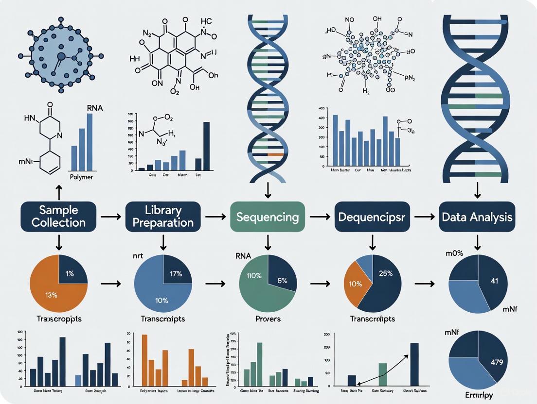
Visual Guide to Experimental Design
The following diagram illustrates the key decision points and recommendations for designing a bulk RNA-Seq experiment.
The Scientist's Toolkit: Essential Reagents and Materials
Successful execution of an RNA-Seq experiment relies on a suite of specialized reagents and materials. The following table details key solutions and their functions.
Table 3: Essential research reagent solutions for RNA-Seq experiments
| Reagent / Material | Function | Application Notes |
|---|---|---|
| Stranded mRNA-Seq Kit | Library preparation that selectively enriches for polyadenylated RNA and preserves strand of origin. | Ideal for standard gene expression studies with high-quality RNA input [8]. |
| Total RNA-Seq Kit with rRNA Depletion | Library preparation that removes ribosomal RNA (rRNA) to enrich for other RNA species (mRNA, lncRNA). | Essential for isoform discovery, non-coding RNA analysis, or when working with degraded RNA (e.g., FFPE) where poly(A) tails may be lost [4] [9]. |
| RNA Integrity Number (RIN) / RQS Assay | Microfluidics-based assay (e.g., Bioanalyzer, TapeStation) to quantitatively assess RNA quality. | A critical QC step; RIN > 8 is recommended for mRNA-Seq, while rRNA-depletion protocols are more tolerant of lower RIN values [4] [6]. |
| Unique Molecular Identifiers (UMIs) | Short random nucleotide tags added to each RNA molecule during library prep before amplification. | Corrects for PCR amplification bias and duplicates, crucial for accurate quantification, especially with low-input or degraded samples [4]. |
| Spike-in RNA Controls | Synthetic RNA molecules added in known quantities to the sample. | Serves as an internal control for monitoring technical performance, including sensitivity, dynamic range, and quantification accuracy across samples [8]. |
| RNA Stabilization Reagents | Reagents (e.g., RNAlater) that immediately stabilize cellular RNA to prevent degradation. | Preserves RNA integrity during sample collection, storage, and transportation, especially from remote locations [8]. |
The Critical Link Between Biological Replicates and Sequencing Depth for Statistical Power
Bulk RNA sequencing (RNA-seq) is a foundational tool in transcriptome analysis, yet many studies struggle with achieving sufficient statistical power for reliable results. Due to considerable financial and practical constraints, RNA-seq experiments often employ limited biological replication, with surveys indicating approximately 50% of studies on human samples use six or fewer replicates, a figure that rises to 90% for non-human samples [10] [11]. This tendency toward underpowered designs directly threatens the replicability of research findings. Recent large-scale replication projects in preclinical cancer biology have reported success rates as low as 46% [10] [11]. This application note examines the critical, and often misunderstood, relationship between biological replicates and sequencing depth, providing structured guidance and protocols to optimize experimental design for robust differential expression analysis.
Quantitative Guidelines for Experimental Parameters
The choice of replicates and sequencing depth is not one-size-fits-all but must be aligned with the specific goals of the study. The tables below summarize evidence-based recommendations for these parameters.
Table 1: Recommended Sequencing Depth for Bulk RNA-Seq Experiments
| Experimental Goal | Recommended Mapped Reads | Key Considerations and Rationale |
|---|---|---|
| Basic Differential Gene Expression (DGE) | 5 - 15 million [2] | A good bare minimum for a snapshot of highly expressed genes. |
| Standard Gene-Level DGE | 20 - 50 million [2] [4] | Provides a more global view of gene expression; a common standard in many published human RNA-Seq experiments [2]. |
| Robust Gene-Level DGE (Sweet Spot) | 25 - 40 million (paired-end) [4] | Stabilizes fold-change estimates across expression quantiles without wasting reads on already-well-sampled transcripts. |
| Isoform Detection & Alternative Splicing | ≥ 100 million (paired-end) [4] | Comprehensive isoform coverage requires significantly greater depth to capture a full range of splice events. |
| Fusion Detection | 60 - 100 million (paired-end) [4] | Ensures sufficient split-read support for breakpoint resolution by fusion callers. |
| Allele-Specific Expression (ASE) | ~100 million (paired-end) [4] | Essential depth to accurately estimate variant allele frequencies and minimize sampling error. |
Table 2: Recommended Number of Biological Replicates
| Scenario | Recommended Replicates per Condition | Rationale and Evidence |
|---|---|---|
| Absolute Minimum | 5 - 7 [10] [11] | Caution is advised with fewer than seven replicates due to high heterogeneity in results [11]. |
| Robust DEG Detection | ≥ 6 [10] | Considered necessary for robust detection of differentially expressed genes (DEGs) [10]. |
| Identifying Majority of DEGs | ≥ 12 [10] [11] | Required when it is important to identify the majority of DEGs across all fold changes [10]. |
| Target for Power (≥80%) | ~10 [11] | Suggested to achieve sufficient statistical power under budget constraints [11]. |
| ENCODE Standard | 2 or more [12] | Minimum standard; must demonstrate high replicate concordance (Spearman correlation >0.9) [12]. |
Experimental Protocols for Power Assessment and Replicability
Power Analysis Using the PROPER Tool
Power analysis is a critical step for planning a statistically sound RNA-seq experiment. The PROPER (PROspective Power Evaluation for RNAseq) Bioconductor package provides a comprehensive solution for complex RNA-seq data [13].
Detailed Methodology:
- Pilot Data Input: The procedure requires a pilot dataset, which can be real or simulated. For simulation, PROPER uses parameters estimated from public data sets (e.g., library size, normalized gene expression, dispersion, percent of genes that are differentially expressed, and log-fold changes of DEGs) [14].
- Simulation and Modeling: PROPER performs Monte Carlo simulations based on the negative binomial distribution, which accurately models the over-dispersion characteristic of RNA-seq count data. It simulates full RNA-seq experiments under various design scenarios (e.g., paired or unpaired samples, different numbers of replicates, different sequencing depths) [14] [13].
- Power Calculation: For each simulated experiment, PROPER runs differential expression analysis using established methods (e.g., DESeq2, edgeR) and calculates statistical power. Power is defined as the proportion of true DEGs that are correctly identified as significant at a specified False Discovery Rate (FDR) threshold [14] [13].
- Stratified Analysis: A key feature is its ability to stratify power calculations by gene expression level and fold-change, allowing researchers to see if their design is adequate for detecting DEGs of biological interest, which often have modest fold-changes and/or low expression [13].
- Output and Visualization: The tool generates comprehensive visualizations, such as the
plotAllfunction, to display stratified power, enabling researchers to make an informed decision on the optimal balance between replicate number and sequencing depth for their specific goals and budget [13].
Bootstrapping for Estimating Replicability
For researchers who already have a dataset, a bootstrapping procedure can estimate the expected replicability and precision of their results, which is particularly valuable for small cohort sizes [10] [11].
Detailed Methodology:
- Subsampling: From the full dataset, repeatedly (e.g., 100 times) randomly select a small cohort of N biological replicates per condition, mirroring the size of the original or planned study [10] [11].
- Differential Expression Analysis: For each subsampled "experiment," perform a full differential expression analysis to identify a list of significant DEGs [10] [11].
- Metric Calculation: Calculate replicability and precision metrics by comparing the results across all subsampled experiments. Common metrics include the Jaccard index (overlap of DEG lists) and precision-recall curves [10] [11].
- Performance Prediction: The level of agreement (or disagreement) between the subsampled results serves as a strong predictor for the real-world replicability of the analysis. This procedure helps diagnose whether a small-scale study is prone to a high rate of false positives or is likely to yield precise, albeit potentially incomplete, results [10] [11].
Visualizing the Relationship Between Key Parameters
The following diagram illustrates the logical workflow for designing a powered RNA-seq experiment and how key parameters influence the final outcomes of statistical power and replicability.
The Scientist's Toolkit: Essential Research Reagents and Materials
Successful execution of a powered RNA-seq experiment relies on several key reagents and materials throughout the workflow.
Table 3: Essential Research Reagent Solutions for Bulk RNA-Seq
| Item | Function/Application | Specifications & Notes |
|---|---|---|
| Stranded Library Prep Kit | Converts RNA into a sequencing-ready library. Preserves strand orientation of transcripts. | A stranded kit is essential for accurate transcriptome annotation. Must be compatible with RNA input amount and quality (e.g., for degraded FFPE samples) [4] [15]. |
| RNA Spike-In Controls | External RNA controls for technical validation and normalization. | The ENCODE consortium standardizes on the Ambion ERCC Spike-In Mix. Spike-in sequences are added to the genome index and used by quantification tools like RSEM [12]. |
| RNA Integrity Assay | Assesses RNA quality to inform sequencing depth requirements. | Use RIN (RNA Integrity Number) or DV200 (% of RNA fragments >200 nucleotides). DV200 >50% is suitable for standard protocols; 30-50% may require 25-50% more reads [4]. |
| Unique Molecular Identifiers (UMIs) | Tags individual RNA molecules to correct for PCR duplication bias. | Crucial for experiments with limited input (≤10 ng) or when sequencing very deeply (>80M reads) to distinguish biological expression from technical amplification [4]. |
| rRNA Depletion Reagents | Removes abundant ribosomal RNA to enrich for mRNA and non-coding RNA. | Preferred over poly(A) selection for degraded RNA samples (DV200 <30%) or when studying non-polyadenylated RNAs [4]. |
| Alignment & Quantification Software | Maps reads to a reference and estimates gene/transcript abundance. | STAR is recommended for splice-aware genome alignment and QC. Salmon (in alignment-based mode) or RSEM is recommended for accurate quantification, handling uncertainty in read assignment [15] [12]. |
| Differential Expression Tools | Identifies statistically significant changes in gene expression. | DESeq2 and edgeR are widely used and show top performance. They model count data using a negative binomial distribution to account for biological variability [14] [15]. |
The Encyclopedia of DNA Elements (ENCODE) Consortium has established comprehensive experimental guidelines and data standards to ensure the production of high-quality, reproducible genomic data. These standards provide a critical framework for researchers designing functional genomics experiments, offering evidence-based recommendations developed through extensive consortium-wide testing and validation. For scientists embarking on bulk RNA-seq research, ENCODE guidelines provide definitive baseline requirements for experimental parameters including sequencing depth, replicate numbers, and quality metrics, thereby reducing costly trial-and-error approaches. Adherence to these standards ensures data interoperability across studies and facilitates meaningful comparisons between datasets generated in different laboratories. This application note synthesizes the current ENCODE recommendations for bulk RNA-seq experiments, with particular emphasis on sequencing depth requirements within the broader context of experimental design.
Bulk RNA-seq Experimental Design
Core Sequencing Requirements
ENCODE has established specific technical standards for bulk RNA-seq experiments to ensure data quality and reproducibility. These requirements address key parameters that significantly impact data utility and reliability. The consortium recommends a minimum read length of 50 base pairs for all RNA-seq experiments, with support for both single-end and paired-end sequencing across all Illumina platforms [12]. The selection between these approaches depends on the experimental aims: paired-end sequencing is particularly recommended for isoform-level differential expression analysis as it provides better mapping across splice junctions.
Library preparation specifics are also addressed in the guidelines. Libraries must be generated from mRNA (poly(A)+), rRNA-depleted total RNA, or poly(A)- populations that are size-selected to be longer than approximately 200 bp to ensure focus on longer transcripts [12]. For experiments utilizing spike-in controls, ENCODE has standardized on the Ambion Mix 1 commercially available spike-ins at a dilution of approximately 2% of final mapped reads to create a standard baseline for RNA expression quantification [12].
A critical consideration in experimental design is the balance between sequencing depth and biological replication. The ENCODE consortium emphasizes that biological replicates are significantly more important than excessive sequencing depth for most gene-level differential expression analyses [6]. This principle guides researchers to allocate resources primarily toward adequate biological replication rather than ultra-deep sequencing, as proper replication enables more accurate estimation of biological variation and more robust statistical analysis.
Replicate Design and Statistical Power
Biological replication represents a cornerstone of reliable RNA-seq experimental design. ENCODE guidelines explicitly state that experiments should have two or more biological replicates, with exemptions granted only for exceptional circumstances such as assays using EN-TEx samples where experimental material is severely limited [12]. Biological replicates—where different biological samples of the same condition are used—are essential for measuring biological variation and must be distinguished from technical replicates, which are generally considered unnecessary in modern RNA-seq due to the relatively low technical variation compared to biological variation [6].
The relationship between replicate number and statistical power is a critical consideration. Research has demonstrated that increasing biological replicates provides substantially greater power for detecting differentially expressed genes than increasing sequencing depth [6]. While the absolute minimum is two replicates, best practices suggest three replicates as an absolute minimum, with four replicates representing the optimum minimum for robust differential expression analysis [9]. This replication strategy enables more precise estimates of mean expression levels and biological variation, leading to more accurate modeling and identification of truly differentially expressed genes.
For specialized RNA-seq applications, modified replicate guidelines exist. For shRNA knockdown followed by RNA-seq and CRISPR genome editing followed by RNA-seq, ENCODE specifies that each replicate should have 10 million aligned reads rather than the standard 30 million, and each experiment must include a corresponding control experiment [12]. Similarly, for siRNA knockdown experiments, each replicate requires 10 million aligned reads plus verification of the percentage knockdown of the targeted factor for each replicate relative to the control [12].
Table: ENCODE Bulk RNA-seq Sequencing Depth Requirements by Application
| Experiment Type | Minimum Aligned Reads | Replicate Requirements | Special Considerations |
|---|---|---|---|
| Standard bulk RNA-seq | 30 million | 2+ biological replicates | Spearman correlation >0.9 between isogenic replicates |
| Gene-level DE (limited material) | 15 million | >3 biological replicates | Sufficient with good number of replicates |
| Isoform-level DE (known isoforms) | 30 million | 2+ biological replicates | Paired-end reads recommended |
| Isoform-level DE (novel isoforms) | 60 million | 2+ biological replicates | Deeper sequencing required |
| shRNA/CRISPR knockdown | 10 million | 2+ biological replicates | Target verification required |
| Single-cell RNA-seq | 5 million | 10-20 individual experiments | Not considered biologically replicated |
Quality Assessment Metrics
ENCODE establishes clear quality thresholds for bulk RNA-seq data to ensure analytical reliability. A central quality metric is replicate concordance, measured through Spearman correlation of gene-level quantifications. The guidelines specify that isogenic replicates (replicates from the same donor) should demonstrate a Spearman correlation >0.9, while anisogenic replicates (replicates from different donors) should maintain a correlation >0.8 [12]. These thresholds provide objective criteria for assessing technical quality before proceeding with downstream analysis.
The consortium employs uniform processing pipelines that generate comprehensive quality metrics, including Spearman correlation coefficients and read depth assessments [16] [12]. These pipelines use the STAR program for read alignment and the RSEM program for gene and transcript quantification, with alignment files mapped to standard reference sequences (GRCh38, hg19, or mm10) and gene quantifications annotated to GENCODE versions (V24, V19, or M4) [12]. This standardized approach ensures consistency across datasets generated by different consortium members.
Beyond technical metrics, ENCODE emphasizes the importance of metadata audits and experimental annotation. All experiments must pass routine metadata audits before public release, ensuring adequate documentation of experimental parameters, sample characteristics, and processing steps [12]. This comprehensive approach to quality assessment addresses both technical performance and experimental metadata integrity, providing multiple safeguards for data quality.
Experimental Protocols
Bulk RNA-seq Workflow
The ENCODE bulk RNA-seq pipeline follows a standardized workflow that can be applied to both replicated and unreplicated experiments, accommodating paired-end or single-end designs and both strand-specific and non-strand specific libraries [12]. The protocol begins with library preparation from mRNA sources, followed by sequencing with minimum length requirements, then proceeds through quality control, read alignment, and quantification steps. The workflow generates multiple output formats including BAM alignment files, bigWig signal files, and gene quantification files, providing researchers with both raw and processed data for analysis.
Library Preparation and Sequencing
The library preparation phase requires careful attention to RNA quality and appropriate selection of RNA fractions. The ENCODE protocol specifies that libraries must be generated from mRNA (poly(A)+), rRNA-depleted total RNA, or poly(A)- populations that are size-selected to be longer than approximately 200 bp [12]. For standard coding mRNA analysis, poly(A)+ selection is recommended, while for experiments investigating long non-coding RNA, the total RNA method with rRNA depletion should be employed [9].
RNA quality is a critical factor in library preparation success. The ENCODE guidelines emphasize that high RNA integrity (RIN > 8) is essential for mRNA library preparations [9]. For degraded RNA samples, such as those from clinical specimens, the total RNA method is more appropriate. During library preparation, inclusion of ERCC spike-in controls is standard practice in ENCODE protocols, using precisely defined concentrations that should result in approximately 2% of final mapped reads deriving from spike-in sequences [12].
For the sequencing phase itself, ENCODE recommends 30 million aligned reads per sample for standard bulk RNA-seq experiments [12]. While older projects targeted 20 million reads, the current standard of 30 million provides sufficient depth for most gene-level differential expression analyses. For specialized applications requiring detection of lowly-expressed genes or isoform-level analysis, deeper sequencing of 30-60 million reads is recommended, with the higher end of this range reserved for novel isoform discovery [6].
Data Processing and Analysis
The ENCODE uniform processing pipeline for bulk RNA-seq employs specific tools and standards to ensure consistent data processing across the consortium. The primary alignment is performed using the STAR program, with some historical data also processed using TopHat [12]. Following alignment, gene and transcript quantification is conducted with the RSEM program, which generates multiple expression measures including expected counts, TPM (transcripts per million), and FPKM (fragments per kilobase of transcript per million) [12].
A critical consideration in data analysis is the appropriate use of different quantification types. While gene-level quantifications can be used confidently for downstream analysis, transcript-level quantifications should be treated with caution because quantifications of individual transcript isoforms can differ substantially depending on the processing pipeline employed and are of unknown accuracy [12]. This distinction is important for researchers planning their analytical approach.
The pipeline produces several key output files that facilitate different types of downstream analysis. These include BAM files containing genome alignments, bigWig files containing normalized RNA-seq signal for visualization, and TSV files containing gene quantifications with spike-in measurements [12]. The quality metrics generated by the pipeline, including Spearman correlation values between replicates, provide objective measures for assessing data quality before proceeding with advanced analyses.
The Scientist's Toolkit
Research Reagent Solutions
Successful execution of ENCODE-standard bulk RNA-seq experiments requires specific reagents and materials that have been validated through consortium-wide use. These reagents address key aspects of the experimental workflow from sample preparation to sequencing, ensuring consistency and reproducibility across different laboratories and experimental batches.
Table: Essential Research Reagents for Bulk RNA-seq Experiments
| Reagent/Material | Function | Specifications |
|---|---|---|
| ERCC Spike-in Controls | RNA quantification standards | Ambion Mix 1, ~2% of final mapped reads [12] |
| Poly(A) Selection Beads | mRNA enrichment | For coding mRNA analysis [9] |
| rRNA Depletion Reagents | Total RNA preparation | For lncRNA studies [9] |
| Strand-Specific Library Prep Kit | Library construction | Maintains strand orientation information [12] |
| High-Sensitivity DNA Assay | Library quantification | Accurate quantification for sequencing [9] |
| STAR Aligner | Read alignment | Spliced transcript alignment to reference [12] |
| RSEM Software | Gene quantification | Calculates TPM, FPKM, expected counts [12] |
Antibody Validation Standards
For researchers incorporating chromatin analyses alongside transcriptomic profiling, ENCODE has established rigorous antibody characterization standards. These guidelines are particularly relevant for ChIP-seq experiments investigating transcription factor binding or histone modifications. The consortium requires two validated tests for each antibody—a primary and secondary characterization—with repetition required for each new antibody lot number [17].
For transcription factor antigens, the primary characterization typically involves immunoblot analysis performed on protein lysates from whole-cell extracts, nuclear extracts, or chromatin preparations. The ENCODE standard specifies that the primary reactive band should contain at least 50% of the signal observed on the blot, ideally corresponding to the expected size of the target protein [17]. When immunoblot analysis is unsuccessful, immunofluorescence demonstrating expected nuclear staining patterns serves as an acceptable alternative primary characterization method.
The secondary characterization for transcription factor antibodies involves independent validation such as similar immunoblot patterns across multiple cell types, immunostaining in a different cell type, or comparison with an independent antibody [17]. For histone modification antibodies, the primary test is typically peptide microarray or immunofluorescence, while the secondary test involves histone peptide immunoblot [18] [17]. These comprehensive validation requirements address common issues with antibody specificity and reactivity, ensuring that ChIP-seq data generated alongside RNA-seq profiles target the intended epitopes with minimal cross-reactivity.
The ENCODE standards and baseline recommendations provide an essential foundation for designing robust bulk RNA-seq experiments. By adhering to these evidence-based guidelines for sequencing depth, replicate numbers, quality metrics, and experimental protocols, researchers can generate high-quality, reproducible data that enables meaningful biological insights. The consortium's emphasis on biological replication over excessive sequencing depth, standardized processing pipelines, and clear quality thresholds offers a practical framework for efficient experimental design. As genomic technologies continue to evolve, these standards will undoubtedly be refined, but the core principles of rigor, reproducibility, and data interoperability will remain essential for advancing our understanding of gene regulation.
How RNA Quality (RIN/RQS and DV200) Influences Effective Sequencing Depth
In bulk RNA-seq research, the quality of input RNA is a fundamental determinant of data quality and reliability. The RNA Integrity Number (RIN) or RNA Quality Score (RQS) and the DV200 metric (percentage of RNA fragments >200 nucleotides) serve as crucial indicators of RNA suitability for sequencing applications. While RIN/RQS provides a comprehensive assessment of RNA degradation based on electrophoretic traces, DV200 specifically quantifies the proportion of fragments long enough for successful library construction [19] [20]. These metrics directly influence effective sequencing depth by determining the proportion of usable fragments, library complexity, and ultimately, the number of informative reads obtained per sample. Research demonstrates that RNA quality metrics correlate strongly with sequencing success, particularly for degraded samples from formalin-fixed paraffin-embedded (FFPE) tissues [19] [21]. Understanding these relationships enables researchers to optimize sequencing depth based on sample quality, ensuring cost-effective experimental design while maintaining data integrity.
Comparative Analysis of RNA Quality Metrics
Metric Definitions and Methodological Basis
RIN/RQS: This algorithm-based metric evaluates RNA integrity through analysis of the entire electrophoretic trace, incorporating the presence of ribosomal RNA peaks and degradation products. Scores range from 1 (completely degraded) to 10 (perfectly intact), with RIN ≥7 typically considered high-quality for standard RNA-seq applications [20]. The calculation employs a proprietary algorithm that considers various features of the electropherogram to generate an objective integrity measurement.
DV200: This metric calculates the percentage of RNA fragments exceeding 200 nucleotides in length, representing the fraction theoretically available for successful library preparation. Unlike RIN, DV200 does not depend on ribosomal peak ratios, making it particularly valuable for assessing FFPE-derived RNA where ribosomal peaks are often absent or altered [19] [21]. Related metrics include DV100 (fragments >100 nucleotides) which has shown particular utility for severely degraded FFPE samples [21].
Performance Characteristics Across Sample Types
Recent systematic comparisons reveal distinct performance advantages for each metric depending on sample preservation methods. For high-quality RNA from fresh-frozen specimens, RIN and DV200 show strong correlation and comparable predictive value for sequencing outcomes. However, for FFPE and other compromised samples, DV200 demonstrates superior performance in predicting library preparation efficiency and sequencing success [19]. One comprehensive study found that DV200 showed stronger correlation with the amount of NGS library product than RINe (R² = 0.8208 versus 0.6927), with receiver operating characteristic analysis confirming DV200's better predictive power for efficient library production [19].
Table 1: Comparative Performance of RNA Quality Metrics for Sequencing Applications
| Metric | Sample Type | Correlation with Library Yield | Optimal Cutoff Value | Advantages | Limitations |
|---|---|---|---|---|---|
| RIN/RQS | Fresh-frozen | R² = 0.6927 [19] | >7 for standard protocols [4] | Comprehensive integrity assessment | Less reliable for degraded samples |
| DV200 | FFPE/Degraded | R² = 0.8208 [19] | >66.1% for library efficiency [19] | Independent of ribosomal peaks | May overestimate if cross-linked |
| DV200 | Fresh-frozen | Strong correlation | >70% for standard protocols [4] | Direct measure of usable fragments | Less informative about overall integrity |
| DV100 | Severely degraded FFPE | High predictive value [21] | >80% for gene detection [21] | Better for highly fragmented RNA | Less commonly reported |
Quantitative Relationships Between RNA Quality and Sequencing Parameters
RNA Quality Impact on Library Construction and Sequencing Depth
The integrity of input RNA directly influences library complexity and sequencing requirements through multiple mechanisms. High-quality RNA (RIN >8, DV200 >70%) generates libraries with greater diversity, enabling comprehensive transcriptome coverage at moderate sequencing depths. In contrast, degraded samples produce libraries with reduced complexity, requiring increased sequencing depth to detect the same number of genes [4]. Research demonstrates that for FFPE samples with DV200 values below 50%, increasing sequencing depth by 25-50% can partially compensate for reduced library complexity [4]. The relationship between RNA quality and usable sequencing depth follows a non-linear pattern, with significant reductions in effective depth occurring below specific quality thresholds.
Table 2: Sequencing Depth Recommendations Based on RNA Quality Metrics
| Application | RNA Quality | Recommended Depth | Read Length | Protocol Considerations |
|---|---|---|---|---|
| Differential Expression | RIN ≥8, DV200 >70% | 25-40 million PE reads [4] | 2×75 bp [4] [1] | Standard poly(A) enrichment |
| Differential Expression | DV200 30-50% | +25-50% more reads [4] | 2×75-2×100 bp [4] | rRNA depletion preferred |
| Isoform Detection | High quality (RIN ≥8) | ≥100 million PE reads [4] | 2×100 bp [4] | Stranded, paired-end designs |
| Isoform Detection | Moderate degradation | +25-50% above standard [4] | 2×100 bp [4] | rRNA depletion essential |
| Fusion Detection | DV200 >50% | 60-100 million PE reads [4] | 2×75-2×100 bp [4] | Paired-end required |
| FFPE (DV200 <30%) | Severely degraded | Avoid or sequence very deep [4] | 2×100 bp | Specialized protocols needed |
Evidence-Based Thresholds for Sequencing Success
ROC curve analyses have established specific quality metric thresholds predictive of successful library preparation. For the amount of 1st PCR product per input RNA (>10 ng/µl), the optimal cutoff values were determined to be RIN >2.3 and DV200 >66.1%, with DV200 demonstrating superior predictive power (AUC 0.99 vs. 0.91 for RIN) [19]. For FFPE samples specifically, a DV100 >80% provided the best indication of gene diversity and read counts upon sequencing [21]. These thresholds enable evidence-based sample triage decisions, minimizing resource waste on samples unlikely to yield meaningful data.
Experimental Protocols for RNA Quality Assessment and Sequencing
Standardized RNA Quality Control Protocol
Materials Required:
- Agilent Bioanalyzer 2100 or TapeStation system with appropriate RNA screening kits
- Qubit Fluorometer with RNA HS Assay kit for accurate quantification [20]
- DNase treatment reagents (if not included in extraction kit)
- Nuclease-free water and consumables
Procedure:
- Extract RNA using methods appropriate for sample type (FFPE-specific kits for archived tissues)
- Treat with DNase to remove genomic DNA contamination (critical for accurate quantification) [20]
- Quantify RNA using fluorometric methods (Qubit) for accurate concentration measurement
- Assess integrity using Bioanalyzer/TapeStation electrophoresis systems
- Calculate RIN/RQS and DV200 values from electropherogram traces
- Based on quality metrics, determine suitability for sequencing and appropriate input amounts
For FFPE samples, include additional verification steps such as quantitative PCR to assess amplifiable RNA content, as this better reflects the functional quantity available for library preparation [21].
Library Preparation Strategies for Degraded RNA
Protocol selection must align with RNA quality to optimize outcomes:
For DV200 >50%:
- Standard poly(A) selection or rRNA depletion protocols can be employed
- Input amount: 10-100 ng total RNA depending on degradation level
- TruSeq RNA Access or similar kits designed for varying input quality [19]
For DV200 30-50%:
- rRNA depletion protocols are strongly recommended over poly(A) selection
- Increase RNA input by 1.5-2× to compensate for reduced usable fragments
- Incorporate unique molecular identifiers (UMIs) to account for amplification bias [4]
- Consider specialized FFPE-optimized kits (Roche KAPA RNA HyperPrep with RiboErase) [20]
For DV200 <30%:
- rRNA depletion is essential; poly(A) selection will introduce severe 3' bias
- Maximum recommended RNA input (100-500 ng)
- UMIs are crucial for accurate quantification
- Expect significantly reduced library complexity and increased sequencing requirements [4] [20]
Decision Framework for Sequencing Depth Adjustment
The relationship between RNA quality and required sequencing depth follows predictable patterns that can be formalized into a decision framework. This framework enables researchers to systematically adjust sequencing parameters based on pre-sequence quality metrics.
The Scientist's Toolkit: Essential Reagents and Platforms
Table 3: Essential Research Reagents and Platforms for RNA Quality Assessment and Sequencing
| Category | Product/Platform | Specific Application | Key Features |
|---|---|---|---|
| RNA Quality Assessment | Agilent Bioanalyzer 2100/TapeStation | RIN/RQS and DV200 calculation | Microfluidics-based electrophoresis, standardized metrics |
| RNA Quantification | Qubit Fluorometer with RNA HS Assay | Accurate RNA quantification | RNA-specific fluorescence, minimal DNA/protein interference |
| FFPE RNA Extraction | Promega ReliaPrep FFPE Total RNA Kit | RNA from archived samples | Optimized for cross-link reversal, high yield/quality ratio [22] |
| FFPE RNA Extraction | Roche KAPA RNA HyperPrep with RiboErase | Degraded RNA library prep | Efficient rRNA depletion, compatible with low-quality inputs [20] |
| Library Preparation | Illumina TruSeq RNA Access | Degraded RNA sequencing | Designed for variable input quality, compatible with FFPE RNA [19] |
| NGS Platform | Illumina HiSeq 2500/3000/4000 | RNA-seq applications | 2×100 bp reads optimal for splice junction detection [20] |
RNA quality metrics, particularly DV200 for degraded samples, provide essential guidance for determining appropriate sequencing depth and methodology. The systematic integration of quality assessment into experimental design enables researchers to make evidence-based decisions about sample inclusion, library preparation strategies, and sequencing depth requirements. By aligning sequencing parameters with RNA quality, researchers can maximize data quality while optimizing resource allocation, particularly valuable when working with biobank samples and clinical specimens where quality varies substantially. As RNA-seq applications continue to expand in both basic research and clinical contexts, the rigorous application of these quality-informed sequencing strategies will ensure robust, reproducible, and biologically meaningful results.
Application-Specific Guidelines: Matching Sequencing Strategy to Your Biological Question
In the field of bulk RNA sequencing, researchers continually face the challenge of balancing data quality with budgetary constraints. The selection of appropriate sequencing depth represents a critical design parameter that directly influences both the scientific validity and practical feasibility of transcriptomic studies. As the technology has evolved from a discovery tool into a cornerstone of clinical and translational genomics, best practices have shifted from following a single recipe to making informed choices driven by specific study goals and sample quality [4]. Within this context, a consensus has emerged around a specific range of 25-40 million reads per sample as a cost-effective sweet spot for one of the most common applications in functional genomics: differential gene expression analysis.
This application note examines the technical justification, practical implementation, and economic rationale behind this optimal read depth, providing researchers with evidence-based protocols for designing robust and efficient RNA-seq experiments.
Establishing the Benchmark: The 25-40 Million Read Recommendation
Consensus Across Guidelines
Multiple independent sources from both academic and industry perspectives converge on the 25-40 million read range as optimal for standard differential expression analyses. This consensus spans consortium recommendations, core facility protocols, and manufacturer guidelines, creating a robust foundation for experimental design.
The ENCODE long-RNA data standards remain the most widely referenced public specification for bulk RNA-Seq, recommending sequencing depths of ≥30 million mapped reads for typical poly(A)-selected RNA-Seq [4]. This benchmark is further refined by technical reviews and manufacturer guidelines that specifically converge on 25–40 million paired-end reads per human sample as a sweet spot for robust gene quantification [4]. Core facilities at major research institutions have adopted similar standards, with Northwestern University's NUSeq core recommending 20-25 million reads per sample for general gene expression profiling [23].
Technical Rationale
The 25-40 million read range represents an optimization point where sequencing depth adequately captures the transcriptional landscape without generating redundant data. At this depth, fold-change estimates stabilize across expression quantiles without wasting reads on already-well-sampled transcripts [4]. This depth provides sufficient sampling to ensure that:
- Expression estimates stabilize for moderately to highly expressed genes
- Statistical power increases for detecting biologically meaningful differences
- Technical variability decreases through comprehensive transcript sampling
- Cost efficiency maximizes by avoiding diminishing returns of deeper sequencing
Sequencing Depth Recommendations by Research Application
The optimal sequencing depth varies substantially depending on the specific biological questions being addressed. The table below summarizes recommended read depths and configurations for common research applications in human studies:
Table 1: RNA-Seq Sequencing Recommendations by Research Application
| Research Application | Recommended Depth | Read Configuration | Key Considerations |
|---|---|---|---|
| Differential Gene Expression | 25-40 million reads [4] [23] | PE 75 bp [4] | Cost-effective for robust gene quantification |
| Isoform Detection & Alternative Splicing | ≥100 million reads [4] | PE 100 bp [4] | Requires longer reads to span multiple exons |
| Fusion Gene Detection | 60-100 million reads [4] | PE 75-100 bp [4] | Higher depth needed for split-read support |
| Allele-Specific Expression | ~100 million reads [4] | Paired-end [4] | Essential for accurate variant allele frequency |
| Small RNA Sequencing | 4-5 million reads [23] | SE 50 bp [1] | Sufficient due to small transcriptome size |
| Total RNA-Seq (rRNA-depleted) | 20-25 million reads [23] | SE 50/75 bp or PE [23] | Similar to mRNA-seq for gene expression |
For differential expression studies specifically, the 25-40 million read recommendation applies particularly to high-quality RNA samples with RNA Integrity Number (RIN) ≥8 or DV200 >70% [4]. The selection of paired-end 75 bp reads provides the additional advantage of more accurate transcript mapping compared to single-end protocols, while remaining cost-effective [4] [23].
Economic Considerations in Experimental Design
Cost-Benefit Analysis of Sequencing Depth
The 25-40 million read recommendation emerges not only from technical considerations but also from economic practicality. Beyond approximately 40 million reads, experiments encounter diminishing returns where additional sequencing yields progressively fewer novel transcript discoveries [4]. As one benchmarking study demonstrated, the new detections rate (NDR) - the number of newly detected genes per million additional reads - drops significantly as sequencing depth increases [24].
Recent methodological advances have further refined the cost-benefit calculus. Early barcoding protocols such as Prime-seq demonstrate that library generation costs can be reduced by almost 50-fold compared to standard TruSeq preparations while maintaining equivalent performance for differential expression analysis [25]. Similarly, BRB-seq and related approaches achieve accurate gene expression quantification with only 5 million reads per sample through 3' mRNA-seq multiplexing, though with some trade-offs in isoform-level information [26].
The Critical Role of Biological Replication
A fundamental principle in experimental design is the prioritization of biological replication over excessive sequencing depth. Multiple studies have demonstrated that increasing replicate number provides greater statistical power for detecting differential expression than simply sequencing the same samples more deeply [27].
In toxicogenomics dose-response studies, increasing from 2 to 4 replicates significantly enhanced reproducibility, with over 550 genes consistently identified across most sequencing depths compared to high variability with only 2 replicates [27]. This principle holds particular importance for differential expression studies, where the power to detect true biological differences depends more on replicate number than on extreme sequencing depth.
Table 2: Cost Distribution for mRNA-seq Using Different Library Prep Methods
| Cost Component | Illumina TruSeq | NEBnext Ultra II | BRB-seq/QuantSeq |
|---|---|---|---|
| RNA Extraction & QC | $6.3-$11.2 | $6.3-$11.2 | $6.3-$11.2 |
| Library Preparation | $68.7 | $41.3 | $24.0 |
| Sequencing (S4 Flow Cell) | $36.9 | $25.9 | $4.6 |
| Data Analysis | ~$2.0 | ~$2.0 | ~$2.0 |
| Total Cost Per Sample | ~$113.9 | ~$75.5 | ~$36.9 |
Sample Quality and Experimental Success
RNA Quality Metrics and Their Impact
RNA integrity represents perhaps the most critical factor influencing sequencing outcomes. The recommended 25-40 million read depth assumes high-quality RNA samples with the following characteristics:
- RNA Integrity Number (RIN) ≥8 or RQS ≥8 [4] [23]
- DV200 >70% [4]
- Absence of genomic DNA contamination [23]
For samples with compromised RNA quality, such as those from Formalin-Fixed Paraffin-Embedded (FFPE) tissues, alternative approaches are necessary. The DV200 metric (percentage of RNA fragments >200 nucleotides) becomes particularly valuable for assessing degraded samples [4].
Adapting Protocols for Suboptimal Samples
When working with degraded or low-quality RNA, researchers should consider both protocol modifications and sequencing adjustments:
- DV200 30-50%: Increase sequencing depth by 25-50% and prefer rRNA depletion over poly(A) selection [4]
- DV200 <30%: Avoid poly(A) selection; use capture or rRNA depletion with higher input and ≥75-100 million reads [4]
- FFPE samples: Combine unique molecular identifiers (UMIs) with rRNA-depletion protocols and increase total reads by 20-40% [4]
- Limited input (≤10 ng RNA): Incorporate UMIs to collapse PCR duplicates and consider low-input specialized protocols [4]
Implementation Protocols and Workflow
Experimental Design Workflow
The following diagram outlines the key decision points in designing a cost-effective RNA-seq experiment for differential expression analysis:
The Scientist's Toolkit: Essential Research Reagents and Materials
Table 3: Essential Research Reagents and Materials for Bulk RNA-Seq
| Reagent/Material | Function/Purpose | Examples/Alternatives |
|---|---|---|
| RNA Extraction Reagents | Isolation of high-quality total RNA | TRIzol (solvent-based), QIAgen RNeasy Kit (silica-based column) [26] |
| RNA Quality Assessment | Evaluate RNA integrity and quantity | Bioanalyzer RNA-6000-Nano chip (RIN generation) [26] [23] |
| Poly(A) Selection Beads | Enrichment for mRNA | Oligo(dT) magnetic beads [4] [23] |
| rRNA Depletion Reagents | Removal of ribosomal RNA (for total RNA-seq) | Ribosomal RNA subtraction kits [4] [23] |
| Library Preparation Kits | Construction of sequencing-ready libraries | TruSeq Stranded mRNA, NEBNext Ultra II, Prime-seq [4] [26] [25] |
| Unique Molecular Identifiers (UMIs) | Correction for PCR amplification bias | Random barcodes incorporated during reverse transcription [4] [25] |
| ERCC Spike-in Controls | Technical standards for quantification | Ambion ERCC RNA Spike-In Mix [12] |
The establishment of 25-40 million reads as a sweet spot for differential gene expression analysis represents a maturation point in bulk RNA-seq methodology. This optimized range balances technical robustness with economic practicality, enabling researchers to design studies with appropriate statistical power while maximizing resource utilization. As sequencing technologies continue to evolve and costs decrease further, the fundamental principles of matching sequencing strategy to biological questions and sample quality will remain paramount.
Future developments in early barcoding methods [25], molecular indexing, and multi-modal sequencing integration will continue to refine these recommendations, but the 25-40 million read benchmark serves as a validated starting point for experimental design in differential expression studies. By adhering to these evidence-based guidelines and prioritizing biological replication over excessive depth, researchers can generate statistically robust, reproducible, and interpretable transcriptomic data that advances scientific discovery.
In bulk RNA sequencing (RNA-seq), the choice of sequencing depth is a fundamental determinant of data quality and biological insight. While standard gene expression profiling can be accomplished with moderate depth, comprehensive isoform detection and alternative splicing analysis present a significantly greater challenge. Alternative splicing, a key mechanism for proteomic diversity, allows a single gene to produce multiple distinct mRNA isoforms. It is prevalent in vertebrates, with an estimated 90% of human genes undergoing this process [28]. The accurate identification of these isoforms is essential for understanding cellular differentiation, organismal development, and the molecular basis of diseases, including cancer and neurological disorders [28].
The transition from gene-level to isoform-level analysis necessitates a substantial increase in sequencing depth. This application note establishes the technical basis for employing ≥100 million paired-end reads to achieve robust isoform detection, delineates specific experimental scenarios requiring this depth, and provides detailed protocols for researchers and drug development professionals operating within a bulk RNA-seq framework.
Sequencing Depth Requirements by Biological Application
The required sequencing depth is primarily dictated by the biological question. The following table summarizes the recommended read depths and lengths for key applications in human studies, based on recent community benchmarks and manufacturer guidelines [4] [1].
Table 1: RNA-Seq Sequencing Recommendations for Different Research Aims
| Research Aim | Recommended Depth (Mapped Reads) | Recommended Read Length | Key Rationale |
|---|---|---|---|
| Differential Gene Expression | 25 - 40 million [4] | 2x75 bp paired-end [4] | Cost-effective stabilization of fold-change estimates for highly expressed genes. |
| Isoform Detection & Alternative Splicing | ≥100 million [4] | 2x75 bp or 2x100 bp paired-end [4] | Ensures sufficient coverage to resolve low-abundance isoforms and splice junctions. |
| Fusion Gene Detection | 60 - 100 million [4] | 2x75 bp (2x100 bp optimal) [4] | Provides cleaner junction resolution and adequate split-read support for breakpoint anchoring. |
| Allele-Specific Expression (ASE) | ~100 million [4] | 2x75 bp paired-end [4] | Essential to accurately estimate variant allele frequencies and minimize sampling error. |
For isoform detection, conventional depths used for differential expression capture only a fraction of splice events [4]. Deeper sequencing (≥100 million reads) ensures that lowly expressed but biologically critical transcripts are sampled, enabling the construction of a complete and quantitative picture of the transcriptome's complexity.
Wet Lab Workflow: From Sample to Sequencer
A meticulous wet lab protocol is critical for successful high-depth isoform studies. The following workflow details the key steps from sample preparation to library qualification.
Sample Quality Assessment and Input
- RNA Integrity Measurement: Quantify RNA Integrity Number (RIN) or RNA Quality Score (RQS) using an instrument such as the Agilent Bioanalyzer. Acceptable threshold: RIN/RQS ≥ 8. Alternatively, use the DV200 metric (percentage of RNA fragments >200 nucleotides). Proceed with standard protocols if DV200 > 70% [4].
- Input RNA Mass: For high-quality RNA (RIN≥8, DV200>70%), use 100 ng - 1 µg of total RNA as input for library preparation. For degraded samples (e.g., FFPE), higher input (e.g., 200 ng - 500 ng) may be required to compensate for reduced intact RNA content [4] [8].
- gDNA Removal: Treat all RNA samples with DNase I to eliminate genomic DNA contamination, which can lead to false-positive intron retention calls [29].
Library Preparation Protocol
This protocol is optimized for stranded, paired-end libraries to preserve strand-of-origin information, which is crucial for accurate isoform annotation.
- RNA Selection: Perform poly(A) selection for mRNA enrichment if studying polyadenylated transcripts and RNA is high-quality (DV200 > 50%). For degraded samples (DV200 < 50%) or to capture non-polyadenylated RNA, use ribosomal RNA (rRNA) depletion kits [4].
- cDNA Synthesis: Generate double-stranded cDNA using reverse transcriptase and random hexamer primers, followed by second-strand synthesis. Avoid excessive PCR cycles to minimize duplication artifacts.
- Adapter Ligation: Ligate platform-specific sequencing adapters to the blunt-ended, A-tailed cDNA fragments.
- Library Amplification & Size Selection: Amplify the library with a limited-cycle PCR (e.g., 8-12 cycles). Perform a clean-up and size selection step (e.g., using SPRI beads) to remove adapter dimers and select for inserts of the desired length.
- Library QC: Quantify the final library using a fluorescence-based method (e.g., Qubit). Assess library size distribution and confirm the absence of primer dimers using a high-sensitivity DNA kit on the Agilent Bioanalyzer or TapeStation. Validate library molarity via qPCR compatible with your sequencing platform.
Incorporating Controls
- Spike-in Controls: Use synthetic RNA spike-ins (e.g., ERCC RNA Spike-In Mix). These controls serve as an internal standard for assessing technical performance, dynamic range, and quantification accuracy across samples [8] [12].
- Unique Molecular Identifiers (UMIs): For studies with limited input (≤10 ng) or highly degraded RNA, use library protocols that incorporate UMIs. UMIs enable accurate correction for PCR duplicates, which is critical when sequencing deeply (>80M reads) to distinguish biological signal from technical artifact [4].
A Decision Framework for High-Depth Sequencing
The following diagram illustrates the logical workflow for determining when to deploy ≥100 million paired-end reads in your bulk RNA-seq experiment.
The Scientist's Toolkit: Essential Reagents and Solutions
Successful high-depth RNA-seq experiments rely on a suite of specialized reagents and computational tools.
Table 2: Essential Research Reagents and Tools for Isoform Detection
| Category | Item | Function and Application Notes |
|---|---|---|
| Sample QC | Bioanalyzer RNA Nano Kit | Assesses RNA Integrity (RIN) and sample quality. Critical for determining the appropriate library prep protocol. |
| Library Prep | Poly(A) Selection Beads | Enriches for polyadenylated mRNA. Use with high-quality RNA (RIN≥8). |
| rRNA Depletion Kit | Removes ribosomal RNA. Essential for degraded samples (FFPE) or for capturing non-polyA RNA. | |
| Stranded cDNA Synthesis Kit | Generates sequencing libraries that preserve strand information, crucial for accurate isoform annotation. | |
| Sequencing Controls | ERCC RNA Spike-In Mix | Synthetic RNA controls added to the sample pre-library prep. Used to monitor technical performance and normalize quantification. |
| Specialized Reagents | UMI Adapters | Unique Molecular Identifiers (UMIs) are short random sequences ligated to each molecule pre-amplification, allowing for precise removal of PCR duplicates. |
| Computational Tools | IsoQuant [28] | A highly effective tool for isoform detection with long-read sequencing, also applicable for short-read data analysis. Excels in precision and sensitivity. |
| Bambu [28] | A machine learning-based tool for transcript discovery and quantification that demonstrates strong performance in benchmarks. | |
| StringTie2 [28] | A widely used and computationally efficient tool for transcript assembly and quantification from RNA-seq data. |
The strategic selection of sequencing depth is a cornerstone of effective bulk RNA-seq experimental design. For researchers aiming to move beyond gene-level expression and delve into the complex world of isoform diversity, alternative splicing, and allele-specific regulation, committing to ≥100 million paired-end reads is a necessary investment. This depth, coupled with robust laboratory protocols, careful sample quality control, and advanced computational tools, unlocks a higher-resolution view of the transcriptome. By adhering to these guidelines, scientists and drug developers can ensure their data possesses the complexity and precision required to uncover meaningful biological insights and advance therapeutic discovery.
This application note details experimental and computational protocols for detecting two critical molecular features in cancer and genetic research: fusion genes and allele-specific expression (ASE). The accurate identification of fusion gene breakpoints and the resolution of allelic imbalances from bulk RNA-Seq data present distinct challenges, with sequencing depth and library preparation choices being paramount. Framed within the broader context of establishing sequencing depth requirements for bulk RNA-Seq, this guide provides detailed methodologies, data standards, and bespoke workflows to empower researchers and drug development professionals in generating reliable, analytically valid results for these specific applications.
In the era of precision medicine, moving beyond standard gene expression profiling to a more nuanced analysis of the transcriptome is essential. The detection of fusion genes, hybrid genes formed from previously independent genes, is crucial for cancer diagnosis, prognosis, and therapeutic targeting [30]. Concurrently, allele-specific expression (ASE) analysis, which measures the relative expression of parental alleles, serves as a powerful tool for uncovering cis-regulatory variation that often eludes genome-wide association studies (GWAS) and standard differential expression analyses [31] [32].
The resolution required to detect these features—namely, the precise mapping of genomic breakpoints for fusions and the quantitative assessment of allelic ratios for ASE—imposes specific and demanding requirements on bulk RNA-Seq experimental design. A critical consideration is the inherent trade-off between sequencing depth and the number of biological replicates; studies have demonstrated that increasing replicate count from 2 to 6, even at a moderate depth of 10 million reads per sample, boosts statistical power more significantly than increasing depth from 10 million to 30 million reads with fewer replicates [2]. This note outlines tailored strategies that balance these factors to optimize the detection of fusion breakpoints and ASE variants.
Sequencing Depth and Experimental Design Requirements
Successful detection is contingent upon appropriate sequencing depth and library construction. The requirements differ based on the primary analytical goal.
Table 1: Recommended Sequencing Specifications for Detection Goals
| Detection Goal | Recommended Mapped Reads (Per Sample) | Key Considerations | Recommended Library Type |
|---|---|---|---|
| Fusion Genes (Basic DGE context) | 20 - 50 million [2] [1] | Sufficient for exon-to-exon fusion discovery in poly(A)+ data. | poly(A)+ or rRNA-depleted |
| Fusion Genes (Breakpoint Resolution) | 30 - 60 million [1] | Higher depth improves resolution of intronic and intergenic breakpoints from intronic reads. | rRNA-depleted (Total RNA) [33] |
| Allele-Specific Expression (ASE) | 30 million+ (aligned) [12] | Higher depth reduces noise in quantifying allelic ratios, especially for lowly expressed genes. | poly(A)+ or rRNA-depleted |
| Transcriptome Assembly & Novel Splice Variants | 100 - 200 million [1] | Extreme depth required for de novo reconstruction of complex transcripts. | Stranded, paired-end |
Beyond depth, other experimental parameters are critical:
- Read Length: For gene expression and fusion detection, 50-75 bp single-end reads may suffice. For novel transcript assembly and improved splice junction mapping, longer paired-end reads (e.g., 2x100 bp) are beneficial [1].
- Replicates: The ENCODE consortium mandates a minimum of two biological replicates, with a Spearman correlation of >0.9 between them for gene quantifications [12].
- RNA Integrity: A RNA Integrity Number (RIN) > 8 is recommended for high-quality library preparation [34].
Protocol for Fusion Gene and Breakpoint Detection
Experimental Workflow and Reagents
The following workflow and toolkit are designed for the sensitive detection of fusion transcripts, including their precise genomic breakpoints, from patient tissue or cell line samples.
Table 2: Research Reagent Solutions for Fusion Detection
| Item | Function | Example & Notes |
|---|---|---|
| Total RNA Isolation Kit | Purifies all RNA species, including pre-mRNA with intronic sequences. | RNeasy Mini Kit (Qiagen) with DNase I treatment to remove genomic DNA [34]. |
| rRNA Depletion Reagents | Removes abundant ribosomal RNA, enriching for pre-mRNA and other non-coding RNAs. | Illumina Stranded Total RNA Prep Kit, with Illumina Unique Dual (UD) indexes for sample multiplexing [33] [34]. |
| High-Output Sequencing Platform | Generates the required sequencing depth and read length. | Illumina NextSeq 2000 system (P3 flowcell) for 2x101 bp paired-end sequencing [34]. |
| Bioanalyzer / TapeStation | Assesses RNA quality and library fragment size. | Agilent Bioanalyzer 2100 to confirm RIN > 8 [34]. |
Detailed Computational Analysis Protocol
The following steps correspond to the computational phase of the workflow above.
- RNA Sequencing Data Acquisition: Sequence libraries to a depth of 30-60 million paired-end reads per sample to ensure sufficient coverage for breakpoint resolution [1]. Data formats are typically demultiplexed FASTQ files.
- Pre-processing and Quality Control: Use tools like Trim Galore (wrapper for Cutadapt and FastQC) to remove adapter sequences and low-quality bases (Q-score < 30) and discard short reads (< 20 bp) [34]. This step is critical for clean data.
- Genome Alignment: Align high-quality reads to a reference genome using a splice-aware aligner such as STAR [12]. For fusion detection, it is vital that the alignment step is configured to output chimeric or split reads, which are the primary evidence of fusion events.
- Fusion Calling with Breakpoint Resolution: Employ specialized algorithms designed to detect fusion transcripts and their underlying genomic breakpoints.
- Dr. Disco: This algorithm is specifically highlighted for its ability to leverage intronic and intergenic reads from rRNA-depleted RNA-Seq data to identify exact genomic breakpoints, moving beyond simple exon-to-exon junctions [33]. It uses the entire reference genome as its search space.
- FindDNAFusion: A combinatorial pipeline that integrates multiple fusion-calling tools (e.g., JuLI, Factera) to improve detection accuracy to ~98% for genes with intronic bait probes [35]. It includes a blacklist for filtering common artifacts and criteria for selecting reportable fusions.
- Visualization and Validation: Manually inspect candidate fusions using integrated genome browsers. Crucially, all bioinformatic predictions, especially novel or complex rearrangements, must be validated by orthogonal methods such as Sanger sequencing, RT-PCR, or fluorescence in situ hybridization (FISH) [30] [35].
Protocol for Allele-Specific Expression Analysis
Experimental Workflow and Reagents
This protocol focuses on detecting allelic imbalance from bulk RNA-Seq data, which acts as a proxy for cis-regulatory variation.
Detailed Computational Analysis Protocol
The following steps correspond to the computational phase of the ASE workflow.
- Sequencing and Alignment: Generate RNA-Seq data according to standard gene expression profiling recommendations (>30 million aligned reads) [12]. Align reads using the STAR aligner as part of a standardized Bulk RNA-seq pipeline (e.g., the ENCODE pipeline) [12].
- Variant Calling and Quality Control: Process the aligned BAM files using the Genome Analysis Toolkit (GATK) best practices for RNA-seq short variant discovery (SNPs and indels) [31]. This step identifies heterozygous sites within expressed regions.
- Allele-Specific Expression Quantification: Count the reads supporting each allele at heterozygous loci. Several tools are available for this step:
- ASEP: Utilizes a generalized linear mixed-effects model to detect ASE across a population of individuals, accounting for correlations of SNPs within the same gene [34].
- MBASED and GeneiASE: Perform ASE analysis at the gene level by aggregating evidence from multiple heterozygous SNPs within a single sample [34].
- Statistical Analysis and Interpretation:
- ASE Score Calculation: Represent ASE as the absolute deviation from the expected heterozygous biallelic frequency of 0.5. An ASE score threshold of 0.966 can be used to distinguish true heterozygous loci from sequencing artifacts when genotype data is available [31].
- Population-level Analysis: Identify genes with significant shared imbalance across a cohort. For example, in a dilated cardiomyopathy study, genes like ABLIM1, TNNT2, and AKAP13 showed imbalance in a large majority of samples, highlighting their potential regulatory role [31].
- Differential ASE (dASE): Compare allelic imbalance between phenotypically distinct groups (e.g., disease subtypes) using non-parametric tests like the Mann-Whitney U test to find cis-regulatory differences associated with specific phenotypes [31].
Integrating fusion and ASE analysis with genomic data provides a more comprehensive biological picture. For instance, identifying a fusion gene's genomic breakpoint via Dr. Disco [33] can be complemented by investigating whether it leads to allelic imbalances in nearby genes or itself via ASE analysis [31]. Furthermore, tools like FusionAI demonstrate the potential of deep learning to predict fusion breakpoints directly from DNA sequence, offering a new avenue for understanding the genomic context of breakage [36].
In conclusion, the resolution of fusion gene breakpoints and allelic imbalances demands a deliberate and informed approach to bulk RNA-Seq experimental design. Key to this is selecting the appropriate library type (rRNA-depleted for full breakpoint resolution) and committing to adequate sequencing depth (30-60 million reads) and biological replication. The protocols and standards detailed herein provide a robust framework for generating high-quality data capable of uncovering these critical molecular events, thereby advancing our understanding of cancer genetics, complex traits, and personalized therapeutic strategies.
Choosing Between Poly(A) Selection and rRNA Depletion Based on Sample Integrity and Goals
In bulk RNA-Seq experiments, the initial choice of library construction method is a critical determinant of success. The two primary strategies—Poly(A) selection and rRNA depletion—fundamentally shape the transcriptome you measure, influencing data quality, analytical possibilities, and biological conclusions [37]. This protocol provides a structured framework for selecting the optimal method based on sample integrity, organism, and research objectives, ensuring robust and interpretable results.
Core Principles and Method Comparison
Underlying Mechanisms
- Poly(A) Selection: This method uses oligo-dT primers or probes to hybridize and enrich for RNA molecules possessing a poly(A) tail. It specifically targets mature eukaryotic messenger RNAs (mRNAs) and many long non-coding RNAs (lncRNAs) with poly(A) tails, while excluding ribosomal RNA (rRNA), transfer RNA (tRNA), and other non-polyadenylated species [37] [38].
- rRNA Depletion (Ribo-Depletion): This method employs sequence-specific DNA probes that bind to cytosolic and mitochondrial ribosomal RNAs. The rRNA-probe hybrids are subsequently removed, typically via RNase H digestion or affinity capture. This retains both polyadenylated and non-polyadenylated RNAs, including pre-mRNAs, many lncRNAs, histone mRNAs, and some viral RNAs [37] [38].
Comparative Workflow and Data Output
The following diagram illustrates the procedural and outcome differences between the two methods.
Decision Framework and Guidelines
The choice between methods hinges on three primary filters: the organism, RNA integrity, and the biological question regarding which RNA species are of interest [37].
Method Selection Table
The table below summarizes key scenarios and the recommended library preparation method.
| Situation | Recommended Method | Rationale | Potential Limitations |
|---|---|---|---|
| Eukaryotic RNA, good integrity, coding-mRNA focus | Poly(A) Selection | Concentrates sequencing reads on exons of mature mRNAs, boosting statistical power for gene-level differential expression [37]. | Coverage skews strongly toward the 3' end as RNA integrity decreases; long transcripts may be undercounted [37]. |
| Degraded or FFPE RNA | rRNA Depletion | More tolerant of RNA fragmentation and cross-links, preserving coverage across the 5' regions of transcripts better than poly(A) capture [39] [37]. | Intronic and intergenic read fractions increase; requires confirmation of probe specificity for the organism [37]. |
| Need for non-polyadenylated RNAs | rRNA Depletion | Retains both poly(A)+ and poly(A)- species (e.g., histone mRNAs, many lncRNAs, nascent pre-mRNA) in a single assay [37] [40]. | Residual rRNA can be high if depletion probes are off-target, wasting sequencing reads [37]. |
| Prokaryotic transcriptomics | rRNA Depletion | Poly(A) capture is unsuitable as prokaryotic mRNA polyadenylation is sparse and often marks transcripts for decay [37]. | Requires species-matched rRNA probes for efficient depletion. |
| Isoform, splicing, or fusion detection | Whole Transcriptome (rRNA Depletion) | Provides full-length transcript coverage necessary for resolving alternative splicing events, novel isoforms, and gene fusions [39] [4]. | Requires higher sequencing depth and more complex data analysis compared to 3' mRNA-Seq [39]. |
| High-throughput gene expression screening | 3' mRNA-Seq (PolyA) | A streamlined, cost-effective workflow ideal for profiling large numbers of samples; simpler data analysis via direct read counting [39]. | Provides little information on isoform usage or structural variants; relies on well-curated 3' UTR annotations [39]. |
Decision Pathway
The following flowchart provides a step-by-step guide for selecting the appropriate method based on your experimental conditions.
Technical Specifications and Experimental Protocols
Sample Quality Assessment and Input Requirements
- RNA Integrity Measurement: Quantify RNA integrity using methods such as RIN (RNA Integrity Number) or DV200 (percentage of RNA fragments >200 nucleotides). Good integrity is generally defined as RIN ≥ 7 or DV200 ≥ 50% [37] [4].
- Input Requirements: Standard library prep kits require 100 ng - 1 µg of total RNA. Ultra-low input and single-cell protocols are available for inputs down to 10 pg [41].
Detailed Protocol for Poly(A) Selection
This protocol is based on widely used commercial kits for stranded mRNA sequencing.
- Step 1: RNA Purification. Isolate total RNA using a method that preserves RNA integrity and does not bias against the RNA species of interest.
- Step 2: Poly(A) RNA Enrichment. Incubate total RNA with oligo-dT magnetic beads. Polyadenylated RNAs hybridize to the beads.
- Step 3: Washing. Wash the beads to remove non-polyadenylated RNA, rRNA, tRNA, and other contaminants.
- Step 4: Elution. Elute the purified poly(A) RNA from the beads in a low-salt elution buffer or nuclease-free water.
- Step 5: Library Construction. Fragment the eluted RNA, synthesize cDNA, add adapters, and amplify the library for sequencing. The fragmentation step is crucial for controlling insert size.
Detailed Protocol for rRNA Depletion
This protocol is typical for total RNA-based, strand-specific library preparation.
- Step 1: RNA Purification. Isolate total RNA. The quality requirements are less stringent than for poly(A) selection.
- Step 2: rRNA Hybridization. Incubate the RNA with biotinylated DNA probes that are complementary to the dominant rRNA sequences of the target organism (e.g., cytoplasmic and mitochondrial rRNA).
- Step 3: rRNA Removal. Add streptavidin-coated magnetic beads, which bind to the biotinylated probe-rRNA hybrids. Use a magnet to separate and discard the beads along with the bound rRNA.
- Step 4: RNA Recovery. The supernatant contains the rRNA-depleted RNA, which is purified and concentrated using magnetic beads or ethanol precipitation.
- Step 5: Strand-Specific Library Construction. Synthesize cDNA from the depleted RNA using random primers. During second-strand synthesis, incorporate dUTP to allow for strand specificity. Proceed with adapter ligation and amplification.
Sequencing Depth and Read Length Recommendations
The optimal sequencing parameters depend on the chosen method and research goals.
| Application | Recommended Sequencing Depth (Mapped Reads) | Recommended Read Length | Rationale |
|---|---|---|---|
| Gene Expression Profiling (3' mRNA-Seq) | 5 - 25 million reads [39] [1] [2] | 50 - 75 bp, single-end [1] | Lower depth is sufficient as reads localize to 3' ends; shorter reads are cost-effective for counting. |
| Differential Expression (WTS) | 25 - 40 million reads [4] | 2x75 bp - 2x100 bp, paired-end [4] | Moderate depth and paired-end reads provide a global view of expression and some splicing information. |
| Isoform Detection & Splicing | ≥ 100 million reads [4] | 2x75 bp - 2x100 bp, paired-end [1] [4] | High depth is required for confident detection of low-abundance isoforms and alternative splicing events. |
| Fusion Gene Detection | 60 - 100 million reads [4] | 2x75 bp - 2x100 bp, paired-end [4] | High depth and long paired-end reads aid in identifying split-reads and mapping breakpoints accurately. |
| Transcriptome Assembly | 100 - 200 million reads [1] [2] | 2x100 bp or longer, paired-end [1] | Maximum depth and long reads are needed for comprehensive coverage and reconstruction of novel transcripts. |
The Scientist's Toolkit: Essential Reagents and Materials
| Item | Function in Protocol |
|---|---|
| Oligo-dT Magnetic Beads | For selective binding and purification of polyadenylated RNA from total RNA [37]. |
| Biotinylated rRNA Depletion Probes | Sequence-specific probes that hybridize to ribosomal RNA for its subsequent removal [37]. |
| Streptavidin Magnetic Beads | Used in rRNA depletion to bind and remove biotinylated probe-rRNA complexes [37]. |
| Stranded cDNA Synthesis Kit | For converting RNA into cDNA while preserving strand-of-origin information, crucial for accurate annotation. |
| Unique Molecular Identifiers (UMIs) | Short random nucleotide sequences used to tag individual RNA molecules pre-amplification, enabling accurate digital counting and removal of PCR duplicates [4] [41]. |
| ERCC Spike-In Controls | Synthetic RNA controls of known concentration used to monitor technical performance, sensitivity, and quantification accuracy across samples [41]. |
There is no universally superior method for RNA-Seq library preparation. Poly(A) selection offers a cost-effective, focused approach for high-quality eukaryotic samples where the objective is robust quantification of protein-coding gene expression. In contrast, rRNA depletion provides a more comprehensive and flexible view of the transcriptome, which is essential for working with degraded samples, non-model organisms, prokaryotes, or when investigating non-polyadenylated RNAs and transcript isoform diversity. By applying the decision framework and technical specifications outlined in this application note, researchers can make an informed choice that aligns with their specific sample characteristics and scientific goals, thereby ensuring the generation of high-quality, biologically meaningful data.
Troubleshooting and Optimization Strategies for Challenging Samples and Budget Constraints
Formalin-fixed, paraffin-embedded (FFPE) tissues represent one of the most accessible and valuable resources for clinical and translational research, particularly in cancer studies, due to their widespread use in pathology archives. However, RNA derived from FFPE samples is often fragmented, chemically modified, and degraded, posing significant challenges for reliable gene expression profiling. The inherent degradation compromises sequencing quality and impacts the reliability of downstream differential expression analysis, requiring optimized strategies to maximize utility of these low-integrity RNA samples. Successfully leveraging these samples requires careful adjustments to library preparation protocols, sequencing depth, and bioinformatic processing to overcome quality limitations and generate biologically meaningful data.
Library Preparation Protocols for FFPE and Low-Quality RNA
Selecting an appropriate library preparation method is the most critical wet-lab decision for FFPE-derived RNA. Protocols specifically designed for degraded RNA can dramatically improve outcomes by accommodating lower input requirements and more effectively handling fragmented templates.
Comparative Performance of FFPE-Optimized Kits
A direct comparison of two FFPE-compatible stranded RNA-seq library preparation kits—TaKaRa SMARTer Stranded Total RNA-Seq Kit v2 (Kit A) and Illumina Stranded Total RNA Prep Ligation with Ribo-Zero Plus (Kit B)—reveals distinct performance characteristics suited to different research scenarios [42]. Both kits generate high-quality sequencing data, but with important trade-offs:
Kit A (SMARTer) demonstrates a remarkable ability to work with extremely low RNA input, requiring 20-fold less RNA input than Kit B while achieving comparable gene expression quantification [42]. This advantage comes at the cost of increased sequencing depth requirements to compensate for higher duplication rates (28.48% vs. 10.73%) and substantially higher ribosomal RNA (rRNA) content (17.45% vs. 0.1%) [42]. Kit A's resilience to low input makes it particularly valuable for precious samples where material is severely limited, such as small biopsies or samples requiring pathologist-assisted macrodissection that further reduces available RNA.
Kit B (Illumina) demonstrates superior library preparation efficiency with better rRNA depletion and significantly lower duplication rates, leading to more informative reads [42]. However, it requires substantially more input RNA, making it less suitable for limited samples. Kit B also showed markedly better alignment performances in terms of uniquely mapped reads and a greater proportion of reads mapping to intronic regions (61.65% vs. 35.18%) [42].
Despite these technical differences, both kits show high concordance in downstream analyses, with a 91.7% overlap in differentially expressed genes and nearly identical pathway enrichment results [42]. This suggests that protocol choice should be driven by sample availability rather than data quality concerns.
Alternative Methods for Limited RNA Input
For scenarios with severely limited RNA, several specialized approaches exist. The SHERRY (Sequencing HEteRo RNA-DNA-hYbrid) protocol enables library preparation from just 200 ng of total RNA through RNA-cDNA hybrid tagmentation, providing a robust and economical method for gene expression quantification [43]. For large-scale drug screens based on cultured cells, 3'-Seq approaches (such as QuantSeq and LUTHOR) allow library preparation directly from lysates, omitting RNA extraction entirely, thus saving time and money while enabling handling of larger sample numbers [8].
Table 1: Library Preparation Methods for Degraded and Low-Input RNA
| Method/Kit | Recommended Input | Key Advantages | Best Application Context |
|---|---|---|---|
| TaKaRa SMARTer Stranded Total RNA-Seq Kit v2 | 20-fold lower than standard kits | Excellent performance with limited material; comparable expression quantification | Small biopsies; macrodissected samples; precious archives |
| Illumina Stranded Total RNA Prep Ligation with Ribo-Zero Plus | Standard input requirements | Superior rRNA depletion; lower duplication rate; better intronic mapping | Samples with adequate RNA quantity; studies requiring isoform information |
| SHERRY Protocol | 200 ng total RNA | Cost-effective; direct RNA-cDNA hybrid tagmentation | Low-input applications with budget constraints |
| 3'-Seq Methods (e.g., QuantSeq) | Can work with lysates (no extraction) | High throughput; minimal sample processing; cost-efficient | Large-scale drug screens; cell line studies |
Adjusting Sequencing Depth for Degraded RNA Samples
Sequencing depth requirements must be carefully calibrated based on RNA quality and experimental aims. Degraded RNA exhibits reduced complexity and higher duplication rates, necessitating adjustments to standard depth recommendations.
Depth Guidelines Based on RNA Quality Metrics
RNA integrity metrics, particularly the DV200 score (percentage of RNA fragments >200 nucleotides), provide critical guidance for determining appropriate sequencing depth [4]. The DV200 value correlates strongly with library complexity and should directly influence sequencing decisions:
- DV200 > 50%: Standard sequencing depths are typically sufficient (25-40 million paired-end reads for differential expression) [4]
- DV200 30-50%: Increase sequencing depth by 25-50% compared to standard recommendations [4]
- DV200 < 30%: Avoid poly(A) selection methods; use rRNA depletion or capture-based protocols with ≥75-100 million reads [4]
For FFPE samples, the relationship between DV200 and expected outcomes is well-established. Samples with DV200 ≥ 30% are generally considered viable for RNA-seq, while those below this threshold may require additional optimization and significantly increased sequencing depth [44] [45].
Depth Requirements by Analysis Objectives
The optimal sequencing depth varies significantly based on the specific biological questions being addressed. Different analytical goals require distinct depth strategies:
- Differential Expression: For high-quality RNA (RIN/RQS ≥8; DV200 >70%), 25-40 million paired-end 2×75 bp reads per human sample represents a cost-effective sweet spot for robust gene quantification [4]
- Isoform Detection and Alternative Splicing: Comprehensive coverage typically requires ≥100 million paired-end reads with 2×75 or 2×100 bp read length [4]
- Fusion Detection: Current best practice favors 60-100 million reads with 2×75 bp as a baseline, with 2×100 bp providing cleaner junction resolution [4]
- Allele-Specific Expression: ~100 million paired-end reads are required for reliable allele-specific profiling, with further increases advisable when tumor purity is low or RNA integrity is compromised [4]
Table 2: Sequencing Depth Recommendations Based on Experimental Goals and RNA Quality
| Analysis Type | High-Quality RNA | Moderately Degraded (DV200:30-50%) | Severely Degraded (DV200<30%) |
|---|---|---|---|
| Differential Expression | 25-40M PE reads | 30-60M PE reads | 75-100M PE reads (not poly(A)) |
| Isoform/Splicing Analysis | ≥100M PE reads | 125-150M PE reads | 150M+ PE reads (capture-based) |
| Fusion Detection | 60-100M PE reads | 75-125M PE reads | 125M+ PE reads (capture-based) |
| Allele-Specific Expression | ~100M PE reads | 125-150M PE reads | 150M+ PE reads (rRNA depletion) |
Sample Size and Replicate Considerations
Appropriate experimental design, particularly sample sizing and replication, is essential for generating statistically robust results from FFPE samples, which often exhibit higher variability.
Determining Adequate Sample Sizes
Recent large-scale empirical studies using mouse models demonstrate that underpowered experiments with insufficient replicates yield highly misleading results [46]. Analysis of N=30 profiling studies compared to smaller subsets revealed that:
- Experiments with N≤4 show high false positive rates and lack of discovery of genes later found with higher N [46]
- For a 2-fold expression difference cutoff, N=6-7 is required to consistently decrease false positive rates below 50% and increase detection sensitivity above 50% [46]
- N=8-12 provides significantly better recapitulation of full experiments [46]
Raising fold-change cutoffs is not an effective substitute for adequate sample sizes, as this strategy results in consistently inflated effect sizes and substantial drops in detection sensitivity [46].
Replication Strategies
The number of replicates has a greater impact on data quality than sequencing depth [46]. Biological replicates (independent samples from the same experimental group) are essential for accounting for natural variation, with at least 3 biological replicates per condition typically recommended as a minimum, though 4-8 replicates per sample group cover most experimental requirements [8]. Technical replicates (same biological sample measured multiple times) are less critical but can help assess technical variation in library preparation and sequencing [8].
Quality Control and Experimental Protocols
RNA Quality Assessment and Extraction Optimization
Rigorous quality control is essential for successful FFPE RNA-seq. The DV200 score serves as the primary quality metric, with a threshold of ≥30% generally indicating sample viability [44] [45]. For extraction, pathologist-assisted macrodissection is often crucial to ensure high tumor content or target specific tissue regions [42]. Samples should have minimum concentrations of 25 ng/μL for FFPE-extracted RNA and 1.7 ng/μL pre-capture library output to achieve adequate RNA-seq data [45].
The following workflow outlines the key decision points for designing a successful FFPE RNA-seq experiment:
Figure 1: Experimental workflow for FFPE RNA-seq, highlighting key decision points for protocol selection based on RNA quality and quantity.
Bioinformatics Processing of FFPE RNA-seq Data
FFPE-derived RNA-seq data requires specialized bioinformatic processing to address unique challenges. A recommended processing pipeline includes:
- Quality Control and Adapter Trimming: Tools like FastQC and multiQC identify technical errors, followed by trimming with Trimmomatic or Cutadapt [47]
- Read Alignment: STAR, HISAT2, or pseudoalignment with Salmon [47]
- Post-Alignment QC: Remove poorly aligned or multi-mapped reads using SAMtools or Picard [47]
- Read Quantification: Generate raw count matrices with featureCounts or HTSeq-count [47]
- Specialized Normalization: For FFPE data, consider upper quartile (UQ) normalization approaches that account for reduced library complexity [48]
For the wet-lab protocol, the following steps outline a standardized approach for processing FFPE samples:
Figure 2: Step-by-step experimental protocol for FFPE RNA-seq, from sample preparation to data analysis.
The Scientist's Toolkit: Essential Research Reagents and Materials
Successfully addressing the challenges of degraded RNA requires specialized reagents and tools throughout the experimental workflow:
Table 3: Essential Research Reagents and Materials for FFPE RNA Studies
| Reagent/Material | Function/Purpose | Example Products/Alternatives |
|---|---|---|
| FFPE-Optimized RNA Extraction Kits | Maximize yield from cross-linked, fragmented tissue | Qiagen FFPE RNA extraction kits |
| DV200 Quality Assessment | Determine sample viability and guide protocol selection | TapeStation, Bioanalyzer |
| Ribo-Depletion Reagents | Remove ribosomal RNA without poly(A) selection | Ribo-Zero Plus, ANYdeplete |
| Low-Input Library Prep Kits | Generate libraries from limited starting material | SMARTer Stranded Total RNA-Seq, NuGEN Ovation |
| RNA Spike-In Controls | Monitor technical variability and normalization | ERCC RNA Spike-In Mix, SIRVs |
| Unique Molecular Identifiers (UMIs) | Correct for PCR duplicates and sequencing errors | Various UMI adapter systems |
| Automated Dissociation Systems | Standardize tissue processing and reduce variability | Miltenyi FFPE Tissue Dissociator |
FFPE and degraded RNA samples present significant but surmountable challenges for RNA-seq studies. Successful profiling requires integrated adjustments across multiple aspects of experimental design: (1) selection of appropriate library preparation methods matched to RNA quality and quantity; (2) careful calibration of sequencing depth based on DV200 metrics and analysis objectives; (3) implementation of adequate sample sizes and replication; and (4) application of specialized bioinformatic processing techniques. By following these evidence-based recommendations, researchers can reliably extract meaningful biological insights from even suboptimal RNA sources, thereby leveraging the vast potential of archival tissue collections for translational research and clinical applications.
Conventional bulk RNA-Seq protocols typically require microgram quantities of total RNA, presenting a significant barrier for researchers working with rare cell populations, fine-needle aspirates, or limited clinical specimens. The emergence of sophisticated amplification technologies has revolutionized this landscape, enabling robust transcriptomic profiling from picogram amounts of starting material—equivalent to the RNA content of merely 1-100 cells [49] [50]. These low-input and single-cell derived bulk RNA-Seq methods now empower scientists to explore previously inaccessible biological questions, from circulating tumor cell characterization to stem cell subpopulation analysis, without compromising data quality or quantitative accuracy.
While single-cell RNA sequencing (scRNA-Seq) provides unparalleled resolution of cellular heterogeneity, its application is constrained by high costs, technical complexity, and specialized computational needs [51] [52]. Low-input bulk RNA-Seq represents a strategic alternative when single-cell variations are not the primary research focus, offering a balanced approach that captures population-averaged gene expression profiles from minimal material while maintaining compatibility with standard bioinformatics pipelines [53]. This Application Note delineates optimized methodologies and analytical frameworks for generating publication-quality data from limited RNA inputs, contextualized within broader considerations for sequencing depth requirements in bulk RNA-Seq research.
Technical Considerations for Low-Input RNA-Seq Workflows
Technology Landscape and Selection Criteria
Choosing an appropriate library preparation method is paramount for successful low-input RNA-Seq experiments. The selection process must consider several interconnected factors: RNA input quantity, sample quality, biological objectives, and available resources. Commercial platforms employ diverse strategies to overcome the fundamental challenge of minimal starting material, primarily through PCR-based pre-amplification or unique molecular identifiers (UMIs) to mitigate amplification biases [54] [53].
For the most challenging samples—including those with moderate RNA degradation—the Revelo RNA-Seq High Sensitivity Assay has demonstrated efficacy with samples having RNA Integrity Number (RIN) scores as low as 2 or DV200 values of 30% [54]. This robustness makes it particularly valuable for clinical specimens such as formalin-fixed, paraffin-embedded (FFPE) tissues or archived samples where RNA integrity is often compromised. When prioritizing cost-effectiveness without sacrificing performance, bulk 3'mRNA-seq technologies like MERCURIUS BRB-seq and QuantSeq provide exceptional value, accommodating inputs from 100 pg to 1 μg of total RNA while maintaining strong quantitative performance [50].
Table 1: Comparison of Low-Input RNA-Seq Technologies
| Technology/Assay | Manufacturer | Input Range (Total RNA) | Key Features | Best Application Fit |
|---|---|---|---|---|
| SMART-Seq mRNA Assay | Takara Bio | 10 pg - 50 pg | Full-length cDNA; polyA selection; requires high RIN (>7) | Ultra-low input requiring complete transcript coverage |
| Ovation RNA-Seq System v2 | Tecan Genomics | 500 pg - 10 ng | Detects polyA+ and non-polyA transcripts | Studies including non-coding RNA or non-polyadenylated transcripts |
| Revelo RNA-Seq High Sensitivity | Tecan Genomics | 250 pg - 10 ng | Works with degraded RNA (RIN >2); includes rRNA/globin reduction | Challenging clinical samples (FFPE, blood) |
| MERCURIUS BRB-seq | Alithea Genomics | 100 pg - 1 μg | 3'mRNA-seq with sample barcoding; cost-effective multiplexing | High-throughput screening studies |
| Lexogen Ultra-low Input | Lexogen | 10 pg - 1 ng | Compatible with cell lysates; detects low-abundance transcripts | Rare cell types; subcellular RNA analysis |
| QIAseq UPXome | QIAGEN | 500 pg - 100 ng | Minimal amplification bias; UMI-based correction | Expression quantification requiring high accuracy |
Sample Preparation and Quality Control
Successful low-input RNA-Seq begins with meticulous sample preparation. When working with tissue samples, optimal dissociation protocols balancing mechanical and enzymatic methods are essential to maximize cell yield and viability while preserving transcriptomic integrity [55]. For fragile cell types or particularly valuable samples, direct lysis approaches compatible with downstream library preparation—such as those offered in the Lexogen platform—can bypass RNA purification steps that often incur significant sample loss [49].
Quality control represents a critical checkpoint before proceeding to library construction. While conventional spectrophotometry methods lack sensitivity for low-concentration samples, technologies such as the Agilent Bioanalyzer or TapeStation provide the necessary precision to quantify and qualify minimal RNA amounts. The Single Cell Genomics Facility (SCGF) emphasizes that quality standards must be assay-specific: the Ovation and SMART-Seq systems require RIN scores >7, whereas the Revelo assay accommodates significantly more degraded samples [54]. Establishing these parameters early in experimental planning prevents costly failures and ensures generation of biologically meaningful data.
Optimized Experimental Protocols
Library Preparation Protocol for Ultra-Low Input Samples
The following protocol has been optimized for RNA inputs ranging from 10 pg to 10 ng total RNA, incorporating best practices from multiple established methodologies [54] [49] [50]. The entire procedure should be performed in a clean, dedicated pre-PCR workspace using RNase-free reagents and consumables to minimize environmental contamination and RNA degradation.
Protocol: Library Construction from Low-Input RNA
Materials Required:
- Purified RNA sample or cell lysate
- Selected low-input RNA-Seq kit (see Table 1 for guidance)
- Magnetic bead-based purification system (e.g., SPRIselect)
- Nuclease-free water
- PCR strips or plates
- Thermal cycler
Procedure:
- Sample Preparation (30 minutes)
- If using purified RNA, dilute to appropriate concentration in nuclease-free water.
- For cell lysates, prepare cells in appropriate lysis buffer according to manufacturer's instructions.
- Transfer calculated input amount to sterile PCR strip tube.
RNA Denaturation and Priming (15 minutes)
- Denature RNA at 65°C for 5 minutes, then immediately place on ice.
- Add reverse transcription primer mix and incubate at 72°C for 3 minutes.
cDNA Synthesis and Amplification (3 hours)
- Add reverse transcription master mix containing reverse transcriptase, nucleotides, and buffer.
- Incubate at 42°C for 90 minutes for first-strand synthesis.
- For second-strand synthesis: Add second-strand synthesis mix and incubate at 16°C for 60 minutes.
- cDNA amplification: Add PCR master mix with limited-cycle amplification (12-18 cycles depending on input).
Library Purification and Quality Control (1 hour)
- Clean up amplified cDNA using magnetic beads at 1.8X ratio.
- Elute in 20-30 μL nuclease-free water.
- Quantify cDNA using fluorometric methods (e.g., Qubit dsDNA HS Assay).
Library Construction (2 hours)
- Fragment purified cDNA to target size of 200-500 bp (if required by platform).
- Add sequencing adapters and sample barcodes via ligation or PCR.
- Perform final library purification with size selection to remove primer dimers.
Final Library QC and Pooling (30 minutes)
- Quantify final libraries using fluorometry.
- Assess size distribution using Bioanalyzer or TapeStation.
- For multiplexed sequencing, pool libraries in equimolar ratios.
Troubleshooting Notes:
- If library yield is low, increase amplification cycles by 2-3 (but not exceeding 25 total cycles).
- If adapter dimer contamination is observed, increase bead:sample ratio during cleanup.
- For degraded RNA samples, consider targeted RNA-seq approaches with probe-based capture.
Experimental Workflow Visualization
The following diagram illustrates the complete workflow for low-input RNA-Seq, from sample preparation through data analysis, highlighting critical decision points and quality control checkpoints:
Low-Input RNA-Seq Experimental Workflow
Sequencing Design and Data Analysis
Determining Optimal Sequencing Depth and Read Length
Sequencing parameters must be carefully calibrated to align with experimental objectives while maintaining cost efficiency. For standard gene expression profiling from high-quality, low-input samples, 25-40 million paired-end reads (2×75 bp) typically provides sufficient coverage for robust differential expression analysis [1] [4]. However, more complex investigative aims necessitate increased depth and read length.
Table 2: Recommended Sequencing Parameters by Research Application
| Research Application | Recommended Depth | Read Length | Key Considerations |
|---|---|---|---|
| Differential Gene Expression | 25-40 million reads | 2×75 bp | Sufficient for most studies; cost-effective |
| Alternative Splicing Analysis | ≥100 million reads | 2×100 bp | Longer reads improve junction detection |
| Novel Transcript Discovery | 100-200 million reads | 2×100 bp | Increased depth enhances isoform resolution |
| Fusion Gene Detection | 60-100 million reads | 2×75-2×100 bp | Paired-end essential for breakpoint mapping |
| Allele-Specific Expression | ≥100 million reads | 2×75 bp | Higher depth reduces sampling error |
| Degraded/Low-Quality RNA | +25-50% additional reads | 2×75 bp | Compensates for reduced complexity |
As highlighted in Table 2, projects focusing on alternative splicing or novel isoform detection require significantly greater sequencing depth—typically ≥100 million paired-end reads—to adequately cover splice junctions and lower-abundance transcripts [4]. For the most challenging samples with moderate degradation (DV200 30-50%), a 25-50% increase in read depth is recommended to offset reduced library complexity, while severely degraded samples (DV200 <30%) perform optimally with rRNA depletion or probe-based capture protocols rather than polyA selection [4].
Bioinformatics Considerations for Low-Input Data
The computational analysis of low-input RNA-Seq data presents unique challenges distinct from conventional bulk sequencing. Preamplification artifacts and increased technical noise require specialized preprocessing approaches. Incorporating unique molecular identifiers (UMIs) during library preparation enables precise correction of PCR duplicates, significantly enhancing quantitative accuracy—particularly crucial for inputs below 1 ng [4] [53].
For reference-based alignment, standard RNA-Seq pipelines (e.g., STAR, HISAT2) generally perform well with high-quality low-input data. However, the higher error rates associated with certain sequencing platforms, or pronounced 3' bias in some protocols, may benefit from specialized aligners like FANSe2splice, which was specifically designed for error-tolerant mapping of low-input datasets [53]. Downstream analysis, including differential expression and pathway analysis, can typically employ established tools (DESeq2, edgeR), though investigators should be mindful of the potential for increased heterogeneity in low-input samples and incorporate appropriate batch correction methods when needed [55].
The Scientist's Toolkit: Essential Research Reagent Solutions
Table 3: Key Reagent Solutions for Low-Input RNA-Seq
| Reagent/Category | Function | Example Products |
|---|---|---|
| Whole Transcriptome Amplification Kits | cDNA synthesis and amplification from minimal input | SMART-Seq v4, Ovation RNA-Seq System v2 |
| 3'mRNA-Seq Kits | 3' digital gene expression with sample barcoding | MERCURIUS BRB-seq, QuantSeq |
| UMI Adapters | Molecular tagging for PCR duplicate removal | Lexogen UMI Second Strand Synthesis Module |
| RNA Degradation Stabilization Reagents | Preserve RNA integrity in minimal samples | RNAlater, DNA/RNA Shield |
| Magnetic Bead Cleanup Kits | Library purification and size selection | SPRIselect, AMPure XP |
| Low-Input QC Assays | Quality assessment of limited material | Bioanalyzer RNA Pico Kit, TapeStation HS RNA Kit |
Application Notes Across Research Domains
The implementation of low-input RNA-Seq methodologies has catalyzed advancements across diverse biological disciplines by enabling transcriptomic studies from previously intractable sample types. In cancer research, these approaches have proven invaluable for characterizing rare cell populations such as circulating tumor cells and therapy-resistant subclones, providing insights into tumor evolution and metastatic mechanisms [51] [52]. The ability to profile minimal specimens obtained via fine-needle aspiration or liquid biopsy creates opportunities for longitudinal monitoring of treatment response and resistance development.
In developmental biology and immunology, low-input methods have illuminated differentiation pathways and immune cell activation states by allowing researchers to isolate and sequence specific cellular subsets without the confounding effects of heterogeneous tissue backgrounds [51] [55]. Similarly, neurological research benefits from the capacity to analyze transcriptomes from small, precisely defined brain regions or rare neuronal populations, advancing our understanding of neural circuitry and neurodegenerative processes.
For ecological and evolutionary studies, these techniques enable investigation of non-model organisms with limited tissue availability, facilitating exploration of adaptive responses to environmental stressors at the molecular level [55]. The growing accessibility of low-input RNA-Seq platforms continues to expand biological discovery across these and numerous other research domains.
Low-input and single-cell derived bulk RNA-Seq technologies have fundamentally transformed the scope of transcriptomic investigation, empowering researchers to extract comprehensive gene expression data from increasingly minimal biological material. As these methodologies continue to evolve, their integration with emerging single-cell and spatial sequencing platforms will further enhance our ability to contextualize population-level expression patterns within architectural and functional frameworks. By adhering to the optimized protocols and strategic considerations outlined in this Application Note, researchers can confidently design and execute robust transcriptomic studies that maximize biological insights from precious, limited samples.
Leveraging Unique Molecular Identifiers (UMIs) to Correct for PCR Duplication Bias
In the realm of bulk RNA sequencing (RNA-Seq), accurate quantification of gene expression is paramount for meaningful biological interpretation, particularly in drug discovery and development workflows. A significant technical challenge in these experiments is the bias introduced by PCR amplification, a necessary step in library preparation to generate sufficient material for sequencing. PCR duplicates are reads that originate from the same original cDNA molecule via PCR amplification rather than from distinct biological molecules [56]. Unique Molecular Identifiers (UMIs) are random oligonucleotide barcodes that are incorporated into individual molecules prior to any PCR amplification steps, providing an elegant solution to accurately identify and correct for this duplication bias [57] [58]. By tagging each original molecule with a unique sequence, UMIs enable bioinformatic tools to distinguish between technical duplicates (arising from PCR) and biologically meaningful identical reads from different molecules, thereby increasing the quantitative accuracy of RNA-Seq data [58] [56]. This application note details the implementation of UMIs in bulk RNA-Seq protocols, framed within the critical context of optimizing sequencing depth requirements for robust and reproducible research.
UMI Principles and Relationship to Sequencing Depth
The Fundamental Principle of UMIs
The core function of UMIs is to provide an absolute molecular count for each original transcript in a sample. In a standard RNA-Seq library preparation without UMIs, a single highly abundant original cDNA molecule can be amplified into thousands of identical copies during PCR. During sequencing, these are indistinguishable from reads derived from different original molecules that happen to map to the same genomic location. UMIs resolve this ambiguity by providing a unique "molecular barcode" for each original molecule. PCR duplicates will therefore share both the alignment coordinates and the UMI sequence, whereas biologically distinct molecules with the same alignment coordinates will have different UMIs [57]. This allows for precise deduplication, leading to a count of unique molecules rather than raw reads, which more accurately reflects the true transcript abundance in the original sample [58].
Interaction with Sequencing Depth and Experimental Goals
The use of UMIs directly influences the determination of optimal sequencing depth. In conventional RNA-Seq without UMIs, a substantial portion of the sequencing budget can be consumed by repeatedly sequencing PCR duplicates, which do not contribute new biological information. By employing UMIs to collapse these duplicates, the effective depth—the number of reads that represent unique molecules—is increased without additional sequencing costs. This is particularly crucial when sequencing depth is naturally limited or when working with samples prone to high duplication rates.
Experiments utilizing degraded RNA, such as from Formalin-Fixed Paraffin-Embedded (FFPE) samples, or those with very low input RNA, inherently exhibit lower library complexity and higher duplication rates [4]. In these cases, incorporating UMIs is highly recommended. When UMIs are used, sequencing can be performed more deeply to ensure adequate sampling of unique molecules, as the bioinformatic pipeline will correctly identify and count only the original molecules [4]. This strategy restores quantitative precision that would otherwise be lost to technical noise. The table below summarizes how UMI usage interacts with various experimental scenarios and the corresponding impact on sequencing strategy.
Table 1: UMI Application and Sequencing Depth Guidance for Different Experimental Conditions
| Experimental Condition | Recommendation for UMIs | Impact on Sequencing Depth & Analysis |
|---|---|---|
| Standard Differential Expression (High-quality RNA) | Beneficial for accurate quantification | Enables confident detection of expression differences; standard depth (20-50M reads) often sufficient [2] [4]. |
| Low Input/High Duplication (e.g., FFPE, rare cells) | Highly recommended [4] | Allows for deeper sequencing (>80M reads) to overcome low complexity without inflation from duplicates [4]. |
| Isoform/Fusion Detection | Valuable for quantitative accuracy | Requires high depth (≥100M reads); UMIs ensure counts reflect true molecule numbers [4]. |
| Bulk B-Cell Repertoire Sequencing | Essential for clonal quantification | Protocol-specific; enables consensus building to correct for PCR and sequencing errors [59]. |
Experimental Protocol: Incorporating UMIs into Bulk RNA-Seq
The following protocol is adapted for bulk RNA-Seq from a specialized B-cell receptor sequencing method [59] and general best practices for UMI implementation [56].
Research Reagent Solutions
Table 2: Essential Materials and Reagents for UMI RNA-Seq
| Item | Function / Description |
|---|---|
| SMART UMI Oligo | An oligonucleotide containing a random UMI sequence and a defined "locator" sequence; primes cDNA synthesis and tags each molecule [59]. |
| Oligo-dT Primer | Initiates reverse transcription from the poly-A tail of mRNAs. |
| SMARTScribe Reverse Transcriptase | A reverse transcriptase that adds non-templated nucleotides to the 3' end of cDNA, facilitating the attachment of the SMART UMI Oligo [59]. |
| Strand-Specific RNA-Seq Adapters | Y-shaped or standard adapters for Illumina sequencing, which can be modified to include UMI sequences [56]. |
| High-Fidelity DNA Polymerase (e.g., PrimeSTAR GXL) | Used for PCR amplification to minimize introduction of errors during library amplification [59]. |
| Indexing Primers | Unique Dual Index (UDI) primers to multiplex samples in a single sequencing run, distinct from UMIs [59] [58]. |
| Nucleic Acid Purification Kits | For RNA extraction and post-PCR clean-up (e.g., NucleoSpin RNA Plus, NucleoMag NGS clean-up) [59]. |
Detailed Wet-Lab Workflow
The graphical workflow below outlines the key steps for a UMI-based bulk RNA-Seq library preparation.
Diagram 1: UMI RNA-Seq experimental workflow
Reverse Transcription and UMI Incorporation
- RNA Prerequisite: Begin with high-quality total RNA. For B-cell specific applications, input can range from 1 ng to 1 μg from purified B cells or PBMCs [59].
- First-Strand cDNA Synthesis: In a nuclease-free tube, mix:
- Total RNA (e.g., 10 ng)
- SMART UMI Oligo (e.g., 1 μL)
- dT Primer (e.g., 1 μL)
- 5× First-Strand Buffer
- RNase Inhibitor
- SMARTScribe Reverse Transcriptase (100 U/μL)
- Nuclease-free water to the final volume [59].
- Incubate: Place the mixture in a thermal cycler with the following program:
- 42°C for 90 minutes (reverse transcription)
- 70°C for 10 minutes (enzyme inactivation)
- Hold at 4°C.
During this step, the SMARTScribe enzyme adds non-templated nucleotides to the 5' end of the completed first-strand cDNA. The SMART UMI Oligo anneals to these nucleotides and is extended, thereby incorporating a unique molecular identifier and a universal PCR handle onto each cDNA molecule [59].
Library Amplification and Indexing
- First PCR (Target Amplification): Use the first-strand cDNA as a template for a PCR reaction with:
- A universal forward primer that binds to the sequence added by the SMART UMI Oligo.
- Gene-specific reverse primers that anneal to the constant regions of the transcripts of interest (e.g., for an immunoglobulin profiling kit, primers for IGH, IGK, and IGL isotypes are used) [59].
- High-fidelity DNA polymerase (e.g., PrimeSTAR GXL with its buffer and dNTPs). The thermal cycling conditions will depend on the polymerase and primer set used.
- Second PCR (Indexing and Full Adapter Addition): Use the product from the first PCR as the template.
- Perform a second, semi-nested PCR with primers that contain the full Illumina adapter sequences, including unique dual indexes (UDIs).
- The forward primers (i7 index) and reverse primers (i5 index) are selected to appropriately multiplex samples [59].
- Library Clean-up: Purify the final PCR product using magnetic beads (e.g., NucleoMag NGS beads) to remove primers, dimers, and non-specific products. Validate the library's size distribution and concentration using an Agilent Bioanalyzer/TapeStation and a fluorometric method like Qubit [59].
Bioinformatic Analysis of UMI Data
The analysis of UMI-tagged sequencing data requires specialized steps to leverage the added information. The core process involves deduplication based on UMI sequences, but this must account for errors that can occur during PCR and sequencing.
Core Analytical Workflow
The following diagram illustrates the key bioinformatic steps for processing UMI data, from raw reads to a deduplicated count matrix.
Diagram 2: UMI data analysis workflow
Key Steps and Tools
Preprocessing and Alignment: Standard quality control (FastQC) and adapter trimming (Trimmomatic, Cutadapt) are performed. Reads are then aligned to a reference genome/transcriptome using a splice-aware aligner like STAR or HISAT2, generating a BAM/SAM file [59].
UMI Extraction and Grouping: Tools like UMI-tools or AmpUMI are used to extract the UMI sequence from each read (based on its position in the read) and append it to the read identifier in the BAM file. Reads are then grouped by their genomic alignment coordinates (e.g., same gene, same start/end position) [57] [60].
Error-Aware Deduplication: This is the most critical step. Simply grouping reads by identical UMIs is insufficient because sequencing errors in the UMI itself can create artifactual, new UMIs. The directional network-based method in UMI-tools is a sophisticated approach to this problem [57]:
- Network Formation: For reads sharing the same alignment coordinates, all UMIs are compared. A network (graph) is formed where each node is a UMI sequence, and edges connect UMIs that are within a small edit distance (e.g., 1 Hamming distance) of each other.
- Directional Resolving: The algorithm then traverses the network, starting from the UMI with the highest read count. It assumes this is the "true" UMI and that connected UMIs with significantly lower counts (meeting a specific threshold, e.g.,
na ≥ 2nb − 1) are likely errors derived from it. These are merged into the central UMI. - Consensus Building: The result is a set of "consensus" UMIs that represent the original molecules. All reads associated with a consensus UMI and its error-derived neighbors are collapsed into a single, high-quality read pair or counted as one molecular observation [57].
This method significantly improves quantification accuracy and reproducibility compared to naive deduplication methods, as demonstrated in iCLIP and single-cell RNA-seq datasets [57].
The integration of Unique Molecular Identifiers (UMIs) into bulk RNA-Seq protocols represents a significant advancement for achieving quantitative accuracy in transcriptome analysis. By enabling the precise identification and correction of PCR duplication bias, UMIs ensure that gene expression counts reflect the true abundance of original molecules in the sample. This is especially critical in contexts with limited starting material, degraded RNA, or when high sequencing depth is required for detecting splice variants or rare transcripts. When framed within the broader thesis of sequencing depth requirements, UMIs provide a powerful strategy to optimize the use of a finite sequencing budget. They increase the effective information content per sequenced read by filtering out technical noise, thereby enhancing the statistical power and reliability of downstream analyses in both basic research and applied drug discovery pipelines.
In the realm of bulk RNA-sequencing (RNA-seq) research, scientists consistently face a fundamental design challenge: how to optimally allocate finite resources between sequencing depth and biological replication. This dilemma is particularly acute in large-scale studies such as those in drug discovery and development, where budget constraints must be balanced against the need for statistically robust results. The prevailing misconception that deeper sequencing automatically translates to more meaningful biological findings often leads to inefficient experimental designs that consume resources without substantially increasing analytical power [61]. Contemporary research demonstrates that beyond a certain point of sequencing depth, the statistical returns diminish significantly, whereas increasing biological replication consistently enhances the power to detect differentially expressed genes [61] [6]. This application note provides a structured framework for designing cost-effective bulk RNA-seq experiments by quantifying the trade-offs between sequencing depth and biological replication, with specific protocols and guidelines tailored for researchers and drug development professionals.
Experimental Evidence: Quantifying the Trade-offs
Key Findings from Empirical RNA-seq Studies
Groundbreaking research directly addressing the depth versus replication trade-off has provided quantitative data to guide experimental design. In a controlled study using MCF7 cells, researchers systematically evaluated the number of differentially expressed (DE) genes detected under varying levels of biological replication and sequencing depth [61]. The results demonstrated unequivocally that increasing biological replicates yields substantially greater returns than increasing sequencing depth beyond a certain threshold.
Table 1: Number of Differentially Expressed Genes Detected Based on Experimental Design
| Biological Replicates | Sequencing Depth (Millions of Reads) | Average DE Genes Detected | Percentage Change from Previous Design |
|---|---|---|---|
| 2 | 10 | 2,011 | - |
| 2 | 15 | 2,139 | +6.4% |
| 3 | 10 | 2,709 | +34.7% |
| 2 | 30 | 2,522 | +25.4% |
| 3 | 30 | 3,447 | +35.0% |
The data reveals a critical pattern: increasing from two to three biological replicates at 10 million reads generated a 34.7% increase in detected DE genes, while merely increasing sequencing depth from 10M to 15M reads with two replicates produced only a 6.4% gain [61]. This trend persisted across multiple sequencing depths, establishing that the marginal benefit of additional replicates consistently exceeds that of additional sequencing depth.
Statistical Power Analysis
Beyond simply counting detected DE genes, the same study evaluated statistical power under different experimental designs. With two replicates at 10 million reads per sample (20 million combined reads), the calculated power was 0.46. Tripling the sequencing to 30 million reads per sample (60 million combined reads) increased power to only 0.55—a modest 19.6% improvement. In contrast, adding one additional biological replicate at 10 million reads (30 million combined reads) boosted power to 0.65, representing a substantial 41.3% increase [61]. This power analysis confirms that financial resources allocated to additional biological replicates provide significantly greater statistical returns than those allocated to deeper sequencing beyond optimal thresholds.
Diagram 1: Relationship between budget allocation, experimental parameters, and outcomes.
Practical Implementation Guidelines
Recommended Designs for Common Research Objectives
Based on empirical evidence and community standards, the following recommendations provide a framework for designing bulk RNA-seq experiments optimized for various research objectives while maintaining cost efficiency.
Table 2: RNA-seq Design Recommendations by Research Objective
| Research Objective | Minimum Recommended Replicates | Recommended Sequencing Depth | Read Length | Key Considerations |
|---|---|---|---|---|
| General Gene-level Differential Expression | 3-4 (≥6 ideal) | 15-30 million mapped reads | ≥50 bp (single-end) | More replicates preferred over depth; follows ENCODE standards [6] [2] |
| Detection of Lowly Expressed Genes | 4-6 | 30-60 million mapped reads | ≥50 bp | Deeper sequencing beneficial but replicates remain priority [6] |
| Isoform-level Analysis & Alternative Splicing | 4-6 | ≥30 million reads (known isoforms) | Paired-end ≥75 bp | Both depth and length increased; biological variation critical [4] [6] |
| Fusion Gene Detection | 3-4 | 60-100 million reads | Paired-end ≥75 bp | High depth needed for split-read support [4] |
| Allele-Specific Expression | 4-6 | ~100 million reads | Paired-end ≥75 bp | High depth essential for variant allele frequency accuracy [4] |
Sample and Library Preparation Protocols
RNA Quality Assessment and Control
RNA integrity is a critical factor in determining sequencing outcomes. Implement the following quality control protocol before library preparation:
- Quantify RNA Integrity: Use RIN (RNA Integrity Number) or RQS (RNA Quality Score) metrics. Proceed only with samples scoring ≥8 for high-quality applications [4].
- Assess Degraded Samples: For FFPE or partially degraded samples, utilize DV200 metrics (percentage of RNA fragments >200 nucleotides):
- DV200 >50%: Proceed with standard poly(A) or rRNA depletion protocols
- DV200 30-50%: Use rRNA depletion and increase sequencing depth by 25-50%
- DV200 <30%: Avoid poly(A) selection; use capture-based protocols with higher input [4]
- Input RNA Requirements: While standard protocols require 100ng-1μg total RNA, low-input methods (e.g., 10ng) are available but require additional PCR cycles and may benefit from Unique Molecular Identifiers (UMIs) to address duplication artifacts [4].
Library Preparation Workflow
The following protocol outlines the optimal library preparation process for bulk RNA-seq studies focused on differential expression analysis:
Diagram 2: Optimal library preparation workflow for bulk RNA-seq studies.
- RNA Selection: Choose appropriate selection method based on research goals and RNA quality:
- Poly(A) Selection: Ideal for high-quality mRNA enrichment from RIN >8 samples
- rRNA Depletion: Preferred for degraded samples or when including non-polyadenylated RNAs
- cDNA Synthesis: Generate double-stranded cDNA using reverse transcriptase with random priming.
- Library Construction: Fragment cDNA, add platform-specific adapters, and incorporate unique barcodes for sample multiplexing.
- Library Quantification and Normalization: Precisely quantify libraries using qPCR and normalize concentrations before pooling.
- Pooled Sequencing: Combine barcoded libraries in equimolar ratios for multiplexed sequencing.
The Scientist's Toolkit: Essential Research Reagents and Materials
Table 3: Key Research Reagent Solutions for Bulk RNA-seq Experiments
| Reagent/Material | Function | Implementation Considerations |
|---|---|---|
| Poly(A) Selection Beads | Enriches for polyadenylated mRNA | Standard for high-quality RNA; avoid with degraded samples (DV200<30) [4] |
| rRNA Depletion Kits | Removes ribosomal RNA | Preferred for degraded samples or bacterial RNA; maintains non-coding RNA [4] |
| Unique Molecular Identifiers (UMIs) | Tags individual molecules to correct PCR duplicates | Essential for low-input protocols (<10ng); improves quantification accuracy [4] |
| Spike-in RNA Controls | External RNA controls consortium (ERCC) standards | Monitors technical performance; enables normalization across samples [8] |
| Strand-Specific Library Kits | Preserves transcript orientation | Critical for antisense transcript detection and accurate isoform quantification [6] |
| Low-Input Protocol Reagents | Specialized chemistry for limited material | Required for precious samples; often combined with UMIs [4] [8] |
Advanced Considerations for Specific Applications
Experimental Design for Drug Discovery Applications
RNA-seq experiments in drug discovery present unique challenges that influence the depth versus replication balance:
- Time Series Experiments: For kinetic studies of drug response, allocate resources to more time points with moderate replication (≥3 biological replicates) rather than deep sequencing at few time points [8].
- Dose-Response Studies: When testing multiple drug concentrations, prioritize biological replication over deep sequencing to adequately capture biological variability across conditions.
- Pooling Strategies: When facing material limitations or extreme budget constraints, consider RNA sample pooling as a cost-effective alternative. Small pools (2-3 samples) can reduce variability while maintaining power when properly designed [62].
Batch Effect Mitigation Protocol
Poor experimental design introducing batch effects can compromise even well-powered studies. Implement this protocol to minimize batch effects:
- Batch Assessment: Identify potential batch factors including RNA isolation date, library preparation personnel, reagent lots, and sequencing lanes [6].
- Randomization: Distribute biological replicates across different batches rather than processing all replicates of one condition together.
- Balanced Design: Ensure each batch contains samples from all experimental conditions in approximately equal numbers.
- Metadata Documentation: Meticulously record all potential batch variables for inclusion in statistical models during analysis [6].
The strategic allocation of sequencing resources between depth and replication represents one of the most consequential decisions in bulk RNA-seq experimental design. Empirical evidence consistently demonstrates that for most gene-level differential expression analyses—the primary goal of many RNA-seq studies—investing in additional biological replicates provides substantially greater statistical power and more differentially expressed genes than pursuing deep sequencing beyond 20-30 million reads. The protocols and guidelines presented here provide a structured framework for designing cost-effective RNA-seq experiments that deliver statistically robust results while optimizing finite research budgets. By implementing these evidence-based recommendations, researchers in both academic and drug development settings can maximize the scientific return on their sequencing investments while maintaining the rigorous standards required for publication and regulatory acceptance.
Validation, Reproducibility, and Comparative Analysis: Ensuring Robust and Interpretable Results
The transition of bulk RNA-Seq from a discovery tool to a cornerstone of clinical and translational genomics necessitates a rigorous understanding of its real-world performance [4]. While theoretical best practices exist, true optimization requires empirical evidence gathered from large-scale, multi-center benchmarking studies. Such investigations quantify the impact of technical variability on data quality and provide evidence-based guidelines for experimental design, ensuring that results are both reliable and reproducible [63]. This application note synthesizes findings from recent major benchmarking efforts to distill actionable protocols and recommendations for researchers and drug development professionals, with a specific focus on sequencing depth requirements within a broader research thesis.
Large-scale studies have systematically evaluated the "bench-to-insight" pipeline, revealing critical factors that influence the accuracy and reproducibility of RNA-Seq data.
The Quartet Project: A Landmark in Real-World Assessment
The Quartet study represents one of the most comprehensive benchmarking efforts to date, involving 45 independent laboratories that generated over 120 billion reads from 1080 RNA-seq libraries using their own in-house protocols and analysis pipelines [63]. This design provided unparalleled insight into inter-laboratory variation under real-world conditions.
Core Findings:
- Significant Inter-Laboratory Variation: The study found substantial variations in data quality and the accuracy of differential expression analysis, particularly when detecting subtle expression differences. The ability to distinguish biological signals from technical noise, measured by the signal-to-noise ratio (SNR), varied widely among laboratories [63].
- Primary Sources of Variation: Experimental factors such as mRNA enrichment methods and library strandedness, alongside every step in the bioinformatics pipeline, were identified as primary contributors to variation in gene expression measurements [63].
- The Challenge of Subtle Differential Expression: Performance assessments based on samples with large biological differences (e.g., traditional MAQC reference materials) may not fully ensure the accurate identification of clinically relevant subtle differential expression, highlighting the need for more sensitive quality controls [63].
Performance Across Research Objectives
A synthesis of community benchmarks and manufacturer guidelines indicates that the optimal sequencing strategy is highly dependent on the specific biological question. The table below summarizes empirical recommendations for different analytical goals.
Table 1: Evidence-Based Sequencing Recommendations for Bulk RNA-Seq Applications
| Research Objective | Recommended Depth (Million Mapped Reads) | Recommended Read Length | Key Considerations and Evidence |
|---|---|---|---|
| Differential Gene Expression | 25 - 40 M [4] | 2x75 bp paired-end [4] | Cost-effective for high-quality RNA (RIN ≥8); stabilizes fold-change estimates [4]. |
| Isoform Detection & Splicing | ≥ 100 M [4] | 2x75 bp or 2x100 bp paired-end [4] | Conventional depths for DE capture only a fraction of splice events; requires longer reads for junction resolution [4]. |
| Fusion Gene Detection | 60 - 100 M [4] | 2x75 bp (baseline), 2x100 bp (improved) [4] | Higher depth ensures sufficient "split-read" support for anchoring breakpoints [4]. |
| Allele-Specific Expression (ASE) | ~100 M [4] | Paired-end [4] | Essential for accurate variant allele frequency estimation, especially with low tumor purity or compromised RNA [4]. |
| De Novo Assembly | 2,000 - 8,000 M (2-8 Gbp) [64] | Platform-dependent | Exomic sequence assembly plateaus in this range; deeper sequencing primarily recovers unannotated, single-exon transcripts [64]. |
Detailed Experimental Protocol: A Robust RNA-Seq Workflow
Based on benchmarking results, the following protocol outlines a robust pipeline for bulk RNA-Seq, from sample preparation to differential expression analysis.
Pre-Sequencing Phase: Sample QC and Library Preparation
Step 1: RNA Quality Assessment and Quantification
- Input Material: 10-1000 ng of purified total RNA per sample. Uniform RNA quantity and quality across all samples are critical for even read distribution [65].
- Quality Control:
- Metrics: Determine RNA Integrity Number (RIN) or RNA Quality Score (RQS) and DV200 value.
- Thresholds: For standard applications, use RNA with RIN/RQS ≥ 8 and DV200 > 70% [4]. For degraded samples (e.g., FFPE), DV200 is a more reliable metric.
- Handling Degraded RNA:
Step 2: Library Preparation
- Method Selection: The choice depends on the research objective, sample quality, and throughput needs.
- Full-Length Protocols: Ideal for isoform detection, fusion discovery, and allele-specific expression. Enables uniform coverage across the gene body [65].
- 3'-End Enriched Protocols (e.g., BRB-seq): Optimal for high-throughput gene expression studies (e.g., drug screens), offering cost and time efficiency with direct preparation from cell lysates [8].
- Incorporation of Spike-Ins and UMIs:
- Spike-in Controls (e.g., ERCC, SIRVs): Use as an internal standard for assessing technical variability, dynamic range, and quantification accuracy [8].
- Unique Molecular Identifiers (UMIs): Essential for samples with limited input (≤ 10 ng) or high degradation (e.g., FFPE) to accurately collapse PCR duplicates and restore quantitative precision when sequencing deeply (>80M reads) [4].
Step 3: Sequencing Configuration
- Follow the depth and length guidelines in Table 1 based on your primary research question.
- Use paired-end sequencing for all applications requiring isoform-level information.
Bioinformatics Phase: From Raw Data to Differential Expression
Step 4: Quality Control and Preprocessing
- Tool: FastQC for quality control of raw sequencing reads, followed by Trimmomatic or similar tools to trim low-quality bases and adapter sequences [66].
- Action: Remove or flag low-quality libraries based on QC metrics.
Step 5: Read Quantification
- Tool: Pseudo-alignment tools like Salmon for fast and accurate estimation of transcript-level abundance [66].
- Annotation: Use a comprehensive and updated gene annotation file (e.g., from GENCODE or Ensembl).
Step 6: Normalization and Batch Effect Correction
- Normalization: Apply the Trimmed Mean of M-values (TMM) method implemented in edgeR to correct for compositional differences across samples [66].
- Batch Correction: If samples were processed in multiple batches, use batch effect detection and correction approaches (e.g., in limma) to remove this technical source of variation [66].
Step 7: Differential Expression Analysis
- Tool Selection: Benchmarking indicates several robust methods are available. A robust pipeline may evaluate and use tools such as:
- dearseq: For complex experimental designs.
- voom-limma: Models the mean-variance relationship for RNA-seq count data.
- edgeR & DESeq2: Well-established methods for count-based data [66].
- Replicates: A minimum of 3 biological replicates per condition is typically recommended, with 4-8 being ideal for achieving robust statistical power [8].
The following workflow diagram summarizes the key steps and decision points in this protocol:
Table 2: Key Research Reagent Solutions for Bulk RNA-Seq
| Reagent / Resource | Function | Application Context |
|---|---|---|
| ERCC Spike-in Controls | Defined mix of synthetic RNA transcripts used to assess technical performance, sensitivity, and dynamic range of the assay. | Quality control for large-scale experiments; enables cross-platform and cross-laboratory performance monitoring [63]. |
| SIRV Spike-in Controls | Spike-in RNA variants with known, complex isoform structures to assess quantification accuracy and isoform detection capability. | Benchmarking for isoform-level analysis and validating bioinformatic pipelines for alternative splicing [67]. |
| Unique Molecular Identifiers (UMIs) | Random nucleotide barcodes added to each molecule before amplification to accurately distinguish biological duplicates from PCR duplicates. | Essential for low-input and degraded RNA samples (e.g., FFPE) to improve quantification accuracy in deep sequencing [4]. |
| rRNA Depletion Kits | Removal of abundant ribosomal RNA to enrich for coding and non-coding RNA of interest, avoiding 3' bias. | Preferred for degraded samples (DV200 < 50%) and total RNA analysis where poly(A) selection is not suitable [4]. |
| Stranded Library Prep Kits | Preserves the strand orientation of the original RNA transcript during cDNA library construction. | Crucial for accurate annotation of transcripts, especially in complex genomes with overlapping genes on both strands [63]. |
| Reference Materials (e.g., Quartet, MAQC) | Well-characterized, stable RNA samples derived from cell lines with known expression profiles and "ground truth" datasets. | Central for multi-center benchmarking, pipeline validation, and quality control at the level of subtle differential expression [63]. |
Empirical evidence from large-scale benchmarking studies unequivocally demonstrates that a one-size-fits-all approach is inadequate for bulk RNA-Seq. The guiding principle for optimal real-world performance is to match the sequencing strategy—particularly depth and read length—to the specific biological question and sample quality, rather than relying on generic norms [4]. The integration of robust experimental protocols, including the use of spike-in controls and UMIs, with standardized bioinformatics pipelines is fundamental to mitigating inter-laboratory variation and ensuring that data generated in research and drug development is reliable, reproducible, and fit for purpose.
In bulk RNA sequencing (RNA-seq) experiments, achieving reliable and replicable results requires careful balancing of sample size (the number of biological replicates) and sequencing depth (the number of reads per sample). These two factors directly control the statistical power to detect genuine biological effects and the rate of false discoveries. The fundamental challenge researchers face is that biological replication accounts for natural variability between samples, while sequencing depth determines the resolution for detecting expressed transcripts, especially those at low abundance. Financial and practical constraints often force trade-offs between these parameters, making it imperative to understand their individual and combined effects on the false discovery rate (FDR)—the proportion of incorrectly identified differentially expressed genes (DEGs) among all genes declared significant.
Recent large-scale empirical studies have quantified the detrimental effects of underpowered experiments, demonstrating that results from small cohorts (e.g., N < 5) are highly variable and unlikely to replicate [11] [68]. Furthermore, evidence suggests that for most applications, increasing biological replication provides greater gains in statistical power and reproducibility than simply sequencing each sample more deeply [27]. This application note synthesizes current evidence and provides detailed protocols for designing robust RNA-seq experiments that minimize false discoveries and maximize replicability within the context of a broader thesis on sequencing depth requirements.
Quantitative Impact of Sample Size and Depth
The Dominant Effect of Biological Replication
Empirical data from multiple studies consistently demonstrates that biological replication is the most critical factor for reducing false discoveries and ensuring replicability.
Table 1: Empirical Guidelines for Sample Size (Biological Replicates per Condition)
| Recommended Minimum N | Observed Outcome | Key Evidence |
|---|---|---|
| N < 5 | High false discovery rate (FDR); low sensitivity; poor replicability; inflated effect sizes ("winner's curse") | In mouse studies, N=4 showed >50% FDR and failed to recapitulate findings from larger cohorts [68]. |
| N = 5-7 | Substantial improvement over lower N; often cited as a pragmatic minimum | Schurch et al. recommend at least six replicates for robust DEG detection [11]. Lamarre et al. suggest five to seven replicates for typical FDR thresholds [11]. |
| N ≥ 8 | Significantly better FDR control and sensitivity; results more reliably recapitulate larger studies | In murine models, N of 8-12 was significantly better at replicating results from an N=30 gold standard [68]. Cui et al. recommend at least ten replicates for reliable results from human data [11]. |
A pivotal 2025 study using genetically modified mice established a gold standard with N=30 per group and then systematically evaluated smaller subsets. The results were striking: with only N=3, over a third of identified differentially expressed genes were false discoveries, meaning they did not meet significance or fold-change thresholds in the full cohort analysis. The false discovery rate showed high variability between trials at low N and only began to stabilize around N=6-8. Similarly, sensitivity—the proportion of true differentially expressed genes that are successfully detected—increased markedly as N increased from 5 to 8 [68].
The Role of Sequencing Depth
While replication is paramount, sequencing depth must be sufficient to quantify the transcripts of interest. Depth requirements are not one-size-fits-all and should be aligned with the specific aims of the study.
Table 2: Recommended Sequencing Depth by Research Objective
| Research Objective | Recommended Read Depth | Additional Considerations |
|---|---|---|
| Gene-level Differential Expression | 25 - 40 million paired-end reads [4] | A sweet spot for robust gene quantification in human samples; stabilizes fold-change estimates [4]. ENCODE standards recommend ≥30 million mapped reads [12]. |
| Splicing Isoform Detection | ≥ 40 - 50 million reads [23] | Comprehensive isoform coverage typically requires ≥100 million paired-end reads for sensitive detection [4]. |
| Fusion Gene Detection | 60 - 100 million reads [4] | Relies on paired-end libraries (e.g., 2x75 bp or 2x100 bp) to anchor breakpoints. |
| Allele-Specific Expression | ~100 million reads [4] | Essential to minimize sampling error and accurately estimate variant allele frequencies. |
| Total RNA-Seq (rRNA-depleted) | 20 - 25 million mappable reads (scaled for transcriptome size) [23] | Used when studying non-coding RNAs or samples with degraded RNA (e.g., FFPE). |
A toxicogenomics dose-response study provided direct evidence on the trade-off between depth and replication. The research concluded that "replication had a greater influence than depth for optimizing detection power." With only two replicates, over 80% of the roughly 2000 identified differentially expressed genes were unique to specific sequencing depths, indicating high variability. Increasing to four replicates substantially improved reproducibility, with over 550 genes consistently identified across most depths. While increasing sequencing depth yielded more differentially expressed genes, the core biological pathways were reliably detected even at lower depths [27].
Figure 1: A strategic workflow for designing a bulk RNA-seq experiment, integrating decisions on sequencing depth and sample size based on the research objective. The pathway emphasizes prioritizing biological replication (N) to enhance reliability.
Protocols for Power Analysis and Sample Size Calculation
ThessizeRNAPackage Protocol for FDR-Controlled Design
Calculating the necessary sample size to control the FDR, rather than the per-hypothesis type I error rate, requires specialized methods. The ssizeRNA package provides an efficient algorithm for this purpose [69].
Protocol Steps:
- Installation: Install the
ssizeRNAR package from the Comprehensive R Archive Network (CRAN) using the commandinstall.packages("ssizeRNA"). - Parameter Estimation: The method relies on the
voommethod of thelimmapackage, which models the mean-variance relationship of log-counts and assigns precision weights to each observation.- Input: Normalized log-counts and associated precision weights from a pilot dataset or a representative dataset from a similar study.
- Estimation: The method estimates the distribution of weighted residual standard deviations of expression levels and, for two-sample experiments, the distribution of effect sizes for differential expression.
- Power Calculation: The procedure approximates the average power across the differentially expressed genes. This is the probability of detecting an effect of a given size, averaged over the set of true positives.
- Sample Size Calculation: The user specifies the desired average power (e.g., 80%) and the FDR level to be controlled (e.g., 5%). The algorithm then calculates the required sample size per condition to achieve these goals.
This method is less computationally intensive than simulation-based approaches, requiring only a one-time simulation, and has been demonstrated to achieve the desired power for several popular tests for differential expression [69].
Empirical Resampling Protocol for Replicability Assessment
For researchers with existing large datasets, a bootstrapping procedure can be used to estimate the expected replicability and precision of results for a given sample size. This method is particularly useful for diagnosing potential issues in underpowered studies [11].
Protocol Steps:
- Input Data: Obtain a large RNA-seq dataset (e.g., from TCGA or GEO) that can serve as a "gold standard" or pseudo-population. The dataset should have a large number of replicates (e.g., N > 15 per condition) [11].
- Subsampling: For a target cohort size n (e.g., n=3, 5, 10), randomly sample n replicates from each condition without replacement. Repeat this process for a large number of Monte Carlo trials (e.g., 100 trials) to account for sampling variability [11] [68].
- Differential Expression Analysis: Perform a full differential expression analysis on each subsampled cohort using a standardized pipeline (e.g., edgeR, DESeq2, or voom/limma).
- Metric Calculation: For each trial, compare the list of significant DEGs from the subsampled cohort to the "gold standard" list of DEGs from the full dataset.
- Sensitivity (Recall): Calculate the proportion of gold standard DEGs that are successfully detected in the subsample.
- False Discovery Rate (FDR): Calculate the proportion of DEGs identified in the subsample that are not present in the gold standard list.
- Precision: Calculate the proportion of DEGs identified in the subsample that are confirmed in the gold standard (1 - FDR).
- Summary and Visualization: Summarize the sensitivity, FDR, and precision metrics across all Monte Carlo trials for each target sample size n. Plotting these metrics against n provides a clear, empirical visualization of how replicability improves with increasing sample size [11] [68].
The Scientist's Toolkit: Essential Research Reagents and Materials
Table 3: Key Reagent Solutions for Bulk RNA-seq Experiments
| Item | Function / Purpose | Example / Specification |
|---|---|---|
| Total RNA Extraction Kit | Isolate high-quality, DNA-free total RNA from biological samples (cells, tissues). | Kits compatible with sample type (e.g., FFPE-specific kits). Include a DNase treatment step. |
| RNA Integrity Assessment | Assess RNA quality; critical for library construction success. | Bioanalyzer or TapeStation to generate RNA Integrity Number (RIN). RIN ≥7 is often required for mRNA-seq [23]. |
| Poly(A) mRNA Enrichment Beads | Select for polyadenylated mRNA, enriching for protein-coding transcripts. | Oligo(dT) magnetic beads. Standard for high-quality RNA from fresh/frozen samples. |
| Ribosomal RNA Depletion Kit | Remove abundant ribosomal RNA (rRNA). | Used for total RNA-seq, essential for studying non-coding RNAs or degraded samples (e.g., FFPE) where poly(A) tails are lost [23]. |
| Stranded RNA Library Prep Kit | Convert RNA into a sequencing-ready library while preserving strand-of-origin information. | Illumina-compatible kits. Stranded information is crucial for accurate transcript annotation. |
| External RNA Controls (Spike-ins) | Monitor technical performance, quantify absolute expression, and normalize samples. | ERCC Spike-in Mix (e.g., from Ambion). Added at a known concentration (~2% of final mapped reads) during extraction [12]. |
| Unique Molecular Identifiers (UMIs) | Tag individual RNA molecules to correct for PCR duplication bias, improving quantification accuracy. | Essential for low-input or degraded RNA applications (e.g., FFPE) where PCR duplication rates are high [4]. |
The collective evidence underscores a fundamental principle for bulk RNA-seq experimental design: prioritize biological replication. While sufficient sequencing depth is necessary to achieve the goals of the study, investing in an adequate number of biological replicates is the most effective strategy for controlling the false discovery rate and ensuring that research findings are replicable.
Synthesized Recommendations:
- Establish a Minimum N: Avoid sample sizes smaller than five per condition. Treat N=3 as a last resort for precious samples, acknowledging that findings will be highly provisional and require independent validation [68].
- Target an Optimal N: For most studies, aim for 8 to 12 biological replicates per condition. This range significantly improves sensitivity and FDR control, making results far more likely to hold up in subsequent studies [11] [68].
- Sequence Smartly, Not Just Deeply: For standard differential expression analysis, 25-40 million paired-end reads is often sufficient. Deeper sequencing (e.g., 50-100 million reads) should be reserved for projects focused on isoform detection, fusions, or allele-specific expression [4] [1] [23].
- Validate with Pilots and Controls: When possible, conduct pilot studies to estimate variability. Always use experimental controls, such as RNA spike-ins, to monitor technical performance and aid in normalization [12] [8].
By adhering to these data-driven guidelines and employing the provided protocols, researchers can design bulk RNA-seq experiments that are not only cost-effective but also robust, reliable, and foundational for meaningful scientific discovery.
In bulk RNA-Seq research, the reliability of biological conclusions is fundamentally dependent on the quality of the underlying sequencing data. Three critical technical metrics—library complexity, mapping rates, and sequence duplication—serve as essential indicators of experimental success and data quality. Library complexity measures the diversity of unique RNA molecules in the original sample that have been successfully captured and sequenced, reflecting the effectiveness of library preparation. Mapping rates quantify the proportion of sequenced reads that can be unambiguously aligned to the reference genome or transcriptome, indicating sample quality and reference suitability. Sequence duplication levels help distinguish between technical artifacts (PCR duplicates) and biological duplicates (natural read duplicates from highly expressed genes), which is crucial for accurate expression quantification. Together, these metrics provide researchers with a comprehensive framework for assessing data quality before proceeding to downstream analysis, ensuring that conclusions about differential expression, alternative splicing, and transcriptome assembly are built upon a solid technical foundation [70].
Table 1: Core Quality Control Metrics in Bulk RNA-Seq
| Metric | Definition | Impact on Data Interpretation | Ideal Range |
|---|---|---|---|
| Library Complexity | Diversity of unique RNA molecules in the sequencing library | Low complexity reduces power to detect differentially expressed genes, especially low-abundance transcripts | High unique molecular content with minimal PCR duplicates |
| Mapping Rate | Percentage of reads that align to the reference genome/transcriptome | Low rates may indicate poor sample quality, contamination, or incorrect reference | Typically >70-80% for standard assemblies [71] |
| Sequence Duplication | Proportion of reads that are exact copies of other reads | High duplication can indicate technical artifacts or dominant gene expression | Varies by experiment; requires distinguishing PCR from natural duplicates [72] |
Library Complexity: The Foundation of Representative Sequencing
Understanding Library Complexity and Its Importance
Library complexity refers to the number of distinct, unique DNA fragments in a sequencing library that represent different original RNA molecules from the biological sample. A highly complex library captures the full diversity of transcripts, enabling comprehensive transcriptome characterization. In contrast, a low-complexity library contains excessive duplicates of the same original molecules, potentially leading to biased expression estimates and reduced power to detect differentially expressed genes, particularly those expressed at low levels [73].
The primary challenge in assessing library complexity lies in distinguishing between two types of duplicates: PCR duplicates (technical artifacts from library amplification) and natural duplicates (biological replicates representing independent fragments from highly expressed genes). While both appear identical in sequencing data, their biological implications differ significantly. Removing all duplicates without distinction can bias expression quantification, particularly for highly expressed genes where natural duplicates are expected [72].
Experimental Protocol: Assessing Library Complexity
Method 1: Computational Estimation Using Heterozygous Variants
This method leverages natural genetic variation to distinguish PCR duplicates from natural duplicates and provides a quantitative estimate of PCR duplication rate [72].
Table 2: Reagents and Tools for Complexity Assessment
| Research Reagent/Tool | Function/Application |
|---|---|
| PCRduplicates Software | Computational estimation of PCR duplication rate using heterozygous variants [72] |
| Unique Molecular Identifiers (UMIs) | Molecular barcodes that label individual RNA molecules prior to amplification [72] |
| RNA Extraction Kits with Stabilization | Preserve RNA integrity during sample collection (e.g., PAXgene for blood) [70] |
| Ribosomal Depletion Kits | Reduce ribosomal RNA content to increase informative sequencing reads [70] |
| Stranded Library Prep Kits | Preserve transcript strand information for accurate transcript identification [70] |
Step-by-Step Protocol:
Sequence Alignment and Duplicate Marking:
- Align reads to the reference genome using a splice-aware aligner (e.g., STAR, HISAT2).
- Identify read duplicates using standard tools (e.g., Picard MarkDuplicates) that group reads with identical outer mapping coordinates.
Heterozygous Variant Identification:
- Identify heterozygous single nucleotide variants (SNVs) in the sample using a variant caller such as GATK UnifiedGenotyper [72].
Variant Overlap Analysis:
- For duplicate read clusters that overlap heterozygous variant positions, examine the allele patterns.
- Natural duplicates from opposite homologous chromosomes will show opposite alleles at heterozygous sites, while PCR duplicates (copies of the same molecule) will show identical alleles.
PCR Duplication Rate Calculation:
- Apply a mathematical model that uses the ratio of clusters with matching versus opposite alleles to estimate the proportion of duplicates that are technical (PCR) versus biological (natural) in origin.
- The method estimates the average number of unique DNA fragments for clusters of different sizes, providing a precise PCR duplication rate [72].
Method 2: Unique Molecular Identifiers (UMIs)
For the most accurate assessment, incorporate UMIs during library preparation:
- Library Preparation with UMIs: Use a protocol that adds a random molecular barcode to each original RNA molecule during reverse transcription.
- Bioinformatic Processing: After sequencing, group reads not only by mapping coordinates but also by their UMI sequence.
- True Unique Molecule Counting: Reads with identical mapping coordinates and identical UMIs are considered PCR duplicates. Reads with identical coordinates but different UMIs represent natural duplicates from highly expressed genes.
Diagram 1: UMI Workflow for Assessing Library Complexity
Mapping Rates: Connecting Sequences to Biological Context
Interpreting Mapping Rate Results
The mapping rate represents the percentage of sequenced reads that successfully align to a reference genome or transcriptome. This metric is influenced by multiple factors, including RNA quality, the appropriateness of the reference, and the presence of contamination or novel sequences not present in the reference.
In bulk RNA-Seq experiments using a well-annotated reference, mapping rates typically exceed 70-80%. Rates significantly lower than this threshold warrant investigation. For example, in a study of non-model insect species using de novo transcriptome assemblies, mapping rates of 30-40% to protein-coding sequences were observed, which the researchers had to evaluate in the context of evolutionary distance and assembly completeness [71].
Experimental Protocol: Mapping with Bowtie2
This protocol provides a standardized approach for read alignment and mapping rate assessment using the Bowtie2 aligner, a widely used tool in RNA-Seq analysis [74].
Step-by-Step Protocol:
Input Data Preparation:
- Obtain FASTQ files containing sequencing reads and corresponding quality scores.
- Perform quality control checks using tools like FastQC to assess base quality, adapter contamination, and GC content.
Reference Genome Selection:
- Select an appropriate reference genome for your organism (e.g., mm10 for mouse, hg38 for human).
- For non-model organisms, consider using a closely related species' genome or constructing a de novo transcriptome assembly.
Bowtie2 Alignment Execution:
- Run Bowtie2 with the following key parameters:
- Set
--sensitiveor--very-sensitivemode for improved alignment accuracy. - For paired-end data: Specify both read files and set appropriate mate orientation if known.
- For spliced alignment: Consider using a splice-aware aligner like STAR or HISAT2 instead.
- Set
- Run Bowtie2 with the following key parameters:
Mapping Statistics Interpretation:
- Examine the Bowtie2 mapping statistics output, which typically includes:
- Overall alignment rate: Percentage of all reads that aligned.
- Uniquely mapped reads: Reads aligned to exactly one location (typically 80-90% in successful experiments).
- Multi-mapped reads: Reads aligned to multiple locations (common in repetitive regions or gene families).
- Unaligned reads: Reads that failed to align [74].
- Examine the Bowtie2 mapping statistics output, which typically includes:
Result Visualization:
- Load the resulting BAM file into a genome browser (e.g., IGV) to visually inspect read alignment, coverage distribution, and potential mapping artifacts.
Diagram 2: Mapping Analysis Workflow and Output Interpretation
Sequence Duplication: Distinguishing Technical Artifacts from Biology
Sequence duplication in RNA-Seq data arises from two distinct sources with different biological implications. PCR duplicates are technical artifacts created during library preparation when identical DNA fragments are amplified and sequenced multiple times. These provide no additional biological information and reduce the effective sequencing depth. Natural duplicates (or sampling duplicates) occur when multiple independent RNA molecules from highly expressed genes are sequenced, accurately reflecting biological abundance [72].
The balance between these duplicate types varies by experiment. Analysis of RNA-seq datasets from the 1000 Genomes project revealed that 70-95% of read duplicates observed in standard RNA-Seq data correspond to natural duplicates sampled from highly expressed genes, while only 5-30% are PCR duplicates [72]. This highlights the importance of proper duplicate classification before filtering.
Experimental Protocol: Duplicate Analysis and Filtering
Step-by-Step Protocol:
Duplicate Identification:
- Use tools like Picard MarkDuplicates to identify reads with identical mapping coordinates and sequence.
Duplicate Classification:
- For standard RNA-Seq without UMIs, apply the computational method described in Section 2.2 to estimate the PCR duplication rate using heterozygous variants [72].
- For UMI-based protocols, classify duplicates based on UMI sequences as shown in Diagram 1.
Strategic Duplicate Handling:
- For differential expression analysis: Retain natural duplicates from highly expressed genes as they represent true biological signal, but consider excluding genes where extreme expression dominates library complexity.
- For variant calling: Remove all duplicates (both PCR and natural) to avoid false positive variant calls from amplified fragments.
- When using UMIs: Confidently remove PCR duplicates while retaining natural duplicates.
Quality Assessment:
- Monitor the proportion of duplicates across samples. Significant variation may indicate technical inconsistencies in library preparation.
- Correlate duplication rates with RNA quality metrics (RIN scores) to identify potential sample degradation issues.
Integration with Experimental Design: The Interplay of Depth and Complexity
Sequencing Depth Guidelines for Bulk RNA-Seq
Sequencing depth requirements in bulk RNA-Seq are intrinsically linked to library complexity and experimental goals. The optimal depth represents a balance between sufficient coverage to detect meaningful biological signals and practical resource constraints.
Table 3: Sequencing Depth Recommendations by Experimental Goal
| Experimental Goal | Recommended Depth (Million Reads) | Rationale | Considerations |
|---|---|---|---|
| Targeted RNA Expression | ~3 million reads | Focused analysis on specific gene panels requires less depth [1] | Compatible with high-plex sample pooling |
| Gene Expression Profiling | 5-25 million reads | Sufficient for snapshot of highly expressed genes [1] [2] | Enables high multiplexing of samples |
| Global Gene Expression & Splicing | 30-60 million reads | Standard for most published mRNA-Seq studies [1] | Balances detection of mid-to-low abundance transcripts with cost |
| Transcriptome Assembly | 100-200 million reads | Required for comprehensive coverage and novel transcript discovery [1] | Necessitates multiple high-output sequencing lanes |
Tissue Complexity and Its Impact on Depth Requirements
Tissue-specific transcriptional characteristics significantly influence sequencing depth requirements. As noted in the GTEx project, "for most tissues, about 50% of the transcription is accounted for by a few hundred genes... in tissues where a few genes dominate expression, fewer RNA-seq reads are comparatively available to estimate the expression of the remaining genes" [75]. This phenomenon, where highly expressed genes "capture" a substantial portion of the sequencing reads, directly reduces the effective depth available for detecting differentially expressed genes at moderate or low abundance.
Experimental Design Considerations:
- For tissues with dominant gene expression (e.g., certain secretory tissues), increase sequencing depth to ensure sufficient coverage of non-dominant genes.
- Prioritize biological replication over excessive depth for differential expression studies. Research has demonstrated that increasing biological replicates from 2 to 6 provides greater statistical power than increasing sequencing depth from 10 million to 30 million reads per sample [2].
- Adjust depth based on organismal complexity, with more complex transcriptomes generally requiring greater sequencing depth.
The Scientist's Toolkit: Essential Reagents and Computational Tools
Table 4: Essential Research Reagents and Computational Tools
| Category | Specific Tool/Reagent | Application in RNA-Seq QC |
|---|---|---|
| Library Preparation | Stranded mRNA-Seq Kits | Preserve strand information for accurate transcript assignment [70] |
| RNA Quality Control | Bioanalyzer/TapeStation | Assess RNA Integrity Number (RIN) and sample degradation [70] |
| Ribosomal Depletion | rRNA Depletion Kits (RNase H-based) | Improve efficiency by reducing ribosomal RNA reads [70] |
| Unique Molecular Identifiers | UMI Adapter Kits | Molecular barcoding for accurate duplicate discrimination [72] |
| Sequence Alignment | Bowtie2, STAR, HISAT2 | Map sequencing reads to reference genomes [76] [74] |
| Duplicate Analysis | Picard Tools, PCRduplicates | Identify and classify sequence duplicates [72] |
| Quality Assessment | FastQC, MultiQC | Comprehensive quality control reporting |
| Visualization | IGV, Integrated Genome Browser | Visual inspection of mapping results [74] |
Effective quality control in bulk RNA-Seq requires an integrated approach that considers library complexity, mapping rates, and duplication in the context of specific experimental goals. By implementing the protocols and metrics outlined in this document, researchers can:
- Systematically assess data quality before proceeding with computationally intensive analyses
- Make informed decisions about the need for additional sequencing depth or technical replication
- Identify potential technical artifacts that could compromise biological interpretations
- Optimize resource allocation by focusing on the most impactful quality metrics
The interplay between these quality metrics and sequencing depth requirements underscores the importance of thoughtful experimental design in bulk RNA-Seq research. By establishing robust QC protocols and understanding the relationships between these technical parameters, researchers can ensure their data provides a solid foundation for meaningful biological discovery.
The Role of Pilot Studies and Spike-In Controls in Validating Experimental Parameters
In bulk RNA sequencing (RNA-Seq), the accurate quantification of gene expression is foundational for drawing meaningful biological conclusions, particularly in critical applications like drug development. However, technical variability stemming from library preparation, sequencing depth, and sample quality can significantly confound these measurements. Two powerful strategies work in concert to mitigate these risks and validate key experimental parameters: carefully designed pilot studies and the incorporation of synthetic spike-in controls. Pilot studies enable the empirical testing of sequencing depth, replication, and sample preparation workflows on a small scale before committing to large, costly experiments. Meanwhile, spike-in controls, which are exogenous RNA sequences of known concentration added to samples, provide an internal standard for monitoring technical performance, normalizing data, and achieving absolute quantification. This application note, framed within the context of optimizing sequencing depth for bulk RNA-Seq, details the protocols and considerations for implementing these essential validation tools.
The Critical Role of Pilot Studies
A pilot study is a small-scale, preliminary experiment conducted to evaluate feasibility, design, and potential variables before investing in a full-scale research project. In the context of bulk RNA-Seq, its primary purpose is to provide empirical data for optimizing sequencing parameters and wet-lab workflows, thereby de-risking the main experiment.
Key Objectives and Design
The central objectives of a pilot study in RNA-Seq are to:
- Assess Biological Variability: Gauge the natural variation in gene expression within and between sample groups to determine the necessary number of biological replicates for sufficient statistical power [8].
- Determine Optimal Sequencing Depth: Identify the point of diminishing returns where additional sequencing reads no longer significantly improve the detection of differentially expressed genes or transcripts of interest [4].
- Validate Sample Preparation Protocols: Test the entire workflow—from RNA extraction to library preparation—especially when working with challenging sample types like FFPE tissue or biofluids [8] [77].
- Estimate Sample Quality Impact: Evaluate how RNA Integrity Number (RIN) or DV200 values affect final data quality and complexity, informing decisions on necessary read depth or the need for protocol adjustments [4].
A well-designed pilot should include a representative subset of samples spanning the expected range of conditions and qualities (e.g., different treatments, tissue types, or RNA integrities). Consulting with a bioinformatician during the design phase is highly recommended to ensure the pilot will yield statistically meaningful results [8].
Protocol: Executing a Sequencing Depth Pilot Study
1. Define Primary Goal: Clearly state the biological question, as this dictates the required sequencing intensity. For example, differential expression analysis requires less depth than isoform or fusion detection [4] [1]. 2. Select Pilot Samples: Choose a minimum of 2-3 biological replicates per key condition that represent the expected biological and quality diversity. 3. Library Preparation and Sequencing: Prepare libraries using the intended full-scale protocol. Sequence the pilot libraries to a very high depth (e.g., 100-150 million reads per sample for a complex mammalian transcriptome) [4]. 4. Computational Down-sampling and Analysis: Bioinformatically sub-sample the sequenced reads to various depths (e.g., 10M, 20M, 30M, 50M, 80M reads). At each depth level, perform key analyses: * Differential Expression: Compare the list of significantly differentially expressed genes and their fold-changes against the list generated from the full, high-depth dataset. * Saturation Analysis: Plot the number of genes detected against sequencing depth. The point where the curve plateaus indicates sufficient depth for transcriptome discovery. * Splice Junction/Isoform Detection: For isoform-level studies, plot the number of detected splice junctions or isoforms against depth [4]. 5. Define Optimal Depth: The optimal depth is the point where adding more reads yields negligible gains in the metrics above, ensuring cost-effectiveness for the full study.
Table 1: Recommended Sequencing Depth Based on Experimental Goal (for high-quality RNA)
| Experimental Goal | Recommended Depth (Million Reads) | Key Considerations |
|---|---|---|
| Targeted Gene Expression | 3 - 5 | Sufficient for targeted panels or 3' mRNA-Seq (e.g., QuantSeq) [77] |
| Differential Gene Expression | 25 - 40 | Stabilizes fold-change estimates for most genes; standard for population-level studies [4] [1] |
| Alternative Splicing & Isoform Analysis | ≥ 100 | Needed for comprehensive coverage of splice junctions and low-abundance isoforms [4] |
| Fusion Gene Detection | 60 - 100 | Provides sufficient split-read support for reliable breakpoint anchoring [4] |
| De Novo Transcriptome Assembly* | 100 - 200 | Enables more complete coverage and reconstruction of novel transcripts [1] |
Note: Requirements can vary based on organism complexity and transcriptome size.
The following workflow diagram outlines the key steps in this pilot study process:
Spike-In Controls for Technical Validation
Spike-in controls are synthetic, exogenous RNA molecules of known sequence and concentration that are added to a sample before library preparation. They serve as an internal reference to monitor technical performance across the entire workflow.
Applications and Benefits
Spike-ins provide a robust solution for several key challenges in RNA-Seq:
- Technical Normalization: They allow for the correction of cell-specific or sample-specific biases in RNA capture, reverse transcription efficiency, and amplification, which is superior to methods that assume constant total RNA content between samples [78].
- Assessment of Technical Performance: They directly measure sensitivity, accuracy, dynamic range, and platform-specific biases (e.g., related to GC content or transcript length) [79].
- Absolute Quantification: By constructing a standard curve from the known input amounts and observed read counts of the spike-ins, researchers can estimate the absolute molecular counts of endogenous transcripts, moving beyond relative measures like FPKM or TPM [80].
- Batch Effect Monitoring: In large-scale or multi-site studies, spike-ins help identify and correct for non-biological variation introduced by different reagent lots, personnel, or sequencing runs [8] [80].
The External RNA Controls Consortium (ERCC) spike-ins are a widely adopted set of 96 synthetic RNAs with varying lengths and GC content, which are compatible with both bulk and single-cell RNA-Seq and are recommended by the ENCODE consortium [79] [12].
Protocol: Implementing Spike-In Controls for Normalization
1. Selection of Spike-In Mix: Choose a commercially available spike-in set, such as the ERCC ExFold RNA Spike-In Mixes, which are designed with a Latin-square concentration design to cover a wide dynamic range [79]. 2. Addition to Sample: Add a small, fixed volume of the diluted spike-in mix to your cell lysate or purified RNA sample before any cDNA synthesis steps. A typical recommendation is an amount that will constitute approximately 2% of the final mapped reads in the library [79] [12]. It is critical to maintain consistency in the volume added across all samples in an experiment. 3. Library Preparation and Sequencing: Proceed with your standard RNA-Seq library preparation protocol. The spike-in RNAs will be processed alongside the endogenous transcripts. 4. Data Analysis and Normalization: * Alignment and Quantification: Map sequencing reads to a combined reference genome that includes both the endogenous genome and the spike-in sequences. Quantify reads aligning to each spike-in transcript and each endogenous gene. * Normalization Factor Calculation: For each sample, calculate a normalization factor based on the spike-in counts. A common method is to use the geometric mean of the spike-in counts or a more robust method like DESeq2's median-of-ratios applied only to the spike-ins [78]. * Application: Divide the counts of each endogenous gene in a sample by that sample's spike-in-derived normalization factor to obtain normalized expression values.
Table 2: Common Spike-In Control Kits and Their Applications
| Spike-In Type | Primary Application | Key Features | Reference |
|---|---|---|---|
| ERCC Spike-Ins | Bulk & Single-Cell RNA-Seq | 96 transcripts with varying GC/length; minimal cross-species homology; enables standard curves. | [79] [12] |
| SIRV Spike-Ins | Complex Isoform Analysis | Defined isoform mixture for validating splice-aware alignment and isoform quantification. | [78] |
| miND Spike-Ins | Small RNA-Seq | Optimized for miRNA and small RNA profiling; brackets expected abundance range. | [80] |
The logical relationship between spike-in addition and data normalization is summarized below:
The Scientist's Toolkit: Key Research Reagent Solutions
The successful implementation of the protocols above relies on several key reagents and tools. The following table details essential materials for validating RNA-Seq parameters.
Table 3: Essential Research Reagents and Tools for RNA-Seq Validation
| Item | Function | Example Use-Case |
|---|---|---|
| ERCC RNA Spike-In Mixes | Exogenous RNA controls for normalization, sensitivity assessment, and dynamic range evaluation in mRNA-seq. | Added to cell lysates to control for technical variation in a differential expression time-course experiment. |
| SIRV Spike-In Mixes | Complex synthetic isoform mixtures for validating alternative splicing analysis and isoform quantification pipelines. | Spiked into an RNA sample to benchmark the performance of a new long-read isoform sequencing protocol. |
| miND Small RNA Spike-Ins | Synthetic oligonucleotides for normalizing and absolutely quantifying microRNA and other small RNA species. | Used in a plasma miRNA biomarker discovery study to account for global shifts in miRNA composition. |
| Unique Molecular Identifiers (UMIs) | Short random nucleotide sequences that tag individual mRNA molecules to correct for PCR amplification bias. | Incorporated during cDNA synthesis for FFPE-derived RNA-seq to accurately count transcripts despite high duplication rates. |
| RNA Quality Assessment Kits | Tools (e.g., Bioanalyzer, TapeStation) to measure RNA Integrity Number (RIN) or DV200, critical for protocol selection. | Used on all pilot study samples to decide between poly(A) selection and rRNA depletion for library prep. |
Concluding Remarks
The integration of pilot studies and spike-in controls represents a best-practice framework for ensuring the validity and reproducibility of bulk RNA-Seq experiments. A well-executed pilot study provides empirical, project-specific data to make informed decisions about sequencing depth and replication, optimizing resource allocation. Concurrently, spike-in controls offer an internal standard that travels with the sample through the entire workflow, enabling robust technical normalization and objective performance monitoring. Together, these strategies empower researchers, particularly those in drug development, to generate high-quality, reliable transcriptomic data that can confidently inform critical decisions on target identification and biomarker discovery.
Conclusion
Optimal bulk RNA-Seq design is not a one-size-fits-all formula but a deliberate balance between biological question, sample quality, and statistical rigor. Foundational principles establish that sequencing depth must be matched to experimental goals, with differential expression requiring different parameters than isoform discovery. Methodological applications demonstrate that 25-40 million reads suffice for gene-level analysis, while complex questions demand ≥100 million reads. Troubleshooting emphasizes that degraded or scarce RNA requires protocol adjustments and increased depth, and validation studies consistently show that adequate biological replicates (N=8-12) are as crucial as raw sequencing depth for reproducible results. Future directions point toward integrating long-read sequencing for complete isoform resolution and standardized quality metrics across platforms. By adopting these evidence-based practices, researchers can generate transcriptomic data that reliably advances both basic research and clinical diagnostics.
References
- 1. Considerations for RNA Seq read length and coverage [https://knowledge.illumina.com/library-preparation/rna-library-prep/library-preparation-rna-library-prep-reference_material-list/000001243]
- 2. What is a good sequencing depth for bulk RNA-Seq? [https://www.ecseq.com/support/ngs/what-is-a-good-sequencing-depth-for-bulk-rna-seq]
- 3. Impact of sequencing depth and read length on single cell ... [https://www.nature.com/articles/s41598-017-12989-x]
- 4. Optimal selection of read depth and length for bulk RNA-Seq [https://www.revvity.com/blog/optimal-selection-read-depth-and-length-bulk-rna-seq-updated-perspective]
- 5. How much sequencing depth is required for RNA-seq ... [https://www.aatbio.com/resources/faq-frequently-asked-questions/how-much-sequencing-depth-is-required-for-rna-seq-experiments]
- 6. Experimental design considerations | Introduction to RNA-seq ... [https://hbctraining.github.io/Intro-to-rnaseq-fasrc-salmon-flipped/lessons/02_experimental_planning_considerations.html]
- 7. The effects of sequencing depth on the assembly of coding ... [https://pmc.ncbi.nlm.nih.gov/articles/PMC9251931/]
- 8. RNA-Seq Experimental Design Guide for Drug Discovery [https://www.lexogen.com/blog/planning-for-success-a-strategic-design-guide-for-rna-seq-experiments-in-drug-discovery/]
- 9. Experimental Design: Best Practices - Bioinformatics [https://bioinformatics.ccr.cancer.gov/ccbr/project-support/experimental-design-best-practices/]
- 10. Replicability of bulk RNA-Seq differential expression and ... [https://journals.plos.org/ploscompbiol/article?id=10.1371/journal.pcbi.1011630]
- 11. Replicability of bulk RNA-Seq differential expression and ... [https://pmc.ncbi.nlm.nih.gov/articles/PMC12077797/]
- 12. Bulk RNA-seq Data Standards and Processing Pipeline [https://www.encodeproject.org/data-standards/rna-seq/long-rnas/]
- 13. Experimental Design and Power Calculation for RNA-seq ... [https://www.rna-seqblog.com/experimental-design-and-power-calculation-for-rna-seq-experiments/]
- 14. Power analysis and sample size estimation for RNA-Seq ... [https://pmc.ncbi.nlm.nih.gov/articles/PMC4201821/]
- 15. [Tutorial] Bulk RNA-seq DE analysis [https://informatics.fas.harvard.edu/resources/tutorials/differential-expression-analysis/]
- 16. Data standards [https://www.encodeproject.org/data-standards/]
- 17. ChIP-seq guidelines and practices of the ENCODE and ... [https://pmc.ncbi.nlm.nih.gov/articles/PMC3431496/]
- 18. Current ENCODE Experiment Guidelines [https://www.encodeproject.org/about/experiment-guidelines/]
- 19. DV200 Index for Assessing RNA Integrity in Next- ... [https://pmc.ncbi.nlm.nih.gov/articles/PMC7063185/]
- 20. Top tips for RNA - sequencing that involves degraded inputs [https://www.news-medical.net/whitepaper/20220802/Top-tips-for-RNA-sequencing-that-involves-degraded-inputs.aspx]
- 21. Enhanced quality metrics for assessing RNA derived from ... [https://pmc.ncbi.nlm.nih.gov/articles/PMC7029400/]
- 22. Systematic comparison of quantity and quality of RNA recovered with... [https://translational-medicine.biomedcentral.com/articles/10.1186/s12967-024-05890-5]
- 23. RNA Sequencing - Center for Genetic Medicine [https://www.cgm.northwestern.edu/cores/nuseq/services/next-generation-sequencing/rna-seq.html]
- 24. Differential expression in RNA-seq: A matter of depth - PMC [https://pmc.ncbi.nlm.nih.gov/articles/PMC3227109/]
- 25. Prime-seq, efficient and powerful bulk RNA sequencing [https://genomebiology.biomedcentral.com/articles/10.1186/s13059-022-02660-8]
- 26. Budgeting for an mRNA-seq project? How much does RNA ... [https://alitheagenomics.com/blog/budgeting-for-an-mrna-seq-project-here-are-the-main-cost-drivers-to-keep-an-eye-on]
- 27. Evaluation of Replicate Number and Sequencing Depth in ... [https://www.sciencedirect.com/science/article/pii/S2468111324000094]
- 28. Comprehensive assessment of mRNA isoform detection ... [https://www.nature.com/articles/s41467-024-48117-3]
- 29. Methods for Analyzing Alternative Splicing and Its Regulation ... [https://pmc.ncbi.nlm.nih.gov/articles/PMC12626760/]
- 30. A Protocol for the Detection of Fusion Transcripts Using ... [https://pubmed.ncbi.nlm.nih.gov/39068367/]
- 31. Allele-specific expression analysis for complex genetic ... [https://www.nature.com/articles/s41598-023-27591-7]
- 32. Allele-specific expression analysis: pipelines, applications, ... [https://www.sciencedirect.com/science/article/pii/S0010482525012429]
- 33. Fusion transcripts and their genomic breakpoints in ... [https://pmc.ncbi.nlm.nih.gov/articles/PMC8673554/]
- 34. A comprehensive analysis of allele-specific expression and ... [https://pmc.ncbi.nlm.nih.gov/articles/PMC12442888/]
- 35. Regular article FindDNAFusion: An Analytical Pipeline with ... [https://www.sciencedirect.com/science/article/pii/S1525157823002751]
- 36. FusionAI: Predicting fusion breakpoint from DNA sequence ... [https://www.sciencedirect.com/science/article/pii/S2589004221011329]
- 37. Choosing Poly(A) Selection or rRNA Depletion for RNA-seq [https://www.revvity.com/blog/polya-selection-vs-rrna-depletion-guide-picking-right-upstream-method]
- 38. Paired rRNA-depleted and polyA-selected RNA ... [https://www.nature.com/articles/s41597-020-00719-4]
- 39. Whole Transcriptome vs 3' mRNA-Seq: How to Choose ... [https://www.lexogen.com/blog/choosing-between-whole-transcriptome-total-rna-seq-and-3-mrna-seq/]
- 40. Comparison of Poly-A+ Selection and rRNA Depletion in ... [https://pmc.ncbi.nlm.nih.gov/articles/PMC7549351/]
- 41. RNA-Seq Frequently Asked Questions [https://web.genewiz.com/faqs/rna-seq]
- 42. Comparative analysis of library preparation approaches for ... [https://www.nature.com/articles/s41598-025-12992-7]
- 43. Protocol for RNA-seq library preparation from low-volume ... [https://www.sciencedirect.com/science/article/pii/S2666166725005878]
- 44. Answering your questions about single cell analysis in ... [https://www.10xgenomics.com/blog/answering-your-questions-about-single-cell-analysis-in-clinical-ffpe-samples]
- 45. Quality control recommendations for RNASeq using FFPE ... [https://www.rna-seqblog.com/quality-control-recommendations-for-rnaseq-using-ffpe-samples/]
- 46. Optimized murine sample sizes for RNA sequencing ... [https://www.nature.com/articles/s41467-025-65022-5]
- 47. a practical guide to RNA sequencing data analysis [https://pmc.ncbi.nlm.nih.gov/articles/PMC12599993/]
- 48. Reliable RNA-seq analysis from FFPE specimens as a means ... [https://journals.plos.org/plosone/article?id=10.1371/journal.pone.0321631]
- 49. Ultra-Low Input RNA Sequencing | Lexogen NGS Services [https://www.lexogen.com/services/ultra-low-input-rna/]
- 50. How Much RNA is Needed for RNA-seq? [https://alitheagenomics.com/blog/how-much-rna-is-needed-for-rna-seq]
- 51. Single-Cell & Low-Input RNA-Seq | Single-cell sequencing methods [https://www.illumina.com/techniques/sequencing/rna-sequencing/ultra-low-input-single-cell-rna-seq.html]
- 52. Applications and emerging challenges of single-cell RNA ... [https://www.sciencedirect.com/science/article/abs/pii/S1359644625000030]
- 53. Low-cost, Low-bias and Low-input RNA-seq with High Experimental Verifiability based on Semiconductor Sequencing | Scientific Reports [https://www.nature.com/articles/s41598-017-01165-w]
- 54. Bulk RNA-Seq for Limited Samples [https://www.cincinnatichildrens.org/research/shared-facilities/single-cell-genomics/services/bulk-rna-sequencing]
- 55. Applications and techniques of single-cell RNA ... [https://pmc.ncbi.nlm.nih.gov/articles/PMC12284766/]
- 56. Elimination of PCR duplicates in RNA-seq and small RNA-seq ... [https://bmcgenomics.biomedcentral.com/articles/10.1186/s12864-018-4933-1]
- 57. UMI-tools: modeling sequencing errors in Unique Molecular ... [https://pmc.ncbi.nlm.nih.gov/articles/PMC5340976/]
- 58. Unique Molecular Identifiers (UMIs) - Sequencing [https://www.illumina.com/techniques/sequencing/ngs-library-prep/multiplexing/unique-molecular-identifiers.html]
- 59. Bulk Sequencing from mRNA with UMI for Evaluation of B-Cell ... [https://www.ncbi.nlm.nih.gov/books/NBK586949/]
- 60. Challenges in UMI-UDI Data Analysis for NGS Workflows [https://www.revvity.com/blog/challenges-analyzing-data-umi-udis-next-generation-sequencing]
- 61. RNA-seq differential expression studies: more sequence or ... [https://pmc.ncbi.nlm.nih.gov/articles/PMC3904521/]
- 62. On the utility of RNA sample pooling to optimize cost and ... [https://bmcgenomics.biomedcentral.com/articles/10.1186/s12864-020-6721-y]
- 63. A real-world multi-center RNA-seq benchmarking study ... [https://www.nature.com/articles/s41467-024-50420-y]
- 64. Impact of sequencing depth and technology on de novo RNA ... [https://bmcgenomics.biomedcentral.com/articles/10.1186/s12864-019-5965-x]
- 65. Full-Length BRB-seq vs Standard Full-Length mRNA-seq [https://alitheagenomics.com/blog/what-is-full-length-brb-seq-and-how-well-does-it-perform]
- 66. A robust pipeline for RNA-seq data and a benchmark ... [https://www.sciencedirect.com/science/article/pii/S2950433325002034]
- 67. A systematic benchmark of Nanopore long-read RNA ... [https://www.nature.com/articles/s41592-025-02623-4]
- 68. Optimized murine sample sizes for RNA sequencing ... [https://pmc.ncbi.nlm.nih.gov/articles/PMC12630869/]
- 69. Sample size calculation while controlling false discovery rate ... [https://bmcbioinformatics.biomedcentral.com/articles/10.1186/s12859-016-0994-9]
- 70. Commentary: a review of technical considerations for ... [https://pmc.ncbi.nlm.nih.gov/articles/PMC12522984/]
- 71. What is an acceptable mapping rate on de novo ... [https://www.biostars.org/p/370254/]
- 72. A computational method for estimating the PCR duplication ... [https://bmcbioinformatics.biomedcentral.com/articles/10.1186/s12859-017-1471-9]
- 73. Understanding NGS Library Complexity: Sources of ... [https://seqonce.com/blog/understanding-ngs-library-complexity-sources-of-sequencing-errors-limits-of-detection/]
- 74. Hands-on: Mapping / Mapping / Sequence analysis [https://training.galaxyproject.org/training-material/topics/sequence-analysis/tutorials/mapping/tutorial.html]
- 75. How should depth change based on tissue complexity for RNA - Seq ? [https://www.biostars.org/p/469477/]
- 76. Mapping Reads on a Genomic Sequence: An Algorithmic ... [https://pmc.ncbi.nlm.nih.gov/articles/PMC3375638/]
- 77. Designing Successful RNA-Seq Experiments for Drug ... [https://alitheagenomics.com/blog/designing-successful-rna-seq-experiments-for-drug-discovery-a-strategic-approach]
- 78. Assessing the reliability of spike-in normalization for analyses ... [https://pmc.ncbi.nlm.nih.gov/articles/PMC5668938/]
- 79. Synthetic spike-in standards for RNA-seq experiments - PMC [https://pmc.ncbi.nlm.nih.gov/articles/PMC3166838/]
- 80. Optimizing Small RNA-Seq with Synthetic Spike-In Controls [https://www.revvity.com/blog/optimizing-small-rna-seq-spike-controls]