Mastering RNA Extraction: A Comprehensive Troubleshooting Guide for High-Quality Sequencing Results
This guide provides researchers and drug development scientists with a systematic framework for overcoming the critical challenges of RNA extraction in preparation for next-generation sequencing.
Mastering RNA Extraction: A Comprehensive Troubleshooting Guide for High-Quality Sequencing Results
Abstract
This guide provides researchers and drug development scientists with a systematic framework for overcoming the critical challenges of RNA extraction in preparation for next-generation sequencing. It covers foundational principles on how RNA integrity dictates data fidelity, guides the selection and execution of optimized methodologies for diverse sample types, and offers a detailed diagnostic manual for common problems like degradation, low yield, and contamination. Furthermore, it addresses essential validation and comparative strategies to ensure reproducibility, minimize batch effects, and select the most reliable extraction method for specific research goals, ultimately empowering robust and conclusive transcriptomic analyses.
The Foundation of Success: Why RNA Quality Dictates Sequencing Outcomes
RNA Sequencing Troubleshooting Center
FAQs & Troubleshooting Guides
Q1: My RNA sequencing library preparation failed. My Bioanalyzer shows a low RIN (RNA Integrity Number). What could be the cause and how do I fix it?
A: Low RIN (<7 for most applications) is the primary cause of library prep failure and biased sequencing data. Causes and solutions:
- Cause: Degradation during extraction due to RNase contamination.
- Solution: Use RNase-free reagents, tips, and tubes. Regularly clean work surfaces with RNase decontaminants. Use fresh, properly aliquoted reagents.
- Cause: Improper tissue handling or storage.
- Solution: Snap-freeze tissues immediately in liquid nitrogen. Store at -80°C. For FFPE samples, optimize de-crosslinking time.
- Cause: Overloading columns during extraction, leading to incomplete DNase digestion and carryover of genomic DNA.
- Solution: Do not exceed the recommended tissue or cell input for your extraction kit. Perform a rigorous DNase digestion step. Verify removal with a gDNA-specific qPCR assay.
Q2: My RNA yield is sufficient, but my sequencing data shows abnormal coverage profiles (e.g., 3’ bias). What parameters should I check?
A: This is a classic symptom of RNA degradation or fragmentation, often not severe enough to drastically lower RIN but enough to skew data. Follow this protocol:
- Re-assess Integrity: Run RNA on a Fragment Analyzer or Bioanalyzer for a higher-resolution profile. Look for a shift in the ribosomal peaks or a smearing of the electrophoretogram.
- Check Purity (A260/A230 & A260/A280): Use UV-Vis spectrophotometry.
- Low A260/A230 (<1.8): Indicates contamination by chaotropic salts (e.g., guanidinium) or phenol. This can inhibit downstream enzymes.
- Protocol for Clean-up: Perform an ethanol-based precipitation. Resuspend the pellet in nuclease-free water.
- Abnormal A260/A280 (<1.8 or >2.0): Indicates protein/phenol contamination or pH imbalance, respectively.
- Solution: Re-purify using a column-based clean-up kit. Ensure elution buffer is at the correct pH.
- Low A260/A230 (<1.8): Indicates contamination by chaotropic salts (e.g., guanidinium) or phenol. This can inhibit downstream enzymes.
- Validate with qPCR: Perform a QC qPCR assay using amplicons at the 5’ and 3’ ends of a long transcript (e.g., GAPDH). A significant difference in Ct values indicates degradation.
Q3: My RNA is pure and intact, but my cDNA synthesis or library amplification efficiency is low. What is the likely culprit?
A: This often points to the presence of inhibitory carryover contaminants from the extraction process.
- Common Inhibitors: Phenol, ethanol, isopropanol, salts, detergents (SDS), heparin, or excessive cellular metabolites.
- Troubleshooting Protocol:
- Quantify via Fluorescence: Use a RNA-binding fluorescent dye (e.g., RiboGreen) for quantification, as it is less affected by common contaminants than UV absorbance.
- Perform a Serial Dilution Test: Use your RNA in a reverse transcription reaction at its standard concentration and at a 1:5 dilution. If the diluted sample performs significantly better, it confirms the presence of inhibitors.
- Solution: Re-purify the RNA using a silica-column-based clean-up kit with an additional wash step containing 80% ethanol. Elute in a larger volume to dilute any persistent inhibitors.
Table 1: Impact of RNA Quality Metrics on Sequencing Outcomes
| Quality Metric | Ideal Value | Acceptable Range | Poor Value | Direct Impact on Sequencing Data |
|---|---|---|---|---|
| RNA Integrity (RIN) | 9 - 10 | ≥ 7 (standard) ≥ 8.5 (single-cell/long-read) | < 7 | Low RIN: Increased 3' bias, false differential expression, reduced library complexity, higher duplicate rates. |
| Purity (A260/A280) | 1.9 - 2.1 | 1.8 - 2.2 | <1.8 or >2.2 | Low: Protein/phenol contamination inhibits enzymes. High: May indicate pH issue or RNA degradation. |
| Purity (A260/A230) | 2.0 - 2.2 | ≥ 1.8 | < 1.8 | Salt, solvent, or carbohydrate carryover; inhibits polymerases and ligases. |
| Quantity (Fluorometric) | Depends on application | >10 ng (bulk RNA-seq) >1 pg (single-cell) | Below input threshold | Low: Insufficient library yield, poor coverage. High (overloading): Contaminant carryover, gDNA contamination. |
Table 2: Recommended QC Checkpoints and Methods
| QC Checkpoint | Method | Target Metric | Action Threshold |
|---|---|---|---|
| Post-Extraction | UV-Vis Spectrophotometry | Concentration, A260/A280, A260/A230 | Proceed if A260/A280 ~2.0 & A260/A230 ≥ 1.8. Clean-up if below. |
| Post-Extraction | Fluorometry (Qubit/RiboGreen) | Accurate RNA Quantity | Use this value for library input, not UV-based concentration. |
| Pre-Library Prep | Capillary Electrophoresis (Bioanalyzer/TapeStation/Fragment Analyzer) | RIN/RQN/DV200, rRNA ratio, fragment profile | Proceed only if RIN ≥ 7 (or DV200 ≥ 70% for FFPE). |
| Post-Library | qPCR (Library Quant) | Amplifiable Library Concentration | Critical for accurate pooling and cluster generation on sequencer. |
Experimental Protocols
Protocol 1: Comprehensive RNA QC Workflow for High-Fidelity Sequencing
Principle: To sequentially assess RNA quantity, purity, and integrity before committing to sequencing.
Materials: See "The Scientist's Toolkit" below.
Procedure:
- Quantification & Purity Check:
- Dilute 2 µL of RNA in 98 µL of nuclease-free water (1:50 dilution).
- Measure absorbance at 230nm, 260nm, and 280nm in a UV-Vis spectrophotometer.
- Calculate A260/A280 and A260/A230 ratios.
- Note: Use this concentration as a rough guide only.
- Accurate Quantification:
- Perform a 1:200 to 1:1000 dilution of the RNA sample in TE buffer.
- Using the fluorometer and its specific assay kit, prepare standards and samples according to the manufacturer's instructions.
- Measure the fluorescent signal and determine the RNA concentration (ng/µL) from the standard curve.
- Integrity Assessment:
- Heat RNA samples (5-100 ng/µL) at 70°C for 2 minutes, then immediately place on ice.
- Load RNA sample and ladder onto the designated chip or capillary system.
- Run the electrophoresis program.
- Analyze the electropherogram. For bioanalyzer, obtain the RIN. For Fragment Analyzer, obtain the RQN and the DV200 value (critical for degraded/FFPE samples).
Protocol 2: SPRI Bead-Based RNA Clean-up for Contaminant Removal
Principle: To remove salts, solvents, and other small molecule inhibitors using size-selective binding of RNA to paramagnetic beads.
Materials: SPRI (Solid Phase Reversible Immobilization) beads, 80% ethanol, nuclease-free water, magnetic stand.
Procedure:
- Bind: Combine RNA sample and SPRI beads at a recommended ratio (typically 1.8x bead-to-sample volume ratio for RNA >100 nt). Mix thoroughly by pipetting. Incubate at room temperature for 5 minutes.
- Separate: Place tube on a magnetic stand until the solution clears. Carefully remove and discard the supernatant.
- Wash: With the tube on the magnet, add 200 µL of freshly prepared 80% ethanol. Incubate for 30 seconds. Remove and discard the ethanol. Repeat this wash step a second time.
- Dry: Air-dry the bead pellet for 2-5 minutes at room temperature until cracks appear. Do not over-dry.
- Elute: Remove tube from magnet. Resuspend the dried beads in nuclease-free water or TE buffer. Incubate at room temperature for 2 minutes. Place tube back on magnet, and transfer the cleared supernatant (containing purified RNA) to a new tube.
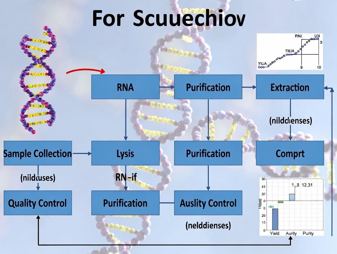
Visualizations
Diagram Title: RNA Quality Control Decision Workflow
Diagram Title: Impact of RNA Integrity on Sequencing Coverage Bias
The Scientist's Toolkit: Essential Research Reagent Solutions
| Item | Function & Role in RNA QC |
|---|---|
| Triazol-Based Lysis Reagent | A monophasic solution of phenol and guanidine isothiocyanate. Simultaneously lyses cells, inactivates RNases, and denatures proteins. The critical first step for preserving RNA integrity. |
| DNase I (RNase-free) | Enzyme that digests contaminating genomic DNA during the extraction process. Essential for obtaining RNA free of gDNA, which can confound RNA-seq mapping and analysis. |
| SPRI (Ampure) Beads | Paramagnetic carboxyl-coated beads used for size-selective purification and clean-up. Remove salts, solvents, primers, and other inhibitors. Crucial for improving RNA purity post-extraction. |
| RiboGreen / Qubit RNA Assay | Fluorescent dye that binds specifically to RNA. Provides accurate quantification independent of common contaminants like salts or protein, unlike UV absorbance. |
| RNA Integrity Assay Kits | (e.g., Agilent RNA 6000 Nano Kit, TapeStation HS RNA Kit). Include gel matrix, dyes, and standards for capillary electrophoresis to generate RIN/RQN scores. |
| RNase Inhibitor | Protein that non-competitively binds and inhibits various RNases. Added to RNA eluates or during cDNA synthesis to prevent trace degradation during storage or handling. |
| Nuclease-Free Water | Water treated to remove nucleases and tested to ensure it will not degrade RNA samples. Used for all dilutions and as an elution buffer. |
Technical Support Center
Troubleshooting Guides & FAQs
Section 1: RNA Integrity Number (RIN)
Q1: My RIN value is low (<7.0), but my RNA yields look good spectroscopically. What could be the cause and how can I fix it? A: Low RIN with good yield often indicates RNA degradation during or after extraction. Causes and solutions:
- Cause: Tissue was not immediately stabilized or snap-frozen after collection.
- Solution: Submerge tissue in at least 10 volumes of RNAlater or flash-freeze in liquid nitrogen immediately upon dissection.
- Cause: Homogenization was inefficient or generated excessive heat.
- Solution: Use a sufficient volume of denaturing lysis buffer and ensure thorough, rapid homogenization using a rotor-stator homogenizer kept cold. Process samples on ice.
- Cause: RNases contaminated the sample during handling.
- Solution: Use RNase-free consumables, change gloves frequently, and use dedicated RNase-decontaminated workstations and pipettes. Include a specific RNase inhibitor in downstream reactions.
Q2: The RIN algorithm fails or gives an error. What does this mean? A: An algorithm failure usually indicates an abnormal electrophoretic trace. Common reasons:
- Sample Overload: The RNA concentration is too high for the assay. Fix: Dilute the RNA in nuclease-free water and re-run the analysis.
- Severe Degradation: The trace shows no distinct ribosomal peaks. Fix: The sample is irreversibly degraded. Check the extraction protocol starting from sample collection.
- Buffer Contamination: Carryover of salts, solvents, or protein from extraction can distort the trace. Fix: Re-precipitate the RNA with ethanol and sodium acetate, wash with 75% ethanol, and re-dissolve in nuclease-free water.
Section 2: Spectrophotometric Ratios (A260/280 & A260/230)
Q3: My A260/280 ratio is too low (<1.8). What contaminant is likely present, and how do I clean up the RNA? A: A low A260/280 ratio typically indicates protein or phenol contamination.
- Protein Contamination: Perform an additional acid-phenol:chloroform extraction. Add an equal volume of acid-phenol:chloroform (pH 4.5), vortex, centrifuge, and recover the aqueous phase. Follow with a chloroform-only back-extraction.
- Phenol Contamination: Ensure proper phase separation during extraction. Do not take any material from the interphase. Perform a chloroform back-extraction on the aqueous phase. Precipitate the RNA and wash the pellet thoroughly with 75% ethanol.
Q4: My A260/230 ratio is unacceptably low (<2.0). What does this signify? A: A low A260/230 ratio signals contamination with chaotropic salts (e.g., guanidinium), carbohydrates, or organic compounds (e.g., phenol, ethanol).
- Primary Fix: Perform an additional ethanol precipitation. Add 0.1 volumes of 3M sodium acetate (pH 5.2) and 2.5 volumes of ice-cold 100% ethanol. Incubate at -20°C for >30 minutes. Centrifuge at max speed (>12,000 g) for 30 minutes at 4°C. Wash the pellet twice with freshly prepared 75% ethanol. Air-dry the pellet briefly (5-10 min) and resuspend in nuclease-free water.
Section 3: Quantification Discrepancies
Q5: My fluorometric quantification (e.g., Qubit) is significantly lower than my spectrophotometric (NanoDrop) concentration. Which one is correct? A: Fluorometric assays are more accurate for RNA. The discrepancy occurs because spectrophotometry (A260) measures all nucleic acids, including degraded RNA and contaminating DNA, while fluorometry measures only intact, double-stranded RNA.
- Interpretation: Trust the fluorometric value. The NanoDrop reading is inflated by contaminants.
- Action: Treat the sample with DNase I (RNase-free) to remove genomic DNA contamination, then re-quantify. Also, check the RNA integrity via RIN.
Q6: My quantification is fine, but my RNA fails in cDNA synthesis or sequencing library prep. Why? A: Residual contaminants invisible to standard QC can inhibit enzymes.
- Protocol for Cleanup: Use a column-based cleanup kit (e.g., silica membrane). Adjust binding conditions per manufacturer's instructions. Elute in a low-EDTA or EDTA-free buffer, as high EDTA can chelate Mg2+ required by enzymes.
- Protocol for Inhibitor Removal: Perform a magnetic bead-based clean-up (e.g., SPRI beads) with an increased bead-to-sample ratio (e.g., 1.8X) to remove small molecular contaminants. Wash beads thoroughly with 80% ethanol.
| Metric | Ideal Range | Indication of Problem | Likely Contaminant |
|---|---|---|---|
| RIN | 8.0 - 10.0 (Sequencing) | < 7.0: Potential library prep issues< 5.0: Severe degradation | N/A (Measures degradation) |
| A260/280 | 2.0 - 2.1 (10mM Tris)~1.8 (Water) | < 1.8 (in water) | Protein, Phenol |
| A260/230 | 2.0 - 2.4 | < 2.0 | Salts, Carbohydrates, Organics |
| Fluoro vs Spec | Difference < 10% | Fluor value << Spec value | DNA, Degraded RNA, Absorbing Contaminants |
Detailed Methodologies
Protocol 1: Acid-Phenol:Chloroform Cleanup for Protein/Phenol Removal
- Thaw RNA sample on ice.
- Add an equal volume of acid-phenol:chloroform (pH 4.5). Vortex vigorously for 30 seconds.
- Centrifuge at 12,000 g for 5 minutes at 4°C to separate phases.
- Carefully transfer the upper aqueous phase to a new tube.
- Add an equal volume of chloroform to the aqueous phase. Vortex and centrifuge as in step 3.
- Transfer the aqueous phase to a fresh tube. Proceed to ethanol precipitation.
Protocol 2: Ethanol Precipitation for Salt/Carbohydrate Removal
- To the aqueous RNA sample, add 0.1 volumes of 3M sodium acetate (pH 5.2) and 2.5 volumes of ice-cold 100% ethanol. Mix thoroughly by inversion.
- Incubate at -20°C for a minimum of 30 minutes (overnight is optimal for low-concentration samples).
- Centrifuge at >12,000 g for 30 minutes at 4°C.
- Decant supernatant. Wash pellet with 750 μL of freshly prepared 75% ethanol.
- Vortex briefly and centrifuge at 12,000 g for 10 minutes at 4°C.
- Carefully remove ethanol. Air-dry pellet for 5-10 minutes (do not over-dry).
- Resuspend in nuclease-free water or TE buffer (pH 8.0).
Protocol 3: DNase I Treatment for Genomic DNA Removal
- Combine in a nuclease-free tube: 1 μg RNA, 1 μL 10X DNase I Buffer, 1 μL RNase-free DNase I, and Nuclease-free water to 10 μL.
- Mix gently and incubate at 37°C for 20-30 minutes.
- Inactivate DNase I by adding 1 μL of 50mM EDTA and incubating at 65°C for 10 minutes.
- Purify RNA using a column-based cleanup kit to remove enzymes and salts.
Visualization of Workflows
Title: RNA QC Metric Troubleshooting Decision Tree
Title: RNA Extraction to QC Workflow for Sequencing
The Scientist's Toolkit: Research Reagent Solutions
| Item | Function & Rationale |
|---|---|
| RNAlater Stabilization Solution | Penetrates tissues to rapidly inhibit RNases, preserving RNA integrity at the moment of collection. Allows storage at 4°C or -20°C before extraction. |
| Denaturing Lysis Buffer (Guanidinium Thiocyanate) | A chaotropic salt that denatures proteins and RNases on contact, ensuring RNA stability during homogenization. |
| Acid-Phenol:Chloroform (pH 4.5) | Organic extraction mixture. The acidic pH partitions RNA to the aqueous phase while DNA and proteins remain in the organic phase or interphase. |
| RNase-free DNase I | Enzyme that digests contaminating genomic DNA without degrading RNA, critical for accurate quantification and sequencing. |
| RNA-binding Silica Columns/Magnetic Beads | Selective binding of RNA in high-salt conditions, allowing efficient washing away of salts, organics, and other contaminants. |
| Fluorometric RNA Assay Dye (e.g., Qubit RNA HS) | RNA-selective dye that fluoresces only when bound to RNA, providing accurate concentration measurements free from common contaminants. |
| RNA Integrity Chip (e.g., Bioanalyzer) | Microfluidic capillary electrophoresis system that separates RNA fragments by size, generating an electropherogram and calculating the RIN algorithm. |
Troubleshooting Guides & FAQs
Q1: My RNA yield is consistently low after extraction. What are the most likely culprits? A: Low RNA yield is frequently caused by incomplete tissue/cell lysis, RNase contamination, or improper handling of the RNA pellet. Ensure immediate homogenization in a denaturing lysis buffer (e.g., containing guanidinium isothiocyanate), use RNase-free consumables, and avoid over-drying the RNA pellet, which makes it difficult to resuspend.
Q2: My RNA has poor purity (A260/280 < 1.8). What does this indicate and how can I fix it? A: A low A260/280 ratio typically indicates protein contamination (e.g., from phenol carryover during phase-separation methods). A high ratio (>2.2) suggests residual chaotropic salts or guanidine. Solutions include: repeating a chloroform extraction and ethanol precipitation for protein, or using a wash buffer with a higher ethanol concentration (e.g., 80%) and allowing the column to dry before elution to remove salts.
Q3: My RNA Integrity Number (RIN) is low, but I worked quickly. What hidden sources of RNases should I suspect? A: Beyond obvious sources like contaminated pipettes, common hidden RNase sources include: 1) User-borne RNases from skin and hair – always wear gloves and change them frequently. 2) Laboratory surfaces and equipment – regularly decontaminate with RNase-inactivating solutions. 3) Endogenous RNases in samples – ensure immediate and thorough sample homogenization directly into the lysis buffer to inactivate RNases instantly.
Q4: My downstream cDNA synthesis or qPCR is inefficient. Could my RNA sample contain inhibitors? A: Yes. Common inhibitors co-purified with RNA include:
- Phenol, Guanidine Salts, and Ethanol from the extraction process.
- Hemoglobin/Heparin from blood samples.
- Polysaccharides and Polyphenols from plant tissues.
- Cellular metabolites and proteins. Mitigation involves using silica-membrane column purification with rigorous wash steps, or performing an additional ethanol precipitation with sodium acetate.
Q5: How can I prevent RNA degradation during storage? A: For short-term (<1 month), store RNA in RNase-free water or TE buffer at -80°C. For long-term storage, adjust pH to slightly alkaline (with TE, pH 7.5-8.0) and store at -80°C. Avoid repeated freeze-thaw cycles; aliquot RNA into single-use quantities. Liquid nitrogen storage is optimal for very long-term preservation.
Table 1: Common RNA Integrity Indicators and Interpretations
| Metric | Optimal Value | Sub-Optimal Value | Likely Cause |
|---|---|---|---|
| A260/A280 | 1.8 - 2.1 (TE) | <1.8 | Protein/Phenol Contamination |
| >2.2 | Chloroform/Guanidine Salt, or Low RNA concentration | ||
| A260/A230 | 2.0 - 2.4 | <1.8 | Carbohydrate, Guanidine, or Ethanol Carryover |
| RIN (Bioanalyzer) | 8.0 - 10.0 | <7.0 | Significant RNA Degradation |
| 28S/18S rRNA Ratio | ~2.0 (Mammalian) | <1.5 | Partial Degradation |
Table 2: RNase Inactivation Efficacy of Common Reagents
| Reagent / Method | Mode of Action | Effectiveness | Notes |
|---|---|---|---|
| Guanidinium Salts | Protein denaturation | Very High | Immediate inactivation in lysis buffer |
| β-Mercaptoethanol | Reducing agent | High | Add to lysis buffers; neutralizes RNases |
| DEPC-treated Water | Alkylating agent | High | Inactivates RNases irreversibly; for solutions only |
| RNaseZap / Commercial Sprays | Chemical denaturation | High | For surface decontamination |
| Dry Heat (Baking) | Protein denaturation | Moderate | 180-250°C for several hours for glass/ metal |
Detailed Experimental Protocols
Protocol 1: Acid Guanidinium Thiocyanate-Phenol-Chloroform (AGPC) Extraction (Single-Step Method) Principle: Simultaneous lysis and inactivation of RNases with a monophasic solution of phenol and guanidinium isothiocyanate, followed by phase separation.
- Homogenize sample in at least 10 volumes of TRIzol/ TRI Reagent. Incubate 5 min at RT for complete dissociation.
- Add 0.2 ml chloroform per 1 ml TRIzol. Cap tightly, shake vigorously for 15 sec. Incubate 2-3 min at RT.
- Centrifuge at 12,000 x g for 15 min at 4°C. The mixture separates into three phases: a red organic phase (phenol-chloroform), an interphase (DNA), and a colorless upper aqueous phase (RNA).
- Transfer the aqueous phase (approx. 50-60% of TRIzol volume) to a new tube.
- Precipitate RNA by adding 0.5 ml isopropyl alcohol per 1 ml TRIzol used. Mix. Incubate 10 min at RT.
- Centrifuge at 12,000 x g for 10 min at 4°C. The RNA pellet is often translucent.
- Remove supernatant. Wash pellet with 1 ml 75% ethanol (in DEPC-water) per 1 ml TRIzol used.
- Vortex briefly, centrifuge at 7,500 x g for 5 min at 4°C. Air-dry pellet for 5-10 min (do not over-dry).
- Resuspend RNA in 20-50 µl RNase-free water or TE buffer (pH 8.0). Heat at 55°C for 10 min to aid dissolution.
Protocol 2: Silica-Membrane Column Purification (Spin-Column) Principle: RNA binding to a silica membrane in the presence of high-concentration chaotropic salt (e.g., guanidine HCl), followed by washes and elution in low-salt buffer.
- Lysate the sample in a buffer containing a strong denaturant (guanidine thiocyanate) and a detergent.
- Add ethanol to the lysate to create optimal binding conditions and apply the mixture to the spin column.
- Centrifuge (≥ 8,000 x g for 15-30 sec). The RNA binds to the membrane; contaminants pass through.
- Wash the membrane with a buffer containing ethanol to remove salts and other impurities. Centrifuge after each wash.
- Perform an optional on-column DNase I digestion (in a buffer containing Mn2+ or Mg2+) for 15 min at RT to remove genomic DNA.
- Perform additional wash steps to remove the DNase and any residual contaminants.
- Crucially, spin the empty column for 1-2 min to dry the membrane and remove residual ethanol, which inhibits downstream reactions.
- Elute the pure RNA with 30-50 µl of RNase-free water or TE buffer by centrifugation.
Visualization: Experimental Workflows & Pathways
Diagram Title: RNA Extraction Workflow with Key Pitfalls
The Scientist's Toolkit: Key Research Reagent Solutions
| Reagent / Material | Function in RNA Work | Critical Notes |
|---|---|---|
| Guanidinium Thiocyanate / HCl | Powerful chaotropic agent. Denatures proteins and RNases, disrupts cells, and promotes nucleic acid binding to silica. | Core component of almost all modern lysis buffers. |
| β-Mercaptoethanol (BME) | Reducing agent. Breaks disulfide bonds in RNases, enhancing denaturation by guanidinium. | Always add fresh to lysis buffer. Use in a fume hood. |
| RNase-free Water (DEPC-treated) | Solvent for resuspending and storing RNA. DEPC alkylates and inactivates RNases. | Do not use on Tris buffers (DEPC reacts with amines). |
| DNase I (RNase-free) | Enzyme that degrades contaminating genomic DNA. Crucial for applications sensitive to DNA (e.g., RNA-seq, qRT-PCR). | Requires incubation in a specific buffer (with Mg2+/Mn2+). Must be subsequently inactivated/removed. |
| RNase Inhibitors (e.g., Recombinant RNasin) | Protein that non-covalently binds to and inhibits a broad spectrum of RNases. Used during cDNA synthesis and other enzymatic reactions. | Protects RNA after it is purified from denaturing conditions. Does not replace careful technique. |
| Silica-membrane Spin Columns | Solid-phase extraction medium. Binds RNA selectively in high-salt, allows contaminants to wash away, and elutes in low-salt. | Enables rapid, reproducible purifications. Membrane drying is a critical step. |
| Anhydrous Ethanol & Isopropanol | Precipitation and wash agents. Reduces solubility of nucleic acids in aqueous solutions; washes away salts. | Use high-purity, molecular biology grade. Ensure correct concentration for wash steps (typically 70-80%). |
This technical support center is a dedicated resource within a broader thesis on RNA extraction troubleshooting for sequencing research. It provides specific, actionable guidance for researchers, scientists, and drug development professionals facing experimental challenges across diverse RNA sequencing applications. The protocols, FAQs, and tools below address common pitfalls from sample preparation to library construction.
Troubleshooting Guides & FAQs
Q1: My total RNA-seq data shows high ribosomal RNA (rRNA) contamination despite using poly-A selection. What are the likely causes and solutions? A: This is often due to RNA degradation or incorrect protocol execution.
- Cause 1: Partial RNA Degradation. Poly-A selection requires intact mRNA with poly-A tails. Degraded RNA has truncated tails that bind inefficiently to poly-T beads.
- Solution: Check RNA Integrity Number (RIN) on a Bioanalyzer or TapeStation. A RIN > 8 is optimal for poly-A selection. For lower-quality samples (e.g., from FFPE), use rRNA depletion kits instead.
- Cause 2: Insufficient Bead Binding or Wash Conditions.
- Solution: Ensure binding buffer contains the correct concentration of salts (e.g., NaCl, MgCl₂) and is at room temperature. Do not over-wash beads. Include a DNase I digestion step during extraction to remove genomic DNA, which can non-specifically bind.
Q2: For targeted RNA-seq (e.g., using hybrid capture panels), my on-target rate is low. How can I optimize this? A: Low on-target rate indicates inefficient capture.
- Cause 1: Insufficient Probe Coverage or Poor Probe Design.
- Solution: Ensure your custom panel uses optimized probe lengths (80-120 nt) and tiles across exons. Verify probe sequences are specific and avoid repetitive regions.
- Cause 2: Suboptimal Hybridization Conditions.
- Solution: Precisely follow hybridization temperature and time. Use a thermal cycler with a heated lid. Increase the amount of input RNA/cDNA if within the kit's linear range. Ensure Cot-1 or other blocking reagents are fresh and used at the recommended concentration to suppress repetitive elements.
Q3: My long-read sequencing (PacBio or Oxford Nanopore) yields are low, and reads are shorter than expected. What steps should I take? A: This typically points to RNA integrity or reverse transcription issues.
- Cause 1: RNA Fragmentation or Damage.
- Solution: For full-length isoform sequencing, use high-integrity RNA (RIN > 9). Minimize freeze-thaw cycles. Store RNA in aliquots in nuclease-free buffers. Avoid vortexing. Perform extraction on ice with fresh RNase inhibitors.
- Cause 2: Inefficient Reverse Transcription for cDNA Synthesis.
- Solution: Use a high-fidelity, processive reverse transcriptase specifically recommended for long-read applications. Optimize reaction time and temperature. Include a template-switching oligo if required by the protocol. Perform a size selection step (e.g., using BluePippin or SageELF) post-cDNA synthesis to remove short fragments.
Experimental Protocols
Protocol 1: Assessing RNA Integrity for Any Sequencing Application
Principle: Electrophoretic analysis of RNA to assign an Integrity Number (RIN).
- Equipment: Agilent Bioanalyzer 2100 or TapeStation, RNA Nano or High Sensitivity RNA chips.
- Procedure: a. Prepare samples and ladder according to manufacturer's instructions. b. Load 1 µL of sample per well. c. Run the appropriate assay (e.g., RNA Nano). d. Analyze electrophoregram: sharp 18S and 28S rRNA peaks (with a 2:1 ratio for mammalian RNA) and a flat baseline indicate high integrity. Software assigns a RIN (1=degraded, 10=intact).
Protocol 2: Ribosomal RNA Depletion for Degraded or Non-Poly-A Samples
Principle: Use sequence-specific probes to remove rRNA.
- Reagents: Commercial rRNA depletion kit (e.g., Illumina Ribo-Zero Plus, QIAGEN FastSelect).
- Procedure: a. Incubate 100-1000 ng of total RNA with biotinylated rRNA-specific probes. b. Add streptavidin-coated magnetic beads to bind probe-rRNA complexes. c. Use a magnetic stand to separate supernatant (rRNA-depleted RNA) from beads. d. Precipitate or clean up the rRNA-depleted RNA. Quantify yield.
Protocol 3: cDNA Synthesis and Size Selection for Long-Read Sequencing
Principle: Generate full-length, amplified cDNA and select optimal fragment sizes.
- Reagents: Clontech SMARTer cDNA Synthesis Kit, AMPure PB beads, BluePippin or SageELF system.
- Procedure: a. Perform first-strand cDNA synthesis using a template-switching oligo. b. Perform LD PCR to amplify the cDNA (optimize cycle number to prevent over-amplification). c. Clean up cDNA with AMPure PB beads. d. Perform size selection using a preparative electrophoresis system (e.g., BluePippin with a 5-9 kb cut-off) according to the system's manual. e. Quantify the size-selected cDNA using a fluorometer (e.g., Qubit).
Data Presentation
Table 1: RNA Sequencing Applications and Their Input Requirements
| Application | Recommended Input Amount | Minimum RNA Integrity (RIN) | Key RNA Requirement | Primary Goal |
|---|---|---|---|---|
| Standard Total RNA-Seq (Poly-A) | 10-1000 ng | 8.0 | Intact poly-A tail | Gene expression profiling |
| Total RNA-Seq (rRNA depletion) | 1-1000 ng | 2.0 (FFPE) to 8.0 | Broad RNA species | Transcriptome without poly-A bias |
| Targeted RNA-Seq | 10-100 ng | 7.0 | Known target sequences | Detect specific transcripts/isoforms |
| Single-Cell RNA-Seq | ~1 pg/cell | N/A (immediately processed) | Minimized amplification bias | Cellular heterogeneity |
| PacBio Iso-Seq | 100-1000 ng | 9.0+ | Full-length transcripts | Full-length isoform discovery |
| Nanopore Direct RNA-Seq | 50-500 ng | 8.5+ | Native RNA with poly-A tail | Direct RNA modification detection |
Table 2: Common RNA Extraction Issues and Impact on Sequencing
| Symptom | Potential Extraction Cause | Impact on Sequencing | Corrective Action |
|---|---|---|---|
| Low RIN / Degraded RNA | RNase contamination, slow processing, harsh lysis | Reduced mapping, 3' bias, failed lib prep | Use fresh RNase inhibitors, process on ice, optimize tissue homogenization |
| Low RNA Yield | Inefficient lysis, poor binding to column, small sample input | Insufficient material for library prep | Add carrier RNA, ensure correct ethanol % in binding buffer, use disruptive lysis (bead beating) |
| Genomic DNA Contamination | Inefficient DNase I treatment | Reads mapping to introns/non-coding regions | Perform on-column DNase digestion, check digestion incubation time/temperature |
| Organic Solvent Carryover (e.g., Phenol) | Incomplete phase separation, inadequate washing | Inhibits enzymatic steps in library prep | Ensure proper centrifugation for phase sep, follow wash buffer volumes, do final 80% ethanol wash |
| A260/A280 Ratio <1.8 | Protein or phenol contamination | Enzyme inhibition in downstream steps | Repeat cleanup with a column-based kit, avoid interphase during aqueous phase transfer |
Mandatory Visualization
Title: RNA Sequencing Experimental Workflow Decision Tree
Title: mRNA Processing Pathway to Sequencing
The Scientist's Toolkit: Research Reagent Solutions
| Item | Function in RNA Sequencing Workflow |
|---|---|
| RNase Inhibitors (e.g., Recombinant RNasin) | Inactivates RNases during extraction and handling to preserve RNA integrity. |
| Magnetic Beads (Silica or Streptavidin) | For nucleic acid binding, cleanup (SPRI beads), and targeted selection (poly-A/rRNA depletion). |
| Template-Switching Reverse Transcriptase | Enables full-length cDNA synthesis by adding a universal sequence to the 5' end, critical for long-read and single-cell protocols. |
| RNA Integrity Assay Kits (Bioanalyzer/TapeStation) | Provides quantitative assessment (RIN/DIN) of RNA degradation prior to costly library prep. |
| Ribosomal RNA Depletion Probes | Biotinylated oligonucleotides that hybridize to rRNA species (cytoplasmic and mitochondrial) for their removal, enabling analysis of non-poly-A transcripts. |
| Size Selection Systems (BluePippin, SageELF) | Precise physical isolation of nucleic acid fragments by size, essential for optimizing long-read sequencing libraries. |
| DNase I (RNase-free) | Digests genomic DNA contamination during or after RNA extraction, preventing false-positive signals in RNA-seq data. |
| PCR Additives (e.g., Betaine, DMSO) | Reduce secondary structure and improve amplification efficiency during cDNA amplification or target enrichment, especially for GC-rich regions. |
From Theory to Bench: Selecting and Executing the Optimal RNA Extraction Protocol
This technical support center is part of a broader thesis on RNA extraction troubleshooting for sequencing research. It provides targeted FAQs and guides to address common pitfalls in RNA extraction using the three dominant methods.
Frequently Asked Questions & Troubleshooting
Q1: My RNA yield from a silica-column kit is consistently low from cultured cells. What could be wrong? A: Low yield often stems from incomplete cell lysis or RNase contamination. Ensure lysis buffer is fresh and added in sufficient volume. For adherent cells, lyse directly on the plate. Always use RNase-free reagents and consumables. For small sample sizes, carrier RNA (if compatible with downstream steps) or switching to a magnetic bead protocol designed for low-input samples may help.
Q2: I see a significant 28S/18S rRNA degradation (ratio <1.5) in my TRIzol extracts. How can I improve integrity? A: This indicates RNase activity or physical shearing. Key fixes: 1) Homogenize immediately after adding TRIzol; do not delay. 2) Keep samples cold and process quickly. 3) Avoid vortexing after the initial homogenization step. 4) Ensure the phase separation is clean; do not take any interphase material. 5) Use fresh, RNase-free glycogen or linear acrylamide during precipitation.
Q3: My magnetic bead-based purification has low RNA recovery. What should I check? A: Magnetic bead performance is highly sensitive to ethanol concentration and bead handling. 1) Verify that the ethanol concentration in the wash buffers is exactly as specified (usually 80%). 2) Do not let beads dry completely during wash steps. 3) Ensure beads are fully resuspended during binding and wash steps. 4) Use the correct bead-to-sample ratio. 5) For the final elution, use warm (e.g., 55°C) RNase-free water and incubate for 2-5 minutes to increase elution efficiency.
Q4: I get contaminating genomic DNA in my silica-column eluate. How do I remove it? A: Most kits include an on-column DNase I digestion step. Ensure you: 1) Prepare the DNase I digestion mix fresh. 2) Apply it directly to the center of the silica membrane. 3) Incubate at room temperature for the recommended time (usually 15 mins). 4) Use the specific wash buffers provided in the kit post-digestion. For TRIzol methods, a follow-up DNase treatment of the eluted RNA is standard.
Q5: My RNA A260/A280 ratio from a column is <1.8, suggesting protein contamination. How to fix? A: This typically indicates carryover of guanidine salts or phenol. For columns: 1) Ensure complete removal of Wash Buffer 1 (often an ethanol-based wash) before proceeding to Wash Buffer 2. 2) Perform an extra wash step with Wash Buffer 2 (usually an ethanol-buffer mix). 3) Centrifuge the empty column for an additional 2 minutes to dry the membrane completely before elution. For TRIzol, ensure no organic phase carryover during aqueous phase collection.
Q6: The magnetic beads are not separating cleanly. What influences this? A: Bead separation is hampered by high viscosity or particulate matter. 1) Centrifuge lysates briefly before adding to beads to remove debris. 2) Ensure adequate mixing during binding (by gentle pipetting or inversion, not vortexing). 3) Use a strong enough magnet and allow sufficient time for a clear supernatant to form (≥2 mins). 4) Check that the sample-to-bead binding buffer ratio is correct.
Table 1: Method Comparison for Key Parameters
| Parameter | Phenol (TRIzol) | Silica-Column | Magnetic Bead |
|---|---|---|---|
| Typical Yield | High | Medium-High | Medium-High |
| Processing Time | 1-3 hours | 30-60 mins | 30-45 mins |
| Cost per Sample | Low | Medium | Medium-High |
| Ease of Automation | Difficult | Moderate | Excellent |
| Scalability | Good (batch) | Good | Excellent |
| DNA Contamination Risk | Higher (req. DNase) | Lower (on-col. DNase) | Low |
| Organic Waste | High | Low | Low |
| Suitability for Small RNAs | Yes (<200 nt) | Varies by kit | Varies by kit |
Table 2: Common Issues and Primary Solutions
| Problem | TRIzol Primary Fix | Silica-Column Primary Fix | Magnetic Bead Primary Fix |
|---|---|---|---|
| Low Yield | Add carrier, ensure precip. | Check lysis, elute with warm H₂O | Check ethanol %, bead drying |
| DNA Contamination | Post-extraction DNase I | On-column DNase I | Use integrated DNase step |
| Protein Contamination | Careful phase separation | Extra wash, dry membrane | Optimize wash buffer volume |
| RNase Degradation | Rapid processing, cold | RNase-free workflow | RNase-free workflow |
| Inhibitor Carryover | 75% Ethanol wash | Extra wash step | Optimize bead washing |
| Poor Bead/Separation | N/A | N/A | Pre-clear lysate, strong magnet |
Detailed Protocol: TRIzol Extraction with DNase Treatment
This is a standard protocol for total RNA isolation, including miRNA.
- Homogenization: Lyse cells/tissue in 500 µL - 1 mL TRIzol reagent per 50-100 mg tissue or 10⁷ cells. Homogenize thoroughly.
- Phase Separation: Incubate 5 min at RT. Add 0.2 mL chloroform per 1 mL TRIzol. Shake vigorously for 15 sec. Incubate 2-3 min at RT. Centrifuge at 12,000 × g for 15 min at 4°C.
- RNA Precipitation: Transfer the clear aqueous phase to a new tube. Add 0.5 mL isopropyl alcohol per 1 mL TRIzol used. Mix. Incubate at RT for 10 min. Centrifuge at 12,000 × g for 10 min at 4°C. RNA pellet forms.
- Wash: Remove supernatant. Wash pellet with 1 mL 75% ethanol. Vortex. Centrifuge at 7,500 × g for 5 min at 4°C.
- Redissolve: Air-dry pellet briefly (5-10 min). Dissolve in 20-50 µL RNase-free water or TE buffer.
- DNase Treatment (Optional but recommended): Add 1 µL DNase I (RNase-free) and 5 µL 10x DNase buffer per 10 µg RNA. Incubate at 37°C for 20-30 min. Purify RNA using a silica-based cleanup column or ethanol precipitation.
Workflow Diagrams
TRIzol RNA Extraction and DNase Treatment Workflow
Decision Tree for Selecting an RNA Extraction Method
The Scientist's Toolkit: Key Research Reagent Solutions
| Item | Primary Function | Key Considerations |
|---|---|---|
| TRIzol/Chloroform | Organic lysis and phase separation for RNA isolation. | Contains phenol; requires proper hazardous waste disposal. Excellent for simultaneous DNA/protein recovery. |
| Silica-Column Membrane | Binds RNA under high-salt conditions for selective purification. | Performance varies by manufacturer. Avoid drying completely before elution. |
| Magnetic Silica Beads | Solid-phase paramagnetic particles for automated RNA binding/washing. | Bead size and surface chemistry impact yield and size selectivity. |
| RNase Inhibitors | Inhibit RNase activity during extraction. | Critical for sensitive samples. Often included in lysis buffers. |
| DNase I (RNase-free) | Degrades contaminating genomic DNA. | Essential for applications sensitive to DNA (e.g., RNA-seq, qRT-PCR). |
| Carrier RNA/Glycogen | Co-precipitates with low-abundance RNA to visualize pellet and improve yield. | Ensure carrier does not interfere with downstream assays (e.g., sequencing). |
| Ethanol (75-80%) | Wash solution to remove salts without eluting RNA from silica. | Concentration is critical; must be made with RNase-free water. |
| β-Mercaptoethanol/DT | Reducing agent added to lysis buffers to inactivate RNases. | Use in a fume hood; add fresh to buffers. |
Technical Support Center: Troubleshooting Guides & FAQs
FFPE (Formalin-Fixed, Paraffin-Embedded) Tissue FAQs
Q1: My RNA yield from FFPE tissue is low and highly fragmented. How can I optimize for sequencing? A: FFPE fixation causes RNA fragmentation and cross-linking. Use a specialized FFPE RNA extraction kit that includes extensive proteinase K digestion (up to 18 hours at 56°C) and a robust de-crosslinking step (often at 80°C). Post-extraction, assess RNA Integrity Number Equivalent (RINe) using a Fragment Analyzer or Bioanalyzer. For sequencing, employ ribosomal RNA depletion instead of poly-A selection, and use library prep protocols designed for degraded RNA (e.g., with random priming and shorter fragment sizes).
Q2: How do I remove paraffin effectively without losing sample? A: Perform two sequential xylene (or xylene-substitute) washes at 50°C for 5-10 minutes, followed by two ethanol washes. Centrifuge thoroughly between steps to pellet tissue. Ensure complete ethanol removal before lysis. For automated systems, verify the deparaffinization module is functioning correctly.
Lipid-Rich Tissue (e.g., Brain, Adipose) FAQs
Q3: My RNA from brain or adipose tissue has low purity (A260/A280 < 1.8). What's the solution? A: Low A260/A280 indicates carryover of organic contaminants like lipids or phenol. Solution: Incorporate a chloroform-based phase separation step during homogenization. After adding the initial lysis buffer (containing a strong chaotropic salt like guanidinium), add 1/5 volume of chloroform, mix vigorously, and centrifuge. The lipids will partition into the organic phase and interphase. Carefully transfer the aqueous (upper) phase containing RNA to a new tube for subsequent binding to silica columns. A second chloroform wash may be necessary.
Q4: Homogenization of fatty tissue is inefficient. Any recommendations? A: Pre-chill all equipment and solutions. For manual disruption, use a motorized homogenizer with disposable plastic probes. For larger samples, a bead mill homogenizer with ceramic beads in a pre-chilled tube is highly effective. Keep samples on ice at all times to inhibit RNases and prevent lipid smearing.
Fibrous Tissue (e.g., Heart, Muscle, Plant) FAQs
Q5: I cannot fully disrupt tough fibrous tissue, leading to inconsistent yields. A: Use a combination of mechanical and enzymatic disruption. First, flash-freeze tissue in liquid nitrogen and pulverize using a mortar and pestle or a cryomill. Transfer the powder to a tube with lysis buffer. Then, consider adding a supplementary proteinase K digestion step. For plant tissues, a CTAB (cetyltrimethylammonium bromide)-based lysis buffer is often essential to break down polysaccharide-rich cell walls.
Q6: My RNA pellets from fibrous tissues are difficult to resuspend. A: Avoid ethanol over-drying. After the final wash, air-dry the pellet for 5-10 minutes only until it appears translucent, not cracked. Resuspend in nuclease-free water or TE buffer by passing the solution up and down a pipette tip repeatedly. Incubating at 55°C for 10 minutes can aid resuspension. Vortexing is not recommended for high molecular weight RNA.
Low Biomass & Single-Cell Samples FAQs
Q7: How can I prevent losing my sample during RNA extraction from low cell numbers? A: Switch to a carrier RNA or linear acrylamide-based protocol. Add 1-2 µL of glycogen or carrier RNA (e.g., 1 µg/µL) during the precipitation step to visualize the pellet and maximize recovery. Use siliconized/low-retention tubes and tips throughout. Consider solid-phase reversible immobilization (SPRI) bead-based cleanups over column-based methods for more consistent recovery of small volumes.
Q8: How do I handle potential contamination in single-cell samples? A: Contamination from ambient RNases or foreign RNA is a critical issue. Implement strict single-cell RNA-seq best practices: work in a UV-equipped laminar flow hood, use RNase decontamination solutions on surfaces and equipment, include negative control (no cell) samples in every batch, and use dedicated reagents and aliquots.
Summarized Quantitative Data
Table 1: Comparative Performance of RNA Extraction Methods Across Tissue Types
| Tissue Challenge | Method / Kit | Avg. Yield (ng/mg tissue) | Avg. RIN/DV200 | Key Limitation Addressed |
|---|---|---|---|---|
| FFPE | Specialized FFPE Kit | 50-200 ng/section | RINe: 2.0-3.5 | De-crosslinking & fragmentation |
| FFPE | Standard Column Kit | 5-50 ng/section | RINe: <1.8 | Inadequate de-crosslinking |
| Lipid-Rich (Brain) | Protocol w/ Chloroform Wash | 800-1500 | RIN: 8.0-9.5 | Lipid/oil removal |
| Lipid-Rich (Brain) | Standard Protocol | 200-700 | RIN: 6.0-7.5 | Low A260/A280 purity |
| Fibrous (Heart) | Cryopulverization + CTAB | 400-800 | RIN: 7.5-9.0 | Incomplete homogenization |
| Fibrous (Heart) | Direct Homogenization | 100-300 | RIN: 5.0-7.0 | Low yield from tough fibers |
| Low Biomass (<10k cells) | Carrier RNA Precipitation | 60-80% recovery | DV200: >80% | Sample loss in handling |
| Low Biomass (<10k cells) | Standard Column | 20-40% recovery | DV200: Variable | Binding inefficiency at low conc. |
Table 2: Impact of Fixation Time on FFPE RNA Quality
| Formalin Fixation Time | RNA Yield (ng/mm³) | Median Fragment Length (nt) | Success Rate in RNA-Seq* |
|---|---|---|---|
| <24 hours | 150-300 | 250-400 | >90% |
| 24-72 hours | 100-200 | 150-300 | 75% |
| >72 hours (overfixed) | 20-80 | 80-150 | <50% |
*Defined as producing >10M mapped reads with expected complexity.
Experimental Protocols
Protocol 1: RNA Extraction from Lipid-Rich Tissues with Phase Separation
- Homogenization: Place up to 30 mg of fresh-frozen tissue in 1 mL of Qiazol (or TRIzol) lysis reagent. Homogenize with a rotor-stator homogenizer for 30 seconds on ice.
- Phase Separation: Incubate homogenate for 5 min at RT. Add 200 µL of chloroform, shake vigorously for 15 sec, incubate 3 min at RT. Centrifuge at 12,000 x g for 15 min at 4°C. The mixture separates into: a lower red phenol-chloroform phase, an interphase (white, containing DNA and lipids), and a colorless upper aqueous phase (containing RNA).
- RNA Precipitation: Transfer the aqueous phase to a new tube. Add 500 µL of 100% isopropanol and 1 µL of glycogen (20 mg/mL). Mix and incubate at -20°C for at least 1 hour.
- Wash & Resuspend: Centrifuge at 12,000 x g for 30 min at 4°C. Carefully remove supernatant. Wash pellet with 1 mL of 75% ethanol. Centrifuge at 7,500 x g for 5 min at 4°C. Air-dry pellet for 5-10 min. Resuspend in 20-50 µL of RNase-free water.
Protocol 2: RNA Extraction from Low Biomass Samples using SPRI Beads
- Lysate Preparation: Lyse cells directly in a tube containing 200 µL of lysis/binding buffer (e.g., from a commercial microRNA kit) with 1% β-mercaptoethanol. Vortex thoroughly.
- Binding: Add 1.8x volumes of room-temperature SPRI (solid-phase reversible immobilization) beads (e.g., AMPure XP). Mix thoroughly by pipetting. Incubate for 5 min at RT.
- Capture: Place tube on a magnetic rack until supernatant is clear (~5 min). Carefully remove and discard supernatant.
- Wash: With tube on magnet, wash beads twice with 200 µL of 80% ethanol. Air-dry beads for 5 min.
- Elute: Remove tube from magnet. Elute RNA in 10-15 µL of nuclease-free water or TE buffer. Mix, incubate for 2 min, then capture beads on magnet. Transfer eluate containing RNA to a new tube.
Protocol 3: Deparaffinization and RNA Extraction from FFPE Sections
- Deparaffinization: Cut 2-4 x 10 µm FFPE sections into a microcentrifuge tube. Add 1 mL of xylene (or substitute). Vortex vigorously. Incubate at 50°C for 5 min. Centrifuge at full speed for 2 min. Carefully remove supernatant. Repeat once.
- Ethanol Washes: Add 1 mL of 100% ethanol to pellet. Vortex. Centrifuge at full speed for 2 min. Remove supernatant. Repeat once. Air-dry pellet for 5-10 min.
- Digestion & De-crosslinking: Resuspend pellet in 200 µL of digestion buffer with 20 µL of proteinase K (20 mg/mL). Incubate at 56°C with agitation (e.g., in a thermomixer) for up to 18 hours. Then incubate at 80°C for 15-60 minutes for de-crosslinking.
- RNA Purification: Proceed with the lysate using an FFPE-optimized silica column kit, following the manufacturer's instructions, typically involving binding conditions optimized for high salt and ethanol concentrations.
Visualizations
Decision Workflow for Challenging RNA Extraction
FFPE RNA Extraction Core Workflow
The Scientist's Toolkit: Key Reagent Solutions
| Reagent / Material | Function in Challenging Tissues |
|---|---|
| Proteinase K (High Concentration) | Digests proteins cross-linked to RNA in FFPE tissues; critical for efficient lysis of fibrous tissues. |
| Qiazol / TRIzol (with Chloroform) | Monophasic lysis reagent for lipid-rich tissues; enables phase separation to remove lipids and proteins. |
| CTAB (Cetyltrimethylammonium Bromide) | Ionic detergent effective for lysing plant and tough fibrous tissues by breaking down polysaccharide walls. |
| Glycogen or Carrier RNA | Co-precipitant for visualizing and maximizing RNA recovery from low biomass and low-concentration samples. |
| SPRI (AMPure) Beads | Magnetic beads for solid-phase reversible immobilization (SPRI) cleanup; superior recovery for low-input samples vs. columns. |
| β-Mercaptoethanol | Reducing agent added to lysis buffers to inhibit RNases, especially important in tissues with high RNase activity (e.g., pancreas). |
| Xylene (or Substitute) | Organic solvent for complete removal of paraffin wax from FFPE tissue sections prior to lysis. |
| RNase Inhibitor (e.g., Recombinant) | Essential additive for reactions post-extraction (e.g., cDNA synthesis) when working with highly degraded or low-input RNA. |
| DNase I (RNase-free) | For on-column or in-solution digestion of genomic DNA contamination, critical for FFPE samples where DNA is also extracted. |
Best Practices for Sample Collection, Stabilization, and Lysis to Inactivate RNases Immediately
Troubleshooting Guides & FAQs
Q1: My RNA yields are consistently low from tissue samples. What are the most critical steps during collection? A: Immediate stabilization is paramount. For tissues, excise a small piece (<0.5 cm thickness) and submerge it in at least 10 volumes of RNase-inactivating stabilization reagent (e.g., RNA-later) immediately. Do not freeze tissue in liquid nitrogen without prior chemical stabilization unless you can guarantee homogenization within minutes. Freeze-thaw cycles without stabilization rapidly degrade RNA.
Q2: I'm working with whole blood. How do I prevent RNA degradation from high endogenous RNase activity? A: For PAXgene Blood RNA tubes: Invert the tube 8-10 times immediately after collection to ensure mixing with the stabilizing reagent. Do not open the tube. Store upright at room temperature for at least 2 hours (up to 3 days) before processing or freezing at -20°C or -80°C. For traditional anticoagulants (e.g., EDTA), process within 30 minutes using a density gradient centrifugation with a compatible RNA stabilization additive in the lysis buffer.
Q3: My RNA Integrity Number (RIN) is poor despite using stabilization reagents. What could be wrong? A: The issue likely lies in the lysis step. Ensure your lysis buffer contains potent RNase inhibitors (e.g., guanidine salts, β-mercaptoethanol, or specific RNase inhibitors). The sample-to-lysis buffer ratio is critical; use at least 5-10 volumes of buffer to sample. Homogenize thoroughly and immediately after combining sample with lysis buffer. For tough tissues, use mechanical homogenization (bead mill or rotor-stator) while keeping samples chilled.
Q4: Can I store stabilized samples before RNA extraction, and if so, under what conditions? A: Yes, but conditions depend on the stabilization method. See the table below for quantitative stability data.
Table 1: Storage Conditions & RNA Stability for Stabilized Samples
| Sample Type | Stabilization Method | Room Temp | 4°C | -20°C | -80°C |
|---|---|---|---|---|---|
| Soft Tissue | RNA-later (immersed) | 1 week | 1 month | 1 year+ | Indefinite |
| Whole Blood | PAXgene Tube | 3 days | N/A* | 1 year+ | 5 years+ |
| Cell Culture | Qiazol Lysis Reagent | 1 hour | 1 week | 1 month | 1 year+ |
| FFPE Tissue | Formalin Fixation | N/A | N/A | Indefinite | Indefinite |
*Not recommended; store at -20°C after 2-hour incubation.
Q5: How do I effectively inactivate RNases during lysis of fibrous or fatty tissues? A: Use a two-step lysis protocol: 1) Mechanical disruption in a chaotropic (guanidinium-based) lysis buffer using a powerful homogenizer. 2) Follow with a chloroform extraction (for phenol-chloroform methods) or a proteinase K digestion step (for column-based methods) to break down the proteinaceous matrix and fully release and protect RNA.
Experimental Protocols
Protocol 1: Immediate Stabilization & Lysis for Mouse Liver Tissue (for High-Quality Total RNA)
- Materials: Dissection tools pre-cooled on dry ice, 1.5 mL RNase-free tubes, RNA-later, TRIzol or Qiazol, bead homogenizer.
- Procedure: a. Euthanize mouse and swiftly excise liver. Within 30 seconds, cut a piece <0.5 cm in any dimension. b. Immediately submerge tissue in 1 mL of RNA-later in a pre-labeled tube. Incubate overnight at 4°C. c. The next day, remove RNA-later and store tissue at -80°C or proceed to lysis. d. For lysis, add 1 mL of TRIzol to the tissue piece. Homogenize using a bead mill for 2 minutes at 25 Hz. e. Incubate the homogenate at room temperature for 5 minutes to ensure complete dissociation. f. Proceed with chloroform addition and phase separation per manufacturer's instructions.
Protocol 2: RNA Stabilization from Whole Blood for Plasma & Cellular RNA Analysis
- Materials: PAXgene Blood RNA Tubes, centrifuge, RNase-free serological pipettes.
- Procedure: a. Draw blood directly into a PAXgene tube. Invert immediately 8-10 times. b. Store tube upright at room temperature (15-25°C) for 2 hours to 3 days. c. For long-term storage, place tube at -20°C or -80°C. d. For processing, thaw (if frozen) and centrifuge at 3000-5000 x g for 10 minutes. e. Carefully decant supernatant. Add recommended lysis buffer to the pellet and proceed with extraction.
Diagrams
Title: Critical Workflow for RNA Sample Integrity
Title: RNase Inactivation Pathways During Lysis
The Scientist's Toolkit: Research Reagent Solutions
Table 2: Essential Reagents for RNA Stabilization & Lysis
| Reagent/Material | Primary Function | Key Consideration |
|---|---|---|
| RNA-later Stabilization Reagent | Penetrates tissue to inactivate RNases rapidly at room temperature. | Optimal for small tissue pieces; not for whole organs or large samples. |
| PAXgene Blood RNA Tubes | Contains proprietary additives that lyse blood cells and stabilize RNA immediately upon draw. | Requires specific downstream purification kits for optimal yield. |
| TRIzol/Qiazol (Acid-Phenol Guanidinium) | Combined lysis and stabilization: chaotropic salt denatures proteins, phenol inactivates RNases. | Contains phenol; requires careful handling and chloroform separation. |
| Bead Mill Homogenizer | Mechanical disruption of tough, fibrous, or frozen tissues in lysis buffer. | Ensures complete lysis; choose bead material (ceramic, steel) compatible with your sample. |
| β-Mercaptoethanol (BME) | Reducing agent added to lysis buffers to denature RNases by breaking disulfide bonds. | Toxic; use in a fume hood. Add fresh to lysis buffer just before use. |
| RNase Inhibitor Protein (e.g., Recombinant RNasin) | Binds non-covalently to RNases to inhibit activity. | Useful in downstream reactions but NOT sufficient for initial sample stabilization/lysis. |
| DNase/RNase-Free Water & Tubes | Provides an RNase-free environment for processed lysates and final RNA elution. | Always use certified consumables; never assume labware is RNase-free without treatment. |
Troubleshooting Guides & FAQs
Q1: Why is my RNA yield low after homogenization? A: Low yield often stems from incomplete tissue disruption or RNase degradation. Ensure homogenization is performed quickly in a cooled, RNase-free environment. For fibrous tissues, increase homogenization time by 15-20 seconds or use a specialized disruption kit. Verify that the homogenizer probe is clean and not degraded. If using a kit, check the lysis buffer-to-sample ratio; too much tissue can overwhelm the capacity of the binding column.
Q2: The aqueous phase after phenol-chloroform separation is cloudy or the interphase is thick. What should I do? A: A cloudy aqueous phase or thick interphase indicates incomplete phase separation, often due to improper homogenate viscosity or incorrect centrifugation. First, ensure centrifugation speed and time are as per protocol (typically 12,000 x g for 15 minutes at 4°C). If the problem persists, do not pipette any cloudy material. Re-centrifuge the tube or add an additional chloroform extraction step (0.2 volumes) to the recovered aqueous phase, mix, and re-centrifuge.
Q3: My RNA pellet is invisible or gelatinous after ethanol precipitation. How can I recover it? A: An invisible pellet suggests very low RNA quantity or co-precipitation of salts. A gelatinous pellet often indicates contamination with genomic DNA. For an invisible pellet, carefully aspirate the supernatant and wash with 70-75% ethanol. Centrifuge again at maximum speed for 10 minutes. For a gelatinous pellet, redissolve the pellet in nuclease-free water and add 0.1 volume of 3M sodium acetate (pH 5.2) and 1 volume of isopropanol. Incubate at -20°C for 30 minutes and re-pellet. Treating the lysate with a DNase step during purification is recommended to prevent gDNA contamination.
Q4: The RNA eluted from the column has low concentration (A260) but a normal 260/280 ratio. What is the issue? A: This typically indicates inefficient elution rather than poor yield. Ensure the elution buffer (nuclease-free water or TE buffer) is pre-heated to 55-60°C before application to the column membrane. After adding the buffer, let the column stand at room temperature for 2 minutes before centrifuging. For maximum yield, perform a second elution with a fresh aliquot of buffer. Also, verify that the binding and wash steps were performed at the correct pH; residual ethanol from washes can inhibit elution.
Q5: How should I store purified RNA for sequencing, and for how long is it stable? A: For short-term use (within a week), store RNA in nuclease-free water or TE buffer at -80°C. For long-term storage, precipitate RNA in ethanol and store at -80°C, or store in a stabilized commercial buffer. Avoid repeated freeze-thaw cycles. Aliquot the RNA to minimize degradation.
Q6: My Bioanalyzer/Fragment Analyzer trace shows degraded RNA (low RIN/ RQN). At which step did degradation likely occur? A: Degradation can occur at multiple points. See the troubleshooting flowchart below for systematic diagnosis.
Diagram Title: RNA Degradation Troubleshooting Flowchart
Key Experimental Protocols
Protocol 1: Optimized Phase Separation for Difficult Tissues (e.g., adipose, plant)
- Homogenize 30 mg of tissue in 1 mL of TRIzol or similar monophasic reagent. Use a bead mill for 2 minutes at 25 Hz for plant tissues.
- Incubate the homogenate for 5 minutes at room temperature.
- Add 0.2 mL of chloroform per 1 mL of TRIzol. Cap the tube securely.
- Shake vigorously by hand for 15 seconds. Do not vortex.
- Incubate at room temperature for 3 minutes.
- Centrifuge at 12,000 x g for 15 minutes at 4°C. The volume of the colorless upper aqueous phase should be ~50% of the TRIzol volume.
- Carefully transfer the aqueous phase to a new tube without disturbing the interphase. If interphase is disturbed, re-extract with an additional 0.1 volume of chloroform.
Protocol 2: On-Column DNase I Digestion for DNA-Free RNA
- After loading the lysate onto a silica membrane column and performing the first wash, prepare the DNase I mix: 10 µL of 10X DNase I Buffer, 5 µL of recombinant DNase I (1 U/µL), and 85 µL of nuclease-free water per sample.
- Apply the 100 µL DNase I mix directly onto the center of the column membrane.
- Incubate at room temperature (20-25°C) for 15 minutes.
- Wash the column with the provided Wash Buffer 1, then proceed with the standard wash and elution steps.
Protocol 3: Ethanol Precipitation for RNA Concentration and Clean-Up
- Measure the volume of your RNA in aqueous solution. Add 0.1 volumes of 3M sodium acetate (pH 5.2) and mix.
- Add 2.5 volumes of ice-cold 100% ethanol. Mix thoroughly by inverting.
- Incubate at -80°C for 30 minutes or -20°C overnight.
- Centrifuge at >12,000 x g for 30 minutes at 4°C. Carefully decant the supernatant.
- Wash the pellet with 500 µL of 75% ethanol (made with nuclease-free water). Centrifuge at 12,000 x g for 5 minutes at 4°C.
- Air-dry the pellet for 5-10 minutes until the ethanol evaporates (do not over-dry).
- Resuspend in the desired volume of nuclease-free water or TE buffer.
Table 1: Impact of Elution Buffer Temperature on RNA Yield from Silica Columns
| Column Type | Elution Buffer Temp. | Average Yield (µg) | % Increase vs. RT | RIN (Avg.) |
|---|---|---|---|---|
| Standard Silica | Room Temp (22°C) | 4.2 | Baseline | 8.5 |
| Standard Silica | 60°C | 5.8 | 38% | 8.4 |
| High-Binding Silica | Room Temp (22°C) | 5.5 | Baseline | 8.6 |
| High-Binding Silica | 60°C | 7.1 | 29% | 8.5 |
Table 2: Stability of Purified RNA Under Different Storage Conditions
| Storage Condition | Concentration Change (1 month) | 260/280 Ratio Change | RIN Drop (After 1 month) |
|---|---|---|---|
| -80°C, Nuclease-free Water | -3% | +/- 0.01 | -0.3 |
| -80°C, TE Buffer (pH 8.0) | -2% | +/- 0.01 | -0.2 |
| -20°C, Nuclease-free Water | -8% | -0.03 | -1.5 |
| 4°C, RNase Inhibitor Solution | -15% | -0.05 | -3.0 |
| -80°C, Ethanol Precipitated | -1% | No change | -0.1 |
The Scientist's Toolkit: Essential Research Reagent Solutions
Table 3: Key Reagents for RNA Extraction & Protocol Optimization
| Reagent / Material | Primary Function | Key Consideration for Optimization |
|---|---|---|
| TRIzol / Qiazol (Acid Phenol-Guanidine) | Simultaneous lysis and inhibition of RNases; initial phase separation. | Ensure freshness; protect from light. Volume must be sufficient for complete lysis (typically 1 mL per 50-100 mg tissue). |
| RNase-free Water (Molecular Grade) | Resuspension and elution of purified RNA. | Use certified nuclease-free, DEPC-treated, or 0.1 µm filtered. For elution, heating to 55-60°C increases yield. |
| DNase I (Recombinant, RNase-free) | Degradation of contaminating genomic DNA during purification. | Must be RNase-free. On-column digestion is most effective. Incubation time (15 min) and temperature (RT) are critical. |
| RNA Storage Solution (Stabilization Buffer) | Long-term stabilization of RNA by preventing degradation and maintaining integrity. | Superior to water or TE for long-term storage (>6 months) at -80°C or for shipping. Does not interfere with downstream applications like reverse transcription. |
| Silica Membrane Spin Columns | Selective binding of RNA in high-salt conditions, washing away impurities. | Binding capacity must not be exceeded. Ensure complete dryness after ethanol washes to prevent carryover. |
| β-Mercaptoethanol or DTT | Reducing agent added to lysis buffers to disrupt disulfide bonds and inactivate RNases. | Add fresh just before use. Use in a fume hood. Critical for tissues high in RNases (e.g., pancreas, spleen). |
| Glycogen or RNase-free Carrier | Co-precipitant to visualize pellet and improve recovery of low-concentration RNA (<50 ng/µL). | Use glycogen that is RNase-free. Add during the ethanol precipitation step before mixing. |
| Sodium Acetate (3M, pH 5.2) | Provides monovalent cations (Na+) required for ethanol precipitation of RNA. | pH is critical (pH 5.2 ensures DNA remains in solution while RNA precipitates). |
Diagram Title: Optimized RNA Extraction Core Workflow
The Role of DNase Treatment and Strategies for Genomic DNA Removal
FAQs and Troubleshooting Guides
Q1: Why is DNase treatment critical for RNA-seq and other downstream RNA applications? A1: Genomic DNA (gDNA) contamination in RNA samples can lead to false-positive signals in qRT-PCR, misalignment of sequencing reads, and inaccurate quantification of gene expression. DNase treatment enzymatically degrades double-stranded DNA, ensuring that only RNA is analyzed.
Q2: My RNA yield dropped significantly after DNase treatment. What went wrong? A2: A drastic drop in yield often indicates contamination with RNases during the DNase treatment or inactivation step. Ensure you are using an RNase-free DNase and that all reagents/equipment are RNase-free. Alternatively, excessive incubation time or temperature can lead to RNA degradation. Follow the manufacturer's recommended protocol strictly.
Q3: How do I confirm that gDNA contamination has been successfully removed? A3: Perform a no-reverse transcription (no-RT) control in your qPCR assay. Use primers that span an exon-intron junction (to detect unspliced genomic DNA) and target a housekeeping gene. A Cq value >5 cycles higher than your +RT sample, or undetectable, typically indicates effective DNA removal.
Q4: What are the pros and cons of on-column vs. in-solution DNase treatment? A4:
| Treatment Type | Pros | Cons |
|---|---|---|
| On-Column | Convenient, minimal hands-on time; DNase is washed away, no need for inactivation; reduces risk of sample cross-contamination. | May be less effective for high gDNA loads; potential for incomplete digestion if flow-through is too rapid. |
| In-Solution | Often more robust and complete digestion, especially for difficult samples with high gDNA. | Requires a separate inactivation step (e.g., with EDTA/heat); extra handling increases risk of RNase contamination and RNA loss. |
Q5: The DNase inactivation step (e.g., adding EDTA) is inhibiting my downstream reaction. What can I do? A5: EDTA chelates Mg2+, which is a cofactor for many enzymes like reverse transcriptase and Taq polymerase. Solutions include:
- Dilution: Dilute the treated RNA to reduce EDTA concentration.
- Repurification: Perform a second ethanol precipitation or clean-up column after DNase inactivation to remove EDTA and salts.
- Optimization: Increase Mg2+ concentration in your downstream reaction buffer to compensate.
Detailed Experimental Protocols
Protocol 1: On-Column DNase I Digestion (During RNA Purification)
Principle: DNase I is applied directly onto the silica membrane of the purification column after RNA binding, digesting co-bound gDNA. The enzyme and digestion products are then washed away.
- Perform standard lysate binding and wash steps per your RNA kit protocol.
- Prepare DNase I incubation mix: 10 µl DNase I, 70 µl Buffer RDD (Qiagen) or equivalent provided buffer.
- Apply the 80 µl mix directly onto the center of the column membrane. Incubate at room temperature (20-25°C) for 15 minutes.
- Proceed with the recommended wash steps and elution.
Protocol 2: In-Solution DNase I Digestion (Post-RNA Purification)
Principle: Purified RNA is digested with DNase I in a buffered solution, followed by chemical inactivation of the enzyme.
- Combine in a nuclease-free tube:
- RNA sample (up to 10 µg): X µl
- 10X DNase I Reaction Buffer: 5 µl
- RNase-free DNase I (1 U/µl): 5 µl
- Nuclease-free water to a final volume of 50 µl.
- Mix gently and incubate at 37°C for 20-30 minutes.
- Inactivate the DNase I by adding 5 µl of 50 mM EDTA and heating at 65°C for 10 minutes.
- (Optional) Purify the RNA using a standard ethanol precipitation or column clean-up protocol to remove EDTA and reaction components.
Table 1: Impact of gDNA Contamination on RNA-seq Metrics
| gDNA Contamination Level | Reads Mapped to Intergenic/Intronic Regions | Apparent Expression of Non-Expressed Genes | Correlation Between Biological Replicates |
|---|---|---|---|
| None (Effective DNase) | <5% | Negligible | High (R² > 0.98) |
| Moderate | 5-15% | Low but detectable | Reduced (R² 0.90-0.95) |
| High | >15% | Significant | Poor (R² < 0.90) |
Table 2: Comparison of Common DNase Inactivation Methods
| Method | Effectiveness | Risk of RNA Degradation | Compatibility with Downstream Apps |
|---|---|---|---|
| EDTA Chelation + 65°C Heat | High | Low if done correctly | May require cleanup if [EDTA] is high |
| Column Purification | Very High | Very Low | High (clean sample) |
| Phenol:Chloroform Extraction | High | Moderate (extra handling) | High (clean sample) |
| Proteinase K + SDS Treatment | High | Low | Requires subsequent cleanup |
Visualization
DNase Treatment Workflow Decision Guide
gDNA Removal Verification via No-RT qPCR
The Scientist's Toolkit: Research Reagent Solutions
| Reagent/Kit | Primary Function | Key Consideration for gDNA Removal |
|---|---|---|
| RNase-free DNase I | Enzyme that hydrolyzes phosphodiester bonds in DNA. | Must be rigorously free of RNase activity. Many are supplied with a proprietary buffer. |
| DNA Removal Columns (e.g., Zymo Spin IC) | Silica-based columns that selectively bind DNA after digestion. | Used post-digestion to remove DNase, EDTA, and digested DNA without ethanol precipitation. |
| RNA Purification Kits with On-Column DNase (e.g., Qiagen RNeasy, Norgen Biotek) | Integrated protocols for simultaneous RNA isolation and gDNA digestion. | Convenience vs. cost. Check digestion efficiency for your tissue type. |
| gDNA Eliminator Spin Columns | Specialized columns designed to remove gDNA during initial lysate cleanup. | Used before RNA binding, often for difficult samples. |
| Inactivation Reagents (e.g., 50 mM EDTA, Proteinase K) | Stop DNase activity to prevent downstream interference. | EDTA concentration post-inactivation must be compatible with subsequent enzymatic steps. |
| PCR Inhibitor Removal Reagents | Remove co-purified contaminants that inhibit RT/qPCR. | Useful if DNase treatment buffer components carry over and inhibit downstream assays. |
Diagnosing and Solving Common RNA Extraction Problems: A Practical Troubleshooting Manual
Technical Support Center: Troubleshooting Guides & FAQs
FAQ: Common Issues and Solutions
Q1: My RNA samples show degraded bands (smearing) on the Bioanalyzer instead of discrete 18S and 28S rRNA peaks. What is the most likely source of contamination? A: The most common source is RNase contamination introduced via improper technique. Key sources include: contaminated reagents (especially water), non-dedicated labware, benchtop surfaces, and user contact (skin, hair). RNases are extremely stable and require active inhibition.
Q2: I always use DEPC-treated water and filter tips, but my RNA still degrades. What am I missing? A: DEPC treatment is ineffective against some RNases and can interfere with downstream applications if not thoroughly inactivated. Your issue may stem from: 1) Equipment: Centrifuges, ice buckets, and tube racks are often overlooked. Wipe with RNase decontamination solutions (e.g., RNaseZap). 2) Storage: Frequent freeze-thaw cycles degrade RNA. Aliquot RNA in nuclease-free tubes. 3) Sample Handling: Working too slowly at room temperature allows endogenous RNases to act.
Q3: How can I systematically identify the specific source of RNase contamination in my workflow? A: Implement a controlled diagnostic experiment. Test each component of your workflow in isolation using a stable RNA control (e.g., a commercially available intact RNA ladder).
Diagnostic Experiment Protocol: Pinpointing RNase Contamination
Objective: To isolate which component (reagent, surface, or instrument) is causing RNA degradation. Materials:
- Intact RNA control (0.1-1 µg/µL)
- Test items: Batches of water, buffer aliquots, new vs. old tube racks, cleaned vs. uncleaned pipettes, etc.
- Nuclease-free microcentrifuge tubes and tips.
- Thermo cycler or water bath.
- Bioanalyzer or gel electrophoresis system.
Method:
- Preparation: Divide your intact RNA control into multiple, single-use aliquots.
- Incubation Test: For each test item (e.g., a suspect water batch), combine 1 µL of RNA with 9 µL of the test item in a nuclease-free tube. Include a positive control (RNA + certified nuclease-free water) and a negative control (RNA + known RNase-contaminated water).
- Challenge: Incubate all mixtures at room temperature (25°C) for 30 minutes or 37°C for 10 minutes to simulate a mild workflow challenge.
- Analysis: Immediately place samples on ice and assess integrity using a Bioanalyzer (RIN score) or formaldehyde-agarose gel electrophoresis.
- Interpretation: Compare the electropherogram or gel image of each test sample to the positive control. Degradation (smearing, lower RIN) in a specific sample identifies the contaminated component.
Q4: What are the critical aseptic techniques specific to RNA work? A:
- Dedicated Space: Use a clean, clutter-free bench area designated for RNA work, if possible.
- Personal Protective Equipment (PPE): Always wear a clean lab coat, gloves, and change gloves frequently. Avoid touching hair, face, or door handles with gloved hands.
- RNase-Decontaminated Surfaces: Thoroughly clean the work area before and after use with an RNase-inactivating solution. Use fresh bench paper.
- Reagent & Tool Dedication: Use a dedicated set of micropipettes for RNA work. Use only sterile, filter-plugged pipette tips and nuclease-free tubes. Never dip used pipette tips into stock reagent bottles.
- Pre-aliquoting: Aliquot all buffers, water, and reagents into small, single-use volumes to minimize contamination of master stocks.
- Temperature Control: Keep samples on dry ice or at -80°C for storage, and on wet ice during thawing and handling. Perform centrifugations at 4°C when possible.
Table 1: Quantitative Assessment of Common RNase Sources and Inactivation Methods
| Contamination Source | Relative Risk (1-5) | Effective Inactivation Method | Time/Effort Required |
|---|---|---|---|
| Pipettes (exterior/internal) | 4 | Wiping with RNase decontaminate; Using filter barriers | Low |
| Lab Water Purification System | 5 | Using certified Nuclease-Free Water; In-lab UV treatment system | Medium |
| User's Bare Skin | 5 | Consistent glove use; No touching of tubes/racks | Low |
| Reagent Contaminants | 3 | Aliquoting; Using RNase inhibitors in buffers | Low |
| Benchtop Surface | 2 | Routine cleaning with RNase decontaminate | Low |
| Centrifuge Rotors/Chambers | 3 | Cleaning with mild detergent & ethanol; Dedicated rotors | Medium |
| Ice Buckets & Tube Racks | 2 | Designated, plastic; Occasional decontamination soak | Low |
Table 2: Research Reagent Solutions for RNase Control
| Item | Function & Importance |
|---|---|
| RNase Decontamination Solution (e.g., RNaseZap) | Ready-to-use spray/wipes to rapidly inactivate RNases on surfaces, glassware, and equipment. Essential for daily bench cleaning. |
| Molecular Biology Grade Water (Nuclease-Free) | The solvent for all RNA work reagents. Must be certified free of nucleases. Do not use DEPC-treated water for downstream sequencing. |
| RNase Inhibitors (e.g., Recombinant RNasin) | Enzyme proteins added to reaction buffers (like RT-PCR) to bind and inhibit common RNases, protecting RNA during manipulation. |
| Filter-Barrier Pipette Tips | Prevent aerosol contaminants and potential RNases within the pipette shaft from entering the sample. Non-negotiable for RNA work. |
| Nuclease-Free Microcentrifuge Tubes | Manufactured to be free of contaminating nucleases and certified not to leach inhibitors. |
| β-Mercaptoethanol or DTT | Reducing agents used in lysis buffers (e.g., RLT) to denature proteins, including RNases, by breaking disulfide bonds. |
Experimental Protocols for Key Cited Techniques
Protocol 1: Rigorous Surface Decontamination Validation
Objective: Verify the efficacy of bench decontamination procedures. Method:
- Swab a ~1 sq inch area of the benchtop before and after cleaning with your RNase decontamination solution.
- Elute the swab in 100 µL of nuclease-free water by vortexing.
- Combine 5 µL of the eluate with 5 µL of intact RNA control.
- Incubate at 37°C for 15 minutes.
- Analyze by Bioanalyzer. A successful cleaning will show an RNA profile identical to the uncontaminated control.
Protocol 2: Small-Scale RNA Integrity Check via Gel Electrophoresis
Method:
- Prepare a 1.2% non-denaturing agarose gel in 1x TAE buffer. (Note: For high sensitivity, use a formaldehyde-agarose denaturing gel).
- Mix 2 µL of RNA sample with 8 µL of nuclease-free water and 2 µL of 6x DNA loading dye.
- Load the mixture alongside an RNA ladder.
- Run the gel at 5-6 V/cm until the dye front has migrated ¾ of the gel length.
- Stain with GelRed or SYBR Gold and visualize under UV. Intact total RNA shows sharp 18S and 28S rRNA bands (28S band approximately twice as intense as 18S).
Workflow Diagrams
Diagram Title: RNA Degradation Troubleshooting Workflow
Diagram Title: Major Sources of RNase Contamination
Technical Support & Troubleshooting Hub
Troubleshooting Guides
Guide 1: Diagnosing Low RNA Yield
| Symptom | Possible Cause | Verification Method | Recommended Action |
|---|---|---|---|
| Low A260 reading | Incomplete tissue lysis or homogenization | Inspect lysate for particulate matter; check homogenizer settings/probe condition. | Increase mechanical disruption (e.g., bead beating time); optimize lysis buffer-to-tissue ratio; pre-treat with proteinase K. |
| Low A260 reading | RNA degradation during processing | Check RNA integrity number (RIN) on Bioanalyzer; A260/A280 < 1.8. | Ensure RNase-free reagents/technique; add fresh RNase inhibitors; reduce processing time on ice. |
| No pellet after precipitation | Inefficient precipitation due to salt or pH | Check pH of precipitation solution; verify final salt concentration. | Ensure correct pH (e.g., ~5.2 for acid-guanidinium methods); add carrier (glycogen, linear acrylamide); increase precipitation time/temp. |
| Pellet visible but low yield | Incomplete resuspension or residual ethanol | Measure A260 of supernatant after resuspension; smell residual ethanol. | Dissolve pellet in RNase-free water or TE buffer, not DEPC-water; ensure complete ethanol removal by air-drying. |
Guide 2: Optimizing Precipitation Efficiency
| Factor | Optimal Condition | Quantitative Impact | Protocol Adjustment |
|---|---|---|---|
| Monovalent Cation Concentration | 0.1 - 0.5 M (e.g., Na+, K+) | Yield drops >60% outside range. | Add 1/10 volume of 3M sodium acetate (pH 5.2) to lysate/supernatant. |
| Precipitation Temperature | -20°C to -80°C for ≥1 hour | -80°C incubation increases yield by ~15% vs -20°C for complex samples. | Precipitate overnight at -80°C for difficult samples. |
| Carrier Addition | 1-5 µg glycogen or linear acrylamide | Improves recovery from dilute samples (<50 ng/µL) by up to 40%. | Add carrier before adding precipitation alcohol. |
| Alcohol Type & Volume | 2.0-2.5 vols ethanol for standard prep; 0.7-1.0 vol isopropanol for small RNAs. | Isopropanol co-precipitates more salt, requiring careful washing. | Use nuclease-free, ice-cold alcohols. For large volume lysates, use isopropanol first, then ethanol wash. |
Frequently Asked Questions (FAQs)
Q1: My tissue is particularly fibrous (e.g., heart, plant) and remains clumpy after homogenization. What can I do? A: Incomplete lysis of fibrous tissues is a common cause of low yield. Implement a combined strategy: 1) Flash-freeze tissue in LN2 and pulverize before lysis. 2) Use a robust mechanical homogenizer (e.g., bead mill) with zirconia/silica beads. 3) Follow homogenization with a vigorous proteinase K digestion (10-15 mg/mL, 55°C for 30 min) before adding alcohol-precipitation reagents.
Q2: I see a gel-like pellet after isopropanol precipitation that is hard to wash. How do I proceed? A: A gel-like pellet often indicates co-precipitation of genomic DNA and polysaccharides. Centrifuge at maximum speed (≥12,000 x g) at 4°C to compact the pellet. Carefully wash with 70-75% ethanol (not 80%) to preserve RNA solubility while removing salts. If DNA contamination is high, include a DNase I digestion step on-column or in-solution prior to final precipitation.
Q3: My precipitation works with cultured cells but fails with tissue samples. Why? A: Tissues have vastly different compositions. The key difference is homogenization efficiency and inhibitor carryover. For solid tissues, the lysis buffer volume must be increased (e.g., 10:1 buffer-to-tissue ratio), and homogenization must be physically disruptive. Furthermore, tissue-rich in lipids (brain, adipose) or polyphenols (plants) require additional cleanup steps (e.g., chloroform extraction, commercial inhibitor removal columns) before the precipitation step.
Q4: Can I re-precipitate my RNA if the yield is too low from a precious sample? A: Yes, but with caution. Combine the aqueous RNA-containing phase and the supernatant from the first ethanol wash. Add 1/10 volume of 3M sodium acetate (pH 5.2) and 2.5 volumes of cold ethanol. Precipitate at -80°C for at least 2 hours. Expect some loss (~10-20%). This is only suitable for RNA that has not been excessively degraded.
Experimental Protocols
Protocol: Acid-Guanidinium-Phenol-Chloroform (AGPC) Extraction with Enhanced Precipitation
- Homogenization: To ≤100 mg tissue, add 1 mL TRIzol or equivalent. Homogenize with a rotor-stator homogenizer (30 sec bursts on ice) or bead mill (5 min at 30 Hz). For fatty tissues, increase reagent volume to 2 mL.
- Phase Separation: Incubate 5 min at RT. Add 0.2 mL chloroform per 1 mL TRIzol. Shake vigorously for 15 sec. Incubate 3 min at RT. Centrifuge at 12,000 x g for 15 min at 4°C.
- RNA Precipitation: Transfer the upper aqueous phase to a new tube. Add 1 µg of glycogen carrier. Add 0.5 volumes of room-temperature isopropanol. Mix thoroughly. Incubate at -80°C for 1 hour (or overnight for maximum yield).
- RNA Wash: Centrifuge at 12,000 x g for 30 min at 4°C. Remove supernatant. Wash pellet with 1 mL of 75% ethanol (in DEPC-treated water). Vortex briefly. Centrifuge at 7,500 x g for 5 min at 4°C.
- RNA Resuspension: Air-dry pellet for 5-10 min (no vacuum). Dissolve in 20-50 µL RNase-free water (pH ~7.0). Heat at 55°C for 5 min to aid dissolution. Quantify via spectrophotometry.
Protocol: Silica-Membrane Column Binding & Elution Optimization Critical step for precipitation efficiency within the column workflow.
- Binding: After lysate preparation, add 1 volume of 70% ethanol to the lysate. Mix immediately by pipetting. Apply the entire mixture to the column. Centrifuge at ≥10,000 x g for 30 sec. Do not exceed binding capacity (usually 100 µg RNA/column).
- Washing: Perform two washes with the provided ethanol-based wash buffer. Centrifuge fully to dry the membrane.
- Elution (Critical): Apply 30-50 µL of pre-heated (65°C) RNase-free water or TE buffer (pH 8.0) directly to the center of the membrane. Incubate at room temperature for 2 minutes. Centrifuge at full speed for 1 minute. Eluting with warm liquid significantly increases elution efficiency by >30% compared to cold water.
Diagrams
Title: RNA Extraction Workflow with Critical Yield Checkpoints
Title: Troubleshooting Guide for RNA Precipitation Failure
The Scientist's Toolkit: Research Reagent Solutions
| Item | Function in RNA Extraction | Key Consideration |
|---|---|---|
| TriZol/Qiazol | Monophasic solution of phenol & guanidine isothiocyanate. Simultaneously lyses cells, inhibits RNases, and denatures proteins. | Effective for most tissues; compatible with downstream phase separation. |
| β-Mercaptoethanol (BME) or DTT | Reducing agent. Disrupts disulfide bonds in proteins like RNases, providing additional RNase inhibition. | Essential for plant and yeast extractions; add fresh to lysis buffer. |
| Glycogen (RNase-free) | Molecular carrier. Provides a visible precipitate to entangle nucleic acids, drastically improving recovery from low-concentration samples. | Use inert, RNase-free grade. Avoid glycogen preparations with contaminating nucleases. |
| Linear Polyacrylamide (LPA) | Inert polymer carrier. Functions like glycogen but is inert to enzymatic reactions, ideal for sensitive downstream applications like sequencing. | Preferred over glycogen for RNA-seq library prep to avoid interference. |
| Sodium Acetate (3M, pH 5.2) | Source of monovalent cations (Na+) and acidic pH. Both are required to neutralize RNA's negative phosphate backbone and enable ethanol precipitation. | pH is critical. Do not substitute with EDTA-containing buffers. |
| RNase Inhibitor (e.g., Recombinant RNasin) | Protein that non-covalently binds to and inhibits RNases. Protects RNA after lysis during handling and incubation steps. | Add to resuspension buffer or during enzymatic steps (DNase, reverse transcription). |
Technical Support Center: Troubleshooting Guides & FAQs
Q1: My RNA pellet appears glossy or doesn't fully resuspend, suggesting organic solvent carryover (e.g., phenol, guanidine, ethanol). How can I improve the wash steps to prevent this? A: Organic solvent carryover severely inhibits downstream enzymatic reactions. Ensure complete removal of the aqueous phase during the initial separation and perform meticulous wash steps.
- Protocol: After phase separation, leave a ~1mm aqueous layer above the interphase to avoid organic phase aspiration. For the ethanol wash, use freshly prepared 75-80% ethanol (in RNase-free water). Vortex or thoroughly flick the pellet in the wash buffer. Centrifuge and remove all supernatant with a fine-tip pipette. Perform a second, identical wash. Air-dry the pellet for 5-10 minutes at room temperature until it appears translucent (not cracked and desiccated). Over-drying makes RNA insoluble.
- Quantitative Data: Residual ethanol >0.5% can reduce reverse transcription efficiency by over 50%.
Q2: My RNA has low A260/A230 ratios (<1.8), indicating salt (e.g., guanidine, EDTA) or organic contaminant carryover. What adjustments to the phase separation and wash can solve this? A: Low A260/A230 typically results from inefficient washing or incomplete phase separation during acidic phenol-chloroform extraction.
- Protocol: For phase separation, ensure the sample is at room temperature before centrifuging (4°C increases viscosity, impairing separation). Centrifuge at 12,000 × g for 15 minutes at 4°C for a definitive phase barrier. Do not disturb the interphase or organic layer when pipetting. For salt removal, consider an additional wash step with a 70% ethanol solution containing 10% (v/v) 3M sodium acetate (pH 5.2). The salt helps displace guanidine contaminants from the RNA pellet.
- Quantitative Data: The following table compares standard and optimized wash conditions:
| Wash Condition | A260/A280 Mean (±SD) | A260/A230 Mean (±SD) | RT-qPCR Efficiency (ΔCt) |
|---|---|---|---|
| Single 75% EtOH Wash | 2.02 (±0.05) | 1.5 (±0.3) | +2.1 cycles |
| Double 75% EtOH Wash | 2.08 (±0.03) | 1.8 (±0.2) | +0.7 cycles |
| 75% EtOH Wash, then Sodium Acetate/EtOH Wash | 2.10 (±0.02) | 2.1 (±0.1) | Baseline |
Q3: The interphase is thick and diffuse, pulling into the aqueous phase and causing protein/DNA contamination (low A260/A280). How can I achieve cleaner phase separation? A: A diffuse interphase is often caused by overloading the organic extraction reagent, improper homogenization, or incorrect pH.
- Protocol: Do not exceed a 1:1 sample-to-organic reagent volume ratio. For TRIzol-like reagents, ensure homogenate is incubated at room temp for 5 min post-lysis for complete protein dissociation. Add chloroform (or BCP) and shake vigorously by hand for 15-30 seconds until the mixture is emulsified and uniformly pink. Incomplete mixing yields poor separation. For tissues high in polysaccharides or protein, a second acid-phenol extraction may be necessary before the final chloroform extraction.
Q4: What are the critical reagent solutions for optimizing RNA purity during phase separation and washing? A: The Scientist's Toolkit: Research Reagent Solutions
| Reagent/Material | Function & Critical Note |
|---|---|
| Acidic Phenol (pH ~4.5) | Denatures proteins and partitions DNA to the interphase/organic phase. Must be pH-balanced. |
| Chloroform or BCP (1-bromo-3-chloropropane) | Organic solvent for phase separation; BCP is less toxic and provides a sharper interphase. |
| 3M Sodium Acetate (pH 5.2) | Critical for salt-ethanol washing. Low pH ensures RNA remains insoluble while co-precipitating salts are washed away. |
| RNase-free 75-80% Ethanol | Wash solvent to remove salts and residual phenol. Must be prepared with RNase-free water and used fresh. |
| Glycogen or Linear Acrylamide (RNase-free) | Carrier to visualize and improve yield of small/nanogram RNA pellets, especially after stringent washes. |
| Phase Lock Gel Heavy Tubes | Polymer barrier that forms a solid seal above the organic phase, preventing interphase carryover during pipetting. |
Method:
- Homogenize sample in TRIzol or similar (guanidine-phenol) reagent. Incubate 5 min at RT.
- Add 0.2 ml chloroform (or BCP) per 1 ml TRIzol. Cap tightly.
- Shake vigorously by hand for 15-30 seconds until emulsified. Incubate 2-3 min at RT.
- Centrifuge at 12,000 × g for 15 minutes at 4°C. Three layers will form.
- Carefully pipette the aqueous (top) layer (typically ~50% of TRIzol volume) into a new tube. Leave ~1mm liquid above the interphase.
- Add 1 volume of 70% ethanol (with 10% 3M NaOAc, pH 5.2) to the aqueous phase, mix by vortexing. This step precipitates RNA from residual guanidine.
- Transfer to a spin column (silica-membrane) or proceed to ethanol precipitation.
- For precipitation: Add 1 vol isopropanol, mix. Incubate 10 min at RT. Centrifuge at 12,000 × g for 10 min at 4°C. Discard supernatant.
- Wash Pellet: Add 1 ml of 75% ethanol (in RNase-free water). Vortex briefly to dislodge pellet. Centrifuge at 7,500 × g for 5 min at 4°C. Remove all supernatant.
- Repeat Wash (Step 9) once.
- Air-dry pellet 5-10 minutes until translucent. Resuspend in RNase-free water or TE buffer.
Visualizing the Troubleshooting Logic
Troubleshooting Low RNA Purity Flowchart
Optimized RNA Extraction Workflow Diagram
Optimized RNA Purification Workflow
Genomic DNA Contamination – Optimizing Lysis Conditions and Utilizing On-Column DNase
Frequently Asked Questions (FAQs)
Q1: How can I definitively confirm that my RNA sample is contaminated with gDNA? A: Perform a no-reverse transcription (no-RT) control in your qPCR assay. Amplification in the no-RT control indicates gDNA contamination. Additionally, analyze your RNA on a 1% agarose gel. A sharp, high-molecular-weight band near the well suggests gDNA, distinct from the ribosomal RNA (18S and 28S) bands.
Q2: My RNA has gDNA contamination even after on-column DNase treatment. What went wrong? A: Common failures include:
- Incomplete Lysis: The DNase cannot access gDNA trapped in un-lysed cellular or nuclear debris.
- Inadequate DNase Incubation: Reducing incubation time below the recommended 15 minutes decreases efficacy.
- Improper Buffer Composition: The presence of chelating agents (like EDTA) from the lysis buffer in the column can inhibit the Mg²⁺-dependent DNase enzyme.
- Overloading the Column: Excess sample can overwhelm the DNase binding capacity.
Q3: What is the single most critical step to minimize gDNA contamination? A: Optimizing the initial lysis and homogenization step to ensure complete and rapid disruption of the nucleus before genomic DNA is released and contaminates the RNA fraction.
Q4: Can I use both on-column DNase and a solution-phase DNase step? A: Yes, for tissues particularly rich in gDNA (e.g., liver, spleen, plants), a rigorous in-solution DNase I digestion post-extraction, followed by a clean-up step, is recommended. However, this increases RNA degradation risk and handling time.
Troubleshooting Guide
Problem: Persistent gDNA contamination from tough tissue samples (e.g., muscle, plant, fibrous tissue).
- Root Cause: Inefficient lysis and homogenization.
- Solution:
- Mechanical Disruption: Use a more vigorous method (e.g., bead beating, rotor-stator homogenizer) in conjunction with a strong, chaotropic lysis buffer (e.g., containing guanidinium isothiocyanate).
- Optimize Lysis Buffer Volume: Increase the lysis buffer-to-tissue mass ratio.
- Pre-homogenization: For frozen tissue, pulverize it under liquid nitrogen before adding lysis buffer.
- Incubate Lysate: After initial homogenization, incubate the lysate at 55°C for 5-10 minutes to further disrupt nucleoprotein complexes.
Problem: Low RNA yield after on-column DNase treatment.
- Root Cause: RNase activity or RNA loss during the DNase step.
- Solution:
- Verify DNase Storage: Ensure the DNase I stock is stored at -20°C and has not been subjected to repeated freeze-thaw cycles.
- Use RNase Inhibitors: Add an RNase inhibitor to the DNase I incubation mixture.
- Strict Protocol Adherence: Ensure the recommended 10 µL of DNase I + 70 µL of Buffer RDD (Qiagen protocol) mixture is prepared correctly and applied directly to the center of the silica membrane. Do not touch the membrane.
Problem: Inconsistent DNase efficiency across multiple samples.
- Root Cause: Variable column loading or uneven incubation.
- Solution:
- Centrifuge Speed & Time: Ensure the column is properly dried after the wash steps preceding DNase application. Residual ethanol can inhibit DNase I.
- Uniform Incubation: Perform the 15-minute room temperature incubation with columns loaded on a rack, not in the centrifuge.
Experimental Protocols
Protocol 1: Optimizing Lysis for Difficult Tissues (e.g., Mouse Heart)
Objective: To achieve complete cellular disruption, minimizing intact nuclei and gDNA release. Materials: Fresh or frozen tissue sample, TRIzol or equivalent, liquid nitrogen, mortar & pestle, homogenizer (e.g., Polytron), refrigerated microcentrifuge. Method:
- Snap-freeze tissue in liquid nitrogen. Pulverize to a fine powder using a mortar and pestle cooled with liquid nitrogen.
- Transfer ~50 mg of powder to a tube containing 1 mL of TRIzol. Vortex immediately.
- Homogenize using a rotor-stator homogenizer at maximum speed for 30-45 seconds on ice.
- Incubate the homogenate at room temperature for 5 minutes to complete dissociation of nucleoprotein complexes.
- Proceed with standard phase-separation for RNA isolation (add chloroform, centrifuge).
- Compare the resulting RNA (by gel electrophoresis and no-RT qPCR) to a sample processed with a standard, non-optimized lysis.
Protocol 2: On-Column DNase I Digestion (Qiagen RNeasy Kit)
Objective: To remove contaminating gDNA during silica-column purification. Materials: RNeasy spin column, RNase-free DNase I (e.g., Qiagen RNase-Free DNase Set), buffers RDD, RW1, and RPE. Method:
- After applying the RNA-containing lysate to the RNeasy column, centrifuge and discard flow-through.
- Wash with Buffer RW1, centrifuge, discard flow-through.
- DNase Treatment: In a sterile tube, mix 10 µL of DNase I stock with 70 µL of Buffer RDD. Mix gently by inversion—do not vortex.
- Apply the entire 80 µL mixture directly to the center of the silica membrane in the column. Place on benchtop (in a clean environment) and incubate at 20-25°C for 15 minutes.
- Wash the column by adding 350 µL Buffer RW1, centrifuge, discard flow-through.
- Proceed with the second wash using 500 µL Buffer RPE (twice). Elute RNA in RNase-free water.
The following data is compiled from recent studies investigating lysis conditions.
Table 1: Effect of Lysis Incubation Time on RNA Integrity (RIN) and gDNA Contamination (Ct value in no-RT control)
| Tissue Type | Lysis Buffer | Incubation Time (min) | Average RIN | No-RT qPCR Ct (GAPDH) | Observation |
|---|---|---|---|---|---|
| HeLa Cells | RLT | 1 | 9.5 | 24.1 | High gDNA |
| HeLa Cells | RLT | 5 | 9.6 | 32.5 | Low gDNA |
| Mouse Liver | TRIzol | 5 | 8.2 | 28.3 | Moderate gDNA |
| Mouse Liver | TRIzol | 10 | 8.1 | 34.8 | Very Low gDNA |
| Plant Leaf | CTAB | 2 | 7.0 | 22.0 | High gDNA |
| Plant Leaf | CTAB | 10 (+55°C) | 6.8 | 32.9 | Low gDNA |
Table 2: Comparison of DNase Treatment Methods for High-gDNA Samples
| Method | Avg. RNA Yield (µg) | gDNA Removal Efficiency* | Time Required | Risk of RNA Degradation |
|---|---|---|---|---|
| On-Column DNase | 4.5 | 95-99% | +15 min | Low |
| In-Solution DNase | 3.8 | >99.9% | +45 min | Medium |
| DNasin in RT Mix | 5.0 | 50-70% | 0 min | Very Low |
| gDNA Eliminator Spin Col. | 3.2 | >99.9% | +10 min | Low |
*As measured by increase in no-RT qPCR Ct value.
Visualizations
Title: On-Column DNase Workflow for gDNA Removal
Title: Troubleshooting gDNA Contamination Decision Tree
The Scientist's Toolkit: Key Research Reagent Solutions
| Item / Reagent | Function & Role in Preventing gDNA Contamination |
|---|---|
| Chaotropic Lysis Buffers (e.g., containing Guanidine salts) | Denature proteins and nucleases, solubilize all cellular components, and allow complete access of DNase to gDNA. |
| RNase-Free DNase I | Enzyme that digests double- and single-stranded DNA. The on-column format localizes digestion to the bound nucleic acids. |
| Buffer RDD (Qiagen) | A specifically formulated buffer providing optimal Mg²⁺ and pH conditions for on-column DNase I activity. |
| Silica-Membrane Spin Columns | Bind RNA while allowing contaminants (including digested gDNA fragments) and buffers to pass through during washes. |
| gDNA Eliminator Columns/ Solutions | Specialized pre-spin columns or solutions designed to selectively bind/remove high molecular weight gDNA prior to RNA binding. |
| RNase Inhibitor | Added during DNase steps to protect RNA from trace RNase activity that may be present in enzyme preparations. |
| Mechanical Homogenizers (Bead beaters, Rotor-Stators) | Essential for tough samples to achieve complete physical disruption, the first critical step for minimizing gDNA contamination. |
Troubleshooting Guide & FAQs
Q1: How can I tell if my RNA pellet is incompletely dissolved, and what are the immediate consequences? A: Incomplete solubilization is often visible as a gelatinous pellet or residual opaque material after gentle vortexing and incubation. The primary immediate consequence is an inaccurate spectrophotometric (A260) quantification, leading to overestimation of yield and subsequent under-loading in downstream reactions like cDNA synthesis or library prep. More critically, it leaves potent inhibitors like guanidinium salts co-precipitated with the RNA, which will severely inhibit reverse transcriptase and polymerase enzymes.
Q2: What is the optimal method to completely redissolve an RNA pellet after ethanol precipitation? A: The key is to use the correct solution and mechanical action.
- Solution: Use nuclease-free water or TE buffer (pH 8.0), not DEPC-water if working with intact RNA for sequencing, as DEPC can modify adenines. For problematic pellets, pre-warm the resuspension buffer to 55-60°C.
- Technique: Do not vortex vigorously. Add a small volume of buffer (e.g., 20-30 µL) and repeatedly pipette the solution over the pellet. Alternatively, flick the tube gently. Incubate at 55-60°C for 5-10 minutes, with brief, gentle vortexing or pipetting every 2-3 minutes.
- Check: Visually inspect the tube. If material remains, repeat step 2. Briefly spin the tube to collect any droplets before quantification.
Q3: How do I ensure the RNA pellet is adequately dried but not over-dried? A: Optimal drying is a critical, often overlooked step.
- Adequate Drying: After ethanol removal, air-dry the pellet for 5-10 minutes at room temperature with the tube cap open. The pellet should transition from white and opaque to translucent and have no visible liquid sheen.
- Avoid Over-drying: Do not use a vacuum centrifuge or excessive heat. Over-drying (where the pellet becomes glassy and cracks) makes the pellet extremely difficult to solubilize, as it creates a dense matrix that traps salts and hinders water penetration.
- Protocol Correction: If the pellet is over-dried, add the warm resuspension buffer and incubate at 55-60°C for a longer period (up to 15 minutes) with periodic gentle agitation.
Q4: What specific inhibitors co-precipitate with RNA, and how can I test for their presence? A: Common co-precipitants include guanidine isothiocyanate (GITC), sodium acetate, and SDS. A simple functional test is a spike-in RT-qPCR assay.
- Dilute your RNA sample to a standard concentration (e.g., 50 ng/µL).
- Perform two parallel RT-qPCR reactions: one with your RNA sample and a known exogenous RNA control (e.g., from another species), and one with a control RNA of known purity.
- A significant delay or failure in the Ct value for the spike-in control in your sample compared to the pure control indicates the presence of enzymatic inhibitors carried over from the pellet.
Q5: If I suspect inhibitors, can I clean up the RNA post-resuspension? A: Yes. The most reliable method is to perform a secondary cleanup using silica-membrane columns or magnetic beads designed for RNA clean-up. This will effectively remove salts, organics, and other inhibitors. Alternatively, for some inhibitors like ethanol, a second ethanol precipitation with a 70% ethanol wash can be effective, but this risks further loss of yield.
Experimental Protocols for Cited Key Experiments
Protocol 1: Assessing RNA Solubilization Efficiency via Spectrophotometry and Electrophoresis
- Objective: Quantitatively and qualitatively assess the completeness of RNA dissolution.
- Method:
- Resuspend the RNA pellet as described in FAQ A2.
- Measure absorbance at A260, A280, and A230. Note any turbidity or inconsistency between replicates.
- Run 100-500 ng of the RNA on a denaturing agarose gel or a Bioanalyzer/TapeStation.
- Key Indicator: Incomplete solubilization often manifests as smearing at the well or an abnormal electrophoregram baseline. Compare the yield calculated from the fluorometric (gel/Bioanalyzer) reading to the spectrophotometric reading. A significantly lower fluorometric yield suggests inaccurate A260 reading due to light scattering from undissolved material.
Protocol 2: Spike-in RT-qPCR Inhibition Assay (Adapted from )
- Objective: Detect the presence of co-precipitated enzymatic inhibitors.
- Materials: Test RNA sample, inhibitor-free control RNA (e.g., commercially purified murine total RNA), exogenous spike-in RNA template (e.g., Arabidopsis thaliana Rubisco mRNA or a synthetic control), one-step RT-qPCR master mix, specific primers for the spike-in sequence.
- Method:
- Normalize both test and control RNA to the same concentration (e.g., 50 ng/µL) based on A260.
- Prepare two RT-qPCR reactions per RNA source. Each reaction contains either the test or control RNA, a fixed amount of spike-in RNA, primers for the spike-in, and master mix.
- Run the RT-qPCR protocol.
- Calculate the ΔCt between the spike-in Ct in the test sample and the spike-in Ct in the pure control sample. A ΔCt > 1.5 cycles indicates significant inhibition.
Table 1: Impact of Pellet Drying Method on Solubilization Time and Downstream Yield
| Drying Method | Avg. Solubilization Time (min) | RT-qPCR Efficiency (%)* | Library Prep Success Rate (%) |
|---|---|---|---|
| Air-dry, 5-10 min (optimal) | 5-10 | 98.2 ± 3.1 | 95 |
| Vacuum centrifuge, 5 min (over-dry) | 25-30 | 45.7 ± 12.4 | 30 |
| Under-dried (visible ethanol) | 2 | 65.3 ± 8.9 | 60 |
Efficiency calculated from a standard curve of a housekeeping gene. *Inhibition primarily from residual ethanol; yields are variable and often inaccurate.
Table 2: Effectiveness of Post-Resuspension Cleanup Methods on Inhibitor Removal
| Cleanup Method | Guanidine Salt Removal (%) | Sodium Acetate Removal (%) | Average RNA Recovery (%) | Recommended Use Case |
|---|---|---|---|---|
| Silica Column (secondary) | >99.9 | >99.5 | 80-90 | Critical applications (sequencing) |
| Ethanol Reprecipitation | >95 | ~70 | 60-75 | High-yield samples, non-critical work |
| Magnetic Beads | >99.9 | >99.0 | 85-95 | High-throughput, automated workflows |
| No Cleanup | 0 | 0 | 100* | Only if no inhibition is detected |
*Recovery is 100% of what was in solution, but inhibitors remain.
Diagrams
Title: RNA Pellet Processing & Inhibition Troubleshooting Flowchart
Title: RT-qPCR Spike-in Assay Protocol for Inhibitor Detection
The Scientist's Toolkit: Research Reagent Solutions
| Item | Function & Rationale |
|---|---|
| Nuclease-free Water (pH ~7.0) | Preferred resuspension buffer for RNA going into enzymatic reactions. Avoids EDTA in TE buffer which can chelate Mg²⁺ required by polymerases. |
| TE Buffer (10 mM Tris, 1 mM EDTA, pH 8.0) | Stabilizes RNA for long-term storage. The alkaline pH protects RNA from acid hydrolysis. EDTA inhibits RNases. |
| RNA Clean-up Kit (Silica Column) | For secondary purification. Selectively binds RNA, allowing thorough washing away of salts, solvents, and other inhibitors. |
| RNase-free Glycogen or Linear Acrylamide (20 µg/µL) | Carrier for low-concentration RNA precipitations. Improves pellet visibility and recovery but does not inhibit enzymes. |
| Warm Incubation Block or Water Bath (55-60°C) | Essential for dissolving difficult pellets. Heat increases solubility of RNA and helps disrupt aggregated material. |
| Fluorometric RNA Assay Kit (e.g., Qubit) | Provides accurate RNA quantification in the presence of common contaminants that skew A260 readings, a key check post-resuspension. |
| Synthetic Spike-in RNA Control | An exogenous, non-competitive RNA sequence used in the RT-qPCR inhibition assay to distinguish between sample degradation and enzyme inhibition. |
| 3M Sodium Acetate (pH 5.2) | The standard salt for ethanol precipitation. Using the correct pH ensures efficient RNA co-precipitation. |
Technical Support Center: RNA Extraction Troubleshooting Guide for Sequencing Research
FAQs and Troubleshooting Guides
Q1: My RNA yield is consistently low after using the modified GITC-Trizol protocol. What are the primary causes? A: Low yield is commonly due to incomplete homogenization or lysis, improper phase separation, or loss during precipitation. Ensure tissue is fully powdered in liquid nitrogen before adding GITC-Trizol. For phase separation, maintain a 1:5 sample-to-Trizol ratio and centrifuge at 4°C for 15 minutes at 12,000 x g. Do not disturb the interphase. Adding 1µl of glycogen (20mg/ml) as a carrier during isopropanol precipitation can improve recovery of low-concentration samples.
Q2: I am getting genomic DNA contamination in my RNA preps for sequencing. How can I mitigate this in the optimized protocol? A: While the classic GITC-T method uses guanidinium isothiocyanate (GITC) to denature nucleases, DNase treatment is often required. In the optimized workflow, incorporate an on-column DNase I digestion step after the first wash. Alternatively, for bulk extractions, add a second acid-phenol:chloroform extraction at pH 4.5-5.0 before the final precipitation. Assess contamination via agarose gel or a genomic DNA qPCR assay targeting intronic regions.
Q3: The purity (A260/A280 and A260/A230 ratios) of my extracted RNA is suboptimal for library prep. What modifications address this? A: A low A260/A280 ratio (<1.8) suggests protein or GITC carryover. Increase the number of wash steps with 75% ethanol made with nuclease-free water. A low A260/A230 ratio (<2.0) indicates contamination by carbohydrates, salts, or organic compounds (e.g., phenol). To resolve this, perform an additional precipitation: redissolve the RNA pellet in nuclease-free water, add 0.1x volume of 3M sodium acetate (pH 5.2) and 2.5x volumes of 100% ethanol, then reprecipitate at -20°C.
Q4: My RNA integrity number (RIN) is poor despite using RNase inhibitors. Which steps in the modified protocol are most critical for preserving integrity? A: Integrity loss occurs during sample collection and lysis. Key modifications:
- Immediate Stabilization: For tissues, use a stabilization reagent or flash-freeze in liquid N₂ immediately.
- Temperature Control: Perform all steps from homogenization through ethanol wash at 4°C or on ice.
- Homogenization: Use a disposable rotor-stator probe treated with RNase decontaminant, and keep the sample in GITC-Trizol for <5 minutes during homogenization.
- Reduced Incubation: Do not let the lysate sit at room temperature after homogenization; proceed directly to phase separation.
Optimized Experimental Protocol: Modified GITC-T Method for High-Quality RNA
Methodology:
- Homogenization: Pulverize 50-100 mg of tissue under liquid N₂. Transfer powder to a tube containing 1 ml of pre-chilled GITC-Trizol reagent. Homogenize with a powered homogenizer for 15-30 seconds on ice.
- Phase Separation: Incubate 5 min at RT. Add 0.2 ml chloroform per 1 ml Trizol. Shake vigorously for 15 sec. Incubate 3 min at RT. Centrifuge at 12,000 x g for 15 min at 4°C.
- RNA Precipitation: Transfer aqueous phase to a new tube. Add 0.5x volume of room-temperature 100% isopropanol and 1µl glycogen (20mg/ml). Mix. Incubate for 10 min at RT. Centrifuge at 12,000 x g for 10 min at 4°C.
- Wash: Remove supernatant. Wash pellet with 1 ml of 75% ethanol (in nuclease-free water). Vortex briefly. Centrifuge at 7,500 x g for 5 min at 4°C. Repeat wash once.
- DNase Treatment & Final Elution: Air-dry pellet for 5-7 min. Redissolve in 50 µl nuclease-free water. Add 5 µl DNase I buffer and 2 µl DNase I (RNase-free). Incubate at 37°C for 15-20 min. Purify using a silica membrane column. Elute in 30 µl nuclease-free water.
- Quality Control: Quantify via fluorometry. Assess purity (A260/280, A260/230) via spectrophotometry. Evaluate integrity via capillary electrophoresis (RIN > 8.0 for sequencing).
Data Presentation
Table 1: Comparison of Classic vs. Modified GITC-T Protocol Performance Metrics
| Performance Metric | Classic GITC-T Protocol | Modified GITC-T Protocol (Optimized) | Improvement |
|---|---|---|---|
| Average Total RNA Yield (from 50mg mouse liver) | 45 ± 8 µg | 62 ± 6 µg | +38% |
| Average A260/A280 Ratio | 1.78 ± 0.10 | 1.95 ± 0.05 | More consistent purity |
| Average A260/A230 Ratio | 1.85 ± 0.25 | 2.15 ± 0.10 | Reduced organic/salt carryover |
| Average RIN Value | 7.5 ± 1.2 | 8.7 ± 0.4 | Enhanced integrity |
| gDNA Contamination (qPCR Ct shift) | 3-5 Ct | <1 Ct | Effective removal |
Table 2: Troubleshooting Summary Table
| Problem | Possible Cause | Recommended Solution |
|---|---|---|
| Low Yield | Incomplete tissue lysis, poor precipitation | Powder tissue in LN₂; add glycogen carrier; ensure correct salt/ethanol ratios. |
| Low A260/280 | Protein or GITC contamination | Increase 75% ethanol washes; reprecipitate RNA. |
| Low A260/230 | Phenol, salt, or carbohydrate carryover | Perform additional acid-phenol:chloroform step; wash with 75% ethanol made with nuclease-free H₂O. |
| Low RIN | RNase degradation, slow processing | Use RNase inhibitors; process samples on ice; minimize room temp incubation. |
| gDNA contamination | Ineffective separation/DNase | Add on-column DNase I digestion step; optimize chloroform ratio. |
Diagrams
Title: Optimized RNA Extraction Workflow
Title: Problem-Modification-Outcome Logic
The Scientist's Toolkit: Research Reagent Solutions
| Item | Function in Optimized Protocol |
|---|---|
| Guanidinium Isothiocyanate (GITC)-Trizol | A monophasic solution of phenol and GITC that simultaneously lyses cells, denatures proteins/nucleases, and stabilizes RNA. |
| Glycogen (Molecular Biology Grade) | An inert carrier that co-precipitates with RNA, dramatically improving the recovery and visibility of microgram or sub-microgram RNA yields. |
| RNase-free DNase I | Enzyme that degrades contaminating genomic DNA during the purification process, critical for downstream sequencing applications. |
| Acid-Phenol:Chloroform (pH 4.5-5.0) | Used for phase separation; the acidic pH partitions DNA and proteins to the interphase/organic phase, leaving RNA in the aqueous phase. |
| Sodium Acetate (3M, pH 5.2) | Provides the necessary cations (Na+) for efficient ethanol precipitation of RNA. The acidic pH favors RNA precipitation. |
| RNase-free 75% Ethanol | Wash solution that removes residual salts, GITC, and other contaminants from the RNA pellet while keeping RNA insoluble. |
| RNA Stabilization Reagent | Pre-homogenization solution that rapidly penetrates tissues to inhibit RNases and stabilize RNA profile at the moment of collection. |
Ensuring Reliability: Validating RNA Quality and Comparing Extraction Methods for Reproducible Research
Troubleshooting Guides & FAQs
Q1: My Bioanalyzer electropherogram shows a large peak or smear below 25 nucleotides. What does this indicate and how do I fix it? A: This indicates significant RNA degradation or contamination with small RNAs/oligonucleotides. Degradation is often caused by RNase contamination during extraction or handling. To fix: Use fresh, certified RNase-free reagents and consumables; ensure proper tissue homogenization and lysis; add RNase inhibitors during extraction; keep samples on ice. For contamination, use purification beads or columns with a stricter small-fragment cutoff.
Q2: My RNA has good RIN/RQN values (>9.0) but consistently fails in RT-qPCR, showing high Ct values or no amplification. What could be the cause? A: High RIN confirms integrity but not purity. Inhibitors from the extraction process (phenol, guanidine salts, alcohols, or heparin) may co-purify and inhibit reverse transcriptase or DNA polymerase. Perform a spectrophotometric check for contaminant ratios (A230, A260, A280). Remediate by: performing an additional ethanol precipitation with sodium acetate (pH 5.2) or using a column-based clean-up kit; diluting the RNA template in the RT reaction; including appropriate controls.
Q3: The Bioanalyzer shows a secondary peak around 40-100 nucleotides, but my RIN is still acceptable (~8.5). Should I be concerned for mRNA-seq? A: Yes. This "shoulder" or secondary peak often represents fragmented rRNA or degraded mRNA, which can bias sequencing results towards the 3' end of transcripts and reduce library complexity. For sequencing, aim for RIN/RQN > 8.8 with a smooth, single-peak electropherogram. Consider optimizing the tissue preservation method (e.g., immediate flash-freezing in LN2, using RNAlater) and verifying that homogenization is rapid and thorough.
Q4: My RT-qPCR shows inconsistent replicate values and poor standard curve efficiency. How do I determine if the issue is with my RNA or the assay? A: Systematically isolate the variable. First, test the assay using a commercial control RNA template. If the problem persists, the issue is in the assay master mix, primer design, or cycler. If the control works, your RNA is likely the culprit. Check RNA quantification by fluorometry (e.g., Qubit) for accuracy vs. spectrophotometry. Ensure RNA is not degraded by running a fresh Bioanalyzer gel. Use an endogenous control gene that is stable in your experimental context.
Q5: For downstream functional assays like sequencing, what specific Bioanalyzer metrics are most critical beyond the RIN number? A: The electropherogram trace itself is paramount. Critical metrics to document include:
- 28S/18S rRNA ratio: Ideal is ~2.0 for eukaryotic total RNA, but this is sample-type dependent.
- Fragment distribution: Look for a smooth decline after the 18S peak; sharp drops or "rattles" indicate degradation.
- Area Under the Curve (AUC) for the "Region of Interest": For mRNA-seq, the area between 200-4000 nt is crucial.
- Presence of a "Fast Region" hump: Indicates genomic DNA contamination.
Table 1: Quantitative RNA Quality Metrics & Interpretation for Sequencing
| Metric | Ideal Value | Acceptable Range | Caution Range | Indication |
|---|---|---|---|---|
| RIN/RQN | 10 | ≥ 8.8 | 7.0 - 8.7 | Integrity score from Agilent/Experion systems. |
| 28S/18S Ratio | 2.0 (Mammalian) | 1.8 - 2.2 | < 1.5 | Sample degradation if low; not reliable for all species/tissues. |
| Concentration (Qubit) | ≥ 50 ng/µL | 20 - 500 ng/µL | < 20 ng/µL | Fluorometric; specific to RNA. Critical for library input. |
| A260/A280 | 2.0 | 1.9 - 2.1 | < 1.8 or > 2.2 | Protein/phenol (<1.8) or EDTA/chloroform (>2.2) contamination. |
| A260/A230 | 2.2 | 2.0 - 2.4 | < 1.8 | Guanidine, phenol, or carbohydrate contamination. |
| Fragment Size (Main Peak) | Sharp at ~2000-4000nt | Clear, single peak | Broad peak or shoulder | Degradation or improper extraction if shifted/broad. |
Experimental Protocols
Protocol 1: Comprehensive RNA Integrity & Purity Assessment
Method: Combined Bioanalyzer and Spectrophotometric Analysis.
- Sample Prep: Dilute 1 µL of RNA in nuclease-free water to ~50 ng/µL.
- Bioanalyzer Run: Use the Agilent RNA 6000 Nano Kit.
- Prepare gel-dye mix, load onto the chip priming station.
- Pipette 5 µL of marker into appropriate wells, then 1 µL of each sample/control.
- Vortex chip for 1 min at 2400 rpm, run in the Bioanalyzer 2100 instrument.
- Analyze electropherogram and gel-like image for peaks, degradation, and contaminants.
- Spectrophotometry: Use a NanoDrop or similar.
- Blank with the same elution buffer used for RNA.
- Apply 1-2 µL of RNA, record A260/A280 and A260/A230 ratios.
- Fluorometric Quantification: Use the Qubit RNA HS Assay.
- Prepare standards and working solution. Add 1-20 µL of RNA to assay tubes.
- Vortex, incubate 2 min, read on Qubit. This is the gold standard for concentration.
Protocol 2: RT-qPCR Validation of RNA for Functional Assays
Method: Two-Step Reverse Transcription Quantitative PCR. A. Reverse Transcription (cDNA Synthesis):
- Use High-Capacity cDNA Reverse Transcription Kit.
- In a nuclease-free tube, mix:
- RNA template (100 ng - 1 µg total RNA).
- 2 µL 10X RT Buffer.
- 0.8 µL 25X dNTP Mix (100 mM).
- 2 µL 10X RT Random Primers (or oligo-dT).
- 1 µL MultiScribe Reverse Transcriptase (50 U/µL).
- Nuclease-free water to 20 µL.
- Thermal cycle: 25°C for 10 min (primer annealing), 37°C for 120 min (synthesis), 85°C for 5 min (enzyme inactivation). Hold at 4°C.
B. Quantitative PCR (qPCR):
- Prepare TaqMan Gene Expression Master Mix or equivalent SYBR Green mix.
- In a 96-well plate, mix per reaction:
- 10 µL Master Mix (2X).
- 1 µL cDNA (diluted 1:5 to 1:20).
- 1 µL TaqMan Gene Expression Assay (20X) or primer pair.
- 8 µL Nuclease-free water.
- Seal plate, centrifuge. Run on real-time PCR system using standard cycling conditions (e.g., 50°C for 2 min, 95°C for 10 min, then 40 cycles of 95°C for 15 sec and 60°C for 1 min).
- Analyze using the ΔΔCt method, ensuring amplification efficiency is between 90-110%.
Diagrams
RNA Validation Workflow
RT-qPCR Inhibition Diagnosis Path
The Scientist's Toolkit: Research Reagent Solutions
Table 2: Essential Reagents & Kits for RNA Validation
| Item | Function & Purpose | Example Product(s) |
|---|---|---|
| RNA Extraction Kit | Isolates intact, pure total RNA from cells/tissues; minimizes genomic DNA carryover. | Qiagen RNeasy, Zymo Research Quick-RNA, Invitrogen TRIzol. |
| DNase I, RNase-free | Digests contaminating genomic DNA during or after RNA purification. | Thermo Fisher DNase I (RNase-free), Qiagen RNase-Free DNase Set. |
| RNA Integrity Number (RIN) Assay | Provides objective, automated assessment of RNA degradation via capillary electrophoresis. | Agilent RNA 6000 Nano Kit, Bio-Rad Experion RNA StdSens Analysis Kit. |
| Fluorometric RNA Quant Kit | Dye-based specific binding to RNA; accurate concentration measurement without contamination interference. | Invitrogen Qubit RNA HS Assay, Promega Quantus RNA Fluorometric System. |
| Reverse Transcription Kit | Synthesizes stable, full-length cDNA from RNA template for downstream amplification. | Thermo Fisher High-Capacity cDNA Kit, Bio-Rad iScript Reverse Transcription Supermix. |
| qPCR Master Mix | Contains optimized buffer, polymerase, dNTPs, and dye (SYBR Green or probe) for real-time PCR. | Applied Biosystems TaqMan Gene Expression Master Mix, Bio-Rad SsoAdvanced SYBR Green Supermix. |
| RNase Inhibitor | Protects RNA samples from degradation during handling and storage. | New England Biolabs Recombinant RNase Inhibitor. |
| Nuclease-free Water | Solvent and diluent free of nucleases that could degrade RNA or interfere with enzymatic reactions. | Various certified suppliers (e.g., Thermo Fisher, Sigma). |
Technical Support Center: RNA Extraction Troubleshooting for Sequencing
Introduction This technical support guide is a component of a broader thesis on RNA extraction optimization for high-throughput sequencing. It addresses common experimental hurdles encountered when comparing RNA yield, integrity (RIN), and cost across different extraction methods for challenging tissues (e.g., fibrous, lipid-rich, or low-cellularity samples). The following FAQs and protocols are synthesized from current literature and best practices.
Troubleshooting Guide & FAQs
Q1: My RNA yield from mouse heart tissue is consistently low with the standard silica-column method. What should I do? A: Fibrous and protein-rich tissues like heart or skeletal muscle require enhanced homogenization and protein removal.
- Troubleshooting Steps:
- Pre-homogenize: Snap-freeze tissue in liquid N₂ and pulverize using a chilled mortar and pestle or a bead mill before adding lysis buffer.
- Increase Lysis Efficiency: Increase the volume of lysis buffer (e.g., QIAzol or TRIzol) by 2x and ensure immediate, thorough homogenization.
- Protein Removal: Add an additional protein precipitation step. After phase separation (in phenol-chloroform methods), add 0.5 volumes of 100% ethanol to the aqueous phase to precipitate residual proteins, centrifuge, and proceed with the supernatant.
- Consider Alternative Kits: Switch to a kit specifically validated for fibrous tissues or a magnetic bead-based system that better handles particulate matter.
Q2: I am getting good yield from brain tissue, but my RIN values are poor (<7), impacting sequencing library quality. A: Neural tissue is rich in RNases. Degradation often occurs during dissection or incomplete inactivation of RNases.
- Troubleshooting Steps:
- Rapid Processing: Isolate and freeze the tissue in <60 seconds. Use RNase inhibitors immediately.
- Lysis Buffer: Ensure the lysis buffer contains a potent denaturant (like guanidine thiocyanate) and is added directly to fresh tissue. For column kits, verify the buffer-to-tissue ratio is sufficient.
- Inhibit RNases: Include β-mercaptoethanol (1% v/v) or a proprietary RNase inhibitor in the lysis buffer.
- DNase Treatment: Perform on-column DNase I digestion to eliminate gDNA contamination, which can interfere with RIN assessment and sequencing.
Q3: When extracting from plant tissues (e.g., Arabidopsis leaves), polysaccharides and phenolics co-purify, inhibiting downstream enzymes. A: This is a classic challenge. Polysaccharides can precipitate with RNA.
- Troubleshooting Steps:
- High-Salt Washes: Use kits or protocols incorporating high-salt (e.g., 2M NaCl) wash buffers to precipitate polysaccharides before RNA binding.
- CTAB-Based Lysis: Use a Cetyltrimethylammonium bromide (CTAB) lysis buffer, which effectively separates polysaccharides and polyphenols from nucleic acids during the chloroform extraction phase.
- Post-Extraction Clean-up: Perform a lithium chloride (LiCl) precipitation (e.g., 2.5M final concentration, incubate at -20°C). RNA precipitates, while many contaminants remain in solution.
Q4: How do I choose between TRIzol/chloroform, silica-column, and magnetic bead methods for a new tissue type? A: The choice involves a trade-off between yield, purity, throughput, and cost. See Table 1 for a comparative summary based on recent case studies.
Q5: My RNA extraction costs are escalating with high-throughput studies. How can I reduce costs without compromising quality? A: For high-throughput applications, magnetic bead methods often offer the best balance.
- Optimization Steps:
- Reagent Scaling: Precisely calculate minimum required lysis buffer volumes. Avoid overuse.
- Automation: Transition to a 96-well magnetic bead platform to reduce hands-on time and improve reproducibility.
- Bulk Reagents: For TRIzol methods, purchase phenol (equilibrated, acidic) and guanidine isothiocyanate in bulk to prepare custom lysis buffers.
Experimental Protocols from Cited Studies
Protocol 1: Modified TRIzol-Chloroform Method for Fibrous Tissue (Adapted from [citation])
- Homogenize 30 mg of snap-frozen, pulverized tissue in 1 mL of TRIzol reagent using a rotor-stator homogenizer (30 sec bursts on ice).
- Incubate for 5 min at room temperature (RT). Add 0.2 mL of chloroform, vortex vigorously for 15 sec.
- Incubate for 3 min at RT. Centrifuge at 12,000 × g for 15 min at 4°C.
- Transfer the upper aqueous phase to a new tube. Add 0.5 mL of 100% isopropanol and 0.5 mL of a high-salt solution (0.8 M sodium citrate, 1.2 M NaCl). Mix and incubate for 10 min at RT.
- Centrifuge at 12,000 × g for 10 min at 4°C to pellet RNA.
- Wash pellet with 1 mL of 75% ethanol (prepared with nuclease-free water). Centrifuge at 7,500 × g for 5 min at 4°C.
- Air-dry pellet for 5-10 min, then resuspend in 30-50 µL of nuclease-free water.
Protocol 2: Silica-Column Protocol with On-Column DNase Digestion (Adapted from [citation])
- Lyse up to 30 mg of fresh tissue in 600 µL of provided lysis buffer (containing β-mercaptoethanol) by vortexing or pipetting.
- Pass lysate through a genomic DNA (gDNA) removal column or filter. Centrifuge at 12,000 × g for 1 min.
- Add 1 volume of 70% ethanol to the flow-through and mix by pipetting.
- Load the mixture onto the RNA-binding column. Centrifuge at 12,000 × g for 30 sec. Discard flow-through.
- Perform an on-column DNase I treatment: Add 80 µL of DNase I incubation mix directly to the column membrane. Incubate at RT for 15 min.
- Wash column with provided wash buffers (typically Wash Buffer 1, then Wash Buffer 2/ethanol).
- Elute RNA in 30-50 µL of nuclease-free water by centrifugation.
Data Presentation
Table 1: Comparative Analysis of RNA Extraction Methods for Specific Tissues
| Tissue Type | Method (Case Study) | Avg. Yield (µg/mg tissue) | Avg. RIN | Cost per Sample (USD) | Notes for Sequencing |
|---|---|---|---|---|---|
| Mouse Heart (Fibrous) | TRIzol + Glycogen Carrier | 0.08 ± 0.02 | 8.2 ± 0.3 | ~$3.50 | Good integrity, suitable for mRNA-seq. |
| Silica-Column (Standard) | 0.04 ± 0.01 | 7.5 ± 0.6 | ~$8.00 | Lower yield, risk of column clogging. | |
| Magnetic Beads (HTS) | 0.07 ± 0.01 | 8.5 ± 0.2 | ~$5.50 | High reproducibility, ideal for automation. | |
| Brain (RNase-rich) | TRIzol (Rapid) | 0.12 ± 0.03 | 8.8 ± 0.2 | ~$3.50 | Best integrity when processed rapidly. |
| Silica-Column (w/ DNase) | 0.10 ± 0.02 | 8.0 ± 0.5 | ~$9.00 | Convenient but sensitive to dissection delay. | |
| Plant Leaf (Polysaccharides) | CTAB-Phenol Method | 0.15 ± 0.05 | 8.0 ± 0.4 | ~$2.00 | High yield, requires LiCl clean-up for best results. |
| Silica-Column (Plant Kit) | 0.09 ± 0.03 | 7.0 ± 0.8 | ~$10.00 | Can have carryover inhibitors; requires QC. |
Note: Cost estimates are for reagents only and may vary. HTS = High-Throughput System.
Mandatory Visualizations
Diagram: RNA Extraction Method Decision Workflow
The Scientist's Toolkit: Essential Research Reagent Solutions
| Item | Function in RNA Extraction | Key Consideration |
|---|---|---|
| TRIzol/ QIAzol | Monophasic lysis reagent containing phenol and guanidine isothiocyanate. Simultaneously lyses cells, denatures proteins, and inactivates RNases. | Universal but requires hazardous phenol-chloroform handling. |
| Guanidine Thiocyanate | A potent chaotropic salt in many lysis buffers; denatures proteins and RNases, and promotes nucleic acid binding to silica. | Critical for RNase-rich tissues. Concentration is key. |
| β-Mercaptoethanol (BME) | A reducing agent that helps break disulfide bonds in proteins like RNases, enhancing their denaturation. | Always add to lysis buffer fresh. Use in a fume hood. |
| RNase Inhibitor (e.g., Recombinant) | Protein that non-competitively binds and inhibits common RNases. Used in post-lysis steps or master mixes. | Essential for low-input or single-cell protocols post-lysis. |
| DNase I (RNase-free) | Enzyme that degrades contaminating genomic DNA. Can be used on-column or in-solution. | Mandatory for RNA-seq to prevent gDNA reads. |
| Glycogen or Linear Acrylamide | A co-precipitant that acts as a carrier to visually aid and improve the recovery of low-concentration RNA pellets. | Use with ethanol/isopropanol precipitation steps. |
| Magnetic Silica Beads | Paramagnetic particles coated with a silica matrix that bind RNA in high-salt conditions. Enables automatable, tube-free washes. | Foundation of most high-throughput, automated extraction systems. |
| LiCl (Lithium Chloride) | Selective precipitant for RNA. Many polysaccharides and proteins remain soluble in LiCl solution. | Effective, inexpensive clean-up step for problematic plant or tissue extracts. |
Troubleshooting Guides & FAQs
FAQ: General Principles & Meta-Analysis Impact
Q1: What is "method-induced bias" in the context of RNA sequencing? A1: Method-induced bias refers to systematic variations in measured transcript abundance directly attributable to the specific protocols, reagents, and equipment used during RNA extraction and library preparation. These biases can distort biological interpretations and compromise the validity of cross-study comparisons in meta-analyses.
Q2: Why does extraction chemistry specifically influence transcript abundance measurements? A2: Different extraction chemistries (e.g., column-based silica vs. organic phase separation) have varying efficiencies at recovering specific RNA types (e.g., small RNAs, long non-coding RNAs, degraded RNA) and at purifying RNA from specific contaminants (e.g., heparin, polysaccharides, melanin). This leads to non-uniform recovery across the transcriptome.
Q3: How can I assess if my extraction method has introduced bias into my samples? A3: Implement quality control measures beyond standard Bioanalyzer/RIN scores:
- Spike-in Controls: Use exogenous, synthetic RNA spikes of known concentration before extraction to calculate recovery rates.
- ERCC Controls: Use External RNA Control Consortium mixes to assess technical variability and dynamic range.
- qPCR for Housekeeping Genes: Perform multi-gene qPCR on pre- and post-extraction samples (if possible) to check for relative shifts.
- Inter-Method Comparison: Split a homogeneous sample and process it with two different extraction kits, followed by sequencing.
Troubleshooting Guide: Common Extraction Issues & Solutions
Issue: Inconsistent yield or integrity across sample types (e.g., tumor vs. normal, different tissues).
- Root Cause: Differential levels of RNases, metabolites, or fibrous content affecting lysis efficiency and RNA stability.
- Solution:
- Optimize lysis: Increase mechanical disruption (bead beating) for fibrous tissues; increase homogenizer power for tough tissues.
- Increase RNase inhibition: Add a supplemental RNase inhibitor to the lysis buffer.
- Protocol: For tough/fibrous tissues (heart, plant), use liquid nitrogen grinding followed by immediate immersion in TRIzol/lysis buffer. Homogenize with a Polytron for 45-60 seconds on ice.
Issue: Low recovery of small RNAs (<200 nt).
- Root Cause: Most column-based kits have a size cutoff; ethanol precipitation steps may lose small RNAs.
- Solution: Switch to a kit specifically validated for small RNA recovery (e.g., miRNeasy, Norgen's microRNA kit). Ensure the ethanol concentration during binding is precisely as recommended.
- Protocol (Modified Ethanol Precipitation for Small RNAs): After phase separation, do not discard the aqueous phase. Precipitate the interphase and organic phase with an additional 100% ethanol to recover RNAs from these fractions. Combine precipitates.
Issue: DNA contamination affecting RNA-seq library metrics.
- Root Cause: Incomplete DNase I digestion or kit failure.
- Solution:
- Perform on-column DNase I digestion with a 15-minute incubation at room temperature.
- Use a rigorous DNase I (RNase-free) in-solution digestion after elution if high sensitivity is required. Heat-inactivate the enzyme with EDTA.
- QC Check: Perform qPCR on the RNA sample using primers for a genomic region with no introns (e.g., ACTB promoter). Cq >35 indicates sufficient removal.
Issue: Inhibition of downstream enzymatic steps (reverse transcription, library prep).
- Root Cause: Carryover of guanidinium salts, phenol, or other kit reagents.
- Solution:
- Perform an extra wash step with 80% ethanol (in nuclease-free water) during column purification.
- Re-precipitate the eluted RNA: add 0.1 volumes 3M sodium acetate (pH 5.2) and 2.5 volumes 100% ethanol. Wash pellet with 75% ethanol.
- Use spectrophotometry (A230/A260 ratio) to detect organic compound contamination.
Table 1: Impact of Extraction Chemistry on Transcript Recovery (Representative Data)
| RNA Type / Property | Silica Column Kit A | Organic Extraction (TRIzol-Chloroform) | Magnetic Bead Kit B | Key Implication for Meta-Analysis |
|---|---|---|---|---|
| Total RNA Yield | High, consistent | Variable, user-dependent | Moderate, automated | Normalize by spike-ins, not total yield. |
| Small RNA Recovery | Low (<5% of spiked-in miR-39) | Moderate (40-60%) | High (>85%) | Kit choice critical for miRNA studies. |
| GC-Bias in RNA-seq | Moderate 3' bias | Minimal | Moderate 5' bias | Confounds differential expression at ends. |
| Inhibitor Carryover | Low (A260/A230 ~2.0) | High risk (phenol) | Very Low | Affects library prep efficiency. |
| Hands-on Time | 45 min | 90 min | 20 min (automated) | Throughput influences batch effects. |
Table 2: Recommended QC Thresholds for Cross-Study Compatibility
| QC Metric | Target Value | Acceptable Range | Method | Purpose in Bias Mitigation |
|---|---|---|---|---|
| RIN / RNA Integrity Number | >8.0 (animal) | >7.0 | Bioanalyzer | Flags degradation bias. |
| DV200 (% >200 nt) | >70% (FFPE) | >50% | Fragment Analyzer | Better for degraded samples. |
| Spike-in Recovery | 95-105% | 80-120% | qPCR for spike | Quantifies extraction efficiency. |
| A260/A280 Ratio | 2.0 (RNA) | 1.8-2.2 | Nanodrop | Detects protein/phenol. |
| A260/A230 Ratio | >2.0 | >1.8 | Nanodrop | Detects salts/organics. |
The Scientist's Toolkit: Key Research Reagent Solutions
| Item | Function & Relevance to Bias Mitigation |
|---|---|
| Exogenous RNA Spike-in Mixes (e.g., ERCC, SIRV, UniSpike) | Added pre-extraction to monitor technical variation and enable normalization across different methods. |
| DNase I, RNase-free | Critical for removing genomic DNA, a major contaminant that can be incorrectly sequenced as intronic reads. |
| RNA Stabilization Reagents (e.g., RNAlater) | Preserves RNA in situ immediately upon sampling, standardizing starting integrity across sample collections. |
| Phase Lock Gels/Tubes | Used during organic extraction to physically separate phases, improving consistency and reducing protein/phenol carryover. |
| Magnetic Stand for Bead-Based Kits | Enables high-throughput, automated processing, reducing user-induced variability and batch effects. |
| Inhibitor Removal Beads/Columns | Specifically designed to remove humic acids, heparin, melanin, etc., common in difficult samples (soil, blood, skin). |
| Non-ionic Carriers (e.g., Glycogen, Yeast tRNA) | Improves precipitation efficiency of low-concentration and small RNA samples, reducing stochastic loss. |
Experimental Protocols
Protocol 1: Systematic Comparison of Extraction Kits for Bias Assessment Objective: To quantitatively evaluate method-induced bias from three common extraction chemistries.
- Sample Preparation: Aliquot a large, homogeneous biological source (e.g., cell pellet, tissue powder) into 6 equal parts.
- Spike-in Addition: Add a known quantity of an exogenous RNA spike-in mix (e.g., ERCC) to each aliquot before lysis.
- Parallel Extraction: Extract RNA from duplicate aliquots using:
- Method A: Silica-membrane spin column kit.
- Method B: Acid-phenol:chloroform (TRIzol) with glycogen carrier.
- Method C: Magnetic bead-based kit on a liquid handler.
- QC & Quantification: Measure yield, integrity (RIN/DV200), and spike-in recovery via qPCR.
- Library Prep & Sequencing: Process all 6 samples in the same library prep batch and sequencing run.
- Analysis: Compare gene body coverage, 3'/5' bias, differential expression calls, and variance between duplicates across methods.
Protocol 2: Detection and Removal of Co-Purified Inhibitors Objective: To identify and mitigate chemical inhibition from extraction reagents.
- Elution Test: Split the final RNA eluate from a suspect sample. Use one portion directly in a downstream reaction (e.g., qPCR). Dilute the other portion 1:5 with nuclease-free water and use it in a parallel reaction.
- Observe Delta Cq: A significant decrease in Cq (e.g., >2 cycles) in the diluted sample indicates the presence of an inhibitor.
- Clean-up Protocol: a. Add 0.1 volumes of 3M Sodium Acetate (pH 5.2) and 1 volume of isopropanol to the contaminated RNA. Incubate at -20°C for 30 min. b. Centrifuge at >12,000 g for 20 min at 4°C. c. Wash pellet twice with 75% ethanol (made with nuclease-free water). d. Air-dry for 5 min and resuspend in nuclease-free water.
- Re-test: Repeat QC and downstream reaction to confirm inhibitor removal.
Visualizations
Diagram 1: RNA Extraction Bias Impact Pathway
Diagram 2: Troubleshooting Workflow for Common Issues
Selecting Stable Reference Genes for Normalization in Gene Expression Studies Post-Extraction
Troubleshooting Guides & FAQs
FAQ 1: Why is my gene expression data highly variable between technical replicates after RNA extraction?
- Answer: This is often a normalization issue, not necessarily an extraction problem. High variability post-extraction can indicate that the reference genes used for normalization are themselves unstable under your experimental conditions. Even with high-quality RNA, using unstable reference genes (e.g., GAPDH, β-actin) that respond to your treatment will distort all your gene expression data.
FAQ 2: How do I validate candidate reference genes from the literature for my specific study?
- Answer: You must experimentally validate them. Do not rely solely on published lists. Follow this protocol:
- Select Candidates: Choose 8-12 genes from different functional classes (e.g., ribosomal, cytoskeletal, metabolic).
- Perform qPCR: Run all candidate genes on all your experimental samples (including different treatments, tissues, time points).
- Analyze Stability: Use dedicated algorithms (e.g., geNorm, NormFinder, BestKeeper) on the resulting Cq values to rank genes by expression stability.
- Determine Number: geNorm will also recommend the optimal number of reference genes required (usually ≥2).
FAQ 3: What are the primary algorithms for reference gene stability analysis and how do their outputs differ?
- Answer: The core algorithms provide complementary stability metrics, summarized below.
Table 1: Comparison of Reference Gene Stability Algorithms
| Algorithm | Key Output Metric | Optimal Value | Primary Consideration |
|---|---|---|---|
| geNorm | Average Pairwise Variation (M) | Lower M = Higher Stability | Ranks genes, suggests optimal number (Vn/n+1 < 0.15 threshold). |
| NormFinder | Intra- and Inter-group Variation | Lower Stability Value = Higher Stability | Better at identifying best single gene; accounts for sample subgroups. |
| BestKeeper | Standard Deviation (SD) & Coefficient of Variance (CV) | Lower SD/CV = Higher Stability | Uses raw Cq values; calculates correlation between genes. |
| ΔCq Method | Mean Standard Deviation (SD) | Lower Mean SD = Higher Stability | Simple comparative method based on relative quantities. |
FAQ 4: I have limited RNA. What is a minimal validation protocol?
- Answer: Use a two-step approach:
- Pilot Study: Use a subset of your most critical samples (e.g., control vs. most extreme treatment) to test 6-8 candidate genes.
- Full Validation: Based on pilot results, take the top 3-4 most stable genes and run them on your full sample set to confirm stability. This conserves precious RNA.
Experimental Protocols
Protocol 1: Comprehensive Reference Gene Validation Workflow
Materials:
- High-quality cDNA synthesized from your experimental RNA samples.
- qPCR primers for 8-12 candidate reference genes (efficiency: 90-110%, amplicon 80-150 bp).
- qPCR master mix (SYBR Green or probe-based).
- Real-time PCR instrument.
Method:
- Plate Setup: Run all candidate genes on all cDNA samples in duplicate or triplicate. Include a no-template control (NTC) for each primer pair.
- qPCR Run: Use a standardized cycling protocol (e.g., 95°C for 2 min, then 40 cycles of 95°C for 5 sec and 60°C for 30 sec, followed by a melt curve).
- Data Export: Export the Quantification Cycle (Cq) values for all wells.
- Pre-analysis: Check primer efficiencies and remove any data with poor amplification or non-specific peaks.
- Stability Analysis:
- Input your Cq data into multiple algorithms (see Table 1).
- For geNorm/NormFinder, use software like RefFinder or the
NormqPCRpackage in R. - Rank genes from most stable (1) to least stable.
- Final Selection: Select the top 2-3 most consistently stable genes across algorithms for normalization of your target genes.
Protocol 2: Normalization of Target Gene Expression Data
Method:
- Calculate the geometric mean of the Cq values from your validated reference genes for each sample.
- Calculate ΔCq for your target gene in each sample: ΔCq = Cq(target) - Cq(geometric mean of references).
- Calculate relative expression using the 2^(-ΔΔCq) method, comparing to your chosen control sample.
Visualizations
Title: Reference Gene Validation & Normalization Workflow
Title: Impact of Reference Gene Stability on Data Accuracy
The Scientist's Toolkit: Research Reagent Solutions
Table 2: Essential Materials for Reference Gene Validation Studies
| Item | Function & Importance |
|---|---|
| High-Quality RNA Extraction Kit | Provides intact, pure RNA free of genomic DNA and inhibitors, which is the foundational material for reliable cDNA synthesis. |
| DNase I (RNase-free) | Critical for removing contaminating genomic DNA post-extraction, preventing false-positive signals in qPCR. |
| Reverse Transcriptase Kit | Converts RNA to cDNA. Use a kit with high efficiency and consistency across different RNA inputs for uniform representation. |
| Validated qPCR Primer Assays | Pre-designed, efficiency-tested primers for common candidate reference genes save time and ensure specific amplification. |
| qPCR Master Mix (SYBR Green) | Contains polymerase, dNTPs, buffer, and fluorescent dye. A robust, consistent master mix reduces well-to-well variability. |
| Intercalating Dye (e.g., SYBR Green I) | Binds double-stranded DNA during amplification, providing the fluorescent signal for quantification and melt curve analysis. |
| Nuclease-Free Water | Used for all dilutions to prevent RNase or DNase contamination that could degrade samples or reagents. |
| Stability Analysis Software | Tools like RefFinder (web tool) or R packages (NormqPCR, SLqPCR) are essential for calculating gene stability rankings. |
Troubleshooting Guides & FAQs
Q1: We validated our RNA extraction protocol on small batches, but upon scaling to 96-well plates for high-throughput, our RNA yield dropped significantly. What could be the cause? A: This is commonly due to inefficient cell lysis or binding in a high-throughput format. Ensure lysate homogenization is thorough for all wells. Check that vacuum or centrifugal pressure is evenly applied across the plate. Pelleted cells may be missed by automated liquid handlers; verify pipette tips are reaching the bottom of deep-well plates.
Q2: Our RNA integrity numbers (RIN) are consistently lower in high-throughput preps compared to manual spin-column preps, despite using the same reagents. How can we improve this? A: This often indicates increased RNase activity due to longer processing times or temperature fluctuations. Key steps:
- Keep plates cold: Use chilled blocks or perform steps in a cold room.
- Add β-mercaptoethanol: Ensure it is fresh and at the correct concentration in the lysis buffer.
- Reduce handling time: Optimize robotic script to minimize pauses between lysis and binding.
- Use an RNase inhibitor: Add a commercial RNase inhibitor to collection plates before elution.
Q3: We see high variability in RNA concentration across wells in a plate, compromising sequencing library uniformity. What should we check? A: Focus on sample input and liquid handling:
- Normalize input: Use a fluorescence-based cell counter or quantify input tissue mass precisely.
- Calibrate liquid handlers: Perform gravimetric calibration for all dispensing steps, especially for viscous lysis buffers.
- Check filter plate integrity: Ensure no clogged wells in the binding plate. Pre-wet filters appropriately.
- Validate elution: Use pre-warmed elution buffer (70°C) and let it incubate on the membrane for 2 minutes before centrifugation.
Q4: Our high-throughput extracts show higher levels of genomic DNA contamination. How can we mitigate this without adding a separate DNase step? A: Optimize lysis conditions and binding chemistry. Increase the ethanol percentage in the binding solution to improve RNA specificity over DNA. Ensure no over-drying of the silica membrane, as this can increase non-specific binding. If using a magnetic bead-based system, optimize the PEG/ salt concentration in the binding mix.
Q5: When processing diverse sample types (e.g., blood, tissue, cells) in the same high-throughput run, yields are inconsistent. Any protocol adjustments? A: Implement sample-type-specific modules in your workflow:
- Tissue: Add a mechanical homogenization step (bead beating) before loading lysate to the plate.
- Blood: Increase the volume of lysis/binding additive to handle hemoglobin inhibitors.
- Adipose tissue: Add a chloroform wash or use a specialized reagent to remove lipids. Consider pre-grouping sample types across plates to apply tailored protocols.
Experimental Protocols
Protocol 1: Validation of High-Throughput RNA Extraction Using a Mock Plate Objective: To identify spatial bias or well-to-well variability in the automated system. Method:
- Prepare a mock plate with a standardized RNA source (e.g., a commercially available RNA pool or a uniform cell lysate) in all 96 wells.
- Process the entire plate using the high-throughput extraction protocol.
- Elute each well in a consistent volume (e.g., 50 µL).
- Quantify RNA yield and quality (RIN) for each well using a plate-based spectrophotometer/fluorometer and bioanalyzer.
- Analyze data for patterns (e.g., edge effects, column/row bias).
Protocol 2: Cross-Contamination Test Objective: To assess the risk of sample carryover in a plate-based format. Method:
- Load alternating wells with a high-concentration RNA sample (e.g., 1 µg/µL) and nuclease-free water.
- Run the full extraction protocol.
- Elute all wells and analyze the "water" wells via a sensitive assay (e.g., qPCR for a specific transcript from the high-concentration sample or a Bioanalyzer trace).
- Any detectable RNA in the water wells indicates carryover, often from plate seals, splashing, or aerosol formation during vacuum steps.
Protocol 3: DNase Treatment Efficiency in High-Throughput Format Objective: To verify complete DNA removal during on-column DNase digestion. Method:
- Spike a known quantity of genomic DNA (e.g., 100 ng) into the lysis buffer of selected wells before extraction.
- Perform the standard protocol, including the on-plate DNase I incubation step.
- Elute RNA and treat an aliquot with a robust DNase (e.g., Turbo DNase) in a tube.
- Perform qPCR on both aliquots (pre- and post-tube DNase) using intron-spanning primers and intergenic primers. The Ct values should be identical (>5-10 Ct shift from spiked DNA) if the on-plate DNase was effective.
Data Presentation
Table 1: Common High-Throughput RNA Extraction Issues and Solutions
| Problem | Potential Cause | Diagnostic Test | Solution |
|---|---|---|---|
| Low Yield (Scale-Up) | Inefficient binding | Mock plate validation (Protocol 1) | Optimize binding buffer:ethanol ratio; ensure no filter drying |
| Low RIN (Scale-Up) | RNase degradation | Incubate sample at 4°C vs. RT for 10 min pre-lysis | Add fresh reducing agents; keep plates on chilled blocks; use RNase inhibitors |
| High Well-to-Well Variability | Liquid handler error | Gravimetric calibration of dispenses | Recalibrate pipetting head; use wider bore tips for viscous buffers |
| gDNA Contamination | Inadequate DNase digestion | qPCR with intron-spanning primers (Protocol 3) | Extend DNase incubation time; ensure proper Mg2+ concentration in buffer |
| Cross-Contamination | Aerosols or seal splashback | Alternating well test (Protocol 2) | Use sealing mats designed for vacuum; reduce vacuum pressure; include empty guard wells |
Table 2: Recommended QC Thresholds for High-Throughput RNA Sequencing Libraries
| QC Metric | Target Range (Bulk RNA-Seq) | Method | Action if Out of Range |
|---|---|---|---|
| Total RNA Input | 10-1000 ng (protocol dependent) | Fluorescent assay (Qubit) | Pool low-yield wells or re-extract |
| RNA Integrity (RIN/RQN) | ≥ 8.0 (mammalian cells/tissue) | TapeStation/Bioanalyzer | For RIN 7-8, use rRNA depletion; <7, re-extract if possible |
| 260/280 Ratio | 1.9 - 2.1 | Spectrophotometer (Nanodrop) | Check for residual guanidine or phenol contamination |
| 260/230 Ratio | ≥ 2.0 | Spectrophotometer (Nanodrop) | Perform an additional wash with 80% ethanol |
| gDNA Contamination | ΔCt > 5 (post- vs. pre-DNase) | qPCR (genomic target) | Repeat DNase treatment or use solid-phase reversible immobilization (SPRI) clean-up |
Visualizations
Title: High-Throughput RNA Extraction & QC Workflow
Title: Root Causes of Low RNA Integrity (RIN)
The Scientist's Toolkit: Research Reagent Solutions
| Item | Function in High-Throughput RNA Extraction |
|---|---|
| Silica-Membrane Filter Plates | Solid-phase for selective RNA binding and washing in a 96-well format. Enable parallel processing. |
| Magnetic Beads (SiO2 or Carboxylated) | Alternative to filter plates. Bind RNA in solution; separated by magnets. Better for automation and viscous samples. |
| Guanidine Thiocyanate-Based Lysis Buffer | Denatures proteins and RNases, maintains RNA integrity while releasing nucleic acids. |
| β-Mercaptoethanol (or alternative) | Reducing agent added to lysis buffer to inactivate RNases by breaking disulfide bonds. |
| DNase I, RNase-Free | Enzyme for on-column/on-bead digestion of genomic DNA contamination. Critical for sequencing. |
| RNA Stabilization Reagents | (e.g., RNAlater). Preserve RNA in tissues/cells between collection and processing, crucial for batch workflows. |
| Solid Phase Reversible Immobilization (SPRI) Beads | Used post-extraction for RNA clean-up, size selection, and normalization before library prep. |
| Plate-Sealing Mats (Pierceable & Non-Pierceable) | Prevent cross-contamination and evaporation during centrifugation, storage, and vacuum steps. |
| Automated Liquid Handler | (e.g., from Hamilton, Tecan, Beckman). Provides precise, reproducible dispensing of reagents across plates. |
| Plate-Compatible Fluorometric QC Kits | (e.g., Quant-iT RiboGreen). Allow accurate RNA quantification directly in plates post-elution. |
Conclusion
Successful RNA sequencing begins long before library preparation—it is fundamentally determined by the quality of the isolated RNA. This guide has synthesized a systematic approach, from understanding foundational quality metrics and selecting context-appropriate methods to diagnosing problems and rigorously validating output. The recurring theme is that there is no universal 'best' method; the optimal choice depends on sample type, desired RNA species, and the specific sequencing application. As the field advances towards analyzing nuanced features like epitranscriptomic modifications via direct RNA sequencing[citation:2], the demand for high-integrity, contamination-free RNA will only intensify. By adopting the troubleshooting and validation frameworks outlined here, researchers can ensure their extraction protocols are a robust and reproducible foundation, transforming raw biological samples into reliable data that drives discovery in biomedical and clinical research.