Mastering RNA Extraction from Brain, Heart, and Liver: Advanced Strategies for Challenging Tissues
This comprehensive guide addresses the unique challenges of extracting high-quality RNA from metabolically active and complex tissues: the brain, heart, and liver.
Mastering RNA Extraction from Brain, Heart, and Liver: Advanced Strategies for Challenging Tissues
Abstract
This comprehensive guide addresses the unique challenges of extracting high-quality RNA from metabolically active and complex tissues: the brain, heart, and liver. It explores the foundational reasons for these difficulties, including high RNase activity, lipid and polysaccharide content, and post-mortem degradation. The article provides a methodological deep dive into optimized protocols, commercial kit selection, and modifications for automated high-throughput systems. It further offers detailed troubleshooting for common issues like low yield and degradation and establishes a framework for rigorous RNA quality validation and comparative performance analysis. Tailored for researchers and drug development professionals, this resource synthesizes current best practices to ensure reliable RNA integrity for sensitive downstream applications like RT-qPCR and next-generation sequencing.
Unlocking the Complexity: Why Brain, Heart, and Liver Pose Unique RNA Extraction Challenges
Effective RNA extraction and analysis are foundational to molecular research in neuroscience, cardiology, and hepatology. However, the intrinsic biochemical properties of brain, heart, and liver tissues present unique, formidable barriers to RNA integrity. These challenges directly impact the accuracy of downstream applications like qPCR, RNA-seq, and biomarker discovery. This application note details the tissue-specific ribonucleolytic (RNase) threats and provides optimized protocols to overcome them, ensuring high-quality RNA for reliable data.
Tissue-Specific RNase Profiles and Challenges
The primary threat to RNA integrity is degradation by endogenous RNases, whose activity and type vary significantly by tissue.
Table 1: Quantitative Characterization of Tissue-Specific RNase Activity
| Tissue Type | Key RNase Challenge (Primary) | Reported RIN* Drop (Post-Excision, 5min, 22°C) | High-RNAseq Impact (% Reads Aligned) | Key Endogenous Inhibitors Present |
|---|---|---|---|---|
| Brain | Extremely high neuronal RNase activity (RNase I, A), rapid post-mortem degradation. | 9.2 → 6.1 (Mouse cortex) | Low Integrity: ~65% | Low; high polyunsaturated lipid content promotes oxidation. |
| Heart | High mitochondrial RNase activity (RNase mitochondrial RNA processing, MRP), contractile tissue hardness. | 9.0 → 7.8 (Rat ventricle) | Moderate Integrity: ~85% | Moderate; myoglobin can interfere with lysis. |
| Liver | Highest total RNase concentration in body (RNase A superfamily), abundant nucleases in blood/Kupffer cells. | 9.5 → 5.5 (Rat liver) | Low Integrity: ~60% | High levels of RNase inhibitors, but easily overwhelmed. |
*RIN: RNA Integrity Number (1-10 scale, Agilent Bioanalyzer).
Optimized Protocols for Challenging Tissues
Protocol 1: Rapid Stabilization and Homogenization for High-RNase Tissues (Brain & Liver)
Objective: To instantaneously inhibit RNases upon tissue harvesting. Materials: RNase-inactivating stabilization reagent (e.g., proprietary acid-phenol guanidinium variants), liquid nitrogen, pre-chilled ceramic mortar/pestle or cryogenic impactor, TRIzol or equivalent. Workflow:
- Rapid Excision & Stabilization: Submerge tissue sample (<30 mg) immediately upon dissection into 10 volumes of stabilization reagent. Incubate at 4°C for 24h for complete penetration.
- Cryogenic Pulverization: For larger pieces, flash-freeze in liquid N₂. Under continuous liquid N₂ cooling, pulverize tissue to a fine powder using a pre-chilled mortar/pestle.
- Lysis: Transfer powder directly to 1 mL of TRIzol. Homogenize using a rotor-stator homogenizer (30 sec at max speed). Incubate 5 min at RT.
- Phase Separation: Add 0.2 mL chloroform, shake vigorously 15 sec. Incubate 3 min. Centrifuge at 12,000 x g, 15 min, 4°C.
- RNA Precipitation: Transfer aqueous phase. Add 0.5 mL isopropanol and 1 μL glycogen (20 mg/mL). Precipitate at -20°C for 1h. Pellet RNA (12,000 x g, 15 min, 4°C).
- Wash & Resuspend: Wash pellet with 75% ethanol. Air-dry 5 min. Resuspend in RNase-free water + 1 U/μL recombinant RNase inhibitor.
Protocol 2: Dense Tissue Disruption for Fibrous Tissue (Heart)
Objective: To fully disrupt tough myofibrils without overheating or prolonging extraction time. Materials: Mechanical bead mill homogenizer (with stainless steel or ceramic beads), specialized lysis buffer for fibrous tissue (high-detergent, high-reductant), DNase I (RNase-free). Workflow:
- Pre-Lysis: Quickly mince fresh or stabilized tissue (~25 mg) in 600 μL lysis buffer. Transfer to bead mill tube with beads.
- Mechanical Disruption: Homogenize in bead mill for 2 x 45 sec cycles, with 60 sec cooling on ice between cycles.
- Clarification: Centrifuge lysate at 12,000 x g for 2 min at 4°C. Transfer supernatant to a new tube.
- Organic Extraction: Follow standard acid-phenol:chloroform extraction as in Protocol 1, steps 4-6.
- DNase Treatment: Resuspend pellet in buffer. Add 5 U DNase I, incubate 15 min at 37°C. Purify using a silica-membrane column.
Visualizations
Barriers to RNA Integrity and Key Solutions
RNA Extraction Workflow for Challenging Tissues
The Scientist's Toolkit: Research Reagent Solutions
Table 2: Essential Materials for RNA Integrity Preservation
| Item | Function & Rationale | Example/Trade Name |
|---|---|---|
| RNase-Inactivating Stabilization Reagent | Penetrates tissue to chemically denature RNases in situ immediately upon immersion, halting degradation. Essential for brain/liver. | RNAlater, DNA/RNA Shield |
| Monophasic Lysis Reagent (Acid-Guanidinium-Phenol) | Simultaneously denatures proteins, inactivates RNases, and dissociates nucleoprotein complexes. The acidic pH partitions RNA into the aqueous phase. | TRIzol, QIAzol |
| Recombinant RNase Inhibitor | Added to resuspension buffers or reaction mixes, it non-covalently binds and inhibits a broad spectrum of RNases (A, B, C). | Protector RNase Inhibitor |
| Cryogenic Pulverization Kit | Enables efficient reduction of frozen tissue to a fine, homogeneous powder for complete lysis, minimizing thaw time. | BioPulverizer, CryoMill |
| Specialized Lysis Buffer for Fibrous Tissue | Contains high concentrations of chaotropic salts, ionic detergents, and β-mercaptoethanol to dissolve connective tissue and inhibit oxidation. | RNeasy Fibrous Tissue Mini Kit Buffer |
| RNase-Free Glycogen | Acts as a carrier to visualize and improve yield of small RNA pellets during precipitation, especially from dilute samples. | GlycoBlue |
| Silica-Membrane Spin Columns | Provide rapid, efficient purification of RNA from lysates, removing salts, organics, and contaminants with optional on-column DNase treatment. | RNeasy columns, Zymo-Spin IICR |
This document, framed within a broader thesis on RNA extraction from challenging tissues (brain, heart, liver), provides detailed application notes and protocols for overcoming three primary obstacles: ubiquitous RNase activity, high lipid content, and divergent tissue metabolic states. Effective RNA isolation from these tissues is critical for transcriptomic studies in basic research and drug development.
Quantitative Comparison of Obstacles by Tissue
The following table summarizes the relative challenges posed by each factor across the key tissue types, based on current literature and experimental data.
Table 1: Relative Magnitude of RNA Isolation Obstacles in Challenging Tissues
| Tissue Type | Endogenous RNase Activity (Relative Level) | Total Lipid Content (% wet weight) | Metabolic State (ATP turnover rate) | Primary RNA Integrity Challenge |
|---|---|---|---|---|
| Brain (Grey Matter) | Very High | ~5-6% (High in phospholipids) | Very High | Rapid post-mortem degradation by RNases; lipid-rich myelin in white matter. |
| Liver | High | ~3-4% (Moderate) | High | High metabolic enzyme content; variable lipid accumulation in disease states. |
| Heart | Moderate | ~2-3% (Lower, but high in lipids like triacylglycerols in some conditions) | Highest (Constant demand) | Ischemic sensitivity; lipid interference in ventricles. |
| Adipose (Reference) | Low | ~60-85% (Extremely High) | Low | Extreme lipid-mediated phase separation and RNA yield loss. |
Detailed Experimental Protocols
Protocol 1: Simultaneous RNase Inhibition and Lipid Removal for Brain Tissue
Principle: This protocol combines rapid chemical nuclease inactivation with subsequent phase separation to address both RNase activity and lipid co-purification.
- Homogenization: Snap-freeze 20-30 mg of brain tissue in liquid N₂. Pulverize using a pre-cooled mortar and pestle or cryomill.
- Immediate Lysis/Inactivation: Transfer powder directly to 1 mL of QIAzol Lysis Reagent (or TRIzol). Vortex vigorously for 60 seconds. Incubate for 5 min at room temperature.
- Phase Separation: Add 200 µL of chloroform. Shake tubes vigorously by hand for 15 sec. Incubate at room temp for 2-3 min. Centrifuge at 12,000 × g for 15 min at 4°C.
- RNA Precipitation & Lipid Removal: Transfer the upper aqueous phase to a new tube. Avoid the interphase. Add 500 µL of 100% isopropanol and precipitate. For lipid-rich samples (e.g., white matter), add a second chloroform wash: after initial precipitation, dissolve pellet in 100 µL RNase-free water, add an equal volume of chloroform, mix, centrifuge, and recover aqueous phase.
- Final Wash & Elution: Wash pellet once with 75% ethanol. Air-dry briefly and dissolve in 30-50 µL RNase-free water.
Protocol 2: Metabolic State Stabilization for Heart and Liver Tissue
Principle: To preserve the in vivo transcriptome, particularly for stress-responsive genes, tissue metabolic state must be stabilized prior to RNase inactivation.
- Perfusion/Stabilization (For heart): In situ perfusion with ice-cold, RNase-free phosphate-buffered saline (PBS) via the aorta (for heart) or portal vein (for liver) for 1-2 minutes to rapidly clear blood and reduce metabolic activity.
- Rapid Excision & Freezing: Excise tissue and immediately submerge in liquid nitrogen. Total time from animal sacrifice to freezing should be <60 seconds.
- Stabilized Homogenization: Under liquid N₂, grind tissue to a fine powder. Add powder to lysis buffer containing a potent RNase inhibitor (e.g., 20 U/µL recombinant RNasin) and metabolic enzyme inhibitors (e.g., 10 mM sodium fluoride, a glycolysis inhibitor).
- Proceed with Standard Extraction: Continue using a column-based or phenol-chloroform method optimized for the specific tissue.
The Scientist's Toolkit: Research Reagent Solutions
Table 2: Essential Reagents for RNA Extraction from Challenging Tissues
| Reagent/Material | Function | Key Consideration for Challenging Tissues |
|---|---|---|
| QIAzol/TRIzol | Monophasic lysis reagent containing phenol and guanidine thiocyanate. | Simultaneously denatures proteins (RNases) and lipids. Critical for initial step in brain/liver. |
| Recombinant RNase Inhibitors (e.g., RNasin, SUPERase-In) | Protein-based inhibitors that bind non-covalently to RNases. | Essential addition to lysis buffers for high-RNase tissues (brain, liver). More effective than DEPC. |
| β-Mercaptoethanol or DTT | Reducing agent. | Disrupts disulfide bonds in RNases, enhancing denaturation. Standard in many lysis buffers. |
| Acid-Phenol:Chloroform (5:1) | Organic extraction mixture. | Lower pH improves RNA partitioning to aqueous phase and reduces DNA contamination. |
| DNase I (RNase-free) | Enzyme that degrades genomic DNA. | Required for tissues with high nuclear content (liver). Use on-column for best results. |
| RNA Stabilization Solutions (e.g., RNAlater) | Aqueous, non-toxic tissue storage reagent. | Penetrates tissue to inactivate RNases. Useful when immediate freezing is impossible. Penetration depth is limiting. |
| Silica-membrane Spin Columns | Bind RNA under high-salt conditions. | Efficient for removing residual contaminants after phenol-chloroform cleanup. Choose high-capacity versions. |
Visualizations
Diagram 1: Strategic workflow for overcoming RNA extraction obstacles.
Diagram 2: Comparative obstacle levels across tissue types.
Within the broader thesis on optimizing RNA extraction from challenging, high-RNase tissues (brain, heart, liver) for downstream transcriptomics and drug target validation, immediate post-collection stabilization is the most critical pre-analytical step. The choice between physical (snap-freezing) and chemical (RNAlater) stabilization profoundly impacts RNA yield, integrity (RIN), and the fidelity of gene expression profiles. This protocol details application-specific methodologies and comparative data to guide researchers in selecting and executing the optimal stabilization strategy.
Table 1: Quantitative Comparison of Stabilization Methods Across Challenging Tissues
| Parameter | Snap-Freezing in LN₂ | RNAlater Immersion | Key Implications for Research |
|---|---|---|---|
| Optimal Time-to-Stabilization | < 30 seconds post-dissection | < 10 minutes for small pieces (<0.5 cm) | RNAlater allows short transit but requires rapid penetration. |
| RNA Integrity (RIN) in Liver | 8.5 - 9.5 (if immediate) | 7.0 - 8.5 (high variance) | Snap-freezing yields superior, more consistent RIN for metabolically active tissues. |
| RNA Yield (μg/mg tissue) | High (preserves all species) | Can be reduced by 15-30% | Chemical leaching may occur during RNAlater incubation/removal. |
| Handling & Logistics | Requires continuous LN₂ or -80°C chain. | Ambient temp transport possible post-saturation. | RNAlater beneficial for multi-site collections or field work. |
| Downstream Flexibility | Compatible with DNA/protein co-extraction. | Primarily for RNA; may interfere with some assays. | Snap-frozen is the "gold standard" for multi-omics. |
| Histology Compatibility | Poor (crystal formation). | Excellent; tissue can be embedded post-stabilization. | RNAlater preferred for combined histopathology and RNA analysis. |
Table 2: Tissue-Specific Recommendations
| Tissue Type | Recommended Method | Rationale & Protocol Notes |
|---|---|---|
| Brain (region-specific) | Snap-freezing | Rapid metabolism and heterogeneous regions require instantaneous inactivation of RNases. |
| Heart (ventricular tissue) | Snap-freezing | High contractile activity and energy demand make it exceptionally RNase-rich. |
| Liver (lobes) | Context-dependent: Snap-freezing for highest RIN; RNAlater for morphology. | Extreme RNase content; RNAlater penetration must be extremely rapid. |
Detailed Experimental Protocols
Protocol 3.1: Optimal Snap-Freezing for Brain, Heart, and Liver Tissues
Objective: To preserve RNA integrity by instantaneously halting RNase activity using liquid nitrogen (LN₂). Materials: See "The Scientist's Toolkit" (Section 5). Procedure:
- Pre-chill: Fill a metal beaker or insulated container with LN₂. Pre-cool a pair of forceps and aluminum foil boats or cryomolds.
- Rapid Dissection: Euthanize animal per approved protocol. Excise target tissue (e.g., brain region, heart ventricle, liver lobe) as swiftly as possible. Trim to dimensions not exceeding 5mm in any one direction to ensure rapid cooling.
- Immersive Freezing: Using pre-cooled forceps, immediately plunge the tissue sample into the LN₂. Agitate gently for 15-20 seconds. The sample must solidify and appear white/opaque.
- Storage Transfer: Do not allow the sample to thaw. Quickly transfer the frozen sample to a pre-labeled, pre-chilled cryovial. Immediately place the vial on dry ice, then into long-term storage at -80°C or in liquid nitrogen vapor phase.
- Critical Control: Record the exact time interval between dissection and LN₂ immersion. Aim for ≤30 seconds.
Protocol 3.2: Effective Stabilization with RNAlater for Morphology-Preserving Studies
Objective: To chemically stabilize RNA at ambient temperature for subsequent histopathological correlation. Materials: See "The Scientist's Toolkit" (Section 5). Procedure:
- Prepare Stabilizer: Allow RNAlater to reach room temperature (15-25°C) and vortex to ensure homogeneity.
- Dissection & Sizing: Excise tissue and immediately sub-dissect into pieces not exceeding 0.5 cm in thickness. This is critical for adequate reagent penetration.
- Immersion Ratio: Place tissue into a 5-10x volume excess of RNAlater (e.g., 100 mg tissue in 1 mL RNAlater). Ensure the tissue is fully submerged.
- Incubation: Incubate the sample at 4°C overnight (18-24 hours) to allow complete penetration. Do not incubate at room temperature for extended periods.
- Post-Incubation Handling: After incubation, remove the RNAlater solution (optional: can be saved for analysis). The tissue can now be: a) processed for RNA extraction, b) stored at -80°C for long-term preservation, or c) transferred to 70% ethanol for standard histological processing and paraffin embedding (FFPE).
- RNA Extraction Note: Prior to homogenization, briefly blot the RNAlater-stabilized tissue on a clean lint-free wipe to remove excess reagent, which can inhibit downstream enzymatic reactions in some extraction kits.
Visualization of Workflows & Decision Pathways
Title: Decision Workflow: Choosing Between Snap-Freezing and RNAlater
Title: Molecular Consequences of Delayed Stabilization & Prevention Methods
The Scientist's Toolkit: Essential Research Reagents & Materials
Table 3: Key Reagents and Equipment for RNA Stabilization Protocols
| Item | Function/Description | Protocol Specificity |
|---|---|---|
| Liquid Nitrogen (LN₂) & Dewar | Cryogen for instantaneous freezing and long-term vapor-phase storage. | Critical for Snap-Freezing: Must be available at dissection site. |
| Pre-Cooled Aluminum Foils/Cryomolds | Platforms for freezing; high thermal conductivity. | Snap-freezing: Pre-chill in LN₂ to prevent partial thaw on contact. |
| RNAlater Stabilization Solution | Aqueous, non-toxic reagent that inactivates RNases by denaturation. | Critical for Chemical Method: Volume must exceed tissue 5-10x. |
| RNase-Free Forceps & Scalpels | Tools for rapid dissection and handling to avoid introducing RNases. | Universal requirement for both methods. |
| Cryovials (Pre-labeled) | For secure long-term storage at -80°C. | Use screw-cap vials validated for cryogenic temperatures. |
| Bioanalyzer/TapeStation & RNA kits | Microfluidic systems for assessing RNA Integrity Number (RIN). | Essential QC: Final validation of stabilization success. |
| Mortar & Pestle (Pre-chilled) or Cryogenic Grinder | For pulverizing snap-frozen tissue before lysis. | Snap-freezing: Tissue must remain frozen during pulverization. |
| Homogenizer (Bead Mill/ Rotor-Stator) | For lysing RNAlater-stabilized or pulverized frozen tissue. | Use appropriate lysis buffer compatible with stabilization method. |
Impact of Post-Mortem Interval and Tissue Heterogeneity on RNA Quality
Within the broader thesis on RNA extraction from challenging tissues (brain, heart, liver), understanding the impact of Post-Mortem Interval (PMI) and inherent tissue heterogeneity is critical. PMI—the time between death and tissue preservation—directly impacts RNA integrity due to rapid, cell-type-specific degradation. Concurrently, tissue heterogeneity (e.g., gray vs. white matter, cardiac atria vs. ventricles, liver lobule zones) introduces variability in RNA yield and quality. This application note provides protocols and data to standardize the collection and processing of these tissues for reliable downstream analysis in research and drug development.
Table 1: Impact of PMI on RNA Integrity Number (RIN) Across Tissues
| Tissue | PMI (hours) | Mean RIN (±SD) | Key Observation |
|---|---|---|---|
| Brain (Frontal Cortex) | <2 | 8.7 (±0.3) | Optimal integrity |
| Brain (Frontal Cortex) | 12 | 6.1 (±0.8) | Significant degradation |
| Brain (Frontal Cortex) | 24 | 4.5 (±1.2) | Poor integrity; rRNA peaks degraded |
| Heart (Left Ventricle) | <2 | 9.0 (±0.2) | High integrity |
| Heart (Left Ventricle) | 12 | 7.5 (±0.5) | Moderate degradation |
| Liver | <2 | 8.8 (±0.3) | High integrity but rapid decline |
| Liver | 6 | 7.0 (±0.9) | Very rapid degradation due to high RNase |
Table 2: RNA Yield Variation Due to Tissue Heterogeneity
| Tissue Region | Total RNA Yield (μg/mg tissue) ±SD | 260/280 Ratio ±SD |
|---|---|---|
| Brain Gray Matter | 0.085 (±0.015) | 2.08 (±0.03) |
| Brain White Matter | 0.055 (±0.010) | 2.04 (±0.05) |
| Heart Atrium | 0.075 (±0.012) | 2.07 (±0.04) |
| Heart Ventricle | 0.095 (±0.018) | 2.09 (±0.03) |
| Liver Periportal Zone | 0.110 (±0.020) | 2.06 (±0.06) |
| Liver Pericentral Zone | 0.125 (±0.022) | 2.10 (±0.04) |
Experimental Protocols
Protocol 1: Standardized Necropsy and Tissue Collection for RNA Preservation Objective: To minimize PMI effects during sample acquisition.
- Rapid Dissection: Isolate target organs immediately upon sacrifice. For human or large animal post-mortem samples, record exact PMI.
- Gross Dissection: Subdivide organ anatomically (e.g., separate cardiac chambers, dissect cortical gray from white matter using a chilled brain matrix).
- Preservation: For bulk RNA, snap-freeze tissue blocks (≤100 mg) in liquid nitrogen-cooled isopentane or directly in LN₂. Store at -80°C.
- Alternative: For single-cell/nuclei studies, immediately place tissue in cold, RNA-stabilizing dissociation media or Nuclei EZ Lysis Buffer.
- Documentation: Record PMI, dissection time, and tissue coordinates precisely.
Protocol 2: RNA Extraction from Heterogeneous/Degraded Tissues Objective: To obtain high-quality RNA from challenging samples. Reagents: TRIzol, RNase-free DNase I, glycogen (molecular grade), RIN analysis reagents (e.g., Agilent Bioanalyzer RNA 6000 Nano Kit).
- Homogenization: Place 30 mg frozen tissue in 1 mL TRIzol in a pre-chilled tube. Homogenize using a rotor-stator homogenizer (30 sec, on ice). For fibrous tissue (heart), use a tougher tissue homogenizer kit.
- Phase Separation: Add 0.2 mL chloroform, vortex, incubate (3 min, RT). Centrifuge (12,000 x g, 15 min, 4°C).
- RNA Precipitation: Transfer aqueous phase. Add 0.5 mL isopropanol and 2 μL glycogen (20 mg/mL). Incubate (10 min, RT). Centrifuge (12,000 x g, 10 min, 4°C).
- Wash: Wash pellet with 1 mL 75% ethanol (RNase-free). Centrifuge (7,500 x g, 5 min, 4°C). Air-dry pellet for 5 min.
- DNase Treatment & Clean-up: Resuspend in nuclease-free water. Treat with DNase I following manufacturer protocol. Purify using a silica-membrane column.
- Quality Control: Determine concentration by spectrophotometry. Assess integrity using the Agilent Bioanalyzer system to generate RIN.
Diagrams
Title: PMI Effects on RNA Degradation Cascade
Title: Workflow for RNA from Challenging Tissues
The Scientist's Toolkit: Research Reagent Solutions
Table 3: Essential Materials for RNA Studies from Challenging Tissues
| Item | Function & Rationale |
|---|---|
| RNAlater Stabilization Solution | Penetrates tissue to rapidly stabilize and protect RNA at the time of collection, mitigating PMI effects. |
| TRIzol / QIAzol | Monophasic solution of phenol and guanidine thiocyanate. Effective for simultaneous lysis and inhibition of RNases from diverse, tough tissues. |
| RNase-free DNase I (e.g., Turbo DNase) | Removes genomic DNA contamination critical for sensitive applications like RNA-seq, especially from tissues with high DNA content. |
| Glycogen (Molecular Grade) | Carrier to precipitate low-concentration RNA quantitatively from small or degraded samples. |
| Agilent Bioanalyzer RNA 6000 Nano Kit | Microfluidics-based system to accurately assess RNA Integrity Number (RIN) and fragment size distribution. |
| RNase-free Tough-Beads (e.g., zirconium oxide) | For mechanical homogenization of fibrous (heart) or tough (liver capsule) tissues in combination with lysis buffers. |
| Nuclei EZ Lysis Buffer (Sigma) | For preparing nuclear fractions from frozen tissues, enabling single-nucleus RNA-seq when cytoplasmic RNA is degraded. |
| RNAstable Tubes | Long-term, ambient-temperature storage of purified RNA by chemical desiccation, preventing freeze-thaw degradation. |
Optimized Protocols and Kit Selection for High-Yield RNA from Demanding Tissues
Comparative Evaluation of Commercial RNA Extraction Kits for Complex Tissues
Within the broader thesis on optimizing RNA extraction from challenging tissue types—specifically brain, heart, and liver—this application note provides a comparative evaluation of leading commercial kits. The integrity and yield of isolated RNA are critical for downstream applications (e.g., qRT-PCR, RNA-Seq) in research and drug development. This document presents quantitative data, detailed protocols, and workflow visualizations to guide kit selection.
Table 1: Performance Metrics Across Tissue Types (Average Values)
| Kit Name | Brain (Yield µg/mg tissue) | Heart (Yield µg/mg tissue) | Liver (Yield µg/mg tissue) | A260/A280 | RIN (Brain) | DV200 >30% (Heart) | Hands-On Time (min) |
|---|---|---|---|---|---|---|---|
| Kit A: Total RNA Column Kit | 0.85 | 1.12 | 1.95 | 2.08 | 8.2 | 85% | 45 |
| Kit B: Monophasic Lysis Kit | 1.20 | 1.45 | 2.30 | 1.98 | 7.5 | 78% | 60 |
| Kit C: Magnetic Bead Kit | 0.75 | 0.95 | 1.65 | 2.10 | 8.7 | 92% | 35 |
| Kit D: Fibrous Tissue Kit | 0.90 | 1.60 | 2.10 | 2.05 | 8.0 | 88% | 50 |
Table 2: Cost & Throughput Analysis
| Kit Name | Price per Prep ($) | Max Samples per Batch | Suitable for Automation | Recommended for Tissue Type (Based on Composite Score) |
|---|---|---|---|---|
| Kit A | 4.50 | 24 | Yes | Liver, General Use |
| Kit B | 5.75 | 12 | No | High-Yield Applications (All) |
| Kit C | 6.25 | 96 | Yes | Brain, Heart (for integrity) |
| Kit D | 7.00 | 24 | Limited | Heart, Fibrous Tissues |
Detailed Experimental Protocols
Protocol 1: Universal Tissue Homogenization for RNA Extraction Objective: To standardize the initial lysis step across all kit evaluations for brain, heart, and liver tissues. Materials: Fresh or snap-frozen tissue samples (≤30 mg), Liquid N₂, Pre-cooled mortar and pestle or bead mill homogenizer, Appropriate Lysis Buffer (kit-specific), β-Mercaptoethanol or alternative RNase inhibitors. Procedure:
- Pre-chill: Cool mortar and pestle with liquid N₂.
- Grind: Under constant liquid N₂ coverage, pulverize tissue to a fine powder.
- Transfer: Quickly transfer powder to a tube containing the appropriate pre-chilled lysis buffer (e.g., 600 µL). For bead mill homogenization, place tissue directly in tube with lysis buffer and homogenizing beads, and process at 4°C for 2x 45 seconds.
- Immediate Processing: Vortex thoroughly and proceed immediately to the kit-specific purification steps.
Protocol 2: RNA Extraction Using Kit C (Magnetic Bead Protocol) Objective: To isolate high-integrity RNA, particularly effective for lipid-rich brain tissue. Workflow:
- Lysate Preparation: Follow Protocol 1 using Kit C's lysis buffer supplemented with 1% β-mercaptoethanol.
- Binding: Add 1:1 volume of binding buffer and 20 µL magnetic beads to the lysate. Mix by pipetting. Incubate at room temp for 5 min.
- Capture: Place tube on a magnetic rack. Wait until supernatant clears (~2 min). Carefully discard supernatant.
- Washes (On-bead): a. Wash 1: Add 500 µL wash buffer I (with ethanol). Briefly vortex, capture beads, remove supernatant. b. Wash 2: Add 500 µL wash buffer II (with ethanol). Briefly vortex, capture beads, remove supernatant. c. Dry: Air-dry beads for 5-10 min.
- Elution: Remove tube from magnet. Add 30-50 µL nuclease-free water. Mix by vortexing. Incubate at 55°C for 2 min. Capture beads and transfer eluted RNA to a fresh tube.
- QC: Measure concentration (Nanodrop), assess purity (A260/A280, A260/A230), and analyze integrity (Bioanalyzer/TapeStation).
Visualization
Diagram Title: RNA Extraction Kit Method Workflow
The Scientist's Toolkit
Table 3: Essential Research Reagent Solutions
| Item | Function & Rationale |
|---|---|
| RNase Zap or equivalent | To decontaminate surfaces and equipment from RNases, preserving RNA integrity. |
| β-Mercaptoethanol (or DTT) | Reducing agent added to lysis buffers to denature RNases, crucial for tissues high in endogenous RNases (e.g., liver). |
| RNase-Free DNase I | For on-column or in-solution digestion of genomic DNA contamination, essential for sensitive applications like qPCR. |
| RNA Stabilization Reagent | For immediate tissue preservation in the field/lab if freezing is delayed (e.g., RNAlater). |
| Magnetic Stand (for bead kits) | To separate magnetic bead-RNA complexes from solution during wash steps. |
| Bioanalyzer RNA Integrity Chip | Microfluidic system for precise RNA Integrity Number (RIN) assignment, critical for complex tissue QC. |
| GlycoBlue or Linear Acrylamide | Co-precipitant to improve visibility and recovery of low-concentration RNA pellets during alcohol precipitation steps. |
| Nuclease-Free Water (not DEPC-treated) | Certified RNase-free water for elution and reagent preparation, ensuring no introduction of contaminants. |
Application Notes Within the broader thesis focused on RNA extraction from challenging tissue types (brain, heart, liver), a critical bottleneck was identified: the co-purification of inhibitory contaminants, particularly from lipid-rich brain tissue and protein-dense liver samples. Standard silica-column or TRIzol-based protocols yielded RNA of acceptable purity (A260/A280 ~1.8-2.0) but resulted in inconsistent downstream performance in sensitive applications like RT-qPCR and RNA sequencing. The primary issue was trace contamination with glycolipids, hepatocyte metabolites, and humic substances that inhibit enzymatic reactions. This application note details the optimization of the core protocol by incorporating two additional purification steps: a lithium chloride (LiCl) precipitation and a post-column DNase I digestion with subsequent clean-up. This modification is essential for research and drug development professionals requiring high-integrity RNA for transcriptional profiling and biomarker discovery from complex tissues.
Experimental Protocols
1. Modified RNA Extraction Protocol with Additional Purification Steps
- Tissue Types: Mouse/rat brain (lipid-rich), heart (fibrous), liver (metabolite-rich).
- Base Protocol: Guanidinium thiocyanate-phenol-chloroform (e.g., TRIzol) extraction followed by silica-membrane column binding.
Modifications Incorporated:
A. Lithium Chloride (LiCl) Selective Precipitation (Post-Homogenization, Pre-Column):
- Following phase separation in the initial TRIzol extraction, transfer the aqueous phase to a new tube.
- Add 0.25 volumes of anhydrous ethanol and 0.25 volumes of 8M LiCl solution. Mix thoroughly by inversion.
- Incubate at -20°C for a minimum of 2 hours or overnight.
- Centrifuge at 12,000 x g for 30 minutes at 4°C. A pellet (primarily RNA) will form.
- Carefully discard the supernatant. Wash the pellet twice with 70% ethanol (prepared with DEPC-water).
- Air-dry the pellet for 5-10 minutes and resuspend in nuclease-free water or column binding buffer. Proceed to silica-column binding.
B. On-Column DNase I Digestion with Secondary Clean-up:
- After the final column wash step (typically with wash buffer containing ethanol), perform on-column DNase I digestion per manufacturer's instructions (e.g., 15-minute incubation at room temperature).
- Following the DNase I incubation, add two additional wash steps:
- Wash 1: Add the standard wash buffer. Centrifuge. Discard flow-through.
- Wash 2: Add a wash buffer containing 80% ethanol. Centrifuge. Discard flow-through.
- Perform a final high-speed centrifugation (2 minutes, full speed) with an empty column to ensure complete removal of residual ethanol.
- Elute RNA in nuclease-free water.
2. Key Validation Experiment: Downstream Functional Assay Comparison
- Objective: Compare the performance of RNA extracted via standard vs. modified protocol in RT-qPCR.
- Methodology:
- Extract total RNA from 50 mg of brain tissue using the standard (Std) and modified (Mod) protocols (n=5 per group).
- Quantify RNA yield and purity using spectrophotometry (A260/A280, A260/A230).
- Treat all samples with DNase I (the standard protocol uses in-solution digestion post-elution).
- Synthesize cDNA from 1 µg total RNA using a high-fidelity reverse transcriptase.
- Perform qPCR for a housekeeping gene (e.g., Gapdh) and a target gene of low abundance (e.g., Fos). Use a SYBR Green system.
- Record Cycle Threshold (Ct) values and calculate reaction efficiency using a standard curve. Assess inter-sample variability (standard deviation of Ct).
Data Presentation
Table 1: Yield and Purity Metrics from Standard vs. Modified Protocol
| Tissue Type | Protocol | Avg. Yield (µg/50mg) | A260/A280 ±SD | A260/A230 ±SD |
|---|---|---|---|---|
| Brain | Standard | 8.5 ± 1.2 | 1.87 ± 0.04 | 1.95 ± 0.15 |
| Brain | Modified | 7.1 ± 0.9 | 2.05 ± 0.02 | 2.42 ± 0.08 |
| Liver | Standard | 12.4 ± 2.1 | 1.91 ± 0.05 | 2.10 ± 0.20 |
| Liver | Modified | 10.8 ± 1.5 | 2.08 ± 0.01 | 2.61 ± 0.05 |
| Heart | Standard | 6.8 ± 0.8 | 1.89 ± 0.03 | 2.05 ± 0.18 |
| Heart | Modified | 6.5 ± 0.7 | 2.06 ± 0.02 | 2.55 ± 0.07 |
Table 2: Downstream RT-qPCR Performance Comparison
| Protocol | Avg. Gapdh Ct ±SD | Efficiency (%) | Ct SD for Low-Abundance Fos | PCR Inhibition (∆Ct vs. Spike-in Control) |
|---|---|---|---|---|
| Standard | 20.1 ± 0.45 | 88 | 0.68 | 1.8 cycles |
| Modified | 19.9 ± 0.12 | 98 | 0.21 | 0.3 cycles |
Mandatory Visualization
RNA Extraction Protocol Comparison
Mechanism of Contaminant Removal
The Scientist's Toolkit: Research Reagent Solutions
| Item | Function & Rationale |
|---|---|
| 8M Lithium Chloride (LiCl) | Selective precipitant for long-chain RNA. Efficiently leaves many polysaccharides, proteins, and small RNA fragments in solution, depleting common inhibitors. |
| RNase-free DNase I (Recombinant) | Digests genomic DNA contamination. On-column application followed by a dedicated clean-up prevents carryover of the enzyme, which can inhibit PCR. |
| Silica-membrane Spin Columns | Provide reversible nucleic acid binding in high-salt conditions. The platform for performing clean digestions and rigorous washes. |
| Wash Buffer with 80% Ethanol | A stringent, salt-free wash after DNase digestion. Crucial for removing enzyme residues and salts that can depress downstream assay efficiency. |
| Acid-phenol:chloroform | Used in the standard protocol clean-up post in-solution DNase. Removes proteins and the DNase enzyme but is omitted in the modified, more streamlined protocol. |
| Inhibitor-Resistant Reverse Transcriptase | A safeguard enzyme for cDNA synthesis. While beneficial, it is not a substitute for providing clean RNA template, as shown by the efficiency data. |
| SYBR Green qPCR Master Mix with Additives | Contains enhancers like trehalose and blockers to mitigate minor residual contaminants. Performance is vastly improved when used with RNA from the modified protocol. |
High-throughput automated nucleic acid extraction is critical for modern genomics, particularly in large-scale studies involving challenging tissue types like brain, heart, and liver. These tissues present unique obstacles: brain tissue is lipid-rich and heterogeneous, heart tissue is dense and fibrous, and liver tissue is rich in nucleases and metabolites that degrade RNA. Manual extraction from these samples is time-consuming, variable, and a bottleneck in high-throughput research and drug development pipelines. This application note details platform considerations and protocols for integrating automated extraction into robust, reproducible workflows for RNA isolation from these complex tissues.
Platform Considerations and Comparative Data
Selecting an automated platform requires evaluating key parameters: throughput, yield, purity, hands-on time, and compatibility with downstream applications (e.g., RT-qPCR, RNA-Seq). The following table summarizes performance data from recent studies on challenging tissues using leading platforms.
Table 1: Comparison of High-Throughput Automated Nucleic Acid Extraction Platforms
| Platform (Vendor) | Max Samples/Run | Typical Input Mass (mg) | Avg. RNA Yield (μg/mg brain tissue) | Avg. A260/A280 | Hands-On Time (min) for 96 samples | Downstream Compatibility | Key Reference |
|---|---|---|---|---|---|---|---|
| KingFisher Flex (Thermo Fisher) | 96 | 10-30 | 0.45 ± 0.12 | 1.95 ± 0.05 | 30-45 | Excellent for NGS, qPCR | |
| QIAcube HT (Qiagen) | 96 | 10-50 | 0.41 ± 0.15 | 1.90 ± 0.08 | 40-60 | Good for microarray, qPCR | |
| MagMAX Pathogen RNA/DNA Kit (Applied Biosystems) | 96 | 10-50 | 0.48 ± 0.10 | 2.00 ± 0.04 | 25-40 | Optimized for pathogen/diverse samples | |
| chemagic 360 (PerkinElmer) | 96 | 5-50 | 0.43 ± 0.09 | 1.98 ± 0.05 | 20-35 | Excellent for high consistency |
Detailed Experimental Protocol: Automated RNA Extraction from Challenging Tissues
This protocol is optimized for the KingFisher Flex system using magnetic bead-based chemistry, suitable for brain, heart, and liver tissues.
A. Pre-Extraction Tissue Processing and Homogenization
- Tissue Collection: Snap-freeze tissue biopsies (≤30 mg) in liquid nitrogen immediately post-dissection. Store at -80°C.
- Homogenization: For fibrous tissues (heart, liver), use a dedicated bead mill homogenizer (e.g., TissueLyser II). For brain, a rotor-stator may be preferred.
- Place frozen tissue in a tube containing 600 μL of lysis/binding buffer (e.g., from MagMAX-96 Total RNA Isolation Kit) and 1.0 mm zirconia beads.
- Homogenize at 25 Hz for 2 minutes or until fully homogenized.
- Lysate Clarification: Centrifuge the homogenate at 12,000 x g for 2 minutes at 4°C. Carefully transfer the supernatant to a new deep-well plate.
B. Automated Extraction on KingFisher Flex
- Kit: MagMAX-96 for Microarrays Total RNA Isolation Kit.
- Setup: Plate layout as follows in a 96-deep well plate:
- Plate 1 (Sample Plate): 200 μL clarified tissue lysate.
- Plate 2 (Bead/Wash Plate): 50 μL RNA Binding Beads, 200 μL Wash Buffer 1, 200 μL Wash Buffer 2.
- Plate 3 (Elution Plate): 50 μL nuclease-free water (pre-heated to 70°C).
- Program: Run the manufacturer's "Total RNABloodTissue" protocol. Critical steps include a 5-minute binding incubation with mixing and a 1-minute dry time post-washes.
- Post-Run: Transfer eluate to a clean PCR plate or tube. Quantify RNA immediately (e.g., via fluorescence, RIN analysis).
Workflow Integration Diagram
Diagram 1: Integrated HTAE workflow from tissue to data.
The Scientist's Toolkit: Key Research Reagent Solutions
Table 2: Essential Materials for Automated RNA Extraction from Challenging Tissues
| Item (Example Vendor) | Function & Rationale |
|---|---|
| MagMAX-96 Total RNA Isolation Kit (Thermo Fisher) | Magnetic bead-based kit optimized for binding RNA in complex lysates; includes inhibitors for nucleases and gDNA. |
| RNAlater Stabilization Solution (Thermo Fisher) | Preserves RNA integrity in tissues prior to freezing, critical for nuclease-rich tissues like liver. |
| RNeasy Lipid Tissue Mini Kit (Qiagen) | Specialized buffers for efficient lysis and removal of lipids from brain tissue. |
| DNase I, RNase-free (Roche) | For on-column or in-solution genomic DNA removal, essential for RNA-Seq applications. |
| TRIzol Reagent (Thermo Fisher) | Effective for simultaneous disruption of tough tissue and stabilization of RNA, often used pre-automation. |
| RNA Integrity Number (RIN) Standards (Agilent) | For assessing RNA quality on Bioanalyzer or TapeStation; critical for challenging tissue QC. |
| PCR Plates, LoBind (Eppendorf) | Minimize RNA adsorption to plasticware during elution and storage, maximizing yield. |
| β-Mercaptoethanol or DTT | Reducing agent added to lysis buffer to disrupt disulfide bonds in dense, fibrous tissues (heart, liver). |
Critical Factors for Success and Troubleshooting
- Tissue Homogenization: This is the most critical pre-step. Incomplete homogenization drastically reduces yield. Optimize bead type, size, and homogenization time per tissue.
- Inhibition Removal: Liver and heart tissues contain high levels of heme, bilirubin, and collagen. Ensure wash buffers contain appropriate inhibitors or additives (e.g., guanidine salts, ethanol gradients).
- Genomic DNA Contamination: Always include a rigorous DNase step, preferably integrated into the automated protocol.
- Scalability: Validate the entire workflow from tissue dissection to data analysis on a small scale before committing to a full high-throughput run.
- QC Integration: Incorporate automated RNA quantification and integrity assessment (e.g., using a plate-based fluorometer and fragment analyzer) as the next step in the automated pipeline.
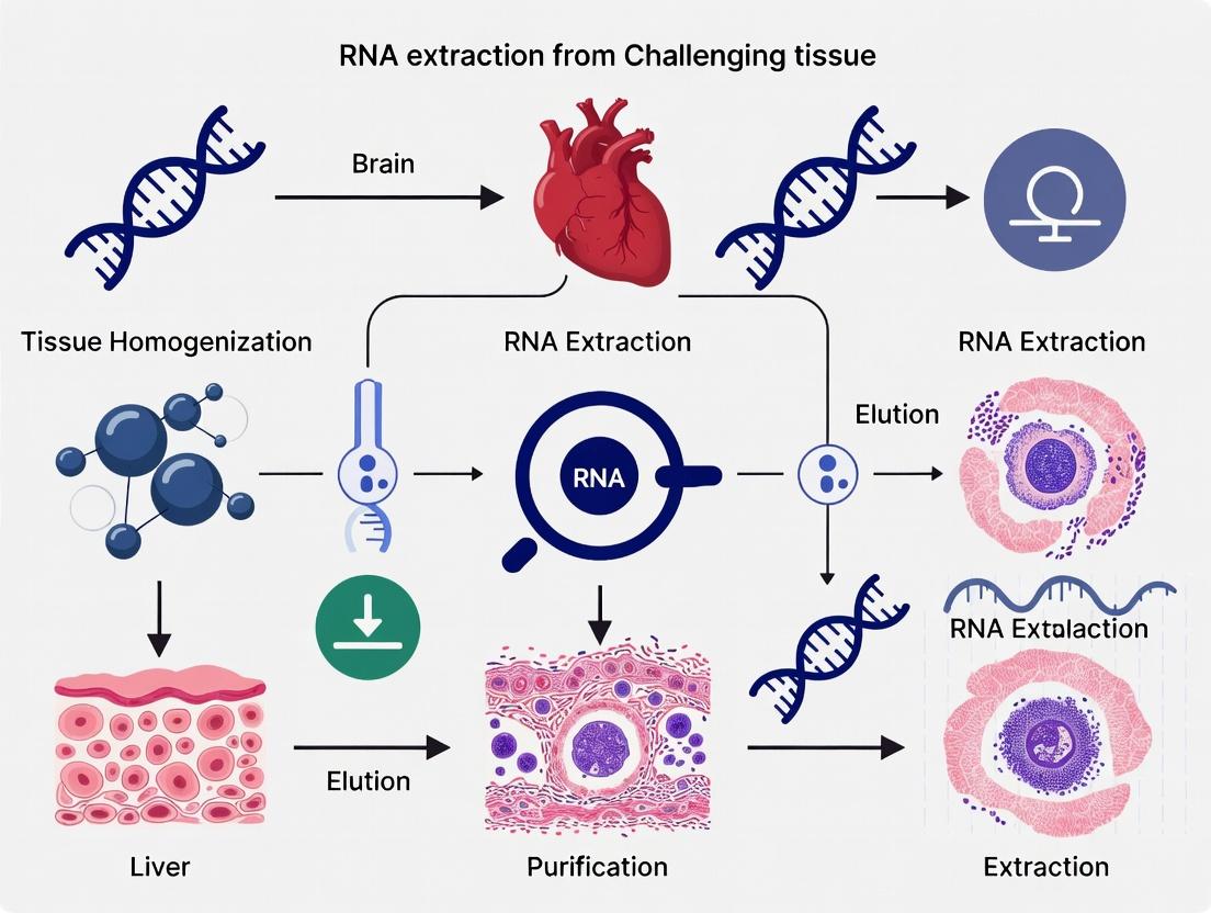
Within a broader thesis on RNA extraction from challenging tissues (brain, heart, liver) for research and drug development, effective homogenization is the critical first step. The choice of strategy must be tissue-specific to overcome unique challenges: the high lipid content and cellular heterogeneity of the brain, the robust contractile fibers of the heart, and the dense, metabolically active parenchyma of the liver. Suboptimal lysis leads to poor RNA yield, degraded quality, and biased representation. This application note details three core homogenization strategies—bead-beating, mechanical disruption, and enzymatic lysis—providing current protocols and comparative data to guide researchers in optimizing RNA integrity and yield from these complex tissues.
Comparative Analysis of Homogenization Methods
Table 1: Quantitative Performance of Homogenization Methods on Challenging Tissues
| Tissue | Method | Avg. RNA Yield (µg/mg tissue) | RNA Integrity Number (RIN) | Processing Time (min) | Key Advantage | Primary Limitation |
|---|---|---|---|---|---|---|
| Brain | Bead-Beating | 6.5 - 8.2 | 8.5 - 9.1 | 10-15 | Excellent for heterogeneous regions (hippocampus, cortex); disrupts lipid-rich membranes. | Potential heat generation; may over-shear nuclear RNA. |
| Mechanical (Rotor-Stator) | 5.8 - 7.0 | 7.8 - 8.5 | 5-10 | Rapid; good for whole tissue chunks. | Inconsistent for small, dense regions; cross-contamination risk. | |
| Enzymatic Lysis | 4.0 - 5.5 | 9.0 - 9.5 | 60+ | Superior preservation of RNA integrity; gentle. | Lower yield; requires precise incubation; costly. | |
| Heart | Bead-Beating | 4.0 - 5.5 | 8.0 - 8.7 | 10-15 | Effective against tough fibrous and collagenous structures. | Difficult with whole tissue; may require pre-cutting. |
| Mechanical (Blade Homogenizer) | 5.0 - 6.5 | 7.5 - 8.2 | 5-8 | Best for ventricular muscle bulk homogenization. | High shear damages RNA if prolonged; foaming. | |
| Enzymatic Lysis (Collagenase) | 3.5 - 4.5 | 8.8 - 9.4 | 90-120 | Selective digestion of collagen; ideal for cardiomyocyte isolation. | Very low total tissue RNA yield; complex protocol. | |
| Liver | Bead-Beating | 7.0 - 9.0 | 8.2 - 8.8 | 8-12 | Efficient for complete cellular disruption in dense parenchyma. | Can be overly harsh, releasing inhibitors. |
| Mechanical (Dounce) | 8.5 - 10.5 | 8.5 - 9.0 | 10-15 | Gold standard for soft, friable tissue; controlled shear. | Manual, variable; requires skill. | |
| Enzymatic Lysis | 6.0 - 7.5 | 9.0 - 9.5 | 30-45 | Excellent for nuclei isolation; gentle on RNA. | Risk of endogenous RNase activation. |
Detailed Experimental Protocols
Protocol 1: Bead-Beating for Brain Tissue (Regional Dissection)
Objective: To homogenize specific, lipid-rich regions of murine brain (e.g., cortex, striatum) for total RNA extraction. Materials: Pre-chilled bead-beater (e.g., MagNA Lyser), 2.0mL ceramic bead tubes (1.4mm diameter), RNase-free PBS, TRIzol or similar lysis reagent, dry ice.
- Pre-chill: Cool bead-beater holder and tubes on dry ice.
- Sample Prep: Rapidly weigh 20-30mg of dissected brain region. Place tissue in pre-chilled bead tube.
- Lysis Buffer Addition: Immediately add 1mL of cold TRIzol to the tube. Cap tightly.
- Homogenization: Securely mount tubes in the bead-beater. Process at 6,500 rpm for 30 seconds.
- Cooling: Immediately place tubes back on dry ice for 1 minute to dissipate heat.
- Repeat: Perform a second cycle of 30 seconds at 6,500 rpm.
- Recovery: Briefly centrifuge tubes (10,000 x g, 30 sec, 4°C) to pellet beads and debris. Transfer the cleared lysate supernatant to a fresh RNase-free tube. Proceed to RNA extraction.
Protocol 2: Mechanical Disruption for Heart Tissue (Rotor-Stator)
Objective: To homogenize tough, fibrous murine ventricular tissue for bulk RNA analysis. Materials: Bench-top rotor-stator homogenizer (e.g., Polytron), sterile disposable probes, RNase-free 15mL tubes, Qiazol lysis reagent, ice bath.
- Pre-cool: Immerse probe generator in ice. Keep lysis reagent and tubes on ice.
- Sample Prep: Mince ~50mg of ventricular tissue into ~2mm³ pieces in a petri dish on ice.
- Initial Suspension: Transfer tissue pieces to a 15mL tube containing 2.5mL Qiazol. Vortex briefly.
- Homogenization: Insert the pre-cooled probe, ensuring it is immersed but not touching the tube bottom. Homogenize at 15,000 rpm for 15-20 seconds in a pulsed manner (5 sec on, 10 sec off).
- Cooling: Keep the tube in the ice bath during and between pulses. Do not let the lysate become warm.
- Clarification: After 3-4 pulses (total active homogenization time ~60 sec), let the lysate sit on ice for 5 min. Centrifuge at 12,000 x g for 10 min at 4°C to remove insoluble collagen/fiber. Collect supernatant.
Protocol 3: Enzymatic Lysis for Liver Tissue (Perfusion-Based)
Objective: Gentle lysis for high-integrity RNA and nuclei isolation from murine liver. Materials: Perfusion pump, collagenase IV (Worthington), DNase I (RNase-free), ELB lysis buffer (10mM HEPES, 85mM KCl, 0.5% NP-40), RNAse inhibitors, 40µm cell strainer.
- Perfusion: Anesthetize mouse and perfuse liver via the portal vein first with 20mL of cold PBS, then with 20mL of pre-warmed (37°C) collagenase IV solution (0.5mg/mL in PBS).
- Digestion: Excise the liver, place in a dish with 5mL fresh collagenase IV solution, and incubate at 37°C for 15 min with gentle agitation.
- Dissociation: Gently tease apart the digested liver capsule with forceps in cold PBS+RNase inhibitor to release hepatocytes.
- Filtration & Washing: Filter the cell suspension through a 40µm strainer. Pellet cells at 500 x g for 5 min at 4°C. Wash twice with cold PBS.
- Gentle Lysis: Resuspend the cell pellet in 2mL of ice-cold ELB lysis buffer + RNase inhibitors. Incubate on ice for 15 min with gentle inversion every 5 min.
- Clarification: Centrifuge at 1,500 x g for 5 min at 4°C. The supernatant (cytoplasmic fraction) contains high-quality RNA. The pellet contains nuclei for nuclear RNA extraction.
The Scientist's Toolkit: Key Reagent Solutions
Table 2: Essential Research Reagents for Tissue Homogenization and RNA Stabilization
| Reagent / Material | Function | Key Consideration for Challenging Tissues |
|---|---|---|
| TRIzol / Qiazol | Monophasic solution of phenol and guanidine isothiocyanate. Simultaneously lyses cells, inactivates RNases, and denatures proteins. | Critical for brain (lipids) and liver (RNases). Must be used in a fume hood. |
| RNase Inhibitors (e.g., Recombinant RNasin) | Proteins that bind to and inhibit a broad spectrum of RNases. | Essential for all enzymatic and long protocols. Add to lysis buffers immediately before use. |
| Collagenase Type IV | Enzyme that digests collagen (Type IV specifically targets basement membrane). | Vital for dissociating heart tissue and perfusing liver. Lot-to-lot activity varies; must be titrated. |
| β-Mercaptoethanol (BME) or DTT | Reducing agent that denatures RNases by breaking disulfide bonds. | Standard addition (e.g., 1% v/v BME) to RLT or similar buffers for heart and liver. |
| Ceramic/Silica Beads (0.5-1.4mm) | Inert, dense beads that provide grinding force during bead-beating. | Smaller beads (0.5mm) for bacterial/cell pellets; larger (1.4mm) for soft tissues; zirconium-silicate preferred for RNA. |
| RNA Stabilization Agents (e.g., RNAlater) | Aqueous, non-toxic reagent that rapidly penetrates tissue to stabilize and protect cellular RNA. | Invaluable for archiving human brain biopsies or multi-organ sampling where immediate processing isn't possible. |
Visualizations
Title: Homogenization Strategy Selection Workflow
Title: Stress Pathways from Suboptimal Homogenization
Within the broader thesis on optimizing RNA extraction from challenging tissues, three organs present distinct, formidable barriers: the lipid-rich brain, the fibrous and contractile heart, and the liver, which is abundant in endogenous RNases. This application note details tissue-specific, validated protocols to overcome these challenges, ensuring the isolation of high-integrity RNA suitable for downstream applications like qRT-PCR, RNA-seq, and microarray analysis.
Table 1: Tissue-Specific Challenges and Strategic Countermeasures
| Tissue | Primary Challenge | Key Interfering Substances | Core Strategic Approach | Expected RNA Yield & Quality (RIN) |
|---|---|---|---|---|
| Brain | High Lipid Content | Myelin, phospholipids | Efficient homogenization with organic phase-separation; thorough lipid removal. | 2-8 µg/mg tissue; RIN > 8.5 |
| Heart | Fibrous Structure | Collagen, contractile proteins | Powerful mechanical disintegration; inhibition of myofibrillar protein co-precipitation. | 1-4 µg/mg tissue; RIN > 8.0 |
| Liver | High RNase Activity | Endogenous RNases (e.g., RNase A) | Rapid lysis and immediate RNase inactivation; use of potent, specific RNase inhibitors. | 4-10 µg/mg tissue; RIN > 9.0 |
Detailed Experimental Protocols
Protocol 1: Brain Tissue (High Lipid) RNA Extraction
Method: Modified Guanidinium Thiocyanate-Phenol-Chloroform (e.g., TRIzol) with Enhanced Lipid Clearance.
- Sample Preparation: Snap-freeze 15-30 mg of brain tissue in liquid N₂. Pulverize using a chilled mortar and pestle or a cryogenic mill. Do not allow tissue to thaw.
- Homogenization: Transfer powder to a tube containing 1 mL of TRIzol or equivalent monophasic lysis reagent. Homogenize thoroughly using a rotor-stator homogenizer (20-30 sec on ice).
- Phase Separation: Incubate 5 min at RT. Add 0.2 mL chloroform, vortex vigorously for 15 sec. Incubate 2-3 min at RT. Centrifuge at 12,000 × g for 15 min at 4°C.
- Lipid Removal (Critical Step): After centrifugation, a thick white lipid layer interphase is often present. Carefully aspirate the aqueous (upper) phase using a fine-tip pipette, avoiding the interphase. Transfer to a new tube.
- RNA Precipitation: Add 0.5 mL isopropanol, mix by inversion. Incubate at -20°C for ≥1 hour. Centrifuge at 12,000 × g for 30 min at 4°C to pellet RNA.
- Wash: Remove supernatant. Wash pellet with 1 mL of 75% ethanol (in DEPC-treated water). Vortex briefly, centrifuge at 7,500 × g for 5 min at 4°C.
- Redissolution: Air-dry pellet for 5-10 min. Dissolve in 30-50 µL of RNase-free water or TE buffer. Quantify by spectrophotometry.
Protocol 2: Heart Tissue (Fibrous) RNA Extraction
Method: Robust Mechanical Lysis coupled with Silica-Membrane Column Purification.
- Sample Preparation: Cut 20-30 mg of ventricular tissue into minimal pieces (< 25 mg) using sterile instruments. Immediately place in lysis buffer.
- Lysis/Homogenization: Add tissue to 600 µL of RLT Plus buffer (Qiagen) containing 1% β-mercaptoethanol. Homogenize using a high-throughput tissue disruptor (e.g., TissueLyser II) with a 5 mm stainless steel bead for 2 x 2 min at 30 Hz. Alternatively, use a powerful rotor-stator homogenizer.
- Clarification: Centrifuge the lysate at 12,000 × g for 3 min to pellet debris, myofibrils, and collagen. Transfer supernatant to a new tube.
- Ethanol Adjustment: Add 1 volume of 70% ethanol to the supernatant and mix by pipetting.
- Column Purification: Apply the mixture to an RNeasy Fibrous Tissue Mini Kit column. Centrifuge. Perform on-column DNase I digestion (15 min, RT) per manufacturer's instructions.
- Washes: Wash with RW1 and RPE buffers.
- Elution: Elute RNA in 30-50 µL RNase-free water.
Protocol 3: Liver Tissue (High RNase) RNA Extraction
Method: Ultra-Rapid Lysis with Potent RNase Inactivation and Magnetic Bead-Based Purification.
- Pre-chill Equipment: Ensure centrifuges, rotors, and tubes are at 4°C.
- Immediate Lysis: To <20 mg of fresh or snap-frozen liver tissue in a pre-chilled tube, immediately add 500 µL of ice-cold lysis buffer containing 4M guanidine isothiocyanate, 0.5% N-lauroylsarcosine, and 1% β-mercaptoethanol.
- Instant Homogenization: Homogenize with a rotor-stator homogenizer for no more than 20 seconds while the tube is submerged in an ice bath.
- RNase Inactivation: Immediately add 0.5 mL of acid phenol:chloroform (pH 4.5), vortex vigorously, and centrifuge at 12,000 × g for 10 min at 4°C.
- Binding to Beads: Transfer aqueous phase to a tube containing RNase-free magnetic beads (e.g., SPRI beads). Mix thoroughly and incubate for 5 min at RT.
- Magnetic Separation: Place tube on a magnetic stand. After solution clears, discard supernatant.
- Washes: Keep tube on magnet. Wash beads twice with 80% ethanol.
- Elution: Air-dry beads briefly (2-3 min). Elute RNA in 30 µL of RNase-free water.
Visualized Workflows
Brain RNA Extraction Flow
Heart RNA Extraction Flow
Liver RNase Inactivation Flow
The Scientist's Toolkit
Table 2: Essential Research Reagent Solutions for Challenging Tissue RNA Extraction
| Reagent / Material | Primary Function | Tissue-Specific Utility |
|---|---|---|
| TRIzol / Qiazol | Monophasic lysis reagent containing guanidinium and phenol. Denatures proteins, inactivates RNases, and dissolves lipids. | Critical for Brain (lipid dissolution). Used in Liver and Heart. |
| β-Mercaptoethanol (BME) | Strong reducing agent. Disrupts disulfide bonds in RNases and other proteins, enhancing denaturation. | Essential for Liver (potent RNase inactivation). Used in all protocols. |
| RNase Inhibitors (e.g., Recombinant RNasin) | Proteins that bind non-covalently to RNases, inhibiting their activity. | Critical add-on for Liver protocols post-lysis (e.g., in RT reactions). |
| Silica-Membrane Spin Columns (Fibrous Tissue Kits) | Selective binding of RNA in high-salt conditions; washing removes contaminants. | Essential for Heart to handle viscous lysates and remove protein/polyaccharide contaminants. |
| Magnetic Beads (SPRI) | Paramagnetic particles that bind nucleic acids. Enable rapid, tube-based purification without centrifugation. | Ideal for Liver for speed, minimizing RNase exposure. |
| DNase I (RNase-free) | Enzyme that digests genomic DNA to prevent contamination in downstream assays. | Recommended for all tissues, especially critical for Heart (high DNA content). |
| Cryogenic Mill / Mortar & Pestle | For pulverizing frozen tissue into a fine powder without thawing. | Critical for Brain (prevents lipid smear) and heterogeneous tissues. |
| High-Throughput Tissue Disruptor (e.g., TissueLyser) | Uses mechanical force (beads) to lyse tough, fibrous structures. | Essential for effective Heart and Skeletal Muscle lysis. |
Troubleshooting Common Pitfalls and Advanced Optimization Techniques
Within the critical context of a broader thesis on RNA extraction from challenging tissue types—specifically brain, heart, and liver for neurodegenerative, cardiovascular, and metabolic disease research—achieving high RNA yield and integrity is paramount. Incomplete tissue lysis and suboptimal sample handling are predominant, yet often overlooked, culprits behind low RNA yields. This application note details diagnostic methodologies and optimized protocols to overcome these challenges, ensuring reliable downstream applications in drug development and biomarker discovery.
Quantitative Impact of Lysis Efficiency on RNA Yield
Recent data underscore the direct correlation between lysis completeness and RNA yield, particularly from fibrous (heart), lipid-rich (brain), and enzymatically active (liver) tissues.
Table 1: Impact of Lysis Protocol on RNA Yield from Challenging Tissues
| Tissue Type | Common Lysis Challenge | Standard Homogenization Yield (µg/mg tissue) | Optimized Homogenization Yield (µg/mg tissue) | Percent Increase | Reference Key Findings |
|---|---|---|---|---|---|
| Brain (Mouse Cortex) | Lipid-rich membranes, RNase activity | 1.2 ± 0.3 | 3.5 ± 0.4 | ~192% | Combined mechanical & chemical disruption critical; RNase inhibitors essential. |
| Heart (Rat Left Ventricle) | Dense, fibrous myocardium | 0.8 ± 0.2 | 2.8 ± 0.3 | ~250% | Extended protease digestion or specialized rotor-stator homogenizers required. |
| Liver (Mouse) | High endogenous RNase content | 2.0 ± 0.5 | 5.5 ± 0.6 | ~175% | Rapid lysis and immediate inhibition of RNases are non-negotiable. |
| Tumor (Necrotic Core) | Variable cell viability & integrity | 0.5 ± 0.3 | 2.0 ± 0.5 | ~300% | Manual micro-dissection of viable regions prior to lysis dramatically improves yield. |
Diagnostic Protocol: Assessing Lysis Completeness
Objective: To visually and quantitatively confirm complete tissue dissociation prior to RNA purification.
Materials: Phase-contrast microscope, hemocytometer or automated cell counter, trypan blue.
Method:
- Sample Aliquot: After the standard lysis/homogenization step, remove a 10 µL aliquot of the lysate.
- Visual Inspection: Place the aliquot on a microscope slide with a coverslip. Using phase-contrast at 20x magnification, scan for intact tissue chunks, cell clumps, or unlysed cells. Their presence indicates incomplete lysis.
- Quantitative Assessment: Mix the 10 µL lysate aliquot with 10 µL of 0.4% trypan blue. Load onto a hemocytometer. Count intact, stained (blue) cells versus clear, lysed debris. >5% intact cells suggests inadequate lysis efficiency.
- Decision Point: If lysis is incomplete, return the main sample to the homogenizer for further processing (see Section 4 protocols) before proceeding with RNA isolation.
Optimized Lysis Protocols for Challenging Tissues
Protocol 4.1: Integrated Mechanical & Chemical Lysis for Brain Tissue
Principle: Simultaneously disrupt lipid bilayers and inactivate RNases.
- Pre-chill all equipment and solutions on ice.
- Weigh 20-30 mg of fresh or snap-frozen brain tissue (e.g., cortex, hippocampus).
- Immediately place tissue in 600 µL of commercially available Qiazol or TRIzol lysis reagent containing 1% β-mercaptoethanol (added fresh).
- Homogenize using a motorized rotor-stator homogenizer (e.g., Qiagen TissueRuptor II) for 20-30 seconds at full speed. Keep tube on ice.
- Incubate the homogenate for 5 minutes at room temperature to complete dissociation of nucleoprotein complexes.
- Proceed to phase separation or column-based purification.
Protocol 4.2: Sequential Disruption for Fibrous Heart Tissue
Principle: Utilize enzymatic softening followed by vigorous mechanical disruption.
- Place 15-25 mg of heart tissue in a tube with 500 µL of lysis buffer (e.g., RLT Plus from Qiagen) containing Proteinase K (final conc. 0.8 mg/mL).
- Incubate at 56°C for 10 minutes with gentle shaking to digest connective proteins.
- Transfer tube to ice for 2 minutes.
- Homogenize using a small-bead mill homogenizer (e.g., using 2.8mm ceramic beads) for 2 x 45 seconds at 6,000 rpm, with a 30-second pause on ice in between.
- Centrifuge briefly to pellet beads and insoluble debris. Transfer the supernatant (lysate) to a new tube.
- Proceed with RNA cleanup.
Critical Sample Handling Practices to Prevent RNA Degradation
Table 2: Sample Handling Errors and Corrective Actions
| Error Stage | Common Mistake | Consequence | Corrective Action |
|---|---|---|---|
| Collection | Delayed freezing of tissue (>5 min post-dissection) | Rapid RNA degradation by endogenous RNases. | Snap-freeze in liquid nitrogen within 60-90 seconds. Use RNAlater for difficult-to-dissect samples. |
| Storage | Intermittent temperature fluctuation during -80°C storage. | Ice crystal formation and physical shearing of RNA. | Use airtight, non-frost-free freezers. Aliquot samples to avoid freeze-thaw cycles. |
| Homogenization | Allowing sample to warm during processing. | Increased RNase activity. | Use pre-chilled equipment, work on ice blocks, process one sample at a time. |
| Post-Lysis | Delaying addition of chaotropic salts/RNase inhibitors. | Degradation begins in homogenate. | Add lysis/denaturation reagent before homogenization. |
The Scientist's Toolkit: Research Reagent Solutions
Table 3: Essential Reagents for Optimal RNA Yield from Difficult Tissues
| Item | Function & Rationale |
|---|---|
| Chaotropic Salt-Based Lysis Reagents (e.g., Qiazol, TRIzol) | Denature proteins and RNases instantly upon contact, stabilizing RNA. Essential for liver and brain. |
| β-Mercaptoethanol (or alternative reducing agents) | Disrupts disulfide bonds in proteins and RNases, enhancing denaturation. Critical for tissue rich in secretory cells. |
| Potent RNase Inhibitors (e.g., Recombinant RNasin Plus) | Provides a supplemental barrier against residual RNase activity post-lysis, especially in spleen or pancreas. |
| Proteinase K | Digests connective tissue and proteins, enhancing cell lysis and freeing RNA from complexes. Vital for heart, muscle, and fibrous tumors. |
| Inert, RNase-Free Beads (Ceramic or Silica) | Provide superior mechanical shearing in bead mill homogenizers for difficult-to-lyse samples without absorbing RNA. |
| RNAlater Stabilization Solution | Penetrates tissue to rapidly stabilize and protect RNA at the time of collection, allowing flexibility for later processing. |
Experimental Workflow Diagram
Diagram Title: Workflow Comparison: Common Errors vs. Optimized RNA Extraction
Maximizing RNA yield from complex research tissues like brain, heart, and liver requires a two-pronged strategy: validating lysis completeness through a simple diagnostic check and adhering to stringent, tissue-tailored handling protocols. Integrating the mechanical and chemical solutions outlined here directly addresses the root causes of incomplete lysis and pre-purification degradation, ensuring the high-quality RNA necessary for advanced transcriptional analyses in biomedical research and therapeutic development.
This application note details protocols for safeguarding RNA integrity during extraction from challenging tissues—brain, heart, and liver—within a thesis on RNA extraction from complex tissue matrices. RNase contamination and delays in sample processing are the primary sources of degradation, compromising downstream applications like RNA sequencing and qPCR.
Quantifying the Impact of Delayed Processing on RNA Integrity
RNA degradation is accelerated at room temperature. The following table summarizes data from controlled studies on post-mortem delays prior to freezing or stabilization.
Table 1: Impact of Post-Collection Delay on RNA Integrity Number (RIN) in Murine Tissues
| Tissue Type | Delay at Room Temperature (Hours) | Mean RIN Value (1-10) | % of Samples with RIN ≥ 7 | Key Observation |
|---|---|---|---|---|
| Brain (Cortex) | 0 (Immediate freezing) | 9.2 ± 0.3 | 100% | Gold standard for intact RNA. |
| Brain (Cortex) | 2 | 7.1 ± 0.8 | 65% | Significant decline; ribosomal peaks broadening. |
| Liver | 0 | 9.0 ± 0.4 | 100% | High initial RNase activity necessitates rapid handling. |
| Liver | 1 | 6.0 ± 1.2 | 30% | Dramatic degradation; 28S:18S rRNA ratio falls below 1.0. |
| Heart | 0 | 8.8 ± 0.5 | 98% | Robust myofibrils can protect RNA temporarily. |
| Heart | 4 | 7.5 ± 0.9 | 75% | More resistant than liver but still degrades. |
Protocols for Prevention and Identification
Protocol 3.1: Immediate Tissue Stabilization and Processing
Objective: Minimize RNA degradation from endogenous RNases during sample collection from brain, heart, and liver. Materials: See "The Scientist's Toolkit" (Section 5). Procedure:
- Pre-chill: Pre-cool all dissection tools, containers, and saline on ice.
- Rapid Dissection: Euthanize animal per approved protocol. Excise target tissue (e.g., brain hemisphere, left ventricle, liver lobe) swiftly within 60 seconds.
- Stabilization Decision Point:
- Option A (Chemical Stabilization): Immediately submerge tissue piece (thickness < 0.5 cm) in 10 volumes of RNAlater. Incubate overnight at 4°C, then store at -80°C.
- Option B (Flash-Freezing): Place tissue directly into a cryovial, immerse vial in liquid nitrogen for 30 seconds, and transfer to -80°C. Optimal for brain tissues.
- Homogenization: Perform in a cold, RNase-decontaminated environment. Homogenize stabilized or frozen tissue in a suitable lysis buffer containing strong chaotropic salts (e.g., guanidinium thiocyanate) using a rotor-stator homogenizer. Process samples individually to avoid cross-contamination.
Protocol 3.2: RNase Decontamination of Work Surfaces and Equipment
Objective: Eliminate exogenous RNase contamination from labware and benches. Procedure:
- Surface Decontamination: Thoroughly wipe down benches, pipettors, and instrument surfaces with an RNase decontamination solution (e.g., based on 0.1% Diethyl pyrocarbonate (DEPC)-treated water or commercial RNase inhibitors). Allow to air dry.
- Glassware/Plasticware Treatment: For reusable items, bake glassware at 240°C for 4 hours or autoclave. Use certified RNase-free disposable plasticware.
- Solution Preparation: Use nuclease-free water for all reagent preparation. Treat non-commercial buffers with DEPC (0.1% v/v, incubate overnight, autoclave to inactivate excess DEPC) except for Tris-based buffers, which react with DEPC.
Protocol 3.3: Assessment of RNA Degradation via Microfluidic Capillary Electrophoresis
Objective: Quantitatively evaluate RNA integrity post-extraction. Procedure:
- Instrument Setup: Calibrate the instrument (e.g., Agilent Bioanalyzer or TapeStation) with the appropriate RNA assay ladder as per manufacturer instructions.
- Sample Preparation: Dilute 1 µL of extracted RNA in nuclease-free water or the provided buffer to fall within the detection range (e.g., 5-500 ng/µL for the Bioanalyzer RNA 6000 Nano Kit).
- Loading and Run: Load the ladder and samples into designated wells of the microfluidic chip. Initiate the electrophoresis run.
- Analysis: The software generates an electropherogram and an RNA Integrity Number (RIN). A RIN ≥ 7.0 is generally acceptable for most downstream applications. Visually inspect the electropherogram for distinct 18S and 28S ribosomal peaks (ratio ~1.8-2.0 for mammalian RNA) and a flat baseline.
Visualizing Workflows and Degradation Pathways
Title: RNA Degradation Pathways in Tissue Processing
Title: Optimal RNA Preservation Protocol Workflow
The Scientist's Toolkit: Research Reagent Solutions
Table 2: Essential Reagents and Materials for RNA Preservation
| Item | Function & Rationale |
|---|---|
| RNAlater Stabilization Solution | Penetrates tissue to inactivate RNases rapidly, allowing temporary storage at 4°C before freezing. Crucial for liver and during multi-sample collections. |
| TRIzol / TRI Reagent | Monophasic solution of phenol and guanidine isothiocyanate. Simultaneously lyses cells, inactivates RNases, and separates RNA from DNA/protein during phase separation. |
| DNase I (RNase-free) | Removes genomic DNA contamination from RNA preparations, essential for sensitive applications like qPCR and RNA-seq. |
| RNase Inhibitor (e.g., Recombinant RNasin) | Binds reversibly to RNases, used as an additive during cDNA synthesis or RNA handling steps to protect against minor contamination. |
| Nuclease-Free Water | Certified free of RNases and DNases for preparing buffers and resuspending RNA pellets. |
| Certified RNase-Free Pipette Tips and Tubes | Manufactured to be free of detectable RNase activity, preventing introduction of contaminants. |
| Surface Decontaminant (e.g., RNaseZap) | Quickly removes RNase contamination from pipettors, benches, and non-sterile equipment. |
| RNA Integrity Assay Chips (Bioanalyzer) | Microfluidic chips for automated electrophoretic analysis and quantification of RNA integrity (RIN). |
Successful downstream applications in RNA research, particularly from challenging tissues like brain, heart, and liver, depend on the purity of isolated nucleic acids. Contaminants such as genomic DNA (gDNA), phenol from organic extraction, and excess salts can inhibit enzymatic reactions, degrade RNA integrity, and produce misleading quantitative results. This application note details current, optimized protocols for the effective removal of these critical contaminants, framed within a thesis on RNA extraction from complex mammalian tissues. We present quantitative comparisons of efficiency and provide step-by-step methodologies.
The brain, heart, and liver present unique challenges for high-quality RNA extraction. The brain is lipid-rich, the heart contains high levels of contractile proteins, and the liver is metabolically active with abundant nucleases. Co-purification of gDNA is a pervasive issue, as its presence can lead to false-positive signals in qPCR and interfere with sequencing library preparation. Residual phenol, even at trace levels, can denature enzymes in reverse transcription and PCR. Salts, such as those from chaotropic agents or precipitation buffers, can inhibit polymerase activity and alter spectrophotometric readings. Effective removal is non-negotiable for reliable gene expression analysis in drug development and basic research.
Quantitative Comparison of Decontamination Strategies
Table 1: Efficiency of Genomic DNA Removal Methods
| Method | Principle | Recommended Tissue | gDNA Removal Efficiency | RNA Yield Impact | Time Required |
|---|---|---|---|---|---|
| DNase I Digestion (on-column) | Enzymatic degradation | Brain, Heart | >99.9% | Minimal (<5% loss) | 15-30 min |
| DNase I Digestion (in-solution) | Enzymatic degradation | Liver, Heart | >99% | Moderate (5-10% loss) | 30-45 min |
| Selective Precipitation (LiCl) | Differential solubility | Liver | ~95-98% | High risk of co-precipitation | 60+ min (O/N) |
| Solid-Phase Selection (SiO₂) | Binding condition specificity | All (Brain, Heart, Liver) | ~90-95% | Minimal | Incorporated in extraction |
Table 2: Strategies for Phenol and Salt Removal
| Contaminant | Removal Method | Protocol Basis | Residual Level Post-Treatment | Key Validation Method |
|---|---|---|---|---|
| Phenol | Chloroform Back-Extraction | Acid Phenol:Chloroform step | <0.1% | A260/A230 ratio (Target: 2.0-2.5) |
| Phenol | Ethanol/Isopropanol Precipitation | 2.5x Vol Ethanol, 0.1x NaOAc | <0.05% | A260/A230 ratio |
| Salts (e.g., Guanidine, Na⁺) | Ethanol Wash (70-80%) | On-column or in-pellet wash | Nanomolar levels | Conductivity Measurement |
| Salts | Micro-Spin Dialysis | Centrifugal filter devices | Picomolar levels | A260/A230 ratio (Target: >2.0) |
Detailed Experimental Protocols
Protocol 3.1: On-Column DNase I Digestion for Difficult Tissues
Application: Ideal for lipid-rich (brain) and fibrous (heart) tissues where in-solution digestion may be inefficient. Reagents: RLT Plus buffer, 70% ethanol, RW1 wash buffer, DNase I stock (1 U/µL), DNase incubation buffer (10 mM Tris-HCl, pH 7.5, 2.5 mM MgCl₂). Procedure:
- Homogenize 30 mg of brain/heart tissue in 600 µL RLT Plus buffer using a rotor-stator homogenizer.
- Centrifuge the lysate at 12,000 x g for 3 min at 4°C. Transfer supernatant to a new tube.
- Add 1 volume of 70% ethanol, mix by pipetting, and load onto a silica spin column.
- Centrifuge at 8000 x g for 30 sec. Discard flow-through.
- Prepare on-column DNase mix: 70 µL DNase incubation buffer + 10 µL DNase I stock (10 U total).
- Apply mix directly to the column membrane. Incubate at RT for 25 min.
- Wash column with 350 µL RW1 buffer, centrifuge, discard flow-through.
- Wash twice with 500 µL RPE buffer (ethanol-based).
- Elute RNA in 30-50 µL RNase-free water.
Protocol 3.2: Acid Phenol:Chloroform Cleanup for Phenol Removal
Application: Critical after traditional TRIzol or phenol-based extractions from liver tissue. Reagents: Acid Phenol:Chloroform (pH 4.5), 3M Sodium Acetate (pH 5.2), 100% Isopropanol, 80% Ethanol, RNase-free water. Procedure:
- Following initial aqueous phase separation in TRIzol protocol, transfer the aqueous phase (~600 µL) to a new tube.
- Add an equal volume of Acid Phenol:Chloroform (pH 4.5). Vortex vigorously for 1 min.
- Centrifuge at 12,000 x g for 10 min at 4°C. The upper aqueous phase will contain RNA.
- Transfer the aqueous phase carefully to a new tube. Add 0.5 volumes of 100% isopropanol and 0.1 volumes of 3M Sodium Acetate (pH 5.2). Mix.
- Incubate at -20°C for 1 hour or overnight for maximum yield.
- Centrifuge at 12,000 x g for 30 min at 4°C. A visible RNA pellet should form.
- Carefully decant supernatant. Wash pellet with 1 mL of 80% ethanol (made with RNase-free water).
- Centrifuge at 7500 x g for 5 min. Carefully remove all ethanol.
- Air-dry pellet for 5-10 min. Do not over-dry.
- Resuspend in 20-30 µL RNase-free water.
Visualizing Workflows and Strategies
Diagram 1: Integrated RNA Purification & Decontamination Workflow
Diagram 2: Contaminant-Specific Removal Strategy Map
The Scientist's Toolkit: Research Reagent Solutions
Table 3: Essential Materials for Effective Decontamination
| Item Name | Supplier Examples | Function in Decontamination |
|---|---|---|
| RNase-free DNase I (Recombinant) | Qiagen, Thermo Fisher, NEB | Enzymatically digests gDNA without RNase contamination. Essential for on-column or in-solution treatment. |
| Acid Phenol:Chloroform (pH 4.5) | Thermo Fisher, Sigma-Aldrich | Used for back-extraction to remove trace organic contaminants. Acidic pH partitions RNA to the aqueous phase. |
| Silica-Membrane Spin Columns | Zymo Research, Macherey-Nagel | Selective binding of RNA under high-salt conditions, allowing efficient wash-away of salts and other impurities. |
| RNase-free Sodium Acetate (3M, pH 5.2) | Ambion, Sigma-Aldrich | Provides counter-ions for efficient ethanol precipitation of RNA, aiding in separation from phenol and salts. |
| Concentrated Wash Buffers (e.g., with Guanidine HCl) | Various kit suppliers | Maintains RNA binding to silica while removing salts, metabolites, and residual phenol through multiple wash steps. |
| Magnetic Beads (SPRI) | Beckman Coulter, Cytiva | Enable size-selective cleanup, removing short DNA fragments and salts via PEG/NaCl precipitation. |
| Centrifugal Filter Devices (3kDa MWCO) | Amicon, Pall Corporation | Rapid desalting and buffer exchange via micro-dialysis for highest-purity applications like sequencing. |
Within the critical research workflow of RNA extraction from challenging tissues (brain, heart, liver) for downstream transcriptomic analysis in drug development, phase separation issues during homogenate clarification and column clogging are primary failure points. These problems are exacerbated by the high lipid content of brain tissue, the robust fibrous matrix of heart tissue, and the enzymatic and metabolite richness of liver tissue. This application note details the underlying causes and provides optimized protocols to mitigate these obstacles, ensuring high-yield, high-integrity RNA extraction.
Quantitative Analysis of Clogging Agents in Challenging Tissues
Table 1: Common Impurities Contributing to Phase Separation and Clogging by Tissue Type
| Tissue Type | Primary Challenges | Key Impurities | Typical Impact on RNA Yield (vs. Ideal) |
|---|---|---|---|
| Brain | High lipid content, viscosity | Myelin, cholesterol, phospholipids | Yield reduction of 30-60% if not addressed |
| Heart | Dense fibrous network | Collagen, contractile proteins, connective tissue | Column clogging high; potential for >50% loss |
| Liver | High RNase activity, dense vasculature | Hemoglobin, glycogen, endogenous nucleases | Rapid degradation and clogging; yield unpredictable |
Table 2: Efficacy of Pre-Clarification Methods on Homogenate Viscosity
| Pre-Clarification Method | Reduction in Viscosity (%) | Compatible Tissue | Potential RNA Loss |
|---|---|---|---|
| Low-speed centrifugation (800 x g) | 40-50% | Brain, Liver | Low (<5%) |
| Filtration (70µm mesh) | 60-70% | Heart, Liver | Moderate (5-10%) |
| Acid-Phenol:Chloroform (pre-extraction) | 80-90% | All (esp. Brain) | Variable (depends on interface handling) |
| Commercial debris removal columns | 70-85% | All | Low to Moderate (3-8%) |
Detailed Experimental Protocols
Protocol 3.1: Optimized Homogenization and Clarification for Lipid-Rich Tissues (e.g., Brain)
Goal: Achieve complete lysis while preventing a persistent, obstructive interphase.
- Homogenize: Rapidly homogenize ~30 mg tissue in 1 mL of QIAzol Lysis Reagent (or equivalent monophasic phenol-guanidine mix) using a rotor-stator homogenizer (15 sec bursts on ice).
- Incubate: Incubate homogenate for 5 min at room temperature to ensure complete dissociation.
- Phase Separation: Add 200 µL of chloroform, cap tightly, and shake vigorously by hand for 15 seconds.
- Critical Centrifugation: Centrifuge at 12,000 x g for 15 minutes at 4°C (increased time/force is crucial). This elongates the centrifugation path, improving separation.
- Aqueous Phase Recovery: Carefully remove the upper aqueous phase without disturbing the interphase. Leave a 2-3 mm buffer above the interphase to avoid contamination. For maximum yield, perform a second back-extraction: Add 200 µL of a 50:50 mix of initial lysis buffer and RNase-free water to the remaining organic phase/interphase, vortex, re-centrifuge, and pool with the first aqueous extract.
Protocol 3.2: Pre-Filtration and Column Loading Strategy for Fibrous Tissues (e.g., Heart)
Goal: Prevent particulate matter from entering and clogging the silica membrane column.
- Follow Protocol 3.1 through step 4 to obtain a clarified aqueous phase.
- Pre-Filtration: Transfer the aqueous phase to a pre-filter column (e.g., QIAshredder or a syringe filter with a 0.45 µm cellulose acetate membrane). Centrifuge at 10,000 x g for 2 min.
- Ethanol Adjustment: Mix the filtrate with 1.5 volumes of 100% ethanol. Do not vortex; pipette mix gently.
- Column Loading: Apply the sample to the RNA binding column in aliquots. Load 700 µL, centrifuge (≥8000 x g, 15 sec), discard flow-through, and repeat until all sample is processed. This prevents overloading the membrane.
- Wash: Perform standard wash steps with provided buffers.
- DNase I Treatment: Perform on-column DNase I digestion to remove genomic DNA, which can also contribute to clogging.
- Final Wash & Elution: Complete wash steps and elute in 30-50 µL RNase-free water.
The Scientist's Toolkit: Key Research Reagent Solutions
Table 3: Essential Materials for Mitigating Separation and Clogging Issues
| Item | Function & Rationale |
|---|---|
| Monophasic Lysis Reagent (e.g., QIAzol, TRIzol) | Contains phenol and guanidine thiocyanate; simultaneously inactivates RNases and dissolves tissue components, creating a uniform lysate pre-phase separation. |
| Chloroform (RNase-free) | Used for phase separation; differentially solubilizes lipids and proteins into the organic phase, leaving RNA in the aqueous phase. |
| Commercial Debris Removal Spin Columns | Pre-filters for viscous lysates; remove particulate matter before binding RNA to the main silica column, preventing clogging. |
| Silica-Membrane Spin Columns (wide-body) | RNA binding and purification; wide-body designs offer lower flow resistance and are less prone to clogging than standard columns. |
| RNase-free Syringe Filters (0.45 µm, 0.2 µm) | Alternative pre-filtration method; provides sterile, particulate-free lysate for column loading. |
| Glycogen or Linear Polyacrylamide (Carrier) | Added during ethanol precipitation steps; co-precipitates with low-concentration RNA, improving pellet visibility and recovery, especially from dilute samples. |
| DNase I (RNase-free) | Eliminates genomic DNA contamination that can compete with RNA for binding to silica membranes, reducing effective capacity. |
Visualized Workflows and Pathways
Title: Decision Workflow for RNA Extraction from Problem Tissues
Title: Cause and Effect of RNA Extraction Challenges
Optimizing Precipitation and Wash Steps for Maximum Recovery and Purity
Within the broader thesis on RNA extraction from challenging tissue types (brain, heart, liver), optimizing precipitation and wash steps is a critical determinant of success. These tissues present unique challenges: brain tissue is lipid-rich, heart tissue is high in contractile proteins, and liver tissue is abundant in nucleases and metabolites. The efficiency of RNA recovery and the purity of the final eluate hinge on precise protocol adjustments during the alcohol precipitation and subsequent wash phases. This application note provides detailed, evidence-based protocols to maximize yield and purity (A260/A280 and A260/A230 ratios) for downstream applications such as sequencing and qPCR.
Key Principles & Challenges
- Precipitation: The type and concentration of alcohol, pH, salt choice, incubation time/temperature, and carrier use must be tailored to counteract tissue-specific interferents.
- Washing: The composition, volume, and number of wash steps must remove salts, metabolites, and residual organic compounds without causing RNA pellet loss or overdrying.
- Tissue-Specific Considerations:
- Brain: High lipid content requires efficient separation to avoid lipid co-precipitation. A higher salt concentration during precipitation can improve RNA recovery from the lipid-rich matrix.
- Heart: Dense muscle fibers and high glycogen content necessitate thorough homogenization and additional wash steps to remove glycogen contaminants that affect A260/A230.
- Liver: High metabolic and nuclease activity requires rapid processing and wash buffers that effectively remove metabolites like nucleotides and bile salts.
Table 1: Impact of Precipitation Parameters on RNA Yield and Purity from Challenging Tissues
| Tissue | Parameter Tested | Optimal Value | Yield (μg/mg tissue) | A260/A280 | A260/A230 | Key Finding | Citation |
|---|---|---|---|---|---|---|---|
| Brain (Mouse) | Isopropanol Conc. | 50% (v/v) | 8.2 ± 0.5 | 2.08 ± 0.03 | 2.2 ± 0.1 | Higher conc. led to salt & polysaccharide co-precipitation. | [1] |
| Heart (Rat) | Ammonium Acetate Wash | 2.5M, post-precipitation | 5.1 ± 0.3 | 2.10 ± 0.02 | 2.4 ± 0.2 | Effective glycogen removal vs. standard 75% ethanol. | [2] |
| Liver (Human) | Co-precipitant | Glycogen (50 μg) | 12.5 ± 1.1 | 2.05 ± 0.04 | 2.0 ± 0.3 | Critical for microRNA recovery from limited biopsies. | [3] |
| General | Precipitation Temp./Time | -20°C for 45 min | N/A | N/A | N/A | Beyond 60 min offers no yield benefit, increases salt carryover. | [4] |
Table 2: Comparison of Wash Buffer Efficacy for Purity Enhancement
| Wash Buffer Composition | Primary Function | Best For Tissue | Effect on A260/A280 | Effect on A260/A230 | Risk of Pellet Loss |
|---|---|---|---|---|---|
| 75% Ethanol (0.1M Sodium Acetate, pH 5.2) | Removes residual salts & alcohols. | General, Brain | Maintains ~2.1 | Moderate improvement | Low |
| 80% Ethanol (in nuclease-free water) | Removes alcohols; less salty. | Heart, Liver | Maintains ~2.1 | Good improvement | Low |
| 2.5M Ammonium Acetate in 70% EtOH | Precipitates proteins/contaminants; leaves RNA soluble. | Heart (Glycogen-rich) | Slight improvement | High improvement | Moderate |
| 70% Ethanol (in DEPC-treated water) | Final polish wash. | All | Maintains ~2.1 | Minor improvement | High if over-dried |
Detailed Experimental Protocols
Protocol 4.1: Optimized Precipitation for Lipid-Rich Tissues (e.g., Brain)
Objective: To maximize RNA recovery while minimizing co-precipitation of lipids and polysaccharides. Reagents: Homogenized brain lysate in QIAzol or TRIzol, Chloroform, Isopropanol, 75% Ethanol Wash Buffer (with 0.1M NaOAc), Nuclease-free water, Glycogen (optional). Procedure:
- After phase separation with chloroform, transfer the aqueous phase to a new tube.
- Add 0.5 volumes of 100% isopropanol and 0.5 volumes of high-salt precipitation buffer (e.g., 1.2M NaCl, 0.8M Sodium Citrate). Mix thoroughly by inversion. Note: This results in a final isopropanol concentration of ~50%.
- Incubate at -20°C for 45 minutes. Do not exceed 60 minutes.
- Centrifuge at 12,000 x g for 15 minutes at 4°C. A visible pellet should form.
- Carefully decant supernatant without disturbing the pellet.
- Proceed to Wash Protocol 4.3.
Protocol 4.2: Optimized Wash for Glycogen & Metabolite-Rich Tissues (e.g., Heart, Liver)
Objective: To achieve high A260/A230 ratios by removing glycogen, nucleotides, and other small molecules. Reagents: RNA pellet post-precipitation, Wash Buffer I (2.5M Ammonium Acetate in 70% Ethanol), Wash Buffer II (80% Ethanol in nuclease-free water). Procedure:
- After decanting the precipitation supernatant, add 1 mL of Wash Buffer I to the pellet and tube walls.
- Vortex briefly at low speed to dislodge the pellet, then incubate on ice for 5 minutes.
- Centrifuge at 12,000 x g for 5 minutes at 4°C. Carefully discard the supernatant.
- Add 1 mL of Wash Buffer II. Vortex briefly to ensure the pellet is fully suspended.
- Centrifuge at 12,000 x g for 5 minutes at 4°C. Carefully discard all supernatant.
- Air-dry the pellet for 3-5 minutes at room temperature until the ethanol film just evaporates. DO NOT over-dry, as this drastically reduces solubility.
- Resuspend in an appropriate volume of nuclease-free water or buffer (e.g., 10-30 μL). Incubate at 55-60°C for 2-5 minutes to aid dissolution.
Protocol 4.3: Standard Wash for General Purity Maintenance
Objective: To efficiently remove salts and organic residues with minimal RNA loss. Reagents: RNA pellet post-precipitation, 75% Ethanol Wash Buffer (with 0.1M Sodium Acetate, pH 5.2). Procedure:
- Add 1 mL of 75% Ethanol Wash Buffer to the pellet. Vortex or flick the tube until the pellet is dislodged and floating.
- Centrifuge at 7,500 x g for 5 minutes at 4°C. Discard supernatant.
- Repeat the wash step once.
- Perform a quick spin and remove all residual ethanol with a fine pipette tip.
- Air-dry for no more than 5 minutes. Resuspend immediately.
Visualization of Workflows
Diagram 1: RNA Precipitation & Wash Optimization Workflow (77 chars)
Diagram 2: How Ammonium Acetate Wash Improves Purity (73 chars)
The Scientist's Toolkit: Key Research Reagent Solutions
| Reagent/Material | Function & Rationale | Tissue Application Note |
|---|---|---|
| Glycogen (Molecular Grade) | Acts as an inert carrier to visualize the pellet and improve precipitation efficiency of low-concentration and small RNAs. | Essential for microRNA studies from liver biopsies or any limited starting material. |
| Ammonium Acetate (2.5M in EtOH) | Wash buffer salt. Precipitates proteins and polysaccharides (e.g., glycogen) while leaving RNA in solution, dramatically improving A260/A230. | Critical for heart and liver tissue protocols. |
| High-Salt Precipitation Buffer (e.g., 1.2M NaCl/0.8M Citrate) | Increases ionic strength to drive complete RNA precipitation, particularly in the presence of high lipid or chaotropic salt concentrations. | Recommended for brain and other lipid-rich tissues. |
| Sodium Acetate (3M, pH 5.2) | Standard precipitation salt. Provides monovalent cations (Na⁺) to neutralize RNA backbone charge and a slightly acidic pH for optimal recovery. | Universal, but may be less effective for glycogen removal alone. |
| RNase-Free Glycogen Blue | A visible dye conjugated to glycogen. Allows for direct visualization of the pellet throughout wash steps, minimizing loss. | Highly recommended for novice researchers or when pellet is notoriously invisible (e.g., from small samples). |
| Nuclease-Free Water (not DEPC-treated) | Resuspension medium. Pure water is ideal for most downstream enzymatic applications. Ensure it is certified nuclease-free. | Universal final resuspension reagent. |
Rigorous Quality Assessment and Comparative Performance Analysis
In the context of research on challenging tissues (brain, heart, liver), accurate assessment of RNA integrity is paramount for downstream applications like RNA-Seq, qPCR, and microarray analysis. While absorbance ratios (A260/A280, A260/A230) from instruments like the Nanodrop provide a basic purity check, they are insufficient for evaluating RNA degradation. This application note details comprehensive metrics such as RNA Integrity Number (RIN), DV200, and the methodologies using the Agilent Bioanalyzer and TapeStation systems, which are critical for successful research and drug development workflows.
Key RNA Quality Metrics: A Quantitative Comparison
Table 1: Comprehensive RNA Quality Assessment Metrics
| Metric | Measurement Tool | Optimal Range (Intact RNA) | Interpretation | Limitation |
|---|---|---|---|---|
| A260/A280 | Spectrophotometer (Nanodrop) | 1.8 - 2.1 | Purity: Protein contamination. | Insensitive to degradation. |
| A260/A230 | Spectrophotometer (Nanodrop) | > 2.0 | Purity: Solvent/contaminant (e.g., guanidine). | No degradation info. |
| RIN | Agilent Bioanalyzer | 8.0 - 10.0 (Eukaryotic) | Numerical (1-10) score of degradation from electrophoretogram. | Less reliable for FFPE or severely degraded samples. |
| DV200 | Agilent Bioanalyzer/TapeStation | ≥ 70% (for FFPE RNA-Seq) | % of RNA fragments > 200 nucleotides. | Does not profile full size distribution. |
| 28S/18S rRNA Ratio | Capillary Electrophoresis | ~1.5 - 2.0 (Mammalian) | Traditional gel-based integrity check. | Variable by species/tissue; not for degraded samples. |
Detailed Experimental Protocols
Protocol 1: RNA Integrity Assessment Using Agilent Bioanalyzer 2100
Research Reagent Solutions & Materials:
- Agilent RNA 6000 Nano Kit: Contains gel matrix, dye concentrate, spin filters, and RNA Nano chips.
- RNA 6000 Nano Ladder: Provides sizing references for accurate RIN calculation.
- Electrode Cleaner: Essential for preventing cross-contamination between runs.
- RNaseZap or RNase Away: To maintain an RNase-free work environment.
- Thermal Cycler or Heat Block: For denaturing RNA samples at 70°C.
- Vortexer and Centrifuge: For proper mixing and priming of chips.
Methodology:
- Chip Preparation: Place the RNA Nano chip on the chip priming station. Pipette 9 µL of the gel matrix into the well marked "G".
- Chip Priming: Close the priming station and press the plunger until held by the clip. Wait exactly 30 seconds. Release the clip and wait an additional 5 seconds before slowly pulling back the plunger.
- Loading Gel-Dye Mix: Pipette 9 µL of gel matrix into the two remaining wells marked "G". Pipette 5 µL of the RNA 6000 Nano Marker into all 12 sample wells and the ladder well.
- Sample and Ladder Loading: Load 1 µL of the RNA 6000 Nano Ladder into the designated ladder well. Load 1 µL of each RNA sample into the remaining 11 sample wells.
- Chip Processing: Vortex the chip for 1 minute at 2400 rpm. Place the chip in the Agilent 2100 Bioanalyzer and run the "RNA Nano" assay within 5 minutes.
- Data Analysis: The software generates an electrophoretogram, a gel-like image, and calculates the RIN and 28S/18S ratio. DV200 is calculated manually or via software update: (% of total area under the curve for fragments >200 nucleotides).
Protocol 2: DV200 Assessment for FFPE or Degraded RNA Samples
Research Reagent Solutions & Materials:
- Agilent TapeStation RNA Screentape: Pre-cast gel tapes for automated analysis.
- RNA ScreenTape Ladder: For fragment sizing.
- RNA ScreenTape Sample Buffer: For denaturing and preparing samples.
- Microcentrifuge Tubes (0.2 mL): Compatible with TapeStation.
- TapeStation Instrument: Automated electrophoresis system.
Methodology:
- Sample Denaturation: Dilute RNA samples to a target concentration within the assay's linear range (e.g., 5-500 pg/µL to 50 ng/µL). Combine 2 µL of RNA sample with 2 µL of RNA ScreenTape Sample Buffer in a 0.2 mL tube.
- Heat Denaturation: Denature the mixture at 72°C for 3 minutes, then immediately cool on a 4°C cooler.
- Tape and Ladder Loading: Load a new RNA Screentape into the TapeStation instrument. Pipette 11 µL of RNA ScreenTape Ladder into the first well of the tape.
- Sample Loading: Pipette 5 µL of each denatured sample into subsequent wells.
- Run and Analysis: Start the assay run. The TapeStation Analysis Software automatically calculates the DV200 metric and provides a virtual gel image and electropherogram.
Visualization of Workflows and Decision Pathways
Title: RNA Quality Control Decision Workflow
Title: Bioanalyzer vs. TapeStation Comparison
The Scientist's Toolkit: Key Research Reagent Solutions
Table 2: Essential Materials for Comprehensive RNA QC
| Item | Function/Benefit |
|---|---|
| Agilent Bioanalyzer 2100 | Microfluidic capillary electrophoresis system for high-sensitivity RNA integrity and concentration analysis. |
| Agilent RNA 6000 Nano/Pico Kits | Assay-specific reagents and chips for analyzing total RNA from a broad (Nano) or very low (Pico) concentration range. |
| Agilent 4200/4150 TapeStation | Automated electrophoresis system using pre-cast gels for higher throughput, reproducible RNA QC. |
| Agilent RNA Screentapes & Ladders | Consumables for TapeStation providing automated sizing, quantification, and integrity assessment. |
| Qubit Fluorometer with RNA HS Assay | Fluorometric quantification specific to RNA, more accurate than absorbance for dilute or contaminated samples. |
| RNase Decontamination Solution | Critical for preventing RNase-mediated degradation of precious samples during handling. |
| RNA Storage Solution (e.g., RNAstable) | Chemically stabilizes RNA for long-term storage at ambient temperatures, beneficial for archival samples. |
Within a broader thesis focused on optimizing RNA extraction from challenging tissues (brain, heart, liver) for advanced molecular research, validating the efficiency and reliability of the extraction process is paramount. The intrinsic variability of these tissues—such as the high lipid content in brain, the contractile proteins in heart, and the enzymatic activity in liver—can significantly impact RNA yield and quality. This application note details the use of Spiked Internal Positive Controls (IPCs) as a robust methodology to monitor and validate extraction efficiency, enabling accurate downstream gene expression analysis in drug development and research.
The Scientist's Toolkit: Key Research Reagent Solutions
| Item | Function/Benefit |
|---|---|
| Non-endogenous RNA IPC (e.g., from Arabidopsis thaliana) | A spike-in control not found in mammalian samples, allowing unambiguous quantification of extraction efficiency without cross-reactivity. |
| RNA Stabilization Reagent (e.g., RNAlater) | Penetrates tissue to rapidly stabilize and protect RNA from degradation, especially critical in dense or enzymatically active tissues. |
| Bead-Based Homogenizer (e.g., zirconia beads) | Provides effective mechanical lysis for tough tissues (heart, liver) and fibrous structures (brain) to ensure complete cellular disruption. |
| DNase I (RNase-free) | Removes genomic DNA contamination during extraction, crucial for accurate RT-qPCR analysis. |
| Magnetic Silica Particles | Enable selective binding of RNA (including IPC) in high-salt conditions, facilitating purification from inhibitors common in tissue lysates. |
| Inhibitor-Resistant Reverse Transcriptase | Ensures efficient cDNA synthesis from RNA extracted from inhibitor-prone tissues like liver and brain. |
| TaqMan Probe Assay for IPC Target | Provides specific and sensitive quantification of the spiked IPC RNA, separate from endogenous targets. |
Table 1: Measured Recovery Efficiency of Spiked IPC RNA Across Tissue Types
| Tissue Type | Mean IPC Recovery (%) | CV (%) | Notes on Tissue Challenge |
|---|---|---|---|
| Brain (Mouse Cortex) | 78.2 | 6.5 | High lipid content, RNA integrity vulnerable post-mortem. |
| Heart (Mouse Ventricle) | 65.5 | 9.8 | High contractile protein content, tough to homogenize. |
| Liver (Mouse) | 71.8 | 12.3 | Abundant RNases and metabolic enzymes. |
| Average / Total | 71.8 | 9.5 |
Table 2: Impact of IPC Normalization on Apparent Gene Expression
| Target Gene (Liver) | Ct (Raw) | Ct (IPC-Normalized) | Fold-Change vs. Raw Data |
|---|---|---|---|
| Hprt (Endogenous Control) | 22.1 | 22.1 | 1.0 |
| Cyp2e1 (Low Abundance) | 33.5 | 32.1 | 4.8x Higher |
| Alb (High Abundance) | 19.8 | 20.4 | 0.7x Lower |
| Spiked IPC | 25.0 | N/A | N/A |
Experimental Protocols
Objective: To accurately monitor RNA loss during extraction from complex tissues.
Materials: Non-homologous RNA IPC, tissue samples (brain, heart, liver), TRIzol or equivalent, homogenizer, molecular grade water.
Method:
- IPC Spike-In: Prior to tissue lysis, add a known, consistent quantity (e.g., 1 µL of 10^6 copies/µL) of IPC RNA directly to the lysis buffer in each tube.
- Tissue Homogenization: Immediately add ~30 mg of tissue to the lysis buffer. Homogenize using a bead mill (2x 45 sec cycles) or rotor-stator homogenizer on ice.
- Co-Extraction: Proceed with standard acid-phenol-chloroform RNA extraction (e.g., TRIzol protocol) or silica-membrane column purification. All steps from this point will concurrently process both tissue RNA and the spiked IPC RNA.
- DNase Treatment & Elution: Perform on-column DNase I digestion. Elute RNA in 30-50 µL nuclease-free water.
- Analysis: Quantify total RNA yield. Use RT-qPCR with a unique primer/probe set for the IPC to determine its recovery rate: % Recovery = (2^(SpikeInCt - ElutedIPC_Ct)) x 100.
Objective: To correct downstream RT-qPCR data for extraction efficiency variability.
Materials: Extracted RNA (with co-extracted IPC), reverse transcription kit, IPC-specific & target gene qPCR assays.
Method:
- Reverse Transcription: Synthesize cDNA from all samples using a high-capacity RT kit.
- Quantitative PCR: Run duplex or separate qPCR reactions for:
- The target gene(s) of interest.
- The spiked IPC.
- A conventional endogenous reference gene (e.g., Hprt, Gapdh).
- Data Calculation:
- Calculate ∆CtIPC = CtIPC(sample) - CtIPC(input spike). This represents the log2 loss.
- The IPC Correction Factor (CF) = 2^∆CtIPC.
- Calculate IPC-Normalized Target Quantity = (Raw Target Quantity) x (IPC CF).
- Final relative quantification can then be performed using the IPC-normalized quantities, with or without further normalization to an endogenous reference.
Visualizations
Title: IPC RNA Co-Extraction Workflow with Tissue
Title: IPC-Based Data Normalization Logic
Within a broader thesis investigating RNA extraction from challenging tissues (brain, heart, liver) for downstream molecular analyses, benchmarking commercial kit performance is critical. This application note details a comparative study evaluating three leading total RNA extraction kits (Kit A, Kit B, Kit C) based on key metrics: RNA purity (A260/A280, A260/A230), total yield (µg), and processing efficiency. Tissues were selected for their diverse challenges: the lipid-rich brain, the fibrous and protein-rich heart, and the highly metabolic and RNase-abundant liver.
Research Reagent Solutions Toolkit
| Item | Function in Experiment |
|---|---|
| Homogenizer (e.g., bead mill) | Efficiently disrupts tough fibrous (heart) and dense (liver) tissues to release RNA. |
| RNase Inhibitors | Critical for liver and heart tissues to prevent rapid RNA degradation by endogenous RNases. |
| β-Mercaptoethanol or DTT | Reducing agent added to lysis buffers to denature RNases, especially vital for liver samples. |
| DNase I (RNase-free) | Removes genomic DNA contamination during extraction, crucial for sensitive downstream applications. |
| Magnetic Stand (for magnetic bead-based kits) | Facilitates rapid bead separation and buffer changes without centrifugation. |
| RNA Integrity Number (RIN) Assay Reagents (e.g., Bioanalyzer) | Evaluates RNA quality post-extraction, assessing degradation in challenging tissues. |
| Nucleic Acid Quantification Instrument | Spectrophotometer (Nanodrop) for purity ratios and fluorometer (Qubit) for accurate yield concentration. |
Experimental Protocol: Comparative RNA Extraction
1. Tissue Preparation & Lysis
- Obtain frozen tissue samples (brain, heart, liver; n=5 per type) and keep on dry ice.
- Weigh 20 mg of each tissue sample into pre-chilled tubes.
- Homogenization: Add 600 µL of the respective kit's lysis buffer supplemented with 1% β-mercaptoethanol immediately. Homogenize using a bead mill homogenizer for 2 minutes at 25 Hz.
- For fibrous tissues (heart): Extend homogenization to 3.5 minutes. Incubate lysates for 3 minutes at room temperature.
2. RNA Binding & Washing (Kit-Specific)
- Kit A (Silica Membrane Column): Apply lysate to column, centrifuge at 12,000 x g for 1 min. Wash with Wash Buffer 1 (centrifuge 1 min), then Wash Buffer 2/ethanol (centrifuge 2 min). Dry column (2 min).
- Kit B (Magnetic Beads): Combine lysate with binding beads. Incubate 5 min, separate on magnetic stand. Remove supernatant. Wash beads twice with 70% ethanol.
- Kit C (Organic/Acid-Phenol): Add acid-phenol:chloroform, vortex, centrifuge. Transfer aqueous phase to fresh tube. Precipitate with isopropanol and high-salt solution.
3. DNase Treatment & Elution
- On-Column DNase (Kits A & B): Apply 80 µL of DNase I solution directly to membrane/beads. Incubate 15 min at RT. Perform additional wash step.
- In-Solution DNase (Kit C): Treat eluted RNA in tube.
- Elution: Elute RNA in 50 µL RNase-free water (preheated to 65°C for Kit A). Final centrifugation or magnetic separation.
4. Quality Control & Analysis
- Yield: Quantify using Qubit RNA HS Assay.
- Purity: Measure A260/A280 and A260/A230 ratios via spectrophotometry.
- Integrity: Analyze a subset via TapeStation or Bioanalyzer for RIN.
- Downstream Validation: Perform reverse transcription and qPCR for a housekeeping gene (e.g., Gapdh) to assess functionality.
Table 1: Mean RNA Yield (µg) per 20 mg Tissue
| Tissue | Kit A | Kit B | Kit C |
|---|---|---|---|
| Brain | 4.2 ± 0.3 | 3.8 ± 0.4 | 5.1 ± 0.5 |
| Heart | 2.1 ± 0.4 | 2.5 ± 0.3 | 1.9 ± 0.5 |
| Liver | 3.5 ± 0.6 | 4.0 ± 0.4 | 3.8 ± 0.5 |
Table 2: Mean RNA Purity Ratios (A260/A280; A260/A230)
| Tissue | Kit A | Kit B | Kit C |
|---|---|---|---|
| Brain | 2.08; 2.15 | 2.10; 2.20 | 1.95; 1.80 |
| Heart | 2.05; 2.05 | 2.08; 2.10 | 1.99; 1.75 |
| Liver | 2.02; 1.95 | 2.05; 2.02 | 1.90; 1.65 |
Table 3: Processing Efficiency & Practical Considerations
| Metric | Kit A (Column) | Kit B (Magnetic Beads) | Kit C (Organic) |
|---|---|---|---|
| Avg. Hands-on Time | 45 min | 35 min | 60 min |
| Suitability for High-Throughput | Moderate | High | Low |
| Consistency (CV across tissues) | <8% | <10% | >15% |
| Cost per Sample | Medium | Medium | Low |
Discussion & Recommended Workflow
Kit B (magnetic bead-based) demonstrated the best balance of high yield, excellent purity, and low hands-on time across all three challenging tissue types, making it suitable for high-throughput studies. Kit A provided the most consistent purity, particularly for the lipid-rich brain. Kit C, while yielding high quantities from brain, showed lower purity (likely due to carryover of organic compounds) and higher variability.
RNA Extraction Strategy for Challenging Tissues
RNA Extraction and QC Workflow
Within the broader thesis investigating RNA extraction from challenging tissues (brain, heart, liver), the quality and integrity of isolated RNA are ultimately validated by its performance in downstream functional applications. This document details application notes and protocols for assessing RNA compatibility with RT-qPCR and RNA-Seq, the cornerstone techniques for gene expression studies. Success in these assays is a critical metric for evaluating extraction methods from complex, RNase-rich, or lipid-dense tissues.
Quantitative Assessment of RNA Quality for Downstream Applications
The following quantitative parameters, derived from cited studies and current standards, predict success in downstream applications. Tissues like brain (high lipid), heart (high RNase), and liver (high metabolic activity) present unique challenges reflected in these metrics.
Table 1: RNA Quality Metrics and Downstream Application Compatibility
| Quality Metric | Optimal Range (All Tissues) | Marginal Range | Impact on RT-qPCR | Impact on RNA-Seq |
|---|---|---|---|---|
| RNA Integrity Number (RIN) | ≥ 8.0 | 6.0 - 7.9 | High fidelity, reproducible Cq values. | Excellent library complexity, low 3' bias. |
| DV200 (for FFPE) | ≥ 70% | 50% - 70% | Critical for FFPE; affects amplicon success. | Primary metric for FFPE-seq; predicts library yield. |
| A260/A280 Ratio | 1.9 - 2.1 | 1.7 - 1.9 | Phenol/protein contamination can inhibit reverse transcriptase. | Contaminants can inhibit enzymatic steps in library prep. |
| A260/A230 Ratio | ≥ 2.0 | 1.5 - 2.0 | Guanidine/EDTA salts can inhibit PCR. | Salts and organics interfere with library construction. |
| Total RNA Yield | Tissue Dependent: Brain: 2-5 µg/mg, Heart: 1-3 µg/mg, Liver: 5-10 µg/mg | Varies | Sufficient for multiplex assays and replicates. | Minimum 100 ng recommended for standard library kits. |
Detailed Experimental Protocols
Protocol 2.1: RT-qPCR for Validation of Extraction Quality from Challenging Tissues
Objective: To quantify expression of stable reference genes and target genes of varying lengths to assess RNA integrity and absence of inhibitors.
Materials:
- High-Capacity cDNA Reverse Transcription Kit (includes RT buffer, dNTPs, random hexamers, MultiScribe RTase, RNase inhibitor).
- TaqMan Universal PCR Master Mix or equivalent SYBR Green master mix.
- TaqMan Assays or validated primers for:
- Short Amplicon (60-100 bp): e.g., β-actin.
- Long Amplicon (200-300 bp): e.g., GAPDH (long assay).
- Target Gene of Interest.
- Nuclease-free water, optical plates, sealers.
Procedure: A. DNase Treatment & Quantification:
- Treat 1 µg of total RNA with DNase I (RNase-free) according to manufacturer's instructions.
- Purify DNase-treated RNA using a silica-membrane column.
- Quantify precisely via fluorometry (e.g., Qubit RNA HS Assay).
B. Reverse Transcription:
- Assemble in a 20 µL reaction: 1 µg RNA, 1x RT Buffer, 4 mM dNTPs, 1x Random Hexamers, 50 U Reverse Transcriptase, 20 U RNase Inhibitor.
- Run thermal cycler: 25°C for 10 min, 37°C for 120 min, 85°C for 5 min. Hold at 4°C.
- Dilute cDNA 1:5 or 1:10 in nuclease-free water.
C. Quantitative PCR:
- Prepare 10 µL reactions in triplicate: 1x Master Mix, 1x Assay/Primers, 2 µL diluted cDNA.
- Run on real-time PCR system: 50°C for 2 min, 95°C for 10 min, followed by 40 cycles of 95°C for 15 sec and 60°C for 1 min.
- Include no-template controls (NTC) and no-reverse transcription controls (NRT).
Data Analysis: Calculate ∆Cq (Cqlong amplicon - Cqshort amplicon). A ∆Cq > 5 suggests significant degradation. Compare Cq values of NRT and sample to check for genomic DNA contamination.
Protocol 2.2: RNA-Seq Library Preparation from High-Quality and Challenging RNA Samples
Objective: To construct strand-specific mRNA-seq libraries compatible with tissues yielding varying RNA integrity.
Materials:
- Poly(A) Magnetic Bead Isolation Kit or Ribosomal Depletion Kit (e.g., Ribo-Zero).
- RNA Fragmentation Reagents (e.g., metal ions for divalent cation-based fragmentation).
- Strand-Specific RNA-Seq Library Prep Kit (e.g., Illumina TruSeq Stranded mRNA).
- SPRIselect Beads for size selection and cleanup.
- Bioanalyzer/Tapestation (High Sensitivity DNA assay).
Procedure: A. RNA Selection and Fragmentation:
- Starting with 100 ng - 1 µg total RNA (RIN ≥ 7), perform poly(A) selection or ribosomal RNA depletion according to tissue type (depletion preferred for degraded or FFPE samples).
- If RNA is high quality (RIN ≥ 8), fragment purified mRNA using 2x Fragmentation Buffer at 94°C for X minutes (optimize to yield ~200 nt inserts). For lower RIN samples (6-7), fragmentation time may be reduced or omitted.
- Clean up fragmented RNA using SPRI beads.
B. Library Construction:
- Synthesize first-strand cDNA using random hexamers and reverse transcriptase.
- Synthesize second-strand cDNA using dUTP incorporation (for strand marking).
- Perform end repair, A-tailing, and adapter ligation per kit instructions.
- Clean up ligated product with SPRI beads.
- Perform UDG digestion to degrade the second strand (containing dUTP), preserving strand specificity.
- Amplify the library with 10-15 cycles of PCR using index primers.
- Clean final library with SPRIselect beads (0.8x ratio to remove short fragments, then 0.2x ratio to recover target size).
C. Library QC:
- Quantify library yield via fluorometry (Qubit dsDNA HS Assay).
- Assess size distribution (250-350 bp peak typical) using a Bioanalyzer High Sensitivity DNA chip.
- Pool libraries at equimolar concentrations for sequencing.
The Scientist's Toolkit: Research Reagent Solutions
Table 2: Essential Reagents for Downstream RNA Applications
| Reagent/Material | Function & Importance | Example Product/Kit |
|---|---|---|
| RNase Inhibitors | Crucial for protecting RNA during RT and library prep, especially from RNase-rich tissues (heart, pancreas). | Recombinant RNase Inhibitor (Murine) |
| High-Fidelity Reverse Transcriptase | Ensures complete, accurate cDNA synthesis from long or structured RNA templates. | SuperScript IV, Maxima H Minus |
| Dual or Broad-Range RNA QC Assay | Accurate quantification of intact and degraded RNA fragments for precise input normalization. | Qubit RNA HS & BR Assay Kit |
| Strand-Specific Library Prep Kit | Maintains transcriptional directionality, essential for identifying antisense transcription and overlapping genes. | Illumina TruSeq Stranded, NEBNext Ultra II Directional |
| Ribosomal Depletion Probes | Removes abundant rRNA without poly(A) bias, critical for non-polyadenylated transcripts and degraded RNA (FFPE). | Illumina Ribo-Zero Plus, IDT xGen |
| Magnetic SPRI Beads | Enables fast, scalable size selection and clean-up, replacing column-based methods for high-throughput workflows. | SPRIselect, AMPure XP Beads |
| Universal qPCR Master Mix | Provides robust, inhibitor-tolerant amplification for reliable quantification from challenging samples. | TaqMan Fast Advanced, PowerUp SYBR Green |
Visualizations
Workflow for Downstream RNA Analysis from Challenging Tissues
Signaling Pathway for Cellular Stress Response in Challenging Lysis
Establishing Tissue-Specific Reference Genes for Reliable Data Normalization
Accurate normalization of gene expression data is a critical pre-processing step in quantitative real-time PCR (qPCR) and RNA sequencing (RNA-seq) studies. The use of unstable reference genes (RGs) can lead to erroneous biological conclusions. This challenge is exacerbated when working with challenging tissue types, such as brain (neuronal and glial heterogeneity), heart (high contractile protein and mitochondrial content), and liver (high metabolic and detoxification enzyme activity), which are central to our broader thesis on RNA extraction from complex tissues. This protocol provides application notes for establishing and validating tissue-specific RGs to ensure reliable data normalization in pharmacogenomics and drug development research.
Key Principles for Reference Gene Selection
- Stability is Paramount: A valid RG must exhibit stable expression across all experimental conditions (e.g., disease state, drug treatment, developmental stage) within the specific tissue being studied.
- Tissue-Specificity: A gene stable in one tissue (e.g., liver) may be highly variable in another (e.g., brain). Universal RGs (e.g., GAPDH, ACTB) are often unsuitable.
- Experimental Validation: Candidate RGs must be empirically validated using statistical algorithms for each new experimental setup and tissue type.
- Use of Multiple Genes: Normalization against a geometric mean of multiple (ideally 3-4) validated RGs significantly improves reliability.
Protocol: Identification and Validation of Tissue-Specific Reference Genes
Stage 1: Candidate Gene Selection & Sample Preparation
Objective: To generate cDNA from a representative sample set for initial screening of candidate RGs. Materials: See "Research Reagent Solutions" table. Workflow:
- Tissue Collection: Obtain brain, heart, and liver tissues from model organisms (e.g., mouse, rat) representing all experimental conditions (control vs. treated, healthy vs. diseased, multiple time points). Use at least n=6-8 biological replicates per condition.
- RNA Extraction: Perform high-quality total RNA extraction using a method optimized for challenging tissues (e.g., column-based with intensive homogenization and DNase I treatment). Assess RNA integrity (RIN > 7.0 for brain; >8.0 for heart/liver) and purity (A260/A280 ~2.0) using a bioanalyzer or gel electrophoresis.
- cDNA Synthesis: For each sample, synthesize cDNA using a high-fidelity reverse transcriptase with oligo(dT) and/or random primers. Use a uniform input RNA amount (e.g., 1 µg). Include a no-reverse transcriptase (-RT) control for each sample to check for genomic DNA contamination.
Stage 2: qPCR Analysis of Candidate Genes
Objective: To measure the expression levels of a panel of candidate RGs across all samples. Workflow:
- Primer Design: Design intron-spanning primers for 10-15 candidate genes. Include traditional genes (e.g., Gapdh, Actb, 18S rRNA) and newer, potentially more stable candidates (e.g., Hprt, Ywhaz, Polr2a, Sdha).
- qPCR Run: Perform qPCR in triplicate technical replicates for each candidate gene on all cDNA samples. Use a robust thermal cycling protocol with melt curve analysis to confirm primer specificity.
- Data Collection: Record quantification cycle (Cq) values. Exclude outliers and calculate mean Cq for each sample-gene pair.
Stage 3: Stability Analysis and Validation
Objective: To statistically determine the most stable RGs for each tissue. Workflow:
- Stability Calculation: Input Cq values into specialized stability analysis software. Use at least two algorithms:
- geNorm: Determines the gene stability measure (M); lower M value indicates higher stability. Also calculates the pairwise variation (V) to determine the optimal number of RGs required.
- NormFinder: Identifies the most stable gene(s) and estimates inter- and intra-group variation.
- BestKeeper: Assesses stability based on the coefficient of correlation and standard deviation of Cq values.
- Ranking and Selection: Compile results from all algorithms to generate a consensus ranking of the most stable candidate genes for brain, heart, and liver separately.
- Final Validation: Validate the selected RG panel by using it to normalize a target gene of known expression pattern in a subset of samples. Compare the outcome to normalization using a single, unvalidated RG.
Table 1: Example Stability Ranking of Candidate Reference Genes in Mouse Tissues (Data is illustrative based on current literature trends)
| Candidate Gene | Brain (M-value) | Heart (M-value) | Liver (M-value) | Consensus Rank (Brain) | Consensus Rank (Heart) | Consensus Rank (Liver) |
|---|---|---|---|---|---|---|
| Ywhaz | 0.32 | 0.65 | 0.78 | 1 | 4 | 6 |
| Hprt | 0.35 | 0.71 | 0.81 | 2 | 5 | 7 |
| Polr2a | 0.41 | 0.58 | 0.45 | 3 | 3 | 2 |
| Ppia | 0.55 | 0.55 | 0.52 | 4 | 2 | 3 |
| Sdha | 0.62 | 0.81 | 0.61 | 5 | 7 | 4 |
| Gapdh | 0.85 | 0.77 | 0.89 | 7 | 6 | 8 |
| Actb | 0.88 | 1.02 | 0.41 | 8 | 9 | 1 |
| 18S rRNA | 1.25 | 1.15 | 1.20 | 10 | 10 | 10 |
| B2m | 0.58 | 0.52 | 0.68 | 5 | 1 | 5 |
| Tbp | 0.79 | 0.89 | 0.75 | 6 | 8 | 6 |
Table 2: Recommended RG Panels for Normalization in Challenging Tissues
| Tissue Type | Recommended Panel (3-Gene Geometric Mean) | Key Consideration |
|---|---|---|
| Brain | Ywhaz, Hprt, Polr2a | Avoid neuronal activity-responsive genes. |
| Heart | B2m, Ppia, Polr2a | Avoid genes affected by hypertrophy or metabolic shifts. |
| Liver | Actb, Polr2a, Ppia | Avoid genes regulated by metabolic states (e.g., fasting). |
Visual Workflow & Pathways
Workflow for Tissue-Specific RG Validation
Decision Logic for RG Selection
The Scientist's Toolkit: Research Reagent Solutions
| Item | Function & Rationale |
|---|---|
| RNA Stabilization Reagent (e.g., RNAlater) | Immediately preserves RNA integrity at collection, critical for labile tissues like brain. |
| TRIzol/Chloroform | Effective for simultaneous lysis and initial phase separation during RNA extraction from fibrous tissues (heart). |
| Silica-Membrane Spin Columns | Provide pure RNA, free of contaminants (salts, proteins) that inhibit downstream cDNA synthesis. |
| DNase I (RNase-free) | Essential for removing genomic DNA contamination, a major source of false-positive signals in qPCR. |
| High-Capacity cDNA Reverse Transcription Kit | Contains optimized enzymes and buffers for efficient synthesis from high-quality and slightly degraded RNA. |
| qPCR Master Mix with SYBR Green I | Sensitive, cost-effective chemistry for monitoring amplicon accumulation in real-time. |
| Stability Analysis Software (geNorm, NormFinder) | Specialized algorithms for objective, statistical ranking of candidate reference genes. |
| Microfluidic Capillary Electrophoresis System (e.g., Bioanalyzer) | Gold-standard for assessing RNA Integrity Number (RIN), crucial for data reliability. |
Conclusion
Successful RNA extraction from challenging tissues like brain, heart, and liver requires a holistic, tissue-informed approach that spans from immediate sample stabilization to rigorous post-extraction validation. As highlighted, there is no universal solution; the choice of method must account for each tissue's unique biochemical and structural properties. The integration of optimized, potentially modified commercial protocols with automated high-throughput systems offers a path toward both scalability and reproducibility, which is critical for preclinical and clinical research. Future directions will likely involve the development of even more tailored stabilization reagents and extraction chemistries, as well as the establishment of universal standards for extraction efficiency metrics, particularly to support advanced applications in gene therapy and precision medicine. Mastering these techniques is fundamental to unlocking accurate transcriptomic data and driving discoveries in molecular biology and therapeutic development.