PolyA Selection vs. Ribosomal Depletion: The Ultimate Guide to Bulk RNA-Seq Methods
This article provides a comprehensive guide for researchers and drug development professionals on choosing between polyA selection and ribosomal depletion for bulk RNA-Seq.
PolyA Selection vs. Ribosomal Depletion: The Ultimate Guide to Bulk RNA-Seq Methods
Abstract
This article provides a comprehensive guide for researchers and drug development professionals on choosing between polyA selection and ribosomal depletion for bulk RNA-Seq. It covers the foundational principles of each method, their specific applications across different sample types (including intact, degraded, and FFPE samples), and practical troubleshooting advice. By synthesizing current research and comparative data, the article delivers actionable insights for experimental design, optimization, and data validation to ensure accurate and reliable transcriptome profiling in both basic research and clinical contexts.
Core Principles: How PolyA Selection and Ribosomal Depletion Shape Your Transcriptome View
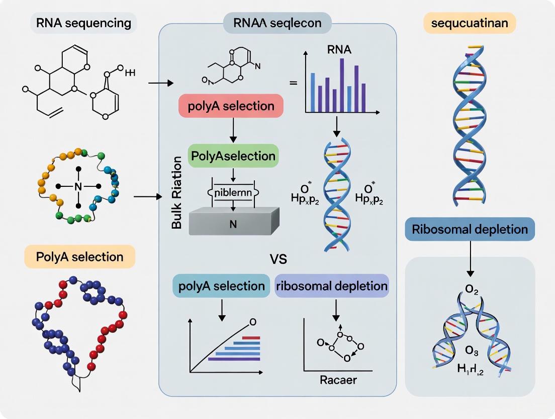
In the field of transcriptomics, bulk RNA sequencing (RNA-seq) has revolutionized our ability to profile gene expression comprehensively. The core of any RNA-seq workflow lies in the critical first step of library preparation: the removal of highly abundant ribosomal RNA (rRNA) which can constitute over 80% of a total RNA sample, thereby allowing efficient detection of informative transcripts [1]. Two principal methodologies have emerged to address this challenge—positive selection for polyadenylated RNA (polyA selection) and negative depletion of rRNA (rRNA depletion). These techniques employ fundamentally different mechanisms that directly influence transcriptome coverage, data interpretation, and experimental outcomes [2].
This technical guide provides an in-depth comparison of these two cornerstone methods, framed within contemporary clinical and research contexts. We delineate their operational mechanisms, comparative performance metrics across different sample types, and provide structured experimental protocols to inform methodological selection for researchers, scientists, and drug development professionals engaged in transcriptome analysis.
Core Mechanistic Principles
Positive Selection (PolyA+ Selection)
The polyA selection method operates on the principle of affinity capture. It utilizes oligo(dT) primers or beads that hybridize specifically to the polyadenylated tails present on mature eukaryotic messenger RNAs (mRNAs) and many long non-coding RNAs (lncRNAs) [2]. This hybridization allows for the direct purification and enrichment of these polyA+ transcripts from the total RNA pool. The process effectively excludes ribosomal RNAs (rRNAs), transfer RNAs (tRNAs), small nuclear RNAs (snRNAs), small nucleolar RNAs (snoRNAs), and other non-polyadenylated transcripts such as replication-dependent histone mRNAs [2]. Consequently, the resulting library is highly enriched for the protein-coding fraction of the transcriptome.
Negative Depletion (rRNA Depletion)
In contrast, the rRNA depletion method functions via subtractive hybridization. This technique employs sequence-specific DNA probes that are complementary to the sequences of various cytoplasmic and mitochondrial ribosomal RNAs [2]. These DNA probes hybridize to the rRNAs within the total RNA sample, forming RNA-DNA hybrids. The hybrids are subsequently removed from the solution through enzymatic digestion (e.g., using RNase H) or magnetic bead-based affinity capture [2]. Unlike polyA selection, this method does not discriminate based on the presence of a polyA tail. The remaining RNA pool after depletion includes both polyadenylated and non-polyadenylated species, such as pre-mRNAs, many lncRNAs, histone mRNAs, and some viral RNAs [2].
The following diagram illustrates the fundamental procedural differences between these two core methodologies:
Performance Comparison and Data Analysis
The choice between polyA selection and rRNA depletion has profound implications for data quality, content, and interpretation. A performance comparison based on clinical samples (human blood and colon tissue) reveals significant methodological differences [1].
Exonic Coverage and Sequencing Efficiency
A primary consideration in RNA-seq experimental design is sequencing efficiency, particularly the proportion of reads that map to exonic regions, which are most informative for gene-level quantification.
Table 1: Exonic Coverage and Sequencing Efficiency
| Sample Type | Method | Usable Exonic Reads | Required Increase in Sequencing Depth* |
|---|---|---|---|
| Blood | PolyA+ Selection | 71% | Baseline |
| Blood | rRNA Depletion | 22% | 220% |
| Colon Tissue | PolyA+ Selection | 70% | Baseline |
| Colon Tissue | rRNA Depletion | 46% | 50% |
*To achieve exonic coverage equivalent to the polyA+ selection method. Data sourced from Zhao et al. (2018) [1].
The data demonstrates that polyA+ selection provides a substantially higher yield of usable exonic reads for gene quantification. The rRNA depletion method requires a significant increase in sequencing depth to achieve comparable exonic coverage, especially for blood-derived RNA [1]. This inefficiency stems from the fact that rRNA-depleted libraries capture a wider array of RNA biotypes, including intronic sequences from immature transcripts.
Transcriptome Feature Detection
While less efficient for exonic coverage, rRNA depletion offers a broader view of the transcriptome by capturing both polyadenylated and non-polyadenylated RNA species.
Table 2: Detected Transcriptome Features by RNA Biotype
| Gene Biotype | Detection by PolyA+ Selection | Detection by rRNA Depletion | Notes |
|---|---|---|---|
| Protein-Coding Genes | Excellent | Excellent | PolyA+ is highly efficient for this class. |
| PolyA+ lncRNAs | Excellent | Excellent | |
| Non-PolyA+ lncRNAs | Not Detected | Detected | Includes histone mRNAs, some viral RNAs. |
| Pre-mRNAs / Nascent Transcripts | Minimal Detection | Significant Detection | Source of high intronic mapping rate. |
| Pseudogenes | Limited | Detected | |
| Small RNAs | Not Detected | Detected |
Analyses show that the rRNA depletion method captures a wider diversity of unique transcriptome features, including non-polyadenylated long non-coding RNAs (lncRNAs), pseudogenes, and small RNAs [1]. This comes at the cost of a significantly higher fraction of reads mapping to intronic regions, which reduces the efficiency of exon-level quantification but can provide valuable information on nascent transcription and transcriptional regulation [2].
Methodological Selection Guide
The decision between polyA selection and rRNA depletion is not a matter of one method being universally superior, but rather of selecting the right tool for the specific experimental context.
Table 3: Guidance for Method Selection in Experimental Design
| Experimental Situation | Recommended Method | Rationale | Technical Considerations |
|---|---|---|---|
| Eukaryotic RNA, High Quality (RIN ≥7) | PolyA+ Selection | Maximizes exonic coverage and power for gene-level differential expression. | Coverage skews toward the 3' end as RNA integrity decreases. |
| Degraded or FFPE RNA Samples | rRNA Depletion | More tolerant of RNA fragmentation; preserves 5' coverage better than polyA capture. | Intronic and intergenic fractions rise; confirm species-specific probe match. |
| Focus on Non-Polyadenylated RNAs | rRNA Depletion | Retains polyA+ and non-polyA species (e.g., histone mRNAs, many lncRNAs, pre-mRNA). | Residual rRNA can increase if probes are off-target. |
| Prokaryotic Transcriptomics | rRNA Depletion | PolyA+ capture is not appropriate for bacteria due to fundamentally different RNA biology. | Use species-matched rRNA probes for optimal depletion. |
| Low-Input RNA Protocols | Specialized Kits (e.g., SMART-Seq) | Methods using random primers (not oligo dT) are more suitable for degraded/low-input RNA [3]. | Performance can be improved by combining with rRNA depletion [3]. |
This guidance is corroborated by a 2024 study which found that for degraded RNA and low-input RNA, such as that from FFPE tissues, methods utilizing random primers (e.g., SMART-Seq) showed superior performance compared to standard polyA selection. Furthermore, the depletion of ribosomal RNA was shown to improve the performance of these methods by increasing expression level detection [3].
The following decision tree encapsulates the key selection criteria:
Detailed Experimental Protocols
To ensure experimental reproducibility, this section outlines standardized protocols for both methods, incorporating best practices from the cited literature.
Protocol for PolyA+ Selection RNA-seq
Principle: Capture of polyadenylated RNA using surface-bound oligo(dT) probes [2] [1].
- Input Material: 10 ng–1 μg of high-quality total RNA (RIN ≥7 or DV200 ≥50%).
- Fragmentation: RNA is fragmented into 200–300 nucleotide pieces using heat and divalent cations.
- cDNA Synthesis: First-strand cDNA is synthesized using random hexamers and reverse transcriptase. Second-strand cDNA is synthesized using DNA Polymerase I and RNase H.
- Library Construction: Double-stranded cDNA undergoes end-repair, adenylation of 3' ends, and ligation of platform-specific sequencing adapters.
- Library Enrichment: Final libraries are enriched and amplified via PCR (typically 10-15 cycles).
- Quality Control: Assess library size distribution (e.g., Bioanalyzer) and quantify (e.g., qPCR).
Protocol for rRNA Depletion RNA-seq
Principle: Removal of ribosomal RNAs via hybridization to sequence-specific probes [1] [4].
- Input Material: 100 ng–1 μg of total RNA. Tolerates a wider range of RNA integrity.
- rRNA Removal: Total RNA is hybridized with biotinylated DNA probes complementary to cytoplasmic (5S, 5.8S, 18S, 28S) and mitochondrial rRNAs.
- Depletion: Probe-bound rRNA is removed using streptavidin-coated magnetic beads.
- Library Construction: The depleted RNA is purified and carried forward. Subsequent steps—fragmentation, cDNA synthesis, and library preparation—mirror the polyA+ selection protocol.
- Quality Control: As per polyA+ selection protocol. Additionally, check for residual rRNA content (e.g., using Bioanalyzer or RNA TapeStation).
The Scientist's Toolkit: Essential Research Reagents
Table 4: Key Research Reagent Solutions for RNA-seq Library Preparation
| Reagent / Kit | Function | Method | Key Considerations |
|---|---|---|---|
| Oligo(dT) Magnetic Beads | Affinity capture of polyA+ RNA | PolyA+ Selection | Efficiency drops significantly with degraded RNA. |
| Biotinylated rRNA Probes (e.g., Ribo-Zero, Globin-Zero) | Hybridize to and deplete rRNA sequences | rRNA Depletion | Probe specificity is critical; check for target organism. |
| Random Hexamer Primers | Initiate first-strand cDNA synthesis | Both | Used in both protocols after enrichment/depletion. |
| Template Switching Oligo | Used in SMART-Seq to generate full-length cDNA from low-input/degraded RNA [3] | Low-Input Methods | Enables sequencing of RNA where the 5' end is compromised. |
| Not-So-Random (NSR) Primers | Designed for more uniform reverse transcription | RamDA-Seq [3] | Aims to reduce bias in cDNA synthesis. |
| RNase H | Digests RNA in RNA-DNA hybrids | rRNA Depletion | Key enzyme in some depletion protocols. |
The strategic choice between positive polyA selection and negative rRNA depletion fundamentally shapes the scope and focus of an RNA-seq study. PolyA+ selection offers superior efficiency and precision for profiling mature, protein-coding mRNA, making it the default choice for intact eukaryotic samples where the research question centers on gene-level differential expression. In contrast, rRNA depletion provides a more comprehensive view of the transcriptome, encompassing non-coding and nascent transcripts, and demonstrates greater resilience with suboptimal sample types like FFPE tissues or samples with mixed RNA integrity. As the field advances, especially in clinical diagnostics where sample quality is often variable, the integration of method-specific performance metrics and the development of robust protocols for challenging samples will be paramount for unlocking the full potential of transcriptome sequencing in research and therapeutic development.
The choice between poly(A) selection and ribosomal RNA (rRNA) depletion in bulk RNA-seq represents a fundamental methodological crossroads that directly dictates the resulting view of the transcriptome. This technical guide examines how each enrichment strategy defines the scope of biological investigation by capturing distinct RNA populations. Poly(A) selection provides a highly efficient, targeted view of protein-coding mRNA but systematically excludes entire classes of non-polyadenylated transcripts. In contrast, rRNA depletion offers a broader, more inclusive perspective of the transcriptional landscape, capturing both coding and non-coding RNA species, yet requires greater sequencing resources and careful optimization. The decision between these methods carries profound implications for experimental outcomes in gene expression studies, particularly in specialized applications involving degraded samples, prokaryotic partners, or non-coding RNA biology. This review synthesizes current evidence to equip researchers with a structured framework for selecting the optimal transcriptome coverage strategy based on specific experimental objectives, sample characteristics, and biological questions.
In bulk RNA-seq experiments, the transcriptome represents the complete set of RNA molecules present in a biological sample at a specific point in time. However, a fundamental challenge arises from the overwhelming abundance of ribosomal RNA (rRNA), which constitutes approximately 80-90% of total RNA in most cells [5]. This predominance of rRNA would consume the majority of sequencing resources if total RNA were sequenced directly, leaving limited capacity for profiling messenger RNA (mRNA) and other informative RNA species. Consequently, effective rRNA removal is a critical first step in most RNA-seq workflows, with two principal strategies employed: poly(A) selection and rRNA depletion.
These two methods operate on fundamentally different principles and consequently reveal different aspects of the transcriptome. Poly(A) selection is a positive enrichment strategy that targets the 3' polyadenylated tails characteristic of mature eukaryotic mRNA [6]. Conversely, rRNA depletion is a negative selection approach that uses hybridization probes to specifically remove rRNA molecules, preserving both polyadenylated and non-polyadenylated RNA species [7]. The choice between these methods directly determines which RNA molecules will be visible in subsequent analyses and which will be systematically excluded, thereby shaping all biological interpretations derived from the data.
Core Methodologies and Biological Basis
Poly(A) Selection: Targeting the 3' Tail
Biological Mechanism of Polyadenylation
The poly(A) selection method leverages a natural post-transcriptional modification process. In eukaryotic cells, mature messenger RNA (mRNA) molecules undergo 3' end processing whereby a stretch of 200-250 adenine nucleotides, known as a poly(A) tail, is added [6]. This tail serves critical biological functions: it protects the mRNA from degradation by exonucleases, facilitates nuclear export, and enhances translation efficiency by interacting with the 5' cap structure through protein intermediaries [6]. This conserved feature provides a molecular handle for selective mRNA capture.
Technical Protocol for Poly(A) Selection
The standard poly(A) selection protocol utilizes the base-pairing specificity between adenine and thymine to isolate polyadenylated RNA [6]. The workflow typically involves several key stages, as visualized in Figure 1.
Figure 1. Poly(A) Selection Workflow. The process involves heat denaturation of total RNA followed by hybridization with oligo(dT) magnetic beads, washing to remove unbound RNA, and final elution of purified poly(A)+ RNA.
Following this general workflow, the specific procedural steps are critical for success:
Bead Preparation and RNA Denaturation: Oligo(dT) magnetic beads are resuspended, and total RNA is mixed with a high-salt binding buffer and heated to 65-70°C. This heat denaturation step is crucial for disrupting RNA secondary structures and making the poly(A) tails accessible for hybridization [6].
Annealing and Hybridization: The denatured RNA is incubated with the oligo(dT) beads at room temperature for 30-60 minutes. During this phase, the poly(A) tails of mRNA specifically bind to the complementary oligo(dT) sequences on the beads through A-T base pairing, which is stabilized by the high-salt buffer [6].
Washing and Elution: The bead-mRNA complex is captured using a magnet, and the supernatant containing non-polyadenylated RNA (rRNA, tRNA, etc.) is discarded. Multiple washes with high-salt buffer remove contaminants. Finally, purified poly(A)+ mRNA is eluted using low-salt buffer or nuclease-free water at 60-80°C, which disrupts the A-T bonds [6].
Protocol variations exist in bead-to-RNA ratios, incubation times, and wash stringency. Some workflows also perform cDNA synthesis directly on the beads to minimize sample loss [6].
rRNA Depletion: A Subtraction-Based Strategy
Biological Basis and Technical Principle
rRNA depletion takes a subtraction-based approach, directly targeting the abundant rRNA molecules for removal rather than positively selecting a specific RNA population. This method is independent of the polyadenylation status of transcripts, making it universally applicable across sample types, including those from prokaryotes [8]. The strategy relies on the design of sequence-specific probes that hybridize to rRNA molecules, followed by their physical removal or enzymatic degradation.
Two primary mechanisms are employed for rRNA removal:
- Probe Hybridization and Physical Removal: Biotinylated DNA oligonucleotides complementary to rRNA sequences are hybridized to total RNA. The resulting RNA-DNA hybrids are then captured and removed using streptavidin-coated paramagnetic beads [8].
- RNase H-Mediated Degradation: After hybridization of DNA probes to rRNA, the enzyme RNase H is introduced. This enzyme specifically cleaves the RNA strand of RNA-DNA hybrids, degrading the rRNA [8].
The efficiency of both methods depends critically on the specificity and comprehensive coverage of the designed probes against the target rRNA sequences (e.g., 18S, 28S, 5.8S, and 5S in eukaryotes) [7].
Detailed Protocol for Probe-Based rRNA Depletion
A robust protocol for customized rRNA depletion using biotinylated oligos is detailed below and illustrated in Figure 2.
Figure 2. rRNA Depletion Workflow. The process involves hybridizing biotinylated DNA probes to rRNA in total RNA, capturing the probe-rRNA complexes with streptavidin beads, and separating the beads to leave an rRNA-depleted sample.
Key experimental steps and considerations include:
Probe Design: Probes (typically 25-50 bp) are designed to be complementary to the 5', middle, and 3' regions of major rRNA transcripts (e.g., 28S alpha, 28S beta, 18S) to ensure depletion of both full-length and degraded rRNA. Specificity must be verified using tools like BLAST to minimize off-target hybridization to non-rRNA transcripts [8].
Hybridization and Capture Optimization: Total RNA is hybridized with a pool of biotinylated oligonucleotide probes. Empirical testing is required to determine the optimal template-to-probe ratio; a mass ratio of 1:2 (RNA:probes) is often effective [8]. For example, 2 µg of total RNA may be hybridized with 4 µg of probes.
Bead-Based Removal and Iterative Depletion: Streptavidin-coated paramagnetic beads are added to capture the biotinylated probe-rRNA complexes. The beads are magnetically separated, and the supernatant containing the rRNA-depleted RNA is recovered. Multiple rounds of depletion (e.g., three rounds) may be necessary to reduce rRNA content to below 5% of total RNA [8].
Comparative Analysis: Coverage, Applications, and Limitations
Direct Comparison of Technical Performance
The choice between poly(A) selection and rRNA depletion involves trade-offs across multiple performance parameters, as summarized in Table 1.
Table 1. Technical Comparison: Poly(A) Selection vs. rRNA Depletion
| Feature | Poly(A) Selection | rRNA Depletion |
|---|---|---|
| Primary Target | Mature poly(A)+ mRNA [2] | Both poly(A)+ and non-poly(A) RNAs [2] |
| Ideal RNA Integrity | Requires high integrity (RIN ≥7) [2] | Works with degraded/FFPE RNA [9] [2] |
| Transcriptome Breadth | Narrow (protein-coding mRNA focus) [6] | Broad (coding & non-coding RNAs) [7] |
| Coverage Uniformity | 3' bias, especially in degraded RNA [7] | More uniform 5' to 3' coverage [7] |
| Sequencing Efficiency | High (fewer reads needed for mRNA) [6] | Lower (more reads required) [6] |
| Organism Applicability | Eukaryotes only [2] | Eukaryotes and prokaryotes [10] |
| Key Limitations | Misses non-poly(A) RNAs (e.g., histone mRNAs, many lncRNAs) [2] [6] | Higher cost per sample; potential residual rRNA [6] |
Quantitative Performance in Experimental Studies
Empirical studies directly comparing these methods provide critical insights for experimental planning. Table 2 summarizes key quantitative findings from published research.
Table 2. Experimental Performance Metrics from Comparative Studies
| Study & Context | Poly(A) Selection Performance | rRNA Depletion Performance | Biological Conclusion |
|---|---|---|---|
| Ma et al., 2019 (Murine Liver) [9] | Detected more differentially expressed genes (DEGs). Read distribution biased toward longer transcripts. | Fewer DEGs detected, but captured key pathways reliably. Less sensitive to transcript length. | Both methods yielded highly similar biological conclusions for pathway enrichment, despite differences in DEG count. |
| Fieth et al., 2022 (Sponge Holobiont) [10] | Effective for eukaryotic host transcriptome. Very poor for capturing bacterial symbiont mRNAs. | Effective for simultaneous capture of both host eukaryotic and bacterial symbiont transcriptomes. | rRNA depletion is the required method for holistic host-symbiont (holobiont) transcriptomic profiling. |
| Wrobel et al., 2019 (Custom Depletion) [8] | N/A in this study. Poly(A) enrichment misses many non-coding and immature transcripts. | Reduced rRNA to <5% with 12 custom probes. 50 non-rRNA transcripts showed co-depletion. | Custom rRNA depletion is highly efficient and specific, providing a viable alternative for non-model organisms. |
| Chung et al., 2015 (Sequencing Efficiency) [6] | ~50% fewer reads needed in colon, ~220% fewer in blood for similar gene-level coverage vs. depletion. | Requires significantly more sequencing depth to achieve exonic coverage comparable to poly(A) selection. | Poly(A) selection is more resource-efficient for standard mRNA profiling. |
Suitability and Limitations by Research Application
Optimal Applications for Poly(A) Selection
- Standard Gene Expression Profiling: When the research question focuses exclusively on differential expression of protein-coding genes in eukaryotic systems with high-quality RNA, poly(A) selection is the most efficient and cost-effective choice [9] [6].
- High-Throughput Studies: For large-scale screening experiments involving many samples (e.g., drug screening, genetic perturbations), the lower sequencing depth requirement makes poly(A) selection economically advantageous [9].
- 3' mRNA-Seq (QuantSeq): Specialized applications like QuantSeq, which use oligo(dT) priming for library construction, are ideal for rapid, cost-effective gene expression quantification, particularly from challenging sample types like FFPE material [9].
When rRNA Depletion is Mandatory or Preferred
- Prokaryotic Transcriptomics or Host-Symbiont Studies: Bacterial mRNAs lack stable poly(A) tails, making rRNA depletion the only viable option. This is also critical for dual RNA-seq of infected tissues or holobiont systems [10].
- Analysis of Non-Polyadenylated RNAs: Studies focusing on long non-coding RNAs (lncRNAs), circular RNAs, histone mRNAs, or other non-coding RNAs that lack poly(A) tails require rRNA depletion [2] [6].
- Degraded or FFPE Samples: When working with archived clinical samples (FFPE) or other degraded RNA where the poly(A) tail may be lost or compromised, rRNA depletion provides more robust and uniform transcript coverage [9] [2].
- Analysis of Transcriptional Dynamics: rRNA depletion retains pre-mRNA and intronic reads, which can be leveraged to study nascent transcription, splicing kinetics, and other transcriptional regulatory mechanisms [2].
Successful implementation of poly(A) selection or rRNA depletion requires specific reagents and careful quality control. Table 3 outlines key components of the experimental toolkit.
Table 3. Research Reagent Solutions for Transcriptome Enrichment
| Reagent / Material | Function | Application Notes |
|---|---|---|
| Oligo(dT) Magnetic Beads | Capture poly(A)+ RNA via hybridization to the poly(A) tail. | Core component of poly(A) selection kits. Bead-to-RNA ratio is critical for yield [6]. |
| Biotinylated DNA Probes | Sequence-specific probes hybridize to rRNA for depletion. | Can be commercial kits or custom-designed for model and non-model organisms [8]. |
| Streptavidin Paramagnetic Beads | Capture and remove biotinylated probe-rRNA complexes. | Used in custom and some commercial rRNA depletion protocols [8]. |
| RNase H | Enzyme that degrades RNA in RNA-DNA hybrids. | Used in RNase H-mediated depletion protocols (e.g., NEB kits) to degrade rRNA [8]. |
| High-Salt Binding Buffer | Stabilizes A-T base pairing during hybridization. | Essential for efficient capture in poly(A) selection [6]. |
| RNA Integrity Analyzer (e.g., Bioanalyzer) | Assesses RNA Quality (RIN/DV200) pre- and post-enrichment. | Critical for QC; DV200 is particularly informative for FFPE samples [2]. |
The decision between poly(A) selection and rRNA depletion is foundational, irrevocably shaping the scope and validity of transcriptomic findings. There is no universally superior method; the optimal choice is dictated by the specific biological question, sample characteristics, and available resources.
For research focused exclusively on protein-coding gene expression in eukaryotes with high-quality RNA, poly(A) selection remains the most efficient and targeted approach. However, for investigations requiring a comprehensive view of the transcriptional landscape—including non-coding RNAs, bacterial transcripts, or samples with compromised RNA integrity—rRNA depletion is the indispensable, albeit more resource-intensive, alternative.
As the field advances with emerging applications in single-cell biology, spatial transcriptomics, and precision medicine, the principles outlined in this guide will continue to inform experimental design. By aligning methodological strengths with specific research objectives, scientists can ensure their chosen path through the transcriptomic landscape reveals the biological insights they seek, rather than the artifacts of their chosen method.
In bulk RNA sequencing (RNA-seq), the choice between polyadenylated (polyA) selection and ribosomal RNA (rRNA) depletion is a fundamental upstream decision that profoundly shapes the composition and interpretation of sequencing data [2]. This choice is particularly decisive for the relative proportions of reads mapping to exonic, intronic, and intergenic regions of the genome. These mapping distributions are not merely technical artifacts; they reflect the underlying biology of the RNA species captured and have direct implications for the accuracy of gene quantification, the detection of novel features, and the biological conclusions that can be drawn.
PolyA selection enriches for mature, protein-coding mRNA by capturing RNAs with polyA tails using oligo-dT primers, thereby focusing the dataset on spliced transcripts. In contrast, rRNA depletion removes abundant ribosomal RNAs from total RNA, preserving a much broader spectrum of RNA species, including pre-mRNA, non-polyadenylated long non-coding RNAs (lncRNAs), and other non-coding RNAs [2] [11]. The core distinction lies in the fact that polyA selection targets a specific RNA feature (the tail), while rRNA depletion targets specific RNA sequences (rRNA). This fundamental difference cascades through the entire data analysis pipeline, resulting in distinctly structured datasets.
Quantitative Impact on Read Distribution
The methodological difference between library preparations leads to starkly divergent profiles in where reads map across the genome. The table below summarizes the typical distribution of reads from polyA-selected and rRNA-depleted libraries in different biological contexts.
Table 1: Comparison of Read Distribution Profiles between Library Prep Methods
| Sample Type | Library Method | Exonic Reads | Intronic Reads | Intergenic Reads | Key Reference |
|---|---|---|---|---|---|
| Human Blood | polyA+ Selection | ~71% | Lower | Lower | [1] |
| Human Blood | rRNA Depletion | ~22% | Substantially Higher (~50% of bases) | Substantially Higher | [1] [12] |
| Human Colon | polyA+ Selection | ~70% | Lower | Lower | [1] |
| Human Colon | rRNA Depletion | ~46% | Substantially Higher (~33% of bases) | Substantially Higher | [1] [12] |
| Human Breast Tumor (FF) | polyA+ Selection | 62.3% | Lower | Lower | [12] |
| Human Breast Tumor (FF) | rRNA Depletion | 20-30% | Majority of reads | Majority of reads | [12] |
| D. melanogaster | polyA+ Selection | Highest | Lower | Higher than intronic | [13] |
| D. melanogaster | rRNA Depletion | Highest (but lower than polyA+) | Higher than polyA+ | Lower than polyA+ | [13] |
The data reveals a consistent pattern: rRNA depletion libraries consistently yield a significantly lower fraction of exonic reads and a concomitantly higher fraction of intronic and intergenic reads compared to polyA-selected libraries. This effect is so pronounced that for a blood sample, 220% more reads must be sequenced with rRNA depletion to achieve the same level of exonic coverage as polyA+ selection; for colon tissue, this number is 50% [1]. This has major implications for sequencing cost and the effective depth of coverage for the target transcriptome.
Biological and Technical Origins of Read Types
The observed data structure is a direct consequence of the RNA species captured by each method.
Intronic reads are a hallmark of rRNA-depleted libraries, but their presence can be attributed to several biological and technical factors:
- Nascent Transcription and Pre-mRNA: A primary source of intronic signal is the capture of unspliced pre-mRNA by rRNA depletion. These transcripts contain intronic sequences that have not yet been removed by the splicing machinery [14]. This is not merely "contamination" but a reflection of transcriptional activity. Studies have shown that longer introns tend to have higher RNA-seq coverage in rRNA-depleted data, supporting the model of co-transcriptional splicing, where splicing occurs while the RNA polymerase is still transcribing the gene [14].
- Genomic DNA Contamination: Trace amounts of genomic DNA (gDNA) contaminating the RNA sample can align to intronic (and intergenic) regions. Unlike pre-mRNA, gDNA contamination typically produces reads mapping to both strands of the intron [14].
- Stable Intronic RNAs and Non-Coding RNAs: Some intronic sequences give rise to functional non-coding RNAs or stable lariat RNAs, which can be captured by rRNA depletion [14].
- Unannotated Transcripts and Exons: Some intronic reads may point to previously unannotated exons or transcript isoforms not present in the reference annotation [14].
Intergenic reads, which map outside of annotated gene boundaries, are also more abundant in rRNA-depleted data. Their origins include:
- Non-polyadenylated Non-Coding RNAs: rRNA depletion captures a wide array of non-coding RNAs (e.g., some lncRNAs, enhancer RNAs) that are not polyadenylated and may be located in intergenic regions [1] [2].
- Novel Transcripts: These reads can indicate the presence of entirely novel, unannotated transcripts.
- Genomic DNA Contamination: Similar to intronic reads, gDNA contamination will also contribute to intergenic signals.
polyA+ Selection Minimizes Non-Exonic Reads
PolyA selection effectively minimizes intronic and intergenic reads by design. Since it captures only RNAs with polyA tails, it selectively enriches for mature, spliced mRNAs, where introns have been removed. It also excludes the majority of non-polyadenylated non-coding RNAs. Consequently, the resulting data is highly concentrated on exonic regions, providing high power for protein-coding gene quantification [1] [2].
Diagram 1: How library prep defines RNA species and read types. polyA+ selection filters for mature mRNA, leading to high exonic reads. rRNA depletion retains a mixed population, resulting in high intronic and intergenic reads.
Experimental Protocols for Comparative Analysis
To ensure reproducible and accurate comparisons between polyA selection and rRNA depletion, a standardized experimental and computational workflow is essential. The following protocol, synthesized from multiple studies, provides a robust framework.
Sample Preparation and Library Construction
- RNA Extraction: Isolate total RNA from the biological material of interest (e.g., human blood, colon tissue, cell lines) using a standardized kit (e.g., Zymo Research RNA Clean and Concentrator [11]). RNA Integrity Number (RIN) or RQS/DV200 values should be assessed (e.g., via Agilent Bioanalyzer) [1] [2].
- Library Preparation (in parallel):
- polyA+ Selection: Use a kit such as SMARTSeq V4 (Takara) that employs oligo-dT primers to capture and reverse-transcribe polyadenylated RNA [11].
- rRNA Depletion: Use a ribodepletion kit such as Ribo-Zero Gold (Illumina) or SoLo Ovation (Tecan). For non-model organisms, ensure the use of species-specific probes (e.g., a custom 200-probe set for C. elegans rRNA sequences) [11].
- Sequencing: Sequence all libraries on a platform such as Illumina to a sufficient depth (e.g., 50 million reads per replicate) using paired-end sequencing [1].
Computational Analysis and QC Pipeline
A unified computational pipeline, such as the Transcriptome Analysis Pipeline (TAP), should be used to process all data uniformly [13].
- Alignment: Perform splicing-aware alignment to the reference genome (e.g., GRCh38 for human) using a aligner such as STAR [13] [15].
- Read Classification: Classify aligned reads based on their overlap with genomic annotations (GTF file). A standard definition is:
- Quantification: Generate gene-level counts using tools like HTSeq or Salmon, considering only exonic reads for classical gene expression analysis [17] [13].
- Quality Control:
Diagram 2: Unified workflow for comparing library methods. Samples are processed in parallel through library prep, then analyzed with a single pipeline to ensure consistent comparison.
The Scientist's Toolkit: Essential Reagents and Software
Table 2: Key Research Reagent Solutions and Computational Tools
| Item Name | Type | Primary Function | Considerations |
|---|---|---|---|
| SMARTSeq V4 | Library Prep Kit | polyA+ selection for full-length cDNA | Optimal for intact, eukaryotic RNA [11]. |
| Ribo-Zero Gold | Library Prep Kit | Depletion of cytoplasmic rRNA from human/mouse/rat | Standard for human clinical samples; less efficient in non-models [1] [12]. |
| Ovation SoLo with Custom AnyDeplete | Library Prep Kit | rRNA depletion with custom probes | Essential for non-model organisms (e.g., C. elegans) [11]. |
| RNA Clean & Concentrator | RNA Purification Kit | Cleanup and concentration of RNA post-extraction | Critical for obtaining high-quality input material [11]. |
| Agilent Bioanalyzer | Instrument | Assesses RNA Integrity (RIN) | RIN ≥7 recommended for polyA+ selection [2]. |
| STAR Aligner | Software | Splicing-aware alignment of RNA-seq reads to a genome | Standard for accurate read mapping and junction detection [15]. |
| Picard Tools | Software Suite | Generates QC metrics (e.g., read distribution) | CollectRnaSeqMetrics is key for data structure analysis [18]. |
| MultiQC | Software | Aggregates results from multiple tools into a single report | Essential for visualizing and comparing QC metrics across samples [18]. |
The choice between polyA selection and rRNA depletion is not a matter of one method being universally superior, but rather of selecting the right tool for the specific research objective. The resulting data structure—defined by the balance of exonic, intronic, and intergenic reads—is a direct and predictable outcome of this choice.
- For primary focus on protein-coding gene expression: polyA+ selection is the recommended method. It delivers superior exonic coverage, higher power for differential expression at a given sequencing depth, and is more cost-effective for this specific goal [1] [2].
- For exploratory transcriptomics or studies of non-coding RNA: rRNA depletion is necessary. It is essential for capturing non-polyadenylated RNAs (lncRNAs, pre-mRNAs, histone mRNAs) and provides a more complete picture of transcriptional activity [1] [11].
- For degraded or FFPE samples: rRNA depletion is strongly preferred due to its resilience to RNA fragmentation, as it does not rely on an intact 3' polyA tail [12] [2].
Ultimately, the decision must be guided by the biological question, the organism, and the quality of the input RNA. Researchers must be aware that the data structure resulting from their chosen protocol directly influences the analytical strategies required and the biological interpretations that can be made.
The Critical Role of RNA Integrity (RIN) in Method Selection
In bulk RNA sequencing (RNA-seq), the choice between poly(A) selection and ribosomal RNA (rRNA) depletion is a foundational experimental decision. The integrity of the input RNA sample, most commonly quantified as the RNA Integrity Number (RIN), is a critical determinant for this choice, directly influencing data quality, coverage, and biological conclusions. RIN scores, typically ranging from 1 (degraded) to 10 (intact), provide a standardized measure of RNA quality by evaluating the ratio of ribosomal RNA bands. When RNA degrades, transcripts fragment and often lose their poly(A) tails, creating a fundamental compatibility issue with poly(A) selection protocols. This technical guide examines how RIN values should guide the selection between poly(A) selection and rRNA depletion, providing a structured framework for researchers to optimize their transcriptomics studies within the broader context of gene expression research.
How RNA Integrity Affects Library Preparation Methods
The Fundamental Mechanisms of Poly(A) Selection and rRNA Depletion
The core mechanisms of the two major enrichment strategies explain their differing dependencies on RNA integrity.
Poly(A) Selection relies on oligo(dT) beads or similar matrices to hybridize and capture RNA molecules bearing polyadenylated tails. This process specifically enriches for mature, polyadenylated messenger RNA (mRNA) by leveraging the base pairing between thymine residues on the beads and adenine residues in the poly(A) tail [6]. The protocol typically involves denaturing total RNA to expose the poly(A) tail, incubating with oligo(dT) beads for hybridization, washing away non-polyadenylated RNA species, and finally eluting the purified mRNA [6]. This mechanism effectively targets a specific biochemical feature—the 3' poly(A) tail—making it highly dependent on the preservation of this terminal structure.
rRNA Depletion operates on a different principle, using sequence-specific DNA probes designed to hybridize to abundant ribosomal RNA sequences (both cytoplasmic and mitochondrial). These probe-rRNA hybrids are subsequently removed through RNase H digestion or affinity capture, leaving behind a complex pool of both polyadenylated and non-polyadenylated RNA species [2]. This "negative selection" approach does not depend on any single RNA feature but rather removes specific unwanted targets, making it more resilient to partial RNA degradation.
Impact of RNA Degradation on Transcript Capture
RNA degradation typically proceeds in a 5'→3' direction, often involving deadenylase enzymes that progressively shorten the poly(A) tail—the very feature targeted by poly(A) selection [6]. As integrity decreases:
- For poly(A) selection: Degraded RNA with shortened or missing poly(A) tails fails to hybridize efficiently with oligo(dT) beads, leading to significant capture bias toward intact transcripts and under-representation of affected molecules [2] [6].
- For rRNA depletion: Since this method does not depend on the 3' tail for capture, fragmented RNA molecules retaining their probe-binding regions can still be effectively depleted of rRNA, allowing more representative sampling of the remaining transcriptome [2] [19].
The following diagram illustrates the core methodological differences and their relationship with RNA integrity:
Quantitative Data Comparison: Method Performance Across RIN Values
Coverage and Bias Metrics
RNA integrity directly impacts sequencing coverage distribution and gene detection capability differently for each method. The following table summarizes key quantitative differences observed across RIN values:
Table 1: Performance Metrics of RNA Enrichment Methods Across RIN Values
| Performance Metric | Poly(A) Selection | rRNA Depletion |
|---|---|---|
| Minimum Recommended RIN | 7-8 [2] | No strict minimum [2] |
| 3' Bias with Degradation | Severe (strong skew toward 3' end) [2] [6] | Minimal (more uniform coverage) [2] |
| Exonic Mapping Rate | High (70-85%) [2] [20] | Moderate (50-70%) with higher intronic reads [2] |
| Residual rRNA | Typically <1% [2] | 5-20% (probe-dependent) [2] [11] |
| Typical Sequencing Depth | 25-40 million reads [4] [20] | 50-80 million reads [4] |
| Detection of Non-polyA Transcripts | No [2] [6] | Yes (lncRNAs, histone mRNAs, etc.) [2] [6] |
Experimental Validation Studies
Empirical studies directly comparing both methods across RNA quality gradients provide compelling evidence for RIN-based selection:
A paired-design study using human CD4+ T cells from 40 donors with RIN values >8.6 demonstrated that while both methods perform well with high-quality RNA, significant divergences emerge with controlled degradation [4]. Poly(A) selection showed progressively stronger 3' bias as integrity decreased, while rRNA depletion maintained more uniform transcript coverage.
Research on low-input C. elegans samples found that rRNA depletion with species-specific probes provided superior performance for degraded samples, detecting an expanded set of noncoding RNAs and showing reduced noise for lowly expressed genes compared to poly(A) selection methods [11].
A comprehensive analysis of fragmented and FFPE (Formalin-Fixed Paraffin-Embedded) RNA samples concluded that rRNA depletion "is more resilient on fragmented and FFPE RNA" and "usually preserves 5′ coverage better than poly(A) capture" [2]. FFPE samples typically have RIN values below 4, making them largely incompatible with poly(A) selection.
Method Selection Framework and Experimental Protocols
Decision Matrix for Method Selection
The following structured framework integrates RIN values with experimental objectives to guide appropriate method selection:
Table 2: RNA-Seq Method Selection Guide Based on RIN and Research Goals
| Situation | Recommended Method | Rationale | Implementation Notes |
|---|---|---|---|
| Eukaryotic RNA, RIN ≥8, coding mRNA focus | Poly(A) selection | Concentrates reads on exons; maximizes power for gene-level differential expression [2] | Check RNA quality using Bioanalyzer/TapeStation; expect high exonic mapping rates |
| RIN 5-7, eukaryotic samples | rRNA depletion | Tolerant of partial fragmentation; preserves coverage of transcript 5' ends [2] | Use species-matched probes; sequence more deeply to compensate for lower efficiency |
| RIN <5, FFPE, or heavily degraded | rRNA depletion | Does not rely on intact 3' tails; most resilient option for compromised samples [2] | Expect higher intronic/intergenic reads; requires careful probe selection |
| Need non-polyadenylated RNAs | rRNA depletion | Retains both poly(A)+ and non-poly(A) species (lncRNAs, histone mRNAs, pre-mRNAs) [2] [6] | Confirms detection of target non-coding RNAs in pilot data |
| Prokaryotic transcriptomics | rRNA depletion | Poly(A) capture inappropriate for bacterial mRNA [2] | Essential to use species-matched rRNA probes |
| Mixed-quality sample cohort | rRNA depletion (for consistency) | Single protocol performs adequately across all quality levels [2] | Avoids protocol-switching artifacts in integrated analysis |
Practical Implementation Protocols
Poly(A) Selection Protocol (adapted from [6]):
- Bead Preparation: Resuspend oligo(dT) magnetic beads thoroughly to ensure even distribution.
- RNA Denaturation: Mix 100ng-5μg total RNA with high-salt binding buffer. Heat to 65-70°C for 2 minutes to disrupt secondary structures, then immediately place on ice.
- Hybridization: Combine denatured RNA with beads. Incubate at room temperature for 30-60 minutes with gentle mixing to allow poly(A)-oligo(dT) hybridization.
- Washing: Place tube on magnet, discard supernatant. Wash beads 2-3 times with high-salt buffer to remove non-specifically bound RNA.
- Elution: Add warm (60-80°C) low-salt buffer or nuclease-free water to release purified poly(A)+ RNA.
- Quality Control: Assess yield and purity (e.g., Bioanalyzer). Proceed to library construction.
Key Considerations: Bead-to-RNA ratio is critical; insufficient beads reduce yield while excess may increase non-specific binding [6]. For low-input samples (<100 ng), consider specialized low-input protocols.
rRNA Depletion Protocol (adapted from [2] [19]):
- Probe Hybridization: Mix total RNA (10ng-1μg) with sequence-specific DNA probes targeting rRNA species. Heat to 70°C for 2 minutes, then incubate at relevant temperature (probe-specific) for 10-15 minutes.
- rRNA Removal:
- RNase H Method: Add RNase H to digest RNA in DNA-RNA hybrids. Follow with purification to remove fragments.
- Affinity Capture Method: Use bead-coupled probes or biotinylated probes with streptavidin beads to physically remove rRNA complexes.
- Purification: Recover depleted RNA using column-based or bead-based clean-up.
- Quality Control: Assess depletion efficiency (e.g., Bioanalyzer, qPCR for residual rRNA). Residual rRNA >20% may indicate probe mismatch.
Key Considerations: Species-specific probe design is essential, particularly for non-model organisms [2] [11]. Pilot testing is recommended when working with novel species or sample types.
Table 3: Key Research Reagents for RNA Selection Methods
| Reagent / Resource | Function | Implementation Notes |
|---|---|---|
| Oligo(dT) Magnetic Beads | Capture polyadenylated RNA via hybridization | Core component of poly(A) selection; enables automation [6] |
| Sequence-Specific rRNA Probes | Hybridize to ribosomal RNA for depletion | Species-matched design critical for efficiency [2] [11] |
| RNase H Enzyme | Digests RNA in DNA-RNA hybrids | Key for enzymatic rRNA depletion methods [2] |
| High-Salt Binding Buffer | Stabilizes A-T base pairing | Critical for poly(A) selection efficiency [6] |
| Magnetic Separation Stand | Immobilizes magnetic beads during washes | Essential for both methods during wash steps |
| Bioanalyzer/TapeStation | Assesses RNA integrity and library quality | Critical for RIN determination and QC pre-/post-selection |
| DNase I | Removes genomic DNA contamination | Important for accurate RNA quantification [20] |
RNA Integrity Number serves as a pivotal decision point in the choice between poly(A) selection and rRNA depletion for bulk RNA-seq. The fundamental relationship is straightforward: as RIN decreases, the advantage shifts decisively toward rRNA depletion.
For researchers designing transcriptomics studies, the following best practices are recommended:
- Always quantify RNA integrity using standardized methods (RIN, DV200) before selecting library preparation method.
- Establish sample quality thresholds specific to your research objectives, with RIN ≥8 for poly(A) selection and no strict minimum for rRNA depletion.
- Employ rRNA depletion for heterogeneous sample cohorts where RNA quality varies, as it provides more consistent performance across quality levels.
- Validate probe specificity when working with non-model organisms or specialized sample types.
- Anticipate sequencing depth needs based on method selection—rRNA depletion typically requires 1.5-2× greater sequencing depth than poly(A) selection for similar gene detection sensitivity.
By aligning method selection with RNA integrity metrics and experimental goals, researchers can optimize data quality, maximize informational yield, and ensure biologically meaningful results from their transcriptomics investments.
Strategic Application: Choosing the Right Method for Your Sample and Research Question
A critical, irreversible first step in any bulk RNA-sequencing experiment is the choice of how to handle the overwhelming abundance of ribosomal RNA (rRNA), which constitutes 80-98% of the total RNA in a typical mammalian cell [21] [22]. This decision determines which RNA molecules enter the sequencing library and fundamentally shapes all downstream data and analyses [2]. The two principal strategies are poly(A) selection, which enriches for polyadenylated transcripts, and rRNA depletion (ribodepletion), which removes rRNA and sequences the remaining transcriptome [2] [23]. This guide provides a structured framework for choosing between these methods based on three core factors: the organism of study, the quality of the input RNA, and the specific transcripts of interest, all within the context of optimizing bulk RNA-seq research.
Core Principles: poly(A) Selection vs. rRNA Depletion
poly(A) Selection: Capturing the Tailed Transcriptome
Mechanism: This method leverages the polyadenylated tails found on most mature eukaryotic messenger RNAs (mRNAs) and some long non-coding RNAs (lncRNAs). Through hybridization with oligo(dT) primers or beads, these tailed transcripts are selectively captured from the total RNA pool [2] [23].
Captured Transcripts:
- Included: Mature eukaryotic mRNA, many polyadenylated lncRNAs.
- Excluded: rRNA, transfer RNA (tRNA), small nuclear/nucleolar RNA (sn/snoRNA), and non-polyadenylated mRNAs such as replication-dependent histone mRNAs [2] [24].
rRNA Depletion: A Broader View of the Transcriptome
Mechanism: This method uses sequence-specific DNA or RNA probes that are complementary to rRNA sequences (e.g., 5S, 5.8S, 18S, 28S). The probe-rRNA hybrids are subsequently removed from the sample, typically via RNase H digestion or magnetic bead capture [2] [25].
Captured Transcripts:
- Included: Both poly(A)+ and non-polyadenylated species. This includes mature mRNA, pre-mRNA (containing introns), many lncRNAs, histone mRNAs, and some viral RNAs [2] [26].
The following diagram illustrates the fundamental workflows and outcomes of these two methods.
The Decision Framework: A Three-Filter Approach
The optimal enrichment method is determined by systematically evaluating the organism, RNA integrity, and target transcripts [2]. The following table provides a consolidated overview for quick comparison.
Table 1: Method Selection Framework for poly(A) Selection vs. rRNA Depletion
| Filter | Criteria | Recommended Method | Key Rationale | Potential Pitfalls |
|---|---|---|---|---|
| Organism | Eukaryotic (good annotation) | poly(A) Selection | Efficiently targets mature, polyadenylated mRNA [2] [23]. | Misses non-poly(A) RNAs; not suitable for prokaryotes [2]. |
| Prokaryotic, Archaeal, or Metatranscriptomic | rRNA Depletion | Necessary as bacterial mRNA lacks stable poly(A) tails [2] [25]. | Requires species-matched probes to avoid high residual rRNA [2] [25]. | |
| RNA Integrity | Intact (RIN/RQS ≥ 7, DV200 ≥ 50%) | poly(A) Selection | Provides high exonic fractions, optimal for gene-level DE analysis [2]. | Coverage skews strongly to the 3' end as integrity drops [2] [9]. |
| Degraded or FFPE | rRNA Depletion | More resilient to fragmentation; better preserves 5' coverage [2] [9]. | Intronic/intergenic fractions rise; may need deeper sequencing [2] [26]. | |
| Target Transcripts | Mature, coding mRNA | poly(A) Selection | Concentrates reads on exons, boosting power for gene-level DE [2]. | Loss of non-coding and nascent transcriptional signal [2] [24]. |
| Non-polyadenylated RNAs (e.g., histone mRNAs, many lncRNAs, pre-mRNA) | rRNA Depletion | Retains both poly(A)+ and poly(A)- species in one assay [2] [23]. | Higher library complexity requires greater sequencing depth [26]. |
Impact on Data Output and Analysis
The choice of method directly shapes your data and its interpretation:
- poly(A) Selection Data: Characterized by high exonic mapping rates and relatively low residual rRNA. This concentrates sequencing power on annotated exons, which is ideal for statistical tests of differential gene expression. A key limitation is 3' bias, where coverage tilts toward the 3' end of transcripts, especially with partially degraded RNA. This can lead to under-representation of long transcripts [2] [26].
- rRNA Depletion Data: Yields a broader transcriptomic profile. The mapping rates will include significant intronic and intergenic fractions. Intronic reads can track nascent transcriptional activity, while exonic reads reflect post-transcriptional processed mRNA. Modeling these signals together allows researchers to separate transcriptional from post-transcriptional regulatory mechanisms [2].
Detailed Methodologies and Experimental Protocols
Protocol for poly(A) Selection and Library Preparation
This protocol is adapted from standard practices for intact eukaryotic RNA [2] [27].
- Total RNA Quality Control: Verify RNA integrity using an Agilent Bioanalyzer. A RNA Integrity Number (RIN) or RQS of ≥7 is recommended [2].
- poly(A) RNA Capture:
- Incubate total RNA with oligo(dT) magnetic beads. The poly(A) tails of mature mRNAs hybridize to the oligo(dT) sequences.
- Use a magnetic stand to separate the bead-bound poly(A)+ RNA from the rest of the total RNA.
- Wash the beads to remove nonspecifically bound RNA, including rRNA and other non-poly(A) species.
- Elution: Elute the purified poly(A)+ RNA from the beads, typically using nuclease-free water or elution buffer at an elevated temperature.
- Stranded Library Preparation:
- Fragment the eluted RNA to a desired size distribution.
- Perform reverse transcription using random primers to generate first-strand cDNA.
- Synthesize the second strand. To preserve strand-of-origin information, incorporate dUTP in place of dTTP during second-strand synthesis [23].
- Perform end-repair, A-tailing, and adapter ligation.
- Treat the library with Uracil-Specific Excision Reagent (USER) enzyme to digest the dUTP-containing second strand, ensuring only the first strand is amplified [23].
- PCR Amplification: Amplify the library with a limited number of PCR cycles using primers complementary to the adapters.
- Library QC: Quantify the final library and validate its size distribution using a Fragment Analyzer or similar system before sequencing.
Protocol for rRNA Depletion and Library Preparation
This protocol, suitable for a wide range of sample types including degraded RNA and prokaryotic samples, is based on hybridization and bead capture methods [2] [25].
- RNA Quality and DNA Digestion: Quality-check the total RNA. Ensure complete removal of genomic DNA with a rigorous DNase treatment, as any contaminating DNA will be sequenced and manifest as high intergenic read alignment [28].
- Hybridization with rRNA Probes:
- Denature the total RNA and hybridize it with a pool of biotinylated DNA oligonucleotides that are complementary to the rRNA sequences (e.g., for 5S, 16S, and 23S rRNAs in bacteria).
- Use species-matched probes for optimal efficiency. Probe mismatch is a common failure mode that leaves high residual rRNA [2] [25].
- rRNA Removal:
- Add streptavidin-coated magnetic beads to the hybridization mix. The biotin on the probes binds tightly to the streptavidin on the beads.
- Use a magnetic stand to capture the bead-probe-rRNA complexes, leaving the desired, rRNA-depleted RNA in the supernatant.
- Recovery of Depleted RNA: Carefully transfer the supernatant, which contains the enriched mRNA and other non-rRNA transcripts, to a new tube.
- Library Preparation:
- Since the RNA pool includes non-polyadenylated transcripts, use random primers for first-strand cDNA synthesis, not oligo(dT) primers [24].
- Follow steps for second-strand synthesis (optionally with dUTP for stranded libraries), adapter ligation, and PCR amplification as described in the poly(A) protocol.
- Library QC: Quantify and validate the final library as before.
Table 2: Research Reagent Solutions for rRNA Depletion
| Kit/Reagent | Function/Basis | Key Application Notes |
|---|---|---|
| riboPOOLs | DNA oligonucleotide probes, biotinylated for bead capture [25]. | Available as species-specific or pan-prokaryotic; shown to be an efficient replacement for discontinued RiboZero [25]. |
| RiboMinus | Biotinylated DNA probes for hybridization-based depletion [25]. | Pan-prokaryotic design; efficiency can be lower than species-specific options [25]. |
| NEBNext rRNA Depletion Kit | Utilizes RNase H to digest DNA probe-rRNA hybrids [21]. | Effective for human/mouse/rat; requires careful handling to minimize off-target digestion [21] [25]. |
| Biotinylated Probes (Self-Designed) | Custom probes designed from rRNA gene sequences for magnetic bead capture [25]. | Allows for fully customized, cost-effective depletion; requires in-house design and validation [25]. |
| scDASH (CRISPR-based) | Post-library depletion using Cas9 nuclease to cleave rRNA cDNA sequences [21]. | Circumvents low-input limitations; applied after cDNA synthesis and amplification [21]. |
Troubleshooting Common Pitfalls
- High Residual rRNA in Depletion Workflows: This is most often caused by probe mismatch, especially when working with non-model organisms or pathogens [2] [25]. Solution: Pilot a few samples with different probe sets if available, and always check the percentage of rRNA reads in the initial sequencing data before scaling up the study. Consider using custom-designed probes [25].
- Strong 3' Bias in poly(A) Data: This indicates that the input RNA was more degraded than anticipated [2]. Solution: Do not try to solve this by simply sequencing deeper. For future samples of similar quality, switch to an rRNA depletion protocol, which is more tolerant of fragmentation [2].
- High Intergenic or Intronic Reads: In rRNA depletion data, this is expected and can be biologically informative (e.g., intronic reads indicate nascent transcription) [2]. However, if the rates are exceptionally high, it may indicate gDNA contamination [28]. Solution: Implement a secondary, rigorous DNase treatment during the RNA extraction or cleanup step [28].
The decision between poly(A) selection and rRNA depletion is a foundational one in bulk RNA-seq experimental design. There is no universal best choice; the optimal path is determined by a logical assessment of the organism being studied, the quality of the sourced RNA, and the specific transcriptional features under investigation. By applying the three-filter framework outlined in this guide—organism, RNA quality, and target transcripts—researchers can make a principled and defensible choice. Consistency is also critical; once a method is selected, it should be applied uniformly across all samples within a study to ensure robust and comparable results [2]. This structured approach ensures that the upstream RNA enrichment strategy is optimally aligned with the downstream biological questions, maximizing the value and reliability of the generated transcriptomic data.
Optimal Use Cases for PolyA Selection in Eukaryotic mRNA Profiling
Polyadenylated (polyA) selection remains a cornerstone technique in eukaryotic transcriptomics, offering a targeted approach for messenger RNA enrichment. This technical guide delineates the optimal use cases for polyA selection within the broader context of RNA sequencing methodologies, particularly in comparison to ribosomal RNA (rRNA) depletion. By examining the underlying mechanisms, experimental protocols, and analytical considerations, we provide researchers and drug development professionals with a comprehensive framework for deploying this method effectively. The analysis reveals that polyA selection is uniquely advantageous for specific applications including quantitative gene expression studies, high-throughput drug screening, and any research context requiring cost-efficient mRNA profiling from high-quality RNA samples.
PolyA selection is a targeted enrichment strategy that leverages the polyadenylated tails present on most mature eukaryotic messenger RNAs (mRNAs). This method specifically captures these molecules using oligo(dT) probes, effectively isolating protein-coding transcripts from the total RNA pool which is dominated by ribosomal RNA (rRNA) and other non-coding RNA species [6]. In the landscape of bulk RNA-seq research, polyA selection and rRNA depletion represent two divergent philosophical approaches to transcriptome assessment: the former offers a focused view of the mature, protein-coding transcriptome, while the latter provides a broader surveillance of both coding and non-coding RNA species [2] [4]. Understanding the technical specifications, advantages, and limitations of polyA selection is fundamental to experimental success, particularly in drug development where resources must be allocated efficiently and conclusions drawn with precision [29].
The biological basis for polyA selection lies in the post-transcriptional modification process of polyadenylation, whereby a stretch of 200-250 adenine nucleotides is added to the 3' end of nascent mRNA molecules by poly(A) polymerase [6]. This poly(A) tail plays crucial roles in mRNA stability, nucleocytoplasmic export, and translation efficiency [30] [31]. From a technical perspective, polyA selection capitalizes on this universal feature of mature eukaryotic mRNAs through hybridization between the poly(A) tail and oligo(dT) probes immobilized on magnetic beads or other solid supports [6]. This binding mechanism forms the foundation of a highly specific enrichment process that effectively removes rRNA, transfer RNA (tRNA), and other non-polyadenylated RNAs, thereby concentrating the mRNA fraction for downstream sequencing applications [2].
PolyA Selection Mechanism and Standardized Protocols
Core Mechanism and Binding Chemistry
The polyA selection process operates through precise molecular interactions between the poly(A) tail of mature mRNAs and complementary oligo(dT) sequences immobilized on solid supports, typically magnetic beads [6]. This mechanism leverages the strong and specific base pairing between adenine (A) and thymine (T) nucleotides, which is further stabilized under high-salt binding conditions [6]. The selection process begins with RNA denaturation through brief heating to 65-70°C, which disrupts secondary structures and makes the poly(A) tail accessible for hybridization [6]. Subsequent incubation with oligo(dT) beads under appropriate buffer conditions allows for specific capture of polyadenylated RNAs, while non-polyadenylated species (including rRNA and tRNA) are removed through washing steps [6]. The final elution, typically using low-salt buffers or nuclease-free water at elevated temperatures (60-80°C), dissociates the A-T bonds and releases purified mRNA for downstream applications [6].
The following diagram illustrates the sequential workflow of the polyA selection protocol:
Standardized Experimental Protocol
The standardized protocol for polyA selection follows a consistent workflow across commercial systems, with minor variations in incubation times and buffer compositions [6]. The table below outlines the critical steps and key considerations for implementation:
Table 1: Standardized PolyA Selection Protocol and Optimization Considerations
| Step | Description | Key Parameters | Technical Considerations |
|---|---|---|---|
| Bead and RNA Preparation | Resuspend oligo(dT) magnetic beads; heat total RNA (65-70°C) in high-salt binding buffer | 100 ng-5 µg input RNA; heating time: 5-10 minutes | RNA quantity and quality critical; ratio of beads to RNA must be optimized |
| Annealing/Hybridization | Mix beads and denatured RNA; incubate for oligo(dT) binding | Room temperature incubation: 5-60 minutes; salt concentration optimization | Longer incubation may increase yield but extends processing time |
| Washing | Magnetic separation followed by 2-3 washes with high-salt buffer | 2-4 washes typically recommended | More washes increase purity but may decrease yield; balance based on application |
| Elution | Release mRNA using low-salt buffer or nuclease-free water at elevated temperature | Temperature: 60-80°C; time: ~2 minutes | Higher temperatures improve elution efficiency but risk RNA degradation |
| Optional On-Bead Workflow | Direct progression to cDNA synthesis without elution | Library preparation directly from beads | Reduces handling loss and processing time; becoming increasingly popular |
Variations in commercial implementations typically focus on bead-to-RNA ratios, with some protocols recommending fixed volumes (e.g., 2 µL beads per 5 µg RNA) while others suggest linear scaling with input amount [6]. Similarly, incubation conditions range from shorter periods (5-10 minutes) leveraging fast hybridization kinetics to longer incubations (60 minutes) aimed at maximizing yield from limited samples [6]. For junior scientists implementing this technique, critical success factors include: adjusting bead volumes when input RNA deviates significantly from standard 5 µg amounts; running pilot tests with varied incubation times to optimize yield; and monitoring RNA quality post-elution using appropriate quality control measures such as Bioanalyzer assessment [6].
Comparative Analysis: PolyA Selection vs. rRNA Depletion
When designing a transcriptomics study, the choice between polyA selection and rRNA depletion represents a fundamental decision point that dictates which RNA species will be captured and analyzed. Each method offers distinct advantages and limitations that must be weighed against experimental objectives [2].
Technical Performance and Coverage Characteristics
The two methods differ significantly in their technical performance and coverage characteristics across the transcriptome:
Table 2: Performance Comparison Between PolyA Selection and rRNA Depletion Methods
| Performance Metric | PolyA Selection | rRNA Depletion |
|---|---|---|
| Target RNA Species | Mature polyadenylated mRNA only [2] [6] | Both polyadenylated and non-polyadenylated RNAs [2] [4] |
| Exonic Coverage | High (concentrates reads on exons) [2] | Lower due to distribution across transcriptome |
| Intronic Coverage | Minimal [2] | Significant (retains pre-mRNA and nascent transcripts) [2] [31] |
| 3' Bias | Pronounced with degraded RNA [2] [6] | More uniform 5' to 3' coverage [2] |
| Sequencing Efficiency | High - fewer reads needed for gene-level coverage [2] [6] | Lower - requires deeper sequencing [2] |
| Detection of Long Genes | May underrepresent long transcripts [32] | Superior for long muscle genes (e.g., TTN, NEB, DMD) [32] |
| RNA Integrity Requirement | Requires high-quality RNA (RIN ≥7) [2] | Tolerant of degraded/FFPE RNA [2] |
The differential detection capabilities between methods extend to specific gene classes. rRNA depletion demonstrates superior performance in capturing long transcripts, particularly relevant in disease contexts such as muscular disorders where genes like titin (TTN), nebulin (NEB), and dystrophin (DMD) exceed 100 kb in length and are significantly under-represented in polyA-based approaches [32]. Additionally, rRNA depletion preserves non-polyadenylated transcripts including many long non-coding RNAs (lncRNAs), replication-dependent histone mRNAs, and nascent pre-mRNAs that are systematically excluded from polyA selection protocols [2] [31].
Decision Framework for Method Selection
The following decision diagram provides a structured approach for selecting between polyA selection and rRNA depletion based on key experimental parameters:
This decision framework highlights the scenarios where polyA selection is unequivocally preferred: when the research question focuses specifically on protein-coding genes, RNA integrity is high, and the experimental design prioritizes sequencing efficiency and cost-effectiveness [9] [2]. Conversely, rRNA depletion is indicated when working with degraded samples, studying non-polyadenylated RNAs, or requiring comprehensive transcriptome coverage including intronic regions [2] [32].
Optimal Application Scenarios for PolyA Selection
Gene Expression Quantification and Drug Discovery
PolyA selection excels in applications requiring precise quantification of gene expression levels, particularly in large-scale studies where cost efficiency and streamlined workflows are paramount [9]. The method's focus on mature mRNAs translates to exceptional exonic coverage and improved statistical power for differential expression analysis at equivalent sequencing depths [2]. In drug discovery pipelines, where hundreds or thousands of samples may be screened under various compound treatments, polyA selection offers significant practical advantages [29]. The method's compatibility with 3' mRNA-Seq approaches, such as QuantSeq, enables ultra-high-throughput expression profiling with minimal sequencing requirements (1-5 million reads per sample) and simplified data analysis through direct read counting without normalization for transcript length [9].
The efficiency of polyA selection in drug discovery extends to mode-of-action studies, where researchers need to identify expression patterns and pathway activation in response to therapeutic candidates [9] [29]. While whole transcriptome approaches may detect more differentially expressed genes due to their broader coverage, biological conclusions regarding affected pathways and processes remain highly consistent between methods [9]. This makes polyA selection particularly valuable for large-scale screening phases, where researchers can efficiently identify conditions of interest before proceeding to more targeted, in-depth investigations using comprehensive transcriptome methods on smaller sample subsets [9].
Specialized Research Contexts Favoring PolyA Selection
Beyond general expression profiling, several specialized research scenarios particularly benefit from polyA selection:
- Studies of cytoplasmic mRNA processing and translation: Since polyA selection specifically targets mature, cytoplasmic mRNAs, it is ideal for research focused on post-transcriptional regulation, translation efficiency, or mRNA stability [30] [6].
- Expression analysis in model organisms with well-annotated 3' UTRs: The effectiveness of polyA selection depends on accurate 3' transcript annotations [9]. In well-characterized systems like human and mouse, where 3' UTR annotations are comprehensive, polyA selection provides excellent quantification accuracy.
- Integration with single-cell RNA sequencing platforms: Most droplet-based single-cell RNA sequencing technologies rely on polyA selection for cell barcoding and mRNA capture, making bulk polyA-selected data highly comparable for integrative analyses [4].
- Viral transcriptomics in eukaryotic systems: Many viruses utilize host polyadenylation machinery, making polyA selection suitable for studying viral gene expression in infected cells [2].
Practical Implementation and Research Toolkit
Essential Research Reagents and Solutions
Successful implementation of polyA selection requires specific reagents and materials optimized for the capture process:
Table 3: Essential Research Reagent Solutions for PolyA Selection
| Reagent/Solution | Function | Technical Specifications | Optimization Tips |
|---|---|---|---|
| Oligo(dT) Magnetic Beads | Capture polyadenylated RNA through hybridization | Bead size: 1-2 µm; oligo(dT) length: 15-25 nt; binding capacity: ~5 µg mRNA/µL beads | Scale bead volume according to input RNA; avoid using expired beads |
| High-Salt Binding Buffer | Stabilize A-T base pairing during hybridization | Typically contains 1M LiCl or similar salt; may include Tris-EDTA and detergent | Maintain precise salt concentration; prepare fresh batches periodically |
| RNA Denaturation Solution | Disrupt RNA secondary structures | May contain dimethyl sulfoxide (DMSO) or formamide; often combined with heating | Limit denaturation time to prevent RNA degradation |
| Low-Salt Elution Buffer | Dissociate mRNA from beads after washing | Nuclease-free water or 1mM EDTA; typically preheated to 60-80°C | Optimize temperature balance: higher temperature improves yield but risks degradation |
| RNase Inhibitors | Prevent RNA degradation during processing | Protein-based or chemical inhibitors; included in commercial kits | Essential for processing low-input samples; add to all solutions |
Quality Control and Troubleshooting
Robust quality control measures are essential throughout the polyA selection process. Input RNA should demonstrate high integrity (RNA Integrity Number ≥7 or DV200 ≥50%) for optimal results [2]. Post-selection assessment should include evaluation of yield, purity (via 260/280 and 260/230 ratios), and size distribution using appropriate methods such as Bioanalyzer or TapeStation electrophoretograms [33]. Common challenges include low yield (often addressed by optimizing bead-to-input ratios and hybridization times), rRNA contamination (indicative of insufficient washing or degraded starting material), and 3' bias (a hallmark of RNA degradation) [2] [6]. For large-scale studies, implementing spike-in controls (such as SIRVs) provides an internal standard for assessing technical performance, normalization, and data quality [29].
PolyA selection remains an indispensable tool in the transcriptomics arsenal, particularly suited for research focused on protein-coding gene expression in eukaryotic systems. Its optimal use cases include quantitative gene expression studies, high-throughput drug screening, and any application where cost-effective mRNA profiling from high-quality RNA samples is desired. While rRNA depletion offers broader transcriptome coverage and greater tolerance for degraded samples, polyA selection provides unmatched efficiency and precision for its intended applications. As transcriptomic technologies continue to evolve, understanding these methodological distinctions enables researchers to align experimental design with biological questions, ensuring scientifically sound and resource-efficient outcomes in both basic research and drug development contexts.
In the realm of bulk RNA-seq research, the critical choice between poly(A) selection and ribosomal RNA (rRNA) depletion defines the transcriptome you measure. While poly(A) selection has been a longstanding method for enriching mature messenger RNAs, ribosomal depletion has emerged as the indispensable technique for a wide array of challenging yet scientifically crucial scenarios. This guide details the specific experimental conditions—degraded RNA samples, Formalin-Fixed Paraffin-Embedded (FFPE) tissues, and studies targeting non-polyadenylated transcripts—where rRNA depletion is not merely an alternative, but a necessity for comprehensive and accurate transcriptome profiling.
Mechanisms of Ribosomal Depletion
Ribosomal depletion strategies work by selectively removing abundant rRNA molecules, which can constitute 80-90% of total RNA, thereby allowing sequencing resources to be focused on informative transcripts [34] [35]. Two primary methodological approaches achieve this:
- Bead-Based Capture Methods: These methods, utilized by kits such as Illumina's Ribo-Zero and Lexogen's RiboCop, employ biotinylated DNA oligonucleotides that are complementary to rRNA sequences [36]. These probes hybridize to the rRNA, and the resulting RNA-DNA hybrids are subsequently captured and removed using streptavidin-coated magnetic beads [34].
- RNase H-Mediated Degradation: This approach, used in kits like NEBNext rRNA Depletion and Kapa RiboErase, involves hybridizing single-stranded DNA probes to rRNA [36]. The enzyme RNase H is then used to specifically degrade the RNA strand of the resulting DNA-RNA hybrids, effectively depleting the rRNA from the sample [34].
The following diagram illustrates the logical decision pathway for selecting the appropriate rRNA depletion method based on experimental parameters:
Key Applications for Ribosomal Depletion
Degraded RNA and FFPE Samples
Formalin-Fixed Paraffin-Embedded (FFPE) tissues represent an invaluable resource for clinical and translational research, with an estimated 50-80 million samples stored globally potentially suitable for NGS analysis [37]. However, RNA from these archives is typically fragmented and cross-linked due to the fixation process [38]. Poly(A) selection performs poorly in this context because it relies on intact poly(A) tails, which are often degraded or obscured in FFPE-derived RNA [2] [12] [39]. Ribosomal depletion, by targeting rRNA sequences directly rather than the 3' tail of mRNAs, demonstrates superior resilience with compromised samples [2] [35].
Experimental Evidence: A 2019 comparative analysis of rRNA depletion kits for FFPE samples confirmed that libraries could be successfully constructed with inputs as low as 50 ng of seriously degraded total RNA, with high concordance in transcript quantification between FFPE and fresh-frozen (FF) sample pairs (R = 0.96-0.98) [39]. Another study found that for intact RNA, most depletion kits successfully reduced rRNA to below 20% of reads, with performance maintained even when samples were artificially degraded [36].
Non-Polyadenylated Transcript Targets
Ribosomal depletion enables comprehensive profiling of the transcriptome by preserving both polyadenylated and non-polyadenylated RNA species. This capability is essential for investigating:
- Long non-coding RNAs (lncRNAs): Many lack poly(A) tails and are differentially expressed in disease conditions [37]
- Replication-dependent histone mRNAs: Which naturally lack poly(A) tails [2]
- Pre-mRNAs and nascent transcripts: Providing insight into transcriptional regulation [2]
- Viral RNAs: Some viral transcripts are non-polyadenylated [2]
- Bacterial transcripts: Prokaryotic mRNAs generally lack stable poly(A) tails, making rRNA depletion essential for metagenomic studies [2] [35]
Prokaryotic and Non-Model Organism Transcriptomics
Poly(A) selection is inappropriate for bacterial transcriptomics because prokaryotic polyadenylation is sparse and often marks RNA for decay rather than stability [2]. Ribosomal depletion is therefore the standard method for prokaryotic studies [2]. Furthermore, for non-model organisms, particularly those with unique rRNA architectures like Drosophila melanogaster (which has fragmented 28S rRNA), specialized depletion approaches are necessary [34]. Custom DNA probes can be designed to target species-specific rRNA sequences, as demonstrated by a 2025 study that achieved ~97% rRNA depletion in Drosophila using tailored ssDNA probes with RNase H treatment [34].
Quantitative Performance Data
The table below summarizes key performance metrics for ribosomal depletion methods compared to poly(A) selection across different sample types:
Table 1: Performance Comparison of rRNA Depletion vs. Poly(A) Selection
| Performance Metric | Poly(A) Selection | rRNA Depletion | Notes |
|---|---|---|---|
| Usable Exonic Reads (Blood) | 71% [40] | 22% [40] | rRNA depletion requires ~220% more reads for equivalent exonic coverage [40] |
| Usable Exonic Reads (Colon) | 70% [40] | 46% [40] | rRNA depletion requires ~50% more reads for equivalent exonic coverage [40] |
| Typical Residual rRNA | <5% [12] | 5-20% [36] | Varies by kit and RNA quality |
| 3' Bias | Pronounced [2] [12] | More uniform coverage [12] [40] | Poly(A) shows strong 3' bias, especially in degraded RNA [2] |
| Gene Detection (FFPE) | Not recommended [2] | ~14,000 protein-coding genes detected [36] | With >1 FPKM threshold |
| Minimum RIN/DV200 | RIN ≥7 [2] | RIN ≥3.5 or DV200 ≥30% [39] [41] | rRNA depletion tolerates lower quality RNA |
Table 2: Ribosomal Depletion Kit Performance Comparison
| Kit Name | Depletion Method | Optimal Input | Residual rRNA | Degraded RNA Performance |
|---|---|---|---|---|
| Illumina Ribo-Zero Plus | Bead capture [36] | 100-1000 ng [38] | ~5% (intact RNA) [36] | Maintains performance [36] |
| Takara/Clontech RiboGone | RNase H [36] | Varies by kit | Low [36] | Consistent intact/degraded [36] |
| NEBNext rRNA Depletion | RNase H [36] | 10-1000 ng | Low [36] | Works well on degraded [36] |
| KAPA RiboErase | RNase H [36] | 25-1000 ng [39] | Low [36] | Good for FFPE [39] |
| Qiagen GeneRead rRNA | Bead capture [36] | 1-100 ng [39] | Variable [36] | Reduced with degradation [36] |
Experimental Design and Protocol Considerations
Sample Quality Assessment
Before proceeding with ribosomal depletion, proper RNA quality assessment is essential:
- RNA Integrity Number (RIN): While poly(A) selection typically requires RIN ≥7, rRNA depletion can work with RIN as low as 3.5 [2] [41]
- DV200 Value: For FFPE samples, DV200 (percentage of RNA fragments >200 nucleotides) should be >30% for reliable results [38] [41]
- Fragment Size Distribution: Analysis using Bioanalyzer or TapeStation profiles helps set appropriate expectations for library complexity [38]
Recommended Workflow for FFPE Samples
- Macrodissection: Pathologist-assisted identification and dissection of regions of interest from FFPE sections [38]
- RNA Extraction: Use optimized FFPE RNA extraction kits to maximize yield and minimize cross-link artifacts [37]
- Quality Control: Assess DV200 values and concentration; typical yields range from 25 ng/μL to 374 ng/μL from a single 5μm section [38]
- rRNA Depletion: Select species-matched depletion method based on input amount and sample quality [39]
- Library Preparation: Use stranded library prep protocols compatible with rRNA-depleted RNA [38]
- Sequencing: Increase sequencing depth to account for lower exonic mapping rates; typically 45-65 million reads needed compared to 14 million for poly(A) selection to detect equivalent genes [12]
The Scientist's Toolkit: Essential Reagents
Table 3: Key Research Reagent Solutions for Ribosomal Depletion
| Reagent/Category | Specific Examples | Function |
|---|---|---|
| rRNA Depletion Kits | Illumina Ribo-Zero Plus, NEBNext rRNA Depletion, Takara RiboGone, KAPA RiboErase [36] | Removes ribosomal RNA from total RNA preparations |
| Species-Specific Probes | riboPOOLs, QIAseq FastSelect, Custom DNA oligos [34] | Target conserved rRNA sequences for specific organisms |
| RNase H Enzyme | Component of RNase H-based depletion kits [34] | Specifically degrades RNA in DNA-RNA hybrids |
| FFPE RNA Extraction Kits | SPLIT One-step FFPE RNA extraction [37] | Optimized for fragmented, cross-linked RNA from archived tissues |
| Stranded Library Prep Kits | NEB Next Ultra II, SMARTer Stranded Total RNA [36] [38] | Preserves strand orientation information during cDNA synthesis |
| RNA Quality Assessment | Bioanalyzer, TapeStation, Qubit [38] | Quantifies RNA concentration and integrity (RIN/DV200) |
Troubleshooting Common Challenges
High Residual rRNA
The dominant failure mode in depletion workflows is probe mismatch, particularly in non-model organisms [2]. If residual rRNA remains high (>20%):
- Verify probe compatibility with your species' rRNA sequences [34]
- For non-model organisms, consider custom-designed probes targeting conserved rRNA regions [34]
- Pilot a few samples and check percent rRNA before scaling [2]
Low Library Complexity
With degraded FFPE samples, library complexity may be reduced:
- Increase input RNA amount when possible [39]
- Use library prep kits specifically optimized for low-input FFPE RNA, such as TaKaRa SMARTer Stranded Total RNA-Seq Kit v2, which can work with as little as 10 ng total RNA [38] [39]
- Incorporate Unique Molecular Identifiers (UMIs) to account for PCR duplicates and improve quantification accuracy [37]
Ribosomal depletion stands as the essential methodological approach when working with degraded RNA specimens, FFPE archives, or when targeting the full spectrum of coding and non-coding transcripts beyond polyadenylated mRNAs. While it requires greater sequencing depth and generates more complex data, its ability to provide comprehensive transcriptome coverage from challenging samples makes it indispensable for clinical research, biomarker discovery, and studies of non-model organisms. As the field advances toward more personalized medicine applications, the strategic implementation of ribosomal depletion will continue to unlock the vast potential of previously inaccessible sample types for transformative RNA research.
RNA sequencing (RNA-seq) begins with a critical choice that determines which RNA molecules are analyzed: enriching for polyadenylated transcripts or depleting ribosomal RNA (rRNA). This decision fundamentally shapes the transcriptome one can measure and is particularly consequential when working with challenging sample types like whole blood and prokaryotes. Within the broader debate of polyA selection versus ribosomal depletion, these samples present unique complexities that make one method markedly superior to the other. While polyA selection efficiently captures polyadenylated mRNA in intact eukaryotic cells, its limitations become starkly apparent with whole blood's high globin mRNA content and prokaryotes' general lack of polyA tails. This technical guide examines the specialized considerations required for these demanding but valuable samples, providing researchers and drug development professionals with evidence-based methodologies to navigate these challenges.
Whole Blood RNA-seq: Overcoming Abundant Contaminants
Whole blood represents a highly informative, easily accessible, and minimally invasive sample source for clinical research and diagnostic development. However, its composition introduces two significant technical hurdles that must be addressed for successful RNA-seq: overwhelming globin mRNA and high ribosomal RNA content.
Key Challenges in Blood Transcriptomics
Blood samples contain exceptionally high levels of globin mRNA from red blood cells, which can constitute 30-80% of all mRNA in the sample [42]. This abundance consumes substantial sequencing space that would otherwise be available for investigating genes of interest. Simultaneously, ribosomal RNAs (rRNAs) represent 80-90% of total RNA, further diluting informative signal [42]. Without addressing these contaminants, gene detection rates are significantly reduced, compromising data quality and statistical power.
Recommended Approaches for Blood RNA-seq
For whole blood RNA-seq, rRNA depletion combined with specific globin reduction strategies outperforms polyA selection alone. The combination effectively frees up sequencing space and dramatically increases gene detection rates [42]. For 3' mRNA-Seq approaches (e.g., QuantSeq), which by design skip polyA enrichment or rRNA depletion, specialized globin blocking during library preparation is still strongly recommended to prevent globin transcripts from dominating the sequencing output [42].
Table 1: Whole Blood RNA-seq Method Comparison
| Method | Globin Handling | rRNA Handling | Key Advantage | Gene Detection |
|---|---|---|---|---|
| PolyA Selection Only | No reduction | No reduction | Simple workflow | Severely reduced due to globin dominance |
| rRNA Depletion + Globin Reduction | Effective removal | Effective removal | Maximizes non-globin signal | Highest detection rate |
| 3' mRNA-Seq with Globin Block | Blocked during library prep | Not applicable | Streamlined workflow | Significantly improved |
Sample Collection and Stabilization
Proper sample handling begins immediately after blood collection due to the high concentration of ribonucleases (RNases) in plasma that can degrade RNA in seconds [42]. Best practices include:
- Using specialized blood collection tubes (PAXgene or Tempus) that stabilize RNA at collection
- Maintaining consistency in tube type throughout a study to avoid platform-induced variability
- Implementing DNase treatment during RNA extraction due to the high DNA:RNA ratio in blood cells [42]
Prokaryotic RNA-seq: Addressing the PolyA Tail Limitation
Prokaryotic transcriptomics presents a fundamental challenge: the polyA selection method used routinely for eukaryotic mRNA is unsuitable because most functional bacterial transcripts lack polyA tails [2] [43]. In bacteria, polyadenylation is sparse and often marks RNA decay rather than stability [2].
Methodological Imperatives for Bacterial Transcriptomics
rRNA depletion is the mandatory foundation for prokaryotic RNA-seq, achieved through several approaches:
- Probe-based "pull-out" methods: Oligonucleotides complementary to rRNA sequences are coupled to magnetic beads to remove rRNA (e.g., MICROBExpress, RiboMinus) [43]
- RNase H-mediated digestion: DNA probes hybridize to rRNAs and are digested with RNase H [43]
- Cas9-based depletion (DASH): Uses CRISPR-Cas9 to cleave rRNA-derived cDNA molecules prior to library amplification, achieving 56-86% rRNA read removal even with subnanogram inputs [43]
The DASH approach is particularly notable for its efficiency, sensitivity, and lower cost (approximately $5 per sample) compared to commercial kits [43].
Computational Considerations for Prokaryotic Data
Processing prokaryotic RNA-seq data requires specialized bioinformatic approaches distinct from eukaryotic pipelines. Standard RNA-seq workflows often fail with bacterial data due to fundamental differences in genomic architecture, particularly the absence of introns and differences in annotation formats [44]. Successful analysis requires:
- Using 'CDS' rather than 'exon' as the feature type for quantification
- Modifying alignment parameters to account for bacterial gene structure
- Ensuring compatibility between reference sequences and alignment files [44]
Experimental Protocols and Methodologies
Integrated Workflow for Whole Blood RNA-seq
Diagram: Whole Blood RNA-seq Workflow
DASH Protocol for Prokaryotic rRNA Depletion
Diagram: Prokaryotic DASH Workflow
The DASH (Depletion of Abundant Sequences by Hybridization) protocol employs Cas9-mediated cleavage for efficient rRNA removal:
- sgRNA Design: Design species-specific guide RNAs targeting conserved regions across rRNA operons using automated tools [43]
- Library Preparation: Convert total RNA to cDNA and preamplify with 2 PCR cycles [43]
- Cas9 Cleavage: Incubate preamplified cDNA with sgRNA pool and SpCas9 nuclease (2 hours, 37°C) at 1000:100:1 molar ratio (sgRNA:Cas9:cDNA) [43]
- Enzyme Removal: Purify reaction with silica-based columns to remove Cas9 nuclease [43]
- Final Amplification: Amplify uncleaved fragments with 16 PCR cycles before sequencing [43]
The Scientist's Toolkit: Essential Research Reagents
Table 2: Key Reagents for Challenging Sample RNA-seq
| Reagent/Tool | Function | Application |
|---|---|---|
| PAXgene/Tempus Tubes | RNA stabilization at collection | Whole blood collection |
| RiboCop HMR+Globin | Combined rRNA and globin mRNA depletion | Whole blood RNA-seq |
| Globin Block (RS-Globin) | Blocks globin mRNA during library prep | 3' mRNA-Seq of blood |
| DASH sgRNA Pools | Species-specific rRNA targeting | Prokaryotic RNA-seq |
| SpCas9 Nuclease | Cleaves rRNA-derived cDNA | Prokaryotic DASH protocol |
| MICROBExpress/RiboMinus | Probe-based rRNA removal | Bacterial RNA-seq |
| Universal Prokaryotic RNA-Seq Kit | Not-so-random hexamer priming | Bacterial transcriptomics |
The specialized requirements of whole blood and prokaryotic samples highlight a critical principle in the polyA selection versus rRNA depletion debate: methodological decisions must be driven by biological reality rather than technical convenience. For these challenging samples, rRNA depletion emerges as the unequivocally superior approach, either alone for prokaryotes or in combination with globin reduction for blood.
For drug development professionals, these considerations extend beyond technical optimization to impact study validity and resource allocation. In blood transcriptomics, failing to implement globin reduction can necessitate 220% more sequencing reads to achieve the same exonic coverage as optimized methods [45]. For microbial studies in drug mechanism research, proper rRNA depletion enables detection of bacterial regulatory networks and noncoding RNAs that would remain invisible with polyA-based approaches [43].
The ongoing evolution of RNA-seq technologies, particularly Cas9-based depletion methods and automated workflows, continues to improve the accessibility and reliability of transcriptomics for these challenging samples. By adopting the specialized protocols outlined in this guide, researchers can confidently leverage whole blood and prokaryotic samples to advance biomarker discovery, drug development, and clinical diagnostics.
Troubleshooting and Optimization: Solving Common Pitfalls and Enhancing Data Quality
Addressing 3' Bias in PolyA Selection with Fragmented RNA
In bulk RNA sequencing (RNA-seq), the accurate representation of full-length transcripts is paramount for comprehensive gene expression analysis. Polyadenylated (polyA) selection, which enriches for mRNA by targeting the polyA tail using oligo(dT) primers, has been a cornerstone method for decades [46]. However, this method presents a critical technical artifact: 3' bias, wherein sequencing coverage is disproportionately skewed toward the 3' end of transcripts [47]. This bias is dramatically exacerbated when working with fragmented or degraded RNA, a common challenge in clinical and archival samples such as Formalin-Fixed Paraffin-Embedded (FFPE) tissues [46] [48]. This technical guide explores the origins and implications of 3' bias, provides a quantitative comparison of mitigation strategies, and details protocols for generating robust data from suboptimal RNA samples within the broader methodological debate of polyA selection versus ribosomal RNA depletion.
Understanding the Mechanisms of 3' Bias
The Biochemical Basis of 3' Enrichment
The fundamental mechanism of 3' bias lies in the biochemistry of polyA selection. In standard polyA-enriched library preparation, oligo(dT) primers hybridize to the polyA tail of mRNAs to initiate reverse transcription. When an RNA transcript is intact, this process can generate cDNA that represents the full length of the original molecule. However, if the RNA molecule is fragmented—a frequent occurrence in degraded samples—the oligo(dT) primer can only bind to fragments that retain a portion of the polyA tail. Consequently, only the 3'-most fragments are captured and subsequently amplified for sequencing, leading to a complete loss of 5' transcript information [46]. This results in a severe coverage bias that compromises the utility of the data for any analysis requiring full-length transcript information.
Practical Consequences for Downstream Analysis
The practical implications of 3' bias extend far beyond uneven coverage plots. This artifact directly impacts multiple facets of transcriptome analysis:
- Isoform Resolution: The inability to distinguish between alternative transcription start sites and splicing events at the 5' end.
- Fusion Gene Detection: Compromised sensitivity for detecting 5' fusion partners.
- Gene Quantification Accuracy: Inconsistent coverage can lead to inaccurate gene-level counts, as the effective number of molecules being counted is reduced to a small portion of each transcript.
- Alternative Polyadenylation (APA) Studies: While 3' bias seemingly benefits APA analysis, the internal priming on genomic polyA stretches can generate false positives and misrepresentation of true APA sites [49].
Quantitative Comparison: PolyA Selection vs. rRNA Depletion
The table below summarizes key performance metrics for polyA selection and rRNA depletion methods, particularly in the context of RNA integrity.
Table 1: Performance Comparison of RNA Enrichment Methods with Varying RNA Quality
| Performance Metric | PolyA Selection (High-Quality RNA) | PolyA Selection (Fragmented RNA) | rRNA Depletion (Fragmented RNA) |
|---|---|---|---|
| Exonic Read Percentage | High (~70% or more) [1] | Severely Reduced | Moderate (e.g., 46% in colon, 22% in blood) [1] |
| 3' Bias | Minimal | Severe | Minimal |
| Required Sequencing Depth | Standard (e.g., 25-40M PE reads) [48] | Higher to compensate for lost transcripts | 50-220% higher than polyA to achieve similar exonic coverage [1] |
| Transcriptome Complexity | Captures polyA+ features well [50] | Largely restricted to 3' ends | Captures both polyA+ and polyA- features [50] [51] |
| Usability for Degraded RNA | Not Recommended (requires RIN >8) [46] | Not Recommended | Recommended [46] [48] |
| Intronic Read Mapping | Low | Low | High (captures nascent transcripts) [1] |
The data reveals a critical trade-off. While rRNA depletion effectively mitigates 3' bias and is the preferred method for degraded RNA, it comes at the cost of sequencing efficiency. A significantly higher number of total reads must be sequenced to achieve gene-level coverage comparable to polyA selection with high-quality RNA, because rRNA-depleted libraries contain a much higher proportion of intronic and other non-exonic sequences [1].
Methodological Approaches for Fragmented RNA
rRNA Depletion as a Primary Strategy
Ribosomal RNA depletion employs species-specific DNA or DNA-RNA probes that hybridize to rRNA sequences, which are then physically removed or enzymatically degraded from the total RNA pool [46] [52]. This process preserves all non-rRNA molecules, including both polyadenylated and non-polyadenylated transcripts, regardless of their fragmentation state.
Detailed Protocol: Probe-Based rRNA Depletion
- Input Material: Begin with 100 ng to 1 μg of total RNA. Assess degradation level using metrics like DV200 (percentage of RNA fragments >200 nucleotides). Proceed with rRNA depletion if DV200 is between 30% and 50%; for DV200 < 30%, increase input amount and anticipate higher sequencing depth requirements [48].
- Hybridization: Denature total RNA at 70°C for 2 minutes to remove secondary structures. Incubate with biotinylated rRNA-specific oligonucleotide probes at a defined hybridization temperature (e.g., 70°C) for 15 minutes to allow specific binding [46] [52].
- rRNA Removal:
- Capture Method: Add streptavidin-coated magnetic beads to the reaction. The biotinylated probe-rRNA complexes bind to the beads. Use a magnet to separate the bead-bound rRNA from the supernatant containing the depleted RNA [52].
- RNase H Method: Use DNA probes hybridized to rRNA. Add RNase H, which specifically cleaves the RNA within RNA:DNA hybrids. Follow with a DNase treatment to remove the DNA probes [46].
- Purification: Recover the rRNA-depleted supernatant and purify using a bead-based clean-up system.
- Library Preparation: Proceed with standard stranded RNA-seq library preparation, using random hexamer primers for cDNA synthesis to ensure uniform coverage across the length of all remaining fragments [51].
Diagram 1: rRNA Depletion Workflow. This probe-based method removes ribosomal RNA regardless of fragmentation state, preventing 3' bias.
DSN Normalization for Abundant Transcript Removal
Duplex-Specific Nuclease (DSN) treatment is an enzymatic method that normalizes transcript abundance by digesting double-stranded cDNA formed by highly abundant transcripts, which re-anneal fastest after denaturation.
Detailed Protocol: DSN Normalization
- cDNA Synthesis: Convert the total RNA (rRNA-depleted or not) to double-stranded cDNA.
- Denaturation and Renaturation: Denature the cDNA at 98°C for 5 minutes, then rapidly cool to 68°C and hold for 5 hours. During this time, abundant cDNA molecules (from rRNA, mitochondrial RNA, etc.) find their complementary strands and form double-stranded DNA faster than low-abundance transcripts.
- DSN Digestion: Add DSN enzyme and incubate at 68°C for 40 minutes. The enzyme preferentially digests the double-stranded (abundant) cDNA fraction.
- Amplification: Purify the remaining single-stranded (low-abundance) cDNA and amplify it by PCR for library sequencing. This method is particularly useful for samples with overwhelming contamination from a specific transcript, such as mitochondrial rRNA in planarian studies [53] [46].
CRISPR-Based Depletion of Specific Contaminants
For scenarios where a single, highly abundant transcript (e.g., mitochondrial 16S rRNA) dominates libraries, the DASH (Depletion of Abundant Sequences by Hybridization) method can be employed post-cDNA synthesis and barcoding.
Detailed Protocol: CRISPR/Cas9 DASH
- Early cDNA Amplification: Generate cDNA libraries with a limited number of PCR cycles (e.g., 10 cycles) to avoid over-amplification.
- sgRNA Complex Formation: Design and pool ~30 single-guide RNAs (sgRNAs) tiling the entire length of the target contaminant transcript. Complex the sgRNAs with Cas9 nuclease.
- Digestion: Incubate the cDNA library with the Cas9-sgRNA complex. The complex will bind to and cleave the cDNA molecules derived from the target transcript.
- Final Amplification: Amplify the digested library with additional PCR cycles to generate the final sequencing library. This method has been shown to deplete >90% of the target sequence, thereby increasing library complexity and the detection of rare transcripts [53].
The Scientist's Toolkit: Essential Reagents and Kits
Table 2: Key Research Reagent Solutions for Mitigating 3' Bias
| Reagent / Kit Name | Type | Primary Function | Considerations for Fragmented RNA |
|---|---|---|---|
| RiboMinus / Ribo-Zero | Probe-Based Depletion | Hybridization and magnetic capture of rRNA from total RNA. | Probe density is critical for degraded RNA; higher density improves efficiency [46] [52]. |
| NEBNext rRNA Depletion Kit | Enzyme-Based Depletion | RNase H-mediated cleavage of rRNA hybridized to DNA probes. | Effective for defined species; works on a wide range of input qualities [51]. |
| Duplex-Specific Nuclease (DSN) | Enzymatic Normalization | Normalizes cDNA populations by digesting abundant dsDNA. | Unspecific; may deplete transcripts of interest. Best for discovery in unannotated genomes [46]. |
| Custom sgRNA Pools | CRISPR-based Depletion | Target-specific depletion of any abundant contaminant post-cDNA synthesis. | Requires prior knowledge of the contaminant sequence; highly specific and customizable [53]. |
| SMARTer Stranded Kit | Total RNA-Seq | Integrates template-switching and rRNA depletion for full-length RNA-seq. | Adapted for long-read sequencing to capture polyA+ and polyA- RNAs [51]. |
| ONT PCR-cDNA Barcoding Kit | PolyA-Selective Seq | Standard long-read polyA selection protocol for cDNA sequencing. | Not recommended for degraded RNA due to severe 3' bias [51]. |
Decision Framework and Best Practices
Diagram 2: Method Selection Workflow. A decision framework for choosing between polyA selection and rRNA depletion based on RNA quality and research goals.
To ensure successful RNA-seq experiments with fragmented RNA, adhere to these best practices:
- Pre-Sequence to Quality Control: Always run the RNA sample on a Bioanalyzer or TapeStation to obtain the DV200 metric. This value is the single most important factor in determining the appropriate library preparation strategy [48].
- Increase Sequencing Depth for rRNA Depletion: Budget for a 50% to over 200% increase in sequencing depth when using rRNA depletion for degraded samples to compensate for the lower exonic mapping rate and to achieve robust gene quantification [1].
- Incorporate UMIs for Duplicate Removal: When working with low-input or highly degraded RNA, incorporate Unique Molecular Identifiers (UMIs) during cDNA synthesis. This allows for accurate computational removal of PCR duplicates, which are prevalent in low-complexity libraries, thereby improving quantification accuracy [48].
- Validate with a Pilot Study: Before scaling to a full cohort, run a pilot study with a representative subset of samples. Measure key outcomes such as duplication rates, exonic fraction, and junction detection to confirm that the chosen strategy meets the analytical goals [48].
Addressing 3' bias in polyA selection is a non-negotiable requirement for deriving biologically meaningful conclusions from fragmented RNA, a common reality in clinical and biomedical research. While polyA selection remains the gold standard for high-quality, intact RNA due to its simplicity and efficiency, rRNA depletion and related methods are unequivocally superior for compromised samples. The choice between these core methodologies must be guided by a careful assessment of RNA integrity and the study's primary objectives. By implementing the detailed protocols and decision framework outlined in this guide, researchers and drug development professionals can confidently navigate the technical challenges of 3' bias, ensuring that their RNA-seq data provides a reliable foundation for scientific discovery and therapeutic innovation.
Mitigating High Intronic/Intergenic Reads in Ribosomal Depletion Data
Ribosomal RNA (rRNA) depletion is a powerful library preparation method for bulk RNA-seq that enables researchers to capture a broad spectrum of coding and non-coding RNAs by removing abundant ribosomal RNAs, which constitute 80-90% of total RNA [52]. Unlike poly(A) selection, which enriches for polyadenylated mature mRNAs, rRNA depletion preserves both polyadenylated and non-polyadenylated transcripts, including pre-mRNA, long non-coding RNAs (lncRNAs), and other non-coding RNA species [2]. While this comprehensive capture enables diverse discovery research, it introduces a significant technical challenge: a high percentage of sequencing reads mapping to intronic and intergenic regions rather than exonic regions typically used for gene quantification.
This phenomenon is well-documented in comparative studies. Research using human blood and colon tissue samples revealed that rRNA-depleted libraries contain substantially fewer usable exonic reads (22% for blood and 46% for colon) compared to poly(A)-selected libraries (71% for blood and 70% for colon) [1]. To achieve the same level of exonic coverage, rRNA depletion requires 220% more sequencing reads for blood samples and 50% more for colon tissue [1] [40]. This technical characteristic positions rRNA depletion as a method that trades sequencing efficiency for transcriptomic comprehensiveness, necessitating specialized approaches for data interpretation and mitigation when the research focus is primarily on mature transcript quantification.
The high percentage of intronic and intergenic reads observed in rRNA depletion data stems from both biological and technical factors that differentiate it from poly(A) selection.
- Immature RNA Transcripts: rRNA depletion captures pre-mRNA and nascent transcripts that still contain introns, as these species lack the poly(A) tails targeted by poly(A) selection methods [1]. These immature RNAs are transcribed directly from the genome but have not undergone complete processing and splicing.
- Non-Polyadenylated Non-Coding RNAs: The method preserves various non-coding RNA species that lack poly(A) tails, including many lncRNAs, pseudogenes, small RNAs, and replication-dependent histone mRNAs [2] [11]. These RNAs are often transcribed from intergenic regions or from the intronic regions of protein-coding genes.
- Enhanced Detection of Transcriptional Activity: The presence of intronic reads can reflect ongoing transcriptional activity, as demonstrated by nascent RNA sequencing methods like scGRO-seq, which specifically capture newly synthesized transcripts with high intronic content [54].
Technical Considerations
- DNA Contamination: While a potential concern, evidence suggests that DNA contamination is not the primary driver of high intronic/intergenic reads. Studies note that DNase treatment often fails to substantially improve exonic mapping rates in rRNA-depleted libraries [55].
- Probe Specificity and Efficiency: Incomplete depletion of rRNA can reduce the effective sequencing depth available for informative transcripts. The efficiency of rRNA removal depends on the specificity of depletion probes to target species [11], with mismatches in non-model organisms potentially exacerbating the problem.
Table 1: Distribution of Sequenced Reads Across Genomic Features in Different RNA-seq Methods
| Library Method | Exonic Reads | Intronic Reads | Intergenic Reads | Primary RNA Types Captured |
|---|---|---|---|---|
| poly(A) Selection | 70-71% [1] | Lower | Lower | Mature mRNA, polyadenylated lncRNAs |
| rRNA Depletion | 22-46% [1] | Higher | Higher | pre-mRNA, lncRNAs, ncRNAs, mature mRNA |
| Single-cell (LUTHOR HD) | ~80% [56] | Balanced | Balanced | mRNA (with specific technology) |
Experimental Design Strategies for Mitigation
Strategic Method Selection
The most effective approach to managing intronic/intergenic reads begins with aligning research goals to the appropriate library preparation method. poly(A) selection is recommended when the primary study objective is quantification of protein-coding genes, as it provides superior exonic coverage and requires less sequencing depth [1] [2]. Conversely, rRNA depletion is the method of choice when research questions involve non-polyadenylated RNAs, degraded samples (such as FFPE tissues), or comprehensive transcriptome characterization that includes non-coding RNAs [2] [40].
For prokaryotic transcriptomics, rRNA depletion is essential as poly(A) capture does not effectively recover bacterial mRNAs due to fundamental differences in RNA processing [2].
Wet-Lab Optimization Techniques
- Probe Specificity Validation: When working with non-model organisms, verify that depletion probes match the target rRNA sequences. Pilot testing a few samples to check residual rRNA levels (≥5% typically indicates effective depletion) can prevent costly sequencing of suboptimal libraries [2] [11].
- Input RNA Quality Assessment: Use the RNA Integrity Number (RIN) or similar metrics to evaluate sample quality. While rRNA depletion performs better than poly(A) selection with degraded samples (RIN <7), maintaining high RNA quality (RIN ≥8) still improves overall data quality [4] [40].
- DNase Treatment Protocol: Implement rigorous DNase digestion during RNA extraction despite evidence suggesting limited impact on intronic reads [55]. This remains essential for ruling out DNA contamination as a contributing factor.
Computational and Bioinformatic Approaches
Read Filtering and Classification
Bioinformatic processing strategies can significantly improve the utility of rRNA-depleted data by distinguishing signal from noise in intronic and intergenic regions:
- Gene Biotype Annotation: Classify reads based on gene biotypes (protein-coding, lncRNA, pseudogene, small RNA) to understand the composition of non-exonic reads. Studies show that in rRNA-depleted libraries, a substantial portion of non-exonic reads originate from lncRNAs and pseudogenes [1].
- Strand-Specific Analysis: Utilize strand-specific information when available to distinguish genuine transcriptional signals from potential artifacts. This approach helps differentiate antisense transcription and overlapping genes [4].
- Integration with Epigenetic Marks: For intergenic reads, correlate with epigenetic datasets (such as H3K4me1 for enhancers) to distinguish functional regulatory regions from background noise [4] [54].
Analytical Frameworks for Intronic Signal
Rather than dismissing intronic reads as noise, researchers can leverage them as biological signals:
- Transcriptional Activity Index: Develop metrics that utilize intronic reads as indicators of nascent transcription, similar to approaches used in single-cell nascent RNA sequencing methods [54].
- Co-Expression Networks: Analyze coordinated expression patterns between intronic and exonic reads to identify relationships between transcriptional and post-transcriptional regulation [2].
- Differential Splicing Analysis: Despite higher noise, rRNA depletion provides more uniform transcript coverage, potentially offering advantages for detecting alternative splicing events compared to 3'-biased poly(A) selection methods [2] [40].
Table 2: Comparison of Key Performance Metrics Between RNA-seq Methods
| Performance Metric | poly(A) Selection | rRNA Depletion | Implications for Experimental Design |
|---|---|---|---|
| Usable Exonic Reads | 70-71% [1] | 22-46% [1] | rRNA depletion requires 50-220% more reads for equivalent exonic coverage |
| Sequencing Depth Requirement | Lower | Higher | Significant cost implications for large studies |
| 3' Bias | Higher | More uniform coverage | poly(A) selection suboptimal for isoform analysis |
| Performance with Degraded RNA | Poor | Robust | rRNA depletion preferred for FFPE samples |
| Non-Coding RNA Detection | Limited | Comprehensive | rRNA depletion essential for lncRNA studies |
The Scientist's Toolkit: Essential Reagents and Methods
Table 3: Research Reagent Solutions for rRNA Depletion Studies
| Reagent/Method | Function | Considerations for Optimal Use |
|---|---|---|
| Species-Specific rRNA Depletion Probes | Hybridize to and remove ribosomal RNA | Verify compatibility with target organism; custom design for non-model organisms [11] |
| DNase I Treatment | Degrades contaminating genomic DNA | Essential control step despite limited impact on intronic reads [55] |
| Strand-Specific Library Kits | Preserves transcript directionality | Critical for accurate annotation of overlapping transcripts [4] |
| RNA Integrity Assessment | Evaluates sample quality (RIN, DV200) | rRNA depletion tolerates lower RIN but high quality still recommended [4] |
| UMI (Unique Molecular Identifiers) | Corrects for PCR amplification bias | Particularly valuable for low-input and single-cell studies [56] |
Mitigating high intronic/intergenic reads in ribosomal depletion data requires a multifaceted approach that begins with appropriate method selection based on research objectives and sample characteristics. When rRNA depletion is necessary for capturing non-polyadenylated transcripts or working with challenging sample types, researchers should anticipate higher sequencing costs and implement both experimental and computational strategies to maximize data utility. By understanding the biological significance of intronic reads and applying appropriate bioinformatic filters, the broader transcriptome coverage provided by rRNA depletion can be leveraged effectively while managing the challenges posed by lower exonic mapping rates. The choice between rRNA depletion and poly(A) selection ultimately depends on balancing the need for comprehensive transcriptome coverage against sequencing efficiency and analytical simplicity, with both methods occupying complementary rather than competing roles in modern transcriptomics research.
In bulk RNA-seq research, the choice between polyA selection and ribosomal RNA (rRNA) depletion is a foundational decision that directly determines the optimal sequencing depth and overall experimental cost. This choice defines the very transcriptome you will measure. PolyA selection uses oligo-dT hybridization to capture polyadenylated RNAs, enriching for mature eukaryotic mRNA and many lncRNAs, while purposefully excluding ribosomal RNA (rRNA), transfer RNA (tRNA), and other non-polyadenylated species [2]. In contrast, rRNA depletion uses sequence-specific probes to remove ribosomal RNAs from total RNA, retaining both polyadenylated and non-polyadenylated transcripts in the resulting library [2].
The strategic balance between cost and coverage hinges on understanding that these methods produce fundamentally different data landscapes from the same amount of sequencing. PolyA selection concentrates reads on informative, exonic regions, while rRNA depletion captures a broader transcriptomic landscape that includes non-coding RNAs and intronic regions, effectively "diluting" the reads across more features [2] [45]. Consequently, failing to align your depth strategy with your enrichment method can lead to either wasted resources or underpowered results. This guide provides a structured framework for achieving this critical balance, with a specific focus on the implications of your choice between polyA selection and rRNA depletion.
How Enrichment Method Defines Your Sequencing Landscape
The mechanism you choose to enrich for transcripts dictates the composition of your sequencing library, which in turn determines how efficiently sequencing reads are converted into biologically meaningful data.
polyA Selection: Targeted Efficiency for Coding Transcripts
- Mechanism: This method utilizes oligo-dT beads or primers to selectively isolate RNA molecules with polyA tails [2].
- Ideal Use Cases: It is the preferred method for intact eukaryotic RNA (RIN ≥ 7), when the primary research goal is gene-level differential expression analysis of coding mRNA [2] [45].
- Coverage Characteristics: Libraries generated with polyA selection exhibit high exonic mapping rates and low residual rRNA, concentrating sequencing power on annotated exons [2]. A key limitation is its susceptibility to 3' bias when RNA is fragmented (e.g., in FFPE samples), as capture depends on an intact polyA tail [2].
- Transcript Omissions: Critically, this method fails to capture non-polyadenylated RNAs, such as replication-dependent histone mRNAs, many non-coding RNAs (lncRNAs), and some viral RNAs [2].
rRNA Depletion: Comprehensive Breadth for Complex Transcriptomes
- Mechanism: This method employs DNA probes or enzymes to hybridize and remove abundant cytosolic and mitochondrial rRNAs from total RNA [2].
- Ideal Use Cases: It is essential for prokaryotic transcriptomics (as bacterial mRNA lacks stable polyA tails), studies of degraded or FFPE material, and any research requiring the detection of non-polyadenylated RNAs [2].
- Coverage Characteristics: It retains both polyA+ and polyA- species, leading to a higher proportion of intronic and intergenic reads, which can be informative for studying nascent transcription and transcriptional regulation [2].
- Technical Considerations: This method is more resilient to RNA fragmentation but requires careful verification that the depletion probes match the target organism to minimize high levels of residual rRNA, which waste sequencing reads [2].
Table 1: Key Differences Between polyA Selection and rRNA Depletion
| Feature | polyA Selection | rRNA Depletion |
|---|---|---|
| Fundamental Mechanism | Positive selection of polyadenylated RNA | Negative depletion of ribosomal RNA |
| Optimal RNA Integrity | High (RIN ≥ 7) [2] | Tolerant of degraded/FFPE RNA [2] |
| Typical Exonic Coverage | High | Lower (requires more reads for same coverage) [45] |
| Handling of Non-polyA RNAs | Excludes them | Retains them (e.g., histone mRNAs, many lncRNAs) [2] |
| Primary Application | Eukaryotic mRNA quantification | Prokaryotic RNA-seq, degraded samples, non-polyA targets [2] |
Quantitative Guidelines for Sequencing Depth
The following recommendations provide a concrete starting point for determining sequencing depth based on your experimental goals and library preparation method. It is crucial to note that these are not one-size-fits-all values, but rather benchmarks that must be adjusted in the context of your chosen RNA enrichment method.
Standard Bulk RNA-seq Depth Recommendations
For conventional bulk RNA-seq experiments, the following depths are considered standard, though the enrichment method can significantly influence the required read count.
Table 2: Recommended Sequencing Depth by Experimental Type
| Experimental Type | Recommended Sequencing Depth | Key Considerations & Impact of Enrichment Method |
|---|---|---|
| Standard Bulk RNA-seq (Coding mRNA focus) | 20-30 million aligned reads per sample [57] [58] | For polyA selection, this depth is typically sufficient. For rRNA depletion, one study noted that 50-220% more reads may be needed to achieve comparable exonic coverage [45]. |
| Total RNA-seq (incl. non-coding RNA) | 25-60 million paired-end reads [58] | This higher depth is often paired with rRNA depletion to adequately capture the broader range of non-polyadenylated transcripts. |
| 3' mRNA-Seq (e.g., QuantSeq) | 3-5 million reads per sample [9] [59] | This is a highly efficient method for gene-level quantification that works with polyA selection. Its lower depth requirement makes it suitable for high-throughput studies [9]. |
| High-Throughput Screening (Pooled) | 200,000 - 1 million reads per sample [59] | Designed for extreme scalability, often using multiplexed 3' mRNA-Seq workflows that rely on polyA selection. |
The Cost-Coverage Trade-off: A Deeper Dive
The relationship between depth, method, and cost is not linear. The law of diminishing returns applies strongly to RNA-seq. Initial increases in depth (e.g., from 10M to 30M reads) dramatically improve the detection of lowly expressed genes and the statistical confidence in differential expression. However, beyond a certain point, the cost of additional reads may outweigh the marginal biological insight gained [60].
The choice of enrichment method directly impacts this calculus. As highlighted in Table 2, an rRNA depletion library may require a significantly higher sequencing depth to achieve the same level of exonic coverage as a polyA selected library [45]. This is because a substantial fraction of reads in an rRNA-depleted library will map to intronic and intergenic regions, or to non-polyadenylated transcripts that are absent from a polyA-selected library [2]. Therefore, for a pure mRNA differential expression study, using rRNA depletion can be inherently less cost-effective, unless the broader transcriptomic information is a specific goal.
The Scientist's Toolkit: Essential Reagents and Controls
A robust RNA-seq experiment relies on more than just sequencing depth and library method. The following tools and controls are essential for generating data that is both reliable and interpretable.
Table 3: Essential Research Reagents and Controls for RNA-seq
| Reagent/Solution | Function | Application Context |
|---|---|---|
| ERCC Spike-in Controls [57] | Exogenous RNA mixes added at known concentrations to serve as an internal standard for quantification normalization and assessment of technical performance (sensitivity, dynamic range). | Critical for experiments where absolute quantification is needed or when comparing samples with potential differences in total RNA content (e.g., drug-treated vs. control cells). |
| SIRV Spike-in Controls [29] [59] | A complex spike-in control comprising synthetic "spike-in RNA variants" with a defined sequence and abundance ratio. Used to measure quantification accuracy, reproducibility, and isoform detection performance. | Particularly valuable for large-scale experiments to ensure data consistency and for validating bioinformatic pipelines for isoform-level analysis. |
| RNase H [2] | An enzyme used in some rRNA depletion protocols to specifically degrade the RNA in DNA-RNA hybrids formed by the rRNA probes. | A key component in enzymatic rRNA depletion workflows, as opposed to probe-based affinity capture methods. |
| Oligo(dT) Beads/Magnetic Particles [2] | The solid-phase support functionalized with oligo-dT sequences used to physically capture and isolate polyadenylated RNA from a total RNA sample. | The core reagent for all polyA selection-based library prep kits. The quality and binding capacity are critical for mRNA yield. |
| RiboMinus Kit / Commercial Depletion Kits [2] [61] | Commercially available solutions containing optimized probes for the removal of cytoplasmic and mitochondrial rRNAs. | Essential for rRNA depletion workflows. Performance can vary by species, so a kit matched to your organism (human, mouse, plant, bacteria) is necessary. |
| Globin Blockers [59] | Specific probes or reagents designed to deplete highly abundant globin mRNAs from whole blood RNA samples. | Used in specialized "Blood RNA-seq" protocols to prevent globin transcripts from dominating the sequencing library and masking other mRNA signals. |
Optimizing sequencing depth is an exercise in strategic resource allocation, and the choice between polyA selection and rRNA depletion is the most significant factor in this equation. PolyA selection offers a cost-efficient path to high-quality mRNA quantification for intact eukaryotic samples, while rRNA depletion provides the necessary breadth for prokaryotes, degraded samples, and studies of non-polyadenylated RNAs at the cost of requiring greater sequencing depth for equivalent exonic coverage.
To maximize the return on your sequencing investment, adhere to these final best practices:
- Pilot Studies Are Paramount: Before committing to a large, costly run, sequence a small subset of samples at your planned depth. Examine the data to ensure your depth and method adequately power your analysis [29] [59].
- Prioritize Biological Replicates: The statistical power gained from more biological replicates (a minimum of 3, ideally 4-8) almost always outweighs the benefits of excessive sequencing depth per sample [58] [29].
- Standardize Your Protocol: For a single study, keep the RNA enrichment method and library preparation protocol constant across all samples to avoid introducing technical batch effects [2].
- Consult Early: Engage with bioinformaticians during the experimental design phase to align your wet-lab choices with the analytical requirements and statistical power needs of your project [29] [59].
In the broader context of polyA selection versus ribosomal depletion for bulk RNA-seq, working with blood presents a unique set of challenges that necessitate specialized depletion strategies. Ribosomal RNA (rRNA) constitutes over 80% of total RNA in mammalian cells, while globin mRNA can account for an astonishing 30-80% of all mRNA in whole blood derived from red blood cells [42] [62]. This overabundance of non-informative transcripts means that without effective depletion, the majority of sequencing resources are wasted on characterizing these highly abundant RNAs rather than biologically relevant transcripts of interest.
The choice between polyA selection and rRNA depletion defines the transcriptome you measure. PolyA selection efficiently captures polyadenylated transcripts including mature mRNA and many lncRNAs, providing high exonic fractions and statistical power for gene-level differential expression analysis in intact eukaryotic RNA [2]. In contrast, rRNA depletion retains both polyadenylated and non-polyadenylated RNAs, making it more resilient for degraded or FFPE samples and enabling detection of non-coding RNAs, pre-mRNA, and histone mRNAs [2] [4]. For blood transcriptomics, however, neither standard approach addresses the critical challenge of globin mRNA overload, necessitating combined depletion strategies to achieve comprehensive transcriptome coverage.
Technical Challenges in Blood RNA Sequencing
The Dominance of Globin Transcripts
In whole blood transcriptomics, globin genes present a particularly formidable challenge. erythrocytes in whole blood contribute massive amounts of globin mRNA, comprising 80-90% of total transcripts [62]. Studies have consistently demonstrated that this overabundance meaningfully impacts data quality, reducing detection sensitivity for thousands of lower-abundance transcripts and potentially masking biologically relevant signals [63] [62]. Without globin reduction, approximately 70-80% of reads in mRNA-seq libraries from blood RNA can map to globin genes (HBA1, HBA2, and HBB), drastically limiting the effective sequencing depth for the remainder of the transcriptome [64].
Impact on Detection Sensitivity and Data Quality
The consequences of inadequate depletion extend beyond simply wasting sequencing resources. Comparative studies have shown that high globin content reduces gene detection rates, impacts measurement accuracy of transcript abundance, and increases technical variability [63] [42]. Globin depletion has been demonstrated to improve the correlation of technical replicates, allow reliable detection of thousands of additional transcripts, and generally increase transcript abundance measures for non-globin genes [63]. One study found that over 3,000 genes were significantly upregulated in detection after globin depletion, dramatically improving the potential for biomarker discovery [63].
Depletion Methodologies: Mechanisms and Applications
Globin Reduction Techniques
Probe Hybridization Methods
Probe hybridization utilizes sequence-specific DNA or RNA probes that bind complementarily to globin transcripts. The GLOBINclear Kit employs biotinylated probes that hybridize to globin RNAs, followed by streptavidin-based magnetic bead capture and physical removal of the probe-RNA complexes [64] [62]. Similarly, the Globin-Zero Gold kit incorporates globin-targeting oligonucleotides alongside rRNA probes for simultaneous depletion in a single step [65] [62]. These methods generally preserve RNA integrity and provide uniform coverage across transcript regions, but may involve multiple cleanup steps that reduce final RNA recovery [62].
RNase H-Based Enzymatic Depletion
RNase H methods utilize DNA oligos that hybridize to target globin sequences, followed by RNase H enzyme treatment that specifically degrades the RNA strand in RNA-DNA hybrids. Kits such as NEBNext Globin & rRNA Depletion and Ribo-Zero Plus employ this strategy [62]. While generally faster and more streamlined than probe hybridization (often occurring in a single tube), enzymatic methods can impart some RNA degradation, potentially leading to 3' bias in resulting sequencing data, particularly for longer transcripts [62].
Blocking-Based Approaches
For 3' mRNA-seq methods like QuantSeq, globin reduction can be achieved through blocking oligos that prevent reverse transcription of globin transcripts rather than physically removing them. Lexogen's Globin Block technology uses oligonucleotides that bind to globin mRNAs and block their amplification during library preparation [42]. This approach is particularly efficient for 3' sequencing designs and integrates seamlessly with specific library prep workflows.
Ribosomal RNA Depletion Strategies
Ribosomal depletion employs similar fundamental approaches but targets the abundant rRNA species. Bead-based capture methods (Illumina RiboZero, Lexogen RiboCop) use biotinylated probes and streptavidin beads to physically remove rRNA [36]. RNase H-based methods (NEBNext rRNA depletion, Kapa RiboErase) employ DNA oligo hybridization and enzymatic degradation [36]. A third strategy, used in Takara/Clontech's SMARTer Pico kit, deploys the ZapR enzyme to remove ribosomal sequences after cDNA synthesis [36]. Each method shows different performance characteristics with intact versus degraded RNA, influencing kit selection based on sample quality.
Combined Depletion Workflows
Modern approaches increasingly combine multiple depletion strategies to maximize sequencing efficiency. The most effective workflows for blood sequentially apply globin reduction and ribosomal depletion, either through separate reactions or integrated kits specifically designed for blood transcriptomics. For example, Lexogen's CORALL mRNA-Seq V2 with RiboCop HMR+Globin simultaneously addresses both rRNA and globin mRNA in a single workflow [42]. Similarly, Illumina's Ribo-Zero Globin kit (originally Globin-Zero Gold) incorporates globin-targeting probes alongside rRNA removal oligos [65]. These integrated approaches streamline the process while efficiently freeing up sequencing resources for biologically informative transcripts.
Comparative Performance of Depletion Methods
Efficiency in Globin and Ribosomal RNA Removal
Table 1: Globin Depletion Efficiency Across Methodologies
| Method Type | Example Kits | Globin Residual % | rRNA Residual % | Key Characteristics |
|---|---|---|---|---|
| Probe Hybridization | GLOBINclear, Globin-Zero Gold | 0.5% (±0.6%) [62] | <2% [62] | Uniform gene body coverage, higher junction reads |
| RNase H Enzymatic | NEBNext Globin & rRNA Depletion | 3.2% (±3.8%) [62] | <1% [62] | Streamlined workflow, potential 3' bias |
| Bead-Based Capture | Ribo-Zero Globin | ~1% [65] | ~5% (intact RNA) [36] | Consistent across samples, robust with degradation |
| Blocking Oligos | Lexogen Globin Block | Significant reduction [42] | N/A (mRNA-seq) | Ideal for 3' mRNA-seq, simple implementation |
Table 2: Ribosomal Depletion Kit Performance Comparison (Intact RNA)
| Kit Name | Method | rRNA % | Protein Coding Genes Detected | Degraded RNA Performance |
|---|---|---|---|---|
| RiboZero Gold | Bead capture | ~5% [36] | ~14,000 (>1 FPKM) [36] | Good [36] |
| NEBNext rRNA Depletion | RNase H | <5% [36] | ~14,000 (>1 FPKM) [36] | Good [36] |
| Takara/Clontech RiboGone | RNase H | <5% [36] | ~14,000 (>1 FPKM) [36] | Good [36] |
| Kapa RiboErase | RNase H | <5% [36] | ~14,000 (>1 FPKM) [36] | Good [36] |
| Lexogen RiboCop | Bead capture | <10% [36] | ~14,000 (>1 FPKM) [36] | Reduced efficiency [36] |
| Qiagen GeneRead | Bead capture | Variable [36] | ~14,000 (>1 FPKM) [36] | Reduced efficiency [36] |
Impact on Transcript Detection and Data Quality
The benefits of effective combined depletion extend far beyond simply reducing unwanted reads. Studies consistently demonstrate dramatic improvements in useful sequencing depth and gene detection. Probe hybridization methods show significantly more junction reads (37-40% of total mapped reads) compared to enzymatic methods (25-36%), indicating better coverage of splicing events [62]. Globin depletion alone can increase detection of non-globin transcripts by 20-60%, with one study reporting detection of thousands of additional transcripts that were otherwise masked [63] [42].
In a direct comparison of blood RNA-seq methods, CORALL mRNA-Seq with combined rRNA and globin depletion increased gene detection rates approximately 3-fold compared to standard mRNA-seq at 1 million uniquely mapping reads [42]. Similarly, QuantSeq with Globin Block showed 2-3 times more genes detected compared to standard QuantSeq in human blood samples [42]. These improvements directly enhance statistical power for differential expression analysis and biomarker discovery.
Experimental Design and Protocol Implementation
RNA Extraction and Quality Control
Successful depletion begins with high-quality RNA extraction. For blood samples, collection directly into RNA-stabilizing tubes (PAXgene or Tempus) is strongly recommended to immediately inactivate RNases [42]. Extraction should include DNase I treatment to remove genomic DNA contamination, particularly important for blood cells with high DNA content [42] [62]. Input RNA quality should be assessed using RIN (RNA Integrity Number) or similar metrics, with values >7.5 generally recommended for optimal depletion efficiency, though modern kits have demonstrated success with more degraded samples [2] [62].
Depletion Protocol Selection Guide
Table 3: Decision Framework for Depletion Method Selection
| Experimental Context | Recommended Approach | Rationale | Potential Limitations |
|---|---|---|---|
| Intact blood RNA (RIN >8), mRNA focus | PolyA selection + globin depletion | Maximizes coding transcript detection, cost-effective for mRNA | Misses non-polyadenylated RNAs |
| Degraded/FFPE blood RNA | rRNA depletion + globin reduction | Tolerant of fragmentation, retains non-polyA transcripts | Higher ribosomal residue possible |
| Non-model organism blood | rRNA depletion + broad specificity globin probes | Works without complete annotation | Potential probe mismatch issues |
| Low input blood samples (<100ng) | Probe-based combined depletion | Higher efficiency with limited material | Reduced complexity possible |
| 3' mRNA-seq focused studies | Globin Block with mRNA-seq | Simple workflow, no physical RNA loss | Limited to 3' coverage |
| Splicing analysis needs | Probe hybridization depletion | Preserves RNA integrity, more junction reads | Multiple cleanup steps |
Detailed Protocol: Combined rRNA and Globin Depletion
The following workflow outlines a standardized approach for comprehensive depletion in blood RNA samples, adapted from optimized commercial protocols [42] [62]:
Input RNA Qualification: Begin with 100ng-1μg of total blood RNA with RIN >7.5. For degraded samples (RIN 3.5-7), increase input to 500ng-1μg if possible.
DNase Treatment (if not included in extraction): Treat with DNase I (1-2U/μg RNA) for 15-30 minutes at 25-37°C, followed by inactivation/removal.
Probe Hybridization: Combine RNA with sequence-specific DNA probes targeting rRNA (cytoplasmic and mitochondrial) and globin transcripts (HBA1, HBA2, HBB). Use manufacturer-recommended hybridization buffer and conditions (typically 10-30 minutes at 68-70°C).
Removal of RNA-Probe Complexes:
- For bead-based capture: Add streptavidin magnetic beads, incubate 5-15 minutes at room temperature, separate on magnet, and recover supernatant.
- For RNase H methods: Add RNase H enzyme following hybridization, incubate 30-60 minutes at 37-45°C.
RNA Cleanup: Purify depleted RNA using RNA cleanup kits (silica membrane or bead-based), adjusting binding conditions for potentially lower RNA concentrations.
Quality Assessment: Check depletion efficiency using fragment analyzer or Bioanalyzer, and assess RNA concentration with fluorescence-based methods (Qubit) as spectrophotometry may be inaccurate.
Library Preparation: Proceed to stranded RNA library prep using manufacturer protocols, typically requiring 1-100ng of depleted RNA input.
Research Reagent Solutions
Table 4: Essential Reagents for Combined Depletion Workflows
| Reagent/Kits | Function | Key Features | Applicable Sample Types |
|---|---|---|---|
| GLOBINclear Kit | Globin-specific depletion | Probe hybridization, >99% globin reduction [64] | Human whole blood RNA |
| Ribo-Zero Globin | Combined rRNA/globin depletion | Single-step removal, robust with degradation [65] | Mammalian whole blood |
| NEBNext Globin & rRNA Depletion | Enzymatic depletion | RNase H-based, single-tube reaction [62] | High-quality blood RNA |
| CORALL mRNA-Seq V2 with RiboCop HMR+Globin | Integrated workflow | Simultaneous rRNA/globin removal, mRNA enrichment [42] | Human/mouse/rat blood |
| QuantSeq with Globin Block | 3' mRNA-seq with blocking | Oligo blocking, simple workflow [42] | Human blood, low input |
| PAXgene Blood RNA Tubes | Sample collection | RNA stabilization at collection [42] | Human whole blood |
| DNase I (RNase-free) | Genomic DNA removal | Prevents DNA contamination [42] | All blood RNA preps |
Data Analysis Considerations
Quality Control Metrics
Post-sequencing QC should include specific assessment of depletion efficiency. Successful globin depletion should yield <1% reads mapping to globin genes, while ribosomal depletion should achieve <10-20% rRNA reads depending on the method [62] [36]. Additional QC should include gene body coverage plots to check for 3' bias (potential indicator of RNA degradation during enzymatic depletion), junction read counts, and analysis of spike-in controls if used [62].
Bioinformatics Adjustment
Even with effective experimental depletion, bioinformatic removal of residual globin reads can provide additional benefits. One study found that computational removal of globin gene counts from non-depleted libraries improved library performance metrics, though not to the level of experimental depletion [65]. For studies combining data from different depletion methods or including non-depleted samples, regression-based normalization approaches can help mitigate batch effects introduced by different globin content.
Integrated Workflow Visualization
Diagram 1: Integrated workflow for blood RNA-seq with depletion options showing critical decision points and method comparisons.
Combined depletion methods represent a significant advancement in blood transcriptomics, addressing the unique challenges posed by high abundance of both ribosomal and globin RNAs. The integration of multiple depletion strategies enables researchers to leverage the accessibility of blood sampling while obtaining comprehensive transcriptome data comparable to more invasive tissue biopsies. As sequencing technologies continue to evolve, further refinement of depletion methods—particularly for challenging samples like FFPE material, low-input clinical specimens, and non-human species—will continue to expand the applications of blood-based transcriptomics in both basic research and clinical diagnostics.
The optimal depletion strategy must be selected through careful consideration of experimental goals, sample quality, and biological questions. While combined depletion requires additional processing steps, the dramatic improvement in useful sequencing depth and gene detection sensitivity makes these techniques essential for rigorous blood transcriptome analysis. As the field moves toward increasingly multi-omic approaches, effective RNA depletion will remain a cornerstone methodology for unlocking the biological information contained in blood samples.
Data Validation and Comparative Analysis: Benchmarking Performance and Clinical Utility
RNA sequencing (RNA-seq) begins with a fundamental choice that determines the transcriptome you measure: enrich for polyadenylated transcripts or deplete ribosomal RNA (rRNA). This decision cannot be undone later and directly impacts which molecules enter your libraries, how tolerant your workflow is to degraded samples, and which biological analyses will be most reliable [2]. Within the broader thesis of polyA selection versus ribosomal depletion in bulk RNA-seq research, this technical guide provides an in-depth comparison of these two mainstream approaches, focusing on their performance in three critical areas: gene detection capability, coverage uniformity across transcripts, and accuracy in gene quantification. Understanding these performance characteristics is essential for researchers, scientists, and drug development professionals who need to optimize their transcriptomic studies for cost, efficiency, and biological validity.
The fundamental distinction lies in their enrichment mechanisms. PolyA selection uses oligo-dT hybridization to capture RNAs with poly(A) tails, enriching mature eukaryotic mRNA and many polyadenylated lncRNAs while excluding most rRNA, tRNA, sn/snoRNA, and tail-less transcripts like replication-dependent histone mRNAs [2]. In contrast, rRNA depletion starts from total RNA and uses sequence-specific DNA probes that hybridize to cytosolic and mitochondrial rRNAs, which are then removed via RNase H digestion or affinity capture. This method retains both poly(A)+ and non-polyadenylated species, including pre-mRNA, many lncRNAs, histone mRNAs, and some viral RNAs [2] [66]. This core methodological difference drives all subsequent variations in performance.
Quantitative Performance Comparison
Direct comparisons of these methods reveal significant differences in their output characteristics. The following tables summarize key performance metrics based on empirical data from comparative studies.
Table 1: Comparative Performance of RNA-seq Methods for Gene Expression Profiling [12]
| Performance Metric | PolyA Selection | rRNA Depletion (Ribo-Zero) | DSN-Seq |
|---|---|---|---|
| rRNA Removal Efficiency | High | High (comparable to mRNA-Seq) | Significantly higher rRNA, more variation |
| Bases Mapping to Transcriptome | 62.3% | 31.5% | 22.7% |
| Bases Mapping to Intronic/Intergenic | 31.6% | 62.5% | >60% |
| Coverage Uniformity (CV) | Lower (more uniform) | Moderate | Highest variation |
| 5'-to-3' Bias | Substantial 3' bias | Less biased, preserves 5' coverage better | Varies |
| Compatibility with FFPE RNA | Poor | Good | Good |
Table 2: Gene Detection and Quantification Performance [2] [12] [45]
| Characteristic | PolyA Selection | rRNA Depletion |
|---|---|---|
| Primary Target | Mature, polyadenylated mRNA | Total RNA minus rRNA |
| Non-PolyA Transcript Detection | Poor (excludes tail-less transcripts) | Excellent (retains histone mRNAs, many lncRNAs, pre-mRNA) |
| Exonic Mapping Rate | High (∼69% of bases) | Moderate (∼20-30% of bases) |
| Intronic/Intergenic Signal | Low | High (informative for nascent transcription) |
| Quantification Accuracy | Higher for coding genes | Broader but with more "noise" |
| Relative Sequencing Depth Required | Lower for same exon coverage | 50-220% more reads for equivalent exonic coverage |
Table 3: Method Recommendation Based on Sample Type and Research Goal [2]
| Situation | Recommended Method | Rationale | Caveats |
|---|---|---|---|
| Eukaryotic, intact RNA (RIN ≥7) | PolyA selection | Concentrates reads on exons, boosts power for gene-level DE | Coverage skews to 3' as integrity falls |
| Degraded/FFPE RNA | rRNA depletion | More tolerant of fragmentation, preserves 5' coverage | Intronic fractions rise; confirm probe match |
| Need non-polyadenylated RNAs | rRNA depletion | Retains both poly(A)+ and non-poly(A) species | Residual rRNA increases if probes off-target |
| Prokaryotic/Archaeal samples | rRNA depletion | PolyA capture not appropriate (sparse prokaryotic polyadenylation) | Use species-matched rRNA probes |
Detailed Experimental Protocols
To ensure reproducibility and provide technical depth, this section outlines the core methodologies as implemented in comparative studies.
PolyA Selection Protocol
The standard polyA selection method follows these key steps [12]:
- Input RNA Quality Control: Verify RNA Integrity Number (RIN) ≥7 or DV200 ≥50% for optimal performance.
- PolyA RNA Enrichment: Incubate total RNA with oligo(dT) magnetic beads to hybridize to poly(A) tails.
- Wash Steps: Remove non-bound RNA through stringent washing, eliminating rRNA, tRNA, and non-polyadenylated transcripts.
- Elution: Release purified polyA RNA from beads using elution buffer.
- Library Preparation: Fragment RNA, synthesize cDNA with random primers, and add sequencing adapters.
This protocol efficiently concentrates sequencing power on annotated exons, making it ideal for gene-level differential expression studies when input RNA is intact [2]. However, it systematically excludes non-polyadenylated transcripts and exhibits strong 3' bias when RNA is fragmented.
rRNA Depletion Protocol
The RNase H-based rRNA depletion method, as optimized for non-model organisms, proceeds as follows [66]:
- Probe Design: Design DNA oligonucleotides complementary to species-specific rRNA sequences (e.g., 5-20 probes targeting 18S, 28S, 5.8S rRNA).
- Hybridization: Mix 500ng size-selected total RNA with 1000ng rRNA-specific probe pool in hybridization buffer (50mM Tris-HCl pH 7.5, 100mM NaCl, 20mM EDTA). Heat to 95°C for 2 minutes, then slowly cool to 65°C.
- RNase H Digestion: Add thermostable RNase H enzyme (e.g., Lucigen Hybridase) and incubate at 65°C for 2.5 minutes to cleave DNA-RNA hybrids.
- DNase I Treatment: Degrade excess DNA probes using TURBO DNase at 37°C for 30 minutes.
- Clean-up: Purify ribodepleted RNA using silica column-based cleanup (e.g., Zymo Research RNA Clean & Concentrator).
- Library Preparation: Prepare sequencing libraries using standard total RNA library prep kits (e.g., KAPA RNA Hyper Prep Kit).
This protocol preserves both polyadenylated and non-polyadenylated RNAs and is more resilient to RNA fragmentation, making it suitable for degraded samples like FFPE material [2] [66]. Success depends critically on probe specificity, particularly for non-model organisms with divergent rRNA sequences.
The Scientist's Toolkit: Essential Research Reagents
Table 4: Key Reagent Solutions for RNA-seq Methods
| Reagent/Category | Example Products | Function in Protocol |
|---|---|---|
| PolyA Enrichment Beads | NEBNext Poly(A) mRNA Magnetic Isolation Module; Dynabeads mRNA DIRECT Purification Kit | Oligo(dT)-coated magnetic beads for polyA RNA selection |
| rRNA Depletion Kits | Illumina Ribo-Zero Plus; Thermo Fisher RiboMinus; NEB NEBNext rRNA Depletion Kit | Commercial probe sets for hybridization-based rRNA removal |
| RNase H Enzymes | Lucigen Hybridase Thermostable RNase H; NEB RNase H | Enzymatic digestion of DNA-rRNA hybrids in custom depletion |
| Library Prep Kits | KAPA RNA HyperPrep Kit; Illumina Stranded Total RNA Prep; TruSeq Stranded mRNA Kit | Post-enrichment/depletion library construction for sequencing |
| RNA Quality Assessment | Agilent Bioanalyzer RNA Nano Kit; Fragment Analyzer | RNA Integrity Number (RIN) and DV200 assessment |
Biological Interpretation and Pathway Analysis
The choice between these methods significantly impacts biological interpretation. PolyA selection provides cleaner data for classical gene-level differential expression analysis, with higher exonic mapping rates improving statistical power for coding gene quantification [45]. However, rRNA depletion captures a more comprehensive biological picture, including regulatory information embedded in intronic reads that reflect nascent transcription [2].
When analyzing pathway enrichment, both methods generally identify the same top biological processes as significantly altered, though with some ranking differences for lower-confidence pathways [9]. For example, a comparative study of mouse liver under high-iron diet found that while whole transcriptome sequencing detected more differentially expressed genes, 3' mRNA-seq (a polyA selection method) robustly captured the same key pathways of iron metabolism, circadian rhythm, and inflammatory response [9].
The choice between polyA selection and rRNA depletion represents a fundamental trade-off in RNA-seq experimental design. PolyA selection provides superior quantification accuracy and exonic coverage for intact eukaryotic samples when the research question focuses on mature, polyadenylated mRNA. Conversely, rRNA depletion offers broader transcriptome visibility, greater resilience to sample degradation, and compatibility with prokaryotes and non-polyadenylated transcripts at the cost of higher sequencing depth requirements and more complex data analysis.
For researchers and drug development professionals, the decision framework should prioritize sample type and quality first, then biological question, and finally practical considerations of cost and analysis complexity. Maintaining consistent methodology across a study is crucial for reproducible results, particularly in large-scale clinical or pharmaceutical applications where batch effects can compromise interpretation. As RNA-seq technologies continue to evolve, understanding these foundational methodological differences remains essential for generating biologically meaningful transcriptomic data.
In the standard toolkit of modern transcriptomics, poly(A) selection has become the dominant method for enriching messenger RNA prior to sequencing. By leveraging oligo(dT) primers to capture RNA molecules with polyadenylated tails, this method efficiently targets mature, protein-coding transcripts while excluding the abundant ribosomal RNA (rRNA) that constitutes approximately 80% of total RNA [4]. However, this targeting creates a significant blind spot: long genes with complex architecture and non-polyadenylated transcripts are systematically underrepresented in poly(A)-selected libraries. This technical bias has profound implications for disease research, particularly in oncology and neurobiology, where these overlooked genes may play critical pathogenic roles.
The fundamental limitation stems from poly(A) selection's dependency on an intact poly(A) tail for capture. While effective for canonical mRNA, this approach misses several biologically important RNA categories: (1) non-polyadenylated coding RNAs (e.g., replication-dependent histone mRNAs); (2) partially degraded transcripts from archived tissues; (3) long genes where the 3' end is disproportionately captured; and (4) various non-coding RNAs that lack poly(A) tails [2] [40]. ribosomal RNA (rRNA) depletion provides a complementary approach that preserves these missing elements by removing ribosomal RNAs regardless of polyadenylation status, thus retaining both polyadenylated and non-polyadenylated transcripts within the sequencing library [4].
Technical Foundations: Methodological Comparisons
Mechanistic Differences in Library Preparation
The core methodological divergence between poly(A) selection and rRNA depletion creates fundamentally different transcriptome representations. Poly(A) enrichment employs oligo(dT)-coated magnetic beads that hybridize to the poly(A) tails of mature mRNAs, physically separating them from other RNA species. In contrast, rRNA depletion uses sequence-specific DNA probes that hybridize to cytoplasmic and mitochondrial rRNA sequences, followed by RNase H digestion or affinity capture to remove these abundant structural RNAs [2].
Diagram: Methodological workflows and coverage differences between RNA enrichment strategies.
Quantitative Performance Comparison
The choice between these methods directly impacts sequencing efficiency, coverage uniformity, and content representation. The table below summarizes key performance differences documented in comparative studies:
Table: Performance Comparison Between Poly(A) Selection and rRNA Depletion
| Parameter | Poly(A) Selection | rRNA Depletion | Experimental Basis |
|---|---|---|---|
| Usable exonic reads | 70-71% | 22-46% | Zhao et al., 2018 analysis of blood/colon tissues [40] |
| Extra reads needed for same exonic coverage | Baseline | +50% (colon) to +220% (blood) | Zhao et al., 2018 [40] |
| Coverage uniformity | Pronounced 3' bias | More uniform transcript coverage | Comparative protocol analysis [2] |
| Non-poly(A) RNA detection | None | Comprehensive capture | Stark et al., 2019 [2] |
| Optimal RNA Integrity Number (RIN) | ≥8 | Tolerates lower values (FFPE compatible) | Adiconis et al., 2013 [2] |
| Sequencing cost per usable read | Lower | Higher (50-220% more sequencing) | Zhao et al., 2018 [40] |
This quantitative comparison reveals the core trade-off: while poly(A) selection provides superior efficiency for targeting canonical mRNA, rRNA depletion offers more comprehensive transcriptome coverage at the cost of greater sequencing depth requirements.
Experimental Evidence: Direct Methodological Comparisons
The Blueprint Project Dataset
A landmark systematic comparison came from the Blueprint Project, which generated paired rRNA-depleted and poly(A)-selected RNA-seq libraries from CD4+ naïve T cells isolated from 40 healthy individuals [4]. This unique dataset enabled direct quantification of how library construction methodology influences downstream analyses including differential expression, alternative splicing, and molecular QTL mapping.
In this carefully controlled study, all RNA samples had high integrity (RIN > 8.6) and cell populations had high purity (average 96%), minimizing confounding variables [4]. Despite these optimal conditions, the two methods produced systematically different profiles. The rRNA-depleted libraries captured a broader spectrum of transcript types, including non-polyadenylated RNAs and pre-mRNAs that were absent from poly(A)-selected libraries. This dataset provides a valuable resource for benchmarking the extent of transcript omission in poly(A)-based approaches.
Case Examples: Documenting Omission of Long Genes
Several studies have documented specific cases where poly(A) selection fails to capture clinically relevant long genes:
- Neurological disease genes: Genes implicated in neurodevelopmental disorders often exceed 100kb in length and show preferential 3' end capture in poly(A) selection, potentially missing critical 5' exons containing pathogenic variants.
- Cancer-associated long non-coding RNAs: Many lncRNAs implicated in oncogenesis either lack poly(A) tails or have unstable polyadenylation, rendering them invisible to poly(A) capture methods [2].
- Histone genes: Replication-dependent histone mRNAs, which lack poly(A) tails, are completely absent from poly(A)-selected libraries but are captured with rRNA depletion [2].
The technical basis for this omission pattern relates to both molecular size and polyadenylation status. Long genes are more susceptible to partial degradation during sample processing, and even minor degradation at the 3' end prevents poly(A) capture. Additionally, some long genes undergo alternative polyadenylation that generates non-polyadenylated isoforms with distinct regulatory functions [67].
Specialized Protocols for Comprehensive Transcriptome Characterization
rRNA Depletion Protocol for Maximum Gene Coverage
For researchers specifically targeting long disease genes, the following rRNA depletion protocol is recommended:
Sample Requirements and Quality Control
- Input: 100ng - 1μg total RNA (quality threshold: DV200 > 30%)
- Quality Assessment: Measure RNA Integrity Number (RIN) or RNA Quality Score (RQS)
- Degraded Samples: For FFPE or low-quality samples (DV200 30-50%), increase input by 25-50% [48]
Library Preparation Workflow
- rRNA Removal: Use species-specific ribo-zero gold probes (Illumina) or similar depletion kits
- RNA Fragmentation: Fragment RNA to 200-300 nucleotides using metal ions at elevated temperature
- cDNA Synthesis: Perform reverse transcription using random hexamers to ensure uniform coverage
- Library Construction: Use strand-specific protocols to maintain directional information
- Sequencing Depth: Target 60-100 million paired-end reads (2×100 bp) for human transcriptomes [48]
Bioinformatic Processing
- Alignment: Use splice-aware aligners (STAR, HISAT2) with appropriate parameters
- Duplicate Marking: Consider transcript-level deduplication rather than coordinate-based
- Expression Quantification: Use tools that account for non-polyadenylated transcripts
Diagram: Recommended experimental workflow for comprehensive transcriptome coverage using rRNA depletion.
Targeted RNA-seq for Specific Gene Sets
For projects focusing on predetermined gene sets (e.g., known long disease genes), targeted RNA-seq panels offer an alternative approach. These panels use probe-based capture to enrich specific transcripts of interest, providing deep coverage even for low-abundance targets [68].
The Afirma Xpression Atlas panel exemplifies this approach, targeting 593 genes covering 905 variants with enhanced sensitivity for clinically relevant mutations [68]. Such targeted approaches are particularly valuable in clinical diagnostics where specific long genes with known disease associations must be reliably detected.
Computational Tools for Alternative Polyadenylation Analysis
Specialized Software for APA Detection
Several computational methods have been developed specifically to identify and quantify alternative polyadenylation (APA) events from RNA-seq data, which is crucial for understanding the full complexity of long gene regulation:
Table: Computational Methods for Alternative Polyadenylation Analysis
| Tool | APA Type Detected | Approach | Novel APA Detection | Differential Analysis |
|---|---|---|---|---|
| PolyAMiner-Bulk | UTR-APA | Deep learning | Yes | Yes [49] |
| DaPars | UTR-APA | Read density modeling | Yes | Yes [67] |
| TAPAS | UTR-APA, IPA | Read density changes | Yes | Yes [67] |
| APAlyzer | UTR-APA, IPA | Annotated PAS-based | No | Yes [67] |
| mountainClimber | UTR-APA, IPA | Read density changes | Yes | Yes [67] |
| IPAFinder | IPA | Read density modeling | Yes | Yes [67] |
These tools enable researchers to extract additional layers of information from existing RNA-seq datasets, particularly regarding 3' end processing variations that may be clinically significant in long disease genes.
Table: Essential Resources for Comprehensive Transcriptome Studies
| Resource Category | Specific Tools/Reagents | Application Purpose | Key Features |
|---|---|---|---|
| Wet Lab Reagents | TruSeq Stranded Total RNA Kit (Illumina) | rRNA depletion | Ribo-Zero Gold depletion [4] |
| NEBNext Poly(A) mRNA Magnetic Isolation Module | Poly(A) selection | Selective poly(A) enrichment [69] | |
| Sequencing Standards | ENCODE long-RNA specifications | Quality control | ≥30M mapped reads, ≥50bp reads [48] |
| Reference Datasets | Blueprint Project paired data | Method comparison | 40 individuals, both protocols [4] |
| Computational Tools | PolyAMiner-Bulk | APA analysis | Deep learning approach [49] |
| FLAIR (Full-Length Alternative Isoform analysis of RNA) | Isoform detection | Long-read transcriptome analysis [69] | |
| Quality Metrics | RIN/RQS, DV200 | RNA quality assessment | Integrity and degradation metrics [48] |
The systematic omission of long genes and non-polyadenylated transcripts in poly(A)-selected RNA-seq represents a significant blind spot in transcriptomics with particular relevance for disease gene discovery. This case study demonstrates that methodological choices in library preparation can directly impact biological conclusions, especially for long genes with complex architecture.
For researchers studying neurological disorders, cancer, and other conditions where long genes are potentially implicated, rRNA depletion provides a more comprehensive approach despite its higher sequencing costs. The future of comprehensive transcriptome analysis likely lies in strategic method selection based on biological questions, and potentially in multi-modal approaches that combine short-read depth with long-read technologies for complete isoform resolution.
As transcriptomic technologies continue to evolve, methodological awareness remains paramount—what we discover is fundamentally shaped by how we look, and seeing the full picture requires using all available lenses.
In precision oncology, DNA-based assays are a necessary but often insufficient component for predicting therapeutic efficacy. While DNA sequencing reveals the presence of mutations, it cannot determine whether these variants are functionally expressed as transcripts. This creates a significant "DNA-to-protein divide" in clinical decision-making. Most cancer drugs target proteins, yet high-throughput proteomic profiling remains challenging and cost-prohibitive for routine clinical use. RNA sequencing (RNA-seq) emerges as a powerful mediator, bridging this gap by revealing which DNA mutations are actually transcribed and expressed within the tumor microenvironment [70].
The fundamental choice between poly(A) selection and ribosomal RNA (rRNA) depletion for library preparation profoundly influences which transcriptomic elements are captured and consequently shapes the validation capacity of RNA-seq data. This technical decision determines the method's ability to detect clinically actionable mutations, splice variants, and fusion transcripts essential for precision medicine applications. As we move toward more comprehensive molecular profiling, understanding the strengths and limitations of each approach becomes paramount for reliable variant validation and clinical interpretation [2] [70].
Technical Foundations: Poly(A) Selection vs. rRNA Depletion
Mechanism of Action and Transcriptomic Coverage
The choice between poly(A) selection and rRNA depletion represents a fundamental branching point in RNA-seq experimental design, with each method employing distinct mechanisms to enrich for meaningful transcriptional signals amid abundant ribosomal RNA background.
Poly(A) Selection utilizes oligo-dT hybridization to capture RNAs containing poly(A) tails, thereby enriching for mature eukaryotic messenger RNA (mRNA) and many polyadenylated long non-coding RNAs (lncRNAs). This method systematically excludes ribosomal RNA (rRNA), transfer RNA (tRNA), small nucleolar RNAs (sn/snoRNAs), and replication-dependent histone mRNAs that lack poly(A) tails. While effective for coding transcripts, this approach inevitably misses substantial portions of the transcriptome that lack polyadenylation [2].
rRNA Depletion employs sequence-specific DNA probes that hybridize to cytosolic and mitochondrial rRNA sequences, followed by removal of these hybrids through RNase H digestion or affinity capture. This strategy preserves both polyadenylated and non-polyadenylated RNA species, including pre-mRNA, many lncRNAs, histone mRNAs, and certain viral RNAs. By targeting rRNA sequences rather than relying on intact 3' tails, depletion methods demonstrate superior resilience when working with degraded or crosslinked RNA samples typical of formalin-fixed paraffin-embedded (FFPE) specimens [2] [21].
Performance Comparison Under Different Experimental Conditions
The analytical performance of these enrichment strategies varies significantly across sample types, RNA integrity conditions, and target organisms. The table below summarizes key performance characteristics and optimal applications for each method.
Table 1: Performance Comparison of RNA Enrichment Methods for Precision Medicine Applications
| Parameter | Poly(A) Selection | rRNA Depletion |
|---|---|---|
| Optimal RNA Integrity | Requires high-quality RNA (RIN ≥7 or DV200 ≥50%) [2] | Tolerates degraded/FFPE RNA; more resilient to fragmentation [2] |
| Transcript Coverage | Mature mRNA, polyadenylated lncRNAs [2] | Both poly(A)+ and non-polyadenylated species (pre-mRNA, lncRNAs, histone mRNAs) [2] |
| Coverage Uniformity | 3' bias increases with RNA degradation [2] | More uniform 5'/3' coverage; preserves 5' coverage better on compromised RNA [2] [32] |
| Organism Compatibility | Eukaryotes only [2] | Eukaryotes and prokaryotes [2] |
| Long Gene Detection | Underrepresents long transcripts (>100kb) like TTN, NEB, DMD [32] | Superior for detecting long muscle genes critical in disease mechanisms [32] |
| Residual rRNA | Very low when RNA is intact [2] | Variable; depends on probe specificity and organism [2] |
| Clinical Utility | Gene-level differential expression for coding mRNA [2] | Splice variant analysis, fusion detection, non-polyadenylated viral RNAs [2] [70] |
Diagram 1: Method Selection Workflow for RNA-seq in Precision Medicine. This decision tree illustrates the key considerations when choosing between poly(A) selection and rRNA depletion, with specific criteria based on organism, RNA quality, and research objectives [2].
RNA-Seq Methodologies for Expressed Mutation Detection
Targeted RNA-Seq Panels for Clinical Mutation Profiling
Targeted RNA-seq approaches represent a sophisticated methodological advancement for precision oncology applications. Unlike whole transcriptome sequencing, targeted panels use customized probes to deeply sequence specific genes of clinical interest, enabling enhanced detection of expressed mutations even at low allele frequencies [70].
Recent research demonstrates that targeted RNA-seq panels can uniquely identify variants with significant pathological relevance that were missed by DNA-seq alone. In a comprehensive 2025 study comparing four targeted panels (Agilent Clear-seq and Roche Comprehensive Cancer panels), researchers established that RNA-seq provides orthogonal validation for DNA-identified mutations while independently detecting additional clinically actionable mutations. The critical advantage lies in RNA-seq's ability to confirm whether a DNA mutation is actually expressed, thereby filtering out genetically present but transcriptionally silent variants that may have limited clinical relevance [70].
The analytical framework for targeted RNA-seq validation typically employs multiple variant callers (VarDict, Mutect2, LoFreq) followed by ensemble approaches like SomaticSeq to improve detection accuracy. Stringent filtering parameters are essential, with recommended thresholds including variant allele frequency (VAF) ≥2%, total read depth (DP) ≥20, and alternative allele depth (ADP) ≥2 to ensure reliable variant detection while controlling false positive rates [70].
Experimental Protocol: Validating DNA Variants with Targeted RNA-Seq
For researchers implementing RNA-seq validation in precision medicine contexts, the following protocol provides a robust framework:
Sample Requirements and Quality Control
- Input: 50-100ng total RNA from tumor specimens (FFPE or fresh frozen)
- Quality Assessment: RIN ≥7 for fresh frozen, DV200 ≥50% for FFPE samples
- DNA Sequencing: Parallel DNA-seq data from matched tumor-normal pairs
Library Preparation
- Enrichment Method: Selection based on sample type and target transcripts (refer to Table 1)
- Targeted Panel: Commercial panels (Afirma Xpression Atlas, Agilent Clear-seq, Roche Comprehensive Cancer) or custom designs
- Probe Design: Include exon-exon junction spanning probes for RNA applications
Sequencing and Bioinformatics
- Sequencing Depth: Minimum 100M reads per sample for whole transcriptome; panel-specific recommendations for targeted approaches
- Alignment: Spliced aligners (STAR) with appropriate reference genomes
- Variant Calling: Multi-caller approach (VarDict, Mutect2, LoFreq) with consensus generation
- Filtering: Expression-based prioritization (FPKM ≥1) and VAF thresholds (≥2%)
Validation and Clinical Reporting
- Orthogonal Validation: Confirmatory testing with RT-PCR or digital PCR for clinically actionable mutations
- Annotation: Integration with clinical databases (COSMIC, ClinVar) for pathogenicity assessment
- Interpretation: Focus on expressed mutations with potential therapeutic implications [70]
Advanced Analytical Frameworks for RNA-Seq Data
Machine Learning Approaches for Alternative Polyadenylation Analysis
Alternative polyadenylation (APA) represents a crucial layer of post-transcriptional regulation that affects mRNA stability, localization, and translation. More than half of human genes undergo APA, generating mRNA transcripts with varying 3' untranslated regions that influence miRNA binding and regulatory element inclusion. Recent advances in computational biology have enabled sophisticated analysis of APA dynamics from standard RNA-seq data [71].
The PolyAMiner-Bulk algorithm represents a significant methodological advancement through its application of attention-based machine learning architecture. This approach utilizes a C/PAS-BERT model to accurately identify cleavage and polyadenylation sites (C/PASs) from bulk RNA-seq data, overcoming limitations of previous tools that relied on incomplete a priori C/PAS annotations or were restricted to specialized 3'UTR sequencing protocols. The system processes raw sequencing reads through a multi-step workflow: (1) alignment to reference genomes using STAR, (2) extraction of de novo C/PASs via softclipped read detection, (3) clustering of C/PASs based on genomic proximity, and (4) filtering through the C/PAS-BERT model to eliminate artifacts while retaining biologically relevant sites [71].
This machine learning framework has demonstrated particular utility in precision medicine contexts, where it has uncovered novel APA dynamics in Alzheimer's Disease through analysis of post-mortem prefrontal cortex samples from the ROSMAP consortium. Similarly, application to scleroderma pathology revealed previously unrecognized APA pathways and identified differential APA in genes independently linked to disease pathogenesis [71].
Integrative Analysis of Intronic and Exonic Reads
rRNA depletion protocols enable a unique analytical advantage through their preservation of pre-mRNA and intronic sequences. This capability allows researchers to distinguish transcriptional regulation from post-transcriptional processing by modeling intronic and exonic reads jointly. Intronic reads primarily reflect transcriptional activity and nascent RNA synthesis, while exonic reads integrate both transcriptional and post-transcriptional processes, including splicing efficiency and mRNA stability [2].
The analytical framework for leveraging this information involves:
- Read Classification: Separating aligned reads into intronic, exonic, and intergenic categories
- Normalization: Applying size factors that account for technical variability in capture efficiency
- Kinetic Modeling: Using mathematical models to distinguish primary drug effects on transcription from secondary effects on RNA processing
- Pathway Analysis: Integrating transcriptional and post-transcriptional regulatory patterns into cohesive biological narratives
This approach provides particular value in drug discovery, where it enables researchers to distinguish primary drug targets from secondary downstream effects, ultimately leading to more accurate mechanism-of-action studies [2] [29].
Implementation in Precision Medicine Workflows
Clinical Validation of miRNA Expression Signatures
RNA-seq has proven invaluable for validating and implementing molecular signatures in clinical diagnostics. A compelling example comes from cutaneous melanoma, where disease-specific microRNA signatures (MEL38 for diagnosis and MEL12 for prognosis) have been successfully validated using RNA-seq technology. These signatures can be assessed in either solid tissue or plasma samples, providing flexibility for clinical implementation [72].
The MEL12 signature demonstrates particular clinical utility for risk stratification, categorizing patients into low-, intermediate-, and high-risk groups with hazard ratios for 10-year overall survival of 2.2 (high-risk vs low-risk, P<0.001) and 1.8 (intermediate-risk vs low-risk, P<0.001). Importantly, this prognostic stratification outperforms other published genomic models and maintains significance independent of standard clinical covariates. The successful translation of these signatures from NanoString profiling to RNA-seq platforms highlights the technology's robustness for clinical validation studies [72].
Research Reagent Solutions for RNA-seq in Precision Medicine
Table 2: Essential Research Reagents and Platforms for RNA-seq Validation
| Reagent/Platform | Function | Application Context |
|---|---|---|
| Oligo(dT) Magnetic Beads | Poly(A) selection via hybridization to mRNA tails | Enrichment of eukaryotic mRNA; requires high-quality RNA [2] |
| Sequence-Specific rRNA Depletion Probes | Hybridization to ribosomal RNAs for removal | Preservation of non-polyadenylated transcripts; degraded/FFPE samples [2] |
| Smart-seq3 | Single-cell full-length RNA-seq protocol | Quantitative transcript counting with isoform reconstruction [21] |
| MATQ-seq with scDASH | Single-cell total RNA-seq with rRNA depletion | Captures total transcriptome including non-polyadenylated RNAs [21] |
| Afirma Xpression Atlas (XA) | Targeted RNA-seq panel (593 genes, 905 variants) | Clinical decision-making for thyroid malignancies [70] |
| Agilent Clear-seq/Roche Panels | Targeted cancer sequencing panels | Deep coverage of cancer-associated genes for mutation detection [70] |
| PolyAMiner-Bulk | Computational APA analysis tool | Identifies alternative polyadenylation dynamics from bulk RNA-seq [71] |
| Spike-in Controls (SIRVs) | RNA standards for quality control | Normalization, technical variability assessment, dynamic range measurement [29] |
Diagram 2: Integrated DNA-to-Protein Validation Workflow. This schematic illustrates the sequential process from DNA variant identification through RNA-based validation to functional assessment, highlighting decision points at the RNA enrichment stage [2] [70].
The integration of RNA-seq into precision medicine workflows represents a paradigm shift from static genetic assessment to dynamic functional validation. By bridging the DNA-to-protein divide, RNA-seq enables clinicians and researchers to distinguish functionally relevant mutations from transcriptionally silent variants, ultimately leading to more accurate therapeutic predictions and improved patient outcomes.
The critical choice between poly(A) selection and rRNA depletion must be guided by sample characteristics, research objectives, and clinical endpoints. While poly(A) selection remains optimal for high-quality eukaryotic samples focused on coding transcripts, rRNA depletion offers superior comprehensive transcriptome capture in degraded samples and for non-polyadenylated RNA species. As precision medicine continues to evolve, the strategic implementation of these RNA-seq methodologies will be essential for unlocking the full potential of functional genomics in clinical decision-making [2] [70] [32].
Future directions will likely see increased adoption of targeted RNA-seq panels for routine clinical profiling, combined with advanced computational methods like PolyAMiner-Bulk for extracting additional regulatory information from standard RNA-seq datasets. Through these technological advancements, RNA-seq will continue to strengthen the evidence base for precision oncology, ensuring that therapeutic decisions are grounded in both genetic potential and functional expression reality.
The choice between polyadenylated (polyA) selection and ribosomal RNA (rRNA) depletion for RNA sequencing represents a critical methodological crossroads in designing multi-omics quantitative trait loci (QTL) studies. These upstream RNA enrichment strategies fundamentally determine which molecular features become visible in subsequent analyses, thereby directly influencing the detection and interpretation of QTLs across omics layers. PolyA selection enriches for mature eukaryotic mRNA and many polyadenylated long non-coding RNAs by capturing the polyA tail, while rRNA depletion removes ribosomal RNAs from total RNA, preserving both polyadenylated and non-polyadenylated transcripts including pre-mRNA, many lncRNAs, and replication-dependent histone mRNAs [2] [4].
Within multi-omics QTL analysis, this methodological distinction carries profound implications for connecting genetic variation to molecular phenotypes. The integrated analysis of genomic, transcriptomic, epigenomic, and proteomic data has emerged as a powerful approach for elucidating the molecular mechanisms underlying complex traits [73] [74]. By systematically dissecting pleiotropic loci associated with traits such as backfat thickness in pigs or Alzheimer's disease risk in humans, researchers can establish causal relationships between genetic variants and molecular phenotypes across biological layers [73] [74]. However, the transcriptional landscape captured by each RNA-seq method directly influences which regulatory relationships become detectable in expression QTL (eQTL) mapping and subsequent multi-omics integration.
Core Principles of Multi-Omics QTL Analysis
The Multi-Omics QTL Framework
Multi-omics QTL analysis integrates molecular quantitative trait loci mapping across different biological layers to establish causal pathways from genetic variation to complex traits. This approach connects genetic variants to intermediate molecular phenotypes including DNA methylation (mQTL), gene expression (eQTL), and protein abundance (pQTL), providing insights into the regulatory architecture linking genetic variation to disease [74]. The fundamental premise is that by intersecting these molecular QTLs with genome-wide association study (GWAS) signals, researchers can prioritize putative causal genes and elucidate biological mechanisms.
The typical multi-omics QTL workflow involves several sequential analytical phases: (1) high-quality molecular phenotyping using appropriate enrichment methods for each omics layer, (2) molecular QTL mapping to identify genetic variants associated with molecular phenotypes, (3) integration of QTL signals across omics layers using statistical colocalization and causal inference methods, and (4) validation of prioritized genes and variants through independent replication and functional studies [73] [74].
Statistical Framework for Multi-Omics Integration
The statistical backbone of multi-omics QTL integration relies primarily on two complementary approaches: summary-data-based Mendelian randomization (SMR) and colocalization analysis. SMR tests whether the genetic effect on a complex trait is mediated through a specific molecular phenotype (e.g., gene expression, DNA methylation, or protein abundance) by integrating GWAS summary statistics with molecular QTL data [74]. The Heterogeneity in Dependent Instruments (HEIDI) test is subsequently applied to distinguish pleiotropy (a single variant affecting both traits) from linkage (distinct but correlated variants affecting each trait) [73] [74].
Colocalization analysis employs Bayesian methods to determine whether two associated traits (e.g., a molecular QTL and a disease GWAS signal) in the same genomic region share a common causal variant [74]. This approach evaluates four competing hypotheses: H0 (no association with either trait), H1 (association with trait 1 only), H2 (association with trait 2 only), H3 (association with both traits but with different causal variants), and H4 (association with both traits with a shared causal variant). A posterior probability for H4 (PP.H4) > 0.5 is typically considered strong evidence of colocalization [74].
Table 1: Key Statistical Methods for Multi-Omics QTL Integration
| Method | Purpose | Key Output | Interpretation |
|---|---|---|---|
| Summary-data-based Mendelian Randomization (SMR) | Tests causal relationship between molecular trait and complex disease | SMR p-value, effect estimate | Suggests potential causal mediation when significant |
| HEIDI Test | Distinguishes pleiotropy from linkage | HEIDI p-value | p > 0.01 suggests true pleiotropy rather than linkage |
| Colocalization Analysis | Determines shared genetic architecture | Posterior probabilities (PP.H0-H4) | PP.H4 > 0.5 indicates shared causal variant |
| Multi-Study Integration | Combines evidence across tissues and studies | Meta-analysis p-values | Confers robustness to findings across contexts |
Experimental Design and Methodologies
RNA-Seq Library Preparation: Method Selection Guidelines
The decision between polyA selection and rRNA depletion should be guided by three primary considerations: organism, RNA integrity, and research question regarding transcriptome coverage. For eukaryotic systems with high-quality RNA (RIN ≥ 7 or DV200 ≥ 50%) focused primarily on coding mRNA dynamics, polyA selection provides optimal coverage of mature transcripts with high exonic mapping rates and minimal ribosomal RNA contamination [2]. Conversely, rRNA depletion is preferred for degraded samples (e.g., FFPE tissues), prokaryotic transcriptomics, or when investigating non-polyadenylated RNAs including many lncRNAs, histone mRNAs, and nascent pre-mRNAs [2].
The profound impact of this methodological choice was demonstrated in a paired-design study comparing both methods in human CD4+ T cells, which revealed significant differences in transcriptome coverage, intronic retention, and subsequent QTL detection [4]. rRNA-depleted libraries captured substantially more intronic and intergenic regions, enabling the detection of nascent transcriptional activity and previously obscured regulatory elements, while polyA-selected libraries provided more focused coverage of exonic regions with higher efficiency for coding gene quantification [4].
Table 2: Comparative Analysis of RNA Enrichment Methods for QTL Studies
| Parameter | PolyA Selection | rRNA Depletion |
|---|---|---|
| Target Transcripts | Mature mRNA, polyadenylated lncRNAs | Total RNA including non-polyA species |
| Ideal RNA Quality | RIN ≥ 7, DV200 ≥ 50% | Tolerant of degradation (FFPE compatible) |
| Intronic Read Coverage | Low | High (enables nascent transcript detection) |
| rRNA Residual Rate | Very low (<1%) | Variable (dependent on probe efficiency) |
| Organism Compatibility | Eukaryotes only | Eukaryotes and prokaryotes |
| 3' Bias in Degraded RNA | Pronounced | Minimal |
| eQTL Detection Power | Optimal for mature transcripts | Enhanced for nascent transcripts & non-polyA genes |
Multi-Omics QTL Experimental Workflow
A comprehensive multi-omics QTL study requires careful experimental design and execution across multiple molecular profiling stages. The following workflow outlines key methodological considerations:
Sample Collection and Quality Control: Collect fresh tissues or cells from a genetically characterized population (n ≥ 100 for sufficient statistical power). For RNA-seq, assess RNA integrity using RIN scores or similar metrics and document any variations in sample quality that might necessitate different library preparation strategies [4] [75].
Molecular Profiling: Conduct coordinated molecular assays on the same biological samples:
- Genotyping: Use high-density SNP arrays or whole-genome sequencing to establish comprehensive genetic backgrounds [73].
- Transcriptomics: Perform RNA-seq using either polyA selection or rRNA depletion based on the considerations in Table 2. The nf-core RNA-seq workflow provides a robust framework for processing both library types, incorporating STAR alignment and Salmon quantification [27].
- Epigenomics: Profile DNA methylation using array-based or sequencing-based methods (e.g., Whole Genome Bisulfite Sequencing) [74].
- Proteomics: Utilize high-throughput platforms such as the SomaLogic SomaScan for protein quantification when possible [74].
Data Processing and Quality Control: Implement rigorous QC pipelines for each data type. For RNA-seq data, this includes adapter trimming, read alignment using splice-aware tools (STAR, HISAT2), gene-level quantification (HTSeq, featureCounts), and normalization [27] [4]. For multi-omics integration, batch effects must be carefully addressed using methods such as ComBat from the sva R package [4].
Computational Analysis Pipelines
Molecular QTL Mapping: For each molecular phenotype (expression, methylation, protein abundance), test associations with genetic variants typically using linear mixed models (GEMMA) or linear models with appropriate covariates [73]. For expression QTL mapping, consider the library preparation method in the model, as polyA-selected and rRNA-depleted data may show systematic differences in gene coverage and variance structure [4].
Multi-Omics Integration: Apply SMR analysis using the SMR software (v1.3.1) to test causal relationships between molecular QTLs and complex traits, followed by HEIDI tests to exclude linkage artifacts [74]. Conduct colocalization analysis using the "coloc" R package (v5.2.3) with region windows of ±1,000 kb for eQTL/pQTL-GWAS and ±500 kb for mQTL-GWAS analyses [74].
Pathway and Functional Enrichment: Perform gene set enrichment analyses using methods such as MAGMA to identify biological pathways enriched for multi-omics QTL signals [73].
Case Studies in Multi-Omics QTL Analysis
Backfat Thickness in Pigs: Integrating GWAS with Epigenomic Annotation
A comprehensive multi-omics study of backfat thickness (BFT) in pigs demonstrated the power of integrating GWAS with fine-mapping and regulatory annotation [73]. Researchers began by performing GWAS on 3,578 pigs with five BFT traits, identifying a 630.6 kb QTL on chromosome 1 significantly associated with fat deposition [73]. Through fine-mapping, they prioritized 34 candidate causal variants, then utilized deep convolutional neural networks (Basenji) integrated with epigenetic profiles to identify SNPs affecting regulatory activity [73].
The integration of high-throughput chromosome conformation capture (Hi-C) data revealed that the key variant rs342950505 interacted with eight genes, while single-cell ATAC-seq demonstrated that this variant resided in a chromatin accessibility peak regulating PMAIP1 expression in inhibitory neurons [73]. This multi-omics approach established a regulatory mechanism whereby genetic variation influences neuronal gene expression potentially affecting energy homeostasis and fat deposition, moving beyond simple association to propose testable biological mechanisms [73].
Alzheimer's Disease: Cross-Tissue Multi-Omics Integration
A landmark multi-omics study of Alzheimer's disease (AD) integrated genomics, transcriptomics, and proteomics data from multiple tissues (blood, cerebrospinal fluid, and brain) to identify novel susceptibility genes [74]. Researchers applied SMR and colocalization analyses to establish causal relationships across omics layers, identifying significant findings for ACE and CD33 genes [74]. For ACE, analyses across methylation, expression, and protein levels revealed a protective effect against AD, with increased methylation at specific CpG sites associated with higher ACE expression [74].
The study demonstrated tissue-specific patterns, with stronger colocalization signals for certain genes in brain tissue compared to blood [74]. Notably, several proteins (TMEM106B, SIRPA, CTSH, CLN5) showed strong colocalization evidence, with genetically predicted protein levels associated with AD risk [74]. This cross-tissue, multi-omics approach provided a comprehensive resource for prioritizing genes for therapeutic development and highlighted the importance of considering tissue context in molecular QTL studies.
Impact of RNA Selection Method on QTL Detection
The direct influence of RNA enrichment method on QTL discovery was systematically investigated in a paired study design using CD4+ T cells from 40 healthy individuals [4]. Researchers prepared both rRNA-depleted and polyA-selected libraries from the same RNA samples, enabling direct comparison of how library construction affects differential expression analysis, alternative splicing, and molecular QTL mapping [4].
The study revealed method-specific biases in transcriptome coverage that subsequently influenced eQTL detection. rRNA-depleted libraries provided greater coverage of intronic regions, enabling detection of regulatory variants affecting nascent transcription that were missed in polyA-selected data [4]. Conversely, polyA selection showed higher efficiency for detecting eQTLs in fully processed transcripts. These findings underscore the importance of aligning RNA-seq methodology with specific research questions in multi-omics studies and suggest that comprehensive QTL mapping may benefit from complementary approaches.
Table 3: Essential Research Reagents and Computational Tools for Multi-Omics QTL Studies
| Category | Resource | Application | Key Features |
|---|---|---|---|
| RNA Library Prep Kits | TruSeq Stranded Total RNA with Ribo-Zero Gold | rRNA depletion | Removes cytoplasmic and mitochondrial rRNA |
| TruSeq RNA Library Prep Kit v2 | PolyA selection | Enriches polyadenylated transcripts | |
| NEBNext Poly(A) mRNA Magnetic Isolation Kit | PolyA selection | High-fidelity mRNA enrichment | |
| Genotyping Platforms | Illumina SNP arrays | Genome-wide genotyping | Established QC metrics, imputation frameworks |
| Whole genome sequencing | Comprehensive variant detection | Identifies rare variants and structural variations | |
| Computational Tools | nf-core/rnaseq | RNA-seq processing | Automated pipeline, supports both library types |
| STAR | Read alignment | Splice-aware genome alignment | |
| Salmon | Expression quantification | Alignment-free quantification, handles uncertainty | |
| SMR software | Multi-omics integration | Tests causal relationships between molecular traits and disease | |
| coloc R package | Colocalization analysis | Bayesian test for shared causal variants | |
| Epigenomic Profiling | Illumina MethylationEPIC array | DNA methylation profiling | Genome-wide CpG coverage, established analysis methods |
| ATAC-seq | Chromatin accessibility | Identifies open chromatin regions | |
| Hi-C | Chromatin conformation | Captures long-range chromosomal interactions |
The integration of paired multi-omics datasets represents a paradigm shift in QTL analysis, moving beyond single-layer association studies toward causal inference and mechanistic understanding. The strategic selection of RNA enrichment methods—polyA selection for focused coding transcript analysis versus rRNA depletion for comprehensive transcriptional landscape mapping—provides complementary lenses through which to view the functional genome. As demonstrated in pioneering studies of complex traits in both agricultural and biomedical contexts, multi-omics QTL integration can resolve regulatory mechanisms, prioritize therapeutic targets, and illuminate the biological pathways connecting genetic variation to phenotype.
Future methodological developments will likely focus on enhancing single-cell multi-omics technologies, improving cross-tissue integration frameworks, and developing more sophisticated causal inference methods that can handle the complexity of biological systems. Furthermore, as long-read sequencing technologies mature, their integration with QTL mapping may resolve isoform-specific regulatory effects that remain challenging with short-read technologies. Through continued refinement of both experimental and computational approaches, multi-omics QTL analysis will remain at the forefront of functional genomics, providing increasingly powerful insights into the genetic architecture of complex traits.
Conclusion
The choice between polyA selection and ribosomal depletion is not one-size-fits-all but a strategic decision with profound implications for data interpretation. PolyA selection excels in gene-level quantification for intact eukaryotic RNA, while ribosomal depletion provides a broader, more resilient view of the transcriptome, crucial for non-coding RNA, degraded samples, and detecting long genes implicated in disease. Future directions point toward more integrated multi-omics approaches, where RNA-seq validates and prioritizes DNA variants for precision oncology, and the development of hybrid or sequential depletion strategies to overcome the limitations of any single method. Ultimately, a deliberate, question-driven selection of the RNA enrichment method is foundational to generating biologically meaningful and clinically actionable insights.
References
- 1. Evaluation of two main RNA-seq approaches for gene ... [https://www.nature.com/articles/s41598-018-23226-4]
- 2. Choosing Poly(A) Selection or rRNA Depletion for RNA-seq [https://www.revvity.com/blog/polya-selection-vs-rrna-depletion-guide-picking-right-upstream-method]
- 3. Comparison of RNA-Sequencing Methods for Degraded RNA [https://www.mdpi.com/1422-0067/25/11/6143]
- 4. Paired rRNA-depleted and polyA-selected RNA ... [https://www.nature.com/articles/s41597-020-00719-4]
- 5. Overview of rRNA Depletion in RNA Sequencing [https://rna.cd-genomics.com/resource/rrna-depletion-in-rna-sequencing.html]
- 6. Understanding Poly(A) Selection: Mechanism, Protocols ... [https://rna.cd-genomics.com/resource/polya-selection-method.html]
- 7. The Ribominus Eukaryote Kit for RNA-Seq [https://www.thermofisher.com/us/en/home/life-science/dna-rna-purification-analysis/rna-extraction/rna-purif-misc/ribominussplashpage/advanced-results-from-sample-to-data.html]
- 8. Efficient and specific oligo-based depletion of rRNA [https://www.nature.com/articles/s41598-019-48692-2]
- 9. Whole Transcriptome vs 3' mRNA-Seq: How to Choose ... [https://www.lexogen.com/blog/choosing-between-whole-transcriptome-total-rna-seq-and-3-mrna-seq/]
- 10. Ribosomal RNA-Depletion Provides an Efficient Method for ... [https://pmc.ncbi.nlm.nih.gov/articles/PMC9385839/]
- 11. A head-to-head comparison of ribodepletion and polyA ... [https://pmc.ncbi.nlm.nih.gov/articles/PMC8495925/]
- 12. Comparison of RNA-Seq by poly (A) capture, ribosomal RNA ... [https://bmcgenomics.biomedcentral.com/articles/10.1186/1471-2164-15-419]
- 13. Transcriptomic Analysis Pipeline (TAP) for quality control ... [https://pmc.ncbi.nlm.nih.gov/articles/PMC10602190/]
- 14. Why Are There Many Rna-Seq Hits To Intronic Regions? [https://www.biostars.org/p/42890/]
- 15. Cell Ranger's Gene Expression Algorithm [https://www.10xgenomics.com/support/software/cell-ranger/latest/algorithms-overview/cr-gex-algorithm]
- 16. Understanding Reads in RNA-Seq Analysis [https://help.partek.illumina.com/partek-flow/white-papers/understanding-reads-in-rna-seq-analysis]
- 17. Investigation of factors affecting RNA-seq gene expression ... [https://pmc.ncbi.nlm.nih.gov/articles/PMC4983432/]
- 18. Quality Control - Introduction to Bulk RNAseq data analysis [https://bioinformatics-core-shared-training.github.io/Bulk_RNASeq_Course_March23/Bulk_RNAseq_Course_Base/Markdowns/04_Quality_Control_practical.html]
- 19. Effective ribosomal RNA depletion for single-cell total ... [https://peerj.com/articles/10717/]
- 20. Prime-seq, efficient and powerful bulk RNA sequencing [https://genomebiology.biomedcentral.com/articles/10.1186/s13059-022-02660-8]
- 21. Effective ribosomal RNA depletion for single-cell total ... [https://pmc.ncbi.nlm.nih.gov/articles/PMC7812930/]
- 22. Essential RNA-seq Metrics: Part 1 [https://www.rna-seqblog.com/essential-rna-seq-metrics-part-1/]
- 23. RNA-Seq Selection Guide: How to Start Designing Your ... [https://blog.genewiz.com/rna-seq-selection-guide-how-to-start-designing-your-project]
- 24. Integrating bulk and single cell RNA-seq refines ... [https://elifesciences.org/reviewed-preprints/106183]
- 25. Comparison of rRNA depletion methods for efficient ... [https://www.nature.com/articles/s41598-022-09710-y]
- 26. Chapter 12 Bulk RNA-seq | Choosing Genomics Tools [https://hutchdatascience.org/Choosing_Genomics_Tools/bulk-rna-seq-1.html]
- 27. [Tutorial] Bulk RNA-seq DE analysis [https://informatics.fas.harvard.edu/resources/tutorials/differential-expression-analysis/]
- 28. Development of an End-to-End Total RNA Sequencing Quality ... [https://pubmed.ncbi.nlm.nih.gov/40578551/]
- 29. RNA-Seq Experimental Design Guide for Drug Discovery [https://www.lexogen.com/blog/planning-for-success-a-strategic-design-guide-for-rna-seq-experiments-in-drug-discovery/]
- 30. Measuring the tail: Methods for poly(A) tail profiling - PMC [https://pmc.ncbi.nlm.nih.gov/articles/PMC10078590/]
- 31. Integration of quantitated expression estimates from polyA ... [https://pmc.ncbi.nlm.nih.gov/articles/PMC5470212/]
- 32. rRNA depletion uncovers long disease genes that polyA ... [https://www.sciencedirect.com/science/article/abs/pii/S0960896625004912]
- 33. Commentary: a review of technical considerations for ... [https://pmc.ncbi.nlm.nih.gov/articles/PMC12522984/]
- 34. EFFICIENT RIBOSOMAL RNA DEPLETION FROM ... [https://pmc.ncbi.nlm.nih.gov/articles/PMC12123297/]
- 35. Which ribosomal RNA depletion method works best? [https://www.rna-seqblog.com/ribo-depletion-in-rna-seq-which-ribosomal-rna-depletion-method-works-best/]
- 36. Cross-site comparison of ribosomal depletion kits for Illumina ... [https://pmc.ncbi.nlm.nih.gov/articles/PMC6389247/]
- 37. FFPE Transcriptomics: 3' mRNA-Seq vs Whole ... [https://www.lexogen.com/blog/transcriptomics-on-ffpe-samples-3-mrna-seq-vs-whole-transcriptome-sequencing/]
- 38. Comparative analysis of library preparation approaches for ... [https://www.nature.com/articles/s41598-025-12992-7]
- 39. A comparative analysis of RNA sequencing methods with ... [https://bmcgenomics.biomedcentral.com/articles/10.1186/s12864-019-6166-3]
- 40. Poly(A) Enrichment vs. rRNA Depletion Strategy [https://rna.cd-genomics.com/resource/choose-polya-vs-rrna-depletion.html]
- 41. RNA Sequencing [https://broadclinicallabs.org/rna-sequencing/]
- 42. Whole Blood RNA-Seq: Best Practice [https://www.lexogen.com/blog/whole-blood-rna-seq-best-practice/]
- 43. Improved bacterial RNA-seq by Cas9-based depletion of ... [https://pmc.ncbi.nlm.nih.gov/articles/PMC7373992/]
- 44. Improvements for processing prokaryotic datasets using ... [https://github.com/nf-core/rnaseq/issues/1512]
- 45. polyA+ selection versus rRNA depletion [https://www.rna-seqblog.com/evaluation-of-two-main-rna-seq-approaches-for-gene-quantification-in-clinical-rna-sequencing-polya-selection-versus-rrna-depletion/]
- 46. RNA Pretreatment: Enrichment or Depletion? | RNA Lexicon [https://www.lexogen.com/blog/rna-lexicon-rna-pretreatment-enrichment-or-depletion/]
- 47. Poly(a) selection introduces bias and undue noise in direct ... [https://bmcgenomics.biomedcentral.com/articles/10.1186/s12864-022-08762-8]
- 48. Optimal selection of read depth and length for bulk RNA-Seq [https://www.revvity.com/blog/optimal-selection-read-depth-and-length-bulk-rna-seq-updated-perspective]
- 49. Protocol for unlocking alternative polyadenylation insights ... [https://www.sciencedirect.com/science/article/pii/S266616672500070X]
- 50. Comparison of Poly-A+ Selection and rRNA Depletion in ... [https://pmc.ncbi.nlm.nih.gov/articles/PMC7549351/]
- 51. Adaptable and comprehensive approaches for long-read ... [https://pmc.ncbi.nlm.nih.gov/articles/PMC11647301/]
- 52. Ribosomal RNA Depletion [https://www.thermofisher.com/us/en/home/life-science/dna-rna-purification-analysis/rna-extraction/rna-applications/ribosomal-rna-depletion.html]
- 53. CRISPR/Cas9-based depletion of 16S ribosomal RNA ... [https://pmc.ncbi.nlm.nih.gov/articles/PMC10246003/]
- 54. Single-cell nascent RNA sequencing unveils coordinated ... [https://www.nature.com/articles/s41586-024-07517-7]
- 55. High percentage of intronic/intergenic reads in RNA-seq [https://www.biostars.org/p/9537725/]
- 56. Introducing High-Definition Single-cell RNA-Seq [https://www.lexogen.com/blog/introducing-high-definition-single-cell-rna-seq/]
- 57. Bulk RNA-seq Data Standards and Processing Pipeline [https://www.encodeproject.org/data-standards/rna-seq/long-rnas/]
- 58. Experimental Design: Best Practices - Bioinformatics [https://bioinformatics.ccr.cancer.gov/ccbr/project-support/experimental-design-best-practices/]
- 59. Designing Successful RNA-Seq Experiments for Drug ... [https://alitheagenomics.com/blog/designing-successful-rna-seq-experiments-for-drug-discovery-a-strategic-approach]
- 60. A Differential Depth Sequencing Method, SPRE‐Seq, for ... [https://pmc.ncbi.nlm.nih.gov/articles/PMC12552348/]
- 61. Introduction to RNA-seq Analysis - SCINet - USDA [https://scinet.usda.gov/events/2025-05-13-rna-seq]
- 62. Comparative evaluation for the globin gene depletion ... [https://bmcgenomics.biomedcentral.com/articles/10.1186/s12864-020-07304-4]
- 63. Variation in RNA-Seq Transcriptome Profiles of Peripheral ... [https://journals.plos.org/plosone/article?id=10.1371/journal.pone.0091041]
- 64. A method to remove highly abundant globin transcripts ... [https://nanoporetech.com/document/requirements/Globin_depletion]
- 65. RNA-Seq of human whole blood: Evaluation of globin ... [https://www.nature.com/articles/s41598-020-62801-6]
- 66. RNaseH-based ribodepletion of total planarian RNA ... [https://pmc.ncbi.nlm.nih.gov/articles/PMC11275719/]
- 67. Computational methods for alternative polyadenylation and ... [https://pmc.ncbi.nlm.nih.gov/articles/PMC12411624/]
- 68. Augmenting precision medicine via targeted RNA-Seq ... [https://pmc.ncbi.nlm.nih.gov/articles/PMC12166063/]
- 69. Long-read RNA sequencing dataset of human pancreatic ... [https://www.nature.com/articles/s41597-025-05939-0]
- 70. Augmenting precision medicine via targeted RNA-Seq ... [https://www.nature.com/articles/s41698-025-00993-8]
- 71. PolyAMiner-Bulk: A Machine Learning Based ... [https://pmc.ncbi.nlm.nih.gov/articles/PMC9900750/]
- 72. RNA-seq validation of microRNA expression signatures for ... [https://pubmed.ncbi.nlm.nih.gov/39456086/]
- 73. Multi-omics integration reveals Chr1 associated QTL ... [https://pmc.ncbi.nlm.nih.gov/articles/PMC12536534/]
- 74. Integration of multi-omics quantitative trait loci evidence ... [https://www.nature.com/articles/s41598-025-12290-2]
- 75. A Beginner's Guide to Analysis of RNA Sequencing Data [https://pmc.ncbi.nlm.nih.gov/articles/PMC6096346/]