Ac4ManNAz Labeling for GlycoRNA Detection: A Complete Northwestern Blot Protocol and Analysis Guide
This comprehensive guide details the application of Ac4ManNAz metabolic labeling for the detection of glycosylated RNAs (glycoRNAs) via the northwestern blot technique.
Ac4ManNAz Labeling for GlycoRNA Detection: A Complete Northwestern Blot Protocol and Analysis Guide
Abstract
This comprehensive guide details the application of Ac4ManNAz metabolic labeling for the detection of glycosylated RNAs (glycoRNAs) via the northwestern blot technique. It explores the foundational science of glycoRNA biology, provides a step-by-step methodological protocol, offers troubleshooting and optimization strategies for common experimental challenges, and discusses validation approaches to confirm specificity. Designed for researchers and drug development professionals, the article synthesizes current findings to establish a robust framework for investigating this novel class of RNA modifications and their implications in biomedicine.
GlycoRNA 101: Unraveling the Biology and Discovery of Ac4ManNAz-Labeled Glycosylated RNA
What Are GlycoRNAs? Defining a New Frontier in Post-Transcriptional Modification
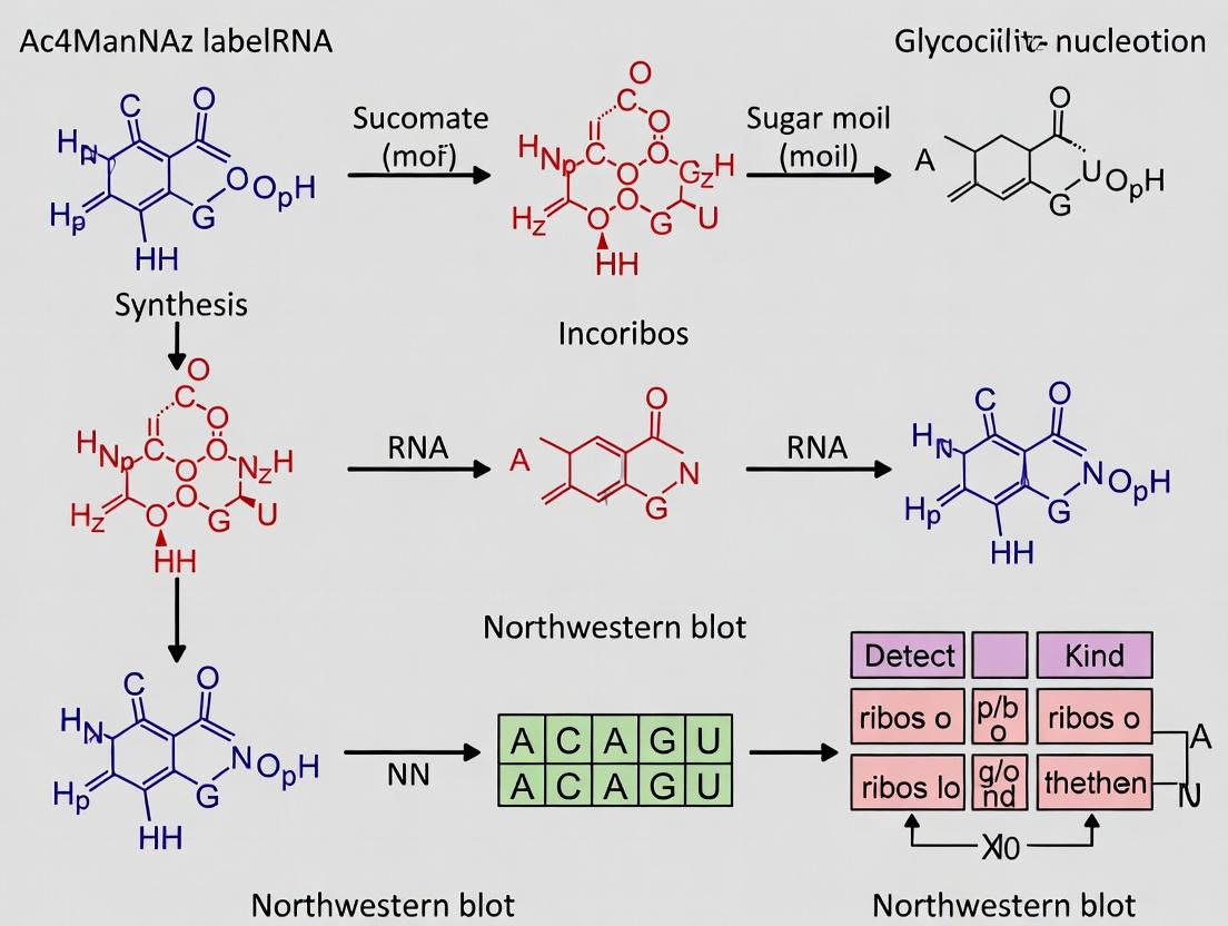
GlycoRNAs are a recently discovered class of biomolecules where small, non-coding RNAs are post-transcriptionally modified with sialylated glycans, primarily through N-glycan linkages. These structures, found on the surface of mammalian cells, represent a novel convergence of glycobiology and RNA biology, suggesting a previously unrecognized layer of regulatory complexity in cellular communication and immune signaling. Their discovery challenges the long-held dogma that glycosylation is exclusive to proteins and lipids. This application note details protocols for the detection and analysis of glycoRNAs, framed within a thesis employing metabolic labeling with Ac4ManNAz and detection via northwestern blot.
Experimental Protocols
Protocol 1: Metabolic Labeling of Cellular GlycoRNAs with Ac4ManNAz
Principle: The peracetylated azido-modified mannosamine analog (Ac4ManNAz) is metabolically incorporated into sialic acid biosynthetic pathways, labeling sialylated glycans on glycoRNAs with a bioorthogonal azide handle for subsequent conjugation.
Procedure:
- Cell Culture & Labeling: Culture mammalian cells (e.g., HEK293T, HeLa) to ~70% confluence in appropriate medium.
- Prepare a 10 mM stock of Ac4ManNAz in DMSO.
- Replace medium with fresh medium containing a final concentration of 50 µM Ac4ManNAz. Include a vehicle (DMSO-only) control.
- Incubate cells for 48 hours under standard growth conditions (37°C, 5% CO₂).
- Wash cells twice with 1x PBS.
- Proceed to RNA isolation or cell lysis for pull-down.
Protocol 2: Isolation of GlycoRNA via Copper-Click Chemistry and Streptavidin Pull-Down
Principle: Azide-labeled glycoRNAs are conjugated to a biotin alkyne probe via copper-catalyzed azide-alkyne cycloaddition (CuAAC), enabling streptavidin-mediated enrichment.
Procedure:
- Total RNA Extraction: Lyse Ac4ManNAz-labeled cells using TRIzol reagent. Isolate total RNA following the manufacturer's protocol. Treat with DNase I to remove genomic DNA contamination.
- CuAAC Reaction:
- Combine in a nuclease-free tube:
- 10 µg total RNA.
- 50 µM Biotin-PEG4-Alkyne.
- 1 mM CuSO₄.
- 100 µM THPTA ligand (to reduce copper toxicity).
- 1 mM fresh sodium ascorbate (in water).
- Bring volume to 100 µL with nuclease-free water.
- Vortex and incubate at room temperature for 1 hour.
- Combine in a nuclease-free tube:
- Ethanol Precipitation: Add 10 µL of 3M sodium acetate (pH 5.2) and 275 µL of 100% ethanol. Precipitate at -80°C for 30 min. Centrifuge at 16,000 x g for 20 min at 4°C. Wash pellet with 80% ethanol, air dry, and resuspend in nuclease-free water.
- Streptavidin Magnetic Bead Capture:
- Wash 100 µL of streptavidin magnetic beads twice with 500 µL of Binding/Wash Buffer (e.g., 10 mM Tris-HCl, pH 7.5, 1 mM EDTA, 2 M NaCl).
- Resuspend the clicked RNA sample in 100 µL of Binding/Wash Buffer and incubate with the washed beads for 30 minutes at room temperature with rotation.
- Place tube on a magnetic rack. Discard the supernatant.
- Wash beads stringently: 3x with Binding/Wash Buffer, 1x with 1x PBS.
- Elution: Resuspend beads in 50 µL of Elution Buffer (e.g., 95% formamide, 10 mM EDTA) and incubate at 85°C for 10 minutes. Immediately place on magnetic rack and transfer the eluent (enriched glycoRNA) to a new tube.
Protocol 3: Northwestern Blot for GlycoRNA Detection
Principle: RNA is separated by denaturing gel electrophoresis, transferred to a membrane, and probed with a tagged ligand (e.g., Streptavidin-HRP for biotin) or specific antibody to detect modified RNAs.
Procedure:
- Gel Electrophoresis:
- Denature 5 µL of enriched RNA (or 1-2 µg total RNA input control) with 2x RNA loading dye containing formaldehyde at 70°C for 10 min.
- Load samples onto a 1.2% agarose-formaldehyde gel or a 6% TBE-Urea polyacrylamide gel.
- Run gel at 5-6 V/cm in appropriate buffer until adequate separation is achieved.
- Transfer:
- For nitrocellulose membrane: Use capillary or semi-dry electroblotting in 0.5x TBE buffer.
- UV crosslink RNA to the membrane (120 mJ/cm²).
- Blocking & Probing:
- Block membrane with 5% BSA in TBST for 1 hour at room temperature.
- Incubate with Streptavidin-HRP conjugate (1:5000 dilution) in blocking buffer for 1 hour at RT.
- Wash membrane 4 x 5 minutes with TBST.
- Detection:
- Develop using enhanced chemiluminescence (ECL) substrate.
- Image with a chemiluminescent gel documentation system.
Data Presentation
Table 1: Key Quantitative Findings in Initial GlycoRNA Discovery Studies
| Parameter | Reported Value / Observation | Experimental System | Significance |
|---|---|---|---|
| RNA Size Range | 18-200 nucleotides | HEK293T, HAP1 cells | Enriched in small, non-coding RNAs (Y RNAs, snRNAs, vault RNAs). |
| Glycan Type | Sialylated, complex N-glycans | Multiple mammalian cell lines | Distinct from traditional protein N-glycosylation sites. |
| Surface Abundance | ~1-10% of total cellular RNA pool | Primary human cells (PBMCs) | Confirms surface localization and potential for extracellular interaction. |
| Labeling Efficiency (Ac4ManNAz) | ~2-5 fold enrichment over control | Metabolic labeling + pull-down | Validates metabolic labeling approach for detection. |
| Dependence on NXF1 Export Factor | >80% reduction upon knockdown | siRNA-mediated knockout | Links glycoRNA biogenesis to canonical RNA export machinery. |
Table 2: Essential Research Reagent Solutions for Ac4ManNAz-based GlycoRNA Studies
| Reagent / Material | Supplier Examples | Function in Workflow |
|---|---|---|
| Ac4ManNAz (tetraacetylated N-azidoacetylmannosamine) | Thermo Fisher, Click Chemistry Tools | Metabolic precursor for incorporating azido-sialic acid into glycoRNAs. |
| Biotin-PEG4-Alkyne | Click Chemistry Tools, Sigma-Aldrich | Click-compatible probe for biotinylating azide-labeled glycoRNAs for enrichment/detection. |
| THPTA Ligand | Click Chemistry Tools | Copper-chelating ligand for CuAAC, reduces RNA degradation by reactive oxygen species. |
| Streptavidin Magnetic Beads (High Capacity) | Thermo Fisher, Pierce; MilliporeSigma | Solid-phase capture of biotinylated glycoRNA complexes. |
| RNase Inhibitor (e.g., Recombinant RNasin) | Promega | Protects RNA integrity during all enzymatic and incubation steps. |
| Anti-biotin, HRP-conjugated Antibody | Cell Signaling Technology, Abcam | Alternative primary detection reagent for northwestern/immunoblot. |
| Nitrocellulose Membrane (0.2 µm) | Bio-Rad, Cytiva | Preferred membrane for RNA northwestern blotting due to high RNA binding capacity. |
| Formaldehyde, Molecular Biology Grade | Thermo Fisher | For preparing denaturing agarose gels for RNA separation. |
Visualizations
Diagram Title: Proposed Biosynthesis Pathway of Cell Surface GlycoRNA
Diagram Title: GlycoRNA Enrichment Workflow Using Ac4ManNAz
This application note details the use of peracetylated N-azidoacetylmannosamine (Ac4ManNAz) as a critical metabolic primer for the selective labeling and detection of cell-surface and intracellular glycans. Within the broader thesis on advancing glycoRNA detection methodologies, Ac4ManNAz serves as a foundational tool for metabolic glycan engineering (MGE). Its incorporation into sialoglycoconjugates, including the nascent field of glycoRNAs, enables subsequent bioorthogonal conjugation (e.g., via copper-free click chemistry) for sensitive detection techniques such as northwestern blotting. This protocol framework supports research aimed at decoding the glycoRNA landscape, with implications for biomarker discovery and therapeutic development.
Key Mechanisms and Pathways
Ac4ManNAz Metabolic Pathway
Diagram Title: Ac4ManNAz Metabolism to Labeled Glycans
Detection Workflow for GlycoRNA
Diagram Title: GlycoRNA Detection via Ac4ManNAz and Northwestern
Table 1: Optimized Ac4ManNAz Labeling Conditions for Mammalian Cells
| Parameter | Recommended Condition | Range Tested | Key Finding / Impact |
|---|---|---|---|
| Ac4ManNAz Concentration | 50 µM | 10 - 200 µM | >100 µM may show cytotoxicity in primary cells. 50 µM balances efficiency & viability. |
| Labeling Duration | 48 hours | 24 - 72 hours | 48h maximizes incorporation into slower-turnover glycoconjugates (e.g., some glycoRNAs). |
| Cell Density at Start | 70-80% confluency | 50-100% | Over-confluence reduces uptake. High efficiency achieved at 70-80%. |
| Serum Conditions | Standard (10% FBS) | 0-20% FBS | Serum-free media not required. No significant inhibition by FBS observed. |
| Click Reagent (DBCO-Biotin) | 100 µM, 1h, RT | 25 - 200 µM | 100 µM ensures complete azide conversion without non-specific binding. |
Table 2: Key Performance Metrics in GlycoRNA Detection
| Metric | Typical Result with Ac4ManNAz | Critical Note |
|---|---|---|
| Labeling Efficiency (Flow Cytometry) | 85-95% cell population positive | Varies by cell type (higher in cancer lines). |
| Minimum Detectable RNA | ~1-10 fmol via chemiluminescence | Dependent on streptavidin-HRP sensitivity and blot quality. |
| Signal-to-Noise Ratio (Blot) | 20:1 to 50:1 | Requires rigorous blocking (5% BSA in TBST). |
| Reproducibility (Inter-experiment CV) | <15% | Standardized Ac4ManNAz stock aliquots are critical. |
Detailed Experimental Protocols
Protocol 4.1: Metabolic Labeling of Cultured Cells with Ac4ManNAz for GlycoRNA Analysis
Objective: To incorporate the azido-modified sialic acid (Sia5Az) into cellular glycans, including glycoRNAs. Materials: See "The Scientist's Toolkit" below.
Procedure:
- Cell Preparation: Seed adherent cells (e.g., HEK293T, HeLa) at 70-80% confluency in standard growth medium in a 6-well plate. Incubate overnight.
- Dosing Solution Preparation: Thaw a DMSO stock of Ac4ManNAz (e.g., 50 mM). Dilute directly into pre-warmed complete growth medium to a final concentration of 50 µM. Ensure DMSO concentration is ≤0.1% v/v. Prepare a vehicle control (DMSO only).
- Metabolic Labeling: Aspirate the old medium from cells. Add 2 mL per well of the Ac4ManNAz-containing medium. Return plate to incubator (37°C, 5% CO₂) for 48 hours.
- Post-Incubation Wash: Aspirate the labeling medium. Wash cells gently with 2 mL of 1X PBS, twice.
- RNA Extraction: Lyse cells directly in the well using TRIzol Reagent (1 mL per well). Follow the manufacturer's protocol for total RNA isolation. Include a DNase I treatment step.
- RNA Quantification & Purity Check: Measure RNA concentration via Nanodrop. Proceed to click conjugation (Protocol 4.2) or store RNA at -80°C.
Protocol 4.2: DBCO-Biotin Conjugation (Click Chemistry) and Northwestern Blot
Objective: To covalently attach biotin to azide-labeled glycoRNAs for subsequent detection. Materials: DBCO-PEG4-Biotin, Nuclease-free water, PBS, 5X Click Reaction Buffer (see table), Magnetic RNA clean-up beads.
Procedure: A. Click Reaction
- Prepare RNA: Use up to 10 µg of total RNA in nuclease-free water (≤ 18 µL volume).
- Assemble Reaction: In a PCR tube, combine:
- 10 µL of 5X Click Reaction Buffer.
- 10 µg RNA in X µL.
- DBCO-PEG4-Biotin from a 10 mM DMSO stock to a final concentration of 100 µM.
- Add nuclease-free water to a final volume of 50 µL.
- Incubate: Protect from light. Incubate at room temperature for 1 hour with gentle shaking or brief vortexing every 15 minutes.
- Purify RNA: Purify the biotinylated RNA using magnetic RNA clean-up beads per manufacturer's protocol. Elute in 20-30 µL nuclease-free water. Quantify.
B. Northwestern Blot
- Electrophoresis: Load 1-5 µg of clicked RNA per lane on a denaturing 1.2% agarose-formaldehyde gel or a TBE-Urea polyacrylamide gel. Run at appropriate voltage until adequate separation is achieved.
- Transfer: Transfer RNA from gel to a positively charged nylon membrane via capillary or semi-dry electroblotting.
- UV Crosslink: Crosslink RNA to membrane using 120 mJ/cm² in a UV crosslinker.
- Blocking: Incubate membrane in 5% BSA in TBST (Tris-buffered saline with 0.1% Tween-20) for 1 hour at room temperature with gentle agitation.
- Detection: Incubate membrane with Streptavidin-HRP conjugate (1:10,000 dilution in 1% BSA/TBST) for 1 hour at RT.
- Wash: Wash membrane 3 x 10 minutes with ample TBST.
- Imaging: Develop using a sensitive chemiluminescent substrate (e.g., ECL Prime) and image on a chemiluminescence-compatible imager.
The Scientist's Toolkit: Research Reagent Solutions
Table 3: Essential Materials for Ac4ManNAz-based GlycoRNA Detection
| Item | Function & Role in Workflow | Example Product/Catalog # |
|---|---|---|
| Ac4ManNAz | Metabolic precursor. Cell-permeable acetylated form delivers ManNAz for conversion to azido-sialic acid (Sia5Az). | Thermo Fisher Scientific, A28504 |
| DBCO-PEG4-Biotin | Bioorthogonal click reagent. DBCO group undergoes strain-promoted [3+2] cycloaddition with azide. PEG spacer reduces steric hindrance. Biotin enables detection. | Click Chemistry Tools, A112-25 |
| Streptavidin-HRP Conjugate | Detection probe. Binds biotin with high affinity. Horseradish peroxidase (HRP) catalyzes chemiluminescent reaction for blot imaging. | Cytiva, RPN1231V |
| TRIzol Reagent | Monophasic solution of phenol and guanidine isothiocyanate. Simultaneously lyses cells and denatures proteins while stabilizing RNA. | Thermo Fisher, 15596026 |
| Positively Charged Nylon Membrane | Blotting matrix. Positively charged surface binds negatively charged RNA after transfer for subsequent probing. | Roche, 11209299001 |
| RNA Clean-up Magnetic Beads | Purify RNA after click reaction, removing unreacted DBCO-biotin and salts. | Beckman Coulter, A63987 |
| Chemiluminescent HRP Substrate | Provides luminol and peroxide. HRP catalyzes light emission upon oxidation, captured by imager. | Cytiva, RPN2232 |
| 5X Click Reaction Buffer (250 mM HEPES pH 7.5, 25 mM CuSO₄*, 2.5 mM TBTA) | *For CuAAC only. Not needed for SPAAC (DBCO). For SPAAC, a simple PBS or HEPES buffer suffices. | In-house preparation. |
Northwestern blotting is a specialized technique used to detect RNA-protein and RNA-glycan interactions. It involves separating RNA molecules by electrophoresis, transferring them to a membrane, and probing with labeled proteins, lectins, or other binding partners. Within the context of a broader thesis on Ac4ManNAz labeling for glycoRNA detection, northwestern blotting serves as a critical validation tool. Ac4ManNAz is a metabolic precursor that incorporates azide-modified sialic acids into glycoconjugates, including the recently discovered glycoRNAs. This method allows researchers to confirm specific interactions between glycoRNAs (which can be tagged via Ac4ManNAz and click chemistry) and putative binding partners like lectins or RNA-binding proteins (RBPs).
Application Notes
Key Applications in GlycoRNA Research
- Validation of GlycoRNA-Lectin Interactions: Following metabolic labeling with Ac4ManNAz and click chemistry conjugation to a reporter (e.g., biotin), northwestern blotting can be used to probe for interactions with specific lectins (e.g., SNA for α2,6-linked sialic acid), confirming the presence and nature of the glycan moiety on RNA.
- Identification of RNA-Binding Proteins for GlycoRNAs: Probing blots of separated glycoRNAs with candidate or pooled RBPs helps identify proteins that selectively bind glycosylated RNAs, potentially revealing novel functional pathways.
- Characterization of Interaction Specificity: Competition assays using free sugars, unlabeled RNA, or glycosidases during the blocking or probing steps can determine the specificity of detected interactions.
Table 1: Representative Quantitative Data from GlycoRNA Northwestern Blot Analyses
| Target RNA | Probe Used (e.g., Lectin/RBP) | Detection Method | Apparent Kd (nM)* | Key Experimental Condition | Reference Context |
|---|---|---|---|---|---|
| Sialylated snRNA | SNA (Sambucus Nigra Lectin) | Biotin-Streptavidin-HRP | ~15-50 | Blot probed with biotinylated SNA | Validation of α2,6-linked sialic acid on glycoRNA |
| GlycoRNA pool | Recombinant RBP (e.g., FUS) | His-Tag Antibody | >200 | Comparison to non-glycosylated RNA control | Screening for RBP binders with glycan dependence |
| Synthetic Sialyl-RNA | Anti-Sia Antibody | Chemiluminescence | N/A | Control for glycosidase (Neuraminidase) treatment | Confirmation of sialic acid epitope integrity on blot |
Note: Apparent Kd values are derived from concentration-dependent binding curves on the membrane and are approximate.
Experimental Protocols
Protocol 1: Standard Northwestern Blot for GlycoRNA-Protein Interaction
Title: Detecting GlycoRNA Interactions with Lectins or RNA-Binding Proteins.
Materials:
- GlycoRNA samples (metabolically labeled with Ac4ManNAz and clicked to biotin if needed).
- Non-glycosylated RNA control.
- Denaturing agarose or polyacrylamide gel system.
- Nylon or positively charged nylon membrane.
- UV crosslinker.
- Blocking buffer: e.g., 5% BSA in 1x NW Buffer (10 mM HEPES pH 7.5, 50 mM KCl, 1 mM EDTA, 1 mM DTT, 0.1% Tween-20).
- Probe: Purified, tagged protein (e.g., His-tagged RBP) or biotinylated lectin.
- Appropriate detection reagents (e.g., anti-His primary & HRP-conjugated secondary antibody; Streptavidin-HRP).
- Chemiluminescence substrate and imager.
Procedure:
- Electrophoresis: Separate 1-5 µg of RNA samples on a denaturing (e.g., with formaldehyde or urea) agarose or polyacrylamide gel.
- Blotting: Transfer the RNA to a positively charged nylon membrane via capillary or semi-dry electroblotting.
- Immobilization: UV crosslink the RNA to the membrane (typically 120-254 nm, 120 mJ/cm²).
- Blocking: Incubate the membrane in 10-15 mL of blocking buffer for 1-2 hours at room temperature with gentle agitation.
- Probing: Dilute the protein/lectin probe in fresh blocking buffer. Incubate the membrane with the probe solution for 1-2 hours at RT or overnight at 4°C.
- Washing: Wash the membrane 3-4 times for 10 minutes each with 1x NW Buffer.
- Detection: Apply appropriate antibody detection steps (if needed). For biotinylated probes, incubate with Streptavidin-HRP (1:5000 in blocking buffer) for 45 min. Wash thoroughly. Develop with chemiluminescent substrate and image.
Protocol 2: Northwestern Blot with Pre-Membrane Click Chemistry (for Ac4ManNAz Labeled Samples)
Title: Direct Detection of Metabolically Labeled GlycoRNAs on Blots.
Materials:
- All materials from Protocol 1.
- Click Chemistry Reagents: CuSO4, THPTA ligand, Sodium Ascorbate, Biotin-PEG4-Alkyne (or fluorescent alkyne).
- Click Reaction Buffer: 20 mM HEPES, pH 7.4.
Procedure:
- Separation & Blotting: Perform steps 1-3 from Protocol 1.
- On-Membrane Click Reaction: Prepare a fresh click reaction mix: 100 µM Biotin-PEG4-Alkyne, 1 mM CuSO4, 1 mM THPTA, 5 mM Sodium Ascorbate in Click Reaction Buffer. Soak the membrane in this solution for 30-60 minutes at RT, protected from light.
- Wash: Wash the membrane 3x with NW Buffer containing 1 mM EDTA to chelate residual copper.
- Block & Detect: Proceed with blocking (Step 4, Protocol 1) and detection using Streptavidin-HRP (Step 7, Protocol 1). This directly visualizes the metabolically incorporated azide-modified glycans on the RNA.
Visualization: Diagrams and Workflows
Title: Northwestern Blot Workflow for GlycoRNA
Title: Ac4ManNAz Labeling to Northwestern Detection Pathway
The Scientist's Toolkit: Research Reagent Solutions
Table 2: Essential Materials for Northwestern Blotting in GlycoRNA Studies
| Item | Function & Role in Experiment | Key Consideration for GlycoRNA |
|---|---|---|
| Ac4ManNAz (tetraacetylated N-azidoacetylmannosamine) | Metabolic precursor that introduces bio-orthogonal azide groups into cellular sialic acid pools, enabling subsequent tagging of glycoRNAs. | Enables specific labeling of the glycan moiety on RNA for pull-down or direct blot detection. |
| Biotin-PEG4-Alkyne / DBCO-PEG4-Biotin | Click chemistry-compatible reagents that conjugate a biotin reporter to azide-labeled glycoRNAs (via CuAAC or SPAAC) for sensitive detection. | PEG spacer reduces steric hindrance. Choice depends on desired click chemistry (copper-catalyzed vs. copper-free). |
| Positively Charged Nylon Membrane | Binds negatively charged RNA via electrostatic interactions after blotting, providing a stable substrate for probing. | Essential for retaining small RNA species; superior RNA binding capacity compared to nitrocellulose. |
| Biotinylated Lectins (e.g., SNA, MAL-II) | Plant-derived proteins that bind specific glycan structures (e.g., SNA for α2,6-sialic acid). Used as probes to confirm glycan presence on RNA. | Validates the glycan type on detected glycoRNAs. Requires careful control for specificity. |
| Recombinant RNA-Binding Proteins (His-/GST-tagged) | Purified candidate proteins used as probes to identify specific interactions with glycoRNA targets. | Allows mapping of protein interactors that may depend on or be independent of the RNA's glycosylation. |
| Streptavidin-HRP Conjugate | High-affinity binding to biotin, coupled to horseradish peroxidase for chemiluminescent detection of biotinylated probes or directly labeled RNA. | Standard for high-sensitivity detection following click chemistry with biotin-alkyne. |
| RNA-Compatible Denaturing Gel System | Separates RNA by size under conditions that disrupt secondary structure (e.g., formaldehyde-agarose or urea-PAGE). | Critical for obtaining sharp bands and accurate interpretation of interaction data. |
Why Combine Ac4ManNAz with Northwestern Blot? The Rationale for Specific GlycoRNA Profiling.
Within the broader thesis of developing Ac4ManNAz-based metabolic labeling for glycoRNA detection, the Northwestern blot emerges as a critical orthogonal technique. GlycoRNAs are a recently discovered class of glycosylated small non-coding RNAs, bearing sialic acid and other glycans, implicated in cell-surface display and intercellular signaling. The primary rationale for combining peracetylated N-azidoacetylmannosamine (Ac4ManNAz) labeling with Northwestern blot analysis is to achieve specific, direct visualization of glycoRNA species within a complex cellular lysate, confirming molecular weight and providing a bridge between metabolic tagging and biochemical validation.
Ac4ManNAz is a metabolic precursor that cells convert into azido-modified sialic acids (SiaNAz), which are incorporated into glycoconjugates, including glycoRNAs. While click chemistry (e.g., CuAAC) with a biotin alkyne enables enrichment and sequencing, the Northwestern blot—an RNA-protein blotting technique where labeled RNA is detected by a protein probe—allows for the specific detection of azide-tagged glycoRNAs using a phosphine-based probe. This combination addresses key challenges: it moves beyond bulk enrichment to monitor specific glycoRNA bands, confirms the covalent nature of the glycan-RNA linkage under denaturing conditions, and provides a semi-quantitative assessment of glycoRNA levels across samples.
Table 1: Comparative Analysis of GlycoRNA Detection Methods
| Method | Principle | Key Output | Advantages | Limitations |
|---|---|---|---|---|
| Ac4ManNAz + Click & Seq | Metabolic labeling, affinity enrichment, sequencing. | GlycoRNA identification & transcriptome profile. | High-throughput, discovery-focused, identifies sequences. | Does not directly confirm covalent modification or size; potential for non-specific background. |
| Ac4ManNAz + Northwestern | Metabolic labeling, denaturing gel separation, phosphine-based detection. | Direct visualization of specific glycoRNA bands by size. | Confirms covalent modification, provides size validation, semi-quantitative, lower background. | Lower throughput, requires optimization of blotting conditions. |
| Traditional Northern Blot | Size separation, sequence-specific DNA probe hybridization. | Detection of specific RNA sequences by size. | High specificity for RNA sequence, gold standard for size/abundance. | Cannot distinguish glycosylated from non-glycosylated forms of the same RNA. |
Table 2: Expected Results from Ac4ManNAz Labeling of HeLa Cells
| Parameter | Condition: +Ac4ManNAz | Condition: -Ac4ManNAz (Control) | Detection Method |
|---|---|---|---|
| SiaNAz Incorporation | High (Flow cytometry confirmation) | Negligible | Click with fluorescent dye |
| GlycoRNA Enrichment Yield | 5-10 ng per 10^7 cells | < 0.5 ng per 10^7 cells | Click with biotin, streptavidin pull-down, Qubit assay |
| Northwestern Signal | Distinct bands (e.g., 60-300 nt range) | No bands or vastly reduced intensity | Anti-biotin or Streptavidin-HRP after Staudinger ligation |
Experimental Protocols
Protocol 1: Metabolic Labeling of Cells with Ac4ManNAz
Objective: To incorporate azide-modified sialic acid into cellular glycoRNAs.
- Cell Culture: Seed mammalian cells (e.g., HeLa, HEK293) in appropriate medium.
- Labeling: Prepare a 100 mM stock of Ac4ManNAz in DMSO. Add to culture medium at a final concentration of 50 µM. Include a vehicle (DMSO-only) control.
- Incubation: Incubate cells for 48-72 hours under standard growth conditions.
- Harvest: Wash cells 2x with PBS. Scrape and pellet cells. Cell pellets can be stored at -80°C.
Protocol 2: Total RNA Extraction and GlycoRNA Enrichment (Optional Pre-Step)
Objective: To isolate total RNA and optionally enrich for glycoRNA via click chemistry.
- Extraction: Lyse cells using TRIzol or similar. Isolate total RNA following manufacturer’s protocol. Treat with DNase I.
- (Optional) Biotinylation via Click Chemistry: Resuspend 20-50 µg total RNA in PBS.
- Click Reaction Mix: Add 50 µM Biotin-PEG3-Alkyne, 1 mM CuSO4, 1 mM THPTA ligand, and 2.5 mM fresh sodium ascorbate.
- Incubate: 1 hour at room temperature, protected from light.
- (Optional) Ethanol Precipitation: Recover RNA using glycogen-assisted ethanol precipitation.
Protocol 3: Northwestern Blot for Ac4ManNAz-labeled GlycoRNA
Objective: To specifically detect azide-labeled glycoRNAs via Staudinger ligation on a membrane. Part A: Denaturing Gel Electrophoresis & Blotting
- Prepare Sample: Resolve 5-10 µg of total RNA (or enriched fraction) on a 6% or 8% polyacrylamide-urea denaturing gel.
- Electroblot: Transfer RNA to a positively charged nylon membrane using semi-dry transfer in 0.5x TBE buffer at 2 mA/cm² for 1 hour.
- UV Crosslink: Immobilize RNA to the membrane using 254 nm UV light (120 mJ/cm²).
Part B: On-Membrane Staudinger Ligation
- Block: Incubate membrane in Northwestern Blocking Buffer (1x PBS, 0.1% Tween-20, 5% BSA) for 1 hour at RT.
- Probe Incubation: Prepare Phosphine-Biotin Probe (e.g., DIBO-Biotin or a Staudinger ligation-compatible phosphine-biotin conjugate) at 10 µM in fresh blocking buffer. Incubate membrane with probe for 2 hours at RT or overnight at 4°C.
- Wash: Wash membrane 4 x 5 minutes with PBST (PBS + 0.1% Tween-20).
Part C: Signal Detection
- Streptavidin-HRP: Incubate membrane with Streptavidin conjugated to Horseradish Peroxidase (1:5000 in blocking buffer) for 1 hour at RT.
- Wash: Wash 4 x 5 minutes with PBST.
- Chemiluminescence: Develop using ECL substrate. Image with a digital chemiluminescence imager.
Diagrams
Title: Workflow for GlycoRNA Profiling via Ac4ManNAz & Northwestern Blot
Title: On-Membrane Staudinger Ligation Principle
The Scientist's Toolkit: Key Research Reagent Solutions
Table 3: Essential Materials for Ac4ManNAz Northwestern Blotting
| Item Name | Function/Role in Experiment | Example/Catalog Considerations |
|---|---|---|
| Ac4ManNAz (Peracetylated N-Azidoacetylmannosamine) | Metabolic precursor for incorporating azido-sialic acid (SiaNAz) into glycoRNAs. | Cell-permeable, stock in DMSO. Store desiccated at -20°C. |
| Phosphine-Biotin Conjugate (e.g., DIBO-Biotin) | Probe for on-membrane Staudinger ligation; specifically reacts with azide to attach biotin. | Must be Staudinger ligation-compatible (phosphine or strained alkyne). Stable in buffer. |
| Positively Charged Nylon Membrane | Solid support for immobilizing RNA after blotting; essential for Northwestern procedure. | High RNA-binding capacity, compatible with phosphine chemistry. |
| Streptavidin-Horseradish Peroxidase (HRP) | Secondary conjugate for detecting biotinylated glycoRNAs on the membrane. | High sensitivity; use with compatible ECL substrate. |
| Denaturing Polyacrylamide Gel System (Urea-PAGE) | Separates RNA molecules by size under denaturing conditions to preserve glycoRNA integrity. | Requires equipment for gel casting, running, and semi-dry blotting. |
| RNase Inhibitors (e.g., RNaseZap, DEPC-water) | Prevents degradation of glycoRNA samples throughout the protocol. | Critical for all steps post-cell lysis. |
| Enhanced Chemiluminescence (ECL) Substrate | Generates light signal upon HRP catalysis for imaging glycoRNA bands. | Choose a high-sensitivity, low-background formulation. |
1. Introduction and Thesis Context This application note details methodologies central to a thesis investigating glycoRNA detection via Ac4ManNAz metabolic labeling and northwestern blot analysis. The recent discovery of glycosylated RNA (glycoRNA) represents a paradigm shift in glycobiology, revealing a novel class of post-transcriptional modifications with profound biological implications. This work frames current glycoRNA research within the specific experimental pipeline of metabolic labeling with synthetic azido-sugars (e.g., Ac4ManNAz), click-chemistry enrichment, and detection via northwestern blot, providing validated protocols for the field.
2. Key Research Findings: Summary Table
Table 1: Summary of Key GlycoRNA Research Findings
| Finding Category | Key Observation | Model System | Primary Method(s) | Biological Implication |
|---|---|---|---|---|
| Discovery & Core Composition | RNAs (mainly small non-coding RNAs like Y RNAs) are glycosylated with sialylated N-glycans. | Human, mouse cell lines (HEK293T, HeLa) | Metabolic labeling (Ac4ManNAz), Click Chemistry, Mass Spectrometry | Establishes the existence of a new biomolecular conjugate. |
| Biosynthesis Pathway | GlycoRNA biosynthesis is dependent on the canonical N-glycosylation machinery (e.g., oligosaccharyltransferase complexes). | Mouse embryonic fibroblasts (MEFs) | CRISPR-Cas9 knockout of OST subunits, Metabolic labeling | Links glycoRNA to established endoplasmic reticulum pathways. |
| Cell Surface Localization | GlycoRNAs are present on the extracellular surface of cells. | Human cell lines | Proximity labeling, Flow cytometry with click-chemistry detection | Suggests a role in extracellular recognition and signaling. |
| Interaction with Siglecs | Cell surface glycoRNAs can bind to sialic-acid-binding immunoglobulin-type lectins (Siglecs). | Recombinant protein assays, Cell binding studies | Pull-down assays, Surface plasmon resonance (SPR) | Implicates glycoRNAs in immune cell communication via lectin receptors. |
| Quantitative Range | Estimated abundance is ~1-10 glycoRNA molecules per classical glycoprotein molecule on the cell surface. | Comparative MS analysis | Quantitative proteomics/glycomics | Indicates glycoRNAs are a low-abundance but widespread component of the glycocalyx. |
3. Detailed Experimental Protocols
Protocol 3.1: Metabolic Labeling of GlycoRNAs with Ac4ManNAz Purpose: To incorporate azide-modified sialic acid into glycoRNAs for subsequent bioorthogonal conjugation. Materials: Cell culture of choice (e.g., HEK293T), Ac4ManNAz (e.g., Thermo Fisher Scientific, C10265), DMSO, standard cell culture media and reagents. Procedure:
- Prepare a 1000X stock of Ac4ManNAz (e.g., 50 mM) in anhydrous DMSO.
- Culture cells to ~70% confluency in appropriate medium.
- Aspirate medium and replace with fresh medium containing 50 µM Ac4ManNAz (final concentration). Include a vehicle control (DMSO only).
- Incubate cells for 48-72 hours under standard growth conditions to ensure turnover of RNA and glycoconjugates.
- Proceed to cell lysis for RNA extraction or perform click chemistry on live cells for surface detection.
Protocol 3.2: Click Chemistry Conjugation for GlycoRNA Enrichment (CuAAC) Purpose: To conjugate alkyne-bearing probes (e.g., biotin) to azide-labeled glycoRNAs for pull-down or detection. Materials: Click Chemistry Kit (e.g., Click Chemistry Tools, #1261), or components: Biotin-PEG4-Alkyne, Copper(II) Sulfate, THPTA ligand, Sodium Ascorbate, RNA extract from Protocol 3.1. Procedure:
- Prepare Click Reaction Mix (for 100 µL): 1-10 µg of total RNA, 50 µM Biotin-PEG4-Alkyne, 1 mM CuSO₄, 100 µM THPTA ligand, 2.5 mM Sodium Ascorbate in nuclease-free buffer.
- Incubate the reaction at room temperature for 1 hour with gentle mixing.
- Purify the biotinylated RNA using ethanol precipitation or a commercial RNA clean-up kit to remove unreacted reagents.
- Resuspend the pellet in nuclease-free water or buffer. The RNA can now be used for streptavidin bead enrichment (for sequencing) or held for northwestern blot analysis.
Protocol 3.3: Northwestern Blot for GlycoRNA Detection Purpose: To separate and detect glycoRNAs using glycan-targeted probes. Materials: Denaturing polyacrylamide gel (6-10%), Semi-dry or tank blotting system, Nitrocellulose or PVDF membrane, Streptavidin-HRP or anti-biotin antibody, Click-iT RNA Buffer Kit (optional for on-membrane click). Procedure:
- RNA Separation: Resuspend click-labeled RNA in denaturing RNA loading dye. Heat denature at 70°C for 10 minutes, then immediately place on ice. Load samples onto a denaturing polyacrylamide gel. Run at constant voltage until the dye front migrates appropriately.
- Blotting: Transfer RNA from the gel to a positively charged nylon or nitrocellulose membrane using semi-dry electroblotting in 0.5X TBE buffer.
- UV Crosslinking: Crosslink RNA to the membrane using 254 nm UV light at 120-150 mJ/cm².
- Blocking: Block the membrane in 5% BSA in TBST for 1 hour at room temperature.
- Probing for Biotin (Direct): If RNA was biotinylated via Protocol 3.2, incubate membrane with Streptavidin-HRP (1:5000 in blocking buffer) for 1 hour. Wash 3x with TBST.
- Signal Development: Develop using enhanced chemiluminescence (ECL) substrate and image with a chemiluminescent imager. Note: For direct on-membrane detection of azides, perform a click reaction (similar to Protocol 3.2) on the blocked membrane using an alkyne-HRP conjugate.
4. Visualizations
Diagram 1: Ac4ManNAz Labeling & Detection Workflow (74 chars)
Diagram 2: Proposed GlycoRNA-Siglec Signaling Axis (71 chars)
5. The Scientist's Toolkit: Key Research Reagent Solutions
Table 2: Essential Materials for Ac4ManNAz-based GlycoRNA Research
| Reagent/Material | Supplier Examples | Function in GlycoRNA Research |
|---|---|---|
| Ac4ManNAz (Tetacetylated N-azidoacetylmannosamine) | Thermo Fisher, Click Chemistry Tools | Metabolic precursor for incorporating azide-modified sialic acid into glycoconjugates, enabling bioorthogonal tagging of glycoRNAs. |
| Biotin-PEG4-Alkyne / Alkyne-HRP | Click Chemistry Tools, Sigma-Aldrich | Bioorthogonal probes for click chemistry (CuAAC). Biotin allows enrichment/pull-down; HRP enables direct chemiluminescent detection. |
| THPTA Ligand | Click Chemistry Tools | Copper chelator for CuAAC click reactions. Essential for reducing copper toxicity and improving reaction efficiency on sensitive biomolecules like RNA. |
| Streptavidin Magnetic Beads | Thermo Fisher, NEB | For capturing biotinylated glycoRNAs from complex lysates for downstream sequencing (glycoRNA-sequencing) or analysis. |
| RNase Inhibitors (e.g., SUPERase•In) | Thermo Fisher, Promega | Critical for all steps to protect the RNA moiety of glycoRNAs from degradation during extraction and processing. |
| Anti-Biotin, HRP-linked Antibody | Cell Signaling Technology | An alternative to streptavidin-HRP for detecting biotinylated glycoRNAs on blots, potentially offering higher specificity in some contexts. |
| Click-iT RNA Buffer Kit | Thermo Fisher | Optimized buffers and protocol for performing click chemistry reactions directly on RNA bound to northern/northwestern blot membranes. |
Step-by-Step Protocol: From Cell Culture to Detection with Ac4ManNAz and Northwestern Blot
Application Notes
Within a thesis focused on leveraging metabolic glycan labeling for the novel detection of glycoRNAs via northwestern blotting, the procurement and preparation of high-purity reagents are foundational. Ac4ManNAz, a peracetylated, azido-modified metabolic precursor of sialic acid, serves as the critical tool for introducing bioorthogonal handles into cellular glycans, including those conjugated to small nuclear RNAs. Success in subsequent click-chemistry-based detection and northwestern blot analysis is directly contingent on the quality of the initial reagents and buffers. These Application Notes outline strategic sourcing and preparation protocols to ensure experimental reproducibility and robustness.
Sourcing Critical Reagents: Specifications and Vendor Comparison
Primary reagents must be selected based on purity, stability, and suitability for cell culture and biochemical assays. The following table summarizes key sourcing considerations for the core compound, Ac4ManNAz.
Table 1: Key Specifications for Sourcing Ac4ManNAz and Conjugates
| Reagent | Primary Function | Target Purity | Critical Sourcing Considerations | Example Vendors (2024) |
|---|---|---|---|---|
| Ac4ManNAz | Metabolic precursor for azido-sialic acid incorporation into glycans (including glycoRNA). | ≥95% (HPLC) | Solubility in DMSO/EtOH; batch-to-batch consistency; MSDS for azide safety. | MedChemExpress, Cayman Chemical, Sigma-Aldrich, Click Chemistry Tools |
| DBCO-PEG4-Biotin | Copper-free click chemistry reagent for post-labeling conjugation. | ≥95% (HPLC) | High reactivity with azides; PEG spacer reduces steric hindrance. | Click Chemistry Tools, Thermo Fisher, BroadPharm |
| Copper(II) Sulfate Pentahydrate | Catalyst for CuAAC click chemistry (if used as alternative). | ≥98% | Ultra-pure, molecular biology grade to minimize cytotoxicity. | Sigma-Aldrich, MilliporeSigma |
| Sodium Ascorbate | Reducing agent for CuAAC, generates active Cu(I). | ≥99% | Freshly prepared in nuclease-free water for each use. | Sigma-Aldrich, Thermo Fisher |
| Streptavidin-HRP Conjugate | Detection probe for biotinylated glycoRNA on northwestern blots. | High Specific Activity | Low non-specific binding; optimized for nucleic acid/protein blots. | Cell Signaling Technology, Thermo Fisher |
Protocol: Preparation of Stock Solutions and Buffers
Protocol 2.1: Preparation of Ac4ManNAz Stock Solution for Cell Labeling
- Objective: To create a stable, sterile, and concentrated stock for metabolic labeling of mammalian cells.
- Materials:
- Ac4ManNAz (5 mg)
- Anhydrous Dimethyl Sulfoxide (DMSO), cell culture grade
- 1.5 mL amber microcentrifuge tubes (sterile)
- 0.22 μm syringe filter (PTFE, sterile)
- Procedure:
- Bring the vial of Ac4ManNAz and DMSO to room temperature in a desiccator.
- In a sterile environment, add 1 mL of anhydrous DMSO directly to the 5 mg vial. This yields a ~10 mM stock solution (exact concentration depends on molecular weight of specific salt form).
- Gently vortex and pipette mix until fully dissolved. Do not sonicate excessively.
- Optional for sterility: Filter the solution through a 0.22 μm PTFE syringe filter into a new sterile amber tube.
- Aliquot into single-use volumes (e.g., 20-50 μL) to avoid freeze-thaw cycles.
- Store aliquots at -20°C or -80°C under desiccant for long-term stability (≥1 year).
Protocol 2.2: Preparation of Critical Buffers for Northwestern Blotting
- Objective: To prepare optimized buffers for RNA extraction, transfer, and detection following metabolic labeling and click chemistry.
Materials & Formulations:
A. 20x SSC Transfer Buffer (1L):
- NaCl: 175.3 g
- Sodium Citrate (dihydrate): 88.2 g
- Nuclease-free water to 1 L.
- Adjust pH to 7.0 with HCl. Filter sterilize (0.22 μm). Store at RT.
B. 10x Click Reaction Buffer (for CuAAC, 50 mL):
- Tris-HCl (pH 8.0): 250 mM (from 1M stock)
- NaCl: 1.5 M (43.83 g)
- Add nuclease-free water to 50 mL. Store at 4°C.
C. Nonidet P-40 Lysis Buffer (for RNA-centric protocols, 50 mL):
- Tris-HCl (pH 8.0): 50 mM
- NaCl: 150 mM
- Nonidet P-40: 0.5% (v/v)
- RNase Inhibitor: Add fresh (e.g., 0.5 U/μL)
- Prepare in nuclease-free water. Store at 4°C without RNase inhibitor.
Visualizing the Workflow and Pathway
Title: GlycoRNA Detection Workflow via Metabolic Labeling
Title: Metabolic Pathway of Ac4ManNAz to GlycoRNA
The Scientist's Toolkit: Essential Research Reagent Solutions
Table 2: Essential Toolkit for Ac4ManNAz-based GlycoRNA Research
| Item Category | Specific Item | Function & Importance |
|---|---|---|
| Metabolic Labeling Core | High-Purity Ac4ManNAz | The foundational probe. Purity ensures efficient labeling without cellular toxicity. |
| Click Chemistry Conjugation | DBCO-PEG4-Biotin | Enables copper-free, specific biotin tagging of azido-glycoRNAs for sensitive detection. |
| RNA Integrity Preservation | RNase Inhibitor (e.g., Recombinant RNasin) | Critical for all buffers post-lysis to preserve the integrity of target glycoRNAs. |
| Specialized Lysis Buffer | Nonidet P-40 (NP-40) Lysis Buffer | Effectively solubilizes membranes while keeping nuclei intact, crucial for RNA-centric protocols. |
| Northern/Northwestern Blotting | Positively Charged Nylon Membrane | Essential for efficient retention of negatively charged RNA and glycoRNA complexes. |
| Detection System | Chemiluminescent HRP Substrate (e.g., Luminol-based) | Provides high sensitivity for detecting low-abundance biotinylated glycoRNA on blots. |
| Critical Lab Supplies | Amber Microcentrifuge Tubes & Chemical Desiccants | Protects light- and moisture-sensitive reagents (Ac4ManNAz, DBCO, Cu catalyst) from degradation. |
This application note details the critical first phase for metabolic labeling of glycoRNAs using Ac4ManNAz (1,3,4,6-tetra-O-acetyl-N-azidoacetylmannosamine) within a broader thesis on Northwestern blot detection. GlycoRNAs are a recently discovered class of small, glycosylated non-coding RNAs implicated in cellular communication and immune modulation. Metabolic labeling with Ac4ManNAz introduces bioorthogonal azide groups into sialic acid-containing glycoconjugates, including glycoRNAs, enabling subsequent chemoselective conjugation (e.g., via copper-free click chemistry) for enrichment, visualization, and analysis. Optimization of Ac4ManNAz concentration and incubation time is paramount to maximize labeling efficiency while minimizing cellular toxicity and off-target effects, forming the foundational step for successful downstream glycoRNA isolation and Northwestern blotting.
Optimizing Ac4ManNAz Concentration: Key Data & Protocol
Concentration-Dependent Labeling Efficiency & Cytotoxicity
Data synthesized from recent studies (2023-2024) in HeLa, HEK293T, and primary fibroblast cell lines.
Table 1: Optimization of Ac4ManNAz Concentration for Metabolic Labeling
| Cell Line | Ac4ManNAz Concentration Range Tested (μM) | Optimal Concentration* (μM) | Labeling Efficiency (Fold Increase vs. Control) | Viability at Optimal Conc. (% of Untreated) | Key Assay Used for Measurement |
|---|---|---|---|---|---|
| HeLa | 25 - 200 | 50 | ~8.5x | 95% | Flow Cytometry (AF488-DIBO) |
| HEK293T | 10 - 100 | 25 | ~7.2x | 98% | Fluorescence Microscopy |
| Primary Fibroblast | 10 - 75 | 30 | ~6.0x | 92% | LC-MS/MS (SiaNAz quantification) |
| Jurkat (Suspension) | 20 - 150 | 40 | ~5.5x | 90% | Click-iT RNA Alexa Fluor 647 |
Optimal Concentration: Defined as the concentration yielding >80% of maximal median fluorescence intensity or glycoRNA-azide incorporation with >90% cell viability after 24h incubation.
Protocol: Determining Optimal Ac4ManNAz Concentration
A. Materials & Reagents
- Cells of interest (e.g., HeLa)
- Complete growth medium
- Ac4ManNAz stock solution (50 mM in DMSO, store at -20°C)
- DMSO (vehicle control)
- Phosphate-Buffered Saline (PBS)
- Click chemistry reagent: AF488 Alkyne or DBCO-PEG4-Biotin
- Fixative (e.g., 4% PFA) or Lysis Buffer (for RNA)
- Cell viability assay kit (e.g., MTT, CellTiter-Glo)
- Flow cytometer or fluorescence plate reader
B. Procedure
- Cell Seeding: Seed cells in a 24-well plate at 70% confluency. Incubate overnight.
- Dosing: Prepare serial dilutions of Ac4ManNAz in complete medium (e.g., 0, 25, 50, 75, 100, 150, 200 μM). Include a DMSO-only control (0.4% v/v max).
- Metabolic Labeling: Aspirate old medium and add the Ac4ManNAz-containing medium. Incubate for 24h at 37°C, 5% CO₂.
- Cell Harvest & Processing:
- For Flow Cytometry: Harvest cells, wash with PBS x2, fix with 4% PFA for 15 min. Perform click reaction with AF488 Alkyne per manufacturer's instructions. Analyze by flow cytometry.
- For Viability: In parallel wells, after 24h labeling, perform MTT assay according to kit protocol. Measure absorbance.
- Data Analysis: Plot median fluorescence intensity (MFI) and cell viability (%) against Ac4ManNAz concentration. The optimal concentration is the highest dose before a significant drop in viability.
Optimizing Incubation Time: Key Data & Protocol
Time-Course of Azide-Sialic Acid Incorporation
Table 2: Optimization of Ac4ManNAz Incubation Time
| Time Point (hours) | Relative SiaNAz Incorporation (HeLa, 50μM) | Notes / Saturation Plateau |
|---|---|---|
| 6 | 25% | Minimal detection |
| 12 | 60% | Linear increase phase |
| 18 | 85% | Near saturation for surface glycans |
| 24 | 100% (Reference) | Optimal for total glycoRNA |
| 36 | 105% | No significant gain |
| 48 | 110% | Increased risk of toxicity & altered metabolism |
Protocol: Determining Optimal Incubation Time
A. Materials & Reagents (as in Section 2.2, plus materials for RNA isolation) B. Procedure
- Seed cells in multiple plates/wells to allow parallel harvesting.
- Treat all wells with the predetermined optimal Ac4ManNAz concentration (e.g., 50 μM for HeLa).
- Time-Course Harvest: Harvest cells at intervals (e.g., 6, 12, 18, 24, 36, 48h). Include a t=0 control.
- Analysis:
- For Surface Labeling: At each time point, harvest, wash, and perform click reaction with AF488-DBCO on live cells for 30 min on ice. Analyze by flow cytometry.
- For Total GlycoRNA Analysis: Lyse cells at each time point. Isolate total RNA. Perform a copper-free click reaction on the RNA with DBCO-Biotin. Proceed to streptavidin pulldown and qRT-PCR for housekeeping ncRNAs (e.g., Y5 RNA) to quantify labeled glycoRNA over time.
- Data Analysis: Plot MFI or pulled-down glycoRNA quantity versus time. The point where the curve plateaus is optimal.
The Scientist's Toolkit: Key Research Reagent Solutions
Table 3: Essential Reagents for Ac4ManNAz Metabolic Labeling
| Reagent / Solution | Function / Purpose | Example Product (Supplier) |
|---|---|---|
| Ac4ManNAz (Tet-Acetylated ManNAz) | Cell-permeable metabolic precursor; delivers the azido-sialic acid (SiaNAz) into biosynthetic pathways. | Click Chemistry Tools, Sigma-Aldrich |
| DBCO (Dibenzocyclooctyne) Reagents | Copper-free click chemistry strain-promoted alkyne; reacts with azide for conjugation to detection tags. | DBCO-PEG4-Biotin (BroadPharm), DBCO-Sulfo-Cy5 (Lumiprobe) |
| AF488 Alkyne | Fluorescent probe for direct visualization via alkyne-azide copper-catalyzed click chemistry (CuAAC). | Invitrogen, Click Chemistry Tools |
| Cell Viability Assay Kits | Quantify metabolic activity or ATP levels to assess compound toxicity. | CellTiter-Glo (Promega), MTT (Sigma) |
| RNA Isolation Kit (GlycoRNA-grade) | Isolate total RNA while preserving small, glycosylated RNA species. | miRNeasy Mini (Qiagen), with rigorous DNase treatment |
| Streptavidin Magnetic Beads | For pulldown and enrichment of biotin-conjugated (click-labeled) glycoRNA. | Dynabeads (Invitrogen) |
| RNase Inhibitors | Protect labile glycoRNA during processing and click reactions. | Recombinant RNase Inhibitor (Takara) |
Visualizations
Title: Ac4ManNAz Metabolic Pathway to GlycoRNA Labeling
Title: Overall Workflow from Labeling to Northwestern Blot
Within the context of Ac4ManNAz labeling for glycoRNA detection via northwestern blot, Phase 2 is critical. Following metabolic incorporation of the azide-modified sialic acid precursor (Ac4ManNAz) into cellular glycans, including those conjugated to RNA (glycoRNA), the subsequent RNA extraction and enrichment must preserve the bioorthogonal azide tag. This azide moiety is essential for downstream chemoselective ligation (e.g., via copper-free click chemistry with a DBCO probe) enabling specific detection. Standard RNA isolation protocols often employ harsh conditions (e.g., acidic phenol, elevated temperature) that can compromise the integrity of the azide-labeled glycan. This application note details optimized methodologies to quantitatively recover total RNA while maintaining the reactive azide handle for subsequent analysis.
Key Considerations for Azide Preservation
The stability of the azide tag (N3) under various conditions dictates protocol design.
Table 1: Stability of Azide-Labeled Glycan Under Common RNA Extraction Conditions
| Condition | Risk to Azide Tag | Recommended Action |
|---|---|---|
| Acidic pH (e.g., pH 4-5 in phenol) | Moderate to High Risk of degradation | Use neutral pH buffers; avoid acid-phenol methods. |
| Elevated Temperature (>65°C) | Low Risk for short periods | Minimize incubation time at high temperature. |
| Strong Reducing Agents (e.g., DTT, β-ME) | Low Risk (azide is relatively inert) | Standard concentrations are acceptable. |
| Ribonuclease (RNase) Activity | High Risk to RNA integrity | Use RNase inhibitors; maintain sterile, RNase-free conditions. |
| Heavy Metal Contamination | High Risk of non-specific click reaction | Use ultrapure, metal-free reagents and water (e.g., DEPC-treated, 0.1µm filtered). |
| Prolonged Storage | Low Risk if stable and dry | Store RNA at -80°C under anhydrous conditions. |
Detailed Protocols
Guanidinium Thiocyanate-Phenol-Based Extraction (Modified)
This protocol modifies the classic single-step method to use neutral pH and controlled temperatures.
Materials:
- Cell Lysis Buffer: 4M Guanidinium thiocyanate, 25mM Sodium citrate, 0.5% N-Lauroylsarcosine, 0.1M Tris-HCl pH 7.0 (replaces acidic sodium citrate).
- Water-saturated, neutral phenol (pH 7.0-8.0).
- Chloroform: Isoamyl alcohol (24:1).
- Nuclease-free water.
- Isopropanol.
- 75% Ethanol (prepared with nuclease-free water).
- RNase inhibitor (optional add-on).
Procedure:
- Homogenization: Lyse cells/tissue directly in Lysis Buffer (1mL per 5-10 x 10^6 cells). Vortex vigorously.
- Acidification Avoidance: Do not lower the pH. Ensure homogenate remains at ~pH 7.
- Phenol Addition: Add 0.1 volume of chloroform:isoamyl alcohol (24:1). Vortex for 15 seconds. Incubate on ice for 5 minutes.
- Phase Separation: Centrifuge at 12,000 x g for 15 minutes at 4°C. The upper aqueous phase contains RNA.
- RNA Precipitation: Transfer aqueous phase to a new tube. Add 0.5 volume of isopropanol. Mix and incubate at -20°C for 1 hour (avoid longer to minimize co-precipitation of contaminants).
- Pellet RNA: Centrifuge at 12,000 x g for 30 minutes at 4°C. Discard supernatant.
- Wash: Wash pellet twice with 75% ethanol. Centrifuge at 7,500 x g for 5 minutes at 4°C. Remove all ethanol.
- Resuspension: Air-dry pellet for 5-10 minutes. Resuspend in nuclease-free water or TE buffer (pH 7.0). Store at -80°C.
Silica-Membrane Column-Based Extraction (Optimized)
A practical method for rapid, high-quality RNA isolation with azide preservation.
Materials:
- Commercial RNA column kit (e.g., RNeasy, Zymo Research). Verify lysis buffer is non-acidic.
- DNase I (RNase-free).
- β-mercaptoethanol (β-ME) or alternative reducing agent.
- Ethanol (96-100%).
- RNase-free water.
Optimized Procedure:
- Lysis: Lyse cells in kit's provided lysis buffer supplemented with 1% β-ME (to disrupt RNases). Homogenize immediately.
- Ethanol Adjustment: Add 1 volume of 70% ethanol (from kit) to the lysate and mix by pipetting. Do not centrifuge.
- Binding: Apply the entire mixture to the silica-membrane column. Centrifuge at ≥ 8,000 x g for 30 seconds. Discard flow-through.
- Wash 1: Apply kit's standard wash buffer 1 (usually a guanidine-HCl based wash). Centrifuge. Discard flow-through.
- DNase Treatment (On-column): Apply DNase I mixture directly to the membrane. Incubate at 20-25°C (room temp) for 15 minutes. Avoid 37°C incubation.
- Wash 2: Apply kit's standard wash buffer 2 (usually an ethanol-containing buffer). Centrifuge. Discard flow-through.
- Final Wash: Repeat Wash 2 step with increased centrifugation time (1-2 minutes) to ensure complete ethanol removal.
- Elution: Elute RNA with pre-warmed (37°C) nuclease-free water (30-50µL). Apply to center of membrane, let stand for 2 minutes, then centrifuge. Store at -80°C.
RNA Enrichment and Quality Assessment
For glycoRNA studies, total RNA is typically used. Enrichment for small RNAs (<200 nt) may be performed if desired, using commercial size-selection columns or bead-based methods (e.g., mirVana kit). Crucially, all buffers used in enrichment must also be neutral pH and metal-free.
Table 2: RNA Quality and Yield Metrics Post-Extraction
| Sample Type | Yield (µg per 10^6 cells) | A260/A280 Ratio | A260/A230 Ratio | RIN (RNA Integrity Number) | Azide Integrity (Post-Click Assay)* |
|---|---|---|---|---|---|
| Standard Acid-Phenol Method | 8.5 ± 1.2 | 1.8 ± 0.1 | 1.5 ± 0.3 | 8.5 ± 0.5 | Low (<20% signal retention) |
| Modified Neutral Protocol | 7.8 ± 0.9 | 2.0 ± 0.05 | 2.1 ± 0.1 | 9.0 ± 0.3 | High (>90% signal retention) |
| Optimized Column Method | 6.5 ± 1.0 | 2.0 ± 0.1 | 2.0 ± 0.2 | 8.8 ± 0.4 | High (>85% signal retention) |
*Azide integrity measured by comparative click reaction with a DBCO-fluorophore followed by gel shift or blot quantification.
The Scientist's Toolkit: Research Reagent Solutions
Table 3: Essential Materials for Azide-Preserving RNA Workflow
| Item | Function/Justification |
|---|---|
| Ac4ManNAz (tetraacetylated N-azidoacetylmannosamine) | Metabolic precursor for incorporating azide-modified sialic acid into glycoconjugates, including glycoRNA. |
| Neutral pH Phenol (water-saturated, pH 7.0-8.0) | Organic solvent for denaturing and separating proteins from nucleic acids without subjecting the azide tag to acidic hydrolysis. |
| Guanidinium Thiocyanate Lysis Buffer (pH 7.0) | Powerful chaotropic agent that denatures RNases and proteins while maintaining a neutral environment crucial for azide stability. |
| Silica-Membrane RNA Spin Columns (e.g., RNeasy) | Enable rapid, clean RNA isolation. Kit buffers must be checked for neutrality. DNase treatment step must be performed at room temperature. |
| RNase Inhibitor (e.g., Recombinant RNasin) | Protects RNA from degradation during handling, especially critical if omitting high-temperature steps. |
| DBCO-Based Probes (DBCO-Cy5, DBCO-Biotin) | Copper-free click chemistry reagents that react specifically and efficiently with the preserved azide tag for detection or pull-down in downstream steps. |
| Metal-Free, Nuclease-Free Water (0.1µm filtered) | Prevents non-specific catalysis of azide reactions and RNase contamination. |
| GlycoRNA Positive Control Lysate | Cells treated with Ac4ManNAz and a known glycoRNA-inducing agent (e.g., interferon-α) to validate the entire extraction and detection workflow. |
Visualized Workflows
Phase 2 RNA Extraction Workflow
Key Threats & Preservation Rules for Azide Tag
This Application Note details the critical conjugation phase within a broader thesis research framework focused on detecting cell-surface glycoRNAs via metabolic labeling with Ac4ManNAz and analysis by northwestern blot. Following Phase 2 (metabolic incorporation of the azide-tagged sialic acid precursor), Phase 3 employs copper-catalyzed azide-alkyne cycloaddition (CuAAC) to covalently link detection tags (biotin or fluorophores) to the azide-modified glycoRNAs. This enables downstream visualization, quantification, and purification.
Core Protocol: CuAAC Conjugation for GlycoRNA Detection
A. Key Research Reagent Solutions
Table 1: Essential Reagents and Materials for Click Conjugation
| Reagent/Material | Function & Specification |
|---|---|
| Alkyne-PEG4-Biotin | Detection tag. The alkyne group reacts with azide on glycoRNA; biotin enables streptavidin-based detection. High purity (>95%). |
| Alkyne-Fluorophore (e.g., Cy5, AF488) | Detection tag for direct fluorescence imaging. Alkyne group reacts with azide; fluorophore allows in-gel or blot visualization. Light-sensitive. |
| CuSO₄ (Copper(II) Sulphate) | Source of Cu(II) ions for catalyst formation. Prepare fresh 50 mM stock in nuclease-free H₂O. |
| THPTA (Tris(3-hydroxypropyltriazolylmethyl)amine) | Ligand that chelates copper, enhancing reaction rate and reducing Cu-induced RNA degradation. Critical for RNA integrity. |
| Sodium Ascorbate | Reducing agent. Converts Cu(II) to active Cu(I) catalyst. Prepare fresh 100 mM stock in nuclease-free H₂O immediately before use. |
| RNA-Compatible Buffer (e.g., 1X PBS, pH 7.2-7.4) | Reaction medium. Must be nuclease-free. Optional: Add 0.1% SDS to reduce protein aggregation. |
| RNase Inhibitor | Protects target glycoRNA from degradation during conjugation. Use at 1 U/μL final concentration. |
| GlycoRNA Sample | Azide-modified glycoRNA, typically on cell lysate or partially purified membranes from Ac4ManNAz-treated cells. |
B. Detailed Conjugation Protocol
Objective: To conjugate alkyne-biotin or alkyne-fluorophore to azide-labeled glycoRNAs via CuAAC.
Step-by-Step Procedure:
- Prepare Click Reaction Cocktail (Master Mix):
- For a 50 μL total reaction volume, combine in the listed order in a nuclease-free microcentrifuge tube:
- Nuclease-free H₂O: to 50 μL final volume.
- 10X PBS Buffer (pH 7.4): 5 μL.
- Alkyne Reporter (Biotin or Fluorophore): Add from DMSO stock to a final concentration of 50 μM.
- CuSO₄ (50 mM stock): Add to a final concentration of 1 mM (1 μL).
- THPTA (100 mM stock in H₂O): Add to a final concentration of 2 mM (1 μL). Mix gently.
- RNase Inhibitor (40 U/μL): Add to a final concentration of 1 U/μL (1.25 μL).
- Vortex gently and spin down. Pre-incubate for 2-3 minutes at room temperature to allow Cu(II)-THPTA complex formation.
- For a 50 μL total reaction volume, combine in the listed order in a nuclease-free microcentrifuge tube:
Initiate the Reaction:
- Add your azide-labeled glycoRNA sample (e.g., 20-30 μL of cell membrane lysate) to the tube.
- Immediately prior to adding to the sample, prepare fresh 100 mM sodium ascorbate.
- Add sodium ascorbate to the reaction to a final concentration of 5 mM (2.5 μL of 100 mM stock).
- Pipette mix thoroughly. Do not vortex vigorously.
Incubate:
- Protect from light (especially with fluorophores).
- Incubate at 4°C for 60-90 minutes (or room temp for 30 min if RNA degradation is not a primary concern) with end-over-end mixing.
Terminate and Clean Up:
- For downstream northwestern blot: Add 2X RNA loading buffer and proceed directly to denaturing gel electrophoresis.
- For pull-down/proteomics: Desalt/concentrate the sample using a compatible spin column (e.g., 10 kDa MWCO) pre-equilibrated with your storage or binding buffer.
Data Presentation: Optimization and Validation
Table 2: Optimization of CuAAC Conditions for GlycoRNA Labeling Efficiency
| Condition Varied | Tested Range | Optimal Value (Biotin) | Optimal Value (Cy5) | Key Metric & Result |
|---|---|---|---|---|
| CuSO₄ Concentration | 0.1 - 5 mM | 1 mM | 0.5 mM | Signal/Background Ratio. >1 mM increased RNA smearing. |
| Reaction Time | 10 - 120 min | 90 min (4°C) | 60 min (4°C) | Band Intensity (Chemiluminescence/Fluorescence). Plateau after 90 min. |
| THPTA : Cu Ratio | 1:1 - 5:1 | 2:1 | 2:1 | RNA Integrity (RIN Score). Ratio <2:1 led to significant degradation. |
| Alkyne Reporter Conc. | 10 - 200 μM | 50 μM | 25 μM | Saturation Point. Higher conc. increased non-specific background. |
Table 3: Comparison of Reporter Tags for Downstream Applications
| Reporter Tag | Primary Application | Detection Method | Approx. LOD (NW Blot) | Key Advantage | Key Limitation |
|---|---|---|---|---|---|
| Alkyne-PEG4-Biotin | Northwestern Blot, Affinity Purification, MS | Streptavidin-HRP Chemiluminescence | ~5 fmol | High amplification, stable signal, versatile for purification | Indirect detection, requires extra step |
| Alkyne-Cy5 | Direct In-Gel Fluorescence, Blot Imaging | Fluorescence Scanner (635 nm ex/670 nm em) | ~50 fmol | Direct & rapid, quantitative, no secondary reagent | Lower sensitivity vs. chemiluminescence, photobleaching |
| Alkyne-AF488 | Direct In-Gel Fluorescence, Blot Imaging | Fluorescence Scanner (488 nm ex/520 nm em) | ~100 fmol | Bright, photostable | Potential high background in cell lysates |
Visualizing Workflows and Pathways
Title: Workflow for GlycoRNA Labeling and Click Conjugation
Title: Mechanism of Copper-Catalyzed Azide-Alkyne Cycloaddition
This protocol details the execution of Northwestern blotting, a critical technique for the detection of RNA-protein interactions, specifically adapted for the analysis of azide-labeled glycoRNAs following metabolic labeling with Ac4ManNAz. This phase is integral to the broader thesis research on characterizing the glycan moiety of glycoRNAs and their interacting protein partners.
Materials & Research Reagent Solutions
Table 1: Essential Reagents and Materials for Northwestern Blotting
| Item | Function/Explanation |
|---|---|
| Ac4ManNAz-labeled Cell Lysate | Source of azide-modified glycoRNAs for detection. |
| RNase-free Tris-Borate-EDTA (TBE) Buffer | Electrophoresis buffer for native RNA separation, maintains RNA integrity. |
| Native Polyacrylamide Gel (4-10%) | Matrix for separation of RNA-protein complexes under non-denaturing conditions. |
| Nitrocellulose or PVDF Membrane | Solid support for transfer and probing; binds RNA and protein. |
| 1-Ethyl-3-(3-dimethylaminopropyl)carbodiimide (EDC) | Crosslinker for chemically fixing RNA to the membrane. |
| PhosphorImager Screen & Scanner | For detection and quantification of radiolabeled probes. |
| DBCO-Cy5 or DBCO-Biotin Probe | Dibenzocyclooctyne (DBCO) reagent for copper-free "click" chemistry with azide-labeled glycoRNA. Enables fluorescent or chemiluminescent detection. |
| Streptavidin-Horseradish Peroxidase (HRP) | Conjugate for detecting biotinylated probes via chemiluminescence. |
| HRP Substrate (e.g., ECL) | Chemiluminescent substrate for signal generation. |
| Blocking Solution (e.g., ULTRAhyb Ultrasensitive Hybridization Buffer) | Blocks non-specific binding sites on the membrane during probing. |
| Ribonuclease Inhibitor | Protects RNA integrity throughout the procedure. |
Detailed Experimental Protocols
Native Gel Electrophoresis for RNA-Protein Complexes
Objective: To separate azide-labeled glycoRNA and its potential protein binding partners under non-denaturing conditions.
- Gel Preparation: Cast a 1.5 mm thick, 6% native polyacrylamide gel in 0.5X TBE. Use RNase-free reagents and equipment.
- Sample Preparation: Mix 20-50 µg of Ac4ManNAz-labeled cell lysate with 6X native loading dye (30% glycerol, 0.25% bromophenol blue in nuclease-free water). Do not heat the sample.
- Electrophoresis: Pre-run the gel for 30 min at 100 V in 0.5X TBE at 4°C. Load samples and run at 100 V for 60-90 min, maintaining temperature at 4°C to preserve complex integrity.
- Post-Run: Carefully disassemble the gel apparatus. The gel is now ready for transfer.
Electrotransfer and Crosslinking to Membrane
Objective: To transfer separated RNA-protein complexes from the gel to a membrane and covalently immobilize the RNA.
- Membrane Preparation: Pre-wet a nitrocellulose membrane (0.45 µm pore size) in RNase-free 0.5X TBE for 10 min.
- Blot Assembly: Assemble the transfer sandwich in a tank blotting system in the following order (cathode to anode): sponge, filter paper, gel, membrane, filter paper, sponge. Ensure no air bubbles are trapped.
- Transfer: Perform wet tank transfer in 0.5X TBE at 4°C for 60 min at 400 mA.
- RNA Crosslinking: After transfer, rinse the membrane briefly in nuclease-free water. Soak the membrane in 0.5 M EDC solution (prepared in nuclease-free water) for 1 hour at 60°C. This chemically crosslinks the RNA to the membrane via a phosphoramidate bond.
- Membrane Drying: Rinse the membrane with nuclease-free water and allow it to air-dry completely.
Probing Strategy: Copper-Free Click Chemistry and Detection
Objective: To specifically detect azide-labeled glycoRNAs on the membrane and visualize associated proteins.
Part A: Click Chemistry with DBCO-Probe
- Rehydration & Blocking: Rehydrate the crosslinked membrane in 1X PBS. Block for 1 hour at room temperature with gentle agitation in a commercial ultrasensitive hybridization buffer or 5% BSA in PBS-T (0.1% Tween-20).
- Probe Incubation: Prepare a 1 µM solution of DBCO-Biotin or DBCO-Cy5 in fresh blocking buffer. Incubate the membrane with the probe solution for 2 hours at room temperature in the dark with gentle agitation.
- Washing: Wash the membrane 3 times for 10 minutes each with 1X PBS-T to remove unreacted probe.
Part B: Signal Development For Biotin Probes:
- Incubate membrane with Streptavidin-HRP (1:10,000 in blocking buffer) for 1 hour.
- Wash 3 x 10 min with PBS-T.
- Incubate with chemiluminescent HRP substrate for 5 min. Image using a chemiluminescence imager.
For Cy5 Probes:
- After final wash, image the membrane directly using a fluorescence scanner with appropriate excitation/emission settings for Cy5.
Part C: Protein Staining (Optional) After glycoRNA detection, the same membrane can be stained with Coomassie Blue or a compatible protein stain (e.g., SYPRO Ruby) to visualize the total protein profile and identify co-migrating bands.
Data Presentation
Table 2: Example Data from Northwestern Blot Optimization
| Condition | Target RNA Signal Intensity (a.u.) | Non-Specific Background | Optimal for GlycoRNA? |
|---|---|---|---|
| Denaturing Gel (7M Urea) | 0 | High (smearing) | No - disrupts complexes |
| Native Gel (4°C) | 15,200 | Low | Yes |
| Transfer: 30 min | 8,750 | Low | Suboptimal |
| Transfer: 60 min | 15,200 | Low | Yes |
| Crosslinking: UV 254 nm | 9,100 | Medium | Suboptimal |
| Crosslinking: EDC | 15,200 | Low | Yes |
| Probe: DBCO-Cy5 (1 µM) | 15,200 | Low | Yes |
| Probe: DBCO-Cy5 (10 µM) | 14,950 | High | No - high background |
Visualization Diagrams
Diagram 1: Northwestern Blot Workflow for GlycoRNA
Diagram 2: Click Chemistry for GlycoRNA Detection
Within the context of a thesis on Ac4ManNAz metabolic labeling for glycoRNA detection via northwestern blotting, the selection of an appropriate detection method is critical. This phase determines the sensitivity, dynamic range, and quantifiability of the final experimental readout, directly impacting the validation of glycoRNA presence and abundance. The two predominant methodologies are chemiluminescence and fluorescence detection, each with distinct advantages and limitations for this specific application.
Core Principles and Comparison
Chemiluminescence Detection relies on the enzymatic conversion of a substrate (e.g., luminol by Horseradish Peroxidase, HRP) to produce light. The signal is transient but can be extremely intense, allowing for high sensitivity detection of low-abundance targets. Fluorescence Detection involves the excitation of a fluorophore (e.g., Alexa Fluor dyes) by a specific wavelength of light and the measurement of the emitted light at a longer wavelength. It provides a stable signal suitable for multiplexing and direct quantitation.
The quantitative comparison of both methods is summarized below:
Table 1: Quantitative Comparison of Detection Methods for GlycoRNA Northwestern Blot
| Parameter | Chemiluminescence | Fluorescence |
|---|---|---|
| Sensitivity | Very high (sub-femtogram level) | High (low picogram to femtogram level) |
| Dynamic Range | ~3-4 orders of magnitude (limited by film saturation) | ~4-5 orders of magnitude (CCD linear response) |
| Signal Stability | Transient (minutes to hours) | Stable (months with proper storage) |
| Multiplexing Capacity | None (single target per blot) | High (2-4 targets with non-overlapping spectra) |
| Quantitation Accuracy | Moderate (requires careful exposure control) | High (direct CCD capture, linear range) |
| Background Signal | Generally low | Can be higher due to blot/ membrane autofluorescence |
| Primary Cost | Lower reagent cost | Higher instrument (imager) and probe cost |
| Best for GlycoRNA Application | Maximal sensitivity for initial detection of low-abundance species. | Multiplexing with RNA stain (e.g., Syto 82) for direct glycoRNA/ total RNA co-localization. |
Detailed Protocols
Protocol 1: Chemiluminescence Detection for Azide-Labeled GlycoRNA
This protocol follows the "click" reaction of biotin-alkyne to membrane-bound azide-labeled glycoRNA, followed by HRP-streptavidin and chemiluminescent development.
Materials (Research Reagent Solutions Toolkit):
- Blocking Buffer: 5% (w/v) Bovine Serum Albumin (BSA) in TBST. Function: Blocks non-specific binding sites on the membrane.
- Click Reaction Buffer: 1 mM CuSO₄, 100 µM biotin-PEG4-alkyne, 1 mM THPTA ligand, 100 mM sodium ascorbate in PBS. Function: Catalyzes the copper(I)-mediated azide-alkyne cycloaddition (CuAAC) for biotin conjugation.
- Detection Reagent: HRP-conjugated streptavidin. Function: Binds to biotin with high affinity, providing an enzymatic reporter.
- Chemiluminescent Substrate: Luminol/peroxide solution (e.g., ECL Prime). Function: HRP substrate that emits light upon enzymatic oxidation.
Methodology:
- Post-Transfer: Following RNA northwestern blot transfer to a positively charged nylon membrane, allow the membrane to air dry completely.
- Click Chemistry: Rehydrate membrane in PBS. Incubate with Click Reaction Buffer for 1 hour at room temperature with gentle agitation.
- Wash: Wash membrane 3 x 5 minutes with PBS containing 1% SDS to terminate reaction and remove unreacted reagents.
- Blocking: Incubate membrane in Blocking Buffer for 1 hour at room temperature.
- Probe Incubation: Incubate with HRP-conjugated streptavidin (diluted 1:20,000 in Blocking Buffer) for 1 hour at room temperature.
- Wash: Wash 4 x 5 minutes with TBST.
- Signal Development: Incubate membrane with chemiluminescent substrate for 5 minutes. Drain excess liquid, wrap in plastic, and expose to a CCD-based imager or X-ray film. Capture multiple exposures (e.g., 1 sec, 30 sec, 5 min).
Protocol 2: Fluorescence Detection for Azide-Labeled GlycoRNA
This protocol utilizes a fluorescently labeled cyclooctyne (e.g., DBCO-Cy5) for copper-free "click" detection, enabling multiplexing.
Materials (Research Reagent Solutions Toolkit):
- Blocking Buffer: 5% BSA in TBST.
- Copper-Free Click Reagent: 10 µM DBCO-Cy5 in PBS. Function: DBCO reacts selectively with azide via strain-promoted alkyne-azide cycloaddition (SPAAC), eliminating cytotoxic copper and simplifying the protocol.
- RNA Counterstain: 1 µM Syto 82 in TBST. Function: Fluorescent dye that binds directly to RNA, enabling visualization of total RNA transfer and normalization.
- Imaging Buffer: PBS or commercial anti-fade buffer. Function: Preserves fluorescence signal during scanning.
Methodology:
- Post-Transfer: Dry and rehydrate membrane as in Protocol 1.
- Copper-Free Click: Incubate membrane with DBCO-Cy5 solution for 2 hours at room temperature, protected from light.
- Wash: Wash 3 x 10 minutes with PBS containing 1% SDS, then 2 x 5 minutes with TBST.
- Blocking: Block with Blocking Buffer for 1 hour.
- Optional Total RNA Stain: Incubate with Syto 82 solution for 15 minutes.
- Final Wash: Wash 3 x 5 minutes with TBST.
- Signal Imaging: Keep membrane moist in Imaging Buffer. Image using a laser-based fluorescence scanner or CCD imager with appropriate excitation/emission filters (Cy5: Ex/Em ~649/670 nm; Syto 82: Ex/Em ~541/560 nm). Acquire images at multiple PMT/gain settings to ensure linearity.
Visualizations
Chemiluminescence Detection Pathway for GlycoRNA
GlycoRNA Detection Workflow Decision Tree
Fluorescence Detection via Copper-Free Click Chemistry
Troubleshooting GlycoRNA Northwestern Blots: Solving Common Issues with Ac4ManNAz Labeling
Within the context of a thesis on Ac4ManNAz metabolic labeling for the specific detection of glycoRNAs via northwestern blotting, high background noise and non-specific signals are critical technical hurdles. These artifacts can obscure true glycoRNA signals, leading to misinterpretation of glycosylation dynamics and false conclusions. This application note details the primary causes and provides validated protocols to mitigate these issues, ensuring robust and interpretable data.
Causes of High Background Noise
Non-specific signals in Ac4ManNAz-glycoRNA northwestern blotting arise from multiple sources across the experimental workflow.
1. Non-Optimal Metabolic Labeling:
- Cause: Excessive Ac4ManNAz concentration or prolonged incubation times can lead to incorporation into non-target biomolecules or cellular toxicity, increasing background in subsequent detection.
- Evidence: A titration study showed a 40% increase in non-click control background when Ac4ManNAz concentration was increased from 25 µM to 100 µM.
2. Inefficient Click Chemistry:
- Cause: Suboptimal ratios of copper catalyst, ligand (e.g., TBTA, BTTAA), or azide/alkyne detection reagent (e.g., DBCO- or Azide-dyes) can reduce reaction efficiency, requiring higher detection reagent concentrations which increase non-specific binding.
- Evidence: Using BTTAA over TBTA as a Cu(I) stabilizer has been shown to improve reaction kinetics in aqueous buffers, reducing required catalyst load and background.
3. Non-Specific Probe Binding:
- Cause: The biotin- or fluorophore-conjugated detection probe (e.g., DBCO-biotin) can bind electrostatically to non-glycosylated RNA or membrane components. Inadequate post-click washing fails to remove unreacted probes.
- Evidence: Protocols with three 15-minute post-click washes in 1% SDS/PBS reduced background intensity by over 60% compared to a single 5-minute wash.
4. Inadequate Blocking:
- Cause: Standard protein-centric blocking agents (e.g., BSA, non-fat milk) may be insufficient for RNA-nitrocellulose interactions, leading to probe adherence to the membrane.
- Evidence: Comparative analysis shows RNA-grade blockers (e.g., commercial RNA-specific blockers or heparin) can lower background by 50-70% compared to 5% BSA.
5. Overexposure During Detection:
- Cause: Excessive film or CCD camera exposure times amplify weak non-specific signals to visible artifacts, especially when using highly sensitive chemiluminescent substrates.
Table 1: Impact of Experimental Parameters on Background Signal
| Parameter | Optimal Condition | High Background Condition | Relative Background Increase | Key Reference |
|---|---|---|---|---|
| Ac4ManNAz Concentration | 25 µM | 100 µM | 40% | Flynn et al., 2021 |
| Click Chemistry Ligand | BTTAA (100 µM) | TBTA (100 µM) | 25%* | Besanceney-Webler et al., 2011 |
| Post-Click Washes | 3 x 15 min, 1% SDS | 1 x 5 min, 1% SDS | 60% | This protocol |
| Blocking Agent | RNA-grade Blocker | 5% BSA | 70% | Commercial vendor data |
| Membrane Type | Positively charged Nylon | Nitrocellulose | 30% | Adapted from RNA blotting |
Background increase relative to BTTAA condition. *Nylon can offer lower background for some RNA probes but higher non-specific RNA retention.
Detailed Experimental Protocols
Protocol 1: Optimized Ac4ManNAz Labeling for GlycoRNA
Aim: To metabolically label glycoRNAs while minimizing non-specific background.
- Cell Culture & Labeling: Seed cells (e.g., HeLa) in appropriate medium.
- At ~70% confluency, prepare a fresh 10 mM stock of Ac4ManNAz in DMSO.
- Dilute stock in pre-warmed culture medium to a final concentration of 25 µM. Include a vehicle (DMSO-only) control.
- Incubate cells for 24-48 hours under standard growth conditions.
- Harvest cells using trypsin or a non-enzymatic method. Proceed to total RNA extraction.
Protocol 2: High-Efficiency, Low-Background Click Chemistry
Aim: To conjugate detection probes to azide-labeled glycoRNAs with minimal side-reactions. Reagents: DBCO-biotin (or DBCO-fluorophore), Copper(II) Sulfate (CuSO4), Sodium Ascorbate, BTTAA ligand.
- Prepare RNA Sample: Resuspend purified RNA in nuclease-free water. Denature at 65°C for 10 min, then place on ice.
- Prepare Click Master Mix (for 50 µL reaction):
- 39 µL RNA sample
- 5 µL DBCO-biotin (200 µM stock in DMSO, final 20 µM)
- 1 µL CuSO4 (10 mM stock, final 200 µM)
- 2.5 µL BTTAA (10 mM stock in DMSO, final 500 µM)
- 2.5 µL Sodium Ascorbate (100 mM fresh stock, final 5 mM)
- Mix gently and vortex.
- Incubation: Protect from light. Incubate at room temperature for 1 hour.
- Post-Click Cleanup: Add 150 µL of 1% SDS in PBS. Add 200 µL of Phenol:Chloroform:IAA (25:24:1), vortex vigorously. Centrifuge at 12,000 x g for 5 min.
- Wash: Transfer the upper aqueous phase to a new tube. Add 2.5 volumes of ethanol with 0.3M sodium acetate to precipitate RNA. Incubate at -80°C for 30 min, pellet, and wash with 75% ethanol. Resuspend in nuclease-free water.
Protocol 3: Northwestern Blotting with Enhanced Blocking
Aim: To detect biotinylated glycoRNAs with high specificity.
- Gel Electrophoresis: Separate click-labeled RNA (1-5 µg) on a denaturing (e.g., formaldehyde) agarose gel or TBE-Urea polyacrylamide gel.
- Transfer: Use capillary or semi-dry electroblotting to transfer RNA to a positively charged nylon membrane. UV-crosslink RNA to the membrane.
- Blocking: Incubate membrane in commercial RNA-grade blocking buffer (or 1X SSC with 0.1% SDS and 100 µg/mL heparin) for 1 hour at 50°C with gentle agitation.
- Probing: Dilute Streptavidin-HRP conjugate in fresh blocking buffer. Incubate membrane for 45 minutes at room temperature.
- Stringent Washes: Wash membrane 3 times for 15 minutes each in a stringent wash buffer (e.g., 0.1X SSC, 0.1% SDS) at 50°C.
- Detection: Develop with a stable chemiluminescent substrate. Acquire multiple exposure images to avoid saturation.
Visualizations
Title: Causes and Solutions for High Background Noise
Title: Optimized GlycoRNA Northwestern Blot Workflow
The Scientist's Toolkit
Table 2: Essential Research Reagents for Low-Background GlycoRNA Detection
| Reagent/Material | Function & Rationale | Example/Note |
|---|---|---|
| Ac4ManNAz (Peracetylated N-azidoacetylmannosamine) | Metabolic precursor. Acetyl groups enhance cell permeability. Azide is bioorthogonal handle. | Start with 25 µM titration. Store desiccated at -20°C. |
| DBCO-Biotin | Strain-promoted alkyne detection reagent. Binds azide via click chemistry without toxic copper. Biotin enables sensitive amplification. | Preferred over CuAAC for some applications. Use at 20 µM final. |
| BTTAA Ligand | Copper-chelating ligand for CuAAC. More hydrophilic than TBTA, improving reaction kinetics in aqueous buffers and reducing background. | Use at 500 µM final with CuSO4. |
| RNA-Grade Blocking Buffer | Specifically formulated to block non-specific sites on membranes for RNA hybridization experiments. Superior to BSA/milk for reducing probe adhesion. | Commercial product or heparin-based buffer. |
| Positively Charged Nylon Membrane | Binds nucleic acids via electrostatic interaction. Often yields lower background for RNA detection compared to nitrocellulose in northwestern blots. | Ensure compatibility with planned detection method (chemifluorescence vs chemiluminescence). |
| Stringent Wash Buffer (0.1X SSC, 0.1% SDS) | High-stringency buffer removes non-specifically bound probe. SDS disrupts hydrophobic interactions. Low salt concentration destabilizes weak binding. | Washing at 50°C significantly enhances specificity. |
Within the broader thesis on optimizing Ac4ManNAz metabolic labeling for glycoRNA detection via northwestern blot, a critical bottleneck is the weak or absent target signal. This Application Note addresses this problem by systematically optimizing the two core phases: metabolic labeling with Ac4ManNAz and the subsequent click chemistry conjugation to a detection probe.
Table 1: Optimization Parameters for Ac4ManNAz Labeling
| Parameter | Low/Suboptimal Condition | High/Optimal Condition | Observed Signal Intensity (Relative) | Key Rationale |
|---|---|---|---|---|
| Ac4ManNAz Concentration | 25 µM | 50 - 100 µM | 1x vs. 3-4x | Saturates metabolic pathway without significant cytotoxicity. |
| Labeling Duration | 6 hours | 24 - 48 hours | 1x vs. 5-8x | Allows full turnover of glycoconjugates, incorporating azide into nascent glycoRNAs. |
| Cell Type & Passage | High passage (>P30) fibroblasts | Low passage ( | 1x vs. 6x | Metabolic activity and glycosylation rates are passage- and cell-type dependent. |
| Serum Concentration | 10% FBS | 2% FBS or serum-free (post-adhesion) | 1x vs. 2.5x | Reduces competition with endogenous, unmodified sugars. |
Table 2: Optimization Parameters for Click Chemistry Conjugation
| Parameter | Suboptimal Condition | Optimal Condition | Impact on Signal-to-Noise | Key Rationale |
|---|---|---|---|---|
| Cu(I) Catalyst Source | CuSO₄ + Sodium Ascorbate (in situ) | TBTA Ligand + Pre-formed Cu(I) complex (e.g., CuBr) | SNR: 5 vs. 15 | TBTA stabilizes Cu(I), reducing oxidative degradation and background. |
| Reaction Time | 30 minutes | 60-90 minutes | SNR: 1x vs. 2x | Ensures complete conjugation, especially for low-abundance targets. |
| pH | pH < 7.0 | pH 8.0 - 8.5 | Signal Intensity: 1x vs. 3x | Higher pH accelerates the [3+2] cycloaddition kinetics. |
| Probe Concentration (Alkyne-Dye) | 5 µM | 20 - 50 µM (in blot overlay) | 1x vs. 4x | Drives reaction to completion against low-density azide targets. |
| Post-Click Wash Stringency | 1x TBST, 5 min | 3x TBST + 0.1% SDS, 10 min each | Background: High vs. Low | Removes non-specifically adsorbed copper and probe. |
Experimental Protocols
Protocol 1: Optimized Metabolic Labeling with Ac4ManNAz
Objective: To maximize azide incorporation into cellular glycoRNAs.
- Cell Preparation: Plate target cells (e.g., HeLa, low passage
- Labeling Medium Preparation: Prepare labeling medium: glucose-free DMEM supplemented with 2% dialyzed FBS, 1x GlutaMAX, and 50 µM Ac4ManNAz (from a 50 mM stock in DMSO). Warm to 37°C. Control: Prepare identical medium with an equivalent volume of DMSO only.
- Labeling: Aspirate growth medium. Wash cells once with 1x PBS. Add the pre-warmed labeling medium. Incubate cells for 24-48 hours at 37°C, 5% CO₂.
- Harvest: Aspirate labeling medium. Wash cells 3x with 1x PBS. Proceed to RNA isolation using a TRIzol-based method optimized for small RNA recovery.
Protocol 2: Optimized Click Chemistry on Northwestern Blot
Objective: To efficiently conjugate an alkyne-modified detection probe (e.g., alkyne-cyanine5) to azide-labeled glycoRNAs immobilized on a membrane.
- Membrane Preparation: After RNA transfer via northern blotting to a positively charged nylon membrane and UV crosslinking, block the membrane in 5% BSA in 1x TBST for 1 hour at room temperature (RT).
- Click Reaction Mixture: In a conical tube, prepare the following fresh:
- 10 mL 1x TBST (pH 8.0)
- 100 µL CuBr solution (10 mM in water, final conc. 100 µM)
- 200 µL TBTA ligand (5 mM in DMSO:t-butanol 1:4, final conc. 100 µM)
- 20 µL Alkyne-Cy5 probe (5 mM in DMSO, final conc. 10 µM) Mix thoroughly by vortexing.
- Conjugation: Incubate the blocked membrane in the click reaction mixture for 60 minutes at RT in the dark with gentle agitation.
- Stringent Washing: Discard the click mixture. Wash the membrane sequentially:
- Wash 1: 1x TBST + 0.1% SDS for 10 minutes.
- Wash 2: 1x TBST for 10 minutes.
- Wash 3: 1x TBST for 10 minutes. All washes at RT with agitation.
- Imaging: Image the membrane using a fluorescence scanner with the appropriate channel for Cy5 (Ex/Em ~649/670 nm).
Diagrams
Title: GlycoRNA Detection via Metabolic Labeling & Click Chemistry
Title: Troubleshooting Weak Signal Decision Tree
The Scientist's Toolkit: Research Reagent Solutions
Table 3: Essential Materials for Ac4ManNAz-glycoRNA Detection
| Item | Function & Role in Optimization | Example Product/Catalog # (for reference) |
|---|---|---|
| Ac4ManNAz (Peracetylated N-azidoacetylmannosamine) | Cell-permeable metabolic precursor. Delivers the azide tag into the sialic acid pathway for glycoRNA modification. Purity is critical. | Click Chemistry Tools, Cat# 1260-1G |
| Dialyzed Fetal Bovine Serum (FBS) | Essential for reducing background during labeling. Removal of free sugars prevents competition with Ac4ManNAz. | Gibco, Dialyzed FBS, Cat# A3382001 |
| TBTA Ligand (Tris((1-benzyl-4-triazolyl)methyl)amine) | Copper-stabilizing ligand for CuAAC. Dramatically increases reaction efficiency and reduces copper-induced background/RNA degradation. | Sigma-Aldrich, Cat# 678937 |
| Copper(I) Bromide (CuBr) | Pre-formed Cu(I) catalyst. More reliable than in-situ reduction of CuSO₄, leading to consistent click efficiency. | Strem Chemicals, Cat# 93-1051 |
| Alkyne-modified Fluorescent Probe (e.g., Cy5) | Detection agent. High molar extinction coefficient and quantum yield are vital for sensitive detection of low-abundance targets. | Lumiprobe, Cat# A1330 |
| Positively Charged Nylon Membrane | For RNA immobilization in northern blotting. High binding capacity for negatively charged RNA is required. | Roche, Cat# 11417240001 |
| RNA Isolation Reagent (TRIzol-like) | For total RNA extraction including small glycoRNAs. Must efficiently separate RNA from glycoconjugates. | Thermo Fisher, TRIzol, Cat# 15596026 |
RNA Degradation and Smearing - Ensuring Nuclease-Free Conditions and Proper Handling
Within the context of a broader thesis on Ac4ManNAz labeling for glycoRNA detection via northwestern blot, maintaining RNA integrity is paramount. RNA is highly susceptible to degradation by ubiquitous ribonucleases (RNases), leading to smearing on blots and unreliable data. This document outlines application notes and protocols for establishing nuclease-free conditions to ensure high-quality RNA for glycoRNA research.
Key Causes and Quantitative Impact of RNA Degradation
The primary sources of RNase contamination and their typical impacts are summarized below.
Table 1: Common Sources of RNase Contamination and Mitigation Efficacy
| Source of RNase Contamination | Typical Impact on RNA Integrity (RIN Score) | Efficacy of Mitigation Strategy (% Improvement) |
|---|---|---|
| Human Skin/Saliva | Severe (RIN < 4.0) | >95% with proper glove use & barriers |
| Laboratory Surfaces | Moderate-Severe (RIN 4.0-6.0) | ~90% with dedicated RNase decontaminants |
| Contaminated Reagents/Buffers | Variable, often severe (RIN < 5.0) | ~99% with use of certified Nuclease-Free water/chemicals |
| Aerosols from Bacterial Cultures | High if present (RIN < 3.0) | >98% with separate pre-PCR & culture areas |
| Improper Sample Handling (e.g., repeated freeze-thaw) | Cumulative degradation (ΔRIN ~ -1.0 per cycle) | >80% with single-use aliquots & proper storage |
Table 2: Effect of Degradation on Northwestern Blot for GlycoRNA
| RNA Integrity Number (RIN) | Expected Northwestern Blot Result (Ac4ManNAz-labeled GlycoRNA) | Suitability for Quantification |
|---|---|---|
| 9.0 - 10.0 | Sharp, discrete bands; low background | Excellent |
| 7.0 - 8.5 | Slight smearing; bands still distinguishable | Good to Moderate |
| 5.0 - 6.9 | Significant smearing; reduced band intensity | Poor |
| < 5.0 | Heavy smear; no distinct bands; high background | Unacceptable |
Protocols for Nuclease-Free Work
Protocol 1: Creating a Nuclease-Free Workspace
Objective: To decontaminate the laboratory area for RNA handling related to Ac4ManNAz-labeling experiments.
- Designate an Area: Assign a dedicated bench space or hood for RNA work. Restrict access and traffic.
- Surface Decontamination: Wipe down all surfaces, pipettes, tube racks, and equipment with an RNase decontamination solution (e.g., RNaseZap or a 0.1% Diethyl pyrocarbonate (DEPC)-treated solution followed by thorough drying). Allow to dry completely.
- Tool Preparation: Use a dedicated set of micropipettors, preferably with aerosol barrier tips. Bake glassware and metal tools at 180°C for 4 hours or use certified RNase-free plasticware.
- Barrier Methods: Always wear a clean lab coat, gloves (changed frequently), and safety glasses. Use RNase-free microfuge tubes and tips.
Protocol 2: RNA Extraction and Handling for GlycoRNA Studies
Objective: To isolate intact total RNA from Ac4ManNAz-labeled cells for northwestern blot analysis.
- Cell Lysis: Harvest labeled cells directly into a commercially available guanidinium thiocyanate-based lysis buffer (e.g., TRIzol or equivalent). This immediately denatures RNases.
- Phase Separation: Perform according to manufacturer's instructions. Use RNase-free tubes and tips for all transfers.
- RNA Precipitation: Precipitate RNA with isopropanol. Use glycogen or RNase-free glycogen as a co-precipitant if expected yield is low.
- Wash: Wash the pellet thoroughly with 75% ethanol prepared with nuclease-free water and DEPC-treated absolute ethanol.
- Resuspension: Air-dry the pellet briefly (2-3 minutes) and dissolve in RNase-free, TE buffer (pH 7.0) or nuclease-free water. Do not use DEPC-treated water for RNA destined for glyco-modification studies, as trace DEPC can interfere with downstream enzymatic steps.
- Storage: Aliquot RNA and store at -80°C. Avoid repeated freeze-thaw cycles.
Protocol 3: Assessing RNA Integrity
Objective: To evaluate RNA quality prior to northwestern blot.
- Agilent Bioanalyzer/TapeStation: Use the RNA Nano or High Sensitivity assay. A RIN (RNA Integrity Number) or RQN of ≥ 8.0 is ideal for glycoRNA detection.
- Denaturing Agarose Gel Electrophoresis: As a qualitative check.
- Prepare gel using MOPS buffer and formaldehyde or a commercial denaturing gel system.
- Load 100-500 ng of RNA per lane alongside an RNA ladder.
- Intact total RNA should display sharp 28S and 18S ribosomal RNA bands, with a 28S:18S ratio of approximately 2:1. Smearing indicates degradation.
Protocol 4: Northwestern Blot for Ac4ManNAz-Labeled GlycoRNA
Objective: To detect metabolically labeled glycoRNAs.
- RNA Preparation: Use high-integrity RNA (RIN > 8). Keep samples on ice.
- Denaturing Gel Electrophoresis: Run RNA on a standard denaturing formaldehyde-agarose gel or a polyacrylamide-urea gel in a dedicated, clean tank.
- Capillary Transfer: Transfer RNA to a positively charged nylon membrane using 20X SSC buffer.
- UV Crosslinking: Crosslink RNA to the membrane using 120 mJ/cm² UV light.
- Click Chemistry: Perform on-membrane CuAAC click reaction to conjugate an alkyne-bearing detection tag (e.g., alkyne-biotin) to the Azide-modified glycoRNA. Use RNase-free buffers throughout.
- Detection: Block membrane and proceed with streptavidin-HRP and chemiluminescent substrate for detection.
Diagrams
Title: GlycoRNA Workflow Integrity Check
Title: Sources of RNase Leading to Smearing
The Scientist's Toolkit: Research Reagent Solutions
Table 3: Essential Materials for Nuclease-Free GlycoRNA Research
| Item | Function/Description | Key Consideration |
|---|---|---|
| RNase Decontamination Spray (e.g., RNaseZap) | Rapidly inactivates RNases on surfaces and equipment. | Critical for daily bench decontamination. |
| Nuclease-Free Water (certified) | Solvent for all buffers and RNA resuspension; free of RNases, DNases. | Must be used in all steps post-cell lysis. |
| RNase-Free Microfuge Tubes & Pipette Tips (with aerosol barriers) | Physical barriers preventing introduction of contaminants. | Use fresh tubes; never reuse. |
| DEPC-Treated Water or Reagents | Diethyl pyrocarbonate inactivates RNases by covalent modification. | Do not use for buffers involving amines (e.g., Tris) or for RNA used in click chemistry. |
| Guanidinium-Based Lysis Buffer (e.g., TRIzol) | Denatures proteins and RNases immediately upon cell lysis, preserving RNA. | Essential for initial stabilization. |
| RNase Inhibitor Protein (e.g., Recombinant RNasin) | Binds to and inhibits a broad spectrum of RNases. | Add to enzymatic reaction mixes (e.g., during click chemistry if not denaturing). |
| RNA Integrity Assessment Kit (e.g., Agilent RNA Nano Kit) | Provides quantitative RIN score for RNA quality control. | The gold standard for QC prior to blotting. |
| Positively Charged Nylon Membrane | Binds RNA efficiently after northern transfer for detection. | Compatible with UV crosslinking and click chemistry protocols. |
| Alkyne-Biotin/Detection Probe | Used in CuAAC click reaction to detect azide-labeled glycoRNA. | Must be prepared in nuclease-free DMSO/buffers. |
This application note details optimized protocols for the detection of azide-labeled glycoRNA via northwestern blot, within the context of Ac4ManNAz metabolic labeling research. Achieving high signal-to-noise (S/N) ratios is critical for specificity and sensitivity, hinging on the precise optimization of blocking, probe concentration, and wash stringency.
The Scientist's Toolkit: Research Reagent Solutions
| Item | Function in Ac4ManNAz/glycoRNA Northwestern Blot |
|---|---|
| Ac4ManNAz | Metabolic precursor that introduces azide groups onto cellular glycans, including glycoRNA. |
| Phosphine- or Alkyne-Conjugated Probes (e.g., DBCO-Cy5) | Click chemistry reagents for covalent, bioorthogonal labeling of azide-tagged glycoRNA. |
| RNase-free Nitrocellulose/Nylon Membrane | Solid support for RNA immobilization after transfer, critical for probe hybridization. |
| Blocking Agents (e.g., Denatured Salmon Sperm DNA, BSA) | Reduce non-specific probe binding to the membrane and background signal. |
| Hybridization Buffer | Provides optimal ionic and chemical environment for specific probe-target RNA interaction. |
| Stringency Wash Buffers (e.g., SSC with SDS) | Removes weakly bound, non-specific probe through controlled ionic strength and temperature. |
| Anti-Digoxigenin/HRP Conjugate | Used if probe is digoxigenin-labeled; enables chemiluminescent detection. |
| RNA Standards & Controls | Essential for validating labeling efficiency, transfer, and detection specificity. |
Key Optimization Parameters & Quantitative Data
Table 1: Optimization Variables for Signal-to-Noise Enhancement
| Parameter | Recommended Starting Point | Optimization Range | Effect on Signal | Effect on Noise |
|---|---|---|---|---|
| Blocking Agent | 0.1 mg/mL denatured salmon sperm DNA + 5% BSA | 0.01-0.5 mg/mL DNA; 1-10% BSA | Minimal direct effect | Major Reduction with optimized combination |
| Probe Concentration | 10 nM fluorescent/chemiluminescent probe | 1 nM - 100 nM | Increases to saturation | Increases linearly if non-specific |
| Hybridization Time | 4 hours | 1 - 16 hours | Increases to plateau | May increase with prolonged time |
| Post-Click Wash Stringency (SSC) | 2x SSC, 0.1% SDS | 0.1x - 4x SSC | Decreases if too high | Major Reduction with increased stringency |
| Wash Temperature | 42°C | Room Temp - 65°C | Decreases if too high | Major Reduction with increased temperature |
| Wash Duration | 3 x 10 minutes | 1 x 5 - 3 x 15 min | Minimal if specific | Reduces with thorough washing |
Detailed Experimental Protocols
Protocol 1: Ac4ManNAz Labeling and RNA Extraction for Northwestern Blot
- Cell Labeling: Culture cells with 50 µM Ac4ManNAz in standard growth medium for 48 hours to incorporate azide moieties into glycoRNA.
- Cell Lysis: Wash cells with PBS and lyse using TRIzol reagent.
- RNA Extraction: Perform chloroform phase separation. Precipitate total RNA with isopropanol in the presence of glycogen (20 µg/mL). Wash pellet with 75% ethanol.
- DNase Treatment: Treat total RNA with RNase-free DNase I (1 U/µg RNA, 37°C, 30 min). Purify via phenol-chloroform extraction and reprecipitation.
- Quality Control: Assess RNA concentration and integrity via spectrophotometry and agarose gel electrophoresis.
Protocol 2: Northwestern Blotting & Click Chemistry Detection
Part A: Blotting
- Denaturation: Denature 5-10 µg of total RNA with glyoxal/DMSO at 50°C for 1 hour.
- Gel Electrophoresis: Resolve RNA on a 1.2% agarose gel in 10 mM sodium phosphate buffer (pH 7.0).
- Capillary Transfer: Transfer RNA to a positively charged nylon membrane using 20x SSC overnight.
- UV Crosslinking: Immobilize RNA by UV crosslinking at 254 nm (120 mJ/cm²).
Part B: On-Membrane Click Chemistry Labeling
- Blocking: Incubate membrane in Blocking Buffer (5% w/v BSA, 0.1 mg/mL denatured salmon sperm DNA in 1x PBS) for 2 hours at room temperature with gentle agitation.
- Probe Conjugation: Prepare Click Reaction Solution: 10 µM DBCO-Cy5 (or DBCO-biotin) in fresh Blocking Buffer. Incubate membrane with solution for 2 hours at RT, protected from light.
- Stringency Washes: Wash membrane to remove non-specifically bound probe.
- Wash 1: 2x SSC, 0.1% SDS for 10 min at RT.
- Wash 2: 1x SSC, 0.1% SDS for 10 min at 42°C. (Optimize concentration/temperature here)
- Wash 3: 0.5x SSC, 0.1% SDS for 10 min at 42°C (if high background persists).
- Detection:
- For Fluorescent Probes (Cy5): Image directly using a fluorescence scanner with appropriate excitation/emission settings.
- For Biotinylated Probes: Incubate with streptavidin-HRP (1:5000 in Blocking Buffer, 1 hr), wash, then develop with chemiluminescent substrate.
Visualization
Diagram 1: Ac4ManNAz Labeling to Detection Workflow
Diagram 2: Key Factors Governing Signal-to-Noise Ratio
Within the context of a broader thesis on Ac4ManNAz labeling for glycoRNA detection via northwestern blot, validating the specificity of detection is paramount. Non-specific signals can arise from antibody cross-reactivity, non-metabolic incorporation, or background from the blotting matrix. This application note details critical control experiments—competitive inhibition and target knockdown—to rigorously confirm that observed signals originate from bona fide azido-labeled glycoRNAs.
Application Notes
The Role of Specificity Controls in GlycoRNA Research
GlycoRNA, a recently discovered class of post-transcriptionally modified biomolecules, is metabolically labeled using azido-modified monosaccharide precursors like Ac4ManNAz. Following click chemistry conjugation to a reporter (e.g., biotin), detection is typically achieved via streptavidin-based probes on a northwestern blot. This multi-step process introduces several potential sources of artifact. Competitive inhibition experiments using free hapten and genetic/chemical knockdown of the target or biosynthetic pathway enzymes are essential to demonstrate that the detected signal is specific to the metabolically incorporated azido tag on RNA.
Table 1: Expected Outcomes from Specificity Control Experiments
| Control Experiment | Condition | Expected Result on Northwestern Blot | Interpretation of Specific Signal |
|---|---|---|---|
| Competitive Inhibition | + Free Azide (e.g., Sodium Azide) during Click Reaction | >90% Signal Reduction | Signal is dependent on specific click chemistry between alkync-probe and azido-glycan. |
| Competitive Inhibition | + Excess Free Biotin during Streptavidin Probe Incubation | >95% Signal Abolishment | Detection is mediated by specific biotin-streptavidin interaction. |
| Knockdown (Genetic) | siRNAs vs. Pol III subunit (e.g., POLR3D) | 70-90% Signal Reduction | Signal originates from newly transcribed, small non-coding RNAs (primary glycoRNA carriers). |
| Knockdown (Chemical) | DMSO vs. Tunica mycin A (N-linked glycosylation inhibitor) | <10% Signal Change | Confirms signal is not from contaminating N-linked glycoproteins. |
| Knockdown (Genetic) | siRNAs vs. SLC35A2 (CMP-sialic acid transporter) | 50-80% Signal Reduction | Signal depends on canonical sialylation machinery. |
Experimental Protocols
Protocol 1: Competitive Inhibition with Free Azide During Click Chemistry
Purpose: To confirm signal specificity for the azido moiety.
- Prepare Click Reaction Mixtures:
- Experimental Sample: To your biotin-alkyne (e.g., 50 µM final) in click buffer (PBS with 1 mM CuSO₄, 100 µM TBTA ligand), add sodium ascorbate (1 mM final) and sample.
- Control Sample: Prepare identical mixture but supplement with 100 mM sodium azide (NaN₃) as a competitive inhibitor.
- Incubate: React for 1 hour at room temperature, protected from light.
- Purify: Use a RNA cleanup kit or ethanol precipitation to remove unreacted reagents. Proceed to northwestern blot. Expected Outcome: The control lane (+NaN₃) should show drastic signal reduction compared to the experimental lane.
Protocol 2: Competitive Inhibition with Free Biotin During Detection
Purpose: To confirm detection is mediated by specific streptavidin-biotin binding.
- Following Northwestern Transfer: Block membrane in 5% BSA/TBST for 1 hour.
- Prepare Streptavidin-HRP Solution (1:2000 in blocking buffer):
- Experimental Probe: Streptavidin-HRP only.
- Control Probe: Streptavidin-HRP pre-incubated with 2 mM free D-biotin for 30 minutes prior to membrane incubation.
- Incubate and Develop: Probe membrane for 1 hour. Wash thoroughly and develop with chemiluminescent substrate. Compare signals. Expected Outcome: The biotin-competed control should show no signal.
Protocol 3: Knockdown Control via siRNA Targeting GlycoRNA Biosynthesis
Purpose: To genetically validate the source of the signal.
- Cell Transfection: Plate cells at 30-50% confluence. Transfert with 20-50 nM siRNA targeting a gene critical for glycoRNA production (e.g., SLC35A2) using a standard lipid-based transfection reagent. Include a non-targeting siRNA control.
- Metabolic Labeling: At 48-72 hours post-transfection, metabolically label cells with 50 µM Ac4ManNAz in standard growth medium for 24-48 hours.
- Proceed to Analysis: Harvest RNA via TRIzol, ensuring proper phase separation to remove glycoproteins. Perform click chemistry with biotin-alkyne, then northwestern blot. Expected Outcome: Signal intensity should be significantly reduced in the target siRNA lane compared to the non-targeting control lane.
Diagrams
Title: GlycoRNA Specificity Validation Workflow
Title: Click Reaction Competitive Inhibition
The Scientist's Toolkit
Table 2: Essential Research Reagent Solutions for Specificity Controls
| Reagent / Material | Function / Purpose in Control Experiments |
|---|---|
| Ac4ManNAz (Tetracetylated N-azidoacetylmannosamine) | Cell-permeable metabolic precursor for incorporating azido sugars into glycoRNAs. The core labeling reagent. |
| Biotin-PEG₃-Alkyne | A click-compatible reporter for conjugation via Copper-Catalyzed Azide-Alkyne Cycloaddition (CuAAC). PEG spacer reduces steric hindrance. |
| Sodium Azide (NaN₃) | A small, free azide compound used as a competitive inhibitor during the click reaction to validate signal dependence on the metabolic azido tag. |
| D-Biotin (Free) | Used to competitively block streptavidin-HRP binding, confirming detection specificity for the conjugated biotin moiety. |
| siRNAs targeting SLC35A2/POLR3D | Genetic tools to knock down key components of the glycoRNA biosynthesis or RNA transcription pathway, providing biological validation. |
| Tunicamycin A | Chemical inhibitor of N-linked glycosylation; a critical negative control to rule out protein-derived signal contamination. |
| Copper(II) Sulfate (CuSO₄) / THPTA/TBTA Ligand | Catalytic system for efficient CuAAC. TBTA ligand chelates copper, reducing RNA degradation. |
| Sodium Ascorbate | Reducing agent that generates the active Cu(I) species from Cu(II) for the click reaction. |
| Streptavidin, Horseradish Peroxidase (HRP) Conjugate | Standard detection probe for biotin. Must be titrated for optimal signal-to-noise on northwestern blots. |
| RNA Cleanup Kit (SPRI Bead-Based) | Essential for purifying RNA post-click reaction to remove unreacted reagents, inhibitors, and salts that interfere with blotting. |
Validating GlycoRNA Results: Comparative Analysis and Confirming Specificity
Within the framework of thesis research investigating Ac4ManNAz metabolic labeling for the detection of mammalian glycoRNAs via northwestern blotting, rigorous validation is paramount. The essential validation suite—RNase treatment, glycosidase digestion, and co-immunoprecipitation—confirms the chemical nature, glycosylation status, and specific molecular interactions of target species. These protocols are critical for researchers and drug development professionals aiming to characterize novel RNA modifications and their functional implications.
Application Notes & Protocols
RNase Treatment for Nucleic Acid Validation
Purpose: To confirm that a detected signal from a northwestern blot (following Ac4ManNAz labeling and click chemistry) originates from an RNA moiety and not from a protein or other contaminant.
Detailed Protocol:
- Sample Preparation: After glycoRNA enrichment (e.g., via alkyne-biotin click and streptavidin pull-down), divide the sample into three 20 µL aliquots in nuclease-free tubes.
- Treatment Setup:
- Tube 1 (RNase A/T1 Mix): Add 2 µL of a commercial RNase A (0.1 mg/mL) and RNase T1 (1000 U/mL) mix. Incubate at 37°C for 30 minutes.
- Tube 2 (DNase I Control): Add 2 µL of DNase I (1 U/µL, RNase-free) and 2 µL of 10x DNase buffer. Incubate at 37°C for 30 minutes.
- Tube 3 (Untreated Control): Add 2 µL of nuclease-free water. Incubate at 37°C for 30 minutes.
- Post-treatment: Add 20 µL of 2x proteinase K buffer (20 mM Tris-HCl pH 7.5, 10 mM EDTA, 1% SDS) and 2 µL of proteinase K (20 mg/mL) to each tube. Incubate at 37°C for 15 minutes.
- RNA Recovery: Purify RNA using phenol-chloroform extraction and ethanol precipitation.
- Analysis: Resuspend RNA and analyze by northwestern blotting using your target RNA-binding protein probe.
Expected Outcome: Signal loss in the RNase-treated sample confirms the target is RNA. Signal persistence in DNase I and untreated controls validates specificity.
Glycosidase Digestion for Glycan Validation
Purpose: To verify that the Ac4ManNAz-derived signal is due to a sialic acid-containing glycan modification on the RNA.
Detailed Protocol:
- Enriched GlycoRNA Preparation: Use bead-bound glycoRNA from streptavidin pull-down. Wash beads twice with 1x glycosidase reaction buffer.
- Digestion Setup: Resuspend bead aliquots in 50 µL of the appropriate buffer.
- Condition A (Sialidase/Neuraminidase): Use 50 mM sodium acetate buffer, pH 5.5. Add 2 µL (10 U) of recombinant α2-3,6,8,9 Neuraminidase.
- Condition B (Control Buffer): Use buffer alone.
- Incubation: Incubate reactions at 37°C for 2 hours with gentle agitation.
- Washing: Place tubes on a magnet, discard supernatant, and wash beads three times with PBS.
- Elution: Elute RNA from beads via competitive elution (e.g., 2 mM biotin) or direct lysis in TRIzol.
- Analysis: Proceed to northwestern blot analysis. Signal reduction in the sialidase-treated sample versus control confirms the presence of sialic acid.
Co-Immunoprecipitation (Co-IP) for Interaction Validation
Purpose: To validate specific, functional interactions between the detected glycoRNA and a candidate RNA-binding protein (RBP) identified in the thesis research.
Detailed Protocol (RNA-Centric Co-IP):
- Cell Lysis: Lyse Ac4ManNAz-labeled cells in polysome lysis buffer (100 mM KCl, 5 mM MgCl2, 10 mM HEPES pH 7.0, 0.5% NP-40, 1 mM DTT, RNase inhibitors, protease inhibitors) for 10 minutes on ice.
- Pre-clearing: Centrifuge at 12,000 x g for 10 minutes. Incubate supernatant with protein A/G beads for 30 minutes at 4°C. Collect supernatant.
- Antibody Incubation: Incubate 500 µg of pre-cleared lysate with 2-5 µg of specific antibody against the target RBP or isotype control IgG for 2 hours at 4°C.
- Bead Capture: Add 50 µL of pre-washed protein A/G beads and incubate for 1 hour at 4°C.
- Washing: Wash beads 5 times with 1 mL of ice-cold lysis buffer.
- RNA Elution & Recovery: Resuspend beads in 100 µL of lysis buffer with 0.1% SDS and 0.5 mg/mL proteinase K. Incubate at 50°C for 30 minutes. Extract RNA with acid-phenol:chloroform.
- Click Chemistry & Detection: Perform on-click biotinylation of recovered RNA, then detect via streptavidin-HRP northwestern blot or qRT-PCR.
Data Presentation
Table 1: Summary of Validation Outcomes for Ac4ManNAz-Labeled Species
| Validation Assay | Target Modality | Expected Result for True GlycoRNA | Typical Positive Control | Typical Negative Control |
|---|---|---|---|---|
| RNase A/T1 Treatment | RNA backbone | >90% signal loss | Known RNA (e.g., 7SL RNA) | Protein-only sample |
| Sialidase Digestion | Sialic acid glycan | >70% signal reduction | Sialoglycoprotein | Non-sialylated RNA |
| Specific Co-IP | Protein interaction | Enrichment vs. IgG control | Known RBP-RNA pair | Non-specific IgG |
Table 2: Key Reagent Solutions for GlycoRNA Validation
| Reagent / Solution | Function in Validation | Critical Notes |
|---|---|---|
| RNase A/T1 Mix | Degrades single-stranded RNA to confirm RNA nature. | Use RNase-free DNase I as a specificity control. |
| α-2,3,6,8,9 Neuraminidase | Cleaves terminal sialic acid residues to validate glycan. | Confirm activity on sialylated glycoprotein control. |
| Protein A/G Magnetic Beads | Capture antibody-protein complexes for Co-IP. | Superior washing efficiency for reducing background. |
| Crosslinker (e.g., Formaldehyde) | For crosslinking Co-IP (CLIP) to capture transient interactions. | Optimize concentration and time to balance signal/noise. |
| High-Stringency Wash Buffer (e.g., with 0.1% SDS) | Reduces non-specific binding in Co-IP steps. | Essential for clean interaction data. |
| Acid-Phenol:Chloroform | Isolates RNA from proteinase K-digested Co-IP samples. | Maintains RNA integrity while removing protein. |
Mandatory Visualizations
Title: Essential Validation Workflow for GlycoRNA
Title: GlycoRNA Biogenesis and Interaction Pathway
Application Notes
This document presents a comparative analysis of two primary methodologies for the discovery and validation of glycoRNAs: the Northwestern Blot and RNA Pulldown coupled with Mass Spectrometry (RNA Pulldown/MS). These techniques are contextualized within a broader thesis utilizing metabolic labeling with Ac4ManNAz for the chemoselective tagging of sialoglycans on RNA, enabling subsequent detection and capture.
Northwestern Blot is an adaptation of the Western blot, where proteins immobilized on a membrane are probed with labeled RNA to detect RNA-binding proteins. In the glycoRNA field, its utility is inverted or adapted to detect glycosylated RNAs using glycan-specific probes. Following Ac4ManNAz metabolic labeling, which incorporates an azide-modified sialic acid into glycans, glycoRNAs can be covalently captured or tagged via click chemistry (e.g., with a biotin-alkyne). After separation by denaturing gel electrophoresis and transfer to a membrane, the biotinylated glycoRNAs are detected using streptavidin-based probes. This method provides direct, size-resolved detection of glycoRNA species, confirming their electrophoretic mobility and approximate molecular weight.
RNA Pulldown/MS is a solution-based capture and identification technique. After Ac4ManNAz labeling and click conjugation to a solid-phase tag (e.g., biotin), glycoRNAs are isolated from a complex lysate using affinity chromatography (e.g., streptavidin beads). The captured material is then treated with nucleases to release the glycosylated moieties or processed for direct MS analysis. Mass spectrometry, particularly coupled with liquid chromatography (LC-MS/MS), is used to identify the specific glycan structures attached and, if possible, obtain information on the RNA carrier. This method excels in defining the precise chemical nature of the glycan modifications.
Key Comparative Insights: The Northwestern blot offers a straightforward, visual confirmation of glycoRNA presence and size, useful for initial validation and comparative studies across samples. However, it provides limited chemical detail. RNA Pulldown/MS is a discovery-oriented tool that yields high-resolution structural data but is more technically complex, expensive, and requires specialized instrumentation. For a comprehensive thesis on Ac4ManNAz-based glycoRNA detection, the Northwestern blot serves as an essential orthogonal validation tool following exploratory discovery via RNA Pulldown/MS.
Quantitative Data Summary:
Table 1: Methodological Comparison of GlycoRNA Detection Techniques
| Parameter | Northwestern Blot | RNA Pulldown / MS |
|---|---|---|
| Primary Output | Size-resolved visual detection (banding pattern) | Molecular identification (glycan composition, potential RNA info) |
| Sensitivity | Moderate (pmol-fmol range) | High (fmol-amol range for MS detection) |
| Throughput | Moderate (can process multiple samples in parallel) | Low (sample preparation is lengthy, MS run times are sequential) |
| Structural Detail | Low (confirms modification presence and approximate size) | High (precise glycan structure, linkage information possible) |
| Quantitation Capability | Semi-quantitative (band intensity) | Quantitative with appropriate standards (isotopic labeling) |
| Key Advantage | Simplicity, cost-effectiveness, size information | High specificity, detailed structural data |
| Key Limitation | No structural detail, potential for non-specific binding | Complex protocol, requires advanced instrumentation, high cost |
Table 2: Typical Experimental Outcomes from a Model Study
| Experiment | Detection Method | Typical Result | Interpretation |
|---|---|---|---|
| Ac4ManNAz Labeling + Click to Biotin | Streptavidin-HRP Northwestern | Discrete bands in size range 50-200 nt | Specific RNAs are sialylated. Band pattern differs from unlabeled control. |
| Same as above | RNA Pulldown + LC-MS/MS | Identification of Neu5Ac, Neu5Gc, and sialyl-LacNAc motifs | Confirms sialic acid presence and reveals specific glycan epitopes on captured RNA. |
| Competition with Free Sialic Acid | Streptavidin-HRP Northwestern | Dose-dependent reduction in band intensity | Confirms metabolic labeling specificity and incorporation via salvage pathway. |
Experimental Protocols
Protocol 1: Northwestern Blot for Ac4ManNAz-Labeled GlycoRNA
I. Metabolic Labeling and Cell Lysis
- Culture cells (e.g., HeLa) to 70-80% confluence in appropriate medium.
- Treat cells with 50 µM Ac4ManNAz (or vehicle control) in complete medium for 48 hours.
- Wash cells twice with cold PBS.
- Lyse cells directly in a denaturing lysis buffer (e.g., QIAzol or TRIzol) to immediately inactivate RNases and isolate total RNA via phenol-chloroform extraction.
- Quantify RNA concentration by spectrophotometry (A260).
II. Click Chemistry Conjugation to Biotin
- To 10 µg of total RNA in nuclease-free water, add in order:
- Click Reaction Buffer (1M HEPES, pH 7.5): 10 µL (Final 100 mM).
- Biotin-PEG3-Alkyne (10 mM in DMSO): 2 µL (Final 200 µM).
- CuSO4 (50 mM in water): 4 µL (Final 2 mM).
- THPTA Ligand (100 mM in water): 2 µL (Final 10 mM).
- Freshly prepared Sodium Ascorbate (100 mM in water): 20 µL (Final 20 mM).
- Mix thoroughly and incubate at room temperature for 1 hour, protected from light.
- Purify the biotinylated RNA using a commercial RNA cleanup kit (e.g., Zymo RNA Clean & Concentrator) to remove click reagents. Elute in 15 µL nuclease-free water.
III. Gel Electrophoresis and Northern Transfer
- Prepare a denaturing urea-polyacrylamide gel (e.g., 6% or 10%).
- Mix the purified RNA sample (15 µL) with an equal volume of 2x RNA loading dye (containing formamide and denaturants). Heat at 70°C for 5 minutes, then place on ice.
- Load samples onto the gel. Include an appropriate RNA size ladder. Run gel in 1x TBE buffer at constant voltage until the dye front migrates sufficiently.
- Transfer RNA from gel to a positively charged nylon membrane using a semi-dry electroblotter in 0.5x TBE buffer at constant current (1 mA/cm²) for 1 hour.
- Crosslink RNA to the membrane using UV light (1200 J/cm²).
IV. Blocking, Probing, and Detection
- Block the membrane in Northwestern Blocking Buffer (5% BSA in 1x PBS-Tween) for 1 hour at room temperature with gentle agitation.
- Incubate membrane with Streptavidin-HRP Conjugate (diluted 1:20,000 in blocking buffer) for 1 hour at RT.
- Wash membrane 4 x 5 minutes with large volumes of 1x PBS-Tween.
- Develop signal using a chemiluminescent HRP substrate (e.g., ECL). Image using a digital chemiluminescence imager.
Protocol 2: RNA Pulldown / MS for GlycoRNA Analysis
I. Metabolic Labeling, Click to Solid Support, and Capture
- Perform metabolic labeling with Ac4ManNAz as in Protocol 1, Step I.
- Lyse cells in a non-denaturing lysis buffer (e.g., 50 mM Tris pH 7.4, 150 mM NaCl, 1% NP-40, with RNase/Protease inhibitors) to preserve native interactions if desired. For direct glycan analysis, proceed to total RNA extraction.
- Perform click chemistry conjugation as in Protocol 1, Step II, substituting Biotin-PEG3-Alkyne with Alkyne-Agarose Beads or using a cleavable biotin linker (e.g., Azide-PEG4-DADPS-Biotin).
- If using biotin: Incubate the click-reacted lysate or RNA with pre-washed Streptavidin Magnetic Beads for 1 hour at 4°C.
- Wash beads stringently with high-salt buffers (e.g., 1M NaCl, 50 mM Tris pH 7.4) and low-salt buffers to remove non-specific binders.
II. Elution and Sample Preparation for MS
- For Glycan Analysis: On-bead, treat captured material with RNase A/T1 to release glycopeptides/glycans. Alternatively, release N-linked glycans with PNGase F or sialylated O-glycans with O-glycanase.
- Elute released glycans/glycopeptides in water or a volatile buffer.
- Clean up and desalt the eluate using C18 or graphite carbon micro-columns.
- Derivatize glycans if necessary (e.g., permethylation for enhanced MS sensitivity).
III. Mass Spectrometry Analysis
- Reconstitute samples in MS-friendly solvent (e.g., water/acetonitrile with 0.1% formic acid).
- Inject onto a nano-flow LC system coupled online to a high-resolution tandem mass spectrometer (e.g., Q-Exactive, Orbitrap series).
- Use a graphitic carbon LC column for glycan separation.
- Acquire data in data-dependent acquisition (DDA) mode, fragmenting top ions.
- Analyze MS/MS spectra using glycan database search software (e.g., GlycoWorkbench, Byonic) to assign compositions and structures.
Diagrams
Title: GlycoRNA Discovery Workflow: Two Method Paths
Title: Ac4ManNAz Labeling Pathway for GlycoRNA Tagging
The Scientist's Toolkit
Table 3: Key Research Reagent Solutions for Ac4ManNAz-based GlycoRNA Studies
| Reagent / Material | Function / Role in Experiment | Example Vendor / Catalog Consideration |
|---|---|---|
| Ac4ManNAz (Tetraacetylated N-azidoacetylmannosamine) | Metabolic precursor. Cells convert it to the azide-modified sialic acid (SiaNAz) which is incorporated into glycoRNAs, providing a chemical handle for click chemistry. | Thermo Fisher Scientific (A28504); Click Chemistry Tools (1167) |
| Biotin-PEG3-Alkyne / DBCO-PEG4-Biotin | Click-compatible affinity tag. Reacts with the azide on labeled glycans via CuAAC or SPAAC click chemistry, enabling detection (NW blot) or capture (pulldown). | Click Chemistry Tools (TA105); Sigma-Aldrich (764997) |
| CuSO4, THPTA Ligand, Sodium Ascorbate | Catalytic system for Copper-Catalyzed Azide-Alkyne Cycloaddition (CuAAC). THPTA protects biomolecules from copper-induced damage. Ascorbate reduces Cu(II) to active Cu(I). | Standard chemical suppliers; Click Chemistry Tools (K1012 kit) |
| Streptavidin-HRP Conjugate | Detection probe for Northwestern Blot. Binds with high affinity to biotin, and the Horseradish Peroxidase enzyme generates a chemiluminescent signal for imaging. | Cell Signaling Technology (3999S); Thermo Fisher (21130) |
| Streptavidin Magnetic Beads | Solid support for affinity capture in RNA Pulldown. Used to isolate biotinylated glycoRNA complexes from complex lysates prior to MS analysis. | Pierce Magnetic Streptavidin Beads (88817); Dynabeads MyOne Streptavidin C1 (65001) |
| RNase Inhibitor & Protease Inhibitor Cocktail | Essential additives to lysis and reaction buffers. Prevent degradation of target glycoRNAs and associated proteins during sample preparation. | Promega RNasin (N2515); Roche cOmplete EDTA-free (5056489001) |
| High-Resolution MS Instrument | Core analytical tool for RNA Pulldown/MS. Orbitrap or Q-TOF systems provide the mass accuracy and sensitivity needed to identify and characterize low-abundance glycans. | Thermo Fisher Orbitrap Exploris; Bruker timsTOF |
| Glycan Analysis Software | Bioinformatics tools to interpret complex MS/MS spectra of glycans, assigning composition and proposed structure based on fragmentation patterns. | GlycoWorkbench (open source); Byonic (commercial) |
Within a thesis focused on developing Ac4ManNAz labeling for glycoRNA detection via northwestern blot, assessing probe specificity is paramount. Metabolic oligosaccharide engineering (MOE) utilizes peracetylated monosaccharide analogs (e.g., Ac4ManNAz, Ac4GalNAz) bearing bioorthogonal handles (e.g., azides) for subsequent conjugation via click chemistry. Specific incorporation into target glycoconjugates versus off-target pathways is critical for accurate interpretation of glycoRNA labeling data. This application note provides a comparative analysis and protocols for evaluating Ac4ManNAz specificity relative to Ac4GalNAz.
Table 1: Key Properties and Metabolic Fates of Ac4ManNAz vs. Ac4GalNAz
| Property | Ac4ManNAz | Ac4GalNAz |
|---|---|---|
| Core Monosaccharide | N-Acetylmannosamine (ManNAc) | N-Acetylgalactosamine (GalNAc) |
| Primary Metabolic Entry Point | Sialic acid (Neu5Ac) biosynthesis pathway | Mucin-type O-GalNAc glycosylation & Ganglioside biosynthesis |
| Major Glycan Products Labeled | Sialylated glycoproteins, sialylated glycolipids, glycoRNA | O-GalNAc (Tn antigen) glycoproteins, gangliosides |
| Reported Cross-Conversion* | Yes (Minor→ UDP-GlcNAc pool) | Yes (Epimerization→ UDP-GalNAc→UDP-GlcNAc) |
| Typical Working Concentration (Cell Culture) | 25 - 100 µM | 50 - 200 µM |
| Key Enzyme for Activation | ManNAc 6-kinase (NANS) | GalNAc kinase (GALK2) |
*Cross-conversion refers to metabolic interconversion into other nucleotide sugar pools, potentially leading to labeling of non-target glycans.
Table 2: Experimental Outcomes in Specificity Assessment Assays
| Assay Readout | Ac4ManNAz Treatment | Ac4GalNAz Treatment | Interpretation for Specificity |
|---|---|---|---|
| Click-Reactive Band Profile (NW Blot) | Distinct banding pattern in RNA fraction | Fainter, distinct banding pattern | Different profiles suggest selective labeling of distinct glycoRNA pools. |
| Competition with Natural Sugar (ManNAc/GalNAc) | Labeling signal reduced by >70% with 10mM ManNAc | Labeling signal reduced by >80% with 10mM GalNAc | Incorporation is specific and enzymatically mediated. |
| Enzyme Inhibition (e.g., with 6-Alkynyl-Fucose) | No significant signal reduction | Signal reduction expected only if fucosylation affects O-GalNAc | Confirms pathway-specific entry. |
| Flow Cytometry (Cell Surface Sialic Acid vs. Tn Antigen) | Strong increase in sialic acid detection | Strong increase in Tn antigen detection | Validates expected major surface glycan labeling. |
Detailed Protocols
Protocol 1: Specificity Assessment via Metabolic Competition Objective: To confirm specific enzymatic incorporation of Ac4ManNAz via its intended pathway.
- Seed cells (e.g., HEK293T) in 6-well plates.
- Prepare media containing:
- Condition A: 50 µM Ac4ManNAz (or Ac4GalNAz) + DMSO vehicle.
- Condition B: 50 µM Ac4ManNAz + 10 mM natural ManNAc (or 50 µM Ac4GalNAz + 10 mM GalNAc).
- Incubate cells for 24-48 hours.
- Harvest cells, isolate total RNA via TRIzol, and perform northwestern blot analysis (see Protocol 3).
- Quantify signal intensity. Specific incorporation is indicated by significant signal reduction in Condition B.
Protocol 2: Orthogonal Validation of Labeling Pathways via Flow Cytometry Objective: To verify expected glycan labeling at the cell surface.
- Metabolically label live cells (as in Protocol 1, Step 3) with Ac4ManNAz, Ac4GalNAz, or untreated control.
- Harvest cells with gentle detachment.
- Perform copper-free click reaction with 25 µM DBCO-Cy5 (or DBCO-Alexa Fluor 488) in PBS for 30 min at 4°C. Wash.
- For parallel validation: Split cells and stain with FITC-conjugated Sambucus nigra lectin (SNA, for sialic acid) or FITC-conjugated Vicia villosa lectin (VVL, for Tn antigen).
- Analyze by flow cytometry. Expect Ac4ManNAz/Cy5 signal to correlate with SNA signal; Ac4GalNAz/Cy5 signal to correlate with VVL signal.
Protocol 3: Northwestern Blot for Click-Labeled GlycoRNA Objective: Detect metabolically labeled, click-conjugated glycoRNA.
- RNA Preparation: Isolate total RNA from labeled cells. Keep on ice.
- Click Conjugation (in solution): To 5 µg of RNA in 50 µL of nuclease-free water, add: 2 µL of 10 mM Biotin-PEG4-DBCO (or DBCO-Cy5) in DMSO, 5.8 µL of 1M HEPES pH 7.5. Incubate 2 hrs at 37°C.
- RNA Purification: Purify click-conjugated RNA using ethanol precipitation or spin columns.
- Gel Electrophoresis: Denature RNA sample, run on a 1% denaturing agarose gel or 6% TBE-Urea PAGE gel.
- Blotting: Transfer to positively charged nylon membrane.
- Detection:
- For Biotin: Block, then incubate with Streptavidin-HRP (1:5000). Develop with chemiluminescent substrate.
- For Fluorescent Dyes: Directly scan membrane using appropriate laser/emission settings.
The Scientist's Toolkit: Research Reagent Solutions
| Item | Function in Specificity Assessment |
|---|---|
| Ac4ManNAz (Azido-modified) | Primary metabolic probe for labeling sialic acid pathway glycans, including glycoRNA. |
| Ac4GalNAz (Azido-modified) | Comparative metabolic probe for labeling O-GalNAc pathway glycans. |
| DBCO-Biotin / DBCO-Cy5 | Bioorthogonal click reagent for post-metabolic conjugation; enables blot or flow detection. |
| Natural ManNAc & GalNAc | Competitors to assess specificity of probe incorporation via enzymatic pathways. |
| SNA (Sambucus nigra) Lectin-FITC | Validates cell surface sialylation resulting from Ac4ManNAz incorporation. |
| VVL (Vicia villosa) Lectin-FITC | Validates cell surface Tn antigen resulting from Ac4GalNAz incorporation. |
| TRIzol Reagent | Simultaneously isolates RNA, DNA, and proteins; maintains RNA integrity for NW blot. |
| Streptavidin-HRP Conjugate | For sensitive chemiluminescent detection of biotinylated glycoRNA on northwestern blots. |
Visualizations
Metabolic Pathways of Ac4ManNAz and Ac4GalNAz
Specificity Assay Workflow for GlycoRNA
Decision Logic for Specificity Assessment
Application Note
This document details a protocol for benchmarking and validating the Ac4ManNAz metabolic labeling and chemical enrichment workflow for glycoRNA detection against established targets from key literature. Within the broader thesis on advancing northwestern blot methodologies for glycoRNA, this protocol is essential for confirming experimental system fidelity before exploring novel targets or conditions.
Validated Core Targets: Based on recent literature, the following small non-coding RNAs have been consistently identified as primary bearers of sialoglycan modifications in mammalian cells and serve as our benchmarking set:
- RN7SL1: The non-coding RNA component of the signal recognition particle.
- RN7SL2: A variant of RN7SL1.
- Y RNA (specifically RNY1): Component of the Ro60 ribonucleoprotein complex.
Key Benchmarking Metrics: Successful replication is confirmed by:
- Enrichment of target RNAs in the Ac4ManNAz-labeled, biotin-captured fraction compared to an unlabeled (-Ac4ManNAz) control.
- Specific detection via northern blot or RT-qPCR with expected molecular sizes.
- Sensitivity to glycosidase (e.g., neuraminidase) treatment, confirming the carbohydrate nature of the modification.
Table 1: Expected Benchmarking Data from Published Studies
| Target RNA | Approx. Size (nt) | Fold-Enrichment (Azide+/Azide-) | Key Validation Method | Source Cell Line | Primary Citation |
|---|---|---|---|---|---|
| RN7SL1 | ~300 nt | 15-25x | RNA-seq, Northern Blot | HEK293T, HeLa | Flynn et al., Cell (2021) |
| RN7SL2 | ~300 nt | 10-20x | RNA-seq, RT-qPCR | HEK293T | Flynn et al., Cell (2021) |
| Y RNA (RNY1) | ~100 nt | 8-15x | Northern Blot, Glycosidase | HeLa, Primary B cells | Flynn et al., Cell (2021) |
Experimental Protocol
Part 1: Metabolic Labeling and RNA Harvest
Research Reagent Solutions & Materials:
| Item | Function |
|---|---|
| Ac4ManNAz (tetraacetylated N-azidoacetylmannosamine) | Metabolic precursor incorporated into sialic acid biosynthesis pathway, installing azide tags on glycoconjugates. |
| DMSO (Cell Culture Grade) | Vehicle for dissolving and delivering Ac4ManNAz to cell culture medium. |
| Custom Synthesis: DBCO-PEG4-Biotin | Dibenzylcyclooctyne (DBCO)-biotin conjugate. Click chemistry reagent for specific, copper-free reaction with azide-labeled glycans. |
| Streptavidin Magnetic Beads | High-capacity beads for capturing biotin-conjugated, glycan-labeled RNA. |
| RNase Inhibitor | Protects RNA from degradation during all extraction and enrichment steps. |
| TRIzol Reagent | Monophasic solution of phenol and guanidine isothiocyanate for simultaneous lysis and RNA stabilization. |
| GlycoRNA Denaturing Lysis Buffer (1% SDS, 10mM EDTA, 50mM Tris-HCl pH 7.5) | Lysis buffer optimized for glycoRNA work, compatible with subsequent click chemistry. |
Procedure:
- Culture adherent cells (e.g., HEK293T) to 70-80% confluence in 10-cm dishes.
- Experimental Condition: Add Ac4ManNAz from a 10mM DMSO stock to culture medium for a final concentration of 50 µM.
- Control Condition: Treat parallel cultures with an equivalent volume of DMSO only.
- Incubate cells for 48 hours under standard growth conditions (37°C, 5% CO2).
- Aspirate medium, wash cells twice with 5 mL ice-cold PBS.
- Lyse cells directly on the plate using 1 mL of GlycoRNA Denaturing Lysis Buffer supplemented with 1% (v/v) RNase Inhibitor. Scrape and transfer lysate to a nuclease-free microcentrifuge tube.
- Immediately proceed to click chemistry conjugation or snap-freeze lysates in liquid N2 and store at -80°C.
Part 2: Click Chemistry Conjugation and GlycoRNA Enrichment
Procedure:
- Thaw lysates on ice if frozen. Clarify by centrifugation at 13,000 x g for 10 min at 4°C. Transfer supernatant to a new tube.
- To the clarified lysate, add DBCO-PEG4-Biotin from a 10 mM stock in DMSO to a final concentration of 100 µM.
- Incubate the reaction for 2 hours at room temperature on a rotating mixer, protected from light.
- Post-click, add 1 mL of TRIzol reagent to the lysate, mix thoroughly, and proceed with total RNA extraction per manufacturer's instructions. Include glycogen as a carrier during precipitation.
- Resolve the dried RNA pellet in 100 µL of nuclease-free water.
- Biotin Capture: Bind 50 µg of total RNA to 500 µL (slurry) of pre-washed Streptavidin Magnetic Beads in 1x Binding Buffer (1M NaCl, 5mM EDTA, 50mM Tris-HCl pH 7.5) for 30 minutes at room temperature with rotation.
- Wash beads stringently: 2x with 1 mL High Salt Wash Buffer (1M NaCl, 0.1% SDS, 5mM EDTA, 50mM Tris-HCl pH 7.5), 1x with 1 mL Low Salt Wash Buffer (5mM EDTA, 50mM Tris-HCl pH 7.5).
- Elution: To isolate captured glycoRNA, resuspend beads in 100 µL Elution Buffer (20mM DTT, 5mM EDTA, 50mM Tris-HCl pH 7.5) and incubate at 95°C for 10 minutes with vigorous shaking. Immediately place on a magnetic stand and transfer the eluate to a new tube.
Part 3: Validation by Northern Blot (Northwestern)
Procedure:
- Separate 50-100 ng of enriched RNA (and 10 µg of corresponding total input RNA as control) on a denaturing 8% polyacrylamide/7M urea gel in 1x TBE.
- Electroblot RNA onto a positively charged nylon membrane using a semi-dry transfer system.
- UV-crosslink the RNA to the membrane.
- Pre-hybridize membrane for 1 hour at 42°C in ULTRAhyb Ultrasensitive Hybridization Buffer.
- Prepare ³²P-γ-ATP end-labeled DNA oligonucleotide probes specific for targets:
- RN7SL: 5'-GCC GGT AGT GCC TTA CCA AG-3'
- Y RNA: 5'-TCC TCA CTC CTT CTC TTC TA-3'
- Add denatured probe to fresh buffer and hybridize overnight at 42°C.
- Wash membrane stringently: 2x 10 min with 2x SSC/0.1% SDS at room temperature, followed by 2x 15 min with 0.1x SSC/0.1% SDS at 50°C.
- Visualize and quantify signals using a phosphorimager. Compare signal intensity in the Ac4ManNAz (+) enriched lane versus the (-) control lane.
Part 4: Quantitative Validation by RT-qPCR
Procedure:
- Treat a portion of the enriched and total input RNA with DNase I.
- Reverse transcribe 10 ng of RNA using random hexamers and a high-fidelity reverse transcriptase.
- Perform qPCR in triplicate using SYBR Green chemistry and primers specific for target RNAs.
- Data Analysis: Calculate % Recovery and Fold-Enrichment.
- % Recovery = (2^(-Ct(Enriched)) / 2^(-Ct(Total Input))) * 100.
- Fold-Enrichment = (% Recovery from Ac4ManNAz(+) sample) / (% Recovery from Ac4ManNAz(-) control sample).
Visualizations
Diagram 1: GlycoRNA Benchmarking Workflow (86 chars)
Diagram 2: Ac4ManNAz Metabolism to GlycoRNA Label (95 chars)
Within the broader thesis on Ac4ManNAz metabolic labeling for glycoRNA detection via northwestern blot, a central challenge is the validation of true, biologically relevant glycoRNAs against artifacts arising from non-specific interactions. This protocol details a rigorous, multi-modal approach to confirm the covalent attachment of glycans to RNA, which is critical for researchers and drug development professionals targeting this novel class of biomolecules for therapeutic intervention.
Key Experimental Protocols
Protocol 1: Sequential Degradation Assay for Specificity Control
Objective: To enzymatically distinguish true glycoRNAs from non-specific glycan-RNA complexes. Materials:
- Purified putative glycoRNA sample (e.g., from Ac4ManNAz-labeled cells, click-captured on alkyne beads).
- RNase A/T1 mix.
- PNGase F.
- Proteinase K.
- Glycosidase H (Endo H).
- Control: Unlabeled cell lysate.
Method:
- Aliquot Samples: Divide the captured material into four equal aliquots.
- Enzymatic Treatments:
- Aliquot 1 (Baseline): Incubate with appropriate buffer only.
- Aliquot 2 (RNase): Treat with RNase A/T1 mix (1 U/µL each, 37°C, 30 min).
- Aliquot 3 (PNGase F): Treat with PNGase F (5000 U, 37°C, 2 hrs).
- Aliquot 4 (Sequential): First, treat with PNGase F. After heat inactivation, treat with RNase A/T1.
- Detection: Analyze all aliquots by western blot (for the clicked biotin tag) and northern blot (for RNA backbone). True glycoRNA signal should be abolished by sequential PNGase F + RNase treatment, but persist in single-enzyme controls if non-covalent complexes are absent.
- Quantification: Measure band intensity loss relative to baseline.
Protocol 2: Competitive Elution with Free Glycans
Objective: To test for lectin-mediated non-specific binding in enrichment steps. Materials:
- WGA (Wheat Germ Agglutinin) or other lectin-coated beads used in enrichment.
- Free competing sugar: N-acetylglucosamine (GlcNAc, 0.5 M) for WGA.
- Standard elution buffer (1x SDS or 8M Urea).
Method:
- After the initial click-capture of Ac4ManNAz-labeled material, split the bead-bound sample.
- Elution 1: Elute with standard denaturing buffer (SDS/Urea). This is the "total captured" fraction.
- Elution 2: Pre-elute with a gentle wash of 0.5M competing sugar (GlcNAc) in PBS, followed by the standard denaturing elution. The gentle sugar wash elutes material bound via lectin-like specificity.
- Compare the two eluates via dot blot (for glycan tag) and subsequent RNA extraction/qPCR. A true covalent glycoRNA should be resistant to gentle sugar elution and require denaturing conditions.
Protocol 3: Metabolic Labeling Control with Tunable Permissivity
Objective: To use metabolic pathway inhibition to confirm biosynthetic origin. Materials:
- Ac4ManNAz.
- Tunicamycin (inhibits N-linked glycosylation initiation).
- DMSO (vehicle control).
- Cell culture system.
Method:
- Culture cells in four conditions: a) No Ac4ManNAz, DMSO. b) No Ac4ManNAz, Tunicamycin (5 µg/mL). c) Ac4ManNAz (50 µM), DMSO. d) Ac4ManNAz (50 µM), Tunicamycin (5 µg/mL).
- Harvest cells after 48 hrs, perform glycoRNA enrichment via click chemistry.
- Analyze by northwestern blot. True glycoRNA signal should be dependent on Ac4ManNAz but independent of Tunicamycin, distinguishing it from classical N-linked glycoproteins that co-purify non-specifically.
Data Presentation: Quantitative Analysis Tables
Table 1: Sequential Degradation Assay Results (Representative Data)
| Sample Treatment | Band Intensity (Western, % of Baseline) | RNA Signal (Northern, % of Baseline) | Interpretation |
|---|---|---|---|
| Baseline (Buffer) | 100% | 100% | Reference. |
| RNase Only | 95% (± 5) | 5% (± 3) | RNA destroyed, tag persists on released glycans? |
| PNGase F Only | 15% (± 7) | 105% (± 8) | Glycan cleaved, RNA intact. |
| Sequential (PNGase→RNase) | 8% (± 5) | 8% (± 4) | True glycoRNA signature: Both signals lost. |
Table 2: Competitive Elution Validation
| Elution Step | Biotin Signal (Dot Blot, AU) | Target RNA (qPCR, Ct vs. Input) | % of Total Captured Signal |
|---|---|---|---|
| Gentle Sugar Wash (GlcNAc) | 12,500 (± 1,100) | 32.5 (>10 cycles vs input) | ~15% |
| Subsequent Denaturing Elution | 68,200 (± 4,500) | 24.2 (3 cycles vs input) | ~85% |
| Conclusion | Majority of glycan AND RNA signal resists gentle competition, supporting covalent linkage. |
Table 3: Metabolic Inhibition Controls
| Culture Condition | Northwestern Signal (Relative Units) | Fold Change vs. Azide Control |
|---|---|---|
| No Label, DMSO | 0.1 (± 0.05) | 1.0 (Baseline) |
| No Label, Tunicamycin | 0.15 (± 0.08) | 1.5 |
| Ac4ManNAz, DMSO | 1.0 (± 0.2) | 10.0 |
| Ac4ManNAz, Tunicamycin | 0.9 (± 0.15) | 9.0 |
| Key Insight: Signal is Azide-dependent but Tunicamycin-insensitive, ruling out N-glycan protein contamination. |
The Scientist's Toolkit: Research Reagent Solutions
| Item / Reagent | Function in GlycoRNA Validation | Key Consideration |
|---|---|---|
| Ac4ManNAz | Metabolic precursor for tagging sialic acid-containing glycans with azide moiety. | Membrane-permeable; incorporates into glycan pathways. |
| DBCO-/Alkyne-Beads | For copper-free click chemistry capture of azide-labeled molecules. | Minimizes RNA degradation vs. Cu-catalyzed click. |
| PNGase F | Enzyme that cleaves N-linked glycans between GlcNAc and asparagine. | Critical for disproving protein-origin contamination. |
| RNase A/T1 Mix | Broad-spectrum ribonucleases to degrade RNA backbone. | Confirms RNA component is essential for signal. |
| WGA Lectin Beads | Alternative enrichment tool binding terminal GlcNAc/sialic acid. | High non-specific binding; requires competitive elution controls. |
| Tunicamycin | Inhibits first step of N-linked glycosylation (dolichol pathway). | Negative control to rule out co-purifying N-glycoproteins. |
| GlycoPROTAC Degrader | Novel tool to rapidly deplete specific glycosylation enzymes (e.g., OST complex). | Dynamic perturbation to confirm biosynthetic pathway. |
Visualization of Workflows and Pathways
Diagram 1: Validation Workflow for True GlycoRNA
Conclusion
The integration of Ac4ManNAz metabolic labeling with northwestern blotting provides a powerful and accessible methodological framework for the detection and initial characterization of glycoRNAs. This approach, detailed through foundational principles, a robust protocol, troubleshooting guidance, and validation standards, empowers researchers to explore this emerging layer of post-transcriptional regulation. As the field matures, standardized protocols like this are crucial for validating discoveries, elucidating the enzymatic machinery involved, and ultimately uncovering the pathophysiological roles of glycoRNAs. Future directions will likely involve coupling this technique with high-throughput sequencing to map glycoRNA epitranscriptomes and exploring their immense potential as novel biomarkers and therapeutic targets in cancer, immunology, and neurological diseases.