ADAR1 vs ADAR2: Decoding Editing Specificity, Efficiency, and Therapeutic Implications
This article provides a comprehensive analysis of the distinct and overlapping roles of ADAR1 and ADAR2 in RNA adenosine deamination.
ADAR1 vs ADAR2: Decoding Editing Specificity, Efficiency, and Therapeutic Implications
Abstract
This article provides a comprehensive analysis of the distinct and overlapping roles of ADAR1 and ADAR2 in RNA adenosine deamination. We explore their foundational biology, including domain architecture and endogenous targets, before detailing current methodologies for measuring and comparing their editing efficiency and specificity in vitro and in vivo. The article addresses common experimental challenges in dissecting their individual contributions and offers optimization strategies for research and therapeutic applications. Finally, we present a comparative validation of their functions in physiological and pathological contexts, synthesizing key insights to guide the development of next-generation RNA-editing therapeutics and precision medicine approaches.
Core Biology of ADAR1 and ADAR2: Unraveling Domain Architecture and Endogenous RNA Targets
Adenosine Deaminases Acting on RNA (ADARs) are a family of enzymes that catalyze the hydrolysis of adenosine to inosine (A-to-I) in double-stranded RNA (dsRNA) substrates. This RNA editing mechanism is crucial for regulating transcript diversity, modulating immune responses, and maintaining cellular homeostasis. This guide provides a comparative overview of the ADAR family, with a focus on the editing specificity and efficiency of ADAR1 and ADAR2, a central theme in current therapeutic research.
ADAR Family Member Comparison
Table 1: Core Characteristics of Human ADAR Family Members
| Feature | ADAR1 (p150 & p110 isoforms) | ADAR2 | ADAR3 |
|---|---|---|---|
| Gene Locus | ADAR1 (1q21.3) | ADARB1 (21q22.3) | ADARB2 (10p15.3) |
| Primary Localization | Nucleus & Cytoplasm | Nucleus | Nucleus (Neurons) |
| Catalytic Activity | Active (A-to-I editor) | Active (A-to-I editor) | Inactive (No deaminase activity) |
| Essential for Life | Yes (embryonic lethal in KO mice) | No (KO mice have seizures, die post-weaning) | Not required for viability |
| Key Domains | 3x dsRNA Binding Domains (dsRBDs), Z-DNA binding domains, deaminase domain | 2x dsRBDs, deaminase domain | 2x dsRBDs, deaminase domain, R-domain |
| Primary Function | Immune tolerance (editing endogenous dsRNA to avoid MDA5 sensing); transcriptome-wide hyper-editing. | Site-specific editing of pre-mRNAs (e.g., GRIA2, Serotonin 2C receptor). | Proposed negative regulator, binds dsRNA but does not edit. |
Table 2: Comparative Editing Specificity & Efficiency (Key Substrates)
| Substrate/Editing Site | ADAR1 Preference & Efficiency | ADAR2 Preference & Efficiency | Experimental Support & Notes |
|---|---|---|---|
| GRIA2 (GluA2) Q/R Site | Very low efficiency. | High efficiency and specificity. Primarily responsible for this edit. | In vitro editing assays with synthetic GRIA2 RNA; ADAR2 KO abolishes >99% of editing at this site. |
| Serotonin 2C Receptor (5-HT2CR) Site A | Moderate activity. | High efficiency and specificity. Preferred editor. | Transfection assays in HEK293 cells; siRNA knockdown shows ADAR2 contributes ~80% of editing. |
| Broad dsRNA (e.g., Synthetic 500bp dsRNA) | Highly efficient, processive editor (multiple edits). | Less efficient, more selective editing pattern. | In vitro assays with long dsRNA; quantified by RNA-seq or HPLC analysis of nucleosides. |
| Endogenous Alu Elements | Primary editor. High activity, prevents MDA5-mediated interferon response. | Minimal contribution. | RNA-seq from ADAR1-deficient vs. ADAR2-deficient cell lines; interferon signature is elevated only in ADAR1 loss. |
| Bladder Cancer Associated Protein (BLCAP) Y/C Site | Low efficiency. | High efficiency and specificity. | In vitro kinetic analysis (kcat/Km) shows ADAR2 is ~50-fold more efficient than ADAR1 at this site. |
Experimental Protocols for Key Comparisons
Protocol 1:In VitroEditing Assay for Site-Specific Efficiency
Purpose: To directly compare the kinetic parameters (kcat/Km) of purified ADAR1 and ADAR2 on a specific RNA substrate. Methodology:
- Protein Purification: Express and purify recombinant human ADAR1 (p110 isoform) and ADAR2 deaminase domains with N-terminal tags from E. coli or insect cells.
- Substrate Preparation: Synthesize a short, dsRNA oligo (≈ 30-50 bp) containing the specific adenosine editing site (e.g., GRIA2 Q/R site) within a predicted duplex structure. 5'-end label with ³²P.
- Reaction Setup: Set up a series of reactions with a fixed, low concentration of enzyme (e.g., 5 nM) and varying concentrations of RNA substrate (e.g., 10 nM to 1 µM) in an editing buffer (e.g., 20 mM HEPES pH 7.0, 150 mM KCl, 5% glycerol, 1 mM DTT, 0.1 mg/mL BSA).
- Incubation & Quenching: Incubate at 30°C for a time within the linear reaction range (e.g., 10-30 min). Quench with an equal volume of 95% formamide / 10 mM EDTA.
- Analysis: Resolve products on a denaturing urea-PAGE gel. A-to-I editing creates an I:U mismatch, cleavable by treatment with glyoxal or RNase T1, leading to a shorter band. Quantify gel bands via phosphorimaging.
- Data Calculation: Calculate reaction velocity (v) and fit data to the Michaelis-Menten equation using software (e.g., GraphPad Prism) to derive Km and kcat for each enzyme.
Protocol 2: Cellular Editing Specificity via RNA-seq
Purpose: To genome-wide map the editing sites primarily dependent on ADAR1 versus ADAR2 in a relevant cell line. Methodology:
- Cell Model: Use wild-type, ADAR1-knockout, and ADAR2-knockout HEK293T or glioblastoma cell lines (generated via CRISPR-Cas9).
- RNA Extraction: Harvest total RNA using a TRIzol-based method, ensuring minimal DNA contamination (DNase I treatment).
- Library Preparation & Sequencing: Prepare stranded RNA-seq libraries (e.g., Illumina TruSeq). Use poly-A selection or ribo-depletion. Aim for >50 million paired-end 150bp reads per sample.
- Bioinformatic Analysis:
- Map reads to the human reference genome (e.g., GRCh38) using a splice-aware aligner (e.g., STAR).
- Identify A-to-I editing sites using specialized tools (e.g., REDItools2, JACUSA2) that compare RNA-seq data to the genomic reference, requiring: i) A-to-G mismatches, ii) position within Alu or dsRNA regions, iii) strand-specificity.
- Filter out known SNPs (using dbSNP).
- Assignment: Sites where editing is abolished (>90% reduction) in ADAR1-KO are designated "ADAR1-dependent." Sites abolished in ADAR2-KO are "ADAR2-dependent." Sites reduced in both are "shared."
Diagrams
The Scientist's Toolkit: Research Reagent Solutions
Table 3: Essential Reagents for ADAR Editing Research
| Reagent / Solution | Function & Application |
|---|---|
| Recombinant ADAR1/2 Protein (Active) | Purified enzyme for in vitro kinetic assays, substrate specificity profiling, and structural studies. |
| ADAR-Specific Chemical Inhibitors (e.g., 8-Azaadenosine) | Tool compounds to acutely inhibit ADAR activity in cells for functional studies, distinct from genetic knockout. |
| CRISPR-Cas9 ADAR1/2 Knockout Cell Lines | Isogenic cell models to dissect the unique and shared functions of each enzyme without compensatory effects. |
| Synthetic dsRNA Oligonucleotides | Defined substrates for in vitro editing assays. Can incorporate specific flanking sequences, mutations, or fluorescent tags. |
| Anti-ADAR1 / Anti-ADAR2 Antibodies (Validated) | For Western blot, immunofluorescence, and immunoprecipitation to assess protein expression, localization, and interactions. |
| RNA-seq Library Prep Kits (Ribo-depletion) | For total RNA sequencing to capture editing in non-coding and repetitive regions (e.g., Alu elements). |
| Specialized Bioinformatics Pipelines (e.g., REDItools2) | Software suites specifically designed to accurately call and quantify RNA editing events from NGS data. |
| Inosine-Specific PCR/Restriction Assay Kits | Gel-based methods to assess editing levels at a specific known site without requiring full RNA-seq. |
Within the broader thesis on ADAR1 versus ADAR2 editing specificity and efficiency, understanding the structural architecture of these enzymes is paramount. Their function is dictated by a modular domain organization—primarily double-stranded RNA binding domains (dsRBDs) and a catalytic deaminase domain—and the existence of distinct isoforms, chiefly ADAR1 p150, ADAR1 p110, and ADAR2. This guide objectively compares the performance and properties of these isoforms and their domains, supported by experimental data relevant to therapeutic targeting.
Domain Architecture & Functional Comparison
Core Domains: dsRBDs and Deaminase
ADAR enzymes share a common core: a C-terminal catalytic deaminase domain and a variable number of N-terminal dsRBDs that mediate RNA substrate recognition and binding.
Table 1: Comparative Domain Architecture and Key Properties
| Feature | ADAR1 p150 | ADAR1 p110 | ADAR2 |
|---|---|---|---|
| Isoform Origin | Interferon-inducible promoter | Constitutive promoter | Constitutive promoter |
| Localization | Nucleus & Cytoplasm (primarily) | Nucleus | Nucleus |
| # of dsRBDs | 3 | 3 | 2 |
| Unique Domain | Z-DNA/RNA binding domains (Zα, Zβ) at N-terminus | None | - |
| Default Dimer State | Heterodimer or Homodimer | Heterodimer or Homodimer | Homodimer |
| Primary Editing Target | Non-specific, often 3' UTRs, Alu elements | Non-specific, often 3' UTRs, Alu elements | Specific coding sites (e.g., GluA2 Q/R, 5-HT2C R/G) |
Editing Efficiency and Specificity Data
Quantitative studies using reporter assays and deep sequencing reveal distinct performance profiles.
Table 2: Comparative Editing Efficiency at Canonical Sites
| Editing Site (Transcript) | Preferred Editor | ADAR1 p150 Efficiency (%)* | ADAR1 p110 Efficiency (%)* | ADAR2 Efficiency (%)* | Experimental System |
|---|---|---|---|---|---|
| GluA2 Q/R (GRIA2) | ADAR2 | 5-15 | 1-5 | >95 | HEK293T transfection |
| 5-HT2C R/G (HTR2C) | ADAR2 | 10-20 | 5-10 | 80-90 | In vitro editing assay |
| Bladder Cancer APOBEC site | ADAR1 p150 | ~65 | ~40 | <10 | HeLa cell reporter |
| Generic Alu element (3' UTR) | ADAR1 | ~30 | ~25 | ~5 | HEK293 RNA-seq |
*Efficiency values are approximate and represent relative comparison from aggregated literature; absolute values depend on expression levels and cellular context.
Experimental Protocols for Key Comparisons
Protocol: In Vitro Editing Assay for Domain Requirement
Objective: To determine the contribution of individual dsRBDs to editing efficiency and site selection. Methodology:
- Protein Purification: Express and purify recombinant full-length and dsRBD-deletion mutants (e.g., ΔdsRBD1, ΔdsRBD2, ΔdsRBD3 for ADAR1) of ADAR1 p110 and ADAR2 from E. coli or insect cells.
- Substrate Preparation: In vitro transcribe and purify target RNA substrates (e.g., a segment of GluA2 pre-mRNA for ADAR2, an Alu-like sequence for ADAR1).
- Reaction Setup: Incubate 50 nM RNA with a titration series (0-200 nM) of each ADAR protein variant in reaction buffer (20 mM HEPES pH 7.0, 150 mM KCl, 0.5 mM DTT, 0.1 mM EDTA) for 1 hour at 30°C.
- Analysis: Stop reaction with proteinase K. Extract RNA, reverse transcribe, and analyze editing efficiency by Sanger sequencing followed by trace decomposition or by targeted high-throughput sequencing. Calculate kinetic parameters (kcat/KM).
Protocol: Cellular Localization and Editing Competence
Objective: To correlate isoform-specific localization with editing of nuclear vs. cytoplasmic transcripts. Methodology:
- Cell Line Generation: Stably integrate doxycycline-inducible constructs for GFP-tagged ADAR1 p150, p110, and ADAR2 into ADAR1/2 double-knockout HEK293 cells.
- Compartmentalized RNA Analysis:
- Fractionate cells into nuclear and cytoplasmic components using a detergent lysis and centrifugation protocol. Validate purity by western blot (Lamin B1 for nucleus, GAPDH for cytoplasm).
- Isolate RNA from each fraction.
- Targeted Sequencing: Perform RT-PCR and amplicon sequencing for a panel of known ADAR targets (e.g., nuclear: GluA2, 5-HT2C; cytoplasmic: antiviral dsRNA, 3' UTR Alu elements).
- Quantification: Calculate site-specific editing levels in each compartment for each induced isoform.
Visualization of Relationships and Workflows
Title: ADAR Isoform Domain Structure and Functional Output
Title: Experimental Workflow for Comparative ADAR Isoform Analysis
The Scientist's Toolkit: Research Reagent Solutions
Table 3: Essential Reagents for ADAR Domain and Isoform Research
| Reagent/Material | Function in Research | Example/Note |
|---|---|---|
| ADAR1/2 DKO Cell Line | Provides a clean genetic background for isoform-specific rescue experiments, eliminating confounding endogenous editing. | HEK293 ADAR1^-/-/ADAR2^-/- (often via CRISPR-Cas9). |
| Inducible Expression Vectors | Enables controlled, titratable expression of ADAR isoforms and domain mutants, avoiding cytotoxicity from constitutive overexpression. | Doxycycline-inducible pTRIPZ or pcDNA5/TO vectors with GFP/FLAG tags. |
| Recombinant ADAR Proteins | Essential for in vitro biochemical assays (kinetics, structural studies) without cellular factors. | Full-length and domain-deletion mutants purified from insect cells (e.g., Spodoptera frugiperda Sf9). |
| Site-Specific RNA Substrates | Defined substrates to probe editing efficiency and specificity of different ADAR:dsRBD combinations. | In vitro transcribed RNAs containing canonical (e.g., GluA2 R/G) or disease-relevant editing sites. |
| Selective Chemical Inhibitors | Tools to acutely inhibit specific ADAR isoforms to study function and validate therapeutic targets. | ADAR1: 8-azaadenosine derivatives; ADAR2: lack highly selective inhibitors. |
| Anti-ADAR Isoform Antibodies | Critical for immunoblotting, immunofluorescence, and IP to detect endogenous protein localization and expression. | Validated antibodies: ADAR1 p150 (SAB2102183), ADAR1 p110 (sc-73408), ADAR2 (ab-157169). |
| Targeted Amplicon Sequencing Panel | High-sensitivity, quantitative measurement of editing levels at many known sites across the transcriptome. | Custom hybrid-capture or multiplex PCR panel for 100+ known ADAR sites. |
This guide compares the functional implications of ADAR1 and ADAR2 subcellular localization and expression patterns, a critical aspect of their editing specificity and efficiency. Understanding these differences is fundamental for research and therapeutic targeting in fields like oncology and neurology.
Key Comparison: ADAR1 vs. ADAR2 Localization & Expression
Table 1: Comparative Subcellular Localization
| Feature | ADAR1 (p110 isoform) | ADAR1 (p150 isoform) | ADAR2 |
|---|---|---|---|
| Primary Nucleus | Nucleoplasm | Nucleoplasm & Cytoplasm | Nucleoplasm (Nucleoli) |
| Signal-Dependent Shuttling | No | Yes (IFN-inducible, cytoplasmic upon stress) | Limited |
| Key Localization Signals | Nuclear Localization Signal (NLS) | NLS & Nuclear Export Signal (NES) | Strong NLS |
| Functional Site of A-to-I Editing | Primarily nucleus | Nucleus & cytoplasm (e.g., viral dsRNA) | Nucleus |
Table 2: Tissue & Cellular Expression Patterns
| Feature | ADAR1 | ADAR2 |
|---|---|---|
| Ubiquitous Expression | High (constitutive p110) | Restricted |
| Inducible Expression | p150 induced by interferon (IFN) & stress | Not IFN-inducible |
| High-Expression Tissues | All tissues, immune cells | Brain (neurons), heart |
| Low/Null Expression Tissues | None | Low in most peripheral tissues |
Experimental Data on Localization and Editing Efficiency
Table 3: Supporting Experimental Data from Key Studies
| Experiment Focus | ADAR1 Findings | ADAR2 Findings | Assay Used |
|---|---|---|---|
| Editing Efficiency on GluA2 (Q/R site) | Very low efficiency | High efficiency (>95% in brain) | RNA-seq, Sanger sequencing |
| Localization upon IFN-α treatment | p150 accumulates in cytoplasm | No change in nuclear localization | Immunofluorescence (IF) |
| Knockout Phenotype (Mouse) | Embryonic lethal (E12.5), IFN response dysregulation | Seizures, prone to death, neurological deficits | Genotyping, phenotypic analysis |
| Preferred RNA Substrate Context | 5' neighbor: U, A; 3' neighbor: G | 5' neighbor: A; 3' neighbor: C, U | Next-gen sequencing of edited transcripts |
Detailed Experimental Protocols
Protocol 1: Subcellular Localization via Immunofluorescence (IF) and Confocal Microscopy
- Cell Seeding: Seed HeLa or HEK293T cells on poly-L-lysine-coated coverslips in a 24-well plate.
- Treatment (Optional): Treat cells with 1000 U/mL IFN-α for 18-24 hours to induce ADAR1 p150.
- Fixation: Aspirate medium, wash with PBS, and fix with 4% paraformaldehyde (PFA) for 15 min at RT.
- Permeabilization: Permeabilize cells with 0.1% Triton X-100 in PBS for 10 min.
- Blocking: Block with 3% BSA in PBS for 1 hour.
- Primary Antibody Incubation: Incubate with anti-ADAR1 (e.g., sc-73408) and/or anti-ADAR2 (e.g., sc-73409) antibodies diluted in blocking buffer overnight at 4°C.
- Secondary Antibody Incubation: Wash and incubate with fluorophore-conjugated secondary antibodies (e.g., Alexa Fluor 488, 555) for 1 hour at RT in the dark.
- Nuclear Staining: Counterstain with DAPI (1 µg/mL) for 5 min.
- Mounting & Imaging: Mount coverslips and image using a confocal microscope. Analyze co-localization with nuclear/cytoplasmic markers.
Protocol 2: Assessing Editing Efficiency via Deep Sequencing (RNA-seq)
- RNA Extraction: Isolate total RNA from tissues or cell lines of interest (e.g., brain vs. liver) using TRIzol reagent.
- DNase Treatment & Purification: Treat with DNase I and purify RNA.
- Library Preparation: Construct stranded RNA-seq libraries using a kit (e.g., Illumina TruSeq). Include a poly-A selection step for mRNA.
- Sequencing: Perform high-throughput sequencing on an Illumina platform (≥50 million paired-end reads per sample).
- Bioinformatic Analysis:
- Align reads to the reference genome (e.g., GRCh38) using a splice-aware aligner (STAR).
- Identify A-to-I editing sites using specialized tools (e.g., JACUSA2, REDItools) that distinguish true editing from SNPs and sequencing errors.
- Quantify editing efficiency as the percentage of "G" reads at a known adenosine position.
- Correlate editing levels with ADAR1/2 expression from the same RNA-seq data.
The Scientist's Toolkit: Research Reagent Solutions
Table 4: Essential Reagents for ADAR Localization & Function Studies
| Item | Function & Application | Example Product/Catalog # |
|---|---|---|
| Anti-ADAR1 Antibody | Detects ADAR1 protein in WB, IF, IP. Distinguishes isoforms. | Santa Cruz Biotechnology, sc-73408 |
| Anti-ADAR2 Antibody | Detects ADAR2 protein in WB, IF. | Santa Cruz Biotechnology, sc-73409 |
| Recombinant Human IFN-α | Induces expression of ADAR1 p150 isoform for localization studies. | PeproTech, 300-02AA |
| DAPI Stain | Nuclear counterstain for fluorescence microscopy. | Thermo Fisher Scientific, D1306 |
| TRIzol Reagent | Monophasic solution for total RNA isolation from cells/tissues. | Thermo Fisher Scientific, 15596026 |
| RNase III | Digests long dsRNA into short fragments; used in editing assays. | NEB, M0245S |
| ADAR Editing Reporter Plasmid | Fluorescent or luciferase-based plasmid to quantify editing activity in vivo. | Addgene, #111166 (pEGFP-C1-R/G) |
| Specific siRNA/shRNA for ADAR1/2 | Knocks down gene expression to study loss-of-function phenotypes. | Dharmacon ON-TARGETplus siRNA pools |
Visualizing ADAR Regulation and Workflow
Title: ADAR1 & ADAR2 Localization and Activation Pathways
Title: Integrated Workflow for Localization and Editing Analysis
This guide compares the editing profiles of ADAR1 and ADAR2 against canonical and complex endogenous substrates, focusing on specificity and efficiency metrics critical for therapeutic design.
Comparison of ADAR1 vs. ADAR2 Editing Efficiency on Endogenous Substrates
The following table summarizes quantitative data from recent in vitro and cellular studies on key endogenous RNA targets.
| RNA Substrate | ADAR1 (p110/p150) Efficiency (A-to-I %) | ADAR2 Efficiency (A-to-I %) | Key Specificity Determinant | Primary Experimental System |
|---|---|---|---|---|
| GluA2 (Q/R site, intronic dsRNA) | < 5% | > 95% | Perfect duplex structure near editing site; intronic cis-element. | HEK293T transfection; mouse brain tissue. |
| 5-HT2C-R (Site A, exon) | ~15% (p110) | ~80% | Short, imperfect duplex formed by complementary exon sequences. | In vitro editing with synthetic RNA; neuronal cell lines. |
| Alu Element (inverted repeat) | ~30-50% (p150, inducible by IFN) | < 10% | Long, imperfect dsRNA; p150's Z-DNA/α-binding domains facilitate access. | RNA-seq of ADAR1/2 KO cell lines (e.g., HEK293, A549). |
| AZIN1 (Site 1, Alu-like) | ~40% (p150 dominant) | ~5% | 3’ UTR AluSx element; requires long dsRNA binding. | Hepatoma cell lines (HepG2); clinical tumor samples. |
| Bladder Cancer APOBEC3G (3'UTR Alu) | ~25% (p110/p150) | Negligible | AluJb element; editing correlates with ADAR1, not ADAR2, expression. | Paired tumor/normal tissue RNA sequencing. |
| Circular RNA (cIRAK1, intronic Alus) | ~20-35% | Negligible | Back-splicing creates unique dsRNA junctions; bound by ADAR1 p150. | RNase R-treated RNA from ADAR1-KO HEK293 cells. |
Experimental Protocols for Key Findings
1. Protocol: Measuring Editing Efficiency on GluA2 Q/R Site
- Method: Allele-Specific Quantitative PCR (AS-qPCR).
- Steps:
- RNA Isolation: Extract total RNA from transfected HEK293T cells or homogenized mouse brain tissue using TRIzol.
- cDNA Synthesis: Reverse transcribe RNA with random hexamers and a reverse transcriptase lacking RNase H activity.
- AS-qPCR: Design two TaqMan MGB probes: one fluorescently labeled (FAM) complementary to the edited "G" (inosine-read-as-guanosine) sequence, and another (VIC) complementary to the unedited "A" sequence. A common primer pair flanks the Q/R site.
- Quantification: Run qPCR. Editing efficiency is calculated as: % Editing = [FAM signal / (FAM signal + VIC signal)] * 100%. Normalize using a control gene.
2. Protocol: Genome-Wide Identification of ADAR-Specific Alu Editing
- Method: RNA Sequencing with ADAR1/2 Knockout (KO) Cell Lines.
- Steps:
- Cell Model: Use isogenic HEK293 cells with CRISPR-Cas9-generated KO of ADAR1, ADAR2, or both.
- Treatment: Treat ADAR1 p150-inducible cells with interferon-β (IFN-β) for 24h to stimulate p150 expression.
- Library Prep: Isolve total RNA, perform poly-A selection or ribosomal RNA depletion. Prepare stranded RNA-seq libraries.
- Bioinformatics: Align reads to the reference genome (e.g., STAR aligner). Identify A-to-G mismatches (indicative of A-to-I editing) using tools like REDItools or JACUSA2. Filter out SNPs (dbSNP). Assign editing events to ADAR1 or ADAR2 by comparing allele frequencies in the different KO lines. Events lost in ADAR1-KO but present in ADAR2-KO are ADAR2-specific, and vice-versa. Events reduced in both are potentially collaborative.
Visualizations
Title: ADAR2 Editing of GluA2 Controls Calcium Permeability
Title: Substrate Specificity Drives ADAR1 vs. ADAR2 Editing
The Scientist's Toolkit: Key Reagents for ADAR Editing Research
| Reagent / Material | Function in Research | Example / Note |
|---|---|---|
| ADAR1/2 Knockout Cell Lines | Isogenic backgrounds to attribute editing events specifically to ADAR1 or ADAR2. | HEK293 ADAR1-KO (available from many core repositories). |
| Interferon-β (IFN-β) | Induces expression of the ADAR1 p150 isoform to study its specific role. | Used at 100-1000 U/mL for 24-48h. |
| Stranded RNA-seq Library Prep Kits | Preserves strand information, crucial for mapping edits in antisense Alu elements. | Illumina TruSeq Stranded Total RNA. |
| TRIzol/RNA Isolation Kits | High-quality RNA extraction essential for minimizing degradation artifacts in editing analysis. | Acid guanidinium thiocyanate-phenol-chloroform extraction. |
| RNase R | Digests linear RNA to enrich for circular RNAs (circRNAs), which often contain edited Alu junctions. | 3 hrs incubation at 37°C prior to RNA-seq library prep. |
| Allele-Specific qPCR Probes | Quantifies editing percentage at a specific site (e.g., GluA2 Q/R) with high sensitivity. | TaqMan MGB probes with FAM/VIC dyes. |
| Anti-ADAR1 p150 Specific Antibody | Distinguishes the interferon-inducible p150 isoform from constitutive p110 in western blot/IP. | Clone 1.12.1 (Sigma-Aldrich). |
| Synthetic dsRNA Oligos | In vitro substrates for measuring purified ADAR enzyme kinetics and sequence preference. | e.g., 30-bp duplex with a central mismatched A. |
Adenosine-to-inosine (A-to-I) RNA editing, catalyzed by the ADAR (Adenosine Deaminase Acting on RNA) enzyme family, is a fundamental post-transcriptional modification that directly diversifies the transcriptome. Inosine is read as guanosine by cellular machinery, leading to A-to-G changes in cDNA sequences. This process is crucial for regulating neural function, immune response, and cellular homeostasis. The two active mammalian deaminases, ADAR1 and ADAR2, exhibit distinct editing specificity and efficiency, which is a central thesis in therapeutic RNA editing research. This guide compares the performance of ADAR1- and ADAR2-based systems against other transcriptome modification technologies like CRISPR-Cas9 and RNA interference (RNAi).
Performance Comparison: Editing Technologies
Table 1: Comparative Analysis of Transcriptome Modification Platforms
| Feature | ADAR1-Mediated Editing | ADAR2-Mediated Editing | CRISPR-Cas9 (DNA) | RNAi (siRNA/shRNA) |
|---|---|---|---|---|
| Primary Target | RNA (primarily in dsRNA regions) | RNA (specific hairpin structures) | Genomic DNA | mRNA via degradation |
| Editing Type | A-to-I (functional A-to-G) | A-to-I (functional A-to-G) | Indels, precise edits | Knockdown |
| Specificity | Moderate to High (context-dependent) | Very High (structure-dependent) | Very High (gRNA-dependent) | High (seed sequence) |
| Efficiency (Typical Range) | 10-50% (varies by site) | 20-80% (for optimal sites) | 20-90% (varies) | 70-95% (knockdown) |
| Permanent/Reversible | Reversible (RNA turnover) | Reversible (RNA turnover) | Permanent | Reversible |
| Off-Target Risk | Moderate (widespread promiscuous editing) | Lower (more selective) | Moderate (DNA off-targets) | High (seed-mediated) |
| Key Advantage | Native function, no DSBs, transient | High precision for specific codons | Permanent correction | High knockdown efficiency |
| Key Limitation | Limited to A sites, requires dsRNA | Requires engineered guide RNA | Double-strand break risk, PAM limit | Transient, knockdown only |
| Therapeutic Example | Editing in repetitive elements (e.g., Alu) | Correcting Q/R site in GRIA2 mRNA | Correcting sickle cell mutation | Silencing mutant HTT |
Experimental Data on ADAR Specificity and Efficiency
Table 2: Experimental Editing Efficiency Data for Key Substrates (In Vitro/In Vivo)
| Target Gene (Site) | Editing Enzyme / System | Measured Efficiency | Experimental Model | Key Citation (Year) |
|---|---|---|---|---|
| GRIA2 (Q/R Site) | Endogenous ADAR2 | ~100% (essential for survival) | ADAR2-/- mice | Higuchi et al., Nature (2000) |
| GRIA2 (Q/R Site) | Engineered ADAR2 (F488S) with guide RNA | Up to 75% | Human HEK293T cells | Katrekar et al., Nat. Biotech. (2022) |
| Cyclin I (Stop Codon) | ADAR1 (p110) with CRISPR-Cas13 guide | ~35% | Human cell lines | Qian et al., Mol. Cell (2022) |
| AZIN1 (S/G Site) | Endogenous ADAR1 | ~10-20% (cancer-linked) | Hepatocellular carcinoma | Chen et al., Nat. Cell Bio. (2013) |
| BIRC4 (Stop Codon) | ADAR2 DD (E488Q) with antisense oligo | ~40% | Mouse model of Rett syndrome | Sinnamon et al., Cell Rep. (2020) |
| Promiscuous Editing (Alu) | Endogenous ADAR1 (p150) | Widespread (~millions of sites) | Human tissues & cell lines | Bazak et al., Genome Res. (2014) |
Detailed Experimental Protocols
Protocol 1: Measuring Site-Specific A-to-I Editing Efficiency via Sanger Sequencing and Trace Analysis
Purpose: Quantify editing efficiency at a specific genomic locus from RNA samples. Steps:
- RNA Isolation & cDNA Synthesis: Extract total RNA using TRIzol. Treat with DNase I. Synthesize cDNA using a gene-specific reverse primer or random hexamers with reverse transcriptase.
- PCR Amplification: Amplify the target region from cDNA using high-fidelity PCR. Include a parallel reaction from genomic DNA (gDNA) as a non-edited control.
- Purification & Sequencing: Purify PCR products. Perform Sanger sequencing with the forward or reverse PCR primer.
- Analysis: Analyze sequencing chromatograms using software like TraceTuner (from EditR toolsuite) or ICE (Synthego). The software calculates the percentage of 'G' (inosine) signal relative to the combined 'A+G' signal at the position of interest. Formula: Editing Efficiency (%) = [G peak height / (A peak height + G peak height)] * 100.
Protocol 2: Genome-Wide Identification of Editing Sites via RNA-seq
Purpose: Identify and quantify A-to-I editing events transcriptome-wide. Steps:
- Library Preparation: Generate stranded RNA-seq libraries from poly-A selected RNA. Use high-depth sequencing (e.g., Illumina, 100bp paired-end).
- Alignment: Map reads to the reference genome using splice-aware aligners (e.g., STAR or HISAT2). Retain only uniquely mapped reads.
- Variant Calling: Use specialized tools like REDItools2 or JACUSA2 to call RNA-DNA variants. This compares RNA-seq data to the reference genome (or a matched gDNA-seq sample) to find A-to-G/T-to-C mismatches (the latter on the opposite strand).
- Filtering: Apply stringent filters: remove known SNPs (dbSNP), require a minimum read depth (e.g., ≥10), and assess clustering in dsRNA regions (using REDIportal database).
- Quantification: Calculate editing level as the number of G reads divided by total reads covering that adenosine site.
Visualization: Pathways and Workflows
ADAR Editing and Transcriptomic Outcomes
Guide RNA Editing Validation Workflow
The Scientist's Toolkit: Research Reagent Solutions
Table 3: Essential Reagents for A-to-I Editing Research
| Reagent / Material | Function in Research | Example Product / System |
|---|---|---|
| Recombinant ADAR Proteins | In vitro biochemical assays to study kinetics and specificity of purified ADAR1/ADAR2. | His-tagged human ADAR1 p110 (Novoprotein, #CK89). |
| ADAR Expression Plasmids | For overexpression or knockout studies in cell lines to assess functional consequences. | pCMV-ADAR1-Flag (Addgene, #14861); pCMV-ADAR2-Flag (Addgene, #14862). |
| Guide RNA Scaffold Plasmids | To tether ADAR enzymes to specific RNA targets for programmable editing. | pSP-8xADAR aptamer gRNA scaffold (Addgene, #138469). |
| High-Fidelity Polymerase | Accurate amplification of target cDNA/gDNA regions for downstream sequencing analysis. | Q5 High-Fidelity DNA Polymerase (NEB, #M0491). |
| RNA-Seq Library Prep Kit | Preparation of stranded RNA-seq libraries for transcriptome-wide editing analysis. | NEBNext Ultra II Directional RNA Library Prep Kit (NEB, #E7760). |
| Editing Analysis Software | Critical for quantifying editing levels from Sanger or NGS data. | EditR (for Sanger traces); REDItools2 (for RNA-seq). |
| dsRNA-Specific Antibodies | Detect endogenous dsRNA structures, often altered in ADAR knockout cells. | J2 anti-dsRNA antibody (SCICONS, #10010200). |
| Inosine-Specific Chemical Probe | Direct chemical labeling of inosine residues for sequencing-based mapping. | Inosine Chemical Erasing (ICE) seq protocol (β-ethoxy-α-ketobutyraldehyde). |
Measuring Editing: Techniques to Quantify ADAR1 and ADAR2 Activity and Specificity
This comparison guide evaluates methodologies and products central to in vitro studies of ADAR (Adenosine Deaminase Acting on RNA) enzymes, specifically within the context of research comparing the editing specificity and efficiency of ADAR1 versus ADAR2. The purification of active recombinant ADAR proteins and their subsequent application on synthetic double-stranded RNA (dsRNA) substrates are foundational to mechanistic and therapeutic discovery.
Research Reagent Solutions Toolkit
The following table outlines essential reagents and their functions for ADAR in vitro studies.
| Reagent / Material | Function in Experiment |
|---|---|
| Recombinant ADAR1-p110 | Catalytic isoform for in vitro editing assays; lacks the Z-DNA binding domains of p150. |
| Recombinant ADAR2 | Key comparative enzyme for specificity studies; often shows higher efficiency on certain sites. |
| His-tag Purification System | Standard (Nickel/NTA) affinity chromatography for purifying recombinant ADAR proteins. |
| Synthetic dsRNA Oligonucleotide | Defined sequence substrate with a target adenosine; allows precise editing efficiency measurement. |
| Control Inosine-Containing RNA | HPLC-purified RNA standard for calibrating analytical methods (e.g., HPLC). |
| RNase Inhibitor | Protects RNA substrates from degradation during extended incubation periods. |
| Editing Buffer (Optimized) | Typically contains Tris-HCl, KCl, EDTA, glycerol, DTT; maintains protein activity and RNA integrity. |
| HPLC System with C18 Column | Gold-standard method for quantifying adenosine-to-inosine conversion via nucleoside digestion. |
| Fluorophore-labeled RNA | Enables real-time or gel-based assays (e.g., PAGE shift) for rapid editing assessment. |
Comparison of Recombinant ADAR Protein Expression & Purification Systems
The yield and activity of purified ADAR proteins are critical for reproducible assays. Below is a comparison of common expression systems.
Table 1: Comparison of Recombinant ADAR Protein Expression Systems
| Expression System | Typical Yield (ADAR2) | Key Advantage | Key Limitation | Best Use Case |
|---|---|---|---|---|
| E. coli (BL21 DE3) | 0.5 - 2 mg/L culture | Rapid, cost-effective, high yield of protein. | Lacks eukaryotic PTMs; may produce insoluble protein. | Initial activity screens, mutagenesis studies. |
| Baculovirus/Insect Cells (Sf9) | 0.1 - 0.5 mg/L culture | Proper folding, essential eukaryotic PTMs (e.g., phosphorylation). | Slower, more expensive, lower yield. | Functional studies requiring native-like enzyme activity. |
| Mammalian (HEK293T) | 0.05 - 0.2 mg/L culture | Native environment with all correct PTMs and potential partners. | Very low yield, highest cost, complex purification. | Studies where authentic PTM status is paramount. |
Supporting Protocol: His-tag Purification of ADAR2 from E. coli
- Expression: Transform BL21(DE3) cells with pET vector encoding His-tagged ADAR2. Induce with 0.5 mM IPTG at 16°C for 18 hours.
- Lysis: Pellet cells, resuspend in Lysis Buffer (50 mM Tris-HCl pH 8.0, 500 mM NaCl, 10 mM imidazole, 0.1% Triton X-100, 1 mM DTT, protease inhibitors). Lyse via sonication.
- Purification: Clarify lysate. Apply supernatant to Ni-NTA agarose resin. Wash with 10 column volumes of Wash Buffer (50 mM Tris-HCl pH 8.0, 500 mM NaCl, 25 mM imidazole).
- Elution: Elute protein with Elution Buffer (50 mM Tris-HCl pH 8.0, 500 mM NaCl, 250 mM imidazole).
- Dialysis: Dialyze into Storage Buffer (20 mM HEPES pH 7.5, 100 mM KCl, 0.5 mM EDTA, 20% glycerol, 1 mM DTT). Determine concentration and store at -80°C.
Comparison of Editing Assay Performance on Synthetic dsRNA
Quantifying editing efficiency is paramount. The following table compares common assay endpoints.
Table 2: Comparison of ADAR Editing Assay Methodologies
| Assay Method | Detection Principle | Time to Result | Quantitative? | Sensitivity (Lower Limit) | Suitability for ADAR1 vs. ADAR2 Kinetics |
|---|---|---|---|---|---|
| HPLC Digestion | Separation of nucleosides (A vs. I) post-RNA digestion. | 6-8 hours | Yes, absolute. | ~5% editing | Excellent for precise side-by-side efficiency comparison. |
| Sanger Sequencing + TIDE | Deconvolution of sequencing chromatogram traces. | 1-2 days | Yes, relative. | ~1-5% editing | Good for multi-site analysis on longer substrates. |
| PAGE Mobility Shift | Altered migration of RNA cleaved by endonuclease V (cleaves at inosine). | 3-4 hours | Semi-quantitative. | ~10% editing | Fast, inexpensive screen for activity. |
| Next-Gen Sequencing | Deep sequencing of PCR-amplified substrate. | 2-4 days | Yes, absolute. | <0.1% editing | Gold standard for specificity profiling across many sites. |
Supporting Protocol: Standard In Vitro Editing Assay for HPLC Analysis
- Reaction Setup: In 20 µL Editing Buffer (20 mM HEPES pH 7.5, 150 mM KCl, 2 mM MgCl2, 0.5 mM EDTA, 5% glycerol, 1 mM DTT), combine 1 µg synthetic 30-bp dsRNA substrate (containing one target adenosine) and 50-200 nM purified ADAR1 or ADAR2.
- Incubation: Incubate at 30°C for 1-2 hours. A no-enzyme control is essential.
- RNA Recovery: Stop reaction with Proteinase K treatment. Extract RNA with phenol/chloroform and ethanol precipitate.
- Digestion to Nucleosides: Resuspend RNA in 20 µL. Add 2 µL 10x Digestion Buffer (0.5 M Tris-HCl pH 8.0, 20 mM MgCl2) and 1 µL each of Nuclease P1, Snake Venom Phosphodiesterase, and Alkaline Phosphatase. Incubate at 37°C for 6 hours.
- HPLC Analysis: Inject digest onto a reverse-phase C18 column. Use isocratic elution (5 mM ammonium acetate pH 5.3, 5% methanol). Detect nucleosides at 254 nm. Calculate editing efficiency as [I/(I+A)] x 100%.
Experimental Data Summary: A representative experiment comparing ADAR1-p110 and ADAR2 on a canonical "optimal" substrate (5'-GAC-3' loop) showed ADAR2 efficiency of 85% ± 3% (n=3) versus ADAR1-p110 at 42% ± 5% (n=3) under identical conditions (200 nM enzyme, 60 min, 30°C).
Signaling and Workflow Visualization
Title: In Vitro ADAR Editing Assay Core Workflow
Title: ADAR Enzyme Action on dsRNA Substrate
Within the broader thesis investigating the specificity and efficiency profiles of ADAR1 versus ADAR2, precise genome-wide editing analysis is paramount. Next-Generation Sequencing (NGS) offers two primary methodological frameworks: RNA sequencing (RNA-seq) of transcripts and direct sequencing of genomic loci. This guide objectively compares these approaches, supported by recent experimental data, to inform the selection of optimal strategies for profiling adenosine-to-inosine (A-to-I) editing.
Performance Comparison: RNA-seq vs. Direct Targeted Sequencing
The following table summarizes key performance metrics based on recent studies (2023-2024) designed to map A-to-I editomes, directly relevant to ADAR1/ADAR2 research.
Table 1: Comparison of NGS Approaches for A-to-I Editing Analysis
| Metric | RNA-seq (Whole Transcriptome) | Direct Targeted Sequencing (e.g., Amplicon-seq) |
|---|---|---|
| Primary Target | Polyadenylated RNA/cellular transcriptome | Genomic DNA of known editing sites or regions |
| Editing Detection | Indirect, via cDNA from edited RNA. | Direct, via PCR amplification of genomic locus. |
| Context | Endogenous RNA expression levels. | Independent of endogenous gene expression. |
| Throughput & Scale | Genome-wide, discovery-focused. | Targeted, validation/high-depth focused. |
| Quantitative Accuracy | Confounded by RNA expression variance. | High, as DNA copy number is stable. |
| Ability to Distinguish ADAR1 vs. ADAR2 Sites | Possible via sequence motif and context analysis post-hoc. | Enabled by designed probes for specific, known sites. |
| Typical Read Depth | 30-100 million reads/sample (broad coverage). | >5000x per target locus (deep, focused). |
| Key Advantage | Unbiased discovery of novel editing sites. | Ultra-sensitive quantification of known editing events. |
| Key Limitation | Cannot distinguish if RNA editing is constitutive or regulated. | Requires a priori knowledge of editing sites. |
| Best Application | Exploratory analysis of global editome changes upon ADAR1/2 knockdown/overexpression. | Validating and precisely quantifying editing efficiency at candidate sites (e.g., GluA2 Q/R site) in different ADAR backgrounds. |
Experimental Protocols
Protocol 1: RNA-seq for Genome-Wide A-to-I Editing Discovery
Objective: To identify and quantify RNA editing events across the transcriptome in ADAR1-knockout vs. ADAR2-knockout cell lines.
- Sample Preparation: Isolate total RNA from isogenic cell lines (WT, ADAR1-KO, ADAR2-KO) using a column-based kit with DNase I treatment. Assess RNA integrity (RIN > 8.0).
- Library Preparation: Use a strand-specific, ribosomal RNA-depletion library preparation kit. Poly(A) selection is an alternative but loses non-polyadenylated substrates.
- Sequencing: Perform paired-end sequencing (2x150 bp) on an Illumina NovaSeq X platform to a minimum depth of 50 million read pairs per sample.
- Bioinformatic Analysis:
- Align reads to the reference genome (GRCh38) using a splice-aware aligner (STAR).
- Identify mismatches to the genome using dedicated RNA-editing callers (e.g., REDItools2, JACUSA2).
- Filter for A-to-G (T-to-C on antisense strand) mismatches.
- Apply stringent filters: remove known SNPs (dbSNP), require minimum read depth (≥10), and editing frequency (>1%). Clustering of sites is a hallmark of ADAR activity.
- Perform differential editing analysis between genotypes.
Protocol 2: Direct Targeted Amplicon Sequencing for Editing Efficiency Quantification
Objective: To achieve ultra-deep sequencing for accurate quantification of editing rates at pre-defined loci (e.g., canonical ADAR2 sites).
- Sample Preparation: Extract genomic DNA from cell or tissue samples using a silica-membrane kit.
- PCR Amplification: Design primers flanking the editing site of interest (amplicon size 150-250 bp). Perform PCR with high-fidelity polymerase. For multiplexing, add unique dual indices (UDIs) in a second PCR round.
- Library Purification & Quantification: Clean amplicons with magnetic beads. Quantify using fluorometry.
- Sequencing: Pool libraries and sequence on an Illumina MiSeq or iSeq platform (2x300 bp) to achieve >10,000x coverage per amplicon.
- Analysis: Demultiplex reads. Align to the reference amplicon sequence. Calculate editing efficiency as (Number of G reads) / (Number of A + G reads) * 100% at the position of interest.
Experimental Workflow Visualization
Title: NGS Workflow Selection for Editing Analysis
Title: Integrating NGS Methods in ADAR Research
The Scientist's Toolkit: Research Reagent Solutions
Table 2: Essential Reagents for NGS-based Editing Analysis
| Reagent/Material | Function in Experiment | Example Product/Note |
|---|---|---|
| RiboCop rRNA Depletion Kit | Removes ribosomal RNA from total RNA samples, enriching for coding and non-coding transcripts prior to RNA-seq library prep. Critical for detecting non-polyadenylated ADAR substrates. | Lexogen RiboCop V2 |
| NEBNext Ultra II DNA Library Prep Kit | A versatile kit for preparing high-quality Illumina-compatible sequencing libraries from either RNA (after cDNA synthesis) or fragmented gDNA. | New England Biolabs |
| KAPA HiFi HotStart PCR Kit | High-fidelity polymerase essential for error-free amplification of target loci in direct amplicon sequencing. Minimizes PCR-induced mutations that could be mistaken for editing. | Roche KAPA Biosystems |
| Unique Dual Index (UDI) Kits | Provides sets of indexed primers to barcode individual samples during library prep. Allows multiplexing of many samples in one sequencing run, essential for cost-effective targeted sequencing. | Illumina Nextera UD Indexes |
| ADAR1-p110 and ADAR2 Expression Plasmids | For overexpression or rescue experiments to directly compare enzyme activity. Plasmid should have a tag (e.g., FLAG) for validation. | Available from cDNA repositories (Addgene). |
| Validated ADAR1 & ADAR2 siRNA/shRNA | For loss-of-function studies to delineate enzyme-specific editomes. Requires validation of knockdown efficiency (qPCR, western blot). | Dharmacon ON-TARGETplus siRNA |
| Synthetic RNA/DNA Editing Standards | Oligonucleotides with known editing levels used as spike-in controls to calibrate and assess the quantitative accuracy of both RNA-seq and amplicon sequencing pipelines. | Custom synthesized from IDT. |
Within the critical research field of adenosine-to-inosine (A-to-I) RNA editing, understanding the distinct roles and specificities of ADAR1 and ADAR2 is paramount. This knowledge is essential for elucidating their functions in gene regulation, immune response, and neurological development, and for developing therapeutic interventions. To dissect these differences, researchers rely heavily on cell-based reporter systems that allow for real-time, quantitative monitoring of editing events. This guide compares two predominant classes of these assays: fluorescent protein-based reporters and splicing-dependent reporters, providing objective performance data and experimental protocols framed within ADAR1/ADAR2 specificity research.
Comparison of Reporter System Performance
The following tables summarize key performance characteristics of fluorescent and splicing-based reporter systems, based on published experimental data.
Table 1: General Characteristics and Applicability
| Feature | Fluorescent Protein-Based Reporters (e.g., GFP Restoration) | Splicing-Based Reporters (e.g., STOP Codon Removal) |
|---|---|---|
| Primary Readout | Fluorescence intensity (Flow Cytometry, Microscopy) | Luminescence/Bioluminescence (Luciferase) or Fluorescence |
| Temporal Resolution | High (Real-time, live-cell monitoring possible) | Typically endpoint, but can be kinetic with destabilized luciferase |
| Throughput | Very High (amenable to FACS) | High (well-plate luminometer/fluorometer compatible) |
| Sensitivity | Moderate to High | Very High (low background, high signal amplification) |
| Best for Measuring | Editing efficiency kinetics, single-cell heterogeneity | Precise editing efficiency at a specific site, high-throughput screening |
| Background Signal | Can have autofluorescence or incomplete quenching | Typically very low due to efficient splicing repression by STOP codon |
| Ease of Cloning | Moderate (requires careful FP engineering) | Relatively Simple (insertion of intron with editing site) |
Table 2: Performance in ADAR1 vs. ADAR2 Specificity Context
| Parameter | Fluorescent Reporters (Data from Ma et al., Nucleic Acids Res. 2021) | Splicing Reporters (Data from Mizrahi et al., Cell Rep. 2022) |
|---|---|---|
| Dynamic Range (Fold Change) | ~15-25 fold (between edited vs. unedited) | ~50-200 fold (between edited vs. unedited) |
| Z'-Factor (HTS suitability) | 0.5 - 0.7 | 0.7 - 0.9 |
| Assay Time to Result | 24-48h post-transfection (live-cell read) | 24-48h post-transfection (lysed cell read) |
| Discrimination of ADAR1 vs ADAR2 Activity | Moderate. Requires targeting to specific loops; can be confounded by promiscuous editing. | Excellent. Highly dependent on precise base-pairing around site; ideal for testing mutant isoforms and specificity determinants. |
| Correlation with NGS Editing Rates | R² = 0.85 - 0.90 | R² = 0.92 - 0.98 |
| Key Advantage for Specificity Research | Visualizes editing in subcellular compartments (e.g., nucleolus vs. cytoplasm). | Unambiguous link between a single editing event and functional readout; superior for cis vs. trans preference studies. |
Experimental Protocols
Protocol 1: Splicing-Based Luciferase Reporter Assay for ADAR Specificity Objective: Quantify the editing efficiency of ADAR1 or ADAR2 at a specific adenosines embedded within a reconstituted intron.
- Reporter Design: Clone a genomic sequence containing a 5' splice site, a branch point, the adenosine target site, and a 3' splice site into the intron of a dual-luciferase vector (e.g., psiCHECK-2). The target adenosine is placed such that its deamination to inosine (read as guanosine) corrects a STOP codon or a mutated splice site.
- Cell Transfection: Seed HEK293T (low endogenous ADAR) cells in a 96-well plate. Co-transfect with the reporter plasmid (50 ng/well) and an expression plasmid for ADAR1-p110, ADAR1-p150, ADAR2, or a catalytically dead mutant (100 ng/well) using a transfection reagent like PEI Max.
- Incubation: Incubate cells for 24-48 hours at 37°C, 5% CO₂.
- Lysis and Measurement: Lyse cells with Passive Lysis Buffer. Transfer lysate to a white assay plate. Inject Luciferase Assay Reagent II to measure Firefly luciferase (transfection control), then Stop & Glo reagent to measure Renilla luciferase (editing-dependent signal).
- Analysis: Calculate the normalized Renilla/Firefly luciferase ratio. Compare to the ratio from a non-editable control reporter (containing a guanosine) to determine fold activation.
Protocol 2: Flow Cytometry-Based Fluorescent Reporter Assay Objective: Measure real-time editing efficiency and single-cell variability using a GFP restoration reporter.
- Reporter Design: Use a plasmid where a target exon containing the editable site (A) is inserted within a GFP ORF, flanked by complementary intronic sequences that form a hairpin. The editable site is part of a STOP codon (TAG) or disrupts the GFP coding sequence.
- Cell Transfection & Culture: Transfect HeLa cells (which have constitutive ADAR activity) or ADAR-knockout cells with the reporter and an ADAR expression plasmid or siRNA. Include mCherry or similar co-transfection marker.
- Live-Cell Monitoring & Harvest: Monitor fluorescence daily by live-cell imaging. At 48h post-transfection, wash and harvest cells with trypsin-EDTA.
- Flow Cytometry: Resuspend cells in PBS + 2% FBS. Analyze using a flow cytometer. Gate on live, mCherry-positive (transfected) cells. Measure the median fluorescence intensity (MFI) of GFP in this population.
- Analysis: Normalize GFP MFI to the mCherry MFI. Plot the percentage of GFP-positive cells (above a threshold set by negative control) and the mean GFP intensity.
Key Diagrams
Title: Splicing-Based Reporter Mechanism for ADAR Editing
Title: Generic Workflow for Reporter Assays
The Scientist's Toolkit: Research Reagent Solutions
| Reagent / Material | Function in ADAR Reporter Assays |
|---|---|
| psiCHECK-2 Vector | Dual-luciferase reporter plasmid enabling normalization; ideal for inserting synthetic introns for splicing-based assays. |
| ADAR1/ADAR2 Expression Plasmids | Mammalian expression vectors (wild-type and catalytic mutants) for overexpression studies in ADAR-knockout cell lines. |
| HEK293T ADAR1/2 DKO Cells | Double-knockout cell line providing a null background for clean characterization of exogenous ADAR enzyme activity. |
| Dual-Luciferase Reporter Assay Kit | Provides optimized reagents for sequential measurement of Firefly and Renilla luciferase activities from a single sample. |
| PEI Max Transfection Reagent | Cost-effective, high-efficiency polymer for transient transfection of plasmid DNA into adherent cell lines. |
| Flow Cytometry-Compatible 96-Well Plates | Plates designed for cell harvesting and direct acquisition on plate-based flow cytometers, increasing throughput. |
| QuikChange II Site-Directed Mutagenesis Kit | For rapid engineering of specific adenosine-to-guanosine (uneditable control) or other point mutations in reporter constructs. |
| RNeasy Mini Kit & RT-PCR Reagents | For isolating reporter mRNA and confirming editing events via RT-PCR and Sanger sequencing, validating the primary assay. |
Within the broader thesis of ADAR1 versus ADAR2 editing specificity and efficiency, defining the sequence and structural determinants of target adenosine selection is paramount. This guide compares the experimental approaches and findings used to dissect these determinants, focusing on the influence of 5' and 3' nucleotide neighborhoods and higher-order RNA structure.
Comparative Analysis of Experimental Approaches
Table 1: Comparison of Methods for Defining Sequence Determinants
| Method | Core Principle | Key Output | Throughput | Strengths | Limitations |
|---|---|---|---|---|---|
| In Vitro Editing Assays | Incubation of purified ADAR enzyme with synthetic RNA oligos. | Kinetic parameters (kcat, Km) for specific sequences. | Low to Medium | Precise control over sequence and structure; quantitative kinetics. | May not fully reflect cellular context. |
| Deep Sequencing of Cellular/In Vitro Edited Transcripts | High-throughput sequencing of RNA post-editing to identify edit sites. | Comprehensive list of edit sites with sequence context. | Very High | Identifies in vivo relevant sites; generates large datasets for motif analysis. | Correlative; can be confounded by transcript abundance and structure. |
| RNA Thermodynamic Prediction & Mutagenesis | Predicts RNA secondary structure; site-directed mutagenesis to disrupt/alter it. | Correlation between predicted structural accessibility and editing efficiency. | Medium | Directly tests structural hypotheses; causal relationships. | Predictions may be inaccurate for complex in vivo structures. |
| Crosslinking & RNA Structure Profiling (e.g., SHAPE) | Experimental mapping of RNA secondary structure in solution. | Nucleotide-resolution reactivity profiles indicating single-strandedness. | Medium | Empirical structural data; more accurate than prediction alone. | Requires optimization; may require large RNA quantities. |
Key Experimental Protocols
Protocol 1: In Vitro Editing Assay for Kinetic Analysis
- Cloning & Transcription: Clone DNA template encoding target RNA sequence (typically 30-80 nt) with T7 promoter. Perform in vitro transcription to generate homogeneous RNA substrate.
- Protein Purification: Purify recombinant, catalytically active ADAR1 (p110 or p150 isoform) or ADAR2 enzyme.
- Editing Reaction: Incubate fixed enzyme concentration with varying RNA substrate concentrations (e.g., 0-2000 nM) in reaction buffer (e.g., 25 mM Tris-HCl pH 7.5, 150 mM KCl, 2 mM DTT, 0.1 mg/mL BSA). Start reaction with addition of enzyme, incubate at 30-37°C for a time within the linear range.
- Analysis: Stop reactions with proteinase K/ SDS. Purify RNA, reverse transcribe, and analyze by Sanger sequencing or Next-Generation Sequencing (NGS). Quantify % editing via chromatogram peak height (Sanger) or variant calling (NGS).
- Data Processing: Calculate initial velocity (v0) from % edited vs. time. Fit v0 vs. [substrate] to the Michaelis-Menten equation to derive Km and kcat.
Protocol 2: DeterminingIn VivoDeterminants via Comparative NGS
- Sample Preparation: Transfect cells (e.g., HEK293T) with ADAR1 or ADAR2 expression plasmid (or siRNA for knockdown). Include appropriate empty vector/control siRNA.
- RNA Extraction & Sequencing: After 48h, extract total RNA. Enrich for poly-A RNA. Prepare strand-specific RNA-seq libraries. Sequence on an Illumina platform to high depth (>50 million paired-end reads).
- Bioinformatic Analysis:
- Alignment: Map reads to reference genome (e.g., hg38) using a splice-aware aligner (e.g., STAR).
- Variant Calling: Use a specialized RNA-editing caller (e.g., REDItools2, JACUSA2) to identify A-to-G (T-to-C in cDNA) discrepancies, filtering SNPs.
- Motif Extraction: Extract -20 to +20 nt sequences surrounding each high-confidence edit site.
- Motif Analysis: Generate sequence logos (e.g., with WebLogo) for ADAR1- and ADAR2-specific sites. Perform position-specific nucleotide frequency analysis.
Visualization of Determinants and Workflow
Title: Determinants of ADAR Editing Specificity
Title: Experimental Workflow for Specificity Research
The Scientist's Toolkit: Key Research Reagent Solutions
Table 2: Essential Reagents for Specificity Research
| Reagent / Solution | Function & Importance | Example / Notes |
|---|---|---|
| Recombinant ADAR Enzymes | Purified ADAR1 (p110/p150) or ADAR2 for in vitro assays. Essential for controlled kinetic studies. | Commercial sources (e.g., OriGene, Abcam) or in-house purification from HEK/insect cells. |
| Synthetic RNA Oligonucleotides | Defined sequence substrates for in vitro editing and structural probing. Allows systematic mutagenesis of neighborhoods. | HPLC-purified, from IDT or Dharmacon. Include both wild-type and mutant duplexes. |
| Structure Probing Reagents | Chemicals that modify flexible/ single-stranded RNA (e.g., NMIA, 1M7 for SHAPE). Maps RNA secondary structure empirically. | Available from Sigma-Aldrich. Critical for linking structure to editing efficiency. |
| High-Fidelity Reverse Transcriptase | Essential for accurate cDNA synthesis from edited RNA templates, minimizing misincorporation during PCR/NGS prep. | SuperScript IV (Thermo Fisher) or similar. |
| Specialized NGS Analysis Software | Bioinformatics tools for accurate identification of RNA editing sites from RNA-seq data. | GATK (with filters), REDItools2, JACUSA2. Requires Linux/ HPC environment. |
| Inosine-Specific Chemical Reagents | Chemicals like acrylonitrile that cyanoethylate inosine, allowing its specific detection via reverse transcription stop or mutation. | Enables biochemical validation of editing sites. |
Comparison Guide: ADAR1-p150 vs. ADAR2 for Site-Specific RNA Editing
The development of RNA editing therapeutics hinges on the precise and efficient correction of disease-causing mutations. This guide compares the two primary human adenosine deaminase acting on RNA (ADAR) enzymes, ADAR1-p150 and ADAR2, as catalytic domains for therapeutic editing, focusing on specificity and efficiency within the context of common disease-relevant mutations.
Table 1: Comparison of Key Performance Metrics for ADAR1-p150 and ADAR2
| Metric | ADAR1-p150 (with engineered guide) | ADAR2 (with engineered guide) | Notes / Experimental Context |
|---|---|---|---|
| Native Substrate Preference | A:U mismatch in dsRNA | A:C mismatch in dsRNA | Based on canonical editing of endogenous targets like GluA2 (Q/R site for ADAR2). |
| Typical On-Target Editing Efficiency (in vitro, HEK293T) | 40-60% | 60-85% | Measured 48h post-transfection for a model point mutation (e.g., eGFP W58X). |
| Typical Off-Target Adenosine Editing (Transcriptome-wide) | Higher baseline (~100s of sites) | Lower baseline (~10s of sites) | Without engineering; ADAR1's constitutive activity increases background. |
| Specificity (On-target : Off-target ratio) | Moderate (10:1 to 50:1) | High (100:1 to 500:1) | With optimized, high-specificity guide RNAs (e.g., circular ASOs). |
| Preferred Flanking Sequence (5' neighbor) | U > A > G > C | A ≈ G > C > U | Key determinant for engineering guide RNAs; ADAR2 strongly disfavors a C 5' to the target A. |
| 3' neighbor preference | G > U > A > C | G >> A ≈ U > C | ADAR2 has a pronounced preference for a G 3' to the target adenosine. |
| Cellular Localization | Nucleus & Cytoplasm (shuttles) | Primarily Nuclear | ADAR1-p150's cytoplasmic presence is crucial for editing cytoplasmic transcripts and antiviral response. |
| Tolerated Mismatch Types in Guide Duplex | More tolerant | Less tolerant | ADAR2 requires more perfect duplex formation near the edit site for high efficiency. |
Table 2: Performance in Correcting Exemplar Disease Mutations
| Disease Mutation (Example) | Target Gene & Mutation | Optimal ADAR Enzyme | Reported Correction Efficiency (Cellular Model) | Key Challenge / Specificity Note |
|---|---|---|---|---|
| Hurler Syndrome (MPS I) | IDUA, W402X (TGG>TAG) | ADAR2 | ~70% RNA editing, ~30% functional protein rescue | High efficiency due to favorable CAG (underlined A) context for ADAR2. |
| Alpha-1 Antitrypsin Deficiency | SERPINA1, E342K (GAA>AAA) | ADAR1-p150 | ~55% RNA editing | Requires re-coding AAG to GAG; ADAR1 better tolerates the local structure. |
| Rett Syndrome (MECP2) | MECP2, R106Q (CGG>CAG) | ADAR2 | ~40% RNA editing | Editing within a challenging GC-rich region; ADAR2's precision favored. |
| Cystic Fibrosis (CFTR) | CFTR, W1282X (TGG>TAG) | ADAR1-p150 | ~45% RNA editing | Cytoplasmic editing by ADAR1-p150 may be advantageous for this transcript. |
Experimental Protocols for Key Comparisons
Protocol 1: Measuring On-Target Editing Efficiency and Specificity
- Objective: Quantify correction of a reporter or endogenous gene mutation and assess transcriptome-wide off-targets.
- Methodology:
- Construct Design: Clone the target sequence (containing the disease mutation) into a reporter plasmid or target an endogenous locus.
- Guide RNA Design: Design and synthesize chemically modified antisense oligonucleotides (ASOs) of ~70-100 nt that form a duplex with the target RNA, exposing the target adenosine. For specificity, use circular ASOs (circASOs) or incorporate mismatches distal to the edit site.
- Delivery: Co-transfect HEK293T or relevant disease cell lines (e.g., patient-derived fibroblasts) with (a) plasmid expressing the engineered ADAR enzyme (e.g., dADAR1-p150-E1008Q or dADAR2-E488Q fused to a guide-binding domain) and (b) the guide ASO, or deliver a pre-formed ribonucleoprotein (RNP) complex.
- RNA Extraction & Analysis: Harvest cells 48-72 hours post-transfection. Isolate total RNA, perform RT-PCR, and sequence the product via next-generation sequencing (NGS) (amplicon-seq) or Sanger sequencing with decomposition analysis.
- Off-Target Analysis: Perform RNA-seq on treated and control samples. Use pipelines like RESCU or SPLINTER to identify A-to-I editing sites genome-wide, filtering for sites not present in control samples.
Protocol 2: Determining Flanking Sequence Preference
- Objective: Systematically define the nucleotide preference 5' and 3' to the target adenosine for an ADAR enzyme.
- Methodology:
- Library Construction: Create a plasmid library where a target adenosine is flanked by randomized sequences (NNANNN) within a stable hairpin structure.
- In vitro Editing Reaction: Incubate the purified RNA library with purified recombinant ADAR1 deaminase domain or ADAR2 deaminase domain.
- Deep Sequencing: Reverse transcribe and amplify the RNA, then perform NGS.
- Bioinformatic Analysis: Calculate the editing efficiency for each sequence context. Generate position weight matrices (PWMs) to visualize the preference for A, C, G, or U at each flanking position (-2 to +2 relative to the target A).
Visualizations
ADAR RNA Editing Therapeutic Workflow
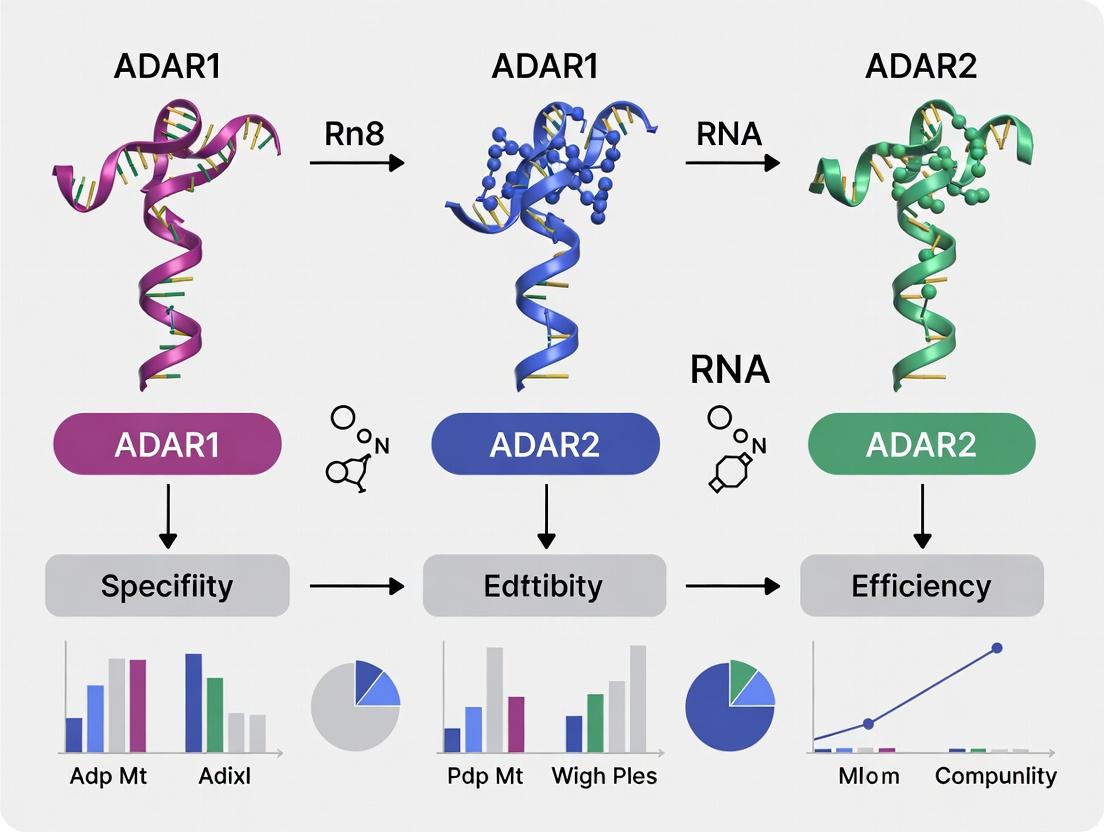
ADAR1 vs ADAR2 Specificity-Efficiency Trade-off
The Scientist's Toolkit: Research Reagent Solutions
Table 3: Essential Reagents for ADAR-mediated RNA Editing Research
| Reagent / Solution | Function / Application | Key Provider Examples (for informational purposes) |
|---|---|---|
| Engineered ADAR Expression Plasmids | Provide the catalytic editing domain (dADAR1 or dADAR2) fused to a guide-binding protein (e.g., λN, BoxB). Essential for cellular editing experiments. | Addgene (deposited by labs of Dr. David Liu, Dr. Thorsten Stafforst). |
| Chemically Modified Antisense Oligonucleotides (ASOs) | Serve as guide RNAs to bind target mRNA and recruit ADAR. Modifications (e.g., 2'-O-methyl, phosphorothioate, LNA) enhance stability and binding affinity. | Integrated DNA Technologies (IDT), Thermo Fisher Scientific, Sigma-Aldrich. |
| Circular RNA (circRNA) Guide Templates | Provide nuclease-resistant, highly specific guide scaffolds for in vivo applications. Can encode both guide and engineered ADAR enzyme. | Custom synthesis services (Circ Bio, etc.). |
| Purified Recombinant ADAR Deaminase Domains | Used for in vitro biochemical studies, determining kinetic parameters (kcat/Km), and flanking sequence preference assays. | Novoprotein, Abcam (some catalytically inactive mutants). |
| Amplicon-seq (NGS) Library Prep Kits | For high-throughput, quantitative measurement of on-target editing efficiency and bystanding edits from bulk cell populations. | Illumina (TruSeq), New England Biolabs (NEBNext). |
| RNA-seq Library Prep Kits | Essential for conducting genome-wide off-target analysis to assess editing specificity. | Illumina (Stranded mRNA Prep), Takara Bio (SMARTer). |
| A-to-I Editing Detection Software (Bioinformatics) | Identify and quantify RNA editing sites from RNA-seq data. Critical for specificity assessment. | RESCU, SPLINTER, REDItools (open-source). |
Experimental Challenges and Optimization Strategies for ADAR Research
Within the broader thesis on ADAR1 versus ADAR2 editing specificity and efficiency, distinguishing their unique and shared biological roles is a critical challenge. ADAR1 and ADAR2 are adenosine deaminases that edit RNA, converting adenosine to inosine. While they share a common catalytic function, their physiological roles, target specificity, and efficiency differ significantly. ADAR1 is essential for preventing aberrant innate immune activation by editing endogenous dsRNA, whereas ADAR2 is critical for neurotransmission through editing of specific neurotransmitter receptor pre-mRNAs. Overlapping editing at some sites complicates functional assignment. This guide compares the experimental approaches of genetic knockout/knockdown models and genetic rescue experiments to disentangle these overlapping functions, providing a framework for precise functional genomics research.
Methodological Comparison: Knockout/Knockdown vs. Rescue
Knockout and Knockdown Models
These approaches aim to reduce or eliminate gene function to observe resulting phenotypes.
- Knockout (KO): Complete, permanent elimination of gene function via genomic editing (e.g., CRISPR-Cas9). Provides a clear null phenotype.
- Knockdown (KD): Partial, often transient, reduction of gene expression (e.g., via siRNA, shRNA). Useful for studying essential genes where total knockout is lethal.
Key Application in ADAR Research: ADAR1 complete knockout is embryonically lethal in mice due to massive interferon response, while ADAR2 knockout mice exhibit seizures and die post-weaning. Knockdown in cell lines helps study acute effects on specific editing sites.
Rescue Experiments
This is a follow-up approach to confirm the specificity of an observed phenotype. The wild-type (or mutant) gene is reintroduced into the knockout/knockdown background to see if it restores normal function.
- Orthologous Rescue: Reintroducing the same gene to confirm phenotype reversal.
- Mutant/Chimera Rescue: Reintroducing specific mutants (e.g., catalytically dead ADAR) or chimeric proteins (e.g., ADAR2 with ADAR1 deaminase domain) to test which protein domains or functions are necessary and sufficient for rescue.
Key Application in ADAR Research: Rescuing ADAR1 knockout embryos with an editing-deficient ADAR1 mutant demonstrates the essential role of its catalytic activity for survival. Expressing ADAR2 in ADAR2 KO neurons can rescue faulty editing of the GluA2 Q/R site.
Quantitative Comparison of Experimental Outcomes
Table 1: Functional Insights from ADAR1 & ADAR2 Manipulation Models
| Experimental Model | Key Phenotypic Readout | ADAR1-Specific Insight | ADAR2-Specific Insight | Overlap/Compensation Insight |
|---|---|---|---|---|
| ADAR1 Full Knockout (Mouse) | Embryonic lethality (E11.5-12.5) | Essential for embryonic development, prevents MDA5-mediated interferonopathy. | Not applicable. | ADAR2 cannot compensate for ADAR1 loss in vivo. |
| ADAR2 Full Knockout (Mouse) | Post-weaning lethality, seizures | Not primary cause of phenotype. | Essential for editing CNS targets like GluA2 Q/R site; critical for neural function. | ADAR1 edits the GluA2 Q/R site inefficiently in vivo, failing to prevent seizures in ADAR2 KO. |
| ADAR1 Knockdown (Cell Line) | Elevated ISG expression, PKR activation | Constitutive role in masking endogenous dsRNA as "self." | Minimal effect on innate immune activation. | At shared sites (e.g., GRIA2 R/G), ADAR2 may partially maintain editing upon ADAR1 KD, but not at immune-critical sites. |
| ADAR2 Knockdown (Cell Line) | Reduced editing at specific CNS targets | Minor changes at a subset of shared sites. | Primary editor for a defined set of synaptic targets. | ADAR1 can partially edit some ADAR2-preferred sites (e.g., 5-HT2CR) in its absence, indicating functional overlap. |
| Rescue in ADAR1 KO Cells | Normalization of ISG expression, viability | Catalytically active ADAR1 p110 or p150 isoforms can rescue. ADAR2 cannot rescue. | Confirms non-redundancy for innate immune function. | Clearly delimits the non-overlapping, essential function of ADAR1. |
| Rescue in ADAR2 KO Neurons | Rescue of GluA2 Q/R editing, normalized electrophysiology | Catalytically active ADAR1 can weakly edit the site but fails to rescue phenotype fully. | Catalytically active ADAR2 efficiently rescues editing and phenotype. | Highlights strong substrate preference (editing efficiency) in a physiological context, limiting functional overlap. |
Detailed Experimental Protocols
Protocol 1: CRISPR-Cas9 Generation of ADAR1/2 Knockout Cell Lines
- Design gRNAs: Target early exons common to all isoforms of human ADAR or ADARB1 (ADAR2) using validated CRISPR design tools.
- Transfection: Co-transfect HEK293T or relevant cell line with a plasmid expressing SpCas9 and the specific gRNA, or deliver as ribonucleoprotein (RNP) complexes.
- Selection & Cloning: Apply puromycin selection (if plasmid-based). Single-cell clone by dilution into 96-well plates.
- Genotype Validation: Isolate genomic DNA from clones. Perform PCR amplification of the target region and analyze by Sanger sequencing or T7 Endonuclease I assay to identify frameshift indels.
- Phenotype Validation: Confirm loss of protein via western blot (anti-ADAR1, anti-ADAR2 antibodies). For ADAR1 KO, validate by upregulation of interferon-stimulated genes (ISGs) via qPCR.
Protocol 2: siRNA-Mediated Knockdown in Primary Neurons
- siRNA Design: Use validated siRNA duplexes targeting ADARB1 (ADAR2) or non-overlapping regions of ADAR (ADAR1).
- Neuron Transfection: At 7-10 days in vitro (DIV), transfect primary cortical or hippocampal neurons using a lipid-based transfection reagent optimized for neurons.
- Efficiency Check: Harvest cells 72-96 hours post-transfection. Assess knockdown efficiency via RT-qPCR for mRNA levels and western blot.
- Editing Analysis: Extract total RNA, synthesize cDNA, and perform PCR amplification around known editing sites (e.g., GluA2 Q/R site in GRIA2). Analyze by Sanger sequencing and quantify editing percentage from chromatograms.
Protocol 3: Genetic Rescue in a Knockout Background
- Construct Design: Clone the cDNA for the rescuing protein (e.g., wild-type ADAR2, catalytic mutant E396A) into a mammalian expression vector with a constitutive or inducible promoter. Include a fluorescent (e.g., GFP) or antibiotic resistance marker.
- Stable Line Generation: Transfect the construct into the validated ADAR2 KO cell line or primary neurons. Select with appropriate antibiotic (e.g., G418) over 2 weeks.
- Rescue Validation:
- Biochemical: Confirm protein re-expression by western blot.
- Functional: Quantify rescue of RNA editing at target sites (e.g., GluA2 Q/R site) via deep sequencing or pyrosequencing.
- Phenotypic: In neurons, perform whole-cell patch clamp to assess recovery of electrophysiological properties (e.g., reduced Ca2+ permeability in AMPA receptors post-rescue).
Signaling Pathway & Experimental Logic
Diagram 1: Logic Flow for Disentangling Gene Function
Diagram 2: Key Pathways & Perturbations for ADAR1 vs ADAR2
The Scientist's Toolkit: Research Reagent Solutions
Table 2: Essential Reagents for ADAR Functional Studies
| Reagent / Material | Function & Application | Example Product/Catalog |
|---|---|---|
| Validated CRISPR gRNAs | For generating knockout cell lines targeting specific ADAR isoforms. | Synthego or IDT predesigned gRNAs for ADAR or ADARB1. |
| ADAR1 & ADAR2 Antibodies | For validating protein knockout/knockdown and rescue expression via western blot or immunofluorescence. | Santa Cruz sc-73408 (ADAR1), Sigma HPA037310 (ADAR2). |
| Site-Specific Editing Assays | For quantifying editing efficiency at key sites (e.g., GluA2 Q/R). | Pyrosequencing assays (Qiagen) or deep sequencing. |
| Interferon Response qPCR Panels | For phenotyping ADAR1 knockout (upregulation of ISGs like ISG15, MX1). | Qiagen Human Interferon & Receptors RT² Profiler PCR Array. |
| cDNA Expression Constructs | For rescue experiments (wild-type, catalytic mutants, chimeric ADAR1/2). | Available from Addgene (e.g., pEGFP-ADAR2). |
| Lipid-Based Transfection Reagents | For delivering siRNA (knockdown) or plasmid DNA (rescue) into hard-to-transfect cells like primary neurons. | Lipofectamine 3000 (Thermo Fisher), Lipofectamine RNAiMax. |
| Primary Neuronal Cultures | Essential system for studying ADAR2's neuronal-specific editing functions and electrophysiological phenotypes. | Isolated from E16-E18 rodent cortices or hippocampi. |
| Selective Culture Media | For stable cell line selection post-transfection (e.g., containing G418 for neomycin resistance). | Thermo Fisher Geneticin (G418) Solution. |
Thesis Context
Within the broader investigation of ADAR1 versus ADAR2 editing specificity and efficiency, a central challenge is the minimization of off-target RNA edits. This guide compares engineered ADAR variants and guide RNA (gRNA) design strategies aimed at achieving high on-target efficiency with reduced off-target activity, a critical requirement for therapeutic development.
Comparative Performance of Engineered ADAR Variants
Table 1: Engineered ADAR Variants for Specificity
| Variant / System | Parent Enzyme | Key Modification | Reported On-Target Efficiency (A-to-I) | Reported Off-Target Reduction | Primary Experimental Model |
|---|---|---|---|---|---|
| hyperADAR | ADAR2 (E488Q) | Mutations in dsRBDs to reduce non-specific binding | ~80% at optimal sites | ~50% reduction vs. wtADAR2 | HEK293T, reporter assays |
| SLR-ADAR | ADAR2 dd | ssRNA-binding σ peptide fused to catalytically dead ADAR; requires λN-box gRNA | 20-70% (gRNA-dependent) | >90% reduction in transcriptome-wide off-targets | HEK293T, RNA-seq |
| REPAIRv2 | ADAR1 dd | Mutations (T375G, E1008G) in deaminase domain for improved specificity | ~50% efficiency on CTNNB1 | 10-fold reduction vs. REPAIRv1 | HEK293T, RNA-seq |
| MINI | ADAR1 dd | Truncated variant (only deaminase domain) fused to λN peptide | Comparable to full-length fusions | Reduced off-targets due to smaller footprint | HeLa, targeted sequencing |
| CIRTS | ADAR1/2 dd | Modular, small fusion protein system | 15-40% | Extremely low background; CRISPR-like gRNA design | Yeast, mammalian cells |
Table 2: Guide RNA (gRNA) Scaffold Comparison
| gRNA Scaffold / Design | Compatible ADAR System | Length & Structure | Specificity Feature | Key Limitation |
|---|---|---|---|---|
| λN-BoxB | SLR, MINI, others | ~20-30nt, stem-loop for λN binding | High-affinity, programmable target site | Immunogenicity concerns for λN peptide |
| MS2/PP7 | Various fusions | ~20nt, aptamer for coat protein binding | Allows multiplexing with different coat proteins | Larger complex size may affect delivery |
| CRISPR-like (CIRTS) | CIRTS | ~70-100nt, resembles sgRNA | Endogenous human proteins; small size | Lower efficiency for some targets |
| Circular gRNA | Various | Covalently closed circle | Increased nuclease resistance, longer half-life | More complex synthesis |
| Chemically Modified gRNA | All | Standard scaffold with modified nucleotides (e.g., 2'-O-methyl) | Reduced immunogenicity, improved stability | Potential impact on binding affinity |
Experimental Protocols for Key Studies
Protocol 1: Measuring Off-Target Editing via RNA-Seq
Objective: Quantify transcriptome-wide off-target adenosine deamination. Method:
- Transfection: Deliver ADAR variant + specific gRNA expression plasmids into HEK293T cells (e.g., 500ng each in 24-well plate using Lipofectamine 3000).
- RNA Isolation: At 48h post-transfection, extract total RNA using TRIzol, include DNase I treatment.
- Library Prep: Deplete ribosomal RNA. Prepare stranded RNA-seq libraries (Illumina TruSeq). High sequencing depth recommended (>50 million paired-end 150bp reads).
- Data Analysis: Map reads to reference genome (STAR aligner). Use A-to-I editing callers (e.g., REDItools2, JACUSA2) to identify sites with significant A-to-G mismatches. Filter out known SNPs (dbSNP).
- Off-Target Metric: Compare number of significant non-target A-to-G changes in experimental vs. untransfected control. Normalize to total editing events at the on-target site.
Protocol 2: In-Cell Specificity Reporter Assay
Objective: Rapid, quantitative comparison of variant specificity. Method:
- Reporter Design: Create a dual-luciferase (Firefly/Renilla) plasmid where the Firefly ORF contains a target adenosine within a premature stop codon (TAG) and multiple, defined off-target A's in its 3'UTR.
- Transfection: Co-transfect reporter plasmid (100ng), ADAR variant plasmid (200ng), and gRNA plasmid (200ng) per well.
- Measurement: At 48h, assay luciferase activity. Firefly signal recovery indicates on-target editing (TAG to TIG, readthrough). Use qRT-PCR on extracted RNA with primers flanking the 3'UTR off-target sites to quantify off-target editing efficiency.
- Specificity Score: Calculate (On-Target Editing %)/(Average Off-Target Editing % in 3'UTR).
Diagrams
Title: Strategic Framework for Minimizing ADAR Off-Targets
Title: RNA-Seq Workflow for Off-Target Quantification
The Scientist's Toolkit: Research Reagent Solutions
| Reagent / Material | Supplier Examples | Function in ADAR Editing Research |
|---|---|---|
| ADAR Expression Plasmids (wt & engineered) | Addgene, custom synthesis | Delivery of ADAR1/2 variants (e.g., hyperADAR, REPAIRv2) into target cells. |
| gRNA Scaffold Plasmids (λN-BoxB, MS2, etc.) | Addgene, IDT, Twist Bioscience | Express guide RNAs that direct ADAR fusion proteins to specific RNA targets. |
| Dual-Luciferase Reporter Assay Systems | Promega | Quantify on-target editing efficiency via stop codon readthrough in high-throughput format. |
| Stranded Total RNA-Seq Kits (with rRNA depletion) | Illumina, NEB, Takara | Prepare sequencing libraries to measure transcriptome-wide editing. |
| A-to-I Editing Detection Software (REDItools2, JACUSA2) | Open source | Bioinformatics tools to identify and quantify editing sites from RNA-seq data. |
| Lipofectamine 3000 Transfection Reagent | Thermo Fisher | Efficient delivery of plasmids into mammalian cell lines (HEK293T, HeLa). |
| RiboRNase Inhibitor | NEB, Thermo Fisher | Protect RNA and editing intermediates from degradation during experiments. |
| Synthetic, Chemically Modified gRNAs | IDT, Dharmacon | For testing stability and specificity of guide RNAs with 2'-O-methyl, phosphorothioate bonds. |
Within the field of RNA editing, the choice between ADAR1 and ADAR2 enzymes for therapeutic applications hinges on their distinct editing specificities and efficiencies. ADAR1 primarily edits adenosines in non-coding regions and is crucial for innate immune regulation, while ADAR2 shows high specificity for specific coding sites, such as the Q/R site in GluA2 pre-mRNA. A critical, practical determinant of successful in vivo editing is the delivery method (viral vs. non-viral) and the administered dosage, which directly impacts editing efficiency, specificity, durability, and safety. This guide objectively compares these delivery paradigms in the context of ADAR-mediated RNA editing.
Comparison of Delivery Methods: Viral vs. Non-Viral Vectors
Table 1: Comparative Analysis of Viral vs. Non-Viral Delivery for ADAR Editing
| Feature | Viral Vectors (AAV, Lentivirus) | Non-Viral Vectors (LNP, PEI) |
|---|---|---|
| Typical Editing Payload | Plasmid encoding engineered ADAR (e.g., hyperactive ADAR2dd) or guide RNA. | Chemically modified guide RNA (e.g., gRNA) with recombinant ADAR protein or mRNA. |
| Max Editing Efficiency (In Vivo) | 50-90% (stable transduction). | 20-70% (transient expression). |
| Onset of Editing | Slow (days to weeks; requires transcription/translation). | Rapid (hours; direct delivery of effector). |
| Duration of Effect | Long-term to permanent (genome integration or episomal persistence). | Transient (days to weeks; degraded). |
| Packaging Capacity | Limited (~4.7 kb for AAV). | Higher, more flexible. |
| Immunogenicity Risk | Moderate to High (pre-existing immunity, capsid response). | Lower (can be mitigated with chemical modifications). |
| Dosage Control | Precise genomic titer (vg/kg); difficult to reverse. | Precise mass-based dosing (mg/kg); titratable. |
| Manufacturing | Complex, costly. | Simpler, more scalable. |
| Key Advantage | Sustained, high-level editing from single dose. | Favorable safety profile, rapid deployment, repeat dosing. |
| Primary Challenge | Off-target editing persistence, immunogenicity. | Lower durability, achieving high in vivo efficiency. |
Impact of Dosage on Editing Outcomes
Dosage is a critical lever for optimizing the therapeutic window. For viral vectors, higher viral genome (vg) doses generally increase the percentage of transduced cells and editing levels but also elevate risks of immune responses and off-target editing. For non-viral LNPs, higher mRNA or protein doses increase editing efficiency transiently but may also lead to saturation effects and increased innate immune sensing.
Table 2: Dosage-Dependent Effects on Editing Parameters
| Parameter | Low Dosage (Sub-optimal) | Optimal Therapeutic Dosage | High Dosage (Toxic) |
|---|---|---|---|
| Editing Efficiency | <20% (viral), <10% (non-viral). | 40-80% (viral), 30-60% (non-viral). | >90% (saturation). |
| Specificity (On:Off Target) | May be high but therapeutically irrelevant. | Maximized (balanced expression). | Decreases due to saturation of ADAR/gRNA. |
| Therapeutic Durability | Short (non-viral), Unstable (viral). | Long-term (viral), Repeatable (non-viral). | Permanent (viral), Potential for chronic toxicity. |
| Immune Activation | Minimal. | Manageable. | High (anti-capsid, anti-transgene, cytokine release). |
| Example (AAV-ADAR2dd) | 1e11 vg/kg (ineffective in large tissue). | 1e12 - 5e12 vg/kg (established range). | >1e13 vg/kg (hepatic toxicity risk). |
| Example (LNP-gRNA/mRNA) | 0.1 mg/kg. | 0.5 - 3 mg/kg. | >5 mg/kg (saturation, infusion reactions). |
Supporting Experimental Data & Protocols
Key Experiment 1: AAV9 Delivery of ADAR2dd for CNS Editing
- Objective: Achieve sustained, high-efficiency RNA editing in the mouse brain.
- Protocol:
- Vector: Package a cDNA sequence for an engineered ADAR2dd (E488Q/T375G) under a neuron-specific promoter (e.g., hSyn) into AAV9 capsids.
- Delivery: Administer via intracerebroventricular (ICV) injection in neonatal mice or systemic intravenous (IV) injection in adult mice.
- Dosage Groups: 1e11 vg, 5e11 vg, 2e12 vg per animal (ICV).
- Analysis: After 4 weeks, harvest brain tissue. Extract RNA, perform RT-PCR, and quantify editing efficiency at the target site (e.g., Gria2 Q/R site) via Sanger or deep sequencing. Assess off-targets by RNA-Seq.
- Typical Result: Editing efficiency shows a dose-dependent increase from ~15% (1e11 vg) to >80% (2e12 vg) at the target site. Off-target editing rates increase non-linearly at the highest dose.
Key Experiment 2: LNP Delivery of ADAR Protein/Guide RNA for Hepatic Editing
- Objective: Achieve rapid, transient RNA editing in the liver.
- Protocol:
- Formulation: Co-encapsulate chemically stabilized, 5’-trisNHA-modified guide RNA and recombinant ADAR1 or ADAR2 protein (with inactivating mutations in deaminase domain for specificity) into biodegradable ionizable LNPs.
- Delivery: Administer via single IV bolus injection in mice.
- Dosage Groups: 0.3 mg/kg, 1 mg/kg, 3 mg/kg total RNA/protein.
- Analysis: Harvest liver at 24h, 48h, and 7 days post-injection. Analyze editing efficiency via sequencing of target transcripts (e.g., PCSK9). Measure serum protein (PCSK9, LDL) levels.
- Typical Result: Peak editing (~50%) occurs at 48h with 1 mg/kg dose, diminishing to <5% by day 7. Dose-dependent reduction in serum PCSK9 is observed.
The Scientist's Toolkit: Key Research Reagent Solutions
Table 3: Essential Reagents for ADAR Delivery Research
| Item | Function in Experiment |
|---|---|
| Ionizable Lipid (e.g., DLin-MC3-DMA, SM-102) | Core component of LNPs; enables efficient encapsulation and endosomal escape of nucleic acid/protein payloads. |
| AAV Serotype Capsids (e.g., AAV9, AAV-PHP.eB) | Determines tropism and transduction efficiency for viral delivery (e.g., CNS, liver, muscle). |
| Chemically Modified Guide RNA (e.g., ms², pseudouridine) | Enhances stability, reduces immunogenicity, and improves editing efficiency of non-viral delivered guides. |
| Engineered ADAR Effector Plasmid (e.g., pAAV-ADAR2dd) | Backbone for viral vector production; contains mutations to enhance catalytic activity and/or specificity. |
| Tissue-Specific Promoters (e.g., hSyn, TBG) | Drives expression of ADAR effector in target cell types, minimizing off-target editing in other tissues. |
| Next-Generation Sequencing (NGS) Kit | For comprehensive on-target and genome-wide off-target editing assessment (RNA-Seq). |
| Droplet Digital PCR (ddPCR) | For absolute quantification of viral vector genome titer and editing efficiency at specific loci. |
Visualizations
Diagram 1: ADAR Editing Delivery Workflow Comparison
Diagram 2: Dosage Effects on Editing Efficiency & Specificity
This guide compares the editing specificity and efficiency of ADAR1 and ADAR2, focusing on the critical experimental variables of cell type, cellular state, and RNA substrate competition. Understanding these context-dependent factors is essential for research and therapeutic applications aiming to leverage or correct A-to-I RNA editing.
Comparative Performance Analysis
Table 1: Summary of ADAR1 vs. ADAR2 Editing Efficiency and Specificity Across Contexts
| Parameter | ADAR1 (p110/p150 isoforms) | ADAR2 | Key Experimental Support & Variability |
|---|---|---|---|
| Primary Substrate Preference | Promiscuous editing of non-coding dsRNA (e.g., Alu elements); some site-selective editing in coding regions. | Highly selective for specific adenosines in structured coding regions (e.g., GluA2 Q/R site). | Cell Type Variability: Editing levels of shared sites (e.g., GRIA2) vary significantly between neuronal and non-neuronal cell lines. |
| Editing Efficiency in Immune-Activated States | Dramatically upregulated by interferon response; p150 isoform essential for editing viral dsRNA and preventing MDA5 activation. | Largely unchanged or slightly downregulated during immune activation. | State Variability: In HEK293T cells treated with IFN-β, ADAR1-mediated Alu editing increases >5-fold, while ADAR2-specific sites show minimal change. |
| Dependence on dsRNA Length & Structure | Efficient on long, perfectly base-paired dsRNA (>100 bp). Requires minimal 5' neighbor specificity. | Prefers short, imperfect dsRNA structures. Strong 5' neighbor preference (U=A>C>>G). | Substrate Competition: In vitro assays with 300bp dsRNA show ADAR1 Kcat/KM ~10x higher than ADAR2. |
| Response to Cellular Stress | Upregulated by ER stress, heat shock. Critical for cellular homeostasis. | Can be downregulated under certain stresses; activity modulated by phosphorylation. | State Variability: Thapsigargin-induced ER stress in HeLa cells increases ADAR1 protein levels by ~2-fold within 8 hours. |
| Nuclear vs. Cytoplasmic Localization | p110: Nuclear. p150: Shuttles between nucleus and cytoplasm (has NES). | Predominantly nuclear. | Cell Type Variability: ADAR1 p150 shows enhanced cytoplasmic localization in dendritic cells upon viral infection. |
| Impact of Substrate Competition | Dominates editing in high dsRNA contexts (e.g., viral infection, Alu-rich transcripts). Can outcompete ADAR2 for shared substrates. | Editing of preferred sites can be suppressed when ADAR1 levels are high, unless ADAR2 expression is also elevated. | Competition Data: Co-expression in HEK293 cells shows 40% reduction in ADAR2-specific editing of a reporter when ADAR1 p110 is overexpressed. |
Detailed Experimental Protocols
Protocol 1: Quantifying Cell-Type Specific Editing Profiles
Objective: Measure baseline and inducible A-to-I editing levels of canonical ADAR1 and ADAR2 sites across different cell lines.
- Cell Culture: Maintain HEK293T (epithelial), SH-SY5Y (neuronal), and Huh-7 (hepatic) cell lines in standard media.
- Treatment (Optional): Split cells and treat one cohort with 1000 U/mL human IFN-β for 24 hours to induce an immune-activated state.
- RNA Extraction & Reverse Transcription: Harvest cells, isolate total RNA using TRIzol, and synthesize cDNA with random hexamers.
- Targeted PCR Amplification: Design primers flanking known editing sites (e.g., GRIA2 Q/R site (ADAR2), Alu element in NLRP1 3'UTR (ADAR1), AZIN1 S/G site (shared)).
- Sequencing & Analysis: Perform Sanger or deep sequencing (RNA-seq). Calculate editing efficiency as (G peak height) / (G + A peak height) at the site of interest from cDNA traces. For RNA-seq, use pipelines like REDItools or JACUSA2.
Protocol 2: In Vitro Substrate Competition Assay
Objective: Determine the relative binding and editing kinetics of ADAR1 and ADAR2 on identical or competing RNA substrates.
- Protein Purification: Purify recombinant human ADAR1 deaminase domain (p110) and full-length ADAR2 from E. coli or using an in vitro transcription/translation system.
- RNA Substrate Preparation: Synthesize and 5'-end label with γ-32P-ATP:
- Substrate A: Short (~50 bp) imperfect dsRNA mimicking the GRIA2 R/G site.
- Substrate B: Long (~300 bp) perfectly base-paired dsRNA.
- Kinetic Assays:
- Single-Enzyme Reactions: Incubate fixed RNA concentration with increasing enzyme concentrations. Quench at time points (e.g., 0, 5, 15, 30, 60 min).
- Competition Reactions: Pre-incubate RNA substrate with a 2-fold molar excess of one enzyme, then add the second enzyme.
- Analysis: Resolve reactions on TLC plates to separate inosine (product) from adenosine (substrate). Quantify conversion to determine initial rates and calculate KM and Vmax.
Experimental Visualizations
Title: Factors Influencing ADAR Editing Outcomes
Title: Protocol for Profiling Context-Dependent Editing
The Scientist's Toolkit: Key Research Reagent Solutions
Table 2: Essential Reagents for ADAR Specificity Research
| Reagent / Material | Function in Experiment | Key Consideration |
|---|---|---|
| Isoform-Specific ADAR Antibodies | Differentiate p110 vs. p150 ADAR1 and ADAR2 protein levels via Western blot. | Validate specificity via siRNA knockdown. Critical for correlating protein level with editing changes. |
| Interferon-beta (IFN-β) | Induce immune-activated cellular state to study ADAR1 p150 upregulation and function. | Use controlled concentrations (e.g., 500-1000 U/mL) and durations (12-48 hr). |
| In Vitro Transcribed (IVT) dsRNA | Defined substrates for kinetic assays (e.g., short imperfect vs. long perfect dsRNA). | Ensure proper annealing and PAGE purification. Labeling (radioactive/fluorescent) enables precise kinetics. |
| Editing-Site Specific qPCR/ddPCR Assays | High-throughput, absolute quantification of editing efficiency at specific loci (e.g., GRIA2 Q/R). | More precise than Sanger for low-frequency or multiplexed analysis. |
| ADAR1/ADAR2 Expression Plasmids (WT & Catalytic Dead) | For overexpression or rescue experiments in KO cell lines. | Catalytic dead mutants (E→A) serve as essential controls for editing-independent effects. |
| ADAR-KO Cell Lines (e.g., HEK293 ADAR1-/-) | Define enzyme-specific editing baselines and remove competition effects. | Enables clean add-back experiments. Available from various knockout repositories. |
| Chemical Stress Inducers (e.g., Thapsigargin, Tunicamycin) | Modulate cellular ER stress state to investigate ADAR1 regulation beyond interferon. | Titrate carefully to avoid excessive cytotoxicity. |
| Next-Generation Sequencing Library Prep Kits | For genome-wide editing analysis (RNA-seq). | Use kits that preserve RNA modifications or employ specialized protocols for editing detection. |
Accurate interpretation of Next-Generation Sequencing (NGS) data is paramount, especially in sensitive applications like quantifying adenosine-to-inosine (A-to-I) RNA editing catalyzed by ADAR1 and ADAR2. This guide compares best-practice bioinformatics tools and pipelines for isolating true biological signal from technical noise, framed within ongoing research on ADAR isoform specificity and efficiency.
Comparison of Primary NGS Analysis Pipelines for RNA Editing
The choice of pipeline significantly impacts the accuracy of RNA editing detection. Below is a comparison of widely used tools, evaluated on key metrics relevant to ADAR research.
Table 1: Comparison of RNA Editing Detection Tools/Pipelines
| Tool/Pipeline | Primary Method | Key Strength for ADAR Research | Key Limitation | Reported Precision (A-to-I) | Reported Recall (A-to-I) |
|---|---|---|---|---|---|
| REDItools2 | Empirical analysis of aligned reads | Excellent for exploratory analysis; good for non-model organisms. | Requires extensive filtering to reduce noise. | ~95% (after filtering) | ~85% |
| GIREMI | Statistical model within-sample | Does not require matched DNA sequence; ideal for human cell line studies. | Lower power in low-coverage regions. | ~92% | ~80% |
| JACUSA2 | Caller-agnostic, based on read pileups | Detects multiple editing types; robust to alignment errors. | Computationally intensive for genome-wide scans. | ~96% | ~88% |
| JACUSA2 (with deep repeat masking) | Caller-agnostic with stringent filtering | Optimal for distinguishing ADAR1 (repeat-rich) from ADAR2 (specific site) signals. | Drastically reduces calls in repetitive regions. | ~99% | ~75% (for ADAR2-like sites) |
Essential Experimental Protocols for Validation
To validate bioinformatics predictions and distinguish true ADAR-mediated editing from noise or other sources, orthogonal experimental validation is required.
Protocol 1: In Vitro Editing Assay for ADAR Specificity
- Purpose: Confirm the catalytic activity and site preference of ADAR1 vs. ADAR2 on candidate RNA substrates.
- Methodology:
- Clone candidate RNA sequences (containing predicted editing sites) into an appropriate vector (e.g., pSP64 Poly(A)).
- Transcribe RNA in vitro using SP6 or T7 RNA polymerase.
- Purify recombinant human ADAR1 (p110 or p150 isoform) and ADAR2.
- Incubate each ADAR protein separately with the RNA substrate in reaction buffer (containing 25 mM Tris-HCl pH 7.5, 100 mM KCl, 5% glycerol, 1 mM DTT, 0.1 mg/mL BSA, 0.5 U/μL RNasin).
- Run reactions at 30°C for 1-2 hours.
- Extract RNA, reverse transcribe, and perform Sanger sequencing or high-fidelity PCR-cloning followed by sequencing.
- Quantify editing efficiency by measuring the ratio of G to A peaks (cDNA) or sequence counts.
Protocol 2: Knockdown/Knockout Validation in Cell Culture
- Purpose: Establish the dependence of an editing event on a specific ADAR enzyme.
- Methodology:
- Transfert target cells (e.g., HEK293T) with siRNA targeting ADAR1, ADAR2, or a non-targeting control. Alternatively, use CRISPR-Cas9 to generate ADAR1/2 knockout cell lines.
- Confirm knockdown/knockout efficiency via western blot (anti-ADAR1, anti-ADAR2) and qRT-PCR 48-72 hours post-transfection.
- Extract total RNA from all conditions and prepare RNA-Seq libraries. Sequence to a high depth (>50M reads per sample).
- Process data through the chosen pipeline (e.g., JACUSA2 with stringent filtering).
- True ADAR1-specific sites will show abolished editing in ADAR1-KD/KO but not in ADAR2-KD/KO, and vice versa. Sites unaffected in both may be technical artifacts.
Visualizing the Workflow and Pathway
Diagram 1: NGS RNA Editing Analysis & Validation Workflow
Diagram 2: ADAR1 vs ADAR2 Substrate Specificity & Functional Outcomes
The Scientist's Toolkit: Research Reagent Solutions
Table 2: Essential Reagents for ADAR Editing Research
| Item | Function in Research | Example/Note |
|---|---|---|
| Recombinant ADAR1/ADAR2 Proteins | For in vitro editing assays to test catalytic activity and site preference directly. | Purified from E. coli or insect cells; ensure deaminase activity is validated. |
| ADAR1 & ADAR2 Specific Antibodies | For validating knockdown/knockout efficiency and protein expression levels via western blot. | Mouse monoclonal anti-ADAR1 (Santa Cruz, sc-73408); Rabbit polyclonal anti-ADAR2 (Sigma, A3233). |
| Validated siRNA or shRNA Sets | For isoform-specific knockdown in cell culture to establish functional dependency. | ON-TARGETplus siRNA pools (Dharmacon) provide high specificity and reduced off-target effects. |
| CRISPR/Cas9 KO Cell Lines | To generate constitutive ADAR1 or ADAR2 null backgrounds for definitive editing assignment. | Commercially available (e.g., from Horizon Genomics) or generated in-house. |
| High-Fidelity Reverse Transcriptase | Critical for accurate cDNA synthesis prior to validation sequencing; reduces introduction of errors. | SuperScript IV (Thermo Fisher) or PrimeScript (Takara). |
| Strand-Specific RNA-Seq Kits | Preserves strand information during NGS library prep, crucial for accurate alignment in repetitive regions. | Illumina Stranded Total RNA Prep or NEBNext Ultra II Directional. |
| Positive Control RNA with Known Editing Sites | To benchmark the sensitivity and specificity of the wet-lab and computational pipeline. | Synthetic RNA spike-ins with known A-to-I edits (e.g., from Seracare). |
Head-to-Head: Validating Functional Divergence and Convergence in Health and Disease
Within the broader investigation of adenosine deaminase acting on RNA (ADAR) family specificity, understanding the distinct yet overlapping roles of ADAR1 and ADAR2 is paramount. This guide objectively compares their editing profiles—efficiency, specificity, and substrate preference—against each other as the primary "alternatives" in cellular RNA editing. The analysis is grounded in recent experimental data, providing a comparative framework for researchers in mechanistic biology and therapeutic development, where engineering these enzymes is a key strategy.
The following table synthesizes key comparative metrics for ADAR1 (primarily the p110 isoform and catalytic domain of p150) and ADAR2, based on recent in vitro and cellular studies.
Table 1: Comparative Editing Profiles of ADAR1 and ADAR2
| Parameter | ADAR1 | ADAR2 | Experimental Context & Key Findings |
|---|---|---|---|
| Primary Function | Immune tolerance, transcriptome diversification | Neurotransmission, neural function | Knockout models: ADAR1-/- is embryonic lethal (IFN response); ADAR2-/- mice have seizures and die young. |
| Catalytic Efficiency (kcat/KM) | Lower (∼10³ M⁻¹s⁻¹) | Higher (∼10⁴ - 10⁵ M⁻¹s⁻¹) | Measured on optimal hairpin substrates in vitro. ADAR2 is inherently more efficient. |
| Sequence & Structure Preference | Prefers 5' neighbor = U, 3' neighbor = G. Less stringent on base pairing 3' to site. | Strongly prefers 5' neighbor = A. Requires specific base pairing 3' to site for catalysis. | High-throughput sequencing of randomized RNA libraries reveals distinct sequence logos. |
| Canonical Site Example | Editing of 5' UAG 3' in dsRNA | Editing of 5' AA 3' in GluA2 Q/R site | ADAR1 inefficiently edits GluA2; ADAR2 exclusively edits this critical site in vivo. |
| Primary Substrate Type | Long, perfectly base-paired dsRNA (viral, Alu elements). | Short, imperfectly base-paired hairpins (coding sites in neuronal transcripts). | CLIP-seq data shows ADAR1 bound to Alu repeats; ADAR2 to specific pre-mRNA structures. |
| Cellular Localization | Nucleus & Cytoplasm (p150); Nucleus (p110). | Predominantly Nucleus. | Determines access to substrate pools (e.g., cytoplasmic Alus vs. nuclear pre-mRNAs). |
| Impact of dsRBDs | Three dsRBDs; crucial for binding long dsRNA, less for sequence specificity. | Two dsRBDs; contribute more directly to site recognition and specificity. | Domain-swap experiments alter editing site selectivity. |
Detailed Experimental Protocols
1. In Vitro Kinetic Analysis of Editing Efficiency
- Objective: Quantify catalytic parameters (kcat, KM) for purified ADAR1 and ADAR2 on defined RNA substrates.
- Methodology:
- Protein Purification: Express and purify recombinant human ADAR1 (deaminase domain) and full-length ADAR2 from HEK293T or Sf9 cells.
- Substrate Preparation: Synthesize and 5'-end label (γ-³²P ATP) short RNA hairpins containing a single target adenosine (e.g., optimal sites from Table 1).
- Reaction Setup: Incubate fixed, low concentrations of labeled RNA with increasing concentrations of purified ADAR enzyme in reaction buffer (20 mM HEPES pH 7.0, 150 mM KCl, 5% glycerol, 1 mM DTT) at 30°C.
- Time Course Assay: For each enzyme concentration, aliquot reactions at multiple time points (e.g., 0, 1, 2, 5, 10, 20 min) and quench with 90% formamide/50 mM EDTA.
- Analysis: Resolve products via denaturing PAGE. Quantify gel bands to determine fraction edited. Plot initial velocity vs. substrate concentration and fit data to the Michaelis-Menten equation using nonlinear regression to extract kcat and KM.
2. High-Throughput Specificity Profiling (SEQREP)
- Objective: Define sequence and structural preferences of ADAR1 and ADAR2 in an unbiased manner.
- Methodology:
- Library Design: Generate a DNA oligonucleotide library encoding a random 8-nucleotide region flanking a central target adenosine, all within a stable hairpin backbone.
- RNA Library Transcription: In vitro transcribe the dsDNA library to generate the pool of RNA substrates.
- Editing Reaction: Incubate the RNA library with purified ADAR1 or ADAR2 under conditions ensuring modest (<30%) overall editing.
- Selection & Sequencing: Reverse transcribe, PCR amplify, and deep sequence both the input and edited pools. Isolate edited molecules via a biochemical trick (e.g., restriction enzyme cleavage if editing creates an I-T base pair).
- Bioinformatic Analysis: Align sequences and compute enrichment scores for each nucleotide at each position (-4 to +4 relative to the edited adenosine). Generate sequence logos to visualize the specificity profile for each enzyme.
Visualization of Key Concepts
Title: ADAR Substrate Selection and Functional Outcomes
Title: Kinetic Assay Protocol for Editing Efficiency
The Scientist's Toolkit: Key Research Reagents
Table 2: Essential Reagents for ADAR Specificity Research
| Reagent / Material | Function / Description |
|---|---|
| Recombinant ADAR Proteins (Purified) | Catalytically active, full-length or deaminase-only domains for in vitro assays. Essential for kinetic and structural studies. |
| Synthetic RNA Hairpin Oligonucleotides | Defined substrates containing specific editing sites for in vitro activity validation and competition assays. |
| High-Throughput RNA Library Kits | Commercial or custom kits (e.g., from Twist Bioscience) for generating randomized sequence libraries for specificity profiling (SEQREP). |
| Anti-ADAR1 / Anti-ADAR2 Antibodies | For immunoprecipitation (CLIP-seq), Western blotting, and immunofluorescence to determine expression and localization. |
| Next-Generation Sequencing Platform | For deep sequencing of edited pools from cellular or in vitro assays (e.g., Illumina MiSeq/NextSeq). |
| ADAR-KO Cell Lines (e.g., HEK293T) | Genetically engineered null backgrounds for clean reconstitution studies with mutant enzymes. |
| Inosine-Specific RNA-seq Protocols | Chemical or enzymatic methods (e.g., ICE) to precisely map inosine sites transcriptome-wide. |
| Molecular Cloning System for dsRBD Swaps | Tools for creating chimeric ADAR proteins to dissect domain contributions to specificity. |
This comparison guide is framed within a broader thesis investigating the specificity and efficiency of human ADAR enzymes. ADAR1 (Adenosine Deaminase Acting on RNA 1) and ADAR2 (ADARB1) are crucial for catalyzing adenosine-to-inosine (A-to-I) editing in double-stranded RNA, a process with profound implications for proteomic diversity, immune response, and neurological function. A central question in this field is quantifying and comparing the intrinsic enzymatic efficiency (via kinetic parameters like kcat/KM) with the observed editing rates in complex cellular environments. This guide benchmarks reported data for ADAR1 and ADAR2, alongside engineered variants and emerging alternatives like Cas13-based RNA editors, providing an objective performance comparison.
Comparative Data Tables
Table 1: In Vitro Kinetic Parameters (kcat/Km) for A-to-I Editing on Model Substrates
| Enzyme / Editor Variant | Substrate (RNA Structure) | kcat (min⁻¹) | KM (nM) | kcat/KM (M⁻¹s⁻¹) | Experimental Conditions (Key Notes) | Primary Source |
|---|---|---|---|---|---|---|
| hADAR1 p110 | Short 30bp dsRNA (fully complementary) | ~0.5 | ~20 | ~4.2 x 10⁵ | 30°C, 150 mM KCl | Eggington et al., 2011 |
| hADAR2 | GluA2 R/G site (short hairpin) | ~20 | ~100 | ~3.3 x 10⁶ | 30°C, 100 mM KCl | Stephens et al., 2004 |
| hADAR2 (E488Q) | GluA2 R/G site (short hairpin) | ~0.3 | ~10 | ~5.0 x 10⁵ | Catalytically impaired mutant; 30°C | Macbeth et al., 2005 |
| TadA-ADAR dd (evoRx) | Specific point mutation in mRNA | N/A | N/A | Estimated 10²-10³ | In vitro kinetics less defined; cellular efficiency driven | RNA-seq based studies, 2023 |
| Cas13b-ADAR2 DD | Targeted mRNA guided by crRNA | N/A | N/A | Lower than ADAR2 | Complex formation reduces effective turnover | Abudayyeh et al., 2019 |
Table 2: Measured Cellular Editing Rates and Efficiencies
| System | Target Site | Reported Editing Efficiency (%) | Apparent Rate / Timeframe | Cell Type | Delivery Method | Key Limitation |
|---|---|---|---|---|---|---|
| Endogenous ADAR1 | Alu repetitive elements | 1-30% (highly variable) | Steady-state | HEK293T | Endogenous | Highly context-dependent |
| Overexpressed ADAR2 | GluA2 Q/R site | Up to ~90% | 24-48 hrs | Primary Neurons | Plasmid transfection | Potential off-targets |
| ADAR1 DD (p150) | Synthetic 3' UTR stem-loop | ~40-60% | 24 hrs | HeLa | Plasmid | Immune activation (p150) |
| Engineered dADAR (SNAP tag) | Specific point mutation | Up to ~80% | 72 hrs | HEK293T | Stable integration | Requires guide RNA |
| REPAIR (Cas13-ADAR2) | Transcriptomic point mutations | ~10-40% | 48 hrs | HEK293T | Plasmid (crRNA + editor) | High RNA off-target editing |
| LEAPER (arRNA-ADAR) | Endogenous transcripts | 10-50% | 72 hrs | Primary cells | ASO delivery | Efficiency varies by site |
Detailed Experimental Protocols
Protocol 1: Determiningkcat andKM for ADAR Enzymes In Vitro
Objective: To measure the steady-state kinetic parameters of a purified ADAR deaminase domain on a defined, short double-stranded RNA substrate. Key Steps:
- Protein Purification: Express and purify recombinant human ADAR deaminase domain (e.g., ADAR2 catalytic domain) using an affinity tag (e.g., His₆) followed by size-exclusion chromatography.
- Substrate Preparation: Synthesize a short (e.g., 30-50 nt) RNA oligonucleotide containing a target adenosine within a known secondary structure. Anneal it to a complementary strand labeled with a 5' fluorescent dye (e.g., FAM) and a 3' quencher.
- Activity Assay: Use a continuous fluorescence-based assay (e.g., change upon deamination) or a discontinuous gel-based assay. For the kinetic assay: a. Set up reactions with a fixed, low concentration of enzyme (0.5-5 nM) in reaction buffer (e.g., 25 mM HEPES pH 7.0, 150 mM KCl, 0.5 mM DTT, 0.1 mg/mL BSA). b. Vary the substrate concentration across a range (e.g., 1 nM to 500 nM). c. Incubate at 30°C and measure initial velocity (v₀) for each substrate concentration [S].
- Data Analysis: Fit the Michaelis-Menten equation (v₀ = (kcat[E]₀[S])/(KM + [S])) to the velocity vs. [S] data using nonlinear regression software (e.g., GraphPad Prism) to extract kcat and KM.
Protocol 2: Quantifying Cellular Editing Rates via Next-Generation Sequencing
Objective: To measure the percentage of A-to-I editing at a specific genomic locus in cultured cells following editor expression. Key Steps:
- Editor Delivery: Transfect or transduce target cells (e.g., HEK293T) with plasmids expressing the ADAR editor (e.g., dADAR variant) and a specific guide RNA, if required.
- Harvesting RNA: At a defined time point (e.g., 48h post-transfection), lyse cells and isolate total RNA using a column-based kit with DNase I treatment.
- Reverse Transcription & PCR Amplification: Design primers flanking the target site. Convert RNA to cDNA using a high-fidelity reverse transcriptase. Perform PCR to amplify the target region.
- Library Preparation & Sequencing: Barcode the amplicons and prepare a next-generation sequencing library (e.g., Illumina MiSeq). Aim for high sequencing depth (>10,000x coverage).
- Bioinformatic Analysis: a. Align sequencing reads to the reference genome. b. Use a variant-calling pipeline (e.g., GATK) or a specialized tool like RESPECT (RNA editing analysis tool) to identify A-to-G mismatches. c. Calculate editing efficiency as: (Number of G reads) / (Number of A reads + Number of G reads) * 100% at the target genomic coordinate.
Diagrams
Diagram 1: ADAR1 vs. ADAR2 Cellular Function & Pathway
Diagram 2: Experimental Workflow for Benchmarking
The Scientist's Toolkit: Key Research Reagent Solutions
| Item / Reagent | Function in ADAR Editing Research | Example Vendor/Cat. No. (Illustrative) |
|---|---|---|
| Recombinant hADAR2 (cat. domain) | Purified protein for in vitro kinetic assays and structural studies. | Sino Biological, ActiveMotif |
| Fluorescent dsRNA Substrates | Labeled RNA duplexes for continuous, real-time monitoring of deaminase activity. | Integrated DNA Technologies (IDT), custom synthesis. |
| dADAR (SNAP-tag) Plasmid Kit | Engineered editor system allowing covalent tagging and guide RNA targeting for precise editing. | Addgene (plasmid #xxxxx). |
| ADAR-specific Antibodies | For immunoblotting (WB) and immunoprecipitation (IP) to detect endogenous or overexpressed ADAR1/ADAR2. | Santa Cruz Biotechnology, Cell Signaling Technology. |
| Ribonuclease T1 | Enzyme used in inosine chemical erasure (ICE) assay to confirm and map A-to-I editing sites by cleaving at inosines. | Thermo Scientific. |
| TriLink CleanCap FLuc mRNA | Control edited mRNA for transfection and functional validation of editing efficiency. | TriLink BioTechnologies. |
| Next-Gen Sequencing Kit (Amplicon) | Library prep kit for deep sequencing of PCR-amplified target regions to quantify editing percentage. | Illumina Nextera XT. |
| RESPECT or REDItools2 Software | Specialized bioinformatics pipeline for accurate identification and quantification of RNA editing events from NGS data. | Open-source (GitHub). |
Within the broader thesis investigating ADAR1 versus ADAR2 editing specificity and efficiency, this comparison guide delineates their distinct, non-overlapping physiological roles. While both enzymes catalyze the deamination of adenosine to inosine in double-stranded RNA, their primary biological functions are segregated: ADAR1 is essential for distinguishing self from non-self in innate immunity, whereas ADAR2 is critical for fine-tuning synaptic transmission in the central nervous system.
Core Functional Comparison
| Feature | ADAR1 | ADAR2 |
|---|---|---|
| Primary Physiological Role | Maintenance of self-tolerance; suppression of aberrant innate immune activation by endogenous dsRNA. | Regulation of synaptic signaling; recoding of key neurotransmitter receptor transcripts. |
| Key Phenotype of Knockout | Embryonic lethality in mice due to chronic type I interferon response and widespread apoptosis. | Seizures, epilepsy, and premature death in mice; impaired synaptic plasticity. |
| Essential Edited Substrate | Endogenous Alu element-containing dsRNAs. | Glutamate receptor subunit GluA2 (GRIA2) pre-mRNA at the Q/R site. |
| Immune Pathway Involvement | Central to MDA5/MAVS/IRF3 signaling suppression; prevents PKR hyperactivation. | Minimal direct role; indirect effects via neuroinflammation. |
| Neurotransmission Role | Minor, largely indirect. | Central; edits critical for receptor function and neuronal viability. |
| Isoforms | Constitutively expressed p110 and interferon-inducible p150. | Single major isoform, constitutively expressed in neurons. |
| Localization | Nucleus and cytoplasm (p150). | Primarily nuclear. |
ADAR1 in Innate Immunity: Mechanism and Experimental Data
Signaling Pathway and Mechanism
ADAR1 editing marks endogenous double-stranded RNA (e.g., from Alu repeats) as "self." Unedited or deficiently edited self-dsRNA is recognized by the cytosolic innate immune sensor MDA5, triggering a signaling cascade that culminates in a potent type I interferon (IFN) response, which is pathogenic if chronic.
Diagram Title: ADAR1 Prevents MDA5-Mediated Recognition of Self-dsRNA
Key Supporting Experimental Data
| Experiment | System/Model | Key Quantitative Result | Conclusion |
|---|---|---|---|
| MDA5 Co-Knockout | Adar1 p150-/- mouse embryonic fibroblasts (MEFs) | 1000-fold reduction in Ifnb1 mRNA levels; rescue from cell death. | MDA5 is the primary sensor of unedited dsRNA in ADAR1 deficiency. |
| PKR Hyperactivation | Adar1 null MEFs | Phospho-PKR levels increased >10-fold vs. wild-type; rescued by Pkr knockout. | Unedited dsRNA also activates the PKR pathway, contributing to translational shutdown. |
| Alu Element Editing | Human cell lines (RNA-seq) | >90% of A-to-I editing occurs in Alu repeats; loss of ADAR1 reduces editing to near-background. | Alu RNAs are the primary immunogenic substrate for ADAR1. |
| Interferon Signature | Adar1 p150-/- mice | Serum IFN-β levels: ~500 pg/ml vs. undetectable in wild-type. | ADAR1 deficiency causes a constitutive, lethal interferonopathy. |
Experimental Protocol: Assessing Immune Activation in ADAR1-Deficient Cells
- Cell Model: Generate Adar1 knockout (e.g., via CRISPR-Cas9) in A549 or HEK293T cells. Include isogenic wild-type control.
- Stimulation: Leave cells unstimulated or transfert with synthetic dsRNA (e.g., poly(I:C)) as a positive control.
- RNA Isolation & qRT-PCR: Harvest cells at 24h. Isolate total RNA, perform cDNA synthesis. Quantify interferon-stimulated gene (ISG) expression (e.g., ISG15, IFI44, MX1) and IFNB1 via TaqMan or SYBR Green qPCR. Normalize to GAPDH or ACTB.
- Protein Analysis: Perform western blot on cell lysates for phospho-PKR (Thr446), total PKR, and cleaved caspase-3.
- Validation: Perform rescue by co-knockout of MDA5 (IFIH1) or MAVS using siRNA or CRISPR.
ADAR2 in Neurotransmission: Mechanism and Experimental Data
Signaling Pathway and Mechanism
ADAR2 site-specifically edits the GRIA2 transcript, changing a genomically encoded glutamine (Q) codon to an arginine (R) in the pore-lining region of the GluA2 AMPA receptor subunit. This Q/R edit is essential for rendering the receptor calcium-impermeable, which governs normal synaptic physiology and prevents excitotoxicity.
Diagram Title: ADAR2 Editing Controls AMPA Receptor Ca2+ Permeability
Key Supporting Experimental Data
| Experiment | System/Model | Key Quantitative Result | Conclusion |
|---|---|---|---|
| GRIA2 Q/R Site Editing | Adar2 -/- mice | Editing at GRIA2 Q/R site: 0% vs. >99% in wild-type. | ADAR2 is solely responsible for this critical edit. |
| Calcium Permeability | Hippocampal neurons from Adar2 -/-;Gria2R/R (edited knock-in) | Ca2+ influx in -/- neurons: ~5x higher; fully rescued in knock-in. | Unedited GluA2(Q) confers pathological Ca2+ permeability. |
| Phenotypic Rescue | Adar2 -/- mice with Gria2R/R allele | Mortality: 100% of Adar2 -/- die by P20; 100% survival in rescued mice. | Seizure and death phenotype is directly caused by lack of GRIA2 editing. |
| Editing Efficiency | In vitro editing assay | ADAR2 Km for GRIA2 R/G site: ~10 nM; ADAR1 Km: >500 nM. | ADAR2 has dramatically higher affinity/efficiency for this synaptic target. |
Experimental Protocol: Measuring GRIA2 Editing and Functional Consequences
- Tissue/Sample: Isolate total RNA from brain regions (e.g., hippocampus, cortex) of wild-type and Adar2 knockout mice.
- Editing Analysis: Perform RT-PCR on GRIA2 region encompassing the Q/R site (exon 11). Purify PCR product and subject to Sanger sequencing. Quantify editing efficiency by measuring the G (inosine) peak height relative to the A (adenosine) peak at the relevant position. For high-throughput: RNA-seq with variant calling.
- Electrophysiology: Prepare acute brain slices. Perform whole-cell patch-clamp recordings on hippocampal CA1 pyramidal neurons.
- IV Relationship: Measure current-voltage (I-V) relationship of AMPAR-mediated EPSCs. Unedited GluA2(Q)-containing receptors exhibit inward rectification (non-linear I-V curve).
- Ca2+ Permeability: Use a philanthotoxin-sensitive current assay to estimate fractional Ca2+ current.
- Behavior: Monitor Adar2 -/- and rescued mice for seizure activity via video-EEG.
The Scientist's Toolkit: Key Research Reagent Solutions
| Item | Function in ADAR1/2 Research | Example/Source |
|---|---|---|
| ADAR1-p150 Specific Antibody | Distinguishes the interferon-inducible p150 isoform from constitutive p110 in western blot/IF. | Rabbit mAb (Clone D8P8G, CST #81246) |
| Phospho-PKR (Thr446) Antibody | Detects activation of the PKR pathway, a key endpoint in ADAR1-deficient immune activation. | Rabbit Ab (CST #3075) |
| Anti-MDA5 (IFIH1) Antibody | For immunoblotting or immunofluorescence to assess MDA5 protein levels upon ADAR1 loss. | Rabbit mAb (Clone D74E4, CST #53212) |
| GRIA2 (GluA2) Antibody | Recognizes the AMPA receptor subunit whose editing status is controlled by ADAR2. | Mouse mAb (Clone 6C4, Millipore MAB397) |
| Poly(I:C) HMW | Synthetic dsRNA analog used to stimulate MDA5/RIG-I pathways as a positive control in innate immunity assays. | InvivoGen (tlrl-pic) |
| Adar1 and Adar2 KO Cell Lines | Isogenic engineered lines (e.g., from HEK293) provide clean backgrounds for rescue and mechanistic studies. | Available from commercial repositories (e.g., Horizon Discovery). |
| A-to-I Editing-Specific RNA-seq Pipeline | Computational tools to identify and quantify editing sites from RNA-seq data, distinguishing them from SNPs. | REDItools2, JACUSA2, SAILOR |
| In Vitro Editing Assay Kit | Contains purified ADAR enzyme and fluorescently labeled RNA substrate to measure editing kinetics and inhibitor screening. | Commercial assay kits (e.g., from Reaction Biology). |
| Philanthotoxin-433 | Selective blocker of Ca2+-permeable, GluA2-lacking AMPA receptors; used in electrophysiology. | Tocris Bioscience (Cat. #1910) |
1. Introduction within Thesis Context This guide compares the disease associations of ADAR1 and ADAR2, framed within the central thesis that their distinct editing specificity and efficiency underpin divergent roles in human pathology. ADAR1, primarily an A-to-I editor of repetitive dsRNA, is critical for preventing aberrant innate immune activation. In contrast, ADAR2 edits specific coding sites in transcripts crucial for neuronal excitability and function. Dysregulation of each enzyme leads to distinct disease spectra: ADAR1 dysfunction is linked to cancer and interferonopathies like Aicardi-Goutières Syndrome (AGS), while ADAR2 deficiency is strongly associated with neurological disorders.
2. Comparative Disease Associations: ADAR1 vs. ADAR2
Table 1: Primary Disease Associations and Molecular Mechanisms
| Feature | ADAR1 (p110/p150 isoforms) | ADAR2 (ADARB1) |
|---|---|---|
| Core Pathogenic Mechanism | Loss of editing leading to MDA5 activation by endogenous dsRNA; or oncogenic gain-of-function editing. | Loss of specific editing events leading to neuronal hyperexcitability and dysfunction. |
| Cancer Association | High. Editing is frequently dysregulated. Hypoediting promotes genomic instability; hyperediting of specific targets (e.g., AZIN1) drives proliferation. | Low. No strong direct oncogenic role identified. |
| Autoimmunity Association | Definitive. Biallelic loss-of-function mutations cause AGS Type 6. Haploinsufficiency can cause dyschromatosis symmetrica hereditaria. | Minimal/None. Not implicated in interferonopathy pathways. |
| Neurological Association | Indirect. Via neuroinflammation in AGS. Some editing sites in neurotransmitters receptors. | Direct. Essential for brain function. KO is lethal in mice (seizures). Linked to epilepsy, ALS, and major depressive disorder in humans. |
| Key Edited Substrate | Repetitive Alu elements in 3'UTRs and introns. | Codon-specific sites in glutamate (GluA2 Q/R) and serotonin (5-HT2C R/G) receptor pre-mRNAs. |
| Immune Signaling Pathway | MDA5/MAVS/IRF3-Type I IFN axis. ADAR1 loss -> unedited dsRNA -> MDA5 sensing -> potent IFN response. | Not applicable. |
Table 2: Supporting Experimental Data from Key Studies
| Disease Model | Target Gene/Substrate | Editing Change (vs. WT) | Measured Outcome | Key Citation (Example) |
|---|---|---|---|---|
| AGS (ADAR1) | Endogenous Alu elements | ~90% reduction | 100-fold increase in ISG expression; perinatal lethality in mice. | Liddicoat et al., 2015 |
| Cancer (ADAR1) | AZIN1 (Antizyme Inhibitor 1) | Site-specific hyperediting (up to 80%) | Increased cell proliferation & invasion in hepatocellular carcinoma. | Chen et al., 2013 |
| Epilepsy (ADAR2) | GluA2 (Gria2) pre-mRNA | Near-complete loss of Q/R site editing | Ca2+-permeable AMPA receptors, neuronal death, lethal seizures in mice. | Higuchi et al., 2000 |
| ALS (ADAR2) | GluA2 Q/R site | Significant reduction in motor cortex | Correlated with TDP-43 pathology and selective motor neuron vulnerability. | Hideyama et al., 2012 |
3. Experimental Protocols for Key Findings
Protocol 1: Assessing Global dsRNA Accumulation and IFN Response (ADAR1-KO)
- Cell Line: Generate ADAR1 knockout lines using CRISPR-Cas9 in HEK293T or appropriate patient-derived fibroblasts.
- dsRNA Detection: Fix cells and perform immunofluorescence using the J2 anti-dsRNA antibody (SCICONS). Quantify mean fluorescence intensity per nucleus.
- IFN Response Quantification: Extract total RNA and perform qRT-PCR for interferon-stimulated genes (ISGs) like ISG15, MX1, and IFI44L. Normalize to GAPDH.
- Protein Confirmation: Perform western blot for phospho-IRF3 and total MDA5.
Protocol 2: Quantifying Site-Specific Editing Efficiency (ADAR2 Substrates)
- Tissue/RNA Source: Isolate RNA from brain regions (e.g., motor cortex, hippocampus) or neuronal cell models.
- Reverse Transcription: Use high-fidelity RT-PCR.
- PCR Amplification: Design primers flanking the editing site of interest (e.g., GluA2 Q/R site in exon 11 of GRIA2).
- Sequencing & Analysis: Clone PCR products and Sanger sequence ≥50 clones, or use high-throughput RNA sequencing. Calculate editing percentage as (G peak height / (G + A peak heights)) at the specific genomic position.
4. Visualizing the Pathogenic Pathways
Diagram 1: ADAR1 and ADAR2 Deficiency Disease Pathways (77 chars)
Diagram 2: A-to-I Editing Analysis Workflow (44 chars)
5. The Scientist's Toolkit: Research Reagent Solutions
Table 3: Essential Reagents for ADAR Editing and Disease Research
| Reagent/Material | Function & Application | Example/Supplier |
|---|---|---|
| J2 Anti-dsRNA Antibody | Gold-standard for detecting immunogenic dsRNA in cells via IF/IHC; critical for validating ADAR1-KO phenotypes. | SCICONS, J2 clone |
| CRISPR-Cas9 KO/KI Kits | For generating isogenic ADAR1 or ADAR2 knockout cell lines, or introducing patient-specific mutations. | Synthego, Horizon Discovery |
| Site-Specific Editing Reporter | Plasmid constructs with a mutant GFP stop codon restored by A-to-I editing; measures editing efficiency at a defined site. | Addgene (e.g., pSARE) |
| MDA5 (IFIH1) Antibody | For detecting MDA5 protein levels and activation state in immune pathway studies. | Cell Signaling Technology |
| Type I IFN Bioassay | Sensitive, functional readout (e.g., ISRE-luciferase reporter) to quantify bioactive interferon in cell supernatants. | InvivoGen, Qiagen |
| High-Fidelity RNA-seq Kit | For transcriptome-wide identification of editing sites (A-to-I "G" mismatches). Must preserve RNA modifications. | Illumina Stranded Total RNA |
| Neuronal Cell Models | iPSC-derived neurons for studying ADAR2 editing in a physiologically relevant context for neurological disorders. | Fujifilm Cellular Dynamics, Axol Bioscience |
| ADAR1/2 Specific Inhibitors/Activators | Small molecule tools (e.g., 8-Azaadenosine derivative inhibitors) for pharmacological modulation. | Merck, Research-focused vendors |
This comparison guide is framed within a broader thesis investigating the distinct roles, editing specificity, and efficiency of ADAR1 (Adenosine Deaminase Acting on RNA 1) versus ADAR2. Synthetic lethality, where the combination of deficiencies in two genes leads to cell death while a deficiency in either alone does not, provides a powerful lens to dissect functional compensation between these paralogs. Insights from genetic models are critical for drug development, particularly in oncology, where targeting specific ADAR vulnerabilities could offer novel therapeutic strategies.
Core Concept Comparison: ADAR1 vs. ADAR2 in Genetic Models
The following table summarizes key differential roles and outcomes derived from loss-of-function genetic models, primarily in mice.
| Feature | ADAR1 (Adar1, ADAR) | ADAR2 (Adarb1, ADARB1) | Experimental Model & Key Insight |
|---|---|---|---|
| Global Knockout Phenotype | Embryonic lethality (E11.5-12.5) due to widespread apoptosis, interferon response, and hematopoietic failure. | Viable but prone to seizures and early death; severe editing deficits in brain transcripts. | Mouse germline knockout models. Demonstrates ADAR1's essential role in innate immune regulation. |
| Primary Editing Function | Primarily edits repetitive dsRNA structures (e.g., Alu elements) in non-coding regions to prevent MDA5 sensing. | Site-specific editing of coding sequences (e.g., GluA2 Q/R site, 5-HT2C receptor) critical for neurophysiology. | Deep sequencing of editomes from knockout tissues. |
| Synthetic Lethality Context | Combined loss of ADAR1 and p53, or ADAR1 and components of the STING pathway, is synthetically lethal in cancer cells. | Synthetic lethality not commonly reported; loss often compensated by ADAR1 activity at shared sites. | Studies in human cancer cell lines (e.g., MDA-MB-231, Mel888). |
| Immune Activation | Loss triggers a dsRNA sensor (MDA5)-driven type I interferon response and apoptosis. | Loss does not induce a significant interferon response. | MEFs (Mouse Embryonic Fibroblasts) from knockout mice; RNA-seq and interferon-beta assays. |
| Functional Compensation | Can edit some critical ADAR2 sites (e.g., GluA2) at low efficiency in vivo when ADAR2 is absent. | Minimal compensation for ADAR1's immune-regulatory function. | Rescue experiments in Adarb1-/- mice expressing editing-competent ADAR1 transgenes. |
| Therapeutic Targeting | High priority for cancer immunotherapy (e.g., ADAR1 inhibitors to enhance immune checkpoint blockade). | Target for correcting specific RNA editing defects in neurological disorders. | Preclinical studies with small-molecule inhibitors (e.g., 8-azaadenosine derivatives) or antisense oligonucleotides. |
Experimental Protocols for Key Findings
Protocol 1: Assessing Synthetic Lethality via Dual Gene Knockdown in Cancer Cell Lines
Aim: To test if combined inhibition of ADAR1 and a second gene (e.g., p53, STING) induces synthetic lethality. Methodology:
- Cell Culture: Use relevant human cancer cell lines (e.g., melanoma, breast adenocarcinoma).
- Gene Silencing: Perform co-transfection with siRNA pools targeting ADAR1 and the gene of interest (GOI). Include single knockdown and non-targeting siRNA controls.
- Viability Assay: 72-96 hours post-transfection, measure cell viability using a resazurin-based (Alamar Blue) or ATP-based (CellTiter-Glo) assay.
- Data Analysis: Calculate percent viability relative to non-targeting control. Synthetic lethality is indicated when the dual knockdown viability is significantly lower than the product of the viabilities from single knockdowns (Bliss independence model).
- Validation: Confirm knockdown efficiency by western blot (ADAR1 p150/p110 isoforms) and qRT-PCR for the GOI.
Protocol 2: Quantifying RNA Editing Efficiency and Specificity
Aim: To compare the editing landscape in ADAR1-deficient vs. ADAR2-deficient systems. Methodology:
- Sample Preparation: Generate isogenic cell lines with CRISPR/Cas9-mediated knockout of ADAR1, ADAR2, or both from a parental line. Alternatively, use tissue from conditional knockout mice.
- RNA Sequencing: Extract total RNA, perform poly-A selection, and prepare stranded RNA-seq libraries. Sequence to high depth (>50 million paired-end reads).
- Bioinformatic Analysis:
- Map reads to the reference genome (e.g., GRCh38).
- Use specialized tools (e.g., JACUSA2, REDItools) to call editing sites, requiring mismatch frequency >10% and coverage >20x.
- Annotate sites to repetitive elements (Alu for ADAR1) and specific coding sites (GluA2 Q/R for ADAR2).
- Specificity Metric: Calculate the percentage of total A-to-I editing events occurring within Alu elements (ADAR1-driven) versus in coding sequences (ADAR2-driven).
Visualizing Key Pathways and Relationships
Diagram 1: ADAR1 Loss Triggers Immune-Mediated Cell Death
Diagram 2: Experimental Workflow for Synthetic Lethality Screening
The Scientist's Toolkit: Key Research Reagent Solutions
| Reagent/Material | Function & Application in ADAR Research |
|---|---|
| ADAR1 p150/p110 Specific Antibodies | For western blot validation of protein knockdown/knockout efficiency and isoform-specific studies. |
| CRISPR/Cas9 Knockout Kits (e.g., for ADAR or ADARB1) | To generate isogenic, genetically engineered cell lines for clean phenotypic comparison. |
| siRNA Pools Targeting ADAR1, ADAR2, or other genes | For transient loss-of-function studies and synthetic lethality screens. |
| Type I Interferon-beta ELISA Kit | To quantitatively measure the interferon response upon ADAR1 loss. |
| Alamar Blue or CellTiter-Glo Viability Assay | Standardized, sensitive assays to measure cell proliferation and death in synthetic lethality experiments. |
| Stranded Total RNA-seq Library Prep Kit | For high-quality RNA preparation to accurately map and quantify RNA editing events. |
| JACUSA2 or REDItools Bioinformatics Software | Critical for identifying and statistically validating RNA editing sites from sequencing data. |
| Selective ADAR1 Inhibitors (e.g., 8-azaadenosine analogs) | Pharmacological tools to probe ADAR1 function and potential therapeutic utility. |
Conclusion
The comparative analysis of ADAR1 and ADAR2 reveals a sophisticated division of labor in RNA editing. ADAR1 serves as a high-efficiency, promiscuous editor crucial for immune tolerance, while ADAR2 acts as a precise, substrate-specific enzyme vital for neurological function. This functional dichotomy directly informs therapeutic strategy: ADAR1 is a primary target for immuno-oncology, whereas engineered ADAR2 variants offer unparalleled precision for correcting point mutations. Future research must focus on mapping the complete editome of each enzyme in diverse tissues, elucidating their regulation, and developing next-generation editors that merge ADAR2's specificity with ADAR1's potency. Overcoming delivery and off-target challenges will be paramount for translating these insights into safe, effective clinical interventions for genetic diseases and cancer.