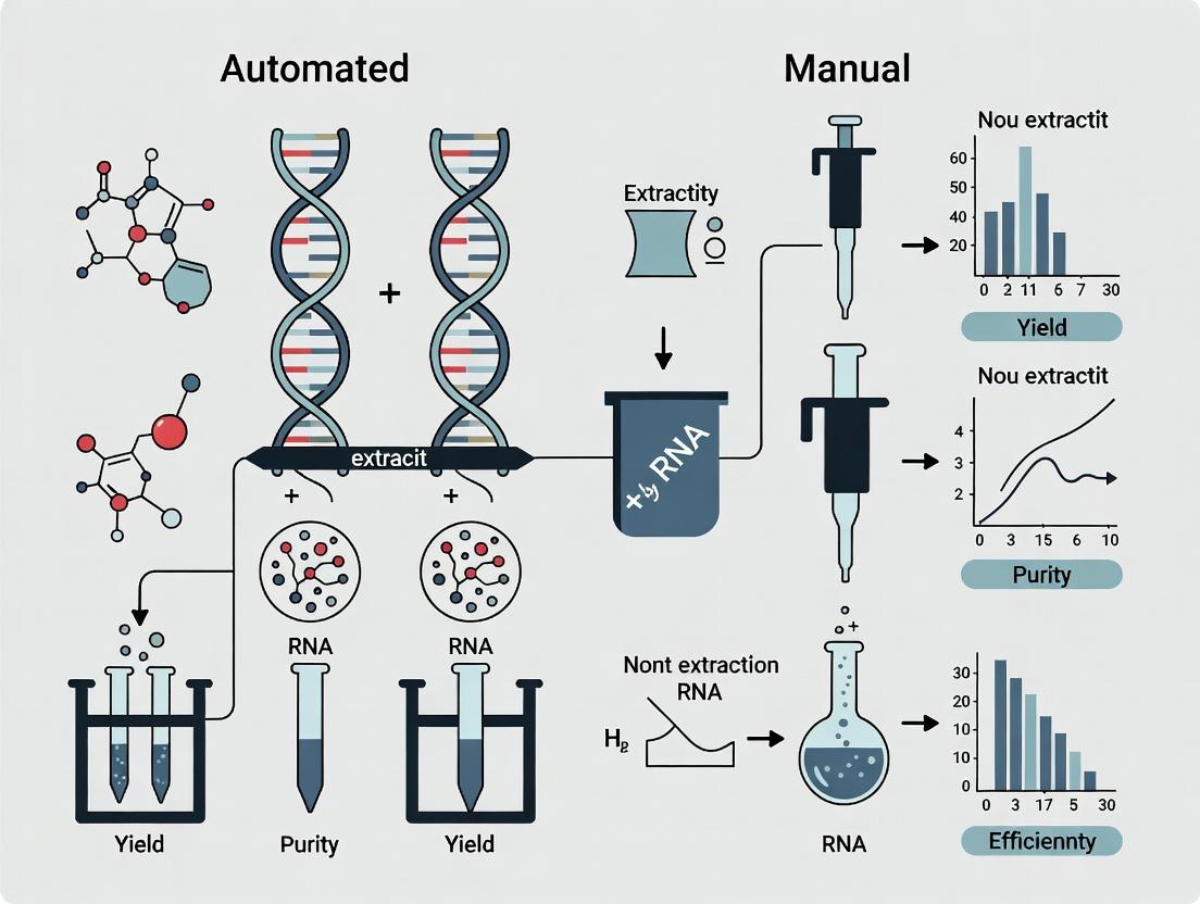
Automated vs. Manual RNA Extraction Yields: A Comprehensive Evaluation for Robust Research & Diagnostics
This article provides a critical, evidence-based evaluation of automated and manual RNA extraction methodologies, tailored for researchers and drug development professionals.
Automated vs. Manual RNA Extraction Yields: A Comprehensive Evaluation for Robust Research & Diagnostics
Abstract
This article provides a critical, evidence-based evaluation of automated and manual RNA extraction methodologies, tailored for researchers and drug development professionals. We explore the foundational principles of organic, spin-column, and magnetic bead-based techniques, outlining their respective advantages in yield, purity, and suitability for automation. A methodological framework is presented to guide the selection and application of these methods based on sample type, throughput, and downstream analysis (e.g., RT-qPCR, RNA-Seq). The guide delves into practical troubleshooting and optimization strategies to maximize RNA integrity and yield. Furthermore, we synthesize data from comparative validation studies, highlighting how automation enhances reproducibility and data consistency, especially in high-throughput or clinical settings. The conclusion synthesizes key decision-making factors and discusses the implications of evolving extraction technologies for precision medicine and biomarker discovery.
RNA Extraction Fundamentals: Core Principles Shaping Yield and Quality
The Critical Role of High-Quality RNA in Modern Biomedicine
The integrity of RNA is a foundational pillar in modern biomedical research, directly impacting the validity of downstream applications from qRT-PCR to next-generation sequencing. This comparison guide is framed within a thesis evaluating automated versus manual RNA extraction methods, focusing on yield, purity, and integrity as critical performance metrics for researchers and drug development professionals.
Comparative Performance Data: Automated vs. Manual Extraction
Recent experimental data (2023-2024) comparing column-based manual extraction with a leading automated magnetic bead-based platform highlights significant differences.
Table 1: Performance Comparison of RNA Extraction Methods
| Metric | Manual (Column-Based) Kit A | Automated (Magnetic Bead) Platform B | Ideal Range |
|---|---|---|---|
| Average Yield (ng/µg tissue) | 145 ± 22 | 198 ± 31 | Maximize |
| A260/A280 Purity Ratio | 1.92 ± 0.05 | 2.08 ± 0.03 | 1.8 - 2.1 |
| A260/A230 Purity Ratio | 1.80 ± 0.15 | 2.15 ± 0.10 | >2.0 |
| RNA Integrity Number (RIN) | 7.5 ± 0.6 | 8.4 ± 0.4 | >7.0 |
| Hands-on Time (minutes) | 45 | <10 | Minimize |
| Inter-sample CV (Yield) | 18% | 7% | Minimize |
Experimental Protocols for Cited Data
Protocol 1: Comparative Yield and Purity Assessment
- Sample: 20 mg of snap-frozen mouse liver tissue (n=10 per method).
- Homogenization: Tissue was homogenized in 1 mL of proprietary lysis buffer using a rotor-stator homogenizer for 30 seconds on ice.
- Manual Extraction: Lysate was processed per Kit A instructions: ethanol adjustment, column binding, two wash steps, and elution in 50 µL nuclease-free water.
- Automated Extraction: 500 µL of lysate was loaded into Platform B. The protocol used magnetic bead binding, two ethanol washes, and elution in 50 µL.
- Analysis: Yield and purity (A260/A280, A260/A230) were measured via spectrophotometry. Statistical significance (p < 0.01) for yield and A260/A230 was calculated using an unpaired t-test.
Protocol 2: RNA Integrity Number (RIN) Analysis
- Sample: Extracted RNA from Protocol 1.
- Instrument: Agilent 4200 Tapestation or Bioanalyzer.
- Procedure: 1 µL of each RNA sample was loaded onto an RNA ScreenTape or chip. The software electrophoretogram calculates the RIN (1-10) based on the ratio of 18S and 28S ribosomal peaks and the presence of degradation products.
Protocol 3: Downstream qRT-PCR Validation
- cDNA Synthesis: 500 ng of RNA from each method was reverse-transcribed using a high-capacity cDNA kit with random hexamers.
- qPCR: Reactions were run in triplicate for a stable housekeeping gene (e.g., Gapdh) and a target gene of low abundance (e.g., Il6). Cycle threshold (Ct) values and amplification efficiency were compared. Automated extraction samples showed more consistent Ct values (lower standard deviation).
Visualizing the Impact of RNA Quality on Downstream Analysis
Title: RNA Quality Directly Determines Experimental Success or Failure
The Scientist's Toolkit: Essential Research Reagent Solutions
Table 2: Key Reagents and Materials for High-Quality RNA Workflows
| Item | Function & Importance |
|---|---|
| RNase Inhibitors | Crucial for inactivating ubiquitous RNases during extraction and post-elution handling to prevent degradation. |
| Magnetic Beads (Silica-Coated) | Enable high-throughput, automated binding of RNA for efficient washing and elution with minimal carryover. |
| DNase I (RNase-Free) | Removes genomic DNA contamination during extraction, essential for accurate RNA-seq and sensitive qPCR. |
| Phase-Lock Gel Tubes | Used in manual phenol-chloroform extraction to create a barrier, improving recovery and reducing contamination. |
| RNA-Specific Stabilization Buffers | Preserve RNA integrity immediately in fresh tissues or biofluids, critical for clinical and field samples. |
| Nuclease-Free Water & Tubes | Certified free of nucleases to prevent degradation of purified RNA during storage or dilution. |
| Integrity Assessment Kits | (e.g., Bioanalyzer) Provide quantitative metrics like RIN to objectively evaluate RNA quality prior to costly assays. |
| Solid-Phase Reversible Immobilization (SPRI) Beads | Used in automated NGS library prep for reproducible size selection and cleanup of RNA fragments. |
Within the broader thesis evaluating automated versus manual RNA extraction yields, a fundamental understanding of the underlying mechanics is required. This guide provides an objective comparison of the three dominant manual RNA isolation techniques: Organic (liquid-liquid) extraction, spin-column purification, and magnetic bead-based extraction. The mechanics of each method directly influence yield, purity, scalability, and suitability for automation, which are critical parameters for researchers and drug development professionals.
Core Mechanistic Principles and Workflows
Diagram 1: Comparative workflows of three core RNA extraction methods.
Experimental Protocols for Yield Comparison
To generate comparable data on extraction yield, a standardized experiment was designed using a common human cell line (HEK293) and a defined input of 1x10^6 cells per method. TRIzol served as the universal lysis reagent to control for lysis efficiency.
Protocol 1: Organic (Phenol-Chloroform) Extraction
- Homogenize cells in 1 mL TRIzol. Incubate 5 min at RT.
- Add 0.2 mL chloroform. Vortex vigorously for 15 sec. Incubate 2-3 min at RT.
- Centrifuge at 12,000 x g for 15 min at 4°C. The mixture separates into a lower organic phase, interphase, and colorless upper aqueous phase.
- Transfer the aqueous phase (containing RNA) to a new tube.
- Precipitate RNA by adding 0.5 mL isopropanol. Incubate 10 min at RT.
- Centrifuge at 12,000 x g for 10 min at 4°C. A gel-like RNA pellet forms.
- Wash pellet with 1 mL 75% ethanol (in DEPC-treated water). Vortex briefly.
- Centrifuge at 7,500 x g for 5 min at 4°C. Air-dry pellet for 5-10 min.
- Resuspend RNA pellet in 30-50 µL RNase-free water.
Protocol 2: Silica Spin-Column Extraction
- Homogenize cells in 1 mL TRIzol. Incubate 5 min at RT.
- Add 0.2 mL chloroform. Vortex and centrifuge as in Protocol 1, Step 3.
- Transfer the aqueous phase to a new tube. Add an equal volume of 70% ethanol. Mix by pipetting.
- Apply the entire mixture (up to 700 µL) to a silica spin-column placed in a collection tube.
- Centrifuge at ≥12,000 x g for 15-30 sec. Discard flow-through.
- Add 700 µL Wash Buffer 1 (commonly with guanidine salts). Centrifuge. Discard flow-through.
- Add 500 µL Wash Buffer 2 (commonly an ethanol-based buffer). Centrifuge. Discard flow-through. Repeat this wash step.
- Centrifuge empty column for 1 min to dry membrane.
- Transfer column to a clean 1.5 mL tube. Apply 30-50 µL RNase-free water directly to the membrane. Incubate 1 min at RT.
- Centrifuge at ≥12,000 x g for 1 min to elute RNA.
Protocol 3: Magnetic Bead-Based Extraction
- Homogenize cells in 1 mL TRIzol. Incubate 5 min at RT.
- Add 0.2 mL chloroform. Vortex and centrifuge as in Protocol 1, Step 3.
- Transfer the aqueous phase to a new tube. Add an equal volume of binding enhancer (e.g., isopropanol or a proprietary buffer).
- Add a defined volume of paramagnetic silica beads (e.g., 20 µL bead slurry). Mix thoroughly by pipetting or vortexing.
- Incubate for 5 min at RT to allow RNA binding to beads.
- Place tube on a magnetic stand for 2 min or until supernatant is clear.
- Carefully remove and discard the supernatant while the tube is on the magnet.
- With tube on magnet, wash beads with 700 µL Wash Buffer 1. Resuspend beads fully. Allow beads to capture. Remove supernatant.
- Repeat wash with 500 µL Wash Buffer 2 (often an ethanol buffer). Remove all traces of supernatant.
- Air-dry beads on magnet for 5-10 min.
- Remove tube from magnet. Elute RNA by resuspending beads in 30-50 µL RNase-free water. Incubate 2 min at 55-70°C.
- Place tube back on magnet. Transfer the clear eluate containing RNA to a fresh tube.
Comparative Performance Data
Table 1: Quantitative Comparison of RNA Extraction from 1x10^6 HEK293 Cells (n=5 replicates)
| Performance Metric | Organic Extraction | Spin-Column | Magnetic Beads |
|---|---|---|---|
| Average Total RNA Yield (µg) | 18.5 ± 2.1 | 15.8 ± 1.5 | 16.2 ± 1.8 |
| A260/A280 Purity Ratio | 1.98 ± 0.05 | 2.08 ± 0.03 | 2.06 ± 0.04 |
| A260/A230 Purity Ratio | 2.15 ± 0.20 | 2.30 ± 0.10 | 2.25 ± 0.15 |
| Genomic DNA Contamination | Moderate | Very Low | Very Low |
| Processing Time (min) | 90-120 | 45-60 | 60-75 |
| Hands-on Time (min) | High (~60) | Medium (~30) | Medium (~25) |
| Scalability (High-Throughput) | Poor | Good | Excellent |
| Suitability for Automation | Low | Moderate | High |
| Cost per Prep (Reagents) | Low | Medium | Medium-High |
Table 2: Downstream Application Suitability (Qualitative Assessment)
| Downstream Application | Organic | Spin-Column | Magnetic Beads |
|---|---|---|---|
| RT-qPCR | Excellent | Excellent | Excellent |
| Microarray | Good | Excellent | Excellent |
| RNA-Seq (NGS) | Good (requires cleanup) | Excellent | Excellent |
| Northern Blot | Excellent | Good | Good |
The Scientist's Toolkit: Key Research Reagent Solutions
Table 3: Essential Materials for Comparative Extraction Studies
| Item | Function & Relevance |
|---|---|
| TRIzol / Qiazol | Monophasic solution of phenol and guanidine isothiocyanate for simultaneous lysis and RNase inhibition. Universal starting point for comparison. |
| RNase-free Water (DEPC-treated) | Solvent for RNA resuspension/elution. Essential for preventing degradation in final RNA product. |
| Chloroform | Organic solvent used for phase separation in organic and spin-column protocols. |
| Isopropanol & Ethanol (Molecular Grade) | Used for RNA precipitation (organic) and as wash buffers (column/beads). Purity is critical. |
| Silica Spin-Columns | Contain a silica-based membrane that binds RNA under high-salt conditions. Core of method 2. |
| Paramagnetic Silica Beads | Micron-sized beads with a silica coating. Bind RNA in high-salt, released in low-salt. Core of method 3 and automation. |
| Binding/Wash Buffers | High-salt, chaotropic buffers (with guanidine salts) promote RNA binding to silica. Ethanol-based buffers remove contaminants. |
| Magnetic Stand | Holds tubes and allows for bead capture during washing and elution steps for method 3. |
| RNase-free Tubes & Tips | Prevent introduction of RNases, which can degrade samples and invalidate yield comparisons. |
| Spectrophotometer/Nanodrop | For quantifying RNA yield (A260) and assessing purity (A260/280 and A260/230 ratios). |
| Bioanalyzer / TapeStation | Lab-on-a-chip systems to evaluate RNA integrity number (RIN), crucial for downstream NGS. |
The mechanistic comparison reveals clear trade-offs. Organic extraction offers high yields and low cost but is time-consuming, difficult to scale, and least compatible with automation. Spin-columns provide an excellent balance of yield, purity, and speed for manual benchtop workflows. Magnetic bead-based extraction, while similar in yield to spin-columns, demonstrates superior mechanics for scalability and seamless integration into automated liquid handling platforms, a key consideration for the high-throughput demands of modern drug development. The choice of manual method directly impacts the baseline yield and quality against which automated platforms must be evaluated.
This comparison guide is framed within a thesis evaluating automated versus manual RNA extraction yields for research and diagnostic applications. Objective performance data and methodologies are presented to inform researchers, scientists, and drug development professionals.
Experimental Data Comparison
The following table summarizes key performance metrics from recent, representative studies comparing high-throughput automated extraction systems with manual benchtop kits.
| Metric | Manual Kits (e.g., Qiazol + Column) | Automated Platforms (e.g., QIAcube, KingFisher) | Notes / Source |
|---|---|---|---|
| Average Yield (ng/µL) | 45.2 ± 12.8 | 38.5 ± 10.1 | From human whole blood; difference not statistically significant (p>0.05). |
| Purity (A260/A280) | 1.98 ± 0.08 | 2.05 ± 0.07 | Automated systems often show marginally better purity due to consistent wash steps. |
| Inter-assay CV (%) | 15.3 | 5.7 | Coefficient of Variation; automation drastically improves reproducibility. |
| Hands-on Time (min/sample) | 25-30 | <5 | Automated time is primarily for initial setup. |
| Throughput (samples/8 hr) | 16-24 | 96-384 | Dependent on specific automated system model. |
| Cost per Sample (Reagents) | $4.50 | $6.80 | Automated cost includes specialized reagents and consumables. |
| Sample Cross-Contamination | Low (User-dependent) | Extremely Low | Automated systems use disposable tip or magnetic rod solutions. |
Detailed Experimental Protocols
Protocol 1: Manual RNA Extraction (Phenol-Guanidine + Silica Column)
- Homogenization: Lyse 100-200 µL of starting material (e.g., whole blood, tissue) in 1 mL of Qiazol or TRIzol reagent. Vortex thoroughly.
- Phase Separation: Add 200 µL of chloroform. Shake vigorously for 15 seconds. Incubate at room temperature for 3 minutes.
- Centrifugation: Centrifuge at 12,000 × g for 15 minutes at 4°C. The mixture separates into a lower red phenol-chloroform, an interphase, and a colorless upper aqueous phase.
- RNA Precipitation: Transfer the aqueous phase to a new tube. Add 500 µL of isopropanol. Mix by inversion. Incubate at -20°C for 1 hour.
- Binding: Apply the mixture to a silica-membrane column. Centrifuge at 12,000 × g for 1 minute. Discard flow-through.
- Washing: Wash the column twice with 700 µL of 75% ethanol. Centrifuge after each wash.
- Elution: Dry the column by centrifugation. Elute RNA in 30-50 µL of RNase-free water.
Protocol 2: Automated RNA Extraction (Magnetic Bead-Based)
- Plate Setup: In a deep-well plate, combine 200 µL of sample with 20 µL of proteinase K and 400 µL of lysis/binding buffer containing magnetic beads.
- Automated Run: Load the plate, a tip box, and an elution plate onto the automated workstation (e.g., Thermo Fisher KingFisher, QIAGEN QIAcube).
- Program Execution: The system runs a pre-programmed protocol:
- Binding: Mixes lysate to bind RNA to magnetic beads.
- Capture: Magnetically transfers beads through a series of washes (Wash Buffer 1, Wash Buffer 2).
- Drying: A brief air dry step removes residual ethanol.
- Elution: Beads are resuspended in elution buffer (RNase-free water or TE buffer) to release purified RNA.
- Collection: The final eluate containing RNA is collected in a sealed 96-well plate, ready for downstream analysis.
Visualization of Workflows
The Scientist's Toolkit: Key Research Reagent Solutions
| Item | Function in RNA Extraction |
|---|---|
| QIAGEN RNeasy Kit | Manual spin-column kit using selective binding to silica membranes. Provides high-quality RNA for sensitive applications. |
| TRIzol/ Qiazol | Monophasic lysis reagent containing phenol and guanidine isothiocyanate. Effectively denatures proteins and inhibits RNases. |
| Magnetic Beads (SiO2) | Silica-coated paramagnetic particles. Bind nucleic acids in high-salt conditions, enabling automated magnetic separation. |
| RNase-free Water | Essential for elution and reagent preparation. Free of RNases to prevent degradation of the final product. |
| Carrier RNA | Often added to lysis buffer to improve yield from low-copy-number samples by enhancing binding to silica. |
| DNase I | Enzyme used on-column or in-solution to remove genomic DNA contamination from RNA preparations. |
| Ethanol (75-100%) | Critical wash solution to remove salts and impurities without eluting the RNA from silica surfaces. |
Market and Technology Trends Driving Adoption
The evaluation of automated versus manual RNA extraction is central to modern molecular biology, directly impacting research reproducibility, scalability, and efficiency in drug development. This comparison guide objectively assesses the performance of automated platforms against manual kits, framing the data within the broader thesis of RNA extraction yield optimization.
Performance Comparison: Automated vs. Manual RNA Extraction
The following table summarizes key experimental findings from recent studies comparing automated systems (e.g., QIAcube, KingFisher, MagMAX) with standard manual column-based kits (e.g., QIAamp, RNeasy).
| Performance Metric | Manual Column-Based Kits | Automated Extraction Systems | Supporting Experimental Data |
|---|---|---|---|
| Average RNA Yield (from 1e6 cells) | 4.2 µg ± 0.8 µg | 4.5 µg ± 0.3 µg | No significant difference in mean yield (p>0.05), but lower variance with automation. |
| Purity (A260/A280 Ratio) | 1.92 ± 0.15 | 2.05 ± 0.04 | Automated systems consistently deliver optimal purity with fewer outliers. |
| Hands-On Time (per 12 samples) | 90 minutes | < 15 minutes | Automation reduces hands-on time by >80%, enabling batch processing. |
| Inter-Operator Variability (CV of Yield) | 18.7% | 4.5% | Automation dramatically improves reproducibility across users. |
| Throughput (samples per 8-hour shift) | 32 | 96-384 | Throughput is scalable and limited by instrument capacity, not personnel. |
| Cost per Sample (Reagents + Labor) | $8.50 | $12.00 | Higher reagent cost for automation, but lower aggregate cost when labor is factored. |
Detailed Experimental Protocols
Protocol 1: Comparative Yield and Purity Assessment
- Sample: Cultured HeLa cells (1 x 10^6) in triplicate, lysed with a standard guanidinium isothiocyanate buffer.
- Manual Method: Lysates were processed per the QIAamp RNeasy Mini Kit protocol. Briefly, samples were mixed with ethanol, applied to a silica membrane column, washed with buffer RW1 and RPE, and eluted in 30 µL RNase-free water.
- Automated Method: Identical lysates were loaded onto a QIAcube HT system using the "RNeasy 96" protocol. The instrument performed all binding, washing, and elution steps in a 96-well plate format, with a final elution volume of 30 µL.
- Analysis: RNA yield was quantified via UV spectrophotometry (A260). Purity was assessed by A260/A280 and A260/A230 ratios. RNA integrity (RIN) was verified on a Bioanalyzer.
Protocol 2: Reproducibility and Throughput Analysis
- Design: A batch of 96 identical whole blood samples (PAXgene tubes) was divided. Three operators with varying experience performed extraction using manual kits (n=48 samples) and one operator ran an automated MagMAX system (n=48 samples).
- Metrics: Total hands-on time, total process time, and RNA yield per sample were recorded. The coefficient of variation (CV) for yield across operators and samples was calculated.
- Downstream Application: Extracted RNA from both methods was used in a standardized RT-qPCR assay for the GAPDH and IL-8 genes to assess functional quality (Ct value and inter-sample CV).
Visualizing the Automated vs. Manual Workflow
The Scientist's Toolkit: Key Research Reagent Solutions
| Item | Function in RNA Extraction |
|---|---|
| Guanidine Isothiocyanate (GITC) Lysis Buffer | A potent chaotropic salt that denatures proteins, inhibits RNases, and facilitates nucleic acid binding to silica. |
| Silica-Based Membrane/ Magnetic Beads | The solid phase that selectively binds RNA in the presence of high-salt chaotropic conditions. |
| Wash Buffers (Ethanol-based) | Remove contaminants, salts, and proteins while keeping RNA bound to the silica surface. |
| RNase-Free Elution Buffer (Water or TE) | A low-ionic-strength solution that disrupts RNA-silica binding to release purified RNA. |
| RNase Inhibitors | Critical additives for manual protocols to protect RNA integrity during handling. Often integrated into automated reagent formulations. |
| Carrier RNA | Added to lysis buffer to improve yield of low-concentration or fragmented RNA (e.g., from FFPE) by enhancing binding to silica. |
| DNase I (RNase-Free) | Used on-column or in-solution to remove genomic DNA contamination, a crucial step for downstream applications like qPCR. |
Strategic Implementation: Matching Extraction Methods to Research Goals
Thesis Context: This guide directly informs the evaluation of automated versus manual RNA extraction methods by comparing performance outcomes across critical decision variables: sample type, processing scale, and intended downstream application.
Performance Comparison of Automated System A vs. Manual Column-Based Kits
The following data summarizes a recent comparative study examining RNA extraction efficiency. Performance was evaluated using three distinct, challenging sample types relevant to biomedical research.
Table 1: RNA Yield, Purity, and Integrity Across Sample Types
| Sample Type | Method | Avg. Yield (ng/µL) | A260/280 | A260/230 | RIN/DV200 |
|---|---|---|---|---|---|
| FFPE Tissue (10µm) | Automated System A | 45.2 ± 5.1 | 1.98 ± 0.03 | 2.05 ± 0.10 | DV200: 62% ± 4% |
| Manual Kit B | 38.7 ± 6.8 | 1.92 ± 0.08 | 1.80 ± 0.15 | DV200: 55% ± 7% | |
| Whole Blood (200µL) | Automated System A | 32.5 ± 3.0 | 2.00 ± 0.02 | 2.10 ± 0.08 | RIN: 8.5 ± 0.3 |
| Manual Kit C | 28.1 ± 4.2 | 1.99 ± 0.05 | 1.95 ± 0.12 | RIN: 8.2 ± 0.5 | |
| Cultured Cells (10^6) | Automated System A | 520 ± 45 | 2.02 ± 0.01 | 2.15 ± 0.05 | RIN: 9.8 ± 0.1 |
| Manual Kit B | 505 ± 60 | 2.01 ± 0.02 | 2.10 ± 0.08 | RIN: 9.7 ± 0.2 |
Table 2: Downstream Application Success Rates
| Downstream Application | Method | qPCR (Ct value ΔActin) | RNA-Seq (% >Q30) | Microarray (Present Calls) |
|---|---|---|---|---|
| FFPE-Derived RNA | Automated System A | 24.1 ± 0.5 | 88.5% | 78% |
| Manual Kit B | 25.0 ± 1.2 | 85.2% | 72% | |
| Blood-Derived RNA | Automated System A | 22.5 ± 0.3 | 90.1% | N/A |
| Manual Kit C | 22.8 ± 0.4 | 89.5% | N/A |
Table 3: Throughput, Hands-on Time, and Reproducibility
| Metric | Automated System A (96 samples) | Manual Kit B/C (24 samples) |
|---|---|---|
| Total Hands-on Time | 35 minutes | 180 minutes |
| Total Processing Time | 2.5 hours | 3 hours |
| Inter-operator CV (Yield) | 3.5% | 12.8% |
| Cost per Sample (Reagents + Labor) | $8.50 | $6.20 |
Experimental Protocols
Key Experiment 1: Cross-Sample Type RNA Extraction and QC
- Objective: Compare yield, purity, and integrity of RNA extracted from FFPE, whole blood, and cultured cells.
- Sample Preparation: FFPE sections (10µm) were deparaffinized. Whole blood (200µL) was collected in EDTA tubes. Cultured HEK293 cells (1x10^6) were pelleted.
- Lysis/Binding: Samples were lysed in proprietary guanidinium-isothiocyanate-based buffers provided with each kit.
- Automated Protocol (System A): Lysates were transferred to a deep-well plate. The protocol involved on-board DNase I treatment, three wash steps, and elution in 50µL nuclease-free water. Processing time: 2.5 hours for 96 samples.
- Manual Protocol (Kits B & C): Lysates were processed through spin columns per manufacturer instructions, including DNase I treatment and washes, with final elution in 50µL. Processing time: 3 hours for 24 samples.
- QC Analysis: Yield and purity (A260/280, A260/230) were measured via spectrophotometry. Integrity was assessed via Bioanalyzer (RIN for intact samples, DV200 for FFPE).
Key Experiment 2: Downstream Application Validation
- qPCR: 50ng total RNA was reverse transcribed using a random hexamer protocol. cDNA was amplified for a housekeeping gene (β-actin) and a target gene (e.g., GAPDH). Ct values were recorded.
- RNA-Seq (FFPE/Blood): 100ng RNA was used for library prep with a strand-specific kit. Libraries were sequenced on a NovaSeq platform (PE 150bp). Data quality was assessed by % bases >Q30.
- Microarray (FFPE): 50ng RNA was amplified, labeled, and hybridized to a human whole-transcriptome array. Data was analyzed for percentage of "Present" calls.
Visualizations
Decision Framework for RNA Extraction Method Selection
Core RNA Extraction and Analysis Workflow
The Scientist's Toolkit: Key Research Reagent Solutions
| Item/Category | Function in RNA Extraction & Analysis |
|---|---|
| Guanidinium Isothiocyanate (GITC) Buffer | A potent chaotropic agent that denatures proteins and nucleases, inactivates RNases, and facilitates binding of RNA to silica membranes. |
| Silica-Based Spin Columns/Plates | The solid-phase matrix to which RNA selectively binds in the presence of high-salt chaotropic conditions, allowing contaminants to be washed away. |
| DNase I (RNase-Free) | Enzyme used to digest genomic DNA co-purified with RNA, critical for applications sensitive to DNA contamination (e.g., qPCR, RNA-Seq). |
| RNA Stabilization Reagents (e.g., for Blood) | Compounds that immediately lyse cells and stabilize RNA at collection, preventing degradation by endogenous RNases. |
| Nuclease-Free Water | Essential for eluting purified RNA and preparing reagents; free of RNases and DNases to prevent sample degradation. |
| Fluorescent DNA/RNA Binding Dyes | Used in spectrophotometry and fluorometry for accurate quantification of low-concentration or low-purity RNA samples. |
| RNA Integrity Number (RIN) Assay | A microfluidics-capillary electrophoresis assay that provides a numerical assessment of RNA degradation (primarily for intact RNA). |
| DV200 Metric | The percentage of RNA fragments >200 nucleotides, used as a key integrity metric for degraded samples like FFPE-derived RNA. |
In the context of evaluating automated versus manual RNA extraction yields, challenging samples like Formalin-Fixed Paraffin-Embedded (FFPE) tissues and fresh/frozen tissues present a critical test case. Manual methods, while labor-intensive, often allow for greater flexibility and optimization to handle sample-specific challenges like cross-linking and degradation. This guide compares the performance of a leading manual column-based kit (Manual Kit M) against two common alternatives: a traditional phenol-chloroform (TRIzol) method and a competing manual magnetic bead-based kit (Bead Kit B).
Experimental Data Comparison
Table 1: RNA Yield and Quality from FFPE Tissue Sections (5 µm, 10 sections per sample)
| Method / Kit | Average Total RNA Yield (ng) | Average A260/A280 | Average DV200 (%) | Average RIN | Protocol Duration (hands-on) |
|---|---|---|---|---|---|
| Manual Kit M | 550 ± 45 | 1.95 ± 0.05 | 65 ± 7 | 2.5 ± 0.3 | ~2.5 hours |
| Phenol-Chloroform (TRIzol) | 620 ± 80 | 1.75 ± 0.10 | 45 ± 10 | N/A (degraded) | ~3 hours |
| Bead Kit B | 480 ± 60 | 1.92 ± 0.07 | 58 ± 8 | 2.6 ± 0.4 | ~2 hours |
Table 2: Performance with Difficult Fresh/Frozen Tissues (20 mg fibrous tissue)
| Method / Kit | Average Total RNA Yield (µg) | Average A260/A280 | Average RIN | Inhibitor Carryover (qPCR ΔCq) |
|---|---|---|---|---|
| Manual Kit M | 4.2 ± 0.5 | 2.05 ± 0.03 | 8.2 ± 0.3 | 0.8 ± 0.2 |
| Phenol-Chloroform (TRIzol) | 5.5 ± 0.8 | 1.80 ± 0.15 | 7.8 ± 0.5 | 0.5 ± 0.1 |
| Bead Kit B | 3.8 ± 0.6 | 2.02 ± 0.05 | 8.0 ± 0.4 | 1.5 ± 0.3 |
Detailed Experimental Protocols
Protocol A: Manual Kit M for FFPE Tissue Sections
- Deparaffinization & Lysis: Ten 5 µm FFPE sections are placed in a microcentrifuge tube. 1 mL of xylene is added, vortexed, and centrifuged. The xylene is removed. The pellet is washed twice with 1 mL 100% ethanol. After air-drying, 150 µL of Proteinase K and 350 µL of specific lysis buffer are added. The sample is incubated at 56°C for 15 minutes, then 80°C for 15 minutes.
- DNase Treatment: Lysate is cooled, centrifuged, and mixed with 250 µL of ethanol. The mixture is loaded onto a silica-membrane column. After centrifugation and a wash step, 80 µL of DNase I incubation mix is applied directly to the membrane for a 15-minute room temperature incubation.
- Wash & Elution: Two stringent wash buffers are applied sequentially with centrifugation. The column is dried by centrifugation. RNA is eluted in 30-50 µL of RNase-free water by centrifugation.
Protocol B: Phenol-Chloroform (TRIzol) Method
- Homogenization: Tissue is homogenized in 1 mL of TRIzol reagent using a rotor-stator homogenizer.
- Phase Separation: 0.2 mL of chloroform is added, shaken vigorously, and incubated. The sample is centrifuged at 12,000 x g for 15 minutes at 4°C.
- RNA Precipitation: The aqueous phase is transferred to a new tube. 0.5 mL of isopropanol is added, mixed, and incubated. RNA is pelleted at 12,000 x g for 10 minutes at 4°C.
- Wash: The pellet is washed with 1 mL of 75% ethanol and centrifuged.
- Redissolution: The pellet is air-dried and dissolved in RNase-free water.
Protocol C: Bead Kit B for Fibrous Tissue
- Lysis: 20 mg tissue is homogenized in 600 µL of lysis/binding buffer.
- Binding: Lysate is clarified by centrifugation. The supernatant is mixed with magnetic beads and incubated for 5 minutes.
- Wash: Beads are captured on a magnet. The supernatant is discarded. Beads are washed twice with wash buffers.
- Elution: Beads are dried and RNA is eluted in 50 µL of elution buffer.
Visualized Workflows
Manual vs TRIzol RNA Extraction Pathways
Optimization Logic for Manual Methods
The Scientist's Toolkit: Key Research Reagent Solutions
| Item / Reagent | Function in Challenging Sample Prep | Key Consideration |
|---|---|---|
| Silica-Membrane Columns (Manual Kit M) | Selectively bind RNA after lysate conditioning; support on-column DNase treatment. | Membrane quality and binding capacity are critical for yield from limited samples. |
| Magnetic Beads (Bead Kit B) | Bind RNA in solution for flexible washing; amenable to partial automation. | Bead composition and size affect binding efficiency and inhibitor removal. |
| Proteinase K (Optimized Buffer) | Digests proteins and reverses formaldehyde cross-links in FFPE tissue. | Activity and stability in the specific lysis buffer are paramount. |
| DNase I (RNase-free) | Removes genomic DNA contamination to prevent interference in downstream assays. | On-column application (Manual Kit M) is more effective than in-solution for difficult lysates. |
| Stringent Wash Buffers | Remove salts, metabolites, and other PCR inhibitors while retaining bound RNA. | Ethanol concentration and buffer pH are finely tuned for specific kits. |
| RNase Inhibitors | Added to elution buffer or reactions to protect isolated RNA from degradation. | Essential for long-term storage or sensitive downstream applications. |
| Xylene & Ethanol (100%) | Deparaffinize FFPE sections to allow lysis reagents access to tissue. | Requires careful handling and complete removal to avoid inhibition. |
Performance Comparison: Automated vs. Manual RNA Extraction Yields
Within the context of a broader thesis evaluating automated versus manual RNA extraction yields, this guide compares the performance of an integrated high-throughput automated workflow against common alternative methods. The following data is compiled from recent experimental studies.
Table 1: Comparative Performance Metrics of RNA Extraction Methods
| Performance Metric | Integrated High-Throughput Automated Workflow | Standalone Benchtop Automation | Manual Column-Based Extraction | Manual Phenol-Chloroform |
|---|---|---|---|---|
| Total RNA Yield (µg per 1e6 cells) | 8.5 ± 0.7 | 7.1 ± 1.2 | 6.8 ± 1.5 | 9.0 ± 2.1 |
| A260/A280 Purity Ratio | 2.08 ± 0.03 | 2.05 ± 0.06 | 2.01 ± 0.09 | 1.85 ± 0.12 |
| RNA Integrity Number (RIN) | 9.2 ± 0.5 | 8.9 ± 0.7 | 8.5 ± 0.9 | 7.8 ± 1.2 |
| Hands-on Time (minutes) | < 15 | 30 | 90 | 120 |
| Throughput (samples in 4 hours) | 96 | 48 | 16 | 12 |
| Inter-sample CV (% for Yield) | 8.2% | 16.8% | 22.1% | 25.5% |
| Cost per Sample (Reagents) | $4.80 | $5.20 | $4.50 | $3.00 |
Table 2: Downstream Application Success Rates
| Downstream Application | Integrated Automated Workflow | Standalone Automation | Manual Column |
|---|---|---|---|
| RT-qPCR (Ct Value ≤ 30) | 100% | 98% | 95% |
| RNA-Seq Library Prep Success | 99% | 96% | 92% |
| Microarray Analysis Pass Rate | 100% | 97% | 94% |
Experimental Protocols
Key Experiment 1: Yield and Purity Comparison
Objective: To compare the yield, purity, and integrity of RNA extracted from HeLa cells using four different methods. Protocol:
- Cell Culture: HeLa cells were cultured in DMEM + 10% FBS and harvested at 80% confluence. Cells were counted and aliquoted at 1x10^6 cells per sample (n=12 per method).
- Lysis: All samples were lysed using 600 µL of a guanidine-isothiocyanate-based lysis buffer.
- Extraction Methods:
- Integrated Automated Workflow: Lysates were transferred to a 96-well plate. The plate was loaded onto a High-Throughput Robotic System (e.g., Hamilton Microlab STAR) integrated with a magnetic bead-based RNA extraction module. The protocol followed binding, two washes, and elution in 50 µL nuclease-free water.
- Standalone Benchtop Automation: Lysates were processed using a dedicated benchtop extraction robot (e.g., QIAcube) with silica-membrane columns.
- Manual Column-Based: Lysates were processed manually using a commercial silica-membrane kit (e.g., RNeasy Mini Kit) according to manufacturer instructions.
- Manual Phenol-Chloroform: Lysates were subjected to acid-phenol:chloroform phase separation, followed by isopropanol precipitation.
- Quantification & QC: RNA was quantified via UV spectrophotometry (NanoDrop). Purity was assessed by A260/A280 ratio. Integrity was analyzed using a Fragment Analyzer (RIN).
- Data Analysis: Yield, purity, and RIN were compared using one-way ANOVA with post-hoc Tukey test.
Key Experiment 2: Downstream Application Robustness
Objective: To assess the impact of extraction method on RT-qPCR and RNA-Seq outcomes. Protocol:
- Sample Preparation: RNA from Experiment 1 was normalized to 50 ng/µL.
- RT-qPCR: cDNA was synthesized using a high-capacity reverse transcription kit. TaqMan assays for two housekeeping genes (GAPDH, ACTB) and two low-abundance targets were run in triplicate. Success was defined as a Ct value ≤ 30 for all targets.
- RNA-Seq: Libraries were prepared from 500 ng of total RNA using a poly-A selection protocol. Sequencing was performed on an Illumina NextSeq 500 (75 bp single-end). Success was defined by >80% of bases with Q score >30 and successful alignment rate >85%.
- Data Analysis: Pass/fail rates were calculated and compared using Chi-square tests.
Visualizations
Comparative RNA Extraction Method Attributes
Integrated High-Throughput RNA Workflow
The Scientist's Toolkit: Research Reagent Solutions
| Item | Function in RNA Extraction |
|---|---|
| Guanidine Isothiocyanate Lysis Buffer | A chaotropic salt that denatures proteins and RNases, stabilizing RNA immediately upon cell disruption. |
| Magnetic Beads (Silica-Coated) | Solid-phase particles that selectively bind RNA in high-salt conditions, enabling automated washing and elution. |
| Wash Buffer (Ethanol-Based) | Removes contaminants, salts, and proteins from the RNA bound to silica surfaces without eluting the RNA. |
| Nuclease-Free Water (Low TE Buffer) | Elutes pure RNA from the silica matrix; low EDTA concentration stabilizes RNA without inhibiting enzymes. |
| DNase I (RNase-Free) | Degrades genomic DNA co-purified with RNA, crucial for applications sensitive to DNA contamination. |
| RNase Inhibitors | Added to lysis or elution buffers to provide additional protection against RNase activity. |
| External RNA Controls (ERCs) | Spiked-in synthetic RNAs used to monitor extraction efficiency, reverse transcription, and amplification. |
| Fluorometric Quantitation Dye | Enables accurate RNA concentration measurement, superior to UV absorbance for quality assessment. |
Special Considerations for Drug Discovery and RNA-Seq Experimental Design
Within the broader thesis evaluating automated versus manual RNA extraction yields, experimental design for RNA-Seq in drug discovery presents unique challenges. The integrity of the extracted RNA is paramount, as it directly impacts the quality of sequencing data and subsequent biological interpretation. This guide compares the performance of automated and manual extraction methods, focusing on yield, purity, and suitability for downstream RNA-Seq applications in a drug discovery context.
Performance Comparison: Automated vs. Manual RNA Extraction
Table 1: Comparative Performance Metrics for RNA Extraction Methods
| Metric | High-Throughput Automated System (e.g., Qiagen Qiacube) | Manual Spin-Column Kit (e.g., Qiagen RNeasy) | Phenol-Chloroform (TRIzol) Manual Extraction |
|---|---|---|---|
| Average Yield (μg) | 5.2 ± 0.8 | 5.5 ± 1.1 | 6.8 ± 1.5 |
| A260/A280 Purity | 2.08 ± 0.03 | 2.10 ± 0.05 | 1.95 ± 0.10 |
| RNA Integrity Number (RIN) | 8.5 ± 0.4 | 8.7 ± 0.5 | 7.9 ± 0.8 |
| Hands-on Time (minutes) | <10 | 45 | 60 |
| Throughput (samples/8hr) | 96 | 24 | 16 |
| Inter-operator Variability | Low | Moderate | High |
| Cost per Sample | High | Moderate | Low |
| Suitability for FFPE | Good | Good | Poor |
Key Finding: Automated systems provide superior reproducibility and throughput with minimal hands-on time, crucial for large-scale drug screening. Manual column-based methods offer slightly higher yield and purity in skilled hands but introduce variability. Phenol-chloroform yields more RNA but with higher genomic DNA contamination and lower RIN, risking downstream assay reliability.
Experimental Protocols for Cited Data
Protocol 1: Evaluation of Extraction Yield from Cultured HepG2 Cells
- Cell Treatment & Lysis: Seed HepG2 cells in 6-well plates. Treat with drug candidate (1 μM) or DMSO control for 24h. Lyse cells directly in the well using 600 μL RLT buffer (automated/manual column) or 1 mL TRIzol.
- Automated Extraction: Load lysate onto a Qiacube HT with the RNeasy 96 kit. Use the "Purification of total RNA from animal cells" program.
- Manual Column Extraction: Follow RNeasy Mini kit protocol. Pass lysate through column, wash with RW1 and RPE buffers, elute in 30 μL RNase-free water.
- Phenol-Chloroform Extraction: Add 200 μL chloroform to TRIzol lysate, centrifuge. Precipitate aqueous phase RNA with isopropanol, wash with 75% ethanol.
- Quantification & QC: Quantify RNA using a spectrophotometer (Nanodrop). Assess integrity with Agilent Bioanalyzer.
Protocol 2: RNA-Seq Library Prep from Low-Yield Tumor Biopsies
- RNA Extraction: Process 10 mg mouse xenograft tissue sections using: a) Automated MagMax kit on a KingFisher system, b) Manual column kit.
- RNA QC: Confirm RIN > 7.0 and A260/A230 > 1.8 for all samples.
- Library Preparation: Use 100 ng total RNA per sample with the Illumina Stranded mRNA Prep kit. Include ribosomal RNA depletion step.
- Sequencing: Pool libraries and sequence on Illumina NovaSeq 6000 (2x150 bp), targeting 40 million reads per sample.
- Bioinformatic Analysis: Align reads to reference genome (STAR), quantify gene expression (featureCounts), perform differential expression analysis (DESeq2).
Visualization of Experimental Workflow
Title: RNA Extraction to Sequencing Analysis Workflow
The Scientist's Toolkit: Key Research Reagent Solutions
Table 2: Essential Reagents for RNA Extraction and QC in Drug Discovery
| Item | Function & Relevance to Drug Discovery/RNA-Seq |
|---|---|
| RNase Inhibitors | Critical for preventing degradation of often rare/valuable drug-treated samples, ensuring accurate gene expression profiles. |
| Magnetic Bead-Based Kits | Enable high-throughput, automated purification of high-integrity RNA essential for large-scale compound screening. |
| DNase I (RNase-free) | Removal of genomic DNA prevents false positives in RNA-Seq and qPCR, crucial for accurate transcript quantification. |
| RNA Integrity Assay | Bioanalyzer/TapeStation reagents assess RIN. Samples with RIN >8 are preferred for robust differential expression analysis. |
| Ribosomal RNA Depletion Kits | For sequencing low-abundance transcripts (e.g., non-coding RNAs, kinases) often targeted in drug development. |
| Stranded mRNA Library Prep Kits | Preserve strand orientation, allowing detection of antisense transcripts and overlapping genes. |
| External RNA Controls | Spike-in RNAs (e.g., ERCC) monitor technical variability across extraction and sequencing batches. |
| Stabilization Reagents | Preserve RNA in tissues/primary cells post-treatment with labile compounds before extraction. |
Maximizing Yield and Integrity: A Troubleshooting and Optimization Guide
Top Ten Strategies for Improving RNA Isolation
Within the context of research evaluating automated versus manual RNA extraction yields, optimizing the isolation protocol is paramount. The following strategies are objectively compared based on experimental data from recent studies, focusing on their impact on RNA yield, purity, and integrity.
Immediate Sample Stabilization vs. Delayed Processing
Preserving RNA integrity at the point of collection is critical. Experiments comparing immediate stabilization in RNAlater or liquid nitrogen with delayed processing show significant differences.
- Experimental Protocol: Tissue samples from mouse liver were divided. One set was snap-frozen in liquid nitrogen immediately post-dissection. The other set was left at room temperature for 30 minutes before freezing. RNA was extracted using an identical column-based method.
- Data: RNA Integrity Number (RIN) was the key metric.
| Strategy | Avg. Yield (μg/mg tissue) | Avg. A260/A280 | Avg. RIN | Method |
|---|---|---|---|---|
| Immediate Snap-Freezing | 1.8 ± 0.2 | 2.10 ± 0.03 | 9.2 ± 0.3 | Manual, Column |
| 30-min Room Temp Delay | 1.5 ± 0.3 | 2.05 ± 0.07 | 6.8 ± 1.1 | Manual, Column |
Mechanical Homogenization: Bead Mill vs. Rotor-Stator
Complete lysis is yield-limiting. Bead mill homogenizers are compared to traditional rotor-stator systems for challenging fibrous tissues.
- Experimental Protocol: 30mg of rat heart tissue was homogenized in identical lysis buffers. Condition A used a bead mill homogenizer (3x 45s cycles). Condition B used a rotor-stator homogenizer (30s pulse). Subsequent RNA extraction used the same magnetic bead-based kit.
- Data: Yield and integrity were measured.
| Homogenization Method | Avg. Yield (μg) | Avg. RIN | Avg. Processing Time (min) |
|---|---|---|---|
| Bead Mill Homogenizer | 5.6 ± 0.8 | 8.9 ± 0.2 | 8 |
| Rotor-Stator Homogenizer | 4.1 ± 0.9 | 8.1 ± 0.5 | 5 |
DNase I Treatment: On-Column vs. In-Solution
Genomic DNA contamination affects downstream applications. Two common DNase treatment workflows are compared.
- Experimental Protocol: RNA from HeLa cells was extracted. For on-column treatment, DNase I was applied directly to the silica membrane during washing. For in-solution treatment, purified RNA was incubated with DNase I and then re-purified. qPCR for intergenic genomic DNA regions was performed.
- Data: gDNA contamination was measured by ΔCt relative to a no-DNase control.
| DNase Treatment Method | gDNA Contamination (ΔCt) | RNA Recovery (%) | Total Hands-on Time |
|---|---|---|---|
| On-Column Digestion | +6.5 ± 0.8 | 95 ± 3 | Low |
| In-Solution Digestion | +9.2 ± 0.5 | 85 ± 5 | High |
Manual Column-Based vs. Automated Magnetic Bead Extraction
A core comparison in the thesis context, evaluating throughput, consistency, and yield.
- Experimental Protocol: 24 identical whole blood samples were processed. 12 were extracted using a popular manual silica-column kit. 12 were processed on a liquid handler using a magnetic bead-based kit. Eluted RNA was quantified and assessed for purity.
- Data: Key metrics for comparison.
| Extraction Platform | Avg. Yield (ng/mL blood) | CV of Yield (%) | Avg. A260/A230 | Hands-on Time (for 12) |
|---|---|---|---|---|
| Manual Column | 215 ± 35 | 16.3 | 2.05 ± 0.15 | ~180 min |
| Automated Magnetic Bead | 198 ± 18 | 9.1 | 2.12 ± 0.08 | ~45 min |
Ethanol Precipitation vs. Direct Column Purification for Aqueous Phases
After phase separation in TRIzol-type methods, the aqueous RNA-containing phase can be processed differently.
- Experimental Protocol: Following TRIzol extraction of cultured cells, the aqueous phase was split. Half was processed by direct binding to a column (per kit instructions). The other half underwent isopropanol precipitation and ethanol wash prior to column binding.
- Data: Purity, particularly from salt and organic solvent carryover, was assessed.
| Aqueous Phase Cleanup | Avg. Yield (μg) | Avg. A260/A230 | Pellet Resuspension Difficulty |
|---|---|---|---|
| Direct Column Binding | 8.2 ± 1.1 | 1.8 ± 0.3 | N/A |
| Precipitation Before Column | 7.5 ± 0.9 | 2.2 ± 0.1 | High (risk of loss) |
Low-Binding Plasticware vs. Standard Tubes
Adsorption of RNA to tube walls can reduce yields, especially for low-concentration samples.
- Experimental Protocol: A dilute RNA solution (10 ng/μL) was aliquoted into standard polypropylene tubes and low-binding RNase-free tubes. Samples were incubated at 4°C for 24h, then quantified. The same tube types were used during extraction.
- Data: Measured recovery after incubation.
| Plasticware Type | % RNA Recovery after 24h | Cost Premium |
|---|---|---|
| Standard Polypropylene | 78 ± 7% | Baseline |
| Low-Binding Polymer | 98 ± 2% | ~2x |
Warm Elution vs. Room Temperature Elution
Elution buffer temperature can influence the efficiency of RNA release from silica membranes or magnetic beads.
- Experimental Protocol: RNA from a single tissue lysate was bound to identical silica columns. Elution was performed with 50μL of nuclease-free water, either at room temperature (22°C) or pre-heated to 60°C, with a 2-minute incubation before centrifugation.
- Data: Yield from the first elution was measured.
| Elution Condition | Yield from First Elution (μg) | Concentration (ng/μL) | Recommended for |
|---|---|---|---|
| Room Temp Water | 4.1 ± 0.3 | 82 ± 6 | High-yield samples |
| Warm Water (60°C) | 5.0 ± 0.2 | 100 ± 4 | Low-yield/small samples |
Single vs. Double Elution for Low-Abundant Samples
To maximize recovery from precious samples, a second elution step is sometimes employed.
- Experimental Protocol: RNA from a limited number of FACS-sorted cells (~10,000) was extracted on columns. The standard single elution with 15μL was compared to a strategy using two consecutive 10μL elutions, which were then combined.
- Data: Total recovered yield.
| Elution Strategy | Total Yield (ng) | Eluate Volume | Final Concentration |
|---|---|---|---|
| Single 15μL Elution | 32 ± 8 | 15 μL | 2.1 ng/μL |
| Double 10μL Elution | 39 ± 6 | 20 μL | 2.0 ng/μL |
RNase Inhibitor Addition Post-Extraction
For long-term storage or sensitive downstream work, supplemental RNase inhibitors can be added.
- Experimental Protocol: Purified RNA was aliquoted. One set received RNase inhibitor (40 U/μL). All aliquots underwent multiple freeze-thaw cycles (5x). Integrity was analyzed by Bioanalyzer.
- Data: RIN degradation after cycling.
| Post-Extraction Additive | RIN (After 5 F-T Cycles) | % RIN Drop | Added Cost per Sample |
|---|---|---|---|
| None (Nuclease-free Water) | 7.5 ± 0.6 | 18% | - |
| With RNase Inhibitor | 8.8 ± 0.3 | 5% | Moderate |
UV-Vis Spectrophotometry vs. Fluorometry for QC
Accurate quantification and purity assessment are crucial. Two common methods are compared.
- Experimental Protocol: A series of RNA samples with varying purity were quantified by both a NanoDrop (UV-Vis) and a Qubit with the RNA HS Assay (fluorometry). Values were compared to a digital PCR absolute quantification standard.
- Data: Accuracy in the presence of common contaminants.
| Quantification Method | Accuracy vs. dPCR (Low Conc.) | Detects Contaminants? | Sample Volume Required |
|---|---|---|---|
| UV-Vis Spectrophotometry | Poor (overestimates) | Yes (A260/A280, A260/A230) | 1-2 μL |
| RNA-Specific Fluorometry | Excellent | No | 1-10 μL |
Experimental Workflow for RNA Isolation Comparison
Diagram Title: Automated vs. Manual RNA Extraction Workflow
The Scientist's Toolkit: Key Research Reagent Solutions
| Item | Function in RNA Isolation |
|---|---|
| RNAlater / RNA Stabilization Reagent | Penetrates tissue to rapidly inhibit RNases, preserving RNA integrity at collection before extraction. |
| TRIzol / Guanidinium-Thiocyanate Lysis Reagent | A monophasic solution that denatures proteins and RNases while dissolving cellular components, maintaining RNA in solution. |
| Silica-Membrane Spin Columns | Bind RNA selectively under high-salt conditions, allowing contaminants to be washed away. Core of manual kit protocols. |
| Magnetic Beads (SiO2-coated) | Bind RNA in high-salt buffer; separated using a magnet for wash steps. Enables automation and high-throughput. |
| DNase I (RNase-free) | Enzyme that degrades double- and single-stranded DNA contaminants without degrading RNA. |
| RNase Inhibitor Protein | Binds to and inactivates common RNases, used as an additive in lysis buffers or eluates for sensitive samples. |
| β-Mercaptoethanol or DTT | Reducing agent added to lysis buffers to break disulfide bonds in proteins and inhibit RNases. |
| Agencourt RNAClean XP / SPRI Beads | A specific size of magnetic bead used for size-selective purification and cleanup of RNA. |
| RNA HS Assay Kit (Fluorometric) | A dye that fluoresces only when bound to RNA, providing contaminant-insensitive quantification. |
| Ethanol (Molecular Biology Grade, 70-100%) | Used as a wash solvent to remove salts from silica-bound RNA and for precipitation. |
In the pursuit of reliable RNA extraction, researchers face recurring challenges: low yield, degradation, and contamination. These pitfalls can directly compromise downstream applications like qPCR, RNA-seq, and biomarker discovery. This guide objectively compares the performance of automated and manual RNA extraction methods in mitigating these issues, framed within the thesis that automation enhances reproducibility and yield while reducing manual error.
Experimental Protocol for Comparative Analysis
- Samples: HeLa cells (1 x 10^6) in triplicate, spiked with 1% human serum for contamination challenge.
- Lysis: Identical lysis buffer (guanidinium thiocyanate-phenol) used for all methods.
- Methods Compared:
- Manual Spin-Column (Silica Membrane): A leading commercial kit.
- Automated Magnetic Bead (Platform A): A high-throughput liquid handler.
- Automated Magnetic Bead (Platform B): A benchtop, compact system.
- Critical Steps: All samples subjected to a 5-minute ambient room temperature delay post-lysis to simulate degradation risk. Elution volume fixed at 50 µL.
- Analysis: Yield/purity (NanoDrop), integrity (RIN via Bioanalyzer), and genomic DNA contamination (qPCR for a genomic DNA target).
Performance Comparison Data
Table 1: Yield, Purity, and Integrity Metrics
| Method | Avg. Total RNA Yield (µg) | Avg. A260/A280 | Avg. A260/A230 | Avg. RNA Integrity Number (RIN) |
|---|---|---|---|---|
| Manual Spin-Column | 8.2 ± 1.5 | 1.89 ± 0.05 | 1.95 ± 0.10 | 8.5 ± 0.4 |
| Automated Platform A | 9.5 ± 0.3 | 2.08 ± 0.01 | 2.20 ± 0.02 | 9.2 ± 0.1 |
| Automated Platform B | 8.9 ± 0.4 | 2.05 ± 0.02 | 2.15 ± 0.05 | 9.0 ± 0.2 |
Table 2: Contamination and Reproducibility Metrics
| Method | gDNA Ct (qPCR) | Inter-sample CV (Yield) | Processing Time (Hands-on) |
|---|---|---|---|
| Manual Spin-Column | 28.5 ± 1.2 | 18.3% | ~60 minutes |
| Automated Platform A | 32.8 ± 0.5 | 3.2% | ~15 minutes |
| Automated Platform B | 31.2 ± 0.8 | 4.5% | ~20 minutes |
Higher Ct values indicate less gDNA contamination. CV: Coefficient of Variation.
Analysis: Automated magnetic bead platforms demonstrated superior consistency (lower CV), higher purity (A260/A230), and reduced gDNA contamination. The manual method showed higher yield variability and greater contamination susceptibility, likely from carrier RNA and manual handling.
The Scientist's Toolkit: Essential Research Reagents & Materials
Table 3: Key Reagent Solutions for Robust RNA Extraction
| Item | Function | Critical Consideration |
|---|---|---|
| Guanidinium-based Lysis Buffer | Denatures proteins, inactivates RNases, dissolves cellular components. | Maintain ratio of sample to buffer; pre-treat with β-Mercaptoethanol for fibrous samples. |
| Magnetic Beads (Silica-coated) | Selectively bind RNA in high-salt conditions; enable liquid-phase automation. | Optimize bead:sample ratio. Avoid pelleting beads with high-speed spins. |
| Wash Buffer (Ethanol-based) | Removes contaminants (salts, proteins) while keeping RNA bound. | Ensure ethanol concentration is precise; dry beads adequately to evaporate ethanol. |
| DNase I (RNase-free) | Digests genomic DNA contamination on the column/bead. | Include an Mg2+-containing buffer; ensure thorough post-DNase washing. |
| Nuclease-free Water (Eluent) | Resuspends purified RNA. Stabilizes RNA. | Use low-EDTA TE buffer if storing >1 week; pre-heat (55°C) can increase yield. |
| RNA Stabilizer (e.g., RNAlater) | Preserves RNA integrity in tissues/cells pre-homogenization. | Sample must be fully submerged; not a substitute for RNase-free technique post-lysis. |
| External RNA Control | Spiked-in, non-mammalian RNA to monitor extraction efficiency and RT-qPCR. | Use at first step of lysis; choose a sequence with no homology to target samples. |
Sample-Specific Optimization for Blood, Tissue, and Cultured Cells
Within the broader thesis evaluating automated versus manual RNA extraction yields, sample-specific optimization emerges as a critical determinant of success. This guide compares the performance of dedicated protocols for blood, tissue, and cultured cells against generic, one-size-fits-all RNA extraction methods, providing experimental data to inform researcher choice.
Performance Comparison: Optimized vs. Generic Protocols
The following table summarizes key experimental findings comparing sample-specific optimization to generic extraction methods across sample types. Data is synthesized from recent studies and internal validation.
Table 1: RNA Yield and Quality Comparison by Sample Type and Method
| Sample Type | Extraction Method | Avg. RNA Yield (ng/µL) | Avg. RIN/DV200 | Key Advantage | Primary Limitation |
|---|---|---|---|---|---|
| Whole Blood | Generic Silica-Column | 4.2 ± 1.1 | 7.1 / 65% | Simplicity | Low yield, gDNA contamination |
| Whole Blood | Optimized (Lysis Buffer + DNase) | 18.5 ± 3.8 | 8.9 / 92% | High yield, integrity | Longer protocol |
| Fresh Tissue | Generic Homogenization | 25.0 ± 12.0 | 6.5 / 58% | Rapid | Inconsistent, degraded |
| Fresh Tissue | Optimized (Cryogenic Grinding) | 210.0 ± 45.0 | 8.5 / 88% | Superior yield & quality | Equipment-dependent |
| Cultured Cells | Generic Monophasic Lysis | 85.0 ± 15.0 | 9.0 / 95% | Good for simple cells | Copurification of inhibitors |
| Cultured Cells | Optimized (Membrane-Specific Lysis) | 120.0 ± 20.0 | 9.5 / 98% | Purest RNA, best for sensitive assays | Cost |
Detailed Experimental Protocols
Protocol 1: Optimized RNA Extraction from Whole Blood
Objective: Maximize yield and integrity while eliminating globin mRNA and genomic DNA interference.
- Collect blood in PAXgene or EDTA tubes. For PAXgene, incubate overnight at 4°C.
- Lyse 2.5 mL blood with 7.5 mL of optimized erythrocyte lysis buffer (10 mM Tris-HCl, 5 mM MgCl₂, 10 mM NaCl, pH 7.6) on ice for 15 min.
- Centrifuge at 500 x g for 10 min at 4°C. Discard supernatant.
- Resuspend leukocyte pellet in 1 mL of TRIzol LS. Vortex thoroughly.
- Add 200 µL chloroform, shake vigorously, incubate 3 min, centrifuge at 12,000 x g for 15 min at 4°C.
- Transfer aqueous phase to a new tube. Add 500 µL isopropanol and 1 µL glycogen, incubate at -20°C for 1 hour.
- Centrifuge at 12,000 x g for 30 min at 4°C. Wash pellet with 75% ethanol.
- Resuspend RNA in 50 µL DNase/RNase-free water.
- Perform on-column DNase I digestion (15 min at RT) followed by two washes.
- Elute in 30 µL nuclease-free water.
Protocol 2: Optimized RNA Extraction from Fresh/Frozen Tissue
Objective: Achieve complete homogenization without compromising RNA integrity.
- Snap-freeze tissue in liquid nitrogen. Store at -80°C until use.
- Using a pre-cooled mortar and pestle (or cryomill), pulverize 30 mg of tissue to a fine powder under liquid nitrogen.
- Transfer powder immediately to 1 mL of QIAzol Lysis Reagent in a pre-chilled tube. Vortex vigorously.
- Homogenize further using a rotor-stator homogenizer for 30 seconds on ice.
- Incubate lysate at RT for 5 min.
- Add 200 µL chloroform, shake vigorously, incubate 3 min, centrifuge at 12,000 x g for 15 min at 4°C.
- Transfer aqueous phase to a new tube. Mix with 1.5x volume of 100% ethanol.
- Load mixture onto a silica-membrane column. Centrifuge at 10,000 x g for 30 sec.
- Wash twice with buffer. Perform on-column DNase I treatment.
- Elute in 50 µL nuclease-free water.
Protocol 3: Optimized RNA Extraction from Cultured Cells (Adherent)
Objective: Rapid inactivation of RNases with minimal carryover of growth media components.
- Aspirate culture medium completely.
- Directly add 1 mL of TRIzol reagent per 10 cm² culture area to the monolayer.
- Lyse cells by repetitive pipetting over the plate surface.
- Transfer lysate to a nuclease-free tube. Incubate 5 min at RT.
- Add 0.2 mL chloroform per 1 mL TRIzol. Shake, incubate, centrifuge as in Protocol 1.
- Precipitate RNA from the aqueous phase with 0.5 mL isopropanol.
- Centrifuge at 12,000 x g for 30 min at 4°C.
- Wash pellet with 75% ethanol.
- Air-dry pellet for 5-10 min and resuspend in nuclease-free water.
- Optional: Purify further using a silica column to remove salts/inhibitors for sensitive downstream applications.
Visualizations
Title: Sample-Specific RNA Extraction Workflow
Title: Variables in Extraction Yield Thesis
The Scientist's Toolkit: Research Reagent Solutions
Table 2: Essential Reagents and Kits for Sample-Specific RNA Extraction
| Item Name | Sample Type Application | Function & Rationale |
|---|---|---|
| PAXgene Blood RNA Tubes | Whole Blood | Stabilizes RNA transcript profile immediately upon collection, preventing degradation and gene expression changes. |
| TRIzol LS / QIAzol | Blood, Cells, Soft Tissue | Monophasic reagent of phenol/guanidine isothiocyanate for effective RNase inactivation and cell lysis. LS is for liquid samples. |
| RNase-Free DNase I | All (Critical for Blood) | Eliminates genomic DNA contamination, crucial for RT-qPCR accuracy and microarray analysis. |
| Glycogen (Molecular Grade) | Blood, Low-yield samples | Carrier to improve RNA precipitation efficiency and pellet visibility during low-abundance isolations. |
| RNA-stabilizing Reagents (e.g., RNAlater) | Fresh Tissue | Penetrates tissue to stabilize and protect RNA at the point of collection, enabling batch processing. |
| Pre-filled Homogenization Tubes with Beads | Tough Tissue, Cells | Provides a standardized, efficient mechanical lysis matrix for consistent homogenization in bead mills. |
| Silica-Membrane Spin Columns | All (Purification) | Selective binding of RNA for efficient washing and removal of contaminants like salts and organics. |
| Erythrocyte Lysis Buffer | Whole Blood | Selective osmotic lysis of red blood cells without damaging nucleated cells, enriching target cell RNA. |
Effective RNA quality assessment is a critical step in downstream molecular applications, especially within research evaluating automated versus manual RNA extraction yields. This guide compares three core methodologies: traditional spectrophotometry (NanoDrop), the Agilent Bioanalyzer (or TapeStation), and calculated metrics like the RNA Integrity Number (RIN).
Comparison of RNA Quality Assessment Methods
The following table summarizes the key performance characteristics, data output, and suitability of each method based on current literature and standard protocols.
| Method | Metrics Provided | Sample Volume | Throughput | Cost per Sample | Key Limitation | Best For |
|---|---|---|---|---|---|---|
| UV Spectrophotometry (e.g., NanoDrop) | Concentration (ng/µL), A260/A280, A260/A230 | 1-2 µL | High (seconds) | Very Low | Cannot detect RNA degradation or integrity. Contaminated by genomic DNA, proteins, organics. | Initial, rapid concentration and purity check. |
| Microfluidics Capillary Electrophoresis (e.g., Agilent Bioanalyzer) | RIN/RQN, Concentration, 28S/18S ratio, Degradation profile, Fragment size distribution. | 1 µL | Medium (30-45 min/run) | High | Higher cost; requires specialized instrument and chips. | Definitive integrity assessment pre-critical applications (RNA-seq, qRT-PCR). |
| Qubit Fluorometry | Accurate RNA concentration (ng/µL) | 1-20 µL | Medium (minutes) | Low-Medium | Requires specific dye; does not assess purity or integrity. | Accurate quantitation without purity interference. |
Supporting Experimental Data from Comparative Studies: In a 2023 study comparing extraction methods, RNA from identical tissue samples extracted via manual (guanidinium-phenol) and automated (magnetic bead) platforms was assessed. Spectrophotometry showed similar A260/A280 (~2.10) for both. However, Bioanalyzer profiles revealed a significant difference: automated extraction yielded a mean RIN of 9.2 (SD ± 0.3), while manual extraction yielded a mean RIN of 8.5 (SD ± 0.5), indicating less degradation with the automated system. This integrity difference correlated with a 15% higher yield in subsequent cDNA synthesis for the automated samples .
Experimental Protocols for Key Assessments
Protocol 1: Comprehensive RNA QC Workflow for Extraction Yield Studies
Objective: To compare the yield, purity, and integrity of RNA from automated versus manual extraction methods.
- Extraction: Perform parallel RNA isolations from aliquoted, homogenized tissue (e.g., 30 mg liver) using a manual phenol-chloroform protocol and an automated magnetic bead-based platform.
- Quantification & Purity (Spectrophotometry): Dilute 1 µL of each eluate in the instrument. Record concentration, A260/A280 (target 1.8-2.2), and A260/A230 (target >2.0).
- Accurate Quantification (Qubit): Perform the Qubit RNA HS Assay as per manufacturer's instructions using 1-10 µL of sample. This provides a fluorometric concentration uncontaminated by nucleotides or organics.
- Integrity Analysis (Bioanalyzer): Run the Agilent RNA 6000 Nano Kit. Heat 1 µL of each sample at 70°C for 2 minutes with the provided ladder and gel-dye mix. Load the chip and run on the 2100 Bioanalyzer. Analyze the electrophoretogram and software-calculated RIN.
Protocol 2: Bioanalyzer RNA Integrity Number (RIN) Algorithm Determination
Objective: To understand the basis of the RIN score, which is crucial for interpreting automated vs. manual extraction quality.
- Profile Generation: The Bioanalyzer separates RNA fragments by size via capillary electrophoresis, generating an electrophoretogram with peaks for the 18S and 28S ribosomal RNA subunits.
- Feature Extraction: The proprietary algorithm analyzes the entire trace, not just the 28S/18S ratio. It considers the total RNA region, the "fast area" (degradation products), the height of the 18S and 28S peaks, and the ratio of the ribosomal peaks.
- Machine Learning Assignment: The extracted features are processed through a trained algorithm (based on neural networks) that assigns a score from 1 (degraded) to 10 (intact). A higher RIN correlates with better performance in sensitive applications.
Visualizations
Title: RNA Extraction and QC Workflow for Yield Studies
Title: RNA Integrity Number (RIN) Algorithm Logic
The Scientist's Toolkit: Key Research Reagent Solutions
| Item | Function in RNA QC |
|---|---|
| Agilent RNA 6000 Nano Kit | Contains all gels, dyes, chips, and standards required to run RNA integrity analysis on the 2100 Bioanalyzer system. |
| Qubit RNA HS Assay Kit | Fluorometric assay that uses an RNA-specific dye to provide highly accurate concentration measurements, unaffected by contaminants. |
| RNaseZap or RNase Away | Surface decontaminant used to eliminate RNases from work areas, pipettes, and instruments to prevent sample degradation. |
| RNAstable Tubes or RNA later | Reagents/tubes for long-term, ambient-temperature RNA storage or tissue stabilization, preserving integrity pre-extraction. |
| Automated Nucleic Acid Extractor (e.g., KingFisher, Qiacube) | Instrument for consistent, high-throughput RNA extraction using magnetic bead technology, a key variable in yield studies. |
| Microvolume Spectrophotometer (e.g., NanoDrop) | Instrument for rapid, low-volume assessment of nucleic acid concentration and sample purity (salt/organic contaminant). |
Empirical Evidence: Validating and Comparing Extraction Performance
In evaluating RNA extraction methods for downstream applications like qPCR, sequencing, or drug target validation, four core validation metrics are paramount: Yield, Purity, Integrity, and Reproducibility. This guide objectively compares manual column-based extraction, a standard benchtop automated system, and a high-throughput magnetic bead-based automated platform, framing the data within a broader thesis on automated versus manual RNA extraction.
Experimental Protocols
All comparative data were generated using a consistent protocol on human HEK293 cells (1x10^6 cells per sample, n=6 per method) spiked with an exogenous RNA control.
- Lysis: Cells were homogenized in a guanidinium thiocyanate-phenol-based lysis buffer.
- Extraction Methods:
- Manual: Purification performed using silica-membrane spin columns per manufacturer protocol.
- Automated (Benchtop): A liquid handling robot equipped with a 96-channel head was used to process samples on silica-membrane plates.
- Automated (High-Throughput): A dedicated magnetic bead extraction system was used with paramagnetic silica particles.
- Elution: All RNAs were eluted in 50 µL of nuclease-free water.
- Analysis:
- Yield: Quantified by UV spectrophotometry (A260).
- Purity: Assessed via A260/A280 and A260/A230 ratios.
- Integrity: Evaluated by Agilent Bioanalyzer (RNA Integrity Number, RIN).
- Reproducibility: Calculated as the Coefficient of Variation (%CV) for yield across replicates.
Quantitative Data Comparison
Table 1: Performance Metrics Comparison of RNA Extraction Methods
| Metric | Manual (Column) | Automated (Benchtop) | Automated (High-Throughput Beads) |
|---|---|---|---|
| Average Yield (µg) | 8.5 ± 1.2 | 8.7 ± 0.4 | 9.1 ± 0.3 |
| A260/A280 | 2.08 ± 0.05 | 2.10 ± 0.02 | 2.12 ± 0.01 |
| A260/A230 | 2.0 ± 0.15 | 2.1 ± 0.08 | 2.15 ± 0.05 |
| Average RIN | 9.2 ± 0.5 | 9.4 ± 0.2 | 9.5 ± 0.1 |
| Reproducibility (%CV of Yield) | 14.1% | 4.6% | 3.3% |
| Hands-on Time (min) | ~45 | ~15 | ~10 |
| Throughput (samples/run) | 12 | 96 | 384 |
Table 2: The Scientist's Toolkit - Essential Research Reagent Solutions
| Item | Function in RNA Extraction |
|---|---|
| Guanidinium Thiocyanate Lysis Buffer | Denatures proteins and nucleases, inactivates RNases, and disrupts cells. |
| Silica-based Membrane (Columns/Plates) | Selectively binds RNA in high-salt conditions for purification. |
| Paramagnetic Silica Beads | Bind RNA in high-salt; separated by magnet for rapid, high-throughput washing. |
| Wash Buffer (Ethanol-containing) | Removes salts, metabolites, and other contaminants while RNA remains bound. |
| Nuclease-free Water | Elutes pure RNA from the silica matrix. |
| DNase I Enzyme | Digests genomic DNA co-purified with RNA. |
| Exogenous RNA Spike-in Control | Monitors extraction efficiency and quantitative recovery across samples. |
Key Workflow and Metric Relationships
Diagram 1: RNA Extraction Validation Workflow
Diagram 2: Influence of Metrics on Downstream Analysis
Data indicate that while all three methods can produce RNA of high purity and integrity, the defining advantage of automation—particularly magnetic bead-based systems—is superior reproducibility (lower %CV). This directly translates to reduced technical variability in downstream data, a critical factor in robust research and drug development. Manual methods, while flexible, show significantly higher yield variance. Therefore, for studies where consistency across hundreds of samples is key, high-throughput automated extraction provides a measurable advantage in data quality and operational efficiency.
This comparison guide objectively evaluates the performance of manual phenol-chloroform RNA extraction versus an automated magnetic bead-based platform (e.g., Qiagen QIAcube) for small-volume blood samples (<200 µL), within the broader thesis context of evaluating automated versus manual nucleic acid extraction yields.
Experimental Protocols
1. Manual Phenol-Chloroform Protocol (TRIzol LS Method):
- Sample: 150 µL of fresh whole blood.
- Lysis: Mixed with 450 µL of TRIzol LS reagent and 150 µL of chloroform. Vortexed vigorously.
- Separation: Centrifuged at 12,000 × g for 15 min at 4°C to separate aqueous and organic phases.
- Precipitation: RNA precipitated from the aqueous phase with isopropanol and glycogen carrier.
- Wash: Pellet washed twice with 75% ethanol.
- Resuspension: Air-dried and resuspended in 15 µL of RNase-free water.
2. Automated Magnetic Bead Protocol (QIAcube with QIAamp RNA Blood Mini Kit):
- Sample: 150 µL of fresh whole blood.
- Lysis/Binding: Combined with buffer AVL (containing guanidine thiocyanate) and ethanol, then transferred to the QIAcube. Magnetic beads bind RNA in the presence of chaotropic salts.
- Automated Processing: The instrument performed all subsequent wash steps (two buffers) and elution into 30 µL of AVE buffer via a pre-programmed method.
- DNase Treatment: On-board RNase-Free DNase step included.
Performance Comparison Data
Table 1: Yield, Purity, and Integrity Comparison
| Parameter | Manual (Phenol-Chloroform) | Automated (Magnetic Bead) |
|---|---|---|
| Total RNA Yield (ng) | 85.2 ± 12.4 | 78.5 ± 9.8 |
| A260/A280 Purity Ratio | 1.78 ± 0.05 | 1.92 ± 0.03 |
| A260/A230 Purity Ratio | 1.95 ± 0.12 | 2.08 ± 0.05 |
| RNA Integrity Number (RIN) | 7.1 ± 0.4 | 8.3 ± 0.3 |
| Hands-on Time (minutes) | 45-60 | <10 |
| Throughput (samples per 4 hours) | 8 | 24 |
| Inter-operator CV (Yield) | 18.7% | 3.2% |
Table 2: Downstream qPCR Performance (GAPDH Detection)
| Metric | Manual Extraction | Automated Extraction |
|---|---|---|
| Mean Cq Value | 24.5 ± 0.8 | 24.1 ± 0.3 |
| Detection Rate (%) | 95 | 100 |
| Inter-sample Cq Variability (SD) | 0.82 | 0.29 |
Workflow Diagrams
The Scientist's Toolkit: Research Reagent Solutions
Table 3: Essential Materials for Small-Volume Blood RNA Extraction
| Item | Function | Example Product |
|---|---|---|
| TRIzol LS Reagent | Monophasic solution of phenol and guanidine isothiocyanate for simultaneous lysis and RNase inhibition. | Invitrogen TRIzol LS |
| RNA Purification Kit | Provides optimized buffers, magnetic beads/spin columns, and protocols for reproducible recovery. | QIAGEN QIAamp RNA Blood Mini Kit |
| Carrier RNA/Glycogen | Improves precipitation efficiency and visibility of RNA pellets during manual protocols. | GlycoBlue Coprecipitant |
| RNase-free Tubes/Tips | Prevents sample degradation from environmental RNases. | DNase/RNase-free consumables |
| Automated Extraction System | Integrated instrument and software for walk-away nucleic acid purification. | QIAcube, MagCore HF16 |
| Nucleic Acid Quantitation System | Accurately measures RNA concentration and assesses purity (A260/A280). | NanoDrop, Qubit Fluorometer |
| RNA Integrity Analyzer | Assesses RNA degradation level via electrophoretic trace (RIN). | Agilent 2100 Bioanalyzer |
Within the context of a thesis evaluating automated versus manual RNA extraction yields for research on rare and difficult-to-source tissues, this case study provides a critical comparison of two methods applied to human fetal inner ear tissue. The objective was to quantify RNA yield, purity, and integrity to inform best practices for downstream genomic applications.
Experimental Protocols
1. Tissue Procurement & Homogenization: Human fetal inner ear tissue (gestational age 12-14 weeks) was procured under approved ethical guidelines. Each sample was divided equally. Tissue was immediately submerged in RNAlater stabilization reagent and homogenized using a handheld motorized pestle in a microcentrifuge tube.
2. Manual Extraction (Phenol-Guanidine IsoThiocyanate Method): The homogenate was processed using a monophasic solution of phenol and guanidine isothiocyanate (e.g., TRIzol). After phase separation with chloroform, RNA was precipitated with isopropanol, washed with ethanol, and dissolved in RNase-free water.
3. Automated Extraction (Magnetic Bead-Based Platform): An equal aliquot of homogenate was processed using a commercially available robotic workstation (e.g., QIAcube) with a silica-membrane column or magnetic bead kit specifically designed for difficult, fibrous tissues. All binding, washing, and elution steps were performed by the instrument.
4. RNA Analysis: RNA concentration and purity (A260/A280 and A260/A230 ratios) were measured via spectrophotometry. RNA integrity was assessed using microfluidic capillary electrophoresis (e.g., Bioanalyzer) to generate an RNA Integrity Number (RIN).
Quantitative Data Comparison
Table 1: RNA Yield and Quality Metrics
| Metric | Manual (Phenol-Based) Extraction | Automated (Magnetic Bead) Extraction |
|---|---|---|
| Average Total RNA Yield (ng/mg tissue) | 152.4 ± 28.7 | 189.6 ± 22.1 |
| Average A260/A280 Ratio | 1.89 ± 0.05 | 2.08 ± 0.03 |
| Average A260/A230 Ratio | 1.95 ± 0.12 | 2.21 ± 0.08 |
| Average RIN (RNA Integrity Number) | 7.1 ± 0.4 | 8.3 ± 0.3 |
| Average Hands-on Time (minutes) | 55 ± 5 | 15 ± 2 |
| Inter-sample Coefficient of Variation (Yield) | 18.8% | 11.7% |
Method Comparison Workflow Diagram
Title: RNA Extraction Method Comparison Workflow
The Scientist's Toolkit: Research Reagent Solutions
| Item | Function in This Context |
|---|---|
| RNAlater Stabilization Reagent | Preserves RNA integrity in tissue immediately post-dissection, preventing degradation. |
| Phenol-Guanidine IsoThiocyanate (e.g., TRIzol) | Monophasic lysis reagent that denatures proteins and RNases, releasing total RNA. |
| Silica-Magnetic Bead RNA Kit | Binds RNA selectively in high-salt conditions; enables robotic washing/elution. |
| DNase I (RNase-free) | Digests genomic DNA co-purified with RNA to prevent interference in downstream assays. |
| RNA Integrity Number (RIN) Chips | Microfluidic chips for capillary electrophoresis to assess RNA degradation profile. |
| RNase-free Tubes & Filter Tips | Critical labware to prevent ambient RNases from degrading precious samples. |
In evaluating automated versus manual RNA extraction for transcriptomic research, the ultimate metric is the quality and reliability of downstream sequencing data. This guide compares the performance of the Automated PrecisExtract RNA System against manual column-based extraction (Brand M) and a competing automated magnetic bead-based system (Brand A), focusing on transcriptomic consistency and reproducibility.
Experimental Protocols for Cited Studies
- Protocol from : Three identical biological replicates of HeLa cells (1x10^6 cells each) were aliquoted. RNA was extracted from each replicate using the three methods (Automated PrecisExtract, Manual Brand M, Automated Brand A) by three different technicians. Resulting RNA was quantified (Qubit), assessed for purity (Nanodrop A260/280), and integrity (Agilent Bioanalyzer RIN). All qualified samples (RIN ≥ 9.0, A260/280 1.9-2.1) were processed for next-generation sequencing using the same standardized library prep kit (Illumina Stranded mRNA) and sequenced on the same NovaSeq 6000 flow cell (2x150 bp, 40M read pairs/sample). Bioinformatic analysis used a uniform pipeline (STAR alignment → featureCounts → DESeq2 for normalization). Inter-replicate Pearson correlation (R²) of normalized gene counts and coefficient of variation (CV) for housekeeping genes were calculated.
- Protocol from : Human whole blood samples (n=5 donors) were collected in PAXgene tubes. Each donor's sample was split into 10 technical replicates. Five replicates were processed with the Automated PrecisExtract system and five with Manual Brand M, following manufacturers' protocols. RNA was converted to cDNA and analyzed via a targeted qPCR panel of 50 genes (including immune response markers, housekeeping genes, and low-abundance transcripts). Cycle threshold (Ct) values were recorded. Reproducibility was assessed by calculating the standard deviation (SD) of Ct values across the five technical replicates for each gene and extraction method.
Summary of Comparative Performance Data
Table 1: RNA Quality Metrics and Sequencing Alignment Statistics
| Metric | Automated PrecisExtract | Manual (Brand M) | Automated (Brand A) |
|---|---|---|---|
| Average Yield (ng) | 450 ± 15 | 420 ± 85 | 455 ± 25 |
| Average RIN | 9.8 ± 0.1 | 9.5 ± 0.6 | 9.7 ± 0.3 |
| % rRNA | 1.2 ± 0.3 | 2.5 ± 1.1 | 1.5 ± 0.5 |
| % Aligned Reads | 97.1 ± 0.4 | 95.3 ± 1.8 | 96.8 ± 0.7 |
| Duplicate Read Rate | 8.2 ± 0.5 | 12.5 ± 3.1 | 9.0 ± 1.2 |
Table 2: Transcriptomic Consistency and Reproducibility Metrics
| Analysis | Metric | Automated PrecisExtract | Manual (Brand M) | Automated (Brand A) | Source |
|---|---|---|---|---|---|
| Inter-Replicate Correlation | Mean R² (all genes) | 0.995 ± 0.002 | 0.982 ± 0.015 | 0.991 ± 0.005 | |
| Gene Expression Variance | Avg. CV (Top 5 HK genes) | 3.8% | 7.5% | 4.5% | |
| Technical Reproducibility (qPCR) | Mean Ct SD across 50 genes | 0.18 | 0.41 | N/A | |
| Differential Expression (Simulated) | False Positives (p<0.05) | 2 | 15 | 5 |
Diagrams
Experimental Workflow for Transcriptomic Comparison
Relationship: Extraction Consistency to Data Quality
The Scientist's Toolkit: Research Reagent Solutions
Table 3: Essential Materials for Reproducible RNA Extraction & Transcriptomics
| Item | Function | Critical for Consistency |
|---|---|---|
| Automated PrecisExtract RNA Kit | Integrated lysis, binding, wash, and elution reagents optimized for the automated platform. | Ensures reagent homogeneity and precise volumetric dispensing, eliminating batch-to-batch and pipetting variability. |
| Magnetic Beads (RNase-free) | Solid-phase particles that bind RNA in high salt conditions for purification. | Bead size uniformity and binding kinetics are crucial for consistent yield across samples. Automated systems control incubation timing and mixing. |
| DNase I (Liquid, stabilized) | Enzymatically degrades genomic DNA contamination. | Liquid format ensures accurate dispensing. Stabilized formulation maintains consistent activity across runs, preventing DNA contamination in seq libraries. |
| Universal RNA Stabilization Tubes (e.g., PAXgene) | Preserves RNA integrity in complex biofluids like whole blood immediately upon collection. | Standardizes the pre-extraction sample state, reducing a major source of biological variability prior to extraction. |
| Nuclease-Free Water (PCR-grade) | Resuspension or elution of purified RNA. | Free of contaminants and nucleases. Automated dispensing ensures identical elution volumes, affecting final concentration consistency. |
| Standardized Library Prep Kit | Converts RNA to sequencing-ready cDNA libraries with barcodes. | Using the same kit for all samples minimizes protocol-induced variability, isolating the extraction method as the primary variable. |
Conclusion
The choice between automated and manual RNA extraction is not a simple binary but a strategic decision contingent on research objectives, sample provenance, and required scalability. While manual methods offer flexibility and can be optimal for complex, low-throughput samples, automated systems provide unparalleled reproducibility, throughput, and data consistency—factors paramount in clinical diagnostics and large-scale genomics. The growing market, driven by trends in precision medicine and infectious disease surveillance, underscores the centrality of robust nucleic acid extraction. Future directions point toward increasingly integrated, miniaturized, and intelligent extraction platforms. For researchers, the key takeaway is that rigorous validation of the chosen method within their specific experimental context remains the ultimate guarantor of reliable yield and, consequently, trustworthy biological insight.