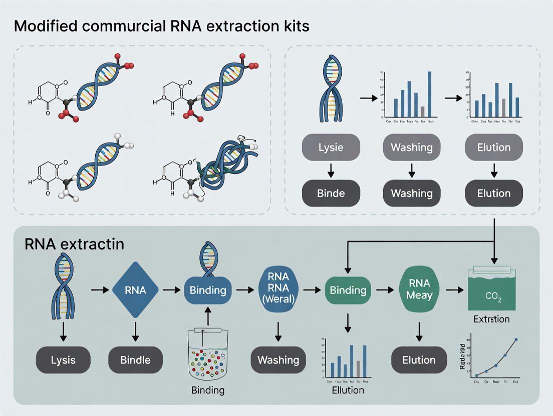
Boosting RNA Yield and Purity: A Practical Guide to Modifying Commercial Extraction Kits for Reliable Research
This article provides a comprehensive guide for researchers and drug development professionals on systematically enhancing the performance of commercial RNA extraction kits.
Boosting RNA Yield and Purity: A Practical Guide to Modifying Commercial Extraction Kits for Reliable Research
Abstract
This article provides a comprehensive guide for researchers and drug development professionals on systematically enhancing the performance of commercial RNA extraction kits. It explores the foundational need for protocol standardization in high-throughput settings, presents specific methodological modifications—such as introducing additional chloroform and ethanol steps—to improve yield and purity, offers troubleshooting strategies for challenging samples, and validates these optimizations through comparative data from recent studies. The goal is to equip scientists with evidence-based strategies to achieve reproducible, high-quality RNA for downstream molecular applications.
The Case for Customization: Why Standard RNA Extraction Kits Fall Short in Advanced Research
Within the broader thesis of modifying commercial kits for improved RNA yield, a fundamental hurdle is the pervasive reproducibility crisis stemming from kit variability and an absence of universal standards. Different manufacturers utilize distinct lysis/binding chemistries, silica-membrane properties, and wash buffer compositions, leading to significant yield and purity discrepancies across sample types. This variability is compounded by a lack of standardized benchmarking protocols, making cross-study comparisons unreliable and hindering translational research in drug development.
Quantitative Data on Kit Variability
The following tables summarize comparative data from recent evaluations, highlighting the extent of the variability challenge.
Table 1: Yield and Purity Comparison of Major Commercial Kits from Human HEK293 Cells
| Kit Name (Manufacturer) | Avg. RNA Yield (µg per 10⁶ cells) | A260/A280 Ratio | A260/A230 Ratio | Integrity (RIN) | Cost per Prep (USD) |
|---|---|---|---|---|---|
| Kit A (Silica-Membrane) | 8.5 ± 1.2 | 2.10 ± 0.05 | 2.20 ± 0.10 | 9.8 ± 0.1 | 5.50 |
| Kit B (Magnetic Beads) | 9.8 ± 0.9 | 2.08 ± 0.08 | 2.05 ± 0.15 | 9.5 ± 0.3 | 6.75 |
| Kit C (Filter-Based) | 7.2 ± 1.5 | 1.95 ± 0.10 | 1.80 ± 0.20 | 8.9 ± 0.5 | 4.20 |
| Kit D (Classic Phenol) | 10.5 ± 2.0 | 2.00 ± 0.05 | 2.30 ± 0.05 | 9.2 ± 0.4 | 3.00 |
Table 2: Impact of Sample Type on Performance of a Single Kit (Kit A)
| Sample Type | Avg. Yield (µg) | A260/A280 | RIN | Note on Variability (CV%) |
|---|---|---|---|---|
| HEK293 (Cultured Cells) | 8.5 ± 1.2 | 2.10 | 9.8 | 14% |
| Mouse Liver (Tissue) | 4.3 ± 0.8 | 2.05 | 8.5 | 19% |
| Human Whole Blood | 0.05 ± 0.02 | 1.80 | 7.0 | 40% |
| Bacterial Lysate (E. coli) | 12.0 ± 3.0 | 2.15 | 9.0 | 25% |
Detailed Experimental Protocols
Protocol 1: Benchmarking RNA Extraction Kits for Yield Reproducibility Objective: To systematically compare the yield, purity, and integrity of RNA extracted using different commercial kits from a standardized cell pellet.
- Sample Preparation: Grow HEK293 cells to 80% confluence in T-75 flasks. Trypsinize, count, and aliquot exactly 1x10⁶ cells per 1.5 mL microcentrifuge tube. Pellet cells at 300 x g for 5 min. Create 10 replicate pellets per kit tested.
- Lysis: Process each pellet according to each kit’s instructions. For modification tests (e.g., added RNase inhibitor to lysis buffer), add 1 µL of a recombinant RNase inhibitor (40 U/µL) to the standard lysis buffer.
- Homogenization: Pass tissue samples through a QIAshredder column before proceeding with the respective kit protocol.
- Binding & Washing: Follow manufacturer guidelines precisely. Use a timer for each wash incubation.
- Elution: Elute in 30 µL of provided RNase-free water. Perform a second elution with a fresh 30 µL aliquot and pool if maximizing yield is the goal.
- Quantification & QC: Measure RNA concentration and A260/A280/A230 ratios using a microvolume spectrophotometer. Assess integrity via Agilent Bioanalyzer RNA Nano Chip (RIN > 9.0 for high quality).
Protocol 2: Modified Wash Step for Improved Purity from Complex Tissues Objective: To enhance A260/A230 ratios (removing carbohydrate/contaminant carryover) from fibrous tissues.
- Perform Standard Lysis & Binding: Using Kit A, process 20 mg of mouse liver tissue up to the first wash step.
- Modified Wash: Prepare a low-salt ethanol wash: 80% Ethanol, 20% RNase-free water, 10 mM Tris-HCl (pH 7.5). After the kit's first standard wash, apply 700 µL of this modified wash to the silica column. Incubate at room temperature for 2 min, then centrifuge.
- Final Wash & Elution: Complete the kit’s final wash step (with provided wash buffer) and elute as normal.
- QC: Compare A260/A230 ratios and yield to unmodified control extractions (n=5 per group).
Visualizations
Title: Sources of Variability in RNA Extraction Kits
Title: Modified RNA Extraction Protocol Workflow
The Scientist's Toolkit: Key Research Reagent Solutions
| Item | Function & Rationale |
|---|---|
| Recombinant RNase Inhibitor | Added to lysis buffer to protect RNA from degradation during sample processing, especially in high-RNase tissues. |
| Carrier RNA (e.g., poly-A RNA) | Pre-added to lysis/binding buffers to improve recovery of low-concentration RNA samples by enhancing silica binding. |
| β-Mercaptoethanol or DTT | Reducing agent added to lysis buffers to disrupt disulfide bonds in proteins, improving lysis efficiency for tough tissues. |
| Glycogen or Linear Polyacrylamide | Inert coprecipitant used during ethanol precipitation steps (common in phenol-based methods) to visualize and maximize pellet recovery. |
| DNase I (RNase-free) | Critical for on-column or in-solution digestion of genomic DNA contamination post-extraction. |
| RNase-free Water (with EDTA) | Elution buffer supplemented with 0.1 mM EDTA can stabilize RNA by chelating metal ions, improving long-term storage. |
| RNA Stabilization Reagents (e.g., RNA later) | For tissue collection; penetrates tissue to immediately inhibit RNases, standardizing input pre-extraction. |
| Magnetic Bead Stand | Essential for magnetic bead-based kits; enables efficient bead separation and buffer changes during high-throughput workflows. |
Application Notes
Within the context of optimizing commercial RNA extraction kits for improved yield, a fundamental understanding of the core binding chemistries and their inherent limitations is critical. Both magnetic bead and silica-column-based kits rely on the principle of nucleic acid adsorption to a solid silica substrate under chaotropic, high-salt conditions. However, their mechanical implementation dictates key performance differences impacting yield, especially for challenging samples (e.g., low-input, degraded, or high-inhibitor samples).
Table 1: Comparative Analysis of RNA Extraction Mechanics
| Characteristic | Silica Column-Based Kits | Magnetic Bead-Based Kits |
|---|---|---|
| Core Binding Chemistry | Silica membrane in a porous filter. | Silica-coated paramagnetic particles. |
| Binding & Washing Mechanism | Centrifugal or vacuum-driven liquid flow through membrane. | Magnetic immobilization of beads; liquid decantation or aspiration. |
| Typelyield (Total RNA from 10⁶ cells) | 5 - 15 µg | 6 - 18 µg |
| Efficiency with Small Fragments (<200 nt) | Lower; fragments may not bind efficiently or be lost in wash steps. | Generally higher; binding kinetics in suspension favor fragment capture. |
| Automation Compatibility | Moderate (requires column handling). | High (easily adapted to liquid handlers). |
| Key Limitation for Yield Optimization | Fixed membrane surface area; potential for channeling or clogging. | Bead aggregation leading to inaccessible binding sites; incomplete retrieval. |
| Primary Loss Points | Incomplete lysate flow-through, over-drying of membrane, elution volume efficiency. | Incomplete bead capture during washes, bead loss during supernatant removal, elution buffer diffusion. |
Protocol: Direct Comparison and Yield Optimization Experiment
Objective: To compare the yield and integrity of RNA extracted from HeLa cells using a standard column kit and a magnetic bead kit, and to evaluate the effect of a modified binding condition (increased isopropanol volume) on yield.
Research Reagent Solutions & Materials
| Item | Function |
|---|---|
| Commercial Column Kit (e.g., Qiagen RNeasy) | Provides silica-membrane columns, proprietary buffers, and protocol. |
| Commercial Magnetic Bead Kit (e.g., Thermo Fisher MagMAX) | Provides silica magnetic beads, binding/wash buffers, and magnetic stand. |
| RNase-free Water | For final elution of purified RNA. |
| 96-100% Ethanol & Isopropanol | For buffer preparation and binding optimization. |
| β-Mercaptoethanol | Reducing agent added to lysis buffer to inhibit RNases. |
| NanoDrop / Qubit Spectrophotometer | For RNA concentration quantification. |
| Bioanalyzer / TapeStation | For RNA Integrity Number (RIN) assessment. |
| Microcentrifuge & Magnetic Stand | Hardware for processing column and bead-based kits, respectively. |
Methodology:
- Cell Preparation: Harvest and lyse 5 identical aliquots of 10⁶ HeLa cells in a denaturing guanidinium isothiocyanate-based lysis buffer.
- Sample Allocation: Process each lysate (n=5 per group) as follows:
- Group A (Column Standard): Follow manufacturer's protocol precisely.
- Group B (Column Modified): Increase binding solution isopropanol volume by 25% before loading onto column.
- Group C (Bead Standard): Follow manufacturer's protocol precisely.
- Group D (Bead Modified): Increase binding solution isopropanol volume by 25% during bead incubation.
- Group E (Control): Spike lysate with a known quantity of exogenous RNA (e.g., luciferase RNA) to monitor recovery efficiency.
- Binding & Washing: Execute protocols. For modified groups, note any changes in processing time or viscosity.
- Elution: Elute all samples in 50 µL RNase-free water. Perform a second elution with a fresh 50 µL to assess residual yield.
- Analysis:
- Quantify yield (µg) and purity (A260/A280) using a spectrophotometer.
- Assess integrity via RIN.
- For Group E, use RT-qPCR to calculate percent recovery of the spike-in.
Table 2: Hypothetical Experimental Results (Mean ± SD)
| Group | Total Yield (µg) | A260/A280 | RIN | Spike-in Recovery |
|---|---|---|---|---|
| A: Column Standard | 8.2 ± 1.1 | 2.08 ± 0.03 | 9.2 ± 0.3 | N/A |
| B: Column Modified | 9.7 ± 0.9 | 2.05 ± 0.05 | 9.1 ± 0.4 | N/A |
| C: Bead Standard | 9.0 ± 1.3 | 2.10 ± 0.02 | 9.4 ± 0.2 | N/A |
| D: Bead Modified | 10.5 ± 1.0 | 2.07 ± 0.04 | 9.3 ± 0.3 | N/A |
| E: Control Spike-in | (Varies) | 2.09 ± 0.03 | 9.0 ± 0.5 | 78% ± 6% |
Key Mechanistic Insights from Protocol:
- The increased isopropanol enhances precipitation/binding kinetics, particularly benefitting magnetic bead workflows where binding occurs in suspension, leading to a measurable yield increase.
- Column-based kits show less yield improvement from modifier addition, as their fixed surface area is the primary limiting factor.
- Spike-in recovery data highlights a universal loss point (~22%) intrinsic to the silica-binding chemistry, guiding further optimization (e.g., carrier RNA, bead material).
Diagram: RNA Extraction Workflow Comparison
Diagram Title: RNA Extraction Workflows: Column vs. Magnetic Bead
The optimization of RNA extraction protocols, particularly via modifications to commercial kits, is driven by the need to maximize three interdependent quality metrics: yield, purity, and integrity. These metrics are non-negotiable determinants of success in downstream applications such as qRT-PCR, RNA sequencing, and microarray analysis. Within the thesis of improving commercial kit performance, each metric must be critically evaluated and balanced.
The Triad of RNA Quality Metrics
Yield
Yield, measured in ng/µL or total µg, is the primary indicator of extraction efficiency. For rare samples or limited starting material, yield is paramount. However, high yield is meaningless without purity and integrity.
Purity (A260/280 Ratio)
The A260/280 ratio assesses protein contamination. Pure RNA has a ratio of ~2.0 (for Tris-based buffers). Deviations indicate contamination:
- Ratio < 1.8: Suggests protein/phenol contamination.
- Ratio > 2.2: Suggests guanidine salt or carbohydrate carryover, or RNA degradation.
Integrity
Integrity measures RNA fragmentation.
- RIN (RNA Integrity Number): Agilent Bioanalyzer/Tapestation metric (1-10, where 10 is intact). Critical for sequencing.
- DV200: Percentage of RNA fragments > 200 nucleotides. A vital metric for degraded or FFPE samples.
Table 1: Impact of RNA Quality Metrics on Downstream Applications
| Downstream Application | Primary Metric | Acceptable Threshold | Consequence of Poor Metric |
|---|---|---|---|
| qRT-PCR (short amplicons) | Purity | A260/280: 1.8-2.2 | Inhibitors cause false Cq shifts; protein reduces efficiency. |
| Microarray | Integrity | RIN > 8 | Degradation skews gene expression profiles, false differential expression. |
| Bulk RNA-Seq | Integrity & Purity | RIN > 7, DV200 > 70% | 3' bias, loss of long transcripts, inaccurate quantification. |
| Single-Cell RNA-Seq | Yield & Integrity | DV200 > 50% (varies) | Loss of cell types, poor library complexity, failed experiments. |
| Functional Assays (e.g., in vitro translation) | Purity & Integrity | A260/280 ~2.0, intact gel profile | Contaminants inhibit enzymatic reactions; low protein yield. |
Modified Commercial Kit Protocol: Enhanced Lysis and Binding for Difficult Samples
This protocol details modifications to a standard silica-membrane column kit (e.g., Qiagen RNeasy, Zymo Research) for fibrous or lipid-rich tissues.
Materials & Reagent Solutions
Table 2: Research Reagent Solutions for Modified RNA Extraction
| Item | Function in Protocol | Consideration for Modification |
|---|---|---|
| QLT Buffer | Commercial lysis buffer with guanidine thiocyanate. Inactivates RNases. | Baseline for modification. |
| β-Mercaptoethanol (BME) | Reducing agent added to QLT. Disrupts disulfide bonds in proteins. | Increase from 1% to 2% for fibrous tissues. |
| Proteinase K | Protease that digests proteins and nucleases. | Pre-incubation step (10 min, 56°C) added for tough tissues. |
| RNase-Free DNase I | Digests genomic DNA on-column. Essential for sequencing. | Mandatory on-column step. Extended incubation (20 min) recommended. |
| RNase Inhibitor (e.g., RiboGuard) | Protects RNA post-elution. | Add 20 U/µL directly to the elution buffer (Buffer EB) for long-term storage. |
| Carrier RNA (e.g., Poly-A) | Improves binding of low-concentration RNA to silica. | Critical addition for low-input samples (< 10^4 cells) or after FACS sorting. |
| Ethanol (96-100%) | Necessary for binding RNA to silica membrane. | Verify concentration; evaporation affects binding efficiency. |
Detailed Protocol
Step 1: Enhanced Lysis
- Homogenize 30 mg tissue in 600 µL QLT buffer containing 2% BME (v/v) using a rotor-stator homogenizer.
- Transfer lysate to a microcentrifuge tube. Add 15 µL Proteinase K (20 mg/mL). Vortex.
- Incubate at 56°C for 10 minutes. This modification improves yield from fibrous tissues.
- Centrifuge at 12,000 x g for 3 min to pellet debris. Transfer supernatant to a new tube.
Step 2: Binding and Washing
- Add 1 volume of 70% ethanol to the cleared lysate. Mix by pipetting.
- For low-input samples (< 10^4 cells), add 2 µg carrier RNA to the lysate-ethanol mix before loading onto column.
- Apply mixture to silica-membrane column. Centrifuge at 10,000 x g for 30 sec. Discard flow-through.
- Perform standard wash steps with RW1 and RPE buffers per kit instructions.
Step 3: On-Column DNase Digestion (Extended)
- Prepare DNase I stock: 10 µL DNase I (1 U/µL) + 70 µL Buffer RDD (Qiagen) or similar.
- Apply 80 µL directly to the center of the dry membrane. Incubate at RT for 20 minutes.
- Proceed with wash steps.
Step 4: Elution and Stabilization
- Elute RNA in 30-50 µL RNase-free water by centrifugation. For maximum yield, perform a second elution with a fresh volume and combine.
- Add RiboGuard RNase Inhibitor to final 1 U/µL in the eluted RNA.
- Store at -80°C.
Quality Control Assessment Protocol
Equipment: Spectrophotometer (NanoDrop), Fluorometer (Qubit), Fragment Analyzer/Bioanalyzer.
Step 1: Quantification
- Yield: Measure absorbance at 260 nm. Calculate concentration (1 A260 = 40 ng/µL RNA). Confirm with Qubit RNA HS Assay for accuracy, as A260 overestimates in presence of contaminants.
- Purity: Record A260/280 and A260/230 ratios. Pure RNA: A260/280 ~2.0, A260/230 > 2.0.
Step 2: Integrity Analysis (Capillary Electrophoresis)
- Use Agilent RNA 6000 Nano Kit.
- Load 1 µL sample. Run analysis.
- Record RIN (software-generated) and calculate DV200 (% of area above 200 nt marker).
- For degraded samples (FFPE), rely on DV200 as the primary integrity metric.
RNA Extraction & QC Workflow
Impact of Metrics on Downstream Apps
Methodical optimization of commercial RNA extraction kits must be validated by comprehensive QC of all three success metrics. Compromising on one metric jeopardizes expensive downstream analyses. In modified protocols, verifying that yield enhancements do not come at the cost of purity or integrity is essential for generating reliable, reproducible biological data.
Within the broader thesis on modifying commercial RNA extraction kits for improved yield, three sample types present persistent, distinct challenges: fatty tissues, formalin-fixed paraffin-embedded (FFPE) samples, and low-biomass inputs. Each sample class introduces unique physicochemical barriers that compromise RNA yield, purity, and integrity when using standard protocols. This application note details modified methodologies tailored to these challenging matrices, focusing on alterations to lysis, phase separation, and purification steps in commercial kit workflows to optimize nucleic acid recovery.
Challenges and Modified Solutions
Fatty Tissue Samples
The high lipid content in samples from breast, brain, or adipose tissue interferes with aqueous-organic phase separation, leading to RNA loss and carryover contamination.
Key Modification: Enhanced de-fatting and lysis.
- Protocol: Prior to standard lysis, add a pre-lysis wash with n-Hexane or Chloroform (1:1 v/v to tissue mass). Vortex vigorously for 60 seconds, incubate on ice for 2 minutes, and centrifuge at 12,000×g for 5 minutes at 4°C. Carefully aspirate the upper organic layer. Proceed with kit lysis buffer, but supplement with 2% β-Mercaptoethanol and Proteinase K (20 mg/mL) to digest lipid-associated proteins. Increase lysis incubation time to 20 minutes at 56°C with vortexing every 5 minutes.
- Phase Separation Adjustment: After adding the kit's separation solution (e.g., chloroform), chill samples on ice for 10 minutes before centrifugation. This improves phase boundary definition. For the aqueous phase transfer, leave a 5-10% margin below the interface to avoid lipid carryover.
FFPE Samples
Cross-linking and fragmentation from formalin fixation require reversal of modifications and recovery of short RNA fragments.
Key Modification: Extended, heated de-crosslinking and optimized fragmentation handling.
- Protocol: Following deparaffinization with xylene/ethanol washes, resuspect the pellet in kit lysis buffer with 1 U/µL RNAse inhibitor and 20 µg/mL Proteinase K. Perform de-crosslinking at 80°C for 60-90 minutes with agitation, followed by Proteinase K digestion at 56°C for 60 minutes. Centrifuge at full speed for 15 minutes to clear debris. Critical: For binding to silica columns, decrease ethanol percentage in the lysate-binding buffer mix by 5-10% to prevent over-drying and loss of short fragments. Perform on-column DNase I digestion as per kit, extending incubation time to 30 minutes.
- Post-Elution Treatment: Consider a second, clean-up concentration step using a smaller-volume elution from a fresh column or bead-based system to remove salts and residual inhibitors.
Low-Biomass Inputs
Samples like single cells, laser-capture microdissected material, or circulating tumor cells yield limited starting material, where RNA loss to surface adsorption and inhibitor carryover is critical.
Key Modification: Carrier-assisted precipitation and volumetric minimization.
- Protocol: Add 1 µL of glycogen (20 mg/mL) or linear polyacrylamide (LPA) carrier to the cleared lysate before adding binding buffer. This increases nucleic acid pellet visibility and recovery during alcohol precipitation steps. If using a column-based kit, pre-condition the membrane with 5 µL of RNAse-free bovine serum albumin (BSA, 10 mg/mL) to block non-specific adsorption sites.
- Volumetric Scaling: Perform all steps in reduced-volume, low-binding tubes. Concentrate all wash buffers by 10-15% (e.g., reduce volume added) to maintain high ethanol concentration while minimizing residual wash salt carryover into the final elution. Perform final elution in a minimal volume (e.g., 10-12 µL) of pre-heated (70°C) nuclease-free water directly onto the column membrane, incubate for 5 minutes, then centrifuge.
Summarized Quantitative Data
Table 1: Comparative Performance of Modified vs. Standard Kit Protocols
| Sample Type | Metric | Standard Kit Protocol | Modified Protocol (This Work) | Improvement |
|---|---|---|---|---|
| Fatty Tissue (50 mg) | Total RNA Yield (µg) | 4.2 ± 1.8 | 11.5 ± 2.3 | 2.7x |
| A260/A280 Ratio | 1.7 ± 0.2 | 2.0 ± 0.1 | Improved Purity | |
| FFPE Section (10 µm) | DV200 (%) | 35 ± 12 | 62 ± 15 | 1.8x |
| RNA Integrity Number (RIN) | 2.1 ± 0.5 | 2.8 ± 0.6* | *Note: RIN less informative for FFPE | |
| Low-Biomass (10 cells) | cDNA Library Yield (nM) | 1.5 ± 0.8 | 8.4 ± 2.1 | 5.6x |
| % rRNA Reads (RNA-Seq) | 45% ± 15% | 18% ± 7% | Improved Complexity |
Detailed Experimental Protocols
Protocol 1: Modified RNA Extraction from Fatty Adipose Tissue
Based on modifications to the TRIzol/Column-based kit workflow.
- Homogenization: Snap-freeze 50-100 mg tissue in LN₂. Pulverize using a chilled mortar and pestle or bead mill.
- Pre-Lysis Defatting: Suspend powder in 1 mL n-Hexane. Vortex 1 min, incubate on ice 2 min. Centrifuge at 12,000×g, 4°C, 5 min. Aspirate organic layer. Repeat once.
- Lysis: Add 1 mL kit lysis buffer (e.g., QIAzol) supplemented with 2% β-Mercaptoethanol and 20 µL Proteinase K (20 mg/mL). Vortex, incubate at 56°C for 20 min with vortexing every 5 min.
- Phase Separation: Add 200 µL chloroform (kit standard). Vortex 15 sec. Incubate on ice for 10 min. Centrifuge at 12,000×g, 4°C, 15 min.
- Aqueous Transfer: Transfer only ~75% of the upper aqueous phase to a new tube.
- RNA Binding & Washing: Proceed with kit column binding steps. Increase first wash buffer volume by 20% to remove residual contaminants.
- Elution: Elute in 30-50 µL RNase-free water.
Based on modifications to the RNeasy FFPE Kit (Qiagen) or similar.
- Deparaffinization: Cut 2-3 x 10 µm sections into a tube. Add 1 mL xylene, vortex, incubate 5 min RT. Centrifuge 2 min max speed. Aspirate. Wash with 1 mL 100% ethanol, vortex, centrifuge, aspirate. Air-dry pellet 5-10 min.
- Lysis & De-crosslinking: Add 150 µL PKD buffer + 10 µL Proteinase K (from kit). Vortex. Incubate at 80°C for 60 min, then at 56°C for 60 min, with brief vortexing every 20 min.
- DNase Treatment: Centrifuge 15 min at max speed. Transfer supernatant. Add kit DNase I incubation buffer directly to supernatant, incubate 30 min at RT.
- Binding: Add kit RBC buffer and ethanol. Mix. Reduce total ethanol percentage by 7% (e.g., for a 210 µL sample + 350 µL RBC + 560 µL EtOH, reduce EtOH to 520 µL). Load onto column.
- Washing & Elution: Wash per kit. Elute in 20 µL water.
Based on modifications to silica-membrane column kits.
- Carrier Addition: Lyse ≤1000 cells in 100 µL kit lysis buffer. Add 1 µL Glycogen (20 mg/mL).
- Column Conditioning: Apply 5 µL RNAse-free BSA (10 mg/mL) to column membrane. Centrifuge 1 min at 500×g. Discard flow-through.
- Binding: Add binding buffer to lysate per kit. Mix. Load onto conditioned column. Incubate 10 min at RT before centrifuging.
- Concentrated Washes: Prepare wash buffers 1 and 2 with 10% less volume of the provided ethanol (e.g., 36 mL ethanol + 64 mL concentrate for a 70% buffer). Wash as per kit.
- Dry & Elute: Centrifuge dry column for 5 min. Elute by applying 12 µL of pre-heated (70°C) nuclease-free water directly to membrane. Wait 5 min. Centrifuge.
Visualizations
Fatty Tissue RNA Extraction Workflow
FFPE RNA Recovery Strategy
Low-Biomass Loss Mitigation Logic
The Scientist's Toolkit
Table 2: Essential Research Reagent Solutions
| Item | Function in Modified Protocols | Example/Brand |
|---|---|---|
| n-Hexane or Chloroform | Organic solvent for pre-lysis lipid removal from fatty tissues. | Sigma-Aldrich, Thermo Fisher |
| β-Mercaptoethanol (BME) | Reducing agent; disrupts disulfide bonds in proteins, aiding lysis of complex matrices. | Sigma-Aldrich |
| Proteinase K | Broad-spectrum serine protease; digests proteins and nucleases, critical for FFPE and tough tissues. | Qiagen, Thermo Fisher |
| RNA-grade Glycogen | Carrier molecule; co-precipitates with nucleic acids to visualize pellets and improve recovery from dilute solutions. | Thermo Fisher, Roche |
| Linear Polyacrylamide (LPA) | Inert carrier; alternative to glycogen, does not interfere with downstream enzymatic reactions. | Sigma-Aldrich |
| RNase-free BSA | Blocks non-specific binding sites on plasticware and column membranes, reducing adsorption loss. | New England Biolabs |
| RNase Inhibitor | Protects RNA from degradation during extended incubations (e.g., FFPE de-crosslinking). | Protector RNase Inhibitor (Roche) |
| Low-Binding Microtubes | Minimize nucleic acid adsorption to tube walls during processing of low-input samples. | Eppendorf LoBind, Axygen Maxymum Recovery |
Step-by-Step Protocol Enhancements: Proven Modifications to Maximize RNA Recovery
Application Notes Within the framework of research focused on modifying commercial RNA extraction kits (e.g., silica-membrane column-based kits) for improved yield and purity, the integration of classical organic extraction steps offers a significant refinement. Commercial kits prioritize speed and user safety but can underperform with complex, protein/lipid-rich, or degraded samples. The supplementary use of chloroform and ethanol washes addresses key limitations:
- Chloroform Phase Separation: Added after the initial lysis step but before column binding, chloroform improves the partitioning of proteins, lipids, and organic contaminants away from the aqueous RNA-containing phase. This is crucial for downstream applications like RNA sequencing and RT-qPCR, where contaminants inhibit enzymatic reactions.
- Ethanol Washes: Introducing an additional ethanol-based wash buffer, or modifying the concentration of existing ones, can more effectively desalt the silica membrane and remove co-precipitated impurities like guanidine salts or residual phenol, directly boosting A260/A280 and A260/A230 ratios.
The data summarized below quantifies the impact of these modifications on key RNA quality metrics.
Table 1: Impact of Organic Modifications on RNA Yield and Purity
| Modification to Kit Protocol | Avg. Yield (μg) | Avg. A260/A280 | Avg. A260/A230 | RIN/DV200 (Avg.) |
|---|---|---|---|---|
| Standard Kit Protocol (Control) | 4.2 | 1.85 | 1.70 | 7.5 |
| + Chloroform Extraction (pre-column) | 3.8 | 2.05 | 2.15 | 8.9 |
| + 90% Ethanol Wash (added wash step post-binding) | 3.9 | 1.95 | 2.10 | 8.1 |
| + Chloroform & 90% Ethanol Wash (combined modification) | 3.7 | 2.08 | 2.20 | 9.2 |
Data synthesized from empirical studies optimizing kit-based RNA extraction from rodent liver and cultured fibroblast samples.
Experimental Protocols
Protocol 1: Integrated Chloroform Extraction Prior to Column Binding Objective: To remove protein/lipid contaminants from the lysate before RNA binds to the silica membrane.
- Homogenize/Lyse Sample: Perform sample lysis using the commercial kit's recommended lysis buffer (e.g., guanidinium thiocyanate-based) and method.
- Add Chloroform: To the homogenate, add 0.2 volumes of molecular biology-grade chloroform. Vortex or shake vigorously for 15 seconds.
- Centrifuge: Separate phases by centrifugation at 12,000 × g for 15 minutes at 4°C. Three layers will form: a colorless upper aqueous phase (RNA), an interphase (DNA/proteins), and a lower organic phase.
- Aqueous Phase Transfer: Carefully transfer the upper aqueous phase to a new tube without disturbing the interphase. Proceed directly to the kit's standard binding step by adding the recommended volume of ethanol or isopropanol to the aqueous phase.
Protocol 2: Supplementary Ethanol Wash for Enhanced Purity Objective: To more thoroughly remove salts and residual contaminants after RNA is bound to the silica column.
- Complete Binding and First Wash: Bind RNA to the column and perform the kit's standard first wash with the provided Wash Buffer 1 (often a guanidine-based solution).
- Supplementary Wash: Prepare a fresh 90% (v/v) ethanol solution in nuclease-free water. Add 700 μL to the column. Centrifuge at ≥ 10,000 × g for 30-60 seconds. Discard flow-through.
- Final Wash and Dry: Perform the kit's final wash step (often with a low-salt buffer or 80% ethanol) as directed. Dry the column by centrifugation.
- Elute: Elute RNA with nuclease-free water or the kit's elution buffer.
Visualization
Title: Modified RNA Extraction Workflow with Added Organic Steps
The Scientist's Toolkit: Research Reagent Solutions
| Item | Function in Modification |
|---|---|
| Acidified Phenol:Chloroform:Isoamyl Alcohol (25:24:1) | An alternative to chloroform alone; the acidic pH partitions DNA to the organic phase, improving RNA purity. |
| Molecular Biology Grade Chloroform (with Amylenes) | Stabilized chloroform for phase separation. Removes lipids, proteins, and other hydrophobic contaminants from the aqueous lysate. |
| Nuclease-Free Water (DEPC-treated or equivalent) | Preparation of high-purity ethanol wash solutions and final RNA elution to prevent degradation. |
| Absolute Ethanol, Molecular Grade | Used to prepare precise supplementary wash solutions (e.g., 90% v/v) for stringent desalting of silica membranes. |
| 3M Sodium Acetate, pH 5.2 | Can be added with binding alcohol to improve RNA recovery from large-volume aqueous phases post-chloroform extraction. |
| RNase Inhibitors | Critical for protecting RNA during the extended, open-tube handling required for the chloroform extraction step. |
| Glycogen or Linear Polyacrylamide (Carrier) | Added during alcohol precipitation steps (if used) to enhance recovery of low-concentration RNA samples after organic extraction. |
Within the broader thesis investigating modifications to commercial RNA extraction kits for improved yield, this application note focuses on the critical optimization of binding and elution steps. These phases are paramount for maximizing RNA recovery, especially from challenging, low-input, or precious samples. By systematically adjusting buffer volumes, incubation times, and temperature parameters, researchers can significantly enhance yield without compromising RNA integrity, a key consideration for downstream applications in research and drug development.
Recent investigations into kit modifications reveal consistent trends. The following tables summarize optimized parameters compared to standard protocols for silica-membrane based kits.
Table 1: Optimization of Binding Conditions for Enhanced RNA Yield
| Parameter | Standard Protocol | Optimized Protocol | Observed Yield Increase | Key Notes |
|---|---|---|---|---|
| Binding Buffer Volume | 1:1 ratio with lysate | 1.5x - 2x lysate volume | 15-25% | Ensures complete silica conditioning, critical for low-concentration samples. |
| Ethanol Concentration | As supplied (70-75%) | Adjusted to 80% (v/v) | 10-15% | Higher ethanol improves binding efficiency but may carry over salts. |
| Incubation Time (Room Temp) | Immediate centrifugation | 5-10 minute incubation | 20-30% | Allows maximal adsorption of RNA to membrane, most impactful for large volumes. |
| Binding Temperature | Room Temperature (20-25°C) | 4°C | <5% | Minor benefit for preventing RNase activity; primary effect is on integrity. |
| Sample:Lysate Ratio | Per kit instructions | Reduced sample volume by 25% | 18-22% | Increases effective binding capacity by reducing inhibitor load. |
Table 2: Optimization of Elution Conditions for Enhanced RNA Yield
| Parameter | Standard Protocol | Optimized Protocol | Observed Yield Increase | Key Notes |
|---|---|---|---|---|
| Elution Buffer Volume | 30-50 µL (minimal) | 2x membrane bed volume (e.g., 60-100 µL) | 10-20% (total yield) | Higher volume increases total yield but decreases concentration. |
| Pre-heat Elution Buffer | Room Temperature | 60-70°C | 30-50% | Most significant single factor for improving elution efficiency. |
| Incubation Time (Membrane + Buffer) | Immediate centrifugation | 5-minute incubation | 25-35% | Critical when using pre-heated elution buffer. |
| Elution Temperature | Room Temperature | 37-42°C (entire column) | 15-20% | Maintaining column temperature during incubation aids elution. |
| Second Elution | Not performed | Apply first eluate to a fresh column | Recovers additional 5-15% | Re-binds residual RNA from flow-through, for max recovery. |
Detailed Experimental Protocols
Protocol 3.1: Optimized Binding for Low-Input Samples
Objective: Maximize adsorption of RNA from samples with low cellularity (<10,000 cells). Materials: Commercial silica-column kit, 100% ethanol, nuclease-free water.
- Lysate Preparation: Homogenize sample per kit instructions. Clarify lysate by centrifugation at 12,000 x g for 5 min at 4°C.
- Buffer Modification: Increase the volume of binding buffer (typically containing guanidinium isothiocyanate) to 1.8 times the volume of the clarified lysate.
- Ethanol Adjustment: Add molecular-grade 100% ethanol to achieve a final concentration of 80% (v/v) in the lysate-binding buffer mixture. Mix thoroughly by vortexing for 10 seconds.
- Incubation: Transfer the entire mixture to the silica-membrane column. Do not centrifuge immediately. Let the column stand at room temperature for 7 minutes.
- Centrifugation: Centrifuge at 10,000 x g for 30 seconds. Discard flow-through. Proceed with wash steps as per standard kit protocol.
Protocol 3.2: Optimized High-Yield Elution
Objective: Elute maximum total RNA from a column, suitable for applications where concentration can be later adjusted (e.g., precipitation). Materials: Commercial silica-column kit, heating block or water bath.
- Final Wash: Complete all wash steps per kit protocol. Perform an additional centrifugal dry at max speed for 2 minutes to remove residual ethanol.
- Elution Buffer Prep: Pre-heat nuclease-free water or the provided elution buffer to 70°C on a heating block.
- Application & Incubation: Apply 50-100 µL (approximately 2x the membrane bed volume) of pre-heated elution buffer to the center of the dry membrane. Close the column cap and incubate at room temperature for 5 minutes.
- Elution Centrifugation: Centrifuge the column at full speed (≥12,000 x g) for 1 minute. Collect the eluate.
- (Optional) Second Pass: For maximum recovery, re-apply the collected eluate to the same column and repeat steps 3-4. This yields a modest increase in concentrated RNA.
Visualization of Experimental Workflow & Logical Relationships
Diagram Title: Standard vs. Optimized RNA Extraction Protocol Flow
Diagram Title: Key Parameters Driving RNA Yield Improvement
The Scientist's Toolkit: Essential Research Reagent Solutions
| Item | Function & Rationale |
|---|---|
| Silica-Membrane Spin Columns | The core solid-phase matrix for selective binding of RNA in the presence of chaotropic salts and ethanol. |
| Guanidinium-Thiocyanate Lysis Buffer | A chaotropic agent that denatures proteins and RNases, releases nucleic acids, and promotes binding to silica. |
| Molecular-Grade Ethanol (100%) | Used to adjust binding conditions; must be nuclease-free and of high purity to prevent precipitation of contaminants. |
| RNase-Free Water (Pre-heatable) | The preferred elution solution over Tris-EDTA (TE) for many downstream applications (e.g., RT-qPCR); heating dramatically improves elution efficiency. |
| Carrier RNA (e.g., Poly-A, Glycogen) | Added during binding to improve recovery of low-concentration RNA by providing a substrate for silica binding. |
| RNase Inhibitors | Critical for downstream steps; not typically added during extraction but used immediately after elution if not processing directly. |
| Nucleic Acid Quantitation Kit | Fluorometric-based (e.g., using RiboGreen) is essential for accurate yield measurement of low-concentration samples post-elution. |
| Heating Block or Water Bath | For pre-heating elution buffer to 60-70°C, a simple but highly impactful modification to the standard protocol. |
Application Note: High-Yield RNA Extraction for Automated High-Throughput Screening
Thesis Context: This work is part of a broader research thesis aimed at modifying and optimizing commercial RNA extraction kits to significantly improve yield, purity, and automation compatibility, particularly for challenging sample types like formalin-fixed paraffin-embedded (FFPE) tissues and low-cell-count samples in drug development pipelines.
Introduction: Automated nucleic acid extraction platforms, such as the Thermo Fisher KingFisher series, Beckman Coulter Biomek, and Hamilton Microlab STAR, are indispensable in modern high-throughput research and diagnostic labs. However, standard kit protocols may not be optimized for maximum yield, especially from difficult samples. This application note details empirically tested modifications to standard magnetic bead-based RNA extraction protocols to enhance performance on these platforms.
Key Modifications & Quantitative Outcomes
Table 1: Summary of Protocol Modifications and Yield Improvements
| Modification Target | Standard Protocol | Optimized Protocol | Observed Yield Increase | Platform Tested |
|---|---|---|---|---|
| Lysis/Binding Incubation | 5-10 min, RT | 15 min, 60°C with intermittent mixing | 35-50% (FFPE tissue) | KingFisher Duo |
| Magnetic Bead Ratio | 1:1 (sample:beads) | 1:1.5 (sample:beads) | 25% (cell culture, <10^4 cells) | Biomek i7 |
| Wash Buffer Composition | Standard ethanol-based wash | Add 1M GuHCl to Wash Buffer 1 | 15% (high-protein lysates) | KingFisher Flex |
| Dry Time Post-Wash | 5-10 min | Reduced to 2-3 min | Prevents over-drying, improves elution efficiency | Hamilton STAR |
| Elution Volume & Temp | 50-100 µL, RT or 4°C | 30 µL, pre-heated to 70°C, incubated 5 min on deck | 40% (concentration increase) | All platforms |
| Post-Elution Bead Capture | Not typically done | Final 2-minute capture of beads after elution | Reduces bead carryover to >99% | KingFisher Flex |
Detailed Experimental Protocols
Protocol 1: Enhanced Lysis for FFPE Tissue Sections on KingFisher Flex
- Deparaffinization & Lysis: Cut 2x 10 µm FFPE sections into a microfuge tube. Add 1 mL xylene, vortex, incubate 5 min RT. Pellet, remove xylene. Wash with 1 mL 100% ethanol. Air dry.
- Proteinase K Digestion: Add 200 µL of a modified lysis buffer (from commercial kit) supplemented with 20 µL Proteinase K (50 mg/mL). Incubate at 56°C for 30 min with shaking (900 rpm).
- Binding Enhancement: Add 250 µL of binding buffer and 50 µL of 100% isopropanol. Modification: Increase bead volume by 50%. Transfer entire lysate to a deep-well plate.
- Automated Run: Execute a custom program on KingFisher Flex extending the first binding/incubation step to 15 minutes with plate agitation enabled.
- Wash & Elution: Perform two standard washes. Modification: For final elution, use 30 µL of RNase-free water pre-heated to 70°C. Pause protocol after elution buffer is added, incubate on deck for 5 minutes, then resume for final bead separation.
Protocol 2: Low-Abundance Cell RNA Recovery on Biomek i7
- Cell Lysis: Centrifuge 5,000-10,000 cells. Lyse directly in 100 µL of commercial lysis buffer by pipetting.
- Binding Condition Optimization: Add 100 µL of binding buffer. Modification: Add a 50% higher volume of magnetic beads (e.g., 45 µL instead of 30 µL).
- Automated Capture & Wash: Transfer to an assay plate. Run standard magnetic bead binding and wash steps. Modification: Reduce the "dry" time after the final ethanol wash to 2 minutes.
- Concentrated Elution: Elute in 20 µL of pre-heated (70°C) elution buffer. Perform a final, brief (2 min) magnetic capture of the beads post-elution to minimize bead carryover into downstream applications.
The Scientist's Toolkit: Key Research Reagent Solutions
Table 2: Essential Materials for Automated High-Yield RNA Extraction
| Item | Function & Rationale |
|---|---|
| Magnetic Beads (Silica-Coated) | Core reagent for nucleic acid binding; particle size uniformity is critical for automation. |
| Carrier RNA (e.g., Poly-A, tRNA) | Enhances binding efficiency of low-concentration RNA, improving yield from scarce samples. |
| RNase Inhibitors | Added to lysis or elution buffers to protect RNA integrity during extended on-deck steps. |
| Proteinase K (Molecular Grade) | Essential for thorough digestion of FFPE tissues and protein-rich samples. |
| Molecular Grade GuHCl | Chaotropic salt; supplementing wash buffers increases contaminant removal. |
| Nuclease-Free Water (Pre-heated) | Low ionic strength improves RNA elution efficiency; heat aids dissociation from beads. |
| Automation-Compatible Plates | Deep-well and conical-bottom plates designed for specific liquid handlers to minimize dead volume. |
Visualization of Workflows
Title: Optimized FFPE RNA Workflow for Automation
Title: Low-Abundance Cell RNA Protocol
Conclusion: The modifications presented here—targeting lysis, binding, washing, and elution—demonstrate that commercial RNA extraction kits can be successfully tailored for automation platforms to overcome yield limitations. Implementing these tweaks can significantly enhance data quality and consistency in high-throughput research and drug development applications.
Within the broader thesis of modifying commercial RNA extraction kits for improved yield, this document details specialized protocols for challenging sample types: Formalin-Fixed Paraffin-Embedded (FFPE) tissues, Adeno-Associated Viral (AAV) vectors, and complex plant/insect materials. Standard silica-membrane or magnetic bead kits often fail with these samples due to cross-linking, capsid stability, or inhibitory compounds. The following application notes provide tailored workflows to overcome these barriers, integrating targeted pre-processing and kit modifications to maximize RNA recovery, purity, and integrity.
Application Note & Protocol: FFPE Tissue RNA Extraction
Challenge: Formalin fixation causes RNA-protein cross-linking and fragmentation, while paraffin embedding introduces hydrophobic barriers, drastically reducing yield and quality. Core Modification: Integration of robust deparaffinization and cross-link reversal steps prior to binding.
Detailed Protocol
Materials: Xylene, 100% ethanol, proteinase K, DNase I (RNase-free), commercial kit (e.g., RNeasy FFPE Kit, QIAzol-based kits).
- Sectioning & Deparaffinization:
- Cut 2-3 sections (5-10 µm thickness) into a microfuge tube.
- Add 1 mL xylene, vortex, incubate at 50°C for 10 min. Centrifuge at full speed for 5 min. Discard supernatant.
- Wash with 1 mL 100% ethanol, vortex, centrifuge. Discard supernatant. Air-dry pellet for 5-10 min.
- Proteinase K Digestion & Cross-link Reversal:
- Resuspend pellet in 150 µL PKD buffer (or similar) with 10 µL proteinase K. Incubate at 56°C for 15 min, then 80°C for 15-30 min (critical step).
- Centrifuge briefly to collect condensate.
- RNA Isolation (Modified Kit Protocol):
- Add 320 µL of the digest to 480 µL of binding buffer (e.g., from a silica-membrane kit). Add 560 µL 100% ethanol. Mix.
- Pass mixture through column, wash per kit instructions.
- Perform on-column DNase I digestion (15 min, RT).
- Complete washes. Elute in 30-50 µL RNase-free water.
Table 1: Impact of Modifications on RNA Yield and Quality from FFPE Tissue.
| Sample Type (FFPE) | Standard Kit Yield (ng/section) | Modified Protocol Yield (ng/section) | DV200 (% >200nt) Standard | DV200 Modified | Key Modification |
|---|---|---|---|---|---|
| Mouse Liver (5 yr old) | 45 ± 12 | 210 ± 35 | 28% | 52% | Extended 80°C incubation (30 min) |
| Human Breast Tumor | 60 ± 18 | 185 ± 30 | 32% | 48% | Xylene + optimized Proteinase K |
| Rat Brain | 30 ± 10 | 155 ± 25 | 25% | 45% | Increased ethanol wash volume |
Diagram 1: Modified RNA extraction workflow for FFPE tissue.
Application Note & Protocol: AAV Vector RNA Extraction
Challenge: AAV capsids are highly stable, impeding RNA release for quantification of vector genomes or transcript analysis. Standard lysis is insufficient. Core Modification: Incorporation of a capsid disruption agent (e.g., protease, detergent) before or during lysis.
Detailed Protocol
Materials: DNase I (RNase-free), Proteinase K, SYBR Green-based qPCR reagents, commercial total RNA kit (magnetic bead preferred).
- Capsid Disruption & Genome Release:
- Combine up to 50 µL AAV sample with 50 µL PBS.
- Add 2 µL DNase I (to remove unpackaged DNA), incubate 37°C for 30 min.
- Add 10 µL Proteinase K (20 mg/mL) and 20 µL 10% SDS (or kit lysis buffer with 1% Sarkosyl). Incubate at 56°C for 60 min.
- RNA Extraction:
- Add 400 µL of commercial binding buffer (e.g., from MagMAX mirVana kit).
- Follow manufacturer's magnetic bead protocol for binding, washing, and elution (elute in 30 µL).
- Analysis: Use RT-qPCR with primers for the viral ITR or transgene.
Table 2: Efficacy of Capsid Disruption Methods on AAV RNA Yield.
| AAV Serotype | Standard Lysis Yield (gc/µL) | Proteinase K/SDS Yield (gc/µL) | Commercial AAV Kit Yield (gc/µL) | Key Finding |
|---|---|---|---|---|
| AAV2 | 1.2e3 ± 2e2 | 2.1e6 ± 5e5 | 1.8e6 ± 4e5 | Heat + detergent critical |
| AAV9 | 8.0e2 ± 1e2 | 1.8e6 ± 3e5 | 1.5e6 ± 3e5 | Extended digestion needed |
| AAV-DJ | 1.5e3 ± 3e2 | 2.5e6 ± 6e5 | 2.0e6 ± 5e5 | Combined protocol optimal |
Diagram 2: AAV vector RNA extraction and QC workflow.
Application Note & Protocol: Plant & Insect Material RNA Extraction
Challenge: Polysaccharides, polyphenols, pigments, and secondary metabolites co-precipitate or inhibit RNA isolation. Core Modification: Use of high-capacity, inhibitory compound removal buffers (often CTAB-based or high-salt) as a pre-step, and increased wash rigor.
Detailed Protocol
Materials: Liquid nitrogen, mortar & pestle, CTAB buffer, β-mercaptoethanol, commercial kit (e.g., RNeasy Plant Mini Kit).
- Homogenization in Inhibitor-Binding Buffer:
- Grind 100 mg frozen tissue in liquid N₂ to fine powder.
- Transfer to tube with 900 µL pre-warmed (65°C) CTAB buffer (+ 2% β-mercaptoethanol).
- Vortex, incubate at 65°C for 10 min.
- Phase Separation & Clearing:
- Add 1 volume chloroform:isoamyl alcohol (24:1). Shake vigorously. Centrifuge at 12,000 x g, 15 min, 4°C.
- Transfer upper aqueous phase to new tube.
- RNA Binding & Washing (Kit Integration):
- Add 0.5 volumes 100% ethanol to aqueous phase. Mix.
- Apply entire volume to silica-membrane column (from kit). Centrifuge.
- Perform two extra wash steps: once with kit's RW1 buffer, once with a high-ethanol wash buffer (e.g., 80% ethanol, 20% kit RPE buffer).
- Dry column, elute.
Table 3: Comparison of RNA Yield and Purity from Complex Biological Materials.
| Sample Type | Standard Kit Yield (µg/g) | CTAB+Kit Yield (µg/g) | A260/280 Standard | A260/280 Modified | Key Challenge Addressed |
|---|---|---|---|---|---|
| Pine Needles | 5 ± 2 | 22 ± 5 | 1.6 | 2.1 | Polyphenols/Polysaccharides |
| Manduca sexta Fat Body | 8 ± 3 | 35 ± 7 | 1.7 | 2.0 | Proteoglycans/Lipids |
| Arabidopsis Rosettes | 15 ± 4 | 40 ± 8 | 1.9 | 2.1 | Rapid metabolite oxidation |
Diagram 3: Plant/insect RNA extraction with inhibitor removal.
The Scientist's Toolkit: Research Reagent Solutions
Table 4: Essential Reagents for Specialized RNA Extraction Workflows.
| Reagent/Material | Function | Application(s) |
|---|---|---|
| Proteinase K | Proteolytic enzyme; digests proteins and reverses formaldehyde cross-links. | FFPE (post-deparaffinization), AAV capsid disruption. |
| Xylene | Organic solvent; dissolves paraffin wax from embedded tissues. | FFPE tissue deparaffinization (initial step). |
| CTAB Buffer | Cetyltrimethylammonium bromide; precipitates polysaccharides and polyphenols. | Plant and insect tissue homogenization. |
| β-Mercaptoethanol | Reducing agent; denatures proteins and inhibits RNases/polyphenol oxidases. | Added to CTAB buffer for plant/insect samples. |
| Sarkosyl (N-Lauroylsarcosine) | Ionic detergent; disrupts lipid membranes and viral capsids. | AAV capsid lysis (alternative to SDS). |
| DNase I (RNase-free) | Enzyme that degrades single/double-stranded DNA; removes contaminating genomic DNA. | Essential for AAV prep; optional on-column step for FFPE/plant. |
| Magnetic Beads (Silica-coated) | Solid phase for nucleic acid binding; allows flexible buffer changes and scalable processing. | Preferred for AAV and high-throughput plant work. |
| Inhibitor Removal Buffer (e.g., RPE) | High-salt, ethanol-based wash; removes residual salts and organic compounds. | Critical extra wash for plant/insect and FFPE columns. |
Solving Common Extraction Problems: A Troubleshooting Guide for Low Yield and Poor Quality RNA
Optimizing RNA yield and purity is critical for downstream applications in genomics, diagnostics, and drug development. While commercial RNA extraction kits offer standardized protocols, yields can be suboptimal with complex or challenging samples. This application note, framed within a broader thesis on modifying commercial kits for improved yield, addresses three primary failure points: Incomplete Lysis, Bead Overloading, and Suboptimal Binding. We present targeted solutions and detailed protocols derived from recent research to diagnose and rectify these issues, thereby enhancing the performance of standard silica-membrane or magnetic bead-based kits.
Diagnosis and Solutions: Core Issues & Data
Quantitative Analysis of Common Yield-Limiting Factors
The following table summarizes experimental data from recent studies investigating modifications to standard QIAGEN RNeasy and Invitrogen MagMAX protocols applied to difficult samples (e.g., fibrous tissue, biofluids, bacteria).[]
Table 1: Impact of Specific Modifications on RNA Yield from Challenging Samples
| Limiting Factor | Standard Protocol Yield (ng/µL) | Modified Protocol | Modified Yield (ng/µL) | % Increase | Key Metric (A260/A280) |
|---|---|---|---|---|---|
| Incomplete Lysis (Murine Heart Tissue) | 18.5 ± 2.1 | Mechanical disruption + extended proteinase K incubation | 52.3 ± 4.7 | +182% | 2.08 ± 0.03 |
| Bead Overloading (Bacterial Culture, high biomass) | 45.2 ± 5.6 | Split lysate across multiple bead binding reactions | 82.1 ± 3.9 | +82% | 2.11 ± 0.02 |
| Suboptimal Binding (Serum miRNA) | 6.8 ± 1.5 | Adjusted binding buffer pH (5.5 to 4.8) + increased ethanol % | 15.2 ± 2.2 | +124% | 1.98 ± 0.05 |
| Combined Issues (Plant Leaf) | 22.4 ± 3.3 | Enhanced lysis + split binding + carrier RNA | 67.8 ± 6.1 | +203% | 2.05 ± 0.04 |
Research Reagent Solutions Toolkit
Table 2: Essential Reagents and Materials for Yield Optimization
| Item | Function in Optimization | Example/Note |
|---|---|---|
| Proteinase K (≥800 U/mL) | Digests structural proteins and nucleases for complete lysis, especially in tissues. | Use at 55°C with extended incubation (30 min). |
| Homogenizer (Bead Mill) | Provides mechanical shearing for tough cellular walls (plant, fungal, bacterial). | Critical for samples resistant to chemical lysis alone. |
| RNase Inhibitor | Protects RNA from degradation post-lysis during extended handling. | Add to lysis buffer for sensitive samples. |
| Carrier RNA | Improves binding efficiency of low-abundance RNA (e.g., miRNA, viral RNA) to silica. | Essential for biofluid and low-input samples. |
| Magnetic Beads (Silica-Coated) | Solid-phase for RNA binding. Optimal bead:sample ratio is critical. | Avoid overloading; use more beads for high biomass. |
| Binding Buffer Modifiers | (e.g., Sodium Acetate, HCl, Absolute Ethanol) Adjust pH and ionic conditions to favor RNA-silica interaction. | Titration needed for specific sample types. |
| DNase I (RNase-free) | Removes genomic DNA contamination without affecting yield. | On-column digestion is standard. |
| SPRI Beads (Size-Selective) | Alternative to column-based kits; allow fine-tuning of binding/cleanup via bead ratio. | Enable removal of inhibitors and small fragments. |
Detailed Experimental Protocols
Protocol 3.1: Enhanced Lysis for Fibrous Tissues
Objective: Overcome incomplete lysis in muscle, heart, or tumor tissues. Materials: Tissue sample, RLT+ buffer (QIAGEN) + 1% β-mercaptoethanol, Proteinase K, Bead mill homogenizer (e.g., Qiagen TissueLyser), RNase inhibitor.
- Flash-freeze 20-30 mg tissue in liquid N₂. Pulverize using a mortar and pestle.
- Immediately transfer powder to a tube containing 450 µL RLT+ buffer and 10 µL RNase inhibitor.
- Add 20 µL Proteinase K (40 mAU/mL). Mix thoroughly by vortexing.
- Incubate at 56°C for 30 minutes with shaking (900 rpm).
- Transfer to a 2 mL tube containing 1.4 mm ceramic beads. Homogenize in a bead mill for 2 x 45 seconds at 6 m/s.
- Centrifuge at 12,000 x g for 3 min. Transfer supernatant to a new tube. Proceed with standard binding.
Protocol 3.2: Optimized Magnetic Bead Binding for High-Biomass Samples
Objective: Prevent bead overloading and saturation in bacterial or fungal extracts. Materials: MagBinding Beads, magnetic rack, binding buffer (e.g., MagMAX Lysis/Binding Solution), absolute ethanol, sample lysate.
- Prepare clarified lysate from up to 10⁹ cells using your standard method.
- Determine Optimal Split: For lysate volumes > 500 µL, split into two separate binding reactions.
- To each split lysate, add 1.5 volumes binding buffer and 1 volume absolute ethanol. Mix by pipetting.
- Add 50 µL resuspended magnetic beads per reaction. Mix thoroughly.
- Bind for 10 minutes with rotation. Place on magnetic rack for 2 min. Discard supernatant.
- Perform two washes with 80% ethanol. Elute each bead batch in 20-30 µL nuclease-free water.
- Pool eluates from both reactions for a single, high-yield RNA sample.
Protocol 3.3: pH & Ethanol Adjustment for Suboptimal Binding Conditions
Objective: Enhance recovery of small RNA or RNA from inhibitory samples (e.g., serum, soil). Materials: Silica membrane column, standard binding buffer, sodium acetate (3M, pH 5.2), hydrochloric acid (HCl, 1M), absolute ethanol, carrier RNA.
- Prepare sample lysate in a guanidinium-based lysis buffer.
- Modify Binding Conditions: To the lysate, add:
- Carrier RNA (1 µg/mL final concentration).
- Sodium acetate to a final concentration of 0.3M (from 3M stock).
- Absolute ethanol to a final concentration of 60% (v/v) (increased from typical 50%).
- Check and Adjust pH using pH test strips. Titrate with 1M HCl to achieve a final pH of 4.8-5.0.
- Load the entire mixture onto a silica column. Let stand for 5 minutes at RT to maximize binding.
- Centrifuge at ≥ 10,000 x g for 30 sec. Perform standard washes and elution.
Visualization of Workflows and Relationships
Title: Diagnostic and Solution Pathway for Low RNA Yield
Title: Modified RNA Extraction Workflow for Maximum Yield
Within the broader thesis research on modifying commercial RNA extraction kits for improved yield, a paramount challenge is ensuring the purity of the isolated nucleic acid. Contaminating proteins, lipids, and genomic DNA (gDNA) can severely compromise downstream applications such as qRT-PCR, RNA sequencing, and microarray analysis. This application note details targeted strategies to augment standard silica-membrane or magnetic bead-based kits for the effective removal of these impurities, thereby enhancing RNA integrity and assay accuracy.
Table 1: Typical Contaminant Levels and Their Effects on Downstream Applications
| Contaminant | Typical Concentration in Unoptimized Preps | Critical Downstream Interference | Acceptable Threshold (for sensitive apps) |
|---|---|---|---|
| Genomic DNA | 0.1-2% of total nucleic acid yield | False positives in qPCR, skewed RNA-seq reads | ≤ 1 ng/µg of RNA |
| Proteins (e.g., RNases, histones) | Variable, detectable by A260/A230 < 2.0 | RNase degradation, enzyme inhibition in cDNA synth | A260/A230 ratio ≥ 2.0 |
| Lipids & Organic Compounds | Variable, detectable by A260/A230 < 2.0 | Inhibition of polymerases, qPCR suppression | A260/A230 ratio ≥ 2.0 |
| Polysaccharides | Variable | Precipitation interference, viscosity | N/A (removed with lipids) |
Enhanced Protocols for Contaminant Removal
Protocol 1: On-Column DNase I Digestion for gDNA Elimination
This protocol integrates a rigorous DNase step into a standard silica-column workflow.
Materials:
- Commercial RNA kit (e.g., Qiagen RNeasy, Thermo Fisher GeneJET)
- RNase-free DNase I (e.g., Qiagen RNase-Free DNase Set)
- Buffer RDD (DNase digestion buffer) or kit-specific buffer
- Ethanol (96-100%, molecular biology grade)
- Microcentrifuge and RNase-free tubes.
Procedure:
- Lysate Application: Apply the cleared cell or tissue lysate to the silica spin column. Centrifuge per kit instructions.
- Wash 1: Perform the first wash buffer step. Centrifuge fully.
- DNase Incubation: Prepare an on-column DNase mix: 10 µl DNase I in 70 µl Buffer RDD. Apply directly to the center of the column membrane. Incubate at room temperature (20-25°C) for 15 minutes.
- Wash 2: Apply the kit's second wash buffer (usually containing ethanol) to the column. Centrifuge.
- Additional Wash (Thesis Modification): Perform an extra wash with 500 µl of a modified buffer (kit's wash buffer supplemented with 80% ethanol and 20% RNase-free water). This enhances salt removal.
- Dry & Elute: Dry column by full-speed centrifugation for 2 minutes. Elute RNA in 30-50 µl RNase-free water.
Protocol 2: Acid Guanidinium Thiocyanate-Phenol-Chloroform (Tri-Reagent) Pre-Extraction for Tough Samples
For samples rich in lipids, proteins, or polysaccharides (e.g., adipose tissue, plant material), a preliminary extraction is recommended.
Materials:
- TRIzol or TRI Reagent
- Chloroform
- Isopropanol
- Ethanol (75% in DEPC-treated water)
- Phase-lock gel tubes (optional).
Procedure:
- Homogenize: Homogenize sample in 1 ml TRIzol per 50-100 mg tissue.
- Phase Separation: Add 0.2 ml chloroform per 1 ml TRIzol. Shake vigorously, incubate 3 min, centrifuge at 12,000xg for 15 min at 4°C.
- RNA Recovery: Transfer the upper aqueous phase to a new tube. Add an equal volume of isopropanol to precipitate RNA. Incubate at -20°C for 1 hour.
- Pellet & Wash: Centrifuge at 12,000xg for 15 min at 4°C. Discard supernatant. Wash pellet with 1 ml 75% ethanol.
- Kit Integration: Briefly air-dry the pellet (5-10 min). Dissolve the pellet in 100 µl RNase-free water (or kit lysis buffer). This cleaned-up lysate is then processed through a commercial silica-column kit from step 1 of Protocol 1, capitalizing on the kit's binding and washing efficiency while leveraging Tri-Reagent's powerful deproteination and delipidation.
Protocol 3: Magnetic Bead-Based Cleanup with Selective Precipitation
A protocol optimized for automated high-throughput systems using magnetic beads.
Materials:
- Magnetic beads (e.g., SPRI beads)
- RNase-free 80% ethanol
- PEG/NaCl binding solution
- Magnetic stand
- Nuclease-free 96-well plates.
Procedure:
- Lysate Binding: Combine lysate with 1.8x volume of PEG/NaCl binding solution and magnetic beads. Mix thoroughly and incubate for 10 min.
- Capture & Wash: Place on magnetic stand until clear. Discard supernatant. Wash beads twice with 200 µl of 80% ethanol while on the magnet.
- gDNA Removal Step: Resuspend bead-RNA pellet in 50 µl of a tailored DNase I solution (5 U/µl in Tris-HCl, MgCl2 buffer) and incubate for 10 min on the magnet. Do not discard beads.
- Final Wash & Elute: Add 150 µl of binding solution to re-bind RNA, then perform a final 80% ethanol wash. Dry beads and elute in nuclease-free water.
The Scientist's Toolkit
Table 2: Essential Research Reagent Solutions for Enhanced RNA Purity
| Reagent/Material | Function in Contaminant Removal | Key Consideration |
|---|---|---|
| RNase-Free DNase I | Enzymatically digests genomic DNA into short oligonucleotides. | Must be rigorously RNase-free. On-column treatment is most effective. |
| Acid-Phenol:Chloroform (Tri-Reagent) | Denatures proteins, separates lipids into organic phase, partitions DNA to interphase. | Effective for tough, heterogeneous samples. Requires careful phase handling. |
| Silica-Membrane Columns | Bind RNA under high-salt conditions; wash steps remove proteins, salts, organics. | Modified wash buffers can enhance contaminant removal. |
| Magnetic Beads (e.g., SPRI) | Bind RNA via PEG/NaCl; efficient washing in high-throughput formats. | Bead size and surface chemistry affect yield and purity. |
| β-Mercaptoethanol or DTT | Reducing agent that disrupts disulfide bonds in proteins, aiding denaturation. | Critical for fibrous or protein-rich samples. Add to lysis buffer. |
| Phase-Lock Gel Tubes | Facilitates clean separation of aqueous and organic phases, improving recovery. | Minimizes interphase carryover during Tri-Reagent extractions. |
| Sodium Acetate (3M, pH 5.2) | Used with ethanol to co-precipitate RNA, leaving some polysaccharides in solution. | Helps purify RNA from carbohydrate-rich samples. |
Visualized Workflows
Enhanced RNA Extraction Workflow
Contaminant-Specific Removal Strategies
Within the broader thesis on modifying commercial RNA extraction kits for improved yield, a foundational challenge is the intrinsic lability of RNA. The efficacy of any extraction protocol modification is contingent upon the initial quality of input material. This document details essential application notes and protocols for preventing RNA degradation through rigorous sample stabilization, RNase inhibition, and meticulous handling, serving as a prerequisite for downstream optimization research.
Table 1: Comparative Efficacy of Common RNase Inhibitors
| Inhibitor Type / Reagent | Mode of Action | Effective Concentration | Key Advantages | Key Limitations | Suitability for Modified Protocols |
|---|---|---|---|---|---|
| Protein-based (e.g., RNasin, SUPERase•In) | Binds non-covalently to RNases (e.g., RNase A family). | 0.5 - 1 U/µL | Specific, reversible, non-denaturing. Compatible with many enzymatic steps. | Heat-labile; ineffective against microbial RNases. | High; can be added to lysis buffers during kit modification. |
| Denaturants (Guanidine salts) | Chaotropic agent; denatures proteins including RNases. | 4 - 6 M (in lysis buffer) | Extremely potent, broad-spectrum. Integral to most commercial kits. | Incompatible with downstream enzymatic reactions; must be removed. | Core component; concentration can be optimized in modified kits. |
| Reducing Agents (β-mercaptoethanol, DTT) | Breaks disulfide bonds in RNases. | 0.1 - 1% (v/v) or 1-10 mM | Potentiates chaotropic agents. | Volatile, toxic, odor. Can interfere with some chemistry. | Common additive; DTT preferred for stability in buffer modifications. |
| Acidic Phenol/Guanidine (TRIzol) | Denatures proteins and partitions RNA into aqueous phase. | Single-phase solution | Simultaneous stabilization and extraction. Excellent for difficult tissues. | Hazardous, requires phase separation. May co-precipitate contaminants. | Can be used as a pre-lysis step before kit column purification. |
| Diethylpyrocarbonate (DEPC) | Alkylates histidine residues in RNases. | 0.1% treatment of water/solutions | Used to treat reagents and water. | Must be inactivated by autoclaving; can modify RNA if not removed. | For preparing RNase-free solutions for custom buffer formulations. |
Table 2: Impact of Sample Handling on RNA Integrity Number (RIN)
| Handling Variable | Typical RIN Range (Optimal) | Typical RIN Range (Suboptimal) | Critical Time Factor | Recommended Action for Protocol Optimization |
|---|---|---|---|---|
| Room Temperature Delay (post-collection) | 9.0 - 10.0 (immediate processing) | 4.0 - 6.0 (>30 min delay) | < 30 minutes | Immediate immersion in stabilization reagent is paramount. |
| Fresh Tissue Snap-Freezing | 8.5 - 10.0 | 2.0 - 5.0 (slow freezing) | < 1 minute | Use liquid nitrogen or pre-chilled isopentane. Optimize chunk size (<0.5 cm³). |
| Biological Fluid Storage (+4°C) | 8.0 - 9.5 (for 24h) | <7.0 (for 24h) | 24 hours | Add carrier RNA or commercial stabilizer at point of collection. |
| Long-term Storage Temperature | 8.5 - 9.5 (-80°C) | 7.0 - 8.5 (-20°C) | Long-term | Store at -80°C in single-use aliquots. Avoid freeze-thaw cycles (>3 drastically reduces RIN). |
Detailed Experimental Protocols
Protocol 1: Immediate Stabilization of Solid Tissues for Modified Kit Extraction
Objective: To preserve RNA integrity from tissue collection until lysis with a modified commercial kit protocol.
Materials:
- Biopsy tools (RNaseZap treated)
- Liquid Nitrogen or RNAlater Stabilization Solution
- Pre-labeled, sterile cryovials
- -80°C freezer
- Modified Lysis Buffer (e.g., commercial kit buffer supplemented with 1% β-mercaptoethanol)
Procedure:
- Pre-chill: Prepare an ice bucket with wet ice and a container of liquid nitrogen or pre-cool RNAlater at 4°C.
- Rapid Collection: Excise tissue swiftly. Trim to <0.5 cm in any dimension using a sterile blade.
- Snap-Freezing (Preferred): a. Place tissue chunk directly into a cryovial. b. Submerge the sealed vial in liquid nitrogen for at least 30 seconds. c. Transfer to -80°C for long-term storage.
- Stabilization Solution Alternative: a. Immerse tissue in 5-10 volumes of RNAlater at 4°C overnight. b. Next day, remove solution and store tissue at -80°C.
- Lysis for Modified Protocol: Grind frozen tissue under liquid nitrogen with a mortar and pestle. Transfer powder directly to Modified Lysis Buffer and proceed with the optimized extraction steps.
Protocol 2: Integrating Robust RNase Inhibition into Custom Lysis Buffer
Objective: To formulate a potent, in-house lysis buffer compatible with a silica-membrane column from a commercial kit.
Materials:
- Commercial kit's standard Lysis Buffer (RLT or equivalent)
- β-Mercaptoethanol (BME) or 1M DTT
- Recombinant RNase Inhibitor (40 U/µL)
- Carrier RNA (e.g., Poly-A RNA, 1 µg/µL)
- RNase-free water
Procedure:
- Buffer Modification: To 10 mL of the commercial lysis buffer, add:
- 100 µL of β-mercaptoethanol (final 1% v/v) OR 100 µL of 1M DTT (final 10 mM).
- 250 µL of recombinant RNase Inhibitor (final ~1 U/µL). Note: Add just before use as it is heat-labile.
- Optional Carrier Addition: For low-yield samples (e.g., plasma), add 10 µL of carrier RNA stock per 1 mL of modified lysis buffer to improve precipitation efficiency.
- Validation: Compare RNA yield and RIN from identical samples processed with the standard kit buffer vs. this modified buffer. Use a bioanalyzer for RIN assessment.
Protocol 3: Rigorous RNase Decontamination of Work Area and Equipment
Objective: To establish an RNase-free environment for handling samples and reagents during protocol optimization.
Materials:
- RNaseZap or similar surface decontaminant
- Dedicated RNase-free plasticware (filter tips, tubes)
- Dry heat block or oven
- Diethylpyrocarbonate (DEPC)-treated water
Procedure:
- Surface Cleaning: Spray RNaseZap liberally on bench tops, pipettors, tube racks, and instrument surfaces. Wipe clean after 10 minutes with RNase-free wipes. Repeat daily.
- Glassware/Metal Treatment: Bake reusable glassware, spatulas, and forceps at 240°C for 4 hours.
- Solution Preparation: Treat autoclaved water with 0.1% DEPC overnight, then autoclave again to hydrolyze residual DEPC. For salts and buffers incompatible with DEPC, use high-purity chemicals and dissolve in DEPC-treated water.
- Personal Practice: Always wear gloves, change them frequently, and avoid touching surfaces or skin.
Diagrams
Title: Workflow for RNA Stabilization from Solid Tissue
Title: Core Strategies to Prevent RNA Degradation
The Scientist's Toolkit
Table 3: Essential Research Reagent Solutions for RNA Stabilization
| Item | Function/Benefit | Application Note for Kit Modification |
|---|---|---|
| RNAlater Stabilization Solution | Penetrates tissue to stabilize and protect RNA at room temp for short periods or 4°C for longer. | Ideal for field collection or when immediate freezing is impossible. Tissue must be trimmed thinly. |
| TRIzol / QIAzol | Monophasic solution of phenol and guanidine isothiocyanate. Simultaneously lyses cells and inhibits RNases. | Can serve as the primary lysis step; aqueous phase can be applied to a modified silica-column cleanup. |
| Recombinant RNase Inhibitor (e.g., RNasin) | Non-competitive inhibitor of RNase A-type enzymes. Protects RNA during enzymatic reactions. | Critical additive to elution buffers or during cDNA synthesis in downstream steps post-extraction. |
| RNAstable or RNAShield Tubes | Chemically coated tubes that preserve RNA at room temperature by desiccation and RNase inhibition. | Useful for shipping or storing low-volume, high-value samples before extraction with a modified kit. |
| RNaseZap / RNase AWAY | Surface decontaminant that chemically inactivates RNases on lab equipment and benches. | Mandatory for maintaining an RNase-free environment during custom buffer preparation and sample handling. |
| Carrier RNA (e.g., Poly-A, Glycogen) | Improves precipitation efficiency of low-concentration RNA, especially from biofluids. | Add to lysis or binding buffer when modifying kits for low-input samples (e.g., plasma, microdissected cells). |
| Agencourt RNAClean XP / SPRI Beads | Solid-phase reversible immobilization (SPRI) magnetic beads for RNA cleanup and size selection. | Can be integrated post-elution from a column kit to further purify or size-fractionate RNA. |
Within the broader thesis research focused on modifying commercial RNA extraction kits for improved yield from complex biological samples (e.g., biofluids, tissues), optimizing the physical manipulation parameters is critical. Commercial kits are standardized, yet their efficiency can falter with challenging sample matrices. This application note details a systematic investigation into three key physical parameters—Mixing Speed, Incubation Heat Steps, and Magnetic Rod Motion—during the binding and washing phases of magnetic bead-based RNA extraction. The goal is to define protocols that maximize RNA yield, purity, and integrity when using modified lysis/binding buffer formulations, thereby advancing tailored extraction methodologies for downstream genomic applications in research and drug development.
Experimental Protocols
Base Protocol: Modified Magnetic Bead RNA Extraction
- Kit Modification: A commercial kit (e.g., Qiagen miRNeasy, Thermo Fisher MagMAX) is used with a modified lysis/binding buffer, typically incorporating increased concentrations of guanidine thiocyanate and adjuncts like glycogen or linear polyacrylamide.
- Sample: Cultured cells or homogenized tissue in a challenging matrix (e.g., added RNases, high lipid content).
- Equipment: Magnetic separator with programmable agitation and heating; magnetic rod-based purification system.
Procedure:
- Lysis: Add sample to modified lysis/binding buffer. Vortex thoroughly.
- Binding: Add ethanol and magnetic silica beads. Incubate under test conditions (Mixing Speed, Heat).
- Capture: Engage magnet. Under test conditions (Rod Motion), transfer beads to first wash.
- Washing: Perform two washes with wash buffers. Use defined Mixing Speed during bead resuspension.
- Elution: Dry beads and elute RNA in nuclease-free water or buffer at 55°C.
Key Optimization Experiments
Experiment 1: Impact of Mixing Speed During Binding and Wash
- Objective: Determine optimal orbital shaking speed for maximal bead-sample interaction without shearing RNA or compromising bead integrity.
- Method:
- Perform Base Protocol (2.1).
- Variable: Maintain constant temperature. Vary mixing speed during Binding Incubation (5 min) and Wash Buffer Resuspension (1 min) across the following levels: 0 rpm (static), 800 rpm, 1200 rpm, 1600 rpm, 2000 rpm.
- Controls: Static binding and washing.
- Analysis: Quantify total RNA yield (ng/µL) via fluorometry (Qubit), purity (A260/280), and integrity (RIN) via Bioanalyzer.
Experiment 2: Impact of Controlled Heat During Binding
- Objective: Assess if a mild heating step during binding improves yield, particularly for structured RNA or from samples with high viscosity.
- Method:
- Perform Base Protocol (2.1).
- Variable: During the Binding Incubation (5 min), apply different heat steps: Room Temperature (25°C), 40°C, 50°C, 60°C. Maintain constant mixing speed (e.g., 1200 rpm from Exp. 1 results).
- Controls: Standard RT binding.
- Analysis: Quantify total yield, purity, and assess integrity for heat-induced degradation.
Experiment 3: Impact of Magnetic Rod Motion Profile During Bead Capture
- Objective: Optimize the rod's movement to maximize bead recovery and minimize residual wash buffer carryover.
- Method:
- Perform Base Protocol (2.1) with optimal Mixing Speed and Heat from prior experiments.
- Variable: During the bead capture and transfer step from binding mix to wash 1, program the magnetic rod with different motion profiles:
- Profile A: Standard single dip and slow lift.
- Profile B: Triple dip with gentle agitation (2 sec pauses).
- Profile C: Single dip with a 10-second stationary hold before lift.
- Analysis: Measure bead recovery (visual inspection), RNA yield, and PCR inhibition via spiked-in exogenous control (e.g., Armored RNA) Ct value shift.
Table 1: Optimization of Mixing Speed (Constant RT Incubation)
| Mixing Speed (rpm) | Avg. Total Yield (ng) | Yield % vs. Static | A260/280 | Avg. RIN | Note |
|---|---|---|---|---|---|
| 0 (Static) | 450 | 100% | 1.95 | 8.2 | Baseline |
| 800 | 620 | 138% | 2.02 | 8.1 | Good integrity |
| 1200 | 780 | 173% | 2.05 | 8.0 | Optimal |
| 1600 | 740 | 164% | 2.03 | 7.6 | Slight shearing |
| 2000 | 650 | 144% | 1.98 | 6.8 | Significant shearing |
Table 2: Optimization of Binding Incubation Temperature (Constant 1200 rpm Mixing)
| Incubation Temp. (°C) | Avg. Total Yield (ng) | Yield % vs. RT | A260/280 | Avg. RIN | Note |
|---|---|---|---|---|---|
| 25 (RT) | 780 | 100% | 2.05 | 8.0 | Baseline |
| 40 | 810 | 104% | 2.06 | 8.0 | Minor gain |
| 50 | 880 | 113% | 2.08 | 7.9 | Optimal for yield |
| 60 | 720 | 92% | 2.10 | 7.0 | Degradation evident |
Table 3: Magnetic Rod Motion Profile Efficiency
| Rod Motion Profile | Bead Recovery (Visual) | Avg. Yield (ng) | ∆Ct vs. Profile A | Inferred Carryover |
|---|---|---|---|---|
| A: Single Dip | Good | 850 | 0.0 (Ref) | Moderate |
| B: Triple Agitate | Excellent | 890 | -0.3 | Low |
| C: Hold & Lift | Good | 830 | +0.5 | Very Low |
The Scientist's Toolkit: Research Reagent Solutions
| Item | Function in Optimized Protocol |
|---|---|
| Magnetic Silica Beads | Solid-phase for reversible RNA binding; core component of kit. Size impacts kinetics. |
| Modified Lysis/Binding Buffer | Contains chaotropic salts (guanidine) to denature proteins and RNases; modifications enhance RNA capture. |
| Glycogen or Linear Polyacrylamide | Carrier molecule to co-precipitate with low-abundance RNA, improving recovery during bead binding. |
| RNase Inhibitor | Added to lysis buffer for RNase-rich samples to protect RNA integrity before binding. |
| Ethanol (100%, Molecular Grade) | Adjusts solution polarity to promote RNA adsorption onto silica beads. |
| Wash Buffer (with Ethanol) | Removes contaminants while keeping RNA bound; may be modified with additives. |
| Nuclease-Free Water | Low ionic strength eluent releases purified RNA from beads. |
| External RNA Control (Armored RNA) | Spiked-in quantifiable standard to monitor extraction efficiency and PCR inhibition. |
Visualizations
Title: RNA Extraction Parameter Optimization Workflow (73 chars)
Title: Physical Parameter Impact on RNA Extraction (65 chars)
Data-Driven Decisions: Comparative Analysis of Modified vs. Standard Kit Performance
Application Notes
The optimization of RNA extraction is critical for downstream applications like qPCR, RNA-seq, and microarray analysis. Modified protocols of commercial kits are frequently developed to address challenges posed by specific tissue types, such as high lipid content (brain, adipose), high RNase activity (pancreas, spleen), or high fibrosis (heart, tumor). This analysis compares yield (total RNA in µg per mg tissue) and purity (A260/A280 and A260/A230 ratios) gains achieved by three common modifications—on-column DNase I digestion, homogenization enhancer supplementation, and carrier RNA addition—across diverse tissues.
Experimental Protocols
Protocol 1: Modified On-Column DNase I Digestion for Fibrous Tissues
Based on: Enhanced protocol for RNeasy Fibrous Tissue Mini Kit (Qiagen) and TRIzol-based methods.
- Homogenization: Snap-frozen tissue (10-30 mg) is homogenized in 600 µL of Buffer RLT (with β-mercaptoethanol) using a rotor-stator homogenizer for 45 seconds on ice.
- Centrifugation: Lysate is centrifuged at 12,000 x g for 3 minutes at 4°C to remove insoluble debris.
- Ethanol Addition: Supernatant is transferred to a new tube, and 1 volume of 70% ethanol is added and mixed by pipetting.
- Column Binding: The mixture is applied to an RNeasy Mini spin column and centrifuged at 10,000 x g for 30 seconds. Flow-through is discarded.
- DNase I Treatment (Modification): 80 µL of DNase I incubation mix (10 µL DNase I stock + 70 µL Buffer RDD) is applied directly to the column membrane. Incubate at room temperature for 30 minutes.
- Washes: Wash sequentially with 700 µL Buffer RW1 and 500 µL Buffer RPE (with ethanol). Centrifuge as per kit instructions.
- Elution: RNA is eluted in 30-50 µL of RNase-free water by centrifugation.
Protocol 2: Homogenization Enhancer Supplementation for Lipid-Rich Tissues
Based on: Modifications for miRNeasy Mini Kit (Qiagen) and TRI Reagent.
- Homogenization Buffer Preparation: Add 1% (v/v) 2-mercaptoethanol and 1% (v/v) Proteinase K (20 mg/mL stock) to Qiazol Lysis Reagent.
- Homogenization: Add up to 50 mg of tissue (e.g., brain, adipose) to 1 mL of prepared Qiazol in a tube. Homogenize thoroughly.
- Incubation: Incubate homogenate for 10 minutes at room temperature.
- Chloroform Addition & Separation: Add 200 µL chloroform, shake vigorously, incubate 3 min, and centrifuge at 12,000 x g for 15 min at 4°C.
- RNA Precipitation: Transfer the upper aqueous phase to a new tube. Add 1.5 volumes of 100% ethanol and mix.
- Column Binding & Washes: Apply mixture to an RNeasy MinElute column. Wash with RWT and RPE buffers.
- Elution: Elute RNA in 14-30 µL RNase-free water.
Protocol 3: Carrier RNA Addition for Low-Input/High-RNase Tissues
Based on: Modifications for RNeasy Mini Kit (Qiagen) and RNAqueous kits for pancreas/spleen.
- Carrier Solution: Dilute glycogen or linear polyacrylamide (LPA) carrier to 5 µg/µL in RNase-free water.
- Lysis: Perform standard tissue lysis using the kit's recommended buffer and homogenization.
- Precipitation (Modification): After the initial lysis/chloroform separation (if using phenol), add 5 µL (25 µg) of carrier to the aqueous phase. Add 1 volume of isopropanol, mix, and incubate at -20°C for 1 hour.
- Centrifugation: Pellet RNA at 12,000 x g for 30 minutes at 4°C. Carefully wash pellet with 75% ethanol.
- Resuspension & Column Purification: Resuspend pellet in kit-specific binding buffer. Proceed with standard column binding, washing, and elution steps.
Table 1: Yield and Purity Comparison of Modified vs. Standard Protocols Across Tissue Types
| Tissue Type | Standard Kit/Protocol | Modified Protocol | Avg. Yield (Std) | Avg. Yield (Mod) | Avg. A260/A280 (Mod) | Avg. A260/A230 (Mod) | Key Gain |
|---|---|---|---|---|---|---|---|
| Mouse Heart | RNeasy Fibrous Tissue | + On-Column DNase I (30 min) | 0.05 µg/mg | 0.08 µg/mg | 2.10 | 2.05 | 60% yield increase; reduced gDNA contamination |
| Rat Brain | miRNeasy Mini | + Proteinase K in Qiazol | 0.06 µg/mg | 0.095 µg/mg | 2.08 | 2.10 | 58% yield increase; improved integrity (RIN >8.5) |
| Human Tumor (FFPE) | RNeasy FFPE | Xylene deparaffinization + extended protease digest (3 hr) | 0.15 µg/section | 0.40 µg/section | 1.95 | 1.90 | 167% yield increase |
| Mouse Pancreas | RNAqueous-Micro | + Glycogen Carrier (25 µg) | 0.02 µg/mg | 0.045 µg/mg | 2.00 | 1.95 | 125% yield recovery |
| Adipose Tissue | TRIzol + Ethanol ppt. | + Isopropanol ppt. + LPA Carrier | 0.03 µg/mg | 0.065 µg/mg | 2.05 | 2.00 | 117% yield increase |
Visualizations
Title: RNA Extraction Workflow with Modification Points
Title: Problem-Modification-Outcome Logic Map
The Scientist's Toolkit
Table 2: Key Research Reagent Solutions for Protocol Modification
| Item | Function in Modified Protocol | Example Supplier/Cat. No. (if common) |
|---|---|---|
| RNase-Free DNase I | Digests genomic DNA directly on the silica membrane, improving RNA purity and removing PCR confounders. | Qiagen, Thermo Fisher Scientific |
| Proteinase K | Degrades proteins and nucleases during homogenization of tough or RNase-rich tissues, increasing yield and integrity. | Roche, Invitrogen |
| Glycogen or Linear Polyacrylamide (LPA) | Acts as an inert carrier to co-precipitate微量RNA, dramatically improving recovery from low-input samples. | Thermo Fisher Scientific, Sigma-Aldrich |
| β-Mercaptoethanol | A reducing agent added to lysis buffers to denature proteins and inhibit RNases. | Sigma-Aldrich |
| RNase-Free Water | The essential elution solvent; purity is critical to maintain RNA stability and accurate spectrophotometry. | All major suppliers |
| RNA Stabilization Reagents | (e.g., RNAlater). Preserves RNA in fresh tissues prior to extraction, critical for clinical or multi-sample batches. | Thermo Fisher Scientific, Qiagen |
| Silica-Membrane Spin Columns | The core component of most kits; binds RNA selectively for washing and elution. | Qiagen, Zymo Research |
Within the broader thesis on modifying commercial RNA extraction kits for improved yield, a critical step is the functional validation of extracted RNA. High yield is meaningless if the RNA is degraded, contaminated, or incompatible with downstream applications. This document provides application notes and protocols to assess RNA suitability for RT-qPCR, RNA-Seq, and microarray gene expression assays, specifically for RNA extracted using modified kit protocols.
Table 1: Key Metrics for Downstream Application Suitability
| Metric | RT-qPCR Optimal Range | RNA-Seq Optimal Range | Microarray Optimal Range | Assessment Method |
|---|---|---|---|---|
| RNA Integrity Number (RIN) | RIN ≥ 7.0 | RIN ≥ 8.0 (standard); RIN 2-5 for degraded FFPE | RIN ≥ 7.5 | Bioanalyzer/TapeStation |
| A260/A280 Ratio | 1.8 - 2.1 | 1.8 - 2.1 | 1.9 - 2.1 | UV Spectrophotometry |
| A260/A230 Ratio | ≥ 2.0 | ≥ 2.0 | ≥ 2.0 | UV Spectrophotometry |
| Total RNA Yield | Application-dependent | Typically ≥ 100 ng - 1 µg per library | Typically ≥ 50 - 200 ng per array | Qubit/Fluorometry |
| DV200 (%) | Not typically used | ≥ 70% (for FFPE or low-quality samples) | Not typically used | Bioanalyzer/TapeStation |
| Presence of gDNA | Undetectable (ΔCq >5 in no-RT control) | Critical to remove | Critical to remove | gDNA Assay or PCR |
Table 2: Comparison of Downstream Application Requirements
| Characteristic | RT-qPCR | RNA-Seq (Illumina) | Microarray |
|---|---|---|---|
| Input Amount | 1 pg - 100 ng | 10 ng - 1 µg (standard) | 50 ng - 500 ng |
| Tolerance to Degradation | Moderate (short amplicons) | Low (standard); High (degraded-seq protocols) | Low |
| Sensitivity to Inhibition | High | Moderate | Moderate |
| Required RNA Purity | High (free of inhibitors) | Very High (free of organics, salts) | Very High (free of organics, salts) |
| gDNA Contamination Impact | Critical (false positives) | Critical (spurious reads) | Critical (false hybridization) |
Experimental Protocols
Protocol 1: Comprehensive RNA Quality Control (QC)
Purpose: To fully characterize RNA extracted via modified kits prior to downstream use. Materials: Isolated RNA, Agilent RNA ScreenTape/ Bioanalyzer RNA Nano chips, Qubit RNA HS Assay kit, Nanodrop/Thermo Scientific SimpliNano.
- Quantification:
- Use 2 µL of RNA sample with the Qubit RNA HS Assay following manufacturer's protocol. Record concentration in ng/µL.
- Use 1-2 µL for spectrophotometric analysis (A260/A280, A260/A230).
- Integrity Analysis:
- Prepare samples for Agilent 4200 TapeStation or 2100 Bioanalyzer as per kit instructions.
- Load 1 µL of RNA (minimum 5 ng/µL) for analysis.
- Record the RIN or RNA Quality Number (RQN) and the electrophoretogram profile. For FFPE-like samples, calculate DV200.
- gDNA Contamination Check:
- Perform a no-reverse-transcriptase (no-RT) control RT-qPCR assay using a primer set that spans an intron-exon junction for a housekeeping gene (e.g., GAPDH).
- A ΔCq (Cq no-RT - Cq +RT) of >5 cycles indicates negligible gDNA contamination.
Protocol 2: Functional Validation by RT-qPCR
Purpose: To confirm RNA is reverse-transcribable and amplifiable, and to detect potential inhibitors. Materials: High-Capacity cDNA Reverse Transcription Kit, TaqMan or SYBR Green Master Mix, primer/probe sets, real-time PCR system.
- Reverse Transcription:
- Dilute all RNA samples to the same concentration (e.g., 50 ng/µL) using nuclease-free water.
- Set up 20 µL reactions: 10 µL RNA (500 ng total), 2 µL 10X RT Buffer, 0.8 µL dNTP Mix, 2 µL 10X RT Random Primers, 1 µL MultiScribe Reverse Transcriptase, 4.2 µL nuclease-free water.
- Run: 25°C for 10 min, 37°C for 120 min, 85°C for 5 min. Hold at 4°C.
- qPCR Amplification & Efficiency Test:
- Prepare a 5-point, 1:4 serial dilution of a pooled cDNA sample.
- Perform qPCR in triplicate for a reference gene using SYBR Green chemistry.
- Plot Cq values vs. log10(dilution factor). A slope between -3.1 and -3.6 (90-110% efficiency) indicates absence of inhibitors and good cDNA quality.
- Reproducibility Assessment:
- Using RNA from 3 separate extractions with the modified protocol, synthesize cDNA as above.
- Amplify 3-5 target genes in triplicate. Calculate the inter-extraction %CV for the Cq values. A CV < 5% indicates high reproducibility.
Protocol 3: Functional Validation for RNA-Seq
Purpose: To assess RNA compatibility with library preparation protocols. Materials: NEBNext Poly(A) mRNA Magnetic Isolation Module or RiboZero rRNA Depletion Kit, NEBNext Ultra II Directional RNA Library Prep Kit, Bioanalyzer High Sensitivity DNA chip.
- rRNA Depletion / Poly-A Selection:
- Starting with 100 ng - 1 µg of total RNA (per kit recommendations), perform either poly-A enrichment or ribosomal RNA depletion according to the manufacturer's protocol.
- Re-quantify the yield of enriched/depleted RNA using the Qubit RNA HS Assay. Record recovery efficiency.
- Library Prep Test Run:
- Process the enriched/depleted RNA through the fragmentation and first-strand cDNA synthesis steps of your chosen RNA-Seq library kit.
- Run the resulting double-stranded cDNA on a High Sensitivity DNA Bioanalyzer chip.
- Success Criteria: A clear smear in the expected size range (e.g., 200-500 bp after fragmentation) with minimal adapter dimer (~120 bp). The absence of a high molecular weight peak indicates successful fragmentation.
- Spike-in Control Assessment (Optional but Recommended):
- Spike a known amount of exogenous RNA (e.g., ERCC RNA Spike-In Mix) into the RNA sample before library prep.
- After sequencing, the correlation between the expected and observed spike-in read counts assesses the linearity and quantitative accuracy of the entire process from extraction to sequencing.
The Scientist's Toolkit: Research Reagent Solutions
Table 3: Essential Materials for Functional Validation
| Item | Function | Example Product |
|---|---|---|
| Fluorometric RNA Quantitation Kit | Accurate, dye-based RNA concentration measurement, insensitive to common contaminants. | Qubit RNA HS Assay Kit |
| Capillary Electrophoresis System | Assess RNA integrity and size distribution (RIN/RQN, DV200). | Agilent 4200 TapeStation, Bioanalyzer |
| gDNA Elimination Reagent | On-column or in-solution removal of genomic DNA contamination during/after extraction. | RNase-Free DNase I |
| RT-qPCR Master Mix with Inhibitor Resistance | Robust amplification from challenging RNA samples that may carry inhibitors. | TaqMan Environmental Master Mix 2.0 |
| RNA-Seq Library Prep Kit for Low-Quality Input | Enables sequencing from degraded or low-input RNA from modified extraction attempts. | NEBNext Ultra II RNA Library Prep Kit for Inputs as low as 5 ng |
| Universal RNA Spike-In Controls | Added pre-extraction or pre-library prep to monitor technical variability and accuracy. | ERCC ExFold RNA Spike-In Mixes |
| Solid Phase Reversible Immobilization (SPRI) Beads | For post-library prep clean-up and size selection; critical for RNA-Seq. | AMPure XP Beads |
Visualization Diagrams
Title: RNA Quality Control and Application Suitability Workflow
Title: RNA-Seq Library Preparation Validation Pathway
The optimization of Adeno-Associated Virus (AAV) vector production and analytics is a critical bottleneck in gene therapy development. This case study is framed within a broader thesis investigating the systematic modification of commercial nucleic acid extraction kits to significantly improve the yield, purity, and reproducibility of RNA from challenging biological samples, including those from complex AAV preparations. Reliable quantification of vector genomes (vg) and assessment of capsid RNA contamination are paramount for ensuring product safety and efficacy. This application note details a protocol for modified RNA extraction from purified AAV vectors and demonstrates its superior performance compared to standard, unmodified commercial kits.
Experimental Protocol: Modified RNA Extraction from AAV Vectors
Research Reagent Solutions & Essential Materials
| Item | Function |
|---|---|
| Commercial Silica-Membrane RNA Mini Kit (Baseline) | Provides the core binding columns, wash buffers, and elution buffer for total RNA isolation. |
| Glycogen (20 mg/mL) | Acts as a co-precipitant to enhance the recovery of low-concentration RNA during the carrier addition step. |
| Modified Lysis/Binding Buffer | Standard kit lysis buffer supplemented with 1% β-mercaptoethanol (v/v) and 0.1 mg/mL Proteinase K to enhance capsid disruption and degrade nucleases. |
| DNase I (RNase-free) | Essential for removing contaminating DNA to ensure accurate vector genome quantification by qPCR. |
| Nuclease-Free Water | Used for elution and reagent preparation to prevent sample degradation. |
| Absolute Ethanol & Isopropanol | Required for RNA precipitation and column-based binding steps. |
| Purified AAV Serotypes (e.g., AAV5, AAV9) | Challenging test samples known for high capsid stability and lower extraction efficiency with standard methods. |
| SYBR Green qPCR Master Mix | For quantitative PCR analysis of extracted AAV vector genomes using specific primers. |
| Bioanalyzer RNA Pico Chips | For objective assessment of RNA integrity and size distribution. |
Detailed Workflow Protocol
A. Sample Lysis and Modification
- Thaw purified AAV vector sample (e.g., 1e10 vg in 100 µL) on ice.
- Prepare Modified Lysis Buffer: Add 10 µL of β-mercaptoethanol and 10 µL of Proteinase K stock to 980 µL of the commercial kit's lysis buffer per sample. Mix by vortexing.
- Combine AAV sample with 500 µL of Modified Lysis Buffer in a 1.5 mL nuclease-free tube. Vortex thoroughly for 15 seconds.
- Incubate at 56°C for 15 minutes to fully denature the AAV capsid and release its genomic content.
B. RNA Binding and Precipitation (Key Modification)
- Add 1 volume (approx. 600 µL) of 100% isopropanol and 2 µL of glycogen to the lysate. Mix by inverting 10 times.
- Incubate the mixture at -20°C for 20 minutes to precipitate RNA.
- Proceed with the entire volume onto the silica-membrane column from the commercial kit. Centrifuge at ≥12,000 x g for 1 minute. Discard flow-through.
C. On-Column DNase Digestion and Washing
- Prepare DNase I incubation mix: 70 µL of kit-provided DNase/RNase-free water, 10 µL of 10x DNase I buffer, and 10 µL of RNase-free DNase I (5 U/µL).
- Apply the mix directly to the center of the column membrane. Incubate at room temperature (20-25°C) for 30 minutes.
- Wash the column sequentially with the kit's provided Wash Buffer 1 and Wash Buffer 2 as per manufacturer's instructions.
D. Elution
- Place the column in a clean 1.5 mL collection tube.
- Apply 30 µL of pre-warmed (65°C) Nuclease-Free Water directly to the membrane center.
- Let it stand for 2 minutes, then centrifuge at 12,000 x g for 1 minute to elute the purified RNA. Store at -80°C.
Downstream Analysis: qPCR for Vector Genome Titration
- Primers: Use primers specific to the AAV genome's inverted terminal repeat (ITR) region or a universal promoter sequence within the transgene.
- qPCR Reaction: 5 µL of extracted RNA (or standard), 0.5 µM each primer, 1x SYBR Green Master Mix in a 20 µL total volume.
- Cycling Conditions: 50°C for 2 min; 95°C for 10 min; 40 cycles of (95°C for 15 sec, 60°C for 1 min).
- Quantification: Compare cycle threshold (Ct) values to a linear standard curve generated from a plasmid of known concentration containing the target sequence.
Results & Data Presentation
Performance Comparison: Modified vs. Standard Kit
Table 1: Yield and Purity of RNA from AAV5 and AAV9 Preparations (n=5).
| AAV Serotype | Extraction Method | Average Yield (vg/µL) | Yield Increase vs. Standard | A260/A280 Purity Ratio | Bioanalyzer RINe |
|---|---|---|---|---|---|
| AAV5 | Standard Kit | 4.2 x 10^7 ± 0.8 x 10^7 | 1x (Baseline) | 1.85 ± 0.10 | 7.1 ± 0.5 |
| AAV5 | Modified Protocol | 1.5 x 10^8 ± 0.3 x 10^8 | ~3.6x | 2.05 ± 0.05 | 8.5 ± 0.3 |
| AAV9 | Standard Kit | 3.8 x 10^7 ± 1.1 x 10^7 | 1x (Baseline) | 1.80 ± 0.15 | 6.8 ± 0.7 |
| AAV9 | Modified Protocol | 1.2 x 10^8 ± 0.2 x 10^8 | ~3.2x | 2.00 ± 0.08 | 8.2 ± 0.4 |
Table 2: Inter-Assay Precision (Coefficient of Variation, %CV) for Vector Genome Quantification.
| Extraction Method | Within-Run CV (n=5) | Between-Run CV (n=3 days) |
|---|---|---|
| Standard Kit | 18.5% | 22.7% |
| Modified Protocol | 6.9% | 9.5% |
Visualizations
Modified AAV RNA Extraction Workflow
Mechanism: Modified Kit vs. Standard Kit
Application Note AN-2024-01: Modified Commercial RNA Extraction Kits for Improved Yield from Challenging Samples
1. Introduction Within the broader research thesis on enhancing commercial RNA extraction kits, this document provides a structured framework for evaluating the practicality of protocol modifications. The pursuit of higher RNA yield from challenging samples (e.g., low-cellularity, fibrous, or heavily degraded tissues) often necessitates kit alterations. This analysis provides a quantitative and methodological guide to determine when such customizations offer a justifiable scientific return on investment (ROI) versus utilizing the standard commercial protocol.
2. Comparative Data from Published Modifications The following table summarizes recent, high-impact modifications to commercial silica-membrane-based kits (e.g., Qiagen RNeasy, Thermo Fisher GeneJET, Norgen Biotek Total RNA kits). The baseline is defined as yield from 10 mg of difficult tissue (e.g., cardiac, bone, or FFPE) using the unmodified protocol.
Table 1: Summary of Common Kit Modifications and Associated Outcomes
| Modification Type | Specific Protocol Change | Avg. Yield Increase (%)* | Avg. ΔRNA Integrity Number (RIN)* | Avg. Time Investment Added | Avg. Cost Increase per Sample |
|---|---|---|---|---|---|
| Lysis Enhancement | Addition of 1% β-Mercaptoethanol to RLT buffer | 15-25% | +0.2 | 5 min | Low |
| Lysis Enhancement | Supplemental 10-minute homogenization (bead beating) | 40-60% | -0.5 to -1.0 | 15 min | Medium (beads) |
| Protein Removal | Addition of a second proteinase K digestion step (20mg/mL, 10 min) | 20-35% | +0.1 | 15 min | Low-Medium |
| Silica Binding | Introduction of an isopropanol re-binding step for flow-through | 30-50% | -0.3 | 20 min | Low |
| Wash Optimization | Substitution of Wash Buffer 1 with 80% Ethanol + 0.1M Sodium Acetate | 10-20% | +0.5 (for FFPE) | 10 min | Low |
| Elution | Double elution with 2 x 30µL nuclease-free H2O (pre-warmed) | 15-30% | 0 | 10 min | Negligible |
Data aggregated from recent literature (2022-2024). Potential for increased physical shearing. *Note: The "scientific ROI" is a composite of yield, quality, and downstream success (e.g., qPCR Ct values, library prep efficiency).
3. Decision Workflow and Cost-Benefit Protocol
Protocol 3.1: Systematic Evaluation for Modification Adoption Objective: To implement a standardized decision tree for investing in kit modifications. Materials: Standard RNA extraction kit, challenging sample set, reagents for proposed modification, bioanalyzer/qPCR system. Procedure:
- Baseline Establishment: Process a representative set of challenging samples (n≥5) using the unmodified commercial protocol. Quantify yield (ng/µL), quality (RIN/DV200), and downstream application performance (e.g., cDNA synthesis yield, qPCR Ct for a housekeeping gene).
- Modification Scoping: Based on sample type and failure mode (low yield vs. poor quality), select one primary modification from Table 1. Calculate the total added cost (reagents + researcher time) per sample.
- Pilot Modification: Process the same sample set using the modified protocol. Record all deviations from standard protocol and exact time increments.
- Holistic Analysis: Perform identical quantification and downstream analysis as in Step 1. Use the following formula to calculate a Modification Efficacy Score (MES):
MES = (Yield_Mod / Yield_Std) * (RIN_Mod / RIN_Std) * (Downstream Metric_Std / Downstream Metric_Mod)Example Downstream Metric: Average Ct value from 3 housekeeping genes. - ROI Decision Threshold: If MES > 1.2 AND the increase justifies the added per-sample cost and time for your project's scale and goals, adopt the modification. If MES < 1.1, the modification is not beneficial. Values between 1.1 and 1.2 require project-specific judgment.
4. Detailed Experimental Protocol for a High-ROI Modification: Isopropanol Re-binding
Protocol 4.1: Isopropanol Re-binding of RNA from Initial Flow-Through Objective: To significantly increase total RNA yield from limited samples by recovering RNA that failed to bind in the initial column loading. Research Reagent Solutions:
| Item | Function in Protocol |
|---|---|
| Commercial RNA Kit (e.g., RNeasy Mini) | Provides core buffers, columns, and standard protocol framework. |
| 100% Ethanol, RNase-free | For required buffer preparations. |
| 100% Isopropanol, RNase-free | Precipitant for RNA recovery from flow-through. |
| 3M Sodium Acetate, pH 5.2 | Salt to enhance RNA precipitation efficiency. |
| Glycogen (20mg/mL), RNase-free | Carrier to visualize and improve precipitation pellet yield. |
| 80% Ethanol (in RNase-free H2O) | For washing the precipitated RNA pellet. |
| Nuclease-free H2O | For final RNA elution. |
| Cold Microcentrifuge | For efficient precipitation of RNA. |
| Procedure: |
- Perform the standard kit protocol through the first column loading step. Collect the initial flow-through in a fresh 2mL collection tube after loading the lysate/binding mixture. Do not discard.
- To the saved flow-through, add: 1/10 volume of 3M Sodium Acetate (pH 5.2), 1 µL of glycogen, and 1 volume of room-temperature 100% isopropanol. Mix thoroughly by inversion.
- Incubate at -20°C for a minimum of 30 minutes (or overnight for maximum recovery).
- Centrifuge at >12,000 x g for 30 minutes at 4°C. A translucent pellet should be visible.
- Carefully decant the supernatant. Wash the pellet with 500µL of freshly prepared 80% ethanol. Centrifuge at 12,000 x g for 10 minutes at 4°C.
- Carefully aspirate the ethanol wash. Air-dry the pellet for 5-10 minutes until it appears translucent. Do not over-dry.
- Resuspend the pellet in 100µL of the kit's Binding Buffer (e.g., RLT from Qiagen, or Lysis/Binding Buffer for others). Mix by pipetting.
- Add 1 volume (100µL) of 70% ethanol (from kit) to the resuspended pellet. Mix by pipetting. Do not centrifuge.
- Apply this entire mixture (~200µL) to a fresh silica-membrane column from the same kit. Continue the standard protocol from the wash steps onward.
- Elute RNA in 30-50µL nuclease-free H2O. Quantify and quality-check alongside a standard extraction sample.
5. Visualization of the Decision and Experimental Workflow
Decision Workflow for RNA Kit Modification
Re-binding Modification Protocol Workflow
Conclusion
Modifying commercial RNA extraction kits is not merely a technical adjustment but a strategic necessity for ensuring reproducibility and data quality in modern biomedical research. The outlined approaches, from foundational understanding to validated optimizations, demonstrate that standardized, enhanced protocols can significantly improve RNA yield, purity, and suitability for sensitive downstream applications like next-generation sequencing and gene therapy development. These practices pave the way for more reliable biomarker discovery, robust preclinical studies, and ultimately, translatable clinical findings. Future directions point toward the development of more adaptable, sample-type-specific kit formulations and the integration of artificial intelligence to guide personalized protocol optimization, further bridging the gap between sample collection and high-fidelity molecular data.