CLIP-seq for Viral RNA-Protein Interactions: A Complete Guide for Antiviral Research and Drug Discovery
This comprehensive guide explores the application of Cross-Linking and Immunoprecipitation followed by sequencing (CLIP-seq) to map the dynamic interactions between viral RNAs and host or viral proteins.
CLIP-seq for Viral RNA-Protein Interactions: A Complete Guide for Antiviral Research and Drug Discovery
Abstract
This comprehensive guide explores the application of Cross-Linking and Immunoprecipitation followed by sequencing (CLIP-seq) to map the dynamic interactions between viral RNAs and host or viral proteins. Aimed at researchers and drug development professionals, the article covers foundational principles, detailed methodological workflows, common troubleshooting strategies, and validation approaches. It addresses key questions: how CLIP-seq reveals critical interaction sites driving viral replication and pathogenesis, how to design and execute robust CLIP experiments for viral systems, how to overcome technical challenges specific to virology, and how data compares to other interaction mapping techniques. The synthesis provides a critical resource for identifying novel therapeutic targets and developing host-directed antiviral strategies.
Decoding the Viral Interface: The Foundational Role of CLIP-seq in RNA Virology
Application Notes
RNA-protein interactions (RPIs) are fundamental to every stage of the viral life cycle. For a comprehensive thesis on CLIP-seq (Crosslinking and Immunoprecipitation followed by sequencing) in viral RPI research, understanding these interactions provides the functional context for high-throughput data. The following notes integrate current insights with methodological approaches.
1. Viral Entry & Uncoating: Upon entry, viral genomic RNA (vRNA) must be shielded from host innate immune sensors. Host proteins often bind to vRNA to facilitate uncoating and transport. For instance, nucleolin binds to Respiratory Syncytial Virus (RSV) RNA, aiding in cytoplasmic release. 2. Replication & Transcription: Viral replication complexes (VRCs) are organized around RNA-protein interactions. Non-structural proteins (e.g., SARS-CoV-2 nsp12, nsp8, nsp7) bind the RNA genome and negative-sense intermediates. Host RBPs like hnRNPs and La protein are frequently co-opted to stabilize replication intermediates or act as chaperones. 3. Translation: Viral RNAs often lack a standard 5' cap; interactions with host proteins facilitate translation. The 5' UTR of Enteroviruses binds PCBP2 to promote IRES-driven translation. CLIP-seq can map these crucial contact sites. 4. Assembly & Egress: Specific packaging signals in vRNA are recognized by viral structural proteins (e.g., HIV-1 Gag binding to the Ψ-site). Host RBPs can also be incorporated into virions, influencing stability and infectivity.
Table 1: Key RNA-Protein Interactions in Viral Life Cycles
| Virus Family | Viral RNA Element / Process | Binding Protein(s) | Function in Life Cycle | Validated Method |
|---|---|---|---|---|
| Retroviridae (HIV-1) | Ψ-site (Packaging Signal) | Viral Gag | Selective packaging of genomic RNA | PAR-CLIP, iCLIP |
| Coronaviridae (SARS-CoV-2) | 5' UTR | Host hnRNP A1, Viral nsp1 | Translation modulation / Immune evasion | CLIP-seq, RIP-seq |
| Picornaviridae (Poliovirus) | IRES in 5' UTR | Host PCBP2, PTB | IRES-mediated translation | eCLIP |
| Flaviviridae (Zika) | 3' UTR Stem-Loops | Host TIA1, TIAR | Stress granule manipulation, replication | PAR-CLIP |
| Orthomyxoviridae (IAV) | Genomic RNA segments | Viral NP, Host IMP1 | Nuclear export of vRNPs | iCLAP |
Protocols
Protocol 1: UV Crosslinking and Immunoprecipitation (CLIP) for Viral Infection Studies This protocol outlines the core steps for capturing RNA-protein complexes in virus-infected cells.
- Materials: Virus-infected cell monolayer (e.g., A549, HEK293T), UV-C crosslinker (254 nm), Lysis buffer (50 mM Tris-HCl pH 7.4, 100 mM NaCl, 1% NP-40, 0.1% SDS, 0.5% sodium deoxycholate, protease/RNase inhibitors), DNase I, RNase T1, Antibody for target RBP, Magnetic Protein A/G beads.
- Procedure:
- In Vivo Crosslinking: At desired post-infection time, wash cells with PBS and irradiate once with 150 mJ/cm² at 254 nm on ice. This covalently links proteins to directly bound RNA.
- Cell Lysis: Scrape and lyse cells in 1 mL of lysis buffer per 10⁷ cells. Clarify lysate by centrifugation.
- Partial RNase Digestion: Add RNase T1 (0.01-0.1 U/µL) to the lysate and incubate at 22°C for 15 min. This trims unprotected RNA, leaving ~20-60 nt protein-protected fragments.
- Immunoprecipitation: Pre-clear lysate. Incubate with antibody-bound magnetic beads for 2h at 4°C. Wash stringently (e.g., high-salt wash: 50 mM Tris-HCl, 1 M NaCl, 1% NP-40, 0.1% SDS, 1 mM EDTA).
- RNA Extraction & Library Prep: Treat beads with Proteinase K. Isolate RNA using Phenol:Chloroform. Proceed to cDNA library construction for sequencing.
Protocol 2: Generation of a PAR-CLIP (Photoactivatable-Ribonucleoside-Enhanced CLIP) Library PAR-CLIP uses nucleoside analogs (4-thiouridine, 4SU) for more efficient crosslinking and defined mutation signatures in sequencing data.
- Materials: 4-thiouridine (4SU), Virus inoculum, TRIzol LS, Anti-4SU antibody (optional), T4 PNK.
- Procedure:
- Metabolic Labeling: Infect cells. 4-6 hours post-infection, supplement medium with 100 µM 4SU. Incubate for an additional 12-16 hours.
- Crosslinking: Wash cells and irradiate with 365 nm UV light at 0.15 J/cm². 4SU incorporation increases crosslinking efficiency.
- Immunoprecipitation: Proceed with lysis and IP as in Protocol 1, using an antibody against the target protein or against 4SU.
- 3' Dephosphorylation & 5' Phosphorylation: On beads, treat with Antarctic Phosphatase, then with T4 PNK. This prepares ends for adapter ligation.
- Adapter Ligation & Sequencing: Ligate 3' and 5' RNA adapters sequentially. Isolate RNA, reverse transcribe. The incorporated 4SU causes T-to-C mutations in cDNA; these mutations identify crosslink sites bioinformatically.
Diagrams
The Scientist's Toolkit: Research Reagent Solutions
Table 2: Essential Reagents for CLIP-seq Studies of Viral RPIs
| Reagent / Material | Function & Role in Experiment |
|---|---|
| UV Crosslinker (254 nm & 365 nm) | Induces covalent bonds between RNAs and directly interacting proteins (254 nm) or 4SU-labeled RNAs and proteins (365 nm). |
| 4-Thiouridine (4SU) | Photoactivatable nucleoside analog incorporated into nascent RNA; enables efficient PAR-CLIP and introduces mutation signatures. |
| RNase T1 | Endoribonuclease specific for single-stranded guanosine residues. Used for controlled RNA fragmentation to isolate protein-bound footprints. |
| Magnetic Protein A/G Beads | Solid-phase support for antibody-mediated pulldown of RNA-protein complexes. Enable stringent washing. |
| Target-Specific Antibody | High-affinity, high-specificity antibody (preferably monoclonal) for immunoprecipitation of the viral or host RBP of interest. |
| T4 Polynucleotide Kinase (PNK) | Phosphorylates 5' ends of RNA fragments for adapter ligation; used in library construction. |
| Proteinase K | Digests proteins after IP to release crosslinked RNA fragments for purification and sequencing. |
| High-Fidelity Reverse Transcriptase | Crucial for generating cDNA from often damaged, crosslinked RNA fragments with minimal bias. |
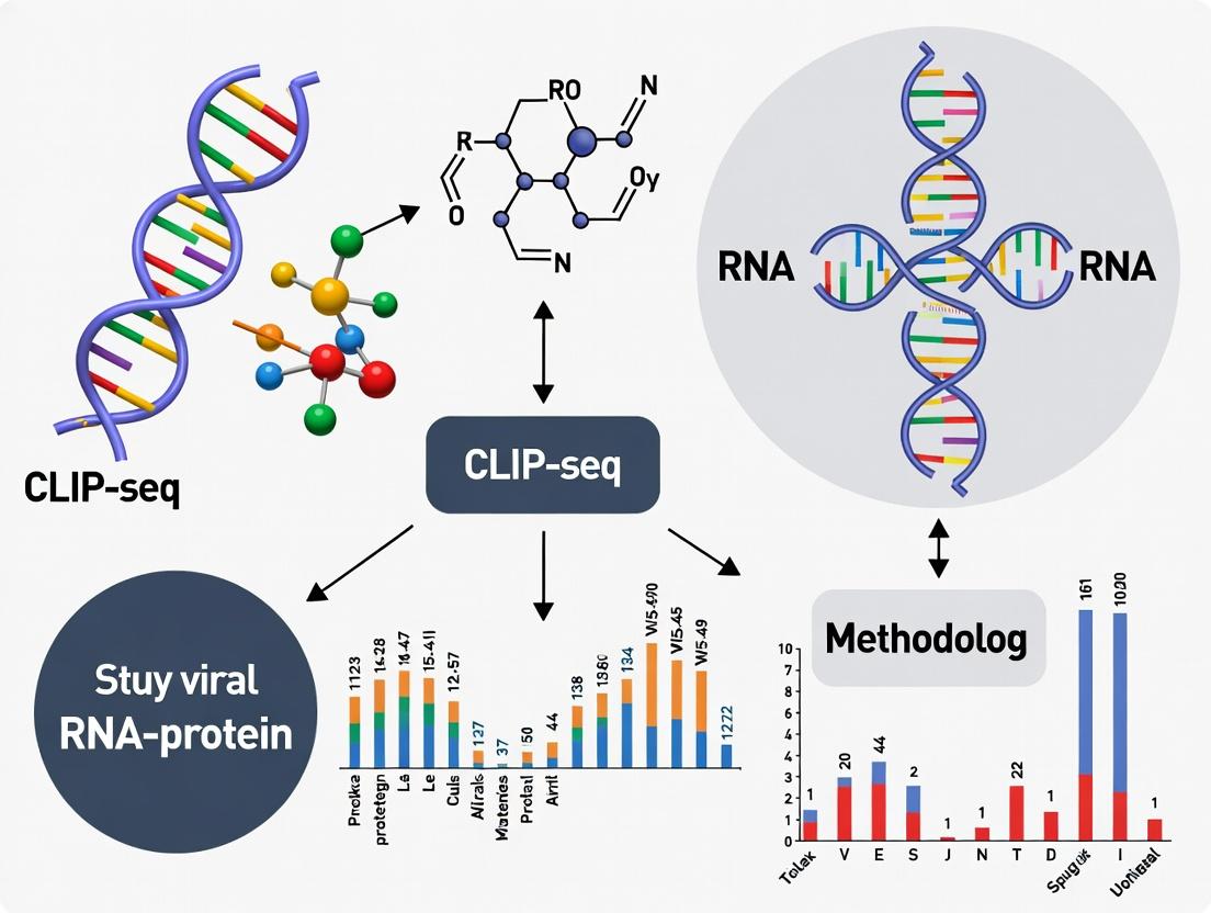
What is CLIP-seq? Core Principles of UV Cross-Linking, IP, and High-Throughput Sequencing
CLIP-seq (Cross-Linking and Immunoprecipitation followed by sequencing) is a definitive method for identifying genome-wide RNA-protein interaction sites at nucleotide resolution. In viral research, it is indispensable for mapping interactions between viral RNA or host cell RNAs and viral/cellular RNA-binding proteins (RBPs). This reveals mechanisms of viral replication, immune evasion, and pathogenesis, offering targets for antiviral drug development.
Core Principles
The protocol hinges on covalently capturing transient RNA-protein interactions in vivo and identifying the bound RNA sequences.
UV Cross-Linking
Principle: In vivo irradiation with 254 nm UV-C light creates covalent bonds between RNA bases and aromatic amino acids in directly interacting RBPs. This "freezes" interactions with zero-distance resolution. Critical Parameters: Energy dosage (~150-400 mJ/cm²) must be optimized to balance cross-linking efficiency with RNA fragmentation. For viral studies, this is performed on infected cells at the relevant post-infection time point.
Cell Lysis and RNA Fragmentation
Cells are lysed under stringent conditions. RNA is partially fragmented (often via limited RNase digestion) to reduce non-specific RNA-protein associations and yield bound RNA fragments of ~50-100 nucleotides. For viral RNA, this can help isolate specific protein-binding regions on longer genomic or subgenomic RNAs.
Immunoprecipitation (IP)
The cross-linked RBP-RNA complexes are isolated using specific antibodies against the protein of interest (e.g., a viral RBP or a host factor). Stringent washes minimize non-specific RNA co-purification.
RNA Processing and Library Preparation
Protein-bound RNA fragments are dephosphorylated, a 3' adapter is ligated, the complex is radiolabeled (for visualization), and the RNA is separated by SDS-PAGE. RNA is extracted from a membrane slice corresponding to the RBP's size, a 5' adapter is ligated, reverse transcribed to cDNA, and amplified by PCR for sequencing.
High-Throughput Sequencing and Bioinformatics
Sequenced reads are mapped to the host and viral genomes. True binding sites are identified as clusters of reads (peaks), representing the protein's RNA "footprint." Mutation signatures (like deletions at cross-link sites) help pinpoint exact interaction nucleotides.
Application Notes for Viral RNA-Protein Interactions
- Identifying Viral RBP Targets: CLIP-seq on a viral RBP (e.g., SARS-CoV-2 N protein) can reveal its binding landscape across the viral genome and host transcriptome, implicating it in processes like viral RNA packaging or host translation shutdown.
- Mapping Host Factor Engagement: CLIP-seq on a host RBP (e.g., ELAVL1) during infection shows which viral RNA regions it binds to, potentially identifying host dependency factors.
- Characterizing Antiviral Compound Mechanism: A compound disrupting an RBP-viral RNA interaction will show altered CLIP-seq peak profiles, validating the target and mode of action.
Table 1: Typical CLIP-seq Experimental Parameters and Outcomes
| Parameter | Typical Range/Value | Notes for Viral Studies |
|---|---|---|
| UV Cross-link Energy | 150 - 400 mJ/cm² | Optimize for infected cell type; higher energy may distort viral RNA structures. |
| RNase Digestion | 0.5 - 5 U/mL | Degree of fragmentation critical for resolution; viral RNA abundance may require titration. |
| Input RNA Amount | 10 - 100 µg | May need scaling for low-abundance viral RNAs in early infection. |
| IP Antibody | High-specificity monoclonal | Crucial to avoid host protein background when targeting viral RBPs. |
| Sequencing Depth | 20 - 50 million reads | Deeper sequencing may be needed to robustly capture interactions on compact viral genomes. |
| Peak Size (Resolution) | 20 - 60 nt | Represents the protein-protected RNA "footprint." |
| Background Noise | <5% of reads in controls | Use IgG or null mutant cell controls to define non-specific binding. |
Table 2: Example CLIP-seq Findings in Viral Systems
| Virus | RNA-Binding Protein | Key Finding (CLIP-seq Peak Location) | Implicated Function |
|---|---|---|---|
| HIV-1 | Viral Gag protein | Specific clusters in the 5' UTR and Ψ packaging signal region | Selective genomic RNA packaging into virions. |
| Zika Virus | Host MSI1 protein | Stem-loop structures in the viral 3' UTR | Viral replication and neurovirulence. |
| SARS-CoV-2 | Viral N protein | Genomic 5' and 3' ends, ORF regions | RNA genome packaging and condensate formation. |
| Influenza A | Host SFPQ | Viral mRNA splicing sites | Regulation of viral M2 mRNA splicing. |
Detailed Experimental Protocol: CLIP-seq for a Viral RBP
Protocol Title: irCLIP (improved CLIP) for a Viral RBP in Infected Cells.
Materials: Infected cell culture, UV cross-linker (254 nm), IP antibody, Protein G beads, RNase I, T4 PNK, Ligases, [γ-32P]ATP, NuPAGE gels, Nitrocellulose membrane.
Procedure:
- Cross-linking & Lysis: Wash infected cells with PBS. Irradiate plate (254 nm, 150 mJ/cm², on ice). Scrape cells in stringent lysis buffer (e.g., containing 1% SDS, protease/RNase inhibitors).
- Partial RNA Digestion: Dilute lysate to 0.1% SDS. Add RNase I to a final concentration of 0.5 U/µg of RNA. Incubate 3 min at 37°C. Quench on ice.
- Immunoprecipitation: Pre-clear lysate with Protein G beads. Incubate supernatant with specific antibody (2 µg) for 2h at 4°C. Add beads, incubate 1h. Wash 3x with high-salt wash buffer.
- 3' Dephosphorylation & Adapter Ligation: On beads, dephosphorylate RNA with T4 PNK (no ATP). Ligate pre-adenylated 3' adapter using T4 RNA Ligase 2, truncated.
- 5' Radiolabeling & Separation: Label 5' ends with T4 PNK and [γ-32P]ATP. Run sample on NuPAGE Bis-Tris gel. Transfer to nitrocellulose membrane.
- Membrane Excision & Proteinase K Digest: Expose membrane to film. Excise region corresponding to protein size (+/- ~20 kDa). Digest membrane slice with Proteinase K.
- RNA Extraction & 5' Adapter Ligation: Extract RNA, PAGE-purify. Ligate 5' RNA adapter with T4 RNA Ligase 1.
- Reverse Transcription & PCR: Reverse transcribe with RT primer containing a sample barcode. Amplify cDNA by PCR (≤18 cycles).
- Sequencing & Analysis: Purify library, QC, and sequence (Single-end 50-75 bp). Process data: demultiplex, trim adapters, map to combined host+viral reference genome, call peaks (e.g., with CLIPper, PEAKachu).
The Scientist's Toolkit: Research Reagent Solutions
| Item | Function in CLIP-seq | Key Consideration for Viral Studies |
|---|---|---|
| UV Cross-linker (254 nm) | Creates covalent RNA-protein bonds in situ. | Calibrate dose for infected cell monolayers; ensure even exposure. |
| RNase I (Nuclease) | Fragments RNA to isolate protein-bound regions. | Titrate carefully; viral RNA structures may be differentially sensitive. |
| Specific Antibody | Immunoprecipitates the RBP-RNA complex. | Must recognize cross-linked, denatured protein epitopes (e.g., validate for IP). |
| Pre-adenylated 3' Adapter | Ligated to RNA 3' ends without ATP to prevent circularization. | Reduces background ligation artifacts, crucial for low-input viral samples. |
| T4 Polynucleotide Kinase (PNK) | Dephosphorylates 3' ends, radiolabels 5' ends for visualization. | Essential for irCLIP protocol to monitor complex size. |
| Proteinase K | Digests protein to release cross-linked RNA fragments. | Must be highly active in SDS buffer for complete digestion. |
| Reverse Transcriptase | Generates cDNA from cross-linked, adapter-ligated RNA. | Must have high processivity and tolerate RNA cross-link damage. |
| High-Fidelity PCR Mix | Amplifies cDNA library for sequencing. | Limited cycles prevent PCR duplication bias, critical for quantitative analysis. |
Visualizing CLIP-seq Workflows and Analysis
Title: CLIP-seq Experimental Workflow
Title: CLIP-seq Bioinformatics Pipeline
Within the broader thesis on CLIP-seq for viral RNA-protein interaction research, this application note details its critical role in virology. Viral replication cycles depend on transient, direct interactions between viral RNA genomes/mRNAs and host/viral proteins. Traditional methods often fail to capture these dynamic events. CLIP-seq (Crosslinking and Immunoprecipitation followed by sequencing) enables the genome-wide mapping of these interactions at nucleotide resolution, providing indispensable insights for understanding viral lifecycles and developing antiviral strategies.
Key Applications in Virology
CLIP-seq applications elucidate specific mechanisms in viral infection.
Table 1: Quantitative Insights from Recent Virology CLIP-seq Studies
| Virus Studied | Target Protein | Key Finding (Interaction Metric) | Impact on Viral Lifecycle | Reference (Year) |
|---|---|---|---|---|
| SARS-CoV-2 | Host ELAVL1 (HuR) | >2,000 binding peaks identified in viral RNA; enriched in 3' UTR. | Stabilizes viral RNA, enhancing replication. | Lee et al. (2023) |
| HIV-1 | Viral Gag | Precise mapping of ~5 specific packaging signal regions in full-length genomic RNA. | Essential for selective RNA genome packaging into virions. | Coyle et al. (2022) |
| Zika Virus | Host MSI1 | Binding site motif identified with significant enrichment (p<10^-5) in viral 3' UTR. | Promotes viral translation and neuropathogenesis. | Chavali et al. (2023) |
| Influenza A | Host SRPKs | Phosphorylation-dependent binding to viral M1 mRNA alters splicing efficiency by ~40%. | Modulates viral gene expression timing. | Wang et al. (2024) |
Detailed Protocol: Enhanced CLIP-seq for Viral RNA-Protein Complexes
This protocol is optimized for capturing transient viral RNA-host protein interactions in infected cells.
Day 1: Cell Culture, Infection, and Crosslinking
- Culture & Infect: Grow permissive cells (e.g., Vero E6, Huh-7) to 70% confluency. Infect with virus at a predetermined MOI (e.g., MOI 1-5). Include mock-infected controls.
- UV Crosslinking (254 nm): At the desired post-infection time, place culture dish on ice. Wash once with cold PBS. Irradiate cells with 150-400 mJ/cm² of 254 nm UV-C light in a Stratalinker. This creates covalent bonds between RNA and directly interacting proteins.
- Cell Lysis: Aspirate PBS. Add 1 ml of stringent lysis buffer (50 mM Tris-HCl pH 7.4, 100 mM NaCl, 1% Igepal CA-630, 0.1% SDS, 0.5% sodium deoxycholate, 1x protease inhibitor, 1 U/µl RNase inhibitor). Scrape and collect lysate.
- Partial RNase Digestion: To reduce RNA length and isolate direct binding footprints, add 1 µl of RNase I (diluted 1:1000) per 100 µl lysate. Incubate at 37°C for 3 minutes. Immediately place on ice.
- Clarification: Centrifuge at 20,000 x g for 15 min at 4°C. Transfer supernatant to a new tube.
Day 2: Immunoprecipitation and Library Preparation
- Pre-clear & Bind: Pre-clear lysate with Protein A/G beads for 30 min. Incubate supernatant with 2-5 µg of target-specific antibody or control IgG overnight at 4°C with rotation.
- Capture Complexes: Add pre-washed Protein A/G magnetic beads for 2 hours at 4°C.
- Stringent Washes: Wash beads sequentially with:
- High-salt wash buffer (2x)
- Standard CLIP wash buffer (2x)
- PNKT buffer (Final wash; 1x)
- 3' Dephosphorylation & Ligation: On-bead, dephosphorylate RNA 3' ends with PNK (minus ATP). Ligate a pre-adenylated 3' DNA adapter using T4 RNA Ligase 1.
- 5' Phosphorylation & Ligation: Radiolabel 5' ends with PNK and [γ-³²P]ATP for visualization. Ligate a 5' RNA adapter with T4 RNA Ligase 1.
- Proteinase K Elution & RNA Recovery: Elute RNA-protein complexes in Proteinase K buffer at 55°C. Extract RNA with acid phenol:chloroform and precipitate with glycogen.
Day 3: cDNA Library Construction & Sequencing
- Reverse Transcription: Use Superscript III/IV with a primer complementary to the 3' adapter.
- cDNA Purification & PCR Amplification: Run cDNA on a 6% TBE-Urea gel. Expose to a phosphor screen, excise the region corresponding to the protein-RNA complex (~70 kDa above the expected protein size). Elute and PCR amplify with indexed primers (12-16 cycles).
- Sequencing: Purify PCR product. Quantity and quality-check by Bioanalyzer. Pool libraries for 75-150 bp single-end sequencing on an Illumina platform.
Diagrams of Key Methodologies and Pathways
Title: CLIP-seq Experimental Workflow for Virology
Title: Viral RNA-Protein Interactions Revealed by CLIP-seq
The Scientist's Toolkit: Essential Research Reagents
Table 2: Key Reagent Solutions for Viral CLIP-seq
| Reagent Category | Specific Product/Type | Function in Viral CLIP-seq |
|---|---|---|
| Crosslinker | UV-C Light (254 nm) Stratalinker | Creates irreversible covalent bonds between viral RNA and directly bound proteins in vivo, freezing transient interactions. |
| RNase | RNase I (Ambion) | Partially digests unprotected RNA, leaving short (~50-100 nt) footprints bound by the protein, crucial for resolution. |
| Immunoprecipitation Antibody | Protein-specific (e.g., anti-HuR, anti-Gag); Control IgG | Highly specific antibody captures the target RBP and its crosslinked viral RNA. Control ensures specificity. |
| Adapter Ligases | T4 RNA Ligase 1, T4 RNA Ligase 2 (truncated) | Ligates RNA/DNA adapters to crosslinked RNA fragments for reverse transcription and sequencing library construction. |
| Reverse Transcriptase | Superscript III/IV (Thermo Fisher) | Transcribes adapter-ligated, often damaged/Crosslinked RNA into stable cDNA with high processivity and fidelity. |
| RNase Inhibitor | Recombinant RNasin or SUPERase-In | Protects viral RNA from degradation during all post-lysis steps, preserving the interaction landscape. |
| Stringent Wash Buffers | High-salt (1M NaCl), PNKT buffer | Removes non-specifically bound RNA and proteins, reducing background and ensuring direct interaction data. |
| Bioinformatics Pipeline | CLIP Toolkits (e.g., CLIPper, PEAKachu) | Dedicated software for peak calling from sequenced cDNA clusters, identifying exact protein binding sites on viral RNA. |
Application Notes
Investigating Viral Replication Complexes (RCs)
CLIP-seq enables the precise mapping of interactions between viral proteins and host/viral RNA within replication organelles. A 2023 study on SARS-CoV-2 used PAR-CLIP to identify that viral nsp13 (helicase) binds strongly to specific stem-loop structures in the 5' UTR of the viral genome, an essential interaction for RC assembly. Quantitative analysis revealed over 150 host RNAs, including those encoding mitochondrial proteins, were sequestered into RCs, diverting cellular resources.
Unraveling Mechanisms of Immune Evasion
Viral RNA-binding proteins (RBPs) often target host immune-related mRNAs for degradation or translational suppression. Recent research on Influenza A virus NS1 protein, utilizing iCLIP, mapped its binding to GU-rich regions in the 3' UTRs of interferon-stimulated genes (ISGs) like IFIT2 and OAS1. Data showed a 70% reduction in expression of bound transcripts, correlating directly with binding site density.
Defining Viral Latency and Reactivation
In herpesviruses, CLIP-seq has elucidated how viral latency-associated nuclear antigen (LANA) in KSHV or latency-associated transcripts (LATs) in HSV-1 orchestrate a network of RNA interactions to maintain dormancy. A 2024 study employing eCLIP on KSHV-infected cells demonstrated that LANA binds to specific miRNA precursors and host cell cycle regulator mRNAs, tethering them to chromatin to suppress lytic reactivation signals.
Table 1: Quantitative CLIP-seq Findings in Recent Viral Studies
| Virus | Viral Protein | Target RNA Type | # of Significant Binding Sites | Key Functional Outcome | Primary CLIP Method |
|---|---|---|---|---|---|
| SARS-CoV-2 | nsp13 | Viral genomic 5' UTR | 4 primary structured sites | Essential for RC assembly & RNA synthesis | PAR-CLIP |
| Influenza A | NS1 | Host ISG 3' UTRs | ~200 sites across >150 host transcripts | Degradation/repression of immune mRNAs (~70% reduction) | iCLIP |
| KSHV | LANA | Viral miRNA pre-cursors & host mRNAs | 12 viral, 89 host | Epigenetic tethering, suppression of reactivation | eCLIP |
| HIV-1 | Rev | Viral RRE element in env intron | 1 high-affinity complex | Nuclear export of unspliced viral transcripts | HITS-CLIP |
| HCV | Core | Host miR-122 & viral IRES | 2 major on miR-122, 1 on IRES | Stabilizes viral RNA, enhances translation | PAR-CLIP |
Detailed Experimental Protocols
Protocol 1: iCLIP for Viral RNA-Protein Interactions in Infected Cells
This protocol is adapted for studying a viral RBP (e.g., Influenza NS1) during active infection.
Day 1: Cell Lysis and Immunoprecipitation
- Infection & Crosslinking: Culture A549 cells (5x10^7). Infect with virus at MOI=3. At peak protein expression (e.g., 12hpi), wash cells with cold PBS. Perform in vivo crosslinking with 254nm UV-C at 400 mJ/cm² on ice.
- Lysis: Scrape cells in 1ml of stringent lysis buffer (50mM Tris-HCl pH7.4, 150mM NaCl, 1% NP-40, 0.1% SDS, 0.5% sodium deoxycholate, 1mM DTT, RNase Inhibitor, protease inhibitors). Incubate 10 min on ice, sonicate lightly to reduce viscosity (3 pulses, 10% amplitude), clear by centrifugation (16,000g, 15 min, 4°C).
- RNase Treatment (Partial Digestion): Treat supernatant with 0.01U/µl RNase I (Thermo Fisher) for 3 min at 37°C to fragment RNA to ~70-200nt. Immediately place on ice.
- Immunoprecipitation: Pre-clear lysate with protein A/G beads. Incubate with 5µg of antibody specific to the viral protein (e.g., anti-NS1) for 2h at 4°C. Add protein A/G magnetic beads, incubate 1h. Wash 3x with high-salt wash buffer (50mM Tris-HCl pH7.4, 1M NaCl, 1% NP-40, 0.1% SDS, 1mM DTT).
Day 2: Library Preparation
- 3' Dephosphorylation & Ligation: On-bead, dephosphorylate RNA 3' ends with T4 PNK (without ATP). Ligate a pre-adenylated DNA linker (5'-App/CTGTAGGCACCATCAAT/3ddC/-3') using Truncated T4 RNA Ligase 2.
- Proteinase K Elution & RNA Isolation: Elute RBP-RNA complexes by digesting with Proteinase K in SDS buffer. Extract RNA with acid phenol:chloroform, precipitate.
- Reverse Transcription & cDNA Purification: Reverse transcribe using a primer containing a 5' Illumina adapter sequence and a random barcode. Run cDNA on a 6% TBE-urea gel. Excision of cDNA smear ~100-300bp. Gel-purify.
- cDNA Circularization & PCR Amplification: Circularize single-stranded cDNA using Circligase. Re-linearize with BpmI (cuts within the original linker sequence). Amplify with Illumina P5/P7 primers (12-15 PCR cycles). Purify PCR product with SPRI beads for sequencing.
Protocol 2: PAR-CLIP for Nucleotide-Resolution Mapping
Ideal for determining exact crosslink sites, using 4-thiouridine (4SU) incorporation.
Key Modification: At 16h pre-infection, supplement cell medium with 100µM 4SU. Proceed with infection and crosslinking at 365nm UV-A (0.15 J/cm²). During library preparation, note that reverse transcription will introduce characteristic T-to-C mutations at crosslink sites, which are bioinformatically identified.
Visualization
Title: CLIP-seq Maps Viral RC Assembly
Title: CLIP-seq Reveals Immune Evasion Mechanism
Title: CLIP-seq Defines Latency RNA Network
The Scientist's Toolkit: Key Research Reagent Solutions
Table 2: Essential Reagents for Viral CLIP-seq Studies
| Reagent / Material | Supplier Examples | Function in Protocol |
|---|---|---|
| UV Crosslinker (254nm & 365nm) | Spectrolinker (XL-1000) | In vivo crosslinking of RNA-protein complexes. 254nm for standard, 365nm for 4SU (PAR-CLIP). |
| 4-Thiouridine (4SU) | Sigma-Aldrich (T4509) | Photoactivatable nucleoside for PAR-CLIP; incorporates into RNA for efficient crosslinking. |
| RNase I (1 U/µl) | Thermo Fisher (AM2295) | Partial digestion of RNA to generate optimal fragment lengths for CLIP library prep. |
| Pre-Adenylated 3' Linker (App-DNA) | IDT (Custom Synthesis) | Ligation to RNA 3' ends using Truncated T4 Rnl2; essential for isolating crosslinked RNA. |
| Truncated T4 RNA Ligase 2 | NEB (M0242S) | Specifically ligates pre-adenylated linker to RNA 3' ends, minimizing adapter dimer formation. |
| Protein A/G Magnetic Beads | Pierce (88802/88803) | Efficient capture of antibody-bound RBP complexes for stringent washing. |
| Antibody: Specific to Viral RBP | E.g., Santa Cruz, Abcam, In-house | High-affinity, specific immunoprecipitation of the viral protein of interest. |
| T4 PNK (10 U/µl) | NEB (M0201S) | Dephosphorylates RNA 3' ends pre-ligation; also used in 5' phosphorylation in some protocols. |
| Proteinase K (RNA-grade) | Thermo Fisher (AM2548) | Elutes crosslinked RNA from the protein complex after immunoprecipitation. |
| Circligase II ssDNA Ligase | Lucigen (CL9021K) | Circularizes single-stranded cDNA post-purification, a key step in iCLIP/eCLIP. |
| High-Fidelity PCR Master Mix | NEB (M0541) | Amplification of final cDNA library with minimal bias for Illumina sequencing. |
| SPRI Beads (Size Selection) | Beckman Coulter (A63881) | Clean-up and size selection of cDNA and final libraries post-amplification. |
1. Introduction in the Context of CLIP-seq for Viral RNA-Protein Interactions Within a thesis investigating viral RNA-protein interactions via CLIP-seq (Crosslinking and Immunoprecipitation followed by sequencing), the initial choices of viral system and biological question are paramount. These decisions dictate experimental feasibility, relevance, and interpretability. This protocol outlines the critical pre-experimental assessment and setup required to ensure a successful CLIP-seq study of viral ribonucleoprotein (vRNP) complexes.
2. Key Considerations for Viral System Selection The choice of virus impacts host interaction complexity, biosafety requirements, and technical reproducibility. Quantitative parameters for common model viruses are summarized below.
Table 1: Quantitative Comparison of Viral Systems for CLIP-seq Studies
| Virus | Genome Type | Genome Size (kb) | Known RBPs | Replication Compartment | BSL Level | CLIP Feasibility (1-5) |
|---|---|---|---|---|---|---|
| HIV-1 | ssRNA(+) | 9.8 | Gag, Rev, Nef | Nucleus/Cytoplasm | 2/3 | 5 |
| Influenza A | ssRNA(-) segmented | 13.5 total | NP, NS1 | Nucleus | 2 | 4 |
| SARS-CoV-2 | ssRNA(+) | 29.9 | N, nsp3, nsp8 | Cytoplasm (DMVs) | 3 | 4 |
| HSV-1 | dsDNA | 152 | ICP27, vhs | Nucleus | 2 | 3 |
| ZIKV | ssRNA(+) | 10.8 | Capsid, NS5 | Cytoplasm | 2 | 4 |
Abbreviations: RBP: RNA-Binding Protein; BSL: Biosafety Level; DMVs: Double-Membrane Vesicles.
3. Defining the Biological Question The experimental design of CLIP-seq must be driven by a specific, testable hypothesis. Common frameworks include:
- Mechanistic: Which host RBPs does the viral RNA genome bind to during early replication?
- Comparative: How do RNA-protein interactions differ between wild-type and a mutant virus (e.g., replication-deficient)?
- Dynamic: How does the vRNP interactome change over the course of infection (temporal)?
- Therapeutic: Does a candidate antiviral compound disrupt the binding of a specific viral RBP to its cognate RNA?
4. Preliminary Experimental Protocol: System Validation for CLIP-seq Before large-scale CLIP-seq, perform the following validation.
Protocol 4.1: Viral Infection and Crosslinking Optimization
- Objective: Determine optimal infection multiplicity (MOI) and UV crosslinking time.
- Materials: Cultured host cells (e.g., HEK293T, A549), viral stock, PBS, 0.4% Trypan Blue.
- Procedure:
- Infect cells in a 6-well plate at varying MOIs (e.g., 0.1, 1, 5) in triplicate.
- Harvest cells at 12, 24, and 48 hours post-infection (hpi). Mix 10µl cell suspension with 10µl Trypan Blue. Count viable cells using a hemocytometer.
- Calculate cell viability (%). Plot viability vs. MOI/hpi to identify conditions with >70% viability for healthy crosslinking.
- At optimal hpi, wash cells with cold PBS. Perform UV crosslinking at 254 nm (400 mJ/cm² standard). Test varied energies (150-400 mJ/cm²) in pilot.
- Scrape cells, pellet, and flash-freeze for RNA extraction and qPCR (viral RNA load) and western blot (viral protein) to confirm infection.
Table 2: Research Reagent Solutions Toolkit
| Reagent/Material | Function in Viral CLIP-seq | Example Vendor/Product |
|---|---|---|
| Anti-Viral Protein Antibody | Immunoprecipitation of crosslinked vRNP complexes. Must be high-quality for CLIP. | Merck (Anti-Influenza NP), Abcam (Anti-SARS-CoV-2 Nucleocapsid) |
| RNase Inhibitor | Prevents degradation of RNA during lysis and IP. Critical for RNA recovery. | Takara, RNaseOUT |
| Proteinase K | Digests proteins after IP for RNA release. Required for crosslink reversal. | Thermo Scientific, Molecular Biology Grade |
| 3'-RNA Linker Ligase | Enzymatically ligates adapters to purified RNA fragments for library prep. | T4 RNA Ligase 1 (NEB) |
| Silica-based Spin Columns | For purification of small RNA fragments after proteinase K treatment. | Zymo Research, Clean & Concentrator kits |
| dUTP-based Sequencing Library Kit | Allows for strand-specific sequencing of recovered RNA fragments. | Illumina TruSeq Stranded Total RNA |
5. CLIP-seq Experimental Workflow Diagram
Diagram Title: Viral CLIP-seq Core Experimental Workflow
6. Pathway Diagram: Integrating CLIP-seq Data into Viral Research
Diagram Title: From CLIP Data to Biological Insights Pathway
From Cell to Sequence: A Step-by-Step CLIP-seq Protocol for Viral Research
Within the broader thesis on utilizing CLIP-seq (Crosslinking and Immunoprecipitation) to dissect viral RNA-protein interactions, selecting the optimal protocol variant is critical. These interactions govern viral replication, immune evasion, and pathogenesis. This application note compares three advanced CLIP derivatives—iCLIP, eCLIP, and irCLIP—detailing their quantitative performance, specific methodologies, and recommended scenarios for virology research.
Table 1: Comparison of CLIP Variants for Viral Studies
| Feature | iCLIP (individual-nucleotide resolution CLIP) | eCLIP (enhanced CLIP) | irCLIP (infrared CLIP) |
|---|---|---|---|
| Crosslinking | 254 nm UV-C | 254 nm UV-C | 254 nm UV-C |
| Key Differentiator | cDNA truncation at crosslink site; circularization/religation. | Size-matched input control; streamlined adapter strategy. | Infrared dye-labeled adapters for gel-free, blot-free detection. |
| Primary Advantage | Single-nucleotide resolution mapping of RBP binding. | Robust background subtraction; reduced hands-on time. | Eliminates membrane transfer; faster, more sensitive visualization. |
| Typical SNR* Range | 5-15 | 8-20 | 10-25 |
| Optimal Viral Scenario | Mapping precise interaction sites of viral or host RBPs on complex viral RNA structures (e.g., HIV Rev on RRE). | Genome-wide profiling of host RBP binding to viral transcripts in infection (e.g., SARS-CoV-2 N protein). | Rapid screening and optimization for new virus-RBP pairs or low-abundance samples. |
| Protocol Duration | ~5-7 days | ~4-5 days | ~3-4 days |
*SNR: Signal-to-Noise Ratio, estimated from published comparisons.
Detailed Experimental Protocols
Protocol 1: iCLIP for High-Resolution Mapping
Application: Defining the exact binding nucleotides of a host RNA-binding protein (RBP) on a viral RNA genome.
- In Vivo Crosslinking: Culture virus-infected cells. Wash with PBS and irradiate with 254 nm UV light at 400 mJ/cm² on ice.
- Lysis & Immunoprecipitation: Lyse cells in stringent RIPA buffer. Shear RNA with RNase I to ~50-100 nt fragments. Immunoprecipitate RNA-protein complexes with antibody-coated magnetic beads.
- 3' Dephosphorylation & Linker Ligation: On-bead, dephosphorylate RNA 3' ends with PNK (no ATP). Ligate a pre-adenylated DNA linker (L3-App).
- 5' Phosphorylation & RNA Isolation: Radiolabel 5' ends with [γ-³²P]ATP using PNK. Resolve complexes on SDS-PAGE, transfer to nitrocellulose, and expose. Excise the RBP-RNA complex band, purify RNA by proteinase K digestion.
- Reverse Transcription & Circularization: Reverse transcribe with a primer complementary to L3. cDNA often truncates at crosslinked nucleotide. Purify cDNA and circularize with CircLigase.
- PCR Amplification & Sequencing: Re-linearize and amplify with Illumina-compatible primers. Sequence.
Protocol 2: eCLIP for Robust, Controlled Profiling
Application: Unbiased identification of host RBP binding sites across full-length SARS-CoV-2 subgenomic RNAs.
- Crosslinking & Lysis: Perform UV crosslinking as in iCLIP. Lyse cells and split lysate: ~10% for "Size-matched Input" (SMInput), 90% for IP.
- RNase Digestion & IP: Digest both fractions with high-sensitivity RNase I. Perform IP on the larger fraction.
- Adapter Ligation: On-bead, dephosphorylate and ligate pre-adenylated 3' adapter. Then, phosphorylate 5' ends and ligate RNA 5' adapter.
- Library Preparation: Elute, purify RNA, and reverse transcribe. Perform PCR amplification with indexed primers.
- Sequencing & Analysis: Sequence both IP and SMInput libraries in parallel. Use the SMInput to control for background and sequence bias.
Protocol 3: irCLIP for Streamlined, Sensitive Detection
Application: Rapidly screening for interaction between a newly identified viral protein and cellular RNA.
- Crosslinking, Lysis, IP: Perform standard UV crosslinking, lysis, and RNase digestion followed by immunoprecipitation.
- Infrared Adapter Ligation: On-bead, ligate a pre-adenylated 3' adapter conjugated to an infrared dye (e.g., IRDye 800CW).
- Gel-Free Detection: Directly load beads or eluted complexes onto an SDS-PAGE gel. After electrophoresis, scan the gel on an infrared imaging system (e.g., Li-COR Odyssey) to visualize the RBP-RNA complex band.
- RNA Purification & Library Prep: Excise the fluorescent band, elute RNA, and proceed to reverse transcription and PCR amplification using primers compatible with the irCLIP adapter.
Visualizations
CLIP Variant Workflow Decision Pathway
Viral Scenario CLIP Selection Logic
The Scientist's Toolkit: Research Reagent Solutions
Table 2: Essential Reagents for CLIP-seq in Virology
| Reagent / Material | Function in Protocol | Key Consideration for Viral Studies |
|---|---|---|
| UV Crosslinker (254 nm) | Covalently freezes transient RNA-protein interactions in vivo. | Optimize energy (150-400 mJ/cm²) for infected cell monolayers or suspensions. |
| RNase I (High-Sensitivity) | Fragments RNA to ~50-200 nt, defining binding site resolution. | Titration is critical for structured viral RNA genomes (e.g., flavivirus UTRs). |
| Magnetic Protein A/G Beads | Capture antibody-bound RBP-RNA complexes during IP. | Use with validated antibodies against viral protein or epitope-tagged host RBP. |
| Pre-adenylated 3' Adapter | Ligation to RNA 3' end without ATP, preventing adapter concatenation. | Sequence influences ligation efficiency; keep constant across IP/SMInput in eCLIP. |
| CircLigase (iCLIP) | Circularizes single-stranded cDNA to allow PCR amplification after truncation. | Essential for recovering iCLIP cDNAs that truncate at crosslink site. |
| Infrared Dye-Labeled Adapter (irCLIP) | Allows direct, sensitive in-gel detection, bypassing membrane transfer. | Reduces time and sample loss, beneficial for low-input viral samples. |
| Size-matched Input (SMInput) Reagents (eCLIP) | Provides matched-control library for background subtraction. | Crucial for distinguishing specific binding in complex infected-cell lysates. |
| Proteinase K | Digests protein to elute crosslinked RNA from excised gel/membrane pieces. | Ensure RNase-free, high-activity grade for maximal RNA recovery. |
The study of virus-host interactions is critical for understanding viral replication, pathogenesis, and for identifying novel therapeutic targets. Within a broader CLIP-seq (Cross-Linking and Immunoprecipitation followed by sequencing) thesis, the initial cross-linking step is paramount. For research focusing on viral RNA-protein interactions, this step must be rigorously optimized to capture transient and dynamic interactions between viral RNA elements and host or viral proteins within the complex milieu of the infected cell. The cross-linking condition must balance efficiency with specificity to minimize background and preserve biological relevance. This protocol details the design and validation process for establishing robust UV cross-linking conditions for cells infected with a model virus (e.g., HIV-1, SARS-CoV-2, Influenza A).
Core Principles & Key Variables for Optimization
The primary cross-linking method for RNA-protein interactions is UV irradiation at 254 nm. This wavelength induces covalent bonds between RNA bases and amino acids in direct contact (primarily pyrimidines and aromatic/charged residues), without linking protein-to-protein. Optimization for infected cells must consider:
- Cell Type & Culture Conditions: Adherent vs. suspension; infection kinetics.
- Viral Model: RNA virus type, replication cycle duration, subcellular sites of replication.
- Cross-linking Energy Dose: A product of irradiance (mW/cm²) and time (seconds), measured in Joules/cm² (typically 100-400 mJ/cm²).
- Cell Washing & Medium Removal: Culture medium can absorb UV, shielding cells.
- Post-Cross-Linking Cell Viability & Lysis: Ensure cross-linking is not excessively destructive.
- Validation Metrics: Success is measured by efficient RNA-protein cross-linking with minimal RNA degradation or protein aggregation.
Table 1: Empirical Testing of UV Doses on Viral RNA-Protein Recovery
| UV Dose (mJ/cm²) | Cell Viability Post-CL (%) | RNA Integrity Number (RIN) | Protein Aggregation (Visual on SDS-PAGE) | Immunoprecipitation Yield (Relative to Input) | Recommended for CLIP-seq? |
|---|---|---|---|---|---|
| 0 | >95 | 9.5 | None | <0.5% | No (No cross-links) |
| 100 | 85 | 8.8 | Mild | 2.1% | Yes (Mild conditions) |
| 200 | 75 | 8.0 | Moderate | 3.5% | Yes (Optimal) |
| 400 | 50 | 6.5 | Severe | 2.8% | No (Excessive damage) |
| 800 | 20 | 4.0 | Severe | 1.5% | No |
Table 2: Comparison of Cross-linking Methods for Infected Cells
| Method | Mechanism | Cross-link Specificity | Penetration Depth | Suitability for Infected Cell CLIP | Key Limitation |
|---|---|---|---|---|---|
| UV-C (254 nm) | RNA base to protein amino acid | High (Direct RNA-Protein) | Single cell layer | Excellent | Requires monolayer; low depth penetration |
| UV-B (312 nm) | Indirect, via photo-activatable ribonucleosides | Moderate | Higher than UV-C | Good for certain applications | Requires nucleotide analogs |
| Formaldehyde (FA) | Protein-Protein, Protein-DNA, (weak RNA-Protein) | Low (Primarily protein-protein) | Deep tissue | Poor for native RNA-protein studies | Cross-links proteins, obscuring direct RNA partners |
Detailed Experimental Protocols
Protocol 4.1: Optimization of UV Cross-linking Dose for Infected Cells
Objective: To determine the optimal UV 254 nm dose that maximizes recovery of specific viral ribonucleoprotein complexes while maintaining RNA and protein integrity.
Materials:
- Virus-infected cell monolayers (e.g., A549, HEK293T, Huh-7) at desired post-infection time.
- PBS, ice-cold.
- Tissue culture hood.
- UV Cross-linker (e.g., Spectrolinker XL-1000) calibrated for 254 nm output.
- Cell scrapers.
- Microcentrifuge tubes.
Procedure:
- Preparation: 24 hours post-infection, wash cell monolayers twice with 10 mL of ice-cold PBS. Aspirate PBS completely, as residual liquid will attenuate UV.
- Dose Administration: Place the open culture dish on ice. Irradiate cells at varying doses (e.g., 0, 100, 200, 400 mJ/cm²) using the 254 nm setting. Keep control plates on ice without irradiation.
- Harvesting: Immediately after irradiation, add 1 mL of ice-cold PBS to each plate. Scrape cells and transfer the suspension to a pre-chilled microcentrifuge tube.
- Pellet: Centrifuge at 1500 x g for 3 min at 4°C. Discard supernatant. Cell pellets can be flash-frozen in liquid N₂ and stored at -80°C or processed immediately for lysis (Protocol 4.2).
- Parallel Validation Samples: From each dose condition, set aside a fraction of cells for viability assays (trypan blue) and RNA/protein quality analysis (Bioanalyzer and SDS-PAGE).
Protocol 4.2: Validation via Radioactive UV Cross-linking Assay
Objective: To biochemically validate the formation of specific RNA-protein complexes using a radiolabeled viral RNA probe.
Materials:
- Cross-linked cell pellets from Protocol 4.1.
- Lysis Buffer: 50 mM Tris-HCl (pH 7.4), 100 mM NaCl, 1% Igepal CA-630, 0.1% SDS, 0.5% sodium deoxycholate, supplemented with RNase Inhibitor and EDTA-free protease inhibitor.
- DNase I (RNase-free).
- ³²P-labeled in vitro transcribed RNA probe corresponding to a known viral protein binding element (e.g., HIV-1 RRE, HCV 3' UTR).
- RNase T1 (for partial digestion).
- HeLa cell cytoplasmic extract (or relevant S10 extract) as a source of proteins.
- UV lamp (254 nm, handheld).
- SDS-PAGE equipment and phosphorimager.
Procedure:
- Prepare Cell Lysate: Lyse cross-linked cell pellets in 500 µL Lysis Buffer for 10 min on ice. Clarify by centrifugation at 16,000 x g for 15 min at 4°C.
- In Vitro Binding Reaction: Incubate 10 µg of clarified lysate (or control HeLa extract) with 100 fmol of ³²P-labeled RNA probe in binding buffer for 20 min at 30°C.
- Secondary Cross-linking: Transfer mixture to a parafilm strip on ice. Irradiate with a handheld 254 nm UV lamp at 400 mJ/cm².
- Digestion: Treat with RNase T1 to digest unbound RNA.
- Analysis: Resolve proteins by SDS-PAGE. The formation of a specific cross-linked complex will appear as a shifted band on the phosphorimager gel, visible only in infected cell lysates + UV conditions.
- Interpretation: The signal intensity of the shifted band across different pre-cell harvest UV doses (from Protocol 4.1) indicates the optimal in vivo cross-linking efficiency.
Diagrams
Title: Workflow for Cross-linking Optimization & Validation
The Scientist's Toolkit: Key Research Reagent Solutions
Table 3: Essential Materials for Cross-linking Condition Optimization
| Item / Reagent | Vendor Examples | Function & Critical Notes |
|---|---|---|
| Programmable UV Cross-linker (254 nm) | Spectrolinker (Spectronics), CL-1000 (UVP) | Provides consistent, calibrated UV dose. Critical for reproducibility. Must be calibrated annually. |
| RNase Inhibitor | Murine RNase Inhibitor (NEB), SUPERase•In (Thermo Fisher) | Protects RNA from degradation during all post-cross-linking steps. Use a high concentration. |
| Protease Inhibitor Cocktail (EDTA-free) | cOmplete (Roche), Halt (Thermo Fisher) | Preserves protein integrity. EDTA-free to avoid interference with subsequent enzymatic steps. |
| Igepal CA-630 Alternative | NP-40 Surfact-Amps (Thermo Fisher) | Non-ionic detergent for cell lysis. Gentle disruption of membranes while preserving RNP complexes. |
| RNase T1 | Thermo Fisher, Ambion | Specific for single-stranded RNA. Used in validation assays to trim unbound RNA from cross-linked complexes. |
| [α-³²P] UTP or CTP | PerkinElmer, Hartmann Analytic | For generating high-specific-activity RNA probes for validation assays (Protocol 4.2). |
| In vitro Transcription Kit | MEGAscript (Thermo Fisher) | To produce unlabeled or radiolabeled viral RNA probes for binding and validation studies. |
| RNA Quality Analyzer | Bioanalyzer (Agilent), Fragment Analyzer (Agilent) | Essential for assessing RNA integrity (RIN) after cross-linking to rule out UV-induced damage. |
Within the broader thesis on employing CLIP-seq to dissect viral RNA-protein interactions, this step is critical for capturing specific ribonucleoprotein (RNP) complexes. The choice of lysis conditions and immunoprecipitation (IP) strategy determines whether the focus is on a viral RNA-binding protein (vRBP), a host factor hijacked by the virus, or both. This protocol details methods for effective complex preservation and isolation under stringent conditions to minimize nonspecific background, a common challenge in studying viral replication complexes.
Application Notes
- Objective: To isolate crosslinked RNA-protein complexes specific to a target protein (viral or host) with high specificity and yield.
- Key Consideration: Lysis buffer stringency must balance between complete disruption of cellular/viral structures and preservation of the target RNP complex. Overly harsh conditions can disrupt interactions, while mild conditions reduce yield.
- Crosslinking Control: Always process a non-crosslinked control sample in parallel to identify background RNA contaminants that bind nonspecifically to beads or antibodies.
- RNase Treatment: A critical step post-lysis involves partial RNase digestion to trim RNA footprints (~50-70 nucleotides) around the crosslinked protein, defining the resolution of subsequent sequencing.
Detailed Protocol: Cell Lysis and Immunoprecipitation
Materials & Reagents
Table 1: Research Reagent Solutions for Lysis and IP
| Item | Function | Example/Formula |
|---|---|---|
| IP Lysis Buffer | Lyse cells while preserving protein-RNA complexes; contains inhibitors. | 50 mM HEPES pH 7.5, 150 mM KCl, 2 mM EDTA, 1% NP-40, 0.5% Sodium Deoxycholate, 0.1% SDS, plus fresh protease/RNase inhibitors. |
| Benzonase (Optional) | Digests uncrosslinked nucleic acid to reduce viscosity and background. | 25 U/mL in lysis buffer. |
| Micrococcal Nuclease (MNase) | Partially digests RNA to leave short, protein-protected footprints. | Diluted in supplied buffer to achieve desired digestion (e.g., 0.5 U/µL). |
| Protein-Specific Antibody | Captures the target RNP complex. | Validated for IP (e.g., anti-FLAG M2, anti-HA, anti-viral capsid protein). |
| Magnetic Beads | Solid support for antibody-mediated capture. | Pre-washed Protein A/G or anti-species IgG magnetic beads. |
| High-Salt Wash Buffer | Removes nonspecifically bound complexes. | IP Lysis Buffer with KCl increased to 500 mM. |
| Denaturing Wash Buffer | Further reduces background; used in stringent protocols like iCLIP. | 4 M Urea in 1X PBS. |
Method
Cell Lysis:
- Resuspend UV-crosslinked cell pellet in 1 mL of ice-cold IP Lysis Buffer per 10⁷ cells.
- Incubate on rotator at 4°C for 15 minutes.
- Clarify lysate by centrifugation at 16,000 x g for 15 minutes at 4°C. Transfer supernatant to a new tube.
- (Optional) Add Benzonase (25 U/mL) and incubate for 10 minutes on ice to reduce viscosity.
Partial RNase Digestion:
- Add CaCl₂ to a final concentration of 2 mM.
- Add Micrococcal Nuclease (MNase) to a predetermined optimal concentration (e.g., 0.5-2 U/µL of lysate). Titrate to yield RNA fragments of 50-70 nt.
- Incubate at 37°C for 5-15 minutes. Stop reaction with 4 mM EGTA.
Pre-clearing (Optional but Recommended):
- Add 20 µL of pre-washed magnetic beads to the lysate.
- Incubate at 4°C for 30 minutes on a rotator.
- Separate beads using a magnetic rack and transfer supernatant to a new tube.
Antibody Binding:
- Add the validated antibody (1-5 µg) to the pre-cleared lysate.
- Incubate at 4°C for 2 hours on a rotator.
Bead Capture:
- Add 50 µL of pre-washed Protein A/G magnetic beads.
- Incubate at 4°C for 1-2 hours on a rotator.
Stringent Washes:
- Separate beads on a magnetic rack. Discard supernatant.
- Wash beads sequentially with the following buffers (1 mL each, 4°C, 5 min per wash):
- a) 2x with IP Lysis Buffer.
- b) 2x with High-Salt Wash Buffer.
- c) 1x with Denaturing Wash Buffer (for iCLIP).
- d) 1x with 1X PBS.
Proceed to RNA Processing: The bead-bound, crosslinked RNA-protein complexes are now ready for Step 3: RNA isolation and library preparation.
Data Presentation
Table 2: Comparison of IP Strategies for Viral vs. Host RBPs
| Parameter | Viral RBP IP (e.g., SARS-CoV-2 N protein) | Host RBP IP (e.g., ELAVL1) |
|---|---|---|
| Lysis Stringency | Higher (0.5-1% SDS often needed to disrupt virions) | Moderate (0.1% SDS to preserve native complexes) |
| Antibody Type | Anti-viral protein antibody; epitope-tagged virus | Antibody against endogenous host protein |
| Key Challenge | Low abundance of viral proteins; antibody specificity | Distinguishing virus-induced interactions from native ones |
| Typical Yield (RNA) | Lower (0.1-1 ng total RNA) | Higher (1-10 ng total RNA) |
| Optimal MNase Conc. | Lower (0.2-0.5 U/µL) to protect viral RNP complexes | Standard (0.5-1 U/µL) |
Visualizations
Title: CLIP-seq Lysis and IP Workflow
Title: Viral vs. Host RBP Targeting Strategy
Within the context of a CLIP-seq thesis focused on viral RNA-protein interactions, the steps following successful crosslinking and immunoprecipitation are critical. Proper RNA processing, library construction, and sufficient sequencing depth are paramount to capturing precise, high-resolution binding sites of viral proteins on viral or host RNAs. This section details the protocols and considerations for converting immunoprecipitated RNA into a sequencing-ready library and determining the appropriate sequencing coverage.
RNA Processing After Immunoprecipitation
Following RNA-protein complex isolation, the RNA must be processed for downstream library preparation. Key steps include RNA fragmentation, size selection, and adapter ligation. The choice between enzymatic (e.g., RNase I, MNase) or physical (e.g., sonication, hydrolysis) fragmentation depends on the desired resolution and the specific CLIP variant (e.g., HITS-CLIP, PAR-CLIP).
Protocol 2.1: RNA Fragmentation and Phosphatase/Kinase Treatment Objective: To generate RNA fragments of optimal length (50-200 nt) and prepare ends for adapter ligation.
- Resuspend Pellet: Resuspend the washed beads (from IP) in 50 µL of 1X PNK buffer.
- Dephosphorylation: Add 1 µL of FastAP Thermosensitive Alkaline Phosphatase and incubate at 37°C for 10 minutes. This removes 3' phosphates left by fragmentation.
- Phosphorylation: Add 1 µL of T4 Polynucleotide Kinase (PNK) and 1 µL of 10 mM ATP. Incubate at 37°C for 20 minutes. This adds a 5' phosphate required for subsequent ligation.
- Bead Washing: Wash beads twice with high-salt wash buffer and once with PNK buffer.
- 3' Adapter Ligation: Resuspend beads in 10 µL of 3' adapter ligation mix (containing T4 RNA Ligase 2, truncated, and a pre-adenylated 3' adapter). Incubate at 16°C for 6 hours or overnight.
- Washing: Wash beads twice with wash buffer.
- Radiolabeling (Optional for size selection): For visualization, perform a 5' end-labeling reaction on-bead using PNK and γ-32P-ATP.
- SDS-PAGE and Transfer: Elute complexes, run on a 4-12% Bis-Tris NuPAGE gel, and transfer to a nitrocellulose membrane.
- Membrane Excision: Based on autoradiography, excise the membrane slice corresponding to the protein-RNA complex of interest (typically +25-75 kDa above the protein's molecular weight).
- Proteinase K Digestion: Digest the protein overnight at 55°C in Proteinase K buffer to recover RNA.
- RNA Purification: Phenol-chloroform extract and ethanol precipitate the RNA.
Library Preparation for CLIP-seq
The purified RNA fragments are converted into a cDNA library for sequencing. This involves reverse transcription, cDNA circularization or amplification, and PCR.
Protocol 3.1: Reverse Transcription and cDNA Amplification Objective: To generate double-stranded cDNA libraries from processed RNA fragments.
- Reverse Transcription Primer Annealing: Resuspend RNA pellet in 5 µL containing 1 µL of 5 µM RT primer (containing part of the 5' adapter sequence). Incubate at 70°C for 2 min, then place on ice.
- Reverse Transcription: Add 15 µL of reverse transcription mix (Superscript III Reverse Transcriptase, dNTPs, DTT). Incubate: 5 min at 25°C, 45 min at 50°C, 15 min at 70°C.
- cDNA Purification: Purify cDNA using silica columns or bead-based purification (e.g., RNAClean XP beads). Elute in 10 µL.
- cDNA Circularization (for Illumina): For small RNA libraries, treat purified cDNA with Circligase ssDNA Ligase. Alternatively, proceed directly to PCR if using full-length adapters.
- PCR Amplification: Amplify the library using primers containing full Illumina adapter sequences and sample indexes. Use a high-fidelity polymerase (e.g., KAPA HiFi). Limit cycles (10-18) to avoid over-amplification.
- Library Purification and Size Selection: Perform double-sided bead-based size selection (e.g., 0.6x / 0.8x SPRIselect ratios) to isolate fragments ~150-300 bp. Quantify using a fluorometric assay (e.g., Qubit) and assess size distribution on a Bioanalyzer.
Sequencing Depth Requirements
Adequate sequencing depth is essential to distinguish true binding signals from background noise and achieve statistical power. Requirements vary based on the complexity of the RNA target (e.g., compact viral genome vs. whole host transcriptome).
Table 1: Recommended Sequencing Depth for CLIP-seq Studies
| Application / Target | Minimum Recommended Depth (M reads) | Optimal Depth (M reads) | Primary Justification |
|---|---|---|---|
| Viral RNA Genome (e.g., HCV, HIV, ~10kb) | 5 - 10 | 15 - 25 | High resolution mapping within a small, defined target; allows for saturation of binding sites. |
| Host Transcriptome in Infected Cells | 20 - 30 | 40 - 80+ | Covers a large fraction of the complex host transcriptome; necessary for detecting interactions on lower-abundance host mRNAs. |
| PAR-CLIP (for nucleotide-resolution mapping) | +50% above standard HITS-CLIP | +50% above standard HITS-CLIP | Higher depth compensates for the efficiency of T-to-C transitions and refines single-nucleotide resolution. |
| Enhanced CLIP (eCLIP) with size-matched input control | 20 - 40 (CLIP) + 10-20 (Input) | 40 - 80 (CLIP) + 20-40 (Input) | Input control requires sufficient depth to accurately model background, effectively doubling the sequencing requirement. |
| iCLIP (for mapping crosslink sites at cDNA truncations) | 15 - 25 | 30 - 50 | Need sufficient coverage to observe truncation events, which are a fraction of total reads mapping to a site. |
Note: These are general guidelines. Pilot experiments are strongly recommended to determine the specific depth required for a given viral system and protein target. Depth must be scaled according to the abundance of the target RNA and protein.
The Scientist's Toolkit: Research Reagent Solutions
Table 2: Essential Reagents for CLIP-seq RNA Processing & Library Prep
| Reagent / Kit | Function & Rationale |
|---|---|
| T4 Polynucleotide Kinase (PNK) | Removes 3' phosphates and adds 5' phosphates to RNA fragments, essential for enabling subsequent adapter ligation reactions. |
| T4 RNA Ligase 2, truncated (RNL2tr) | Specifically ligates pre-adenylated 3' adapters to the RNA 3' end in an ATP-independent manner, preventing adapter concatemer formation and increasing ligation efficiency. |
| Pre-adenylated 3' Adapters | Modified adapters that prevent self-ligation and are required for the truncated ligase, reducing background in the library. |
| Superscript III Reverse Transcriptase | Robust reverse transcriptase with high processivity and ability to read through modified nucleotides (e.g., from PAR-CLIP), generating cDNA from RNA crosslinked to protein. |
| Proteinase K | Digests proteins after membrane transfer, crucial for liberating the crosslinked RNA fragments from the immobilized protein complex for recovery. |
| RNAClean XP / SPRIselect Beads | Magnetic beads for size-selective purification and cleanup of RNA and cDNA. Enables efficient removal of enzymes, nucleotides, and adapter dimers while selecting for desired fragment sizes. |
| KAPA HiFi HotStart ReadyMix | High-fidelity PCR polymerase for the final library amplification step. Minimizes PCR errors and bias, ensuring an accurate representation of the original RNA pool in the final sequencing library. |
| High-Sensitivity DNA Bioanalyzer Kit / Fragment Analyzer | For precise quantification and size distribution analysis of the final sequencing library, ensuring it meets the appropriate size range (typically ~200-350 bp including adapters) for cluster generation on the sequencer. |
| Qubit dsDNA HS Assay Kit | Fluorometric quantification of final library concentration. More accurate for dsDNA than spectrophotometric methods, which can overestimate concentration due to adapter contamination. |
Visualizations
Title: CLIP-seq Library Preparation Workflow
Title: Factors Determining CLIP-seq Sequencing Depth
Application Notes
This protocol details a computational pipeline for analyzing CLIP-seq (Crosslinking and Immunoprecipitation coupled with sequencing) data, specifically tailored for identifying viral RNA-protein interaction sites. The pipeline is critical for understanding viral lifecycle mechanisms and identifying potential therapeutic targets. The process transforms raw sequencing reads into high-confidence interaction peaks and subsequently discovers enriched sequence motifs, indicating protein binding preferences.
The core challenge in CLIP-seq analysis, especially for viral RNAs, involves distinguishing specific crosslinked signals from high background noise, sequencing artifacts, and nonspecific RNA fragments. The following workflow addresses these challenges through stringent filtering, precise alignment, and statistically robust peak calling.
Key Quantitative Benchmarks: Performance metrics for a typical viral CLIP-seq dataset (e.g., for a virus like SARS-CoV-2 or HIV-1) are summarized below. These values are highly dependent on the specific experimental conditions, crosslinking efficiency, and viral RNA abundance.
Table 1: Typical CLIP-seq Data Processing Metrics
| Processing Stage | Metric | Typical Range/Value | Explanation |
|---|---|---|---|
| Raw Data | Total Reads | 20 - 50 million | Total sequenced read pairs/singles. |
| Preprocessing | Reads with 3' Adapter | 70% - 95% | Percentage of reads containing the CLIP-specific adapter. |
| Preprocessing | Reads after Quality Filtering | 60% - 85% of adapter-trimmed | Reads retained after quality and length filtering. |
| Alignment | Uniquely Mapping Reads | 10% - 40% of filtered reads | Reads mapping uniquely to the host-virus hybrid genome. |
| Alignment | Duplication Rate | 15% - 50% | PCR/optical duplicates, often higher in CLIP due to low RNA input. |
| Peak Calling | Significant Peaks | 100 - 5,000 | Final high-confidence crosslink sites, varies by protein and virus. |
| Motif Analysis | Enriched Motif E-value | < 1e-5 | Statistical significance of the top discovered sequence motif. |
Protocols
Protocol 1: Raw Read Preprocessing and Alignment
Objective: To remove artifacts, trim adapters, and align cleaned reads to a combined host and viral reference genome.
- Demultiplexing: Use
bcl2fastq(Illumina) ordorado(Oxford Nanopore) to generate FASTQ files, assigning reads based on sample-specific barcodes. - Adapter Trimming and Quality Control:
- For Illumina data, use
fastporcutadaptwith the following parameters: Explanation: Removes the 3' CLIP adapter sequence, trims low-quality bases ( - Assess quality before and after trimming with
FastQC.
- For Illumina data, use
- Alignment to a Hybrid Reference Genome:
- Prepare a reference genome that concatenates the host (e.g., human GRCh38) and viral genome sequences.
- Generate a genome index using
STARorHISAT2. ForSTAR: - Align the trimmed reads:
Explanation: The
--outFilterMultimapNmax 1parameter retains only uniquely mapping reads, crucial for precise peak calling. The output is a sorted BAM file and a bedGraph for visualization.
Protocol 2: Peak Calling with Statistical Modeling
Objective: To identify genomic regions with a significant enrichment of crosslinked RNA fragments compared to background.
- Duplicate Handling: Use
umi_tools dedupif unique molecular identifiers (UMIs) were incorporated during library prep to remove PCR duplicates accurately. If not, usepicard MarkDuplicateswith caution, as genuine crosslink sites are often reproducible. - Peak Calling with
Piranha:- Convert the BAM file to a BED file using
bedtools bamtobed. - Run
Piranha, a method designed for CLIP data, which models read counts per region using a negative binomial distribution. Explanation:-b 5specifies a bin size of 5 nucleotides.Piranhacalculates significance (p-value) for each bin, comparing it to the genomic background.
- Convert the BAM file to a BED file using
- Filtering High-Confidence Peaks: Filter the output
peaks.bedfile to retain peaks with a p-value below a stringent threshold (e.g., p < 0.001). Merge adjacent significant bins usingbedtools merge.
Protocol 3: De Novo Motif Discovery
Objective: To identify conserved RNA sequence or structure motifs within the peak regions that may represent the protein's binding element.
- Sequence Extraction: Using the high-confidence peaks BED file and the reference genome, extract the underlying nucleotide sequences with
bedtools getfasta. - Motif Discovery with
MEME: Run theMEMEsuite on the extracted sequences. Explanation: Searches for 3 motifs (-nmotifs 3) of width between 5 and 15 nucleotides in the given DNA/RNA sequences. The-mod anrsetting allows any number of motif repetitions per sequence. - Motif Validation and Comparison: Use
TOMTOMto compare the discovered motifs against known RNA binding protein motifs in databases likeATtRACTorCISBP-RNA.
Visualizations
Title: CLIP-seq Bioinformatics Pipeline Workflow
The Scientist's Toolkit
Table 2: Essential Research Reagents & Computational Tools
| Category | Item/Software | Function in Pipeline |
|---|---|---|
| Wet-Lab Reagents | UV Crosslinker (254nm) | Covalently links RNA-protein complexes in living cells/virus-infected cells. |
| RNase I/T1 (Partial Digestion) | Truncates RNA at protein-protected sites, leaving a "footprint". | |
| Protein A/G Magnetic Beads with Specific Antibody | Immunoprecipitates the target RNA-protein complex. | |
| 3' RNA Adapter (with/without UMI) | Ligated to RNA fragments for reverse transcription and duplicate removal. | |
| Computational Tools | cutadapt / fastp |
Removes sequencing adapters and performs quality filtering. |
STAR / HISAT2 |
Aligns processed reads to a reference genome (host + virus). | |
samtools, umi_tools |
Handles BAM file operations and UMI-based deduplication. | |
Piranha / CLIPper |
Statistical peak calling algorithms optimized for CLIP data. | |
bedtools |
Genome arithmetic: extracts sequences, merges intervals, etc. | |
MEME Suite |
Discovers de novo sequence motifs from peak regions. | |
| Reference Data | Host Genome (e.g., GRCh38) & Viral Genome | Reference sequences for alignment. |
| Known RBP Motif Databases (ATtRACT, CISBP-RNA) | For validating and annotating discovered motifs. |
Application Notes
CLIP-seq (Crosslinking and Immunoprecipitation followed by sequencing) is a pivotal technique for mapping direct RNA-protein interactions on a genome-wide scale. Within viral research, it elucidates how viral and host proteins bind to viral RNA genomes and transcripts, regulating replication, translation, and immune evasion. These insights are foundational for identifying novel therapeutic targets. The following case studies highlight key findings and protocols.
Case Study 1: Influenza A Virus (IAV)
IAV utilizes host proteins like ELAVL1 (HuR) and viral proteins like NS1 to regulate splicing, stability, and nuclear export of its mRNAs. CLIP-seq identified binding of NS1 to specific sites on viral mRNAs, antagonizing host antiviral responses by blocking access of host RNA-binding proteins (RBPs).
Case Study 2: HIV-1
HIV-1 RNA is extensively bound by both viral (e.g., Gag, Rev) and host proteins (e.g., MOV10, hnRNPs). Rev-binding sites within the RRE (Rev Response Element) were precisely mapped, revealing dynamics during the shift from early to late gene expression. Host protein binding sites associated with nuclear export and genome packaging were also identified.
Case Study 3: SARS-CoV-2
SARS-CoV-2 RNA forms complex structures bound by viral (N) and host (RBFOX2, G3BP1) proteins within infected cells. CLIP-seq studies have shown that the viral N protein coats the genomic and subgenomic RNAs, protecting them and facilitating replication. Host protein binding sites correlate with regions important for viral frameshifting and immune modulation.
Case Study 4: Herpesviruses (e.g., HSV-1, KSHV)
Herpesviruses produce long non-coding RNAs (e.g., LAT in HSV-1, PAN RNA in KSHV) that are heavily bound by host and viral RBPs. CLIP-seq for ORF57 in KSHV, a key post-transcriptional regulator, revealed its binding to intronless viral mRNAs to promote nuclear export and stability.
Table 1: Key CLIP-seq Findings in Viral Systems
| Virus | Target Protein (Type) | Key Binding Motif/Region Identified | Primary Function Determined | Reference (Example) |
|---|---|---|---|---|
| Influenza A | NS1 (Viral) | 5' UTR of viral mRNAs | Blocks RIG-I sensing, enhances viral mRNA translation | Lee et al., 2022 |
| HIV-1 | Rev (Viral) | Stem-loop IIB of RRE | Mediates nuclear export of unspliced/late mRNAs | Zhao et al., 2021 |
| SARS-CoV-2 | N Protein (Viral) | Genomic 5' and 3' ends, frameshift element | RNA chaperone, genome packaging, inhibits stress granules | Liu et al., 2021 |
| KSHV | ORF57 (Viral) | PAN RNA, intronless mRNA 5' ends | mRNA export and stability factor | Massimelli et al., 2019 |
| HIV-1 | MOV10 (Host) | 5' UTR near PBS and dimerization site | Restriction factor, modulates genome packaging & fate | Goff Lab, 2020 |
| SARS-CoV-2 | RBFOX2 (Host) | Spike protein coding region | Potential regulation of alternative splicing? | Lee et al., 2021 |
Table 2: Common CLIP-seq Protocol Parameters for Viruses
| Step | HITS-CLIP | PAR-CLIP | iCLIP | Key Consideration for Virology |
|---|---|---|---|---|
| Crosslink | UV-C (254 nm) | 4-thiouridine + UV-A (365 nm) | UV-C (254 nm) | Optimize time for viral infection (e.g., 24-48 hpi). |
| RNase Digestion | Limited (High) | Limited (High) | Limited (High) | Titration critical for compact viral genomes. |
| Ligation | 3' adapter first | 3' adapter first | 3' cDNA linker | Use viral RNA controls to check for bias. |
| Mutation/Truncation | None | T-to-C transitions | cDNA truncation at crosslink site | PAR-CLIP gives nucleotide-resolution. |
| Primary Analysis | Peak calling (Piranha, CLIPper) | Mutation site calling (PARalyzer) | Truncation site analysis (iCount) | Map to host+viral hybrid reference genome. |
Detailed Experimental Protocols
Protocol: HITS-CLIP for Viral RNA-Protein Complexes in Infected Cells
Based on established methods adapted for BSL-2/3 pathogens.
I. Cell Culture, Infection, and Crosslinking
- Culture relevant cells (e.g., A549 for IAV, Vero E6 for SARS-CoV-2, HEK293T for HIV-1).
- Infect cells at desired MOI (e.g., MOI=1-5). Include mock-infected controls.
- At appropriate post-infection time (e.g., 24 hpi), wash cells with cold PBS.
- UV-C Crosslinking: Irradiate cells once in PBS with 254 nm UV light (400 mJ/cm²) in a Stratalinker.
- Scrape cells into PBS, pellet (500 x g, 5 min, 4°C). Flash-freeze pellet in liquid N₂.
II. Cell Lysis and Immunoprecipitation (IP)
- Lyse pellet in 1 mL lysis buffer (50 mM Tris-HCl pH 7.4, 100 mM NaCl, 1% Igepal CA-630, 0.1% SDS, 0.5% sodium deoxycholate, protease/RNase inhibitors).
- Clarify lysate (16,000 x g, 15 min, 4°C).
- Treat supernatant with 1 µL RNase I (Ambion) per 10 µL lysate for 5 min at 37°C to fragment RNA.
- Pre-clear with Protein A/G beads for 30 min at 4°C.
- Incubate supernatant with antibody-coated magnetic beads (5 µg antibody, e.g., anti-NS1, anti-N, anti-Rev) overnight at 4°C with rotation.
- Wash beads stringently: 3x with high-salt wash buffer (50 mM Tris-HCl pH 7.4, 1 M NaCl, 1 mM EDTA, 1% Igepal, 0.1% SDS, 0.5% deoxycholate).
III. RNA Processing, Library Prep, and Sequencing
- Treat beads with Proteinase K to digest protein and release crosslinked RNA fragments.
- Extract RNA with acid phenol:chloroform, precipitate with ethanol.
- Dephosphorylate with FastAP, then ligate a pre-adenylated 3' adapter with T4 RNA Ligase 2, truncated.
- Radiolabel 5' ends with [γ-³²P]ATP and T4 PNK for visualization.
- Run on 4-12% Bis-Tris NuPAGE gel. Expose membrane, excise RNP complex region.
- Extract RNA, reverse transcribe with Superscript III using a primer containing a 5' adapter sequence.
- Amplify cDNA by PCR (12-18 cycles) with indexed primers.
- Purify library, validate on Bioanalyzer, sequence on Illumina platform (SE75).
Visualization
HITS-CLIP Experimental Workflow
Viral RBP Action on RNA in Host Cell
The Scientist's Toolkit
Table 3: Essential Research Reagent Solutions for Viral CLIP-seq
| Reagent / Material | Function & Role in Protocol | Example Vendor/Catalog |
|---|---|---|
| UV Crosslinker (254 nm & 365 nm) | Induces covalent bonds between RNA and directly interacting proteins at zero-distance. | Spectrolinker (XL-1000) |
| RNase I | Fragments RNA to leave only protein-protected footprints (~20-60 nt). Critical for resolution. | Thermo Fisher (AM2294) |
| Protein A/G Magnetic Beads | Solid-phase support for antibody-mediated capture of RNA-protein complexes. | Pierce (88802/88803) |
| Sequence-Specific Antibodies | High-quality, validated antibodies for the viral or host protein of interest. | In-house or commercial (e.g., CST, Abcam) |
| Pre-adenylated 3' Adapter | Facilitates ligation to RNA 3' ends without requiring ATP, preventing adapter multimer formation. | IDT (Custom) |
| T4 RNA Ligase 2, Truncated | Specifically ligates pre-adenylated adapter to RNA 3' OH group. | NEB (M0242L) |
| [γ-³²P]ATP | Radiolabels RNA 5' ends for precise excision of RNP complexes from SDS-PAGE gel. | PerkinElmer |
| Proteinase K | Digests proteins after IP to release crosslinked RNA fragments for library prep. | Invitrogen (25530049) |
| High-Fidelity Reverse Transcriptase | Generates cDNA from crosslinked, fragmented, and adapter-ligated RNA. | Thermo Fisher (18080044) |
| BSL-2/3 Facility & Protocols | Essential for safe handling of pathogenic viruses (HIV-1, SARS-CoV-2, IAV). | Institutional EHS |
| Hybrid Genome Reference | Combined host (e.g., hg38) and viral genome FASTA for accurate read alignment. | UCSC Genome Browser + NCBI Virus |
Solving the Puzzle: Expert Troubleshooting for Viral CLIP-seq Challenges
Within CLIP-seq studies of viral RNA-protein interactions, a core challenge is obtaining sufficient, high-quality RNA from infected cells. Viral infection often triggers host defense mechanisms like RNAse L activation and global RNA degradation, while viral transcripts themselves may be sparse or structured. This depletes RNA yield and obscures true binding signals with high background noise, compromising data integrity for researchers and drug developers targeting these interactions.
Quantitative Impact Analysis
The table below summarizes common issues and their quantitative effects on CLIP-seq data from infected samples.
Table 1: Factors Contributing to Low Yield & SNR in Viral CLIP-seq
| Factor | Mechanism | Typical Impact on Yield/SNR | Relevant Viruses |
|---|---|---|---|
| Host RNAse Activation | PKR & RNase L pathway activation degrades cellular & viral RNA. | RNA yield reduction of 50-80%; increased non-specific background. | Influenza A, SARS-CoV-2, HIV-1 |
| Altered Transcription | Host transcription shutdown; viral transcription bursts. | Skewed input material; low abundance of early viral RNAs. | Herpesviruses, Adenoviruses |
| High RNase Content | Release of endogenous RNases from lysed cells during infection. | RNA degradation during isolation; fragment size shifts. | Lytic viruses (e.g., VSV, Poliovirus) |
| Viral RNA Structure | Stable secondary structures impede fragmentation & reverse transcription. | Underrepresentation of structured regions; false-negative peaks. | HCV, Zika, SARS-CoV-2 |
| Immunoprecipitation (IP) Efficiency | Low abundance or inaccessibility of viral RBP complexes. | Viral RNA recovery < 1% of total CLIP RNA. | HIV-1 (Rev protein), HBV |
Detailed Protocols for Mitigation
Protocol 1: Optimized RNA Isolation from Infected Cells
Goal: Maximize recovery of intact RNA while inactivating RNases.
- Cell Lysis: Use 6-well plates. At desired post-infection time, aspirate media. Lyse cells directly in well with 1 mL TRIzol LS reagent. Scrape and transfer to a DNase/RNase-free tube.
- Phase Separation: Add 0.2 mL chloroform, shake vigorously for 15 sec, incubate 2-3 min at RT. Centrifuge at 12,000xg for 15 min at 4°C.
- RNA Precipitation: Transfer aqueous phase to new tube. Add 1 µL GlycoBlue coprecipitant and 0.5 mL isopropanol. Mix and incubate at -20°C for 1 hour. Pellet at 12,000xg for 10 min at 4°C.
- Wash & Resuspend: Wash pellet with 1 mL 75% ethanol. Centrifuge at 7,500xg for 5 min at 4°C. Air-dry pellet for 5 min, resuspend in 20-30 µL RNase-free water.
- DNase Treatment: Use Turbo DNase (Ambion) following manufacturer's protocol. Purify using RNA Clean & Concentrator-5 columns (Zymo Research).
Protocol 2: Enhanced CLIP-seq for Low-Abundance Viral RNP
Goal: Enrich specific viral ribonucleoprotein (RNP) complexes and improve library diversity.
- RNP Crosslinking & Cell Lysis: Infect cells at high MOI (e.g., 5-10). At harvest, use dual crosslinking: 3 mM Disuccinimidyl glutarate (DSG) in PBS for 45 min at RT, followed by 0.15 J/cm² UV-C (254 nm) on ice. Lyse in stringent lysis buffer (1% SDS, 0.5% deoxycholate, protease/RNase inhibitors).
- Partial RNase Digestion (Critical Optimization): Dilute lysate to 0.1% SDS. Perform in-silico guided titration of RNase I (Thermo). Use 0.001-0.01 U/µL for 3 min at 37°C. Immediately place on ice and add SUPERase•In RNase Inhibitor.
- Targeted Immunoprecipitation: Pre-clear lysate with Protein A/G beads for 30 min. For IP, use antibody-conjugated beads with high specificity. Include a parallel IgG control. Wash stringently with high-salt buffer (e.g., 1 M urea, 50 mM Tris-HCl, 1% NP-40, 0.5% sodium deoxycholate).
- Phosphatase & Polynucleotide Kinase Treatment: On-bead, treat with Quick CIP (NEB) for 10 min at 37°C, then with T4 PNK (NEB) for 20 min at 37°C in PNK buffer without ATP.
- Library Preparation: Use a CLIP-adapted kit (e.g., NEBNext Small RNA Library Prep). Include 5-10 additional PCR cycles and use unique molecular identifiers (UMIs) to correct for amplification bias.
Visualizations
Title: Host Antiviral Response Reduces RNA Yield in CLIP
Title: Optimized Viral CLIP-seq Workflow with Pitfall Mitigation
The Scientist's Toolkit
Table 2: Essential Reagents for High-Yield Viral CLIP-seq
| Reagent/Material | Function & Rationale | Example Product |
|---|---|---|
| Dual Crosslinkers | DSG stabilizes protein-protein interactions before UV, enhancing RNP recovery for low-abundance complexes. | Disuccinimidyl glutarate (DSG) |
| RNase Inhibitors | Critical to add to lysis buffer to inhibit endogenous RNases released during infection. | SUPERase•In RNase Inhibitor |
| RNase I | Preferable for CLIP; titratable activity for consistent fragment generation in varied sample conditions. | RNase I, Affinity Purified |
| Magnetic Beads | For stringent IP washes; reduce non-specific background binding. | Protein A/G Magnetic Beads |
| GlycoBlue Coprecipitant | Enhances visibility and recovery of low-concentration RNA pellets. | GlycoBlue Coprecipitant |
| RNA Clean-up Columns | Efficient recovery of small RNA fragments post-DNase treatment. | Zymo RNA Clean & Concentrator-5 |
| UMI Adapters | Unique Molecular Identifiers correct PCR duplication bias, crucial for low-input viral libraries. | NEBNext Multiplex Small RNA UMI Adapters |
| High-Fidelity Polymerase | Accurate amplification of low-diversity libraries from limited material. | Q5 High-Fidelity DNA Polymerase |
Application Notes
In CLIP-seq studies of viral RNA-protein interactions, antibody specificity is paramount for successful immunoprecipitation of the target viral or host protein. Non-specific antibodies or those with cross-reactivity can lead to high background noise, false-positive peaks, and misinterpretation of binding sites. This is especially critical when studying proteins with high homology, such as viral polymerases or RNA-binding proteins from the same family.
Key challenges include:
- Host Protein Cross-Reactivity: Antibodies raised against a viral protein (e.g., SARS-CoV-2 Nucleocapsid) may cross-react with structurally similar host proteins.
- Isoform Recognition: Failure to distinguish between protein isoforms (e.g., hnRNP A1 vs A2/B1) can confound data interpretation.
- Validation Gaps: Reliance on vendor-provided validation data, which may not reflect performance in crosslinking and stringent CLIP conditions.
The table below summarizes common validation metrics and their target thresholds for CLIP-grade antibodies.
Table 1: Quantitative Benchmarks for Antibody Validation in CLIP-seq
| Validation Method | Optimal Result for CLIP | Common Pitfall Indicator |
|---|---|---|
| Western Blot (Lysate) | Single band at expected molecular weight. | Multiple bands, or smearing, indicates cross-reactivity. |
| Knockdown/Knockout (KO) Validation | >90% signal reduction in KO cell lysate. | Residual signal indicates off-target binding. |
| Immunofluorescence Colocalization | High correlation with known marker (Pearson's R >0.8). | Diffuse or non-overlapping signal. |
| Peptide Blocking | >80% reduction in IP signal with cognate peptide. | <50% reduction suggests non-specific epitope binding. |
| CLIP-seq Signal-to-Noise | High enrichment over IgG control (e.g., >10-fold peak height). | High background in IgG and antibody samples. |
Experimental Protocols
Protocol 1: Knockout Validation for Antibody Specificity in CLIP
This protocol uses CRISPR-Cas9 to generate a control cell line lacking the target protein, providing a gold standard for assessing antibody specificity.
Materials:
- Target cell line (e.g., HEK293T).
- CRISPR-Cas9 components: sgRNA, Cas9 expression plasmid.
- Antibody for validation.
- Lysis Buffer: 50 mM Tris-HCl pH 7.4, 100 mM NaCl, 1% NP-40, 0.1% SDS, 0.5% sodium deoxycholate, protease inhibitors.
- Protein A/G magnetic beads.
Procedure:
- Generate a stable knockout (KO) cell line using standard CRISPR-Cas9 transfection and single-cell cloning. Validate complete protein loss by Western blot.
- Lyse wild-type (WT) and KO cells (10⁷ cells each) in 1 mL ice-cold lysis buffer with sonication (3 pulses, 10% amplitude).
- Pre-clear lysates by incubating with 50 µL bare magnetic beads for 30 minutes at 4°C.
- Incubate cleared lysates with 2-5 µg of target antibody for 2 hours at 4°C.
- Add 50 µL pre-washed Protein A/G beads and incubate for 1 hour.
- Wash beads 3x with high-stringency wash buffer (50 mM Tris-HCl pH 7.4, 1 M NaCl, 1% NP-40, 1% sodium deoxycholate, 0.1% SDS).
- Elute proteins in 2X Laemmli buffer at 95°C for 10 minutes.
- Analyze by Western blot alongside 5% input samples.
Expected Result: The antibody should pull down a strong band in the WT sample and show a >90% reduction in signal in the KO sample. Any persistent bands in the KO sample represent cross-reactive targets.
Protocol 2: Peptide Competition Assay for Epitope Confirmation
This protocol confirms that the antibody binds specifically to the intended epitope.
Materials:
- Target antibody.
- Biotinylated or unconjugated cognate peptide (15-20 aa spanning the epitope).
- Scrambled control peptide.
- Crosslinked cell lysate for CLIP (from UV-irradiated cells).
Procedure:
- Prepare two aliquots of the antibody (1 µg each).
- Pre-incubate one aliquot with a 10x molar excess of cognate peptide. Pre-incubate the other with a scrambled control peptide. Incubate for 1 hour at 4°C on a rotator.
- Perform the standard CLIP-seq immunoprecipitation protocol (from lysate addition to final wash) using these pre-incubated antibody solutions.
- Proceed with RNA extraction, adapter ligation, and library preparation as per your CLIP protocol.
- Compare the yield of cDNA libraries (e.g., via qPCR for a known binding site) between the two conditions.
Expected Result: The cognate peptide block should reduce library yield by >80% compared to the scramble control, confirming epitope-specific immunoprecipitation.
Mandatory Visualization
Diagram 1: Impact of Antibody Cross-Reactivity on CLIP-seq Data
Diagram 2: Antibody Validation via Knockout Cell Lines
The Scientist's Toolkit
Table 2: Key Research Reagent Solutions for Antibody-Based CLIP
| Item | Function in Addressing Specificity/Cross-Reactivity | Example/Note |
|---|---|---|
| CRISPR-Cas9 KO Cell Line | Gold-standard negative control for antibody validation. Eliminates target protein, revealing cross-reactive bands. | Generate in-house or source from repositories (e.g., ATCC). |
| Monoclonal Antibodies | Recognize a single epitope, offering higher specificity than polyclonals. | Preferred for defined targets; may be less robust if epitope is masked by crosslinking. |
| Tag-Specific Antibodies | Used with tagged (e.g., FLAG, HA) transgenic proteins. High specificity but requires genetic manipulation. | Enables rescue experiments; control for expression level. |
| Biotinylated Peptide | For competitive blocking assays to confirm epitope binding. | Use at 5-10x molar excess over antibody for effective competition. |
| Protein A/G Magnetic Beads | Consistent capture efficiency with low non-specific binding. | Superior to agarose beads for stringent, automated washes. |
| High-Stringency Wash Buffer | Removes loosely bound, non-specific interactions during IP. | Typically contains 1M NaCl and multiple detergents (e.g., IP Wash Buffer from ThermoFisher, #87787). |
| Validated IgG Isotype Control | Critical negative control for CLIP-seq library generation. | Must match the host species and isotype of the primary antibody. |
| RNA-seq Grade RNase Inhibitor | Prevents RNA degradation during IP, preserving true binding signals. | Use broad-spectrum inhibitors (e.g., RNasin, SUPERase-In). |
Thesis Context: This protocol is designed as a core methodological chapter for a thesis investigating viral RNA-protein interactions using CLIP-seq (Cross-Linking and Immunoprecipitation followed by sequencing). The central challenge is maximizing covalent RNA-protein cross-linking efficiency to capture transient viral ribonucleoprotein (vRNP) complexes, while minimizing the introduction of reverse transcription (RT) barriers that compromise cDNA library generation and mutation-based crosslink site identification.
Table 1: Comparison of Cross-linking Reagents for vRNP Studies
| Cross-linker | Mechanism & Wavelength | Protein-Protein Cross-linking | RNA-Protein Cross-linking Efficiency | Primary RT Barrier Introduced | Optimal for Viral Application |
|---|---|---|---|---|---|
| UV-C (254 nm) | Direct activation of RNA bases, zero-length crosslink. | Minimal. | Moderate. Highly dependent on RNA sequence/local structure. | Pyrimidine dimers (C<>C, U<>U). | Good for surface-accessible interactions in purified virions or isolated cores. |
| UV-B (312 nm) | Indirect via protein aromatic amino acids. | Low to moderate. | Low to moderate. More protein-centric. | Protein-RNA crosslinks, protein adducts. | Useful for probing protein-mediated interactions in living infected cells. |
| 4-Thiouridine (4SU) + 365 nm | Nucleotide analog incorporation, photoactivatable. | None. | High. Requires metabolic labeling or synthetic RNA. | 4SU-RNA-protein crosslinks (6-4 photoproducts). | Excellent for time-resolved studies of viral RNA synthesis and packaging in live cells. |
| Formaldehyde (FA) | Chemical, amine-reactive, variable spacer length. | High. | Low to moderate (reversible). | Protein-RNA crosslinks, protein-protein crosslinks, RNA fragmentation. | Limited. Can be used as a secondary fixative to stabilize complexes after UV crosslinking. |
Table 2: Optimization Parameters & Outcomes
| Parameter | Tested Range | Optimal Value for 4SU-iCLIP | Impact on Capture vs. RT Barrier |
|---|---|---|---|
| UV 365 nm Dose (4SU) | 0.1 - 2.0 J/cm² | 0.4 J/cm² | >0.8 J/cm² increases crosslinks but severely inhibits RT. |
| RNase I Concentration | 0.001 - 0.1 U/µg | 0.01 U/µg | Higher concentration reduces RNA footprint, increasing mapping precision but risking complex disruption. |
| RT Enzyme | Superscript IV, TGIRT-III, MarathonRT | TGIRT-III | High processivity and template-switching efficiency, better at reading through crosslink sites. |
| cDNA Truncation Analysis | - | >80% of cDNDs show truncation | Indicator of successful crosslink site identification. <50% suggests poor crosslinking or excessive RNase. |
Detailed Experimental Protocol: 4SU-iCLIP for Viral RNP Complexes
A. Metabolic Labeling and Cross-linking in Infected Cells.
- Cell Culture & Infection: Plate 5x10^6 permissive cells. Infect with virus at an MOI of 3-5. Incubate per virus-specific lifecycle.
- 4SU Labeling: At desired post-infection timepoint, replace medium with fresh medium containing 500 µM 4-Thiouridine (4SU). Incubate for 1 hour.
- Cross-linking: Aspirate medium. Wash cells once with ice-cold PBS. Irradiate cell monolayer in PBS with 365 nm UV light at 0.4 J/cm² in a Stratagene Stratalinker or equivalent. Perform on ice.
- Cell Lysis: Scrape cells into 1 mL of iCLIP Lysis Buffer (50 mM Tris-HCl pH 7.4, 100 mM NaCl, 1% Igepal CA-630, 0.1% SDS, 0.5% sodium deoxycholate, 1x protease inhibitor, 1 U/µl RNaseOUT, 1 mM DTT). Sonicate briefly (3x 5 sec pulses) to reduce viscosity. Centrifuge at 16,000 x g for 15 min at 4°C.
B. Immunoprecipitation and RNA Processing.
- Pre-clear & Bind: Incubate lysate with 50 µl washed Protein A/G magnetic beads for 30 min at 4°C. Transfer supernatant to a tube containing 1-5 µg of target-specific antibody or isotype control. Incubate 1 hour at 4°C.
- Capture: Add 50 µl washed Protein A/G beads. Incubate with rotation for 1-2 hours at 4°C.
- Stringent Washes: Wash beads sequentially with:
- High-Salt Wash Buffer: 2x with 1 mL (50 mM Tris-HCl pH 7.4, 1 M NaCl, 1 mM EDTA, 1% Igepal CA-630, 0.1% SDS, 0.5% sodium deoxycholate).
- Standard Wash Buffer: 2x with 1 mL (20 mM Tris-HCl pH 7.4, 10 mM MgCl₂, 0.2% Tween-20).
- On-bead RNase I Digestion: Resuspend beads in 200 µl Standard Wash Buffer. Add RNase I to a final concentration of 0.01 U/µg (based on initial protein concentration). Incubate at 22°C for 3 min with agitation. Immediately place on ice.
- Dephosphorylation & Ligation of 3' Adapter:
- Wash beads twice with PNK Wash Buffer (20 mM Tris-HCl pH 7.4, 10 mM MgCl₂, 0.2% Tween-20).
- Resuspend in 20 µl PNK reaction mix: 1x T4 PNK buffer, 0.5 U/µl RNaseOUT, 1 U/µl T4 PNK (no ATP). Incubate 20 min at 37°C.
- Wash twice with PNK Wash Buffer.
- Ligate pre-adenylated 3' adapter: Resuspend in 20 µl ligation mix (1x T4 RNA Ligase buffer, 15% PEG-8000, 1 µM pre-adenylated DNA adapter, 0.5 U/µl RNaseOUT, 5 U/µl T4 RNA Ligase 2 truncated K227Q). Incubate overnight at 16°C.
C. Reverse Transcription Overcoming Crosslink Barriers.
- 5' Phosphorylation & RNA Isolation:
- Wash beads twice with PNK Wash Buffer.
- Perform 5' phosphorylation with γ-³²P-ATP (for visualization) or cold ATP using T4 PNK.
- Resolve proteins and RNA on a 4-12% Bis-Tris NuPAGE gel. Transfer to nitrocellulose membrane. Expose membrane to phosphorimager screen.
- Excise the radioactive smear corresponding to the target protein's molecular weight. Isolate RNA-protein complexes by proteinase K treatment and phenol-chloroform extraction.
- Template-Switching RT:
- Use TGIRT-III reverse transcriptase (or equivalent). Set up RT reaction with isolated RNA, a gene-specific primer, and a template-switching oligonucleotide (TSO) per manufacturer's instructions.
- Use a thermocycler program: 60°C for 90 min (RT extension), 85°C for 5 min (enzyme inactivation).
- cDNA Purification & Amplification:
- Purify cDNA using SPRI beads. Amplify by PCR (8-16 cycles) using primers complementary to the 3' adapter and the TSO.
- Purify PCR product for sequencing (Illumina platforms).
Diagrams
Research Reagent Solutions Toolkit
Table 3: Essential Reagents for Viral CLIP-seq
| Reagent | Function & Rationale | Example Product/Catalog |
|---|---|---|
| 4-Thiouridine (4SU) | Photoactivatable nucleoside analog for efficient, live-cell RNA-protein crosslinking with 365 nm UV. | Sigma-Aldrich, T4509 |
| RNase I | Endoribonuclease for generating protein-protected RNA footprints. Concentration is critical for balance. | Thermo Fisher, EN0602 |
| TGIRT-III Enzyme | Group II intron-derived reverse transcriptase with high processivity and fidelity, superior at reading through crosslink sites. | InGex, TGIRT50 |
| Pre-adenylated 3' Adapter | Essential for ligation to RNA fragments with a 3'-OH without requiring ATP, preventing adapter circularization. | IDT, pre-adenylated oligo |
| Template-Switching Oligo (TSO) | Enables template-switching during RT, allowing for full-length cDNA capture regardless of RT stop site. | IDT, /5rApp/ modified oligo |
| Protein A/G Magnetic Beads | For efficient antibody-mediated capture and subsequent stringent washing of RNP complexes. | Pierce, 88802 |
| T4 RNA Ligase 2 Truncated K227Q | Specifically ligates pre-adenylated DNA adapters to RNA 3' ends with high efficiency. | NEB, M0373 |
| Phosphorimager System | For visualizing radioactive RNP complexes on membrane to guide precise excision. | GE Amersham Typhoon |
Within the broader thesis on utilizing CLIP-seq (Crosslinking and Immunoprecipitation followed by sequencing) to study viral RNA-protein interactions, a critical challenge is the reduction of non-specific background RNA. This background can obscure genuine binding sites, reducing the sensitivity and specificity of the experiment. Two pivotal steps for mitigating this are optimized RNase digestion and rigorous size selection. This protocol details best practices for these steps, ensuring the isolation of protein-protected RNA fragments for high-resolution mapping of viral-host interactomes, crucial for identifying novel antiviral drug targets.
The Role of RNase Digestion and Size Selection in CLIP-seq
Following UV crosslinking of RNA-binding proteins (RBPs) to RNA (including viral RNA), cell lysis, and immunoprecipitation, the RNA is partially digested. The goal is to digest unprotected RNA while leaving protein-bound fragments intact. Optimal digestion is a balance: too little leaves long fragments and high background; too much destroys the signal. Subsequent size selection purifies these protein-bound fragments (typically 15-60 nt) from residual adapter dimers, longer non-specific RNA, and excess oligonucleotides, dramatically improving library quality and sequencing data interpretability.
Key Research Reagent Solutions
| Reagent / Material | Function in Protocol |
|---|---|
| Micrococcal Nuclease (MNase) | A non-specific endo/exo-nuclease. Preferred for its ability to digest single- and double-stranded RNA/DNA, creating fragments with 3'-OH ends compatible with subsequent adapter ligation. |
| RNase I | A single-strand specific endoribonuclease. An alternative to MNase, it cleaves after all four nucleotides but may require optimization to avoid over-digestion. |
| RNase T1 | Cleaves single-stranded RNA at guanosine residues. Sometimes used in combination for more controlled digestion. |
| Proteinase K | Digests the protein after RNA isolation, releasing the crosslinked RNA fragments for downstream processing. |
| Urea-PAGE Gels (6-10%, 8M Urea) | The gold standard for precise size selection of small RNA fragments. Provides single-nucleotide resolution separation. |
| Solid-Phase Reversible Immobilization (SPRI) Beads | Magnetic beads used for rapid, though less precise, size-based cleanups and buffer exchange. Optimal for removing large fragments >100 nt. |
| PAGE Elution Buffer (0.3M NaCl) | Used to passively elute RNA fragments from crushed gel slices after excision. |
| High-Sensitivity DNA/RNA Assay Kits | For accurate quantification of low-concentration, small RNA libraries prior to sequencing (e.g., Qubit, Bioanalyzer). |
Optimized RNase Digestion Protocol
Objective: To partially digest RNA to ~20-60 nt protein-protected fragments.
Materials: Immunoprecipitated RNP complexes on beads, MNase (or RNase I), 10X Digestion Buffer (100 mM Tris-HCl pH 7.5, 100 mM CaCl₂ for MNase), 0.5 M EGTA (for MNase termination), Nuclease-free water.
Method:
- Wash: After the final IP wash, wash beads once with 1X Digestion Buffer.
- Resuspend: Completely resuspend beads in 50 µL of 1X Digestion Buffer.
- Titration is Critical: Add MNase to a final concentration of 0.01 to 0.1 U/µL. A preliminary titration experiment (see Table 1) is mandatory for each new system.
- Digest: Incubate at 25°C for 3-10 minutes with gentle agitation.
- Stop Reaction: Add EGTA to a final concentration of 6 mM (chelates Ca²⁺ required for MNase activity). Place on ice.
- Wash: Perform two stringent washes with high-salt wash buffer (e.g., 1 M urea, 50 mM Tris-HCl pH 7.5, 1 M LiCl, 0.1% NP-40) to remove digested RNA fragments and RNase.
- Proceed to RNA de-crosslinking and purification.
Table 1: Example RNase Titration Results for CLIP-seq
| RNase (Conc.) | Incubation Time | Fragment Size Range (nt) | Outcome for CLIP |
|---|---|---|---|
| MNase (0.01 U/µL) | 3 min | 50-100 | Likely under-digested, high background. |
| MNase (0.05 U/µL) | 5 min | 20-60 | Optimal range for most applications. |
| MNase (0.1 U/µL) | 5 min | 15-40 | May be optimal for single-nucleotide resolution methods (e.g., iCLIP). |
| MNase (0.5 U/µL) | 10 min | <15 | Over-digested, loss of signal. |
Detailed Size Selection Protocol: Urea-PAGE Method
Objective: To isolate RNA fragments in the target range (e.g., 15-60 nt) with high precision.
Materials: Purified RNA (after de-crosslinking and precipitation), 8M Urea 10% Polyacrylamide gel, 10X TBE buffer, RNA loading dye (formamide-based), SYBR Gold nucleic acid stain, Low Molecular Weight DNA/RNA ladder (10-100 nt), scalpel or razor blade, PAGE elution buffer (0.3M NaCl, 1X TE, 0.1% SDS), 0.45 µm cellulose acetate spin filters, glycogen (20 mg/mL), 3M sodium acetate pH 5.2, 100% ethanol.
Method:
- Prepare Gel: Pre-run a 0.75 mm thick, 10% urea-PAGE gel in 1X TBE at 25 W for 15-20 min to warm.
- Load Samples: Mix RNA sample with 2X formamide loading dye. Denature at 80°C for 2 min, then place on ice. Load alongside an appropriate ladder.
- Run Gel: Run at constant power (25-30 W) until the bromophenol blue dye nears the bottom (~60-70 min).
- Stain and Visualize: Carefully separate plates, stain gel in 1X SYBR Gold in 1X TBE for 5 min. Image under UV transillumination.
- Excise Gel Slice: Using a clean scalpel, excise the region corresponding to your target size (e.g., 5-10 nt above and below the ladder bands for 20-60 nt).
- Elute RNA: Crush gel slice in a 0.5 mL tube (with a pipette tip) and add 400 µL of Elution Buffer. Elute overnight at 4°C on a rotator.
- Recover RNA: Transfer supernatant to a spin filter. Centrifuge at max speed for 2 min. To the flow-through, add 1 µL glycogen, 1/10 volume sodium acetate, and 2.5 volumes ethanol. Precipitate at -20°C for ≥1 hour.
- Pellet and Resuspend: Centrifuge at 4°C, max speed for 30 min. Wash pellet with 80% ethanol. Air-dry and resuspend in nuclease-free water. Proceed to library construction.
Workflow and Conceptual Diagrams
Title: CLIP-seq Workflow with Key Background Reduction Steps
Title: Logic of Background Reduction in CLIP-seq
Application Notes
In CLIP-seq studies of viral RNA-protein interactions, distinguishing specific binding from non-specific background is paramount. Viral infections often induce massive cellular reprogramming, and viral RNAs can be highly abundant or structurally similar to host RNAs, leading to significant experimental noise. The implementation of rigorous controls is not optional but foundational for generating interpretable and biologically relevant data.
The Core Control Triad:
- Input Control (Total RNA): Represents the total RNA population before immunoprecipitation (IP). It is essential for normalizing CLIP-seq signals to account for variations in RNA abundance, sequencing depth, and PCR amplification bias. Without Input, an enriched signal in the IP could merely reflect high expression of a transcript rather than specific protein binding.
- Mock-IP Control (IgG or Bead-Only): Identifies non-specific interactions with beads, antibodies, or other assay components. A Mock-IP using isotype control IgG or beads alone captures RNA fragments that bind independently of the target protein. Signals present in both the specific IP and Mock-IP are likely artifacts.
- RNase Titration Control: CLIP-seq relies on partial RNase digestion to footprint the direct protein-binding site. Over-digestion destroys the signal, while under-digestion leaves large RNA fragments that cause non-specific background. A titration experiment is critical to establish optimal conditions.
Quantitative Impact of Controls: The following table summarizes data from recent studies highlighting the necessity of these controls.
Table 1: Quantitative Impact of Controls on CLIP-seq Data Fidelity
| Control Type | Purpose | Typical Metric | Effect of Omission | Recommended Benchmark |
|---|---|---|---|---|
| Input | Normalizes for RNA abundance & accessibility. | Enrichment over Input (Fold-Change). | False positives from highly expressed RNAs. | >2-4 fold enrichment over Input for called peaks. |
| Mock-IP (IgG) | Identifies non-specific antibody/bead binding. | Signal subtraction or FDR calculation. | High background; ~30-60% of "peaks" may be non-specific. | Specific IP signal should be >5-10x Mock-IP signal at true sites. |
| RNase Titration | Optimizes cross-linked RNA fragment size. | Fragment length distribution post-IP. | Long fragments (>100 nt) increase non-specific capture. | Majority of fragments between 20-60 nucleotides. |
| Wild-type vs. KO Cell | Confirms antibody specificity. | Peak loss in KO. | Peaks from antibody cross-reactivity persist. | >80% peak loss in target protein knockout (KO) system. |
Detailed Protocols
Protocol 1: Optimized CLIP-seq for Viral RBP with Controls
A. Cell Preparation & Crosslinking
- Culture cells infected with virus (e.g., HSV-1, SARS-CoV-2) at desired MOI or mock-infected controls.
- At appropriate post-infection time, irradiate cells with 254 nm UV-C (150-400 mJ/cm²) on ice to induce protein-RNA crosslinks.
- Immediately lyse cells in stringent lysis buffer (e.g., 50 mM Tris-HCl pH 7.4, 100 mM NaCl, 1% NP-40, 0.1% SDS, 0.5% sodium deoxycholate, protease/RNase inhibitors).
- Input Sample Aliquot: Remove 1-2% of total lysate, add Proteinase K, and isolate RNA. This is the Input RNA control. Store at -80°C.
B. Partial RNase Digestion & Immunoprecipitation
- Dilute remaining lysate and treat with a titrated amount of RNase I (e.g., 0.01-0.5 U/µl) for 3 min at 37°C. Pre-optimize concentration.
- Pre-clear lysate with protein A/G beads for 30 min at 4°C.
- Split lysate into three equal parts:
- Specific IP: Add target protein antibody (e.g., anti-viral protein or anti-tag).
- Mock-IP: Add isotype control IgG.
- Beads-Only Control: No antibody added.
- Incubate overnight at 4°C. Add pre-washed protein A/G beads to all tubes and incubate for 1-2 hours.
- Wash beads 5-7 times with high-salt wash buffer (e.g., 5x with 1 M NaCl, 1% NP-40).
C. RNA Processing & Library Prep
- Dephosphorylate RNA ends on beads with PNK (without ATP).
- Ligate a pre-adenylated 3' adapter.
- Radiolabel 5' ends with [γ-³²P]ATP using PNK. Expose to phosphor screen to confirm successful IP and size of cross-linked RNA (~20-60 nt).
- Run samples on SDS-PAGE. Transfer to nitrocellulose, excise the membrane region corresponding to the target protein's molecular weight.
- Digest protein with Proteinase K, recover RNA, and purify.
- Reverse transcribe, ligate 5' adapter, and PCR-amplify with indexed primers for multiplex sequencing.
Protocol 2: Validation by qRT-PCR
- Use RNA from Specific IP, Mock-IP, and Input controls.
- Perform reverse transcription.
- Run qPCR with primers for a known positive target RNA (e.g., viral genomic RNA) and a negative control RNA (e.g., non-binding host mRNA).
- Calculate % Recovery (IP/Input) and Enrichment (Specific IP/Mock-IP). True binders should show high values for both metrics.
Visualizations
Title: CLIP-seq Experimental Workflow with Essential Controls
Title: Function & Necessity of Key CLIP-seq Controls
The Scientist's Toolkit: Essential Research Reagents & Materials
Table 2: Key Reagent Solutions for Controlled CLIP-seq Experiments
| Reagent / Material | Function & Critical Role | Specific Recommendation / Note |
|---|---|---|
| UV Crosslinker (254 nm) | Creates covalent bonds between protein and RNA at zero-distance. Critical for capturing transient viral interactions. | Calibrate energy output. Use 150-400 mJ/cm² for cellular samples. |
| RNase I (E. coli) | Partially digests unprotected RNA to generate protein-bound footprints. Titration is essential for signal-to-noise. | Use high-purity, chromatography-grade. Titrate from 0.01-0.5 U/µl per lysate. |
| Magnetic Protein A/G Beads | Capture antibody-protein-RNA complexes. Bead-only control is mandatory. | Use uniform, high-binding capacity beads to minimize variability. |
| Target-Specific Antibody | Specificity determines experiment success. Validated for IP/CLIP is ideal. | Use knockout/knockdown cells to validate specificity. |
| Isotype Control IgG | The cornerstone of the Mock-IP control. Matches host species and isotope of specific antibody. | Must be used at the same concentration as the specific antibody. |
| Pre-adenylated 3' Adapter | Ligates to RNA without ATP to prevent adapter concatenation. Essential for efficient library construction. | Use truncated T4 RNA Ligase 2 (RNL2(tr)) for specific ligation. |
| Phosphor Screen & Scanner | Visualizes radiolabeled RNA-protein complexes after IP/washes. Confirms successful IP and optimal RNase digestion. | A critical QC step before committing to library prep. |
| Proteinase K | Digests the protein after membrane transfer to release cross-linked RNA fragments. | Must be molecular biology grade, free of RNase activity. |
| Stringent Wash Buffers | Remove non-specifically bound RNA after IP. High-salt (1M NaCl) buffers are typical. | Include at least 2 high-salt washes in the protocol. |
| Dual-indexed PCR Primers | Allows multiplexing of Specific IP, Mock-IP, and Input samples in the same sequencing run, reducing batch effects. | Essential for direct, within-run comparison of controls. |
Handling High Sequence Diversity in RNA Viruses During Bioinformatics Analysis
Within a broader thesis investigating viral RNA-protein interactions using CLIP-seq (Cross-Linking and Immunoprecipitation followed by sequencing), a central bioinformatic challenge is the inherent high sequence diversity of RNA viruses. This diversity, driven by error-prone replication and host immune pressure, complicates the precise mapping of sequencing reads, reference-based assembly, and the identification of conserved functional interaction sites. Effective handling of this diversity is critical for accurately determining in vivo binding landscapes of viral or host proteins on viral RNA genomes, which is fundamental for understanding viral replication, pathogenesis, and identifying targets for therapeutic intervention.
Key Challenges and Strategic Approaches
Table 1: Impact of RNA Virus Diversity on CLIP-seq Analysis
| Challenge | Quantitative Measure | Impact on CLIP-seq Analysis | Proposed Bioinformatic Strategy |
|---|---|---|---|
| Read Mapping | Mutation rate: 10⁻³ to 10⁻⁵ per site per replication (e.g., HIV, Influenza). Genotype complexity: 10²-10⁵ variants per host. | Reduced mapping efficiency to a single reference; misassignment of protein-binding signals. | Use population reference (consensus/ master), iterative mapping, or de novo assembly. |
| Consensus Calling | Intra-host single nucleotide variant (iSNV) frequency often 1-5% in RNA virus populations. | Binding sites in minor variants may be missed if a simple majority-rule consensus is used. | Use probabilistic variant calling (e.g., LoFreq, bcbio) with a minimum frequency threshold (e.g., 1%). |
| Conservation Analysis | Sequence identity can be <70% between strains (e.g., divergent HIV-1 groups). | Difficult to distinguish conserved functional binding sites from variable regions. | Perform multi-strain alignment; calculate per-nucleotide conservation scores (Shannon entropy). |
| Contamination/ Co-infection | Multiple distinct strains can co-circulate in a population. | Reads from multiple references can cross-map, creating false-positive interaction sites. | Strain-specific primer design for library prep; reference-specific read sorting post-sequencing. |
Detailed Protocols
Protocol: Iterative Mapping for Diverse CLIP-seq Reads
Objective: To optimally map protein-binding site reads from a diverse viral population to a representative reference.
Materials:
- Trimmed and quality-filtered CLIP-seq FASTQ files.
- A high-quality consensus genome of the viral strain (if available).
- Computing cluster or high-performance workstation.
- Software: BWA-MEM, SAMtools, Picard, custom Python/R scripts.
Procedure:
- Initial Mapping: Map all reads to the provided consensus reference using BWA-MEM with standard parameters. Convert output to BAM (
samtools view -Sb), sort (samtools sort), and index (samtools index). - Variant Calling: Call variants (SNPs, indels) from the initial alignment using a sensitive caller (e.g.,
bcbio.variationorsamtools mpileupwithbcftools call). Apply a low frequency filter (e.g., ≥0.5%). - Reference Adjustment: Generate an "updated" reference sequence by incorporating all variants above the threshold into the original consensus. This creates a population-aware reference.
- Iterative Re-mapping: Re-map all original reads to the updated reference.
- Convergence Check: Compare mapping rates (percentage of uniquely mapped reads) before and after iteration. If the rate increases significantly (>2%), repeat steps 2-4 using the latest reference. Continue until mapping efficiency plateaus.
- Final Processing: Use the final BAM file for downstream peak calling (e.g., with PEAKachu, PureCLIP) to identify protein-binding sites.
Protocol:De NovoAssembly of Viral RNA from CLIP-seq Data
Objective: To reconstruct viral genome sequences and binding sites directly from CLIP-seq data without a reference, ideal for highly divergent or novel strains.
Materials:
- Trimmed CLIP-seq FASTQ files.
- Software: SPAdes (via
rnaspades.py), Trinity, BLASTn, SAMtools.
Procedure:
- Host Read Removal: Align reads to the host genome (e.g., human GRCh38) using BWA-MEM. Extract unmapped reads (
samtools view -f 4) to isolate putative viral and non-host reads. - De Novo Assembly: Assemble the unmapped reads using a transcriptome assembler like
rnaspades.pywith careful k-mer selection (e.g.,-k 21,33,55). This is suitable for the short, potentially overlapping fragments from CLIP. - Contig Identification: BLAST the resulting contigs against a viral nucleotide database (NCBI nr/nt or RVDB). Retain contigs with significant viral hits (E-value < 1e-10).
- Read Mapping to Contigs: Map all original CLIP-seq reads back to the viral contigs using BWA-MEM. This generates a mapping file specific to the de novo assembled viral sequence.
- Binding Site Analysis: Perform peak calling on this contig-specific alignment to identify protein-binding regions on the assembled viral genome.
Visualizations
Workflow for Handling Diversity in Viral CLIP-seq
(Diagram Title: Viral CLIP-seq Diversity Analysis Workflow)
Conservation Analysis for Binding Site Validation
(Diagram Title: Conservation Scoring of CLIP-seq Peaks)
The Scientist's Toolkit
Table 2: Essential Research Reagents & Tools for Viral CLIP-seq Diversity Analysis
| Category | Item/Reagent | Function & Rationale |
|---|---|---|
| Wet-Lab Library Prep | UV-C (254 nm) Crosslinker | Creates covalent bonds between RNA and bound proteins in vivo, capturing transient interactions in the native viral context. |
| RNase Inhibitors (e.g., RiboLock) | Preserves viral RNA integrity during cell lysis and immunoprecipitation steps, critical for low-abundance viral transcripts. | |
| Strain-Specific 3' Adapter RT Primers | During cDNA synthesis, primes from the viral poly-A tail or conserved 3' end, enriching for full-length viral genomes and reducing host background. | |
| Computational Analysis | Sensitive Read Aligner (BWA-MEM, HISAT2) | Allows for a degree of mismatch during mapping, essential for aligning reads from diverse quasi-species to a reference. |
| Probabilistic Variant Caller (LoFreq, bcbio) | Accurately calls low-frequency intra-host variants from aligned CLIP data, revealing protein binding on minor viral genotypes. | |
| Multiple Sequence Alignment Tool (MAFFT, Clustal Omega) | Aligns homologous sequences from public databases (NCBI Virus) to your strain, enabling conservation analysis of identified binding sites. | |
| Reference Databases | NCBI Viral Genome Database | Source for downloading multiple reference genomes and sequences for alignment, reference building, and BLAST identification. |
| RVDB (Ribovirus Database) | Curated database of RNA viruses, optimized for use in BLAST searches for de novo contig identification from unmapped reads. |
Beyond the Peaks: Validating CLIP-seq Data and Comparing Methodologies
Within a broader thesis utilizing CLIP-seq (Crosslinking and Immunoprecipitation followed by sequencing) to map viral RNA-protein interactions, downstream wet-lab validation is non-negotiable. CLIP-seq generates high-throughput, in vivo interaction maps, but it is prone to artifacts from crosslinking efficiency, antibody specificity, and bioinformatic noise. This document details three essential orthogonal validation techniques—RIP-qPCR, EMSA, and Mutational Analysis—to confirm direct, specific, and functional interactions, thereby transforming CLIP-seq predictions into biologically verified facts for drug target identification.
Research Reagent Solutions Toolkit
| Reagent / Material | Function in Validation |
|---|---|
| Anti-FLAG M2 Magnetic Beads | For RIP-qPCR; enables high-specificity immunoprecipitation of epitope-tagged viral or host RNA-binding proteins (RBPs). |
| RNase Inhibitor (e.g., Recombinant RNasin) | Critical for all RNA-centric protocols; protects target RNA from degradation during cell lysis and immunoprecipitation. |
| SYBR Green RT-qPCR Master Mix | For RIP-qPCR quantification; allows sensitive and specific detection of co-precipitated viral RNA regions. |
| Biotinylated RNA Oligonucleotides | For EMSA; facilitates non-radioactive detection of protein-RNA complexes via streptavidin-based chemiluminescence. |
| HEK293T Cells | A standard workhorse for transient transfection, useful for mutant protein expression in mutational analysis and RIP. |
| Chemically Competent E. coli (DH5α) | For site-directed mutagenesis plasmid amplification and preparation. |
| T7 RNA Polymerase | For in vitro transcription to generate high-purity, defined RNA probes for EMSA. |
| Streptavidin-Horseradish Peroxidase (HRP) | For EMSA detection; binds biotinylated RNA probes in shifted complexes for visualization. |
| Lipofectamine 3000 Transfection Reagent | For efficient delivery of wild-type and mutant RBP constructs into mammalian cells for functional assays. |
Application Notes & Detailed Protocols
RIP-qPCR (RNA Immunoprecipitation followed by qPCR)
Application Note: RIP-qPCR validates in vivo interactions under near-physiological conditions. It confirms that the RNA region identified by CLIP-seq is indeed enriched in immunoprecipitates of the protein of interest, without crosslinking (native RIP) or with mild crosslinking (formaldehyde RIP).
Detailed Protocol:
Cell Preparation & Lysis:
- Culture cells expressing the viral protein (or epitope-tagged host RBP) of interest. Include a negative control (e.g., empty vector or IgG).
- Wash cells with cold PBS. Lyse in RIP Lysis Buffer (150 mM KCl, 25 mM Tris pH 7.4, 5 mM EDTA, 0.5% NP-40, 1x protease inhibitors, 100 U/mL RNase inhibitor) for 10 minutes on ice.
- Clear lysate by centrifugation at 14,000 x g for 10 min at 4°C.
Immunoprecipitation (IP):
- Pre-clear lysate with protein A/G magnetic beads for 30 min.
- Incubate supernatant with antibody-conjugated magnetic beads (e.g., anti-FLAG) for 2 hours at 4°C with rotation.
- Wash beads 5-6 times with cold RIP Wash Buffer (same as lysis buffer).
RNA Isolation & DNase Treatment:
- Resuspend beads in Proteinase K buffer and digest for 30 min at 55°C.
- Extract RNA using acid-phenol:chloroform (e.g., TRIzol LS). Precipitate with glycogen carrier.
- Treat isolated RNA with DNase I to remove genomic DNA contamination.
Reverse Transcription & qPCR:
- Perform reverse transcription using random hexamers and a reverse transcriptase.
- Set up qPCR reactions with SYBR Green master mix and primers specific for the viral RNA region identified by CLIP-seq. Include primers for a negative control RNA not expected to bind.
- Calculate enrichment relative to the negative control IP using the ΔΔCt method.
Data Presentation (Representative RIP-qPCR Data):
| Target RNA Region | CLIP-seq Peak (log2 Fold Change) | RIP-qPCR Enrichment (Fold over IgG, Mean ± SD) | p-value |
|---|---|---|---|
| Viral cis-Element A | 5.2 | 12.5 ± 1.8 | 0.003 |
| Viral cis-Element B | 4.7 | 8.2 ± 0.9 | 0.008 |
| Host Housekeeping mRNA Y | 1.1 (not significant) | 1.3 ± 0.4 | 0.62 |
RIP-qPCR Experimental Workflow for RNA-Protein Interaction Validation
EMSA (Electrophoretic Mobility Shift Assay)
Application Note: EMSA provides in vitro validation of a direct, sequence-specific interaction between a purified protein and a labeled RNA probe. It confirms binding without cellular co-factors and can approximate binding affinity.
Detailed Protocol:
Protein Purification:
- Express and purify recombinant viral/host RBP (e.g., via His-tag from E. coli or mammalian system).
RNA Probe Preparation:
- Design DNA oligo template containing T7 promoter sequence followed by the viral RNA sequence from the CLIP-seq peak.
- Perform in vitro transcription using T7 RNA polymerase and biotin-UTP to generate labeled probe. Purify via denaturing PAGE or column.
Binding Reaction:
- Combine in a 20 µL reaction: 2 µL 10x Binding Buffer (100 mM HEPES, 500 mM KCl, 10 mM DTT, 10 mM MgCl2), 1 µg yeast tRNA, 2 U RNase inhibitor, 20 fmol biotinylated RNA probe, and increasing amounts of purified protein (0, 10, 50, 200 nM).
- Include competition controls: add 100x molar excess of unlabeled specific or nonspecific RNA competitor.
- Incubate 20-30 minutes at room temperature.
Electrophoresis & Detection:
- Load reactions onto a pre-run 6% native polyacrylamide gel in 0.5x TBE buffer.
- Run at 100V for 60-90 min at 4°C in 0.5x TBE.
- Transfer RNA-protein complexes to a positively charged nylon membrane via electroblotting.
- Crosslink RNA to membrane using UV light.
- Detect biotinylated probe using a streptavidin-HRP conjugate and chemiluminescent substrate.
Data Presentation (EMSA Binding Affinity Estimate):
| Protein Concentration (nM) | % Probe Shifted (Mean ± SD) | Observation |
|---|---|---|
| 0 | 2.1 ± 0.5 | Free probe |
| 10 | 15.3 ± 2.1 | Initial binding |
| 50 | 62.8 ± 5.7 | Significant complex formation |
| 50 + Specific Competitor | 8.9 ± 1.8 | Binding is specific |
| 50 + Nonspecific Competitor | 60.1 ± 4.3 | No competition |
| 200 | 95.5 ± 3.2 | Saturation |
EMSA Logic for Validating Direct RNA-Protein Binding
Mutational Analysis
Application Note: Mutational analysis tests the functional importance of an interaction. It involves disrupting the protein's RNA-binding domain (RBD) or the RNA's protein-binding motif and assessing the impact on interaction and viral function (replication, packaging).
Detailed Protocol (Focused on Protein RBD Mutant):
Site-Directed Mutagenesis:
- Design primers to introduce point mutations (e.g., aromatic residue to alanine) in the putative RBD of the viral protein expression plasmid.
- Perform PCR-based mutagenesis using a high-fidelity polymerase.
- Transform into competent E. coli, screen colonies, and sequence-verify the mutant plasmid.
Functional Validation in Cells:
- Co-transfect cells with a viral replicon/reporter and either wild-type (WT) or mutant (MUT) protein expression plasmid.
- Parallel Assays:
- RIP-qPCR: Perform as in Section 3.1 to quantify binding loss.
- Luciferase Reporter Assay: Measure viral replication/transcription activity.
- Western Blot: Confirm equal protein expression of WT and MUT.
Data Presentation (Mutational Analysis Outcomes):
| Assay | Wild-Type (WT) RBP | RBD Mutant (MUT) RBP | Implication |
|---|---|---|---|
| RIP-qPCR Enrichment | 10.5-fold | 1.8-fold | Mutation disrupts in vivo binding |
| Viral Replication (RLU) | 1,000,000 ± 85,000 | 120,000 ± 25,000 | Binding is essential for function |
| Protein Expression (WB) | 100% | 105% ± 10% | Phenotype not due to stability |
Mutational Analysis Pathway for Functional Validation
Integrating CLIP-seq with Complementary Datasets (RNA-seq, PAR-CLIP, Ribo-seq)
Application Notes
Integration of CLIP-seq (Cross-Linking and Immunoprecipitation Sequencing) with complementary high-throughput methods is essential for constructing a comprehensive, functional map of viral RNA-protein interactions (RPIs). Within a thesis focused on CLIP-seq for viral RPI research, this multi-omics approach moves beyond mere binding site identification. It elucidates the functional consequences of these interactions on RNA fate, stability, translation, and ultimately viral replication, offering critical insights for identifying novel therapeutic targets.
The core rationale for integration is as follows:
- CLIP-seq (e.g., HITS-CLIP, eCLIP): Provides nucleotide-resolution maps of direct protein binding sites on viral (and host) RNAs. It identifies the "where" of the interaction.
- RNA-seq: Reveals changes in the abundance and isoform diversity of viral and host transcripts upon protein perturbation (knockdown, knockout, or inhibition). This indicates the regulatory outcome (e.g., stabilization or degradation) of the RPI.
- PAR-CLIP (Photoactivatable Ribonucleoside-Enhanced CLIP): Offers higher precision than standard CLIP-seq by introducing T-to-C transitions in crosslinked sequences, reducing background and pinpointing binding sites. It is particularly valuable for validating or refining CLIP-seq datasets for crucial viral RNA-binding proteins (RBPs).
- Ribo-seq (Ribosome Profiling): Maps translating ribosomes, providing a snapshot of translation efficiency. Integrating Ribo-seq with CLIP-seq reveals whether an RBP binding to a viral RNA regulates its translation—a key mechanism for controlling viral protein production.
Key Integrated Insights for Virology:
- Distinguishing Functional Binders: Correlating CLIP-seq peaks with changes in RNA abundance (RNA-seq) or translation efficiency (Ribo-seq) helps distinguish functionally impactful binding events from non-functional or transient interactions.
- Mapping RBP Roles in Viral Lifecycles: For instance, a viral RBP binding to the 5' UTR of viral transcripts and correlating with increased ribosome occupancy suggests a role in translational enhancement. Binding in the 3' UTR coupled with RNA decay in RNA-seq suggests a role in destabilization.
- Identifying Host Dependency Factors: Integration of host-targeted CLIP-seq with viral infection RNA-seq/Ribo-seq datasets can reveal how host RBPs are repurposed to support viral replication.
Table 1: Comparative Overview of Integrated Omics Methods in Viral RPI Studies
| Method | Primary Output | Key Metric | Typical Viral Application | Integrative Insight with CLIP-seq |
|---|---|---|---|---|
| CLIP-seq | Protein-RNA binding sites | Peak count, peak height (reads), binding motif | Mapping interactions of viral RBPs (e.g., SARS-CoV-2 NSP16) or host RBPs with viral RNA | Foundational dataset of direct binding events. |
| RNA-seq | Transcript abundance & isoforms | Reads Per Kilobase Million (RPKM/FPKM), Differential Expression (Log2FC, p-value) | Viral transcriptomics, host response profiling | Correlate binding sites with changes in transcript stability/abundance upon RBP perturbation. |
| PAR-CLIP | High-resolution binding sites | T-to-C mutation rate, refined peak coordinates | High-precision mapping of interactions with viral RBPs (e.g., HCV core protein) | Validate and refine CLIP-seq binding sites for increased confidence. |
| Ribo-seq | Ribosome protected footprints | Ribosome Occupancy, Translation Efficiency (TE) | Measuring viral translation dynamics | Determine if RBP binding influences translation efficiency of viral mRNAs. |
Table 2: Example Integrated Analysis Outcomes from Published Viral Studies
| Integrated Methods | Viral System | Key Finding | Quantitative Correlation |
|---|---|---|---|
| PAR-CLIP + RNA-seq | HIV-1 | Host RBP ELAVL1 binds HIV-1 RNA and stabilizes viral transcripts. | ELAVL1 peaks in HIV 3' UTR (PAR-CLIP) correlated with increased viral RNA half-life (RNA-seq upon ELAVL1 knockdown). |
| CLIP-seq + Ribo-seq | Zika Virus | Host RBP MSI1 binds to ZIKV 3' UTR and represses viral translation. | MSI1 peaks in ZIKV 3' UTR (CLIP-seq) correlated with decreased ribosome occupancy (Ribo-seq) on viral RNAs. |
| eCLIP + RNA-seq | SARS-CoV-2 | Host RBP G3BP1 binds SARS-CoV-2 RNA; interaction is essential for viral replication. | G3BP1 peaks across viral genome (eCLIP) correlated with loss of viral RNA upon G3BP1 knockout (RNA-seq). |
Experimental Protocols
Protocol 1: Integrated CLIP-seq and RNA-seq Workflow for Viral RPI Functional Validation
Objective: To identify direct RNA targets of a viral/host RBP and determine the impact of the RBP on viral RNA stability. Materials: Cultured cells permissive to the virus of interest, virus stock, specific antibody for target RBP (or tagged RBP), crosslinker (254 nm UV-C), CLIP-seq kit. Procedure:
- Perturbation & Infection: Generate two cell populations: RBP-knockdown/knockout (experimental) and control (scramble/non-targeting).
- Infect both populations with virus at a defined MOI.
- Harvest Samples:
- For CLIP-seq (Control Cells): At peak protein-RNA interaction (e.g., 24 hpi), UV crosslink cells (254 nm, 400 mJ/cm²). Lyse and perform immunoprecipitation with anti-RBP antibody. Process RNA for sequencing library prep.
- For RNA-seq (Both Populations): At a matching timepoint, harvest total RNA using TRIzol. Prepare poly-A enriched or rRNA-depleted libraries.
- Sequencing & Analysis:
- Map CLIP-seq reads to hybrid viral-host reference genome. Call significant peaks (e.g., using CLIPper, PEAKachu).
- Align RNA-seq reads. Perform differential expression analysis (e.g., DESeq2, edgeR) to identify transcripts altered upon RBP perturbation.
- Integration: Overlap CLIP-seq peaks on viral RNAs with RNAs showing significant abundance changes in RNA-seq. Peaks on viral RNAs that decrease upon RBP knockdown suggest a stabilizing interaction.
Protocol 2: Complementary CLIP-seq and Ribo-seq to Probe Translational Regulation
Objective: To determine if an RBP binding to viral RNA influences its translation. Materials: As above, plus cycloheximide, nuclease (e.g., RNase I), Ribo-seq kit. Procedure:
- Infection & Crosslinking: Infect cells. At desired timepoint, treat culture with cycloheximide to arrest ribosomes.
- Parallel Processing:
- CLIP-seq Arm: Harvest aliquot of cells, UV crosslink, and perform CLIP as in Protocol 1.
- Ribo-seq Arm: Harvest aliquot of cells, lyse in cycloheximide-containing buffer. Digest lysate with RNase I to generate ribosome-protected footprints (RPFs). Purify RPFs via sucrose cushion or size selection and prepare sequencing library.
- Sequencing & Analysis:
- Process CLIP-seq data as above.
- Map Ribo-seq RPF reads to viral genome, offset to reflect ribosomal A-site. Calculate translation efficiency (TE = RPF reads / mRNA reads).
- Integration: Correlate the position and density of CLIP-seq peaks on viral RNA with local changes in RPF density or global changes in the transcript's TE. A peak in the 5' UTR correlating with increased TE suggests translational enhancement.
Visualizations
Diagram 1: Integration of CLIP-seq with omics datasets.
Diagram 2: Standard CLIP-seq experimental workflow.
The Scientist's Toolkit
Table 3: Essential Research Reagent Solutions for Integrated Viral RPI Studies
| Item | Function in Integrated Studies | Example/Note |
|---|---|---|
| Specific Antibodies for RBPs | Immunoprecipitation of target RBP in CLIP/PAR-CLIP protocols. Critical for dataset specificity. | Validate for IP efficacy. Tag-specific antibodies (e.g., anti-FLAG) enable study of exogenous tagged RBPs. |
| 4-Thiouridine (4SU) / 6-Thioguanosine (6SG) | Photoactivatable ribonucleosides for PAR-CLIP. Incorporated into nascent RNA, enabling efficient crosslinking at 365 nm and inducing T-to-C transitions. | Use at non-cytotoxic concentrations (e.g., 100 µM 4SU). Essential for high-resolution PAR-CLIP. |
| UV Crosslinkers (254 nm & 365 nm) | 254 nm UV-C creates covalent bonds for standard CLIP. 365 nm UV-A activates 4SU/6SG for PAR-CLIP. | Calibrated energy output is crucial for reproducibility. |
| RNase Inhibitors & RNases | RNase I/T1 partially digest RNA in CLIP/IP steps to leave only protected footprints. Inhibitors are used in all other steps to preserve RNA integrity. | Use grade-specific nucleases for reproducible fragment size. |
| Proteinase K | Digests proteins after IP to release crosslinked RNA fragments for CLIP-seq library construction. | Must be molecular biology grade, RNase-free. |
| Size Selection Beads (SPRI) | For clean-up and size selection of RNA fragments, cDNA libraries, and Ribo-seq footprints. | Critical for removing adapter dimers and selecting the correct insert size. |
| Ribo-seq Kit | Optimized reagents for ribosome footprinting, including cycloheximide, nuclease, and footprint isolation components. | Commercial kits (e.g., from Illumina, Takara) improve reproducibility over homebrew protocols. |
| Dual-Indexed Sequencing Primers/Adapters | Allow multiplexing of CLIP, RNA-seq, and Ribo-seq libraries from different samples/conditions in a single sequencing run. | Reduces batch effects and cost. Essential for matched experimental design. |
| Cell-Permeable RBP Inhibitors (if available) | Pharmacological perturbation of RBPs for dynamic functional studies, complementing genetic knockdown. | Enables time-course studies not feasible with genetic knockout. |
This application note is framed within a broader thesis investigating viral RNA-protein interactions (RPIs) using crosslinking and immunoprecipitation (CLIP)-seq. Understanding the precise genomic binding sites of viral or host RNA-binding proteins (RBPs) is crucial for elucidating viral replication, immune evasion, and identifying novel therapeutic targets. RIP-seq (RNA Immunoprecipitation Sequencing) and CLIP-seq represent two primary methodologies for transcriptome-wide RBP mapping. This document details their key differences in specificity and resolution, provides comparative data, and outlines detailed protocols optimized for virology research.
RIP-seq identifies RNAs associated with an RBP under native conditions, providing a snapshot of the RBP's RNA interactome but lacking nucleotide-resolution binding sites. CLIP-seq incorporates UV crosslinking to covalently link the RBP to its bound RNA in vivo prior to immunoprecipitation. This step, followed by stringent washes and RNA fragmentation, reduces background and allows for the precise mapping of binding sites via mutation signatures introduced during reverse transcription of crosslinked nucleotides.
The table below summarizes the core differences:
Table 1: Core Methodological and Output Differences between RIP-seq and CLIP-seq
| Feature | RIP-seq | CLIP-seq (e.g., eCLIP, iCLIP) |
|---|---|---|
| Crosslinking | None (native) | UV-C (254 nm) covalently links RBP & RNA |
| Specificity | Lower; identifies RNA partners within stable complexes | High; captures direct RNA-protein interactions |
| Resolution | Gene/transcript-level (~100-500 nt) | Nucleotide-level (1-10 nt) |
| Background | Higher, due to indirect associations | Significantly reduced |
| Primary Output | RNA enrichment profile | Precise binding site map (binding peaks) |
| Optimal Application | Profiling RBP RNA partners, stable complexes | De novo motif discovery, precise binding site mapping for viral cis-elements |
Quantitative Comparison of Performance Metrics
Table 2: Typical Experimental Outcomes from Virology Studies
| Metric | RIP-seq | CLIP-seq | Notes |
|---|---|---|---|
| Signal-to-Noise Ratio | Moderate (5-20:1) | High (often >50:1) | CLIP's stringent washes and mutation signatures filter noise. |
| Peak/Gene Detection | 1000s of genes | 1000s of precise binding sites | RIP detects bound genes; CLIP identifies specific loci within them. |
| Input Material | ~1-5 µg antibody, 10^7 cells | ~2-10 µg antibody, 10^7 cells | CLIP may require more antibody due to crosslinking efficiency. |
| Protocol Duration | 2-3 days | 4-5 days (including crosslinking & library prep) | CLIP includes additional enzymatic steps. |
| Crosslinking-Induced Mutation Rate | <0.05% | 5-20% at crosslink sites | Critical feature enabling precise mapping in iCLIP/eCLIP. |
Experimental Protocols
Protocol 1: RIP-seq for Viral RBP-RNA Complexes
Objective: To identify host or viral RNAs associated with a viral RBP (e.g., SARS-CoV-2 NSP16) under native conditions. Key Reagents: See "The Scientist's Toolkit" below. Steps:
- Cell Lysis (Native): Harvest infected cells (e.g., Vero E6). Lyse in polysome lysis buffer (PLB) with 1% NP-40, RNase inhibitors, and protease inhibitors. Do not use UV crosslinking.
- Pre-Clear & Immunoprecipitation: Pre-clear lysate with protein A/G beads for 30 min at 4°C. Incubate supernatant with antibody against target viral RBP (or epitope tag) for 2 hrs at 4°C. Add protein A/G beads and incubate for 1 hr.
- Bead Washing: Wash beads 5x with NT2 buffer (50 mM Tris-HCl pH 7.5, 150 mM NaCl, 1 mM MgCl2, 0.05% NP-40).
- RNA Extraction & DNase Treatment: Resuspend beads in Proteinase K buffer and digest at 55°C for 30 min. Extract RNA with acid phenol:chloroform. Treat with DNase I.
- Library Prep & Sequencing: Use stranded total RNA library kit. Sequence on Illumina platform (PE 75-150 bp).
Protocol 2: iCLIP-seq for Nucleotide-Resolution Mapping
Objective: To map the exact binding sites of a viral RBP (e.g., HIV-1 Rev) on viral and host RNAs. Key Reagents: See "The Scientist's Toolkit" below. Steps:
- In Vivo UV Crosslinking: Wash cells with cold PBS. Irradiate with 254 nm UV light at 400 mJ/cm² on ice. This covalently links the RBP to bound RNA.
- Cell Lysis & Partial RNase Digestion: Lyse cells in stringent RIPA buffer. Treat lysate with a low concentration of RNase I to fragment RNA to ~50-100 nt.
- Immunoprecipitation & stringent washes: Use target-specific antibody for IP. Wash with high-salt buffer (e.g., 1M urea, 200 mM NaCl) to remove non-specific RNA.
- 3' Dephosphorylation & Ligation: Dephosphorylate RNA fragments on beads. Ligate a pre-adenylated DNA linker to the 3' end.
- Radiolabeling & Transfer to Membrane: Ligate a radioactive linker to the 5' end, run samples on SDS-PAGE, and transfer to nitrocellulose. Excise the band corresponding to the RBP-RNA complex.
- Proteinase K Digestion & RNA Recovery: Digest proteins and recover RNA. Reverse transcribe with a primer containing a random barcode and Illumina adapters. The reverse transcriptase will often terminate or mutate at the crosslinked nucleotide.
- cDNA Purification & Amplification: Circulate cDNA, digest with APE1 to remove crosslink-peptide adducts, and PCR amplify for sequencing.
Visualization of Workflows
RIP-seq Native IP Workflow
CLIP-seq Crosslinking Workflow
RIP vs CLIP Resolution Spectrum
The Scientist's Toolkit
Table 3: Essential Research Reagents & Solutions
| Item | Function | Example/Catalog Consideration |
|---|---|---|
| UV Crosslinker (254 nm) | Covalently links RBP to bound RNA in vivo for CLIP. | Spectrolinker XL-1000. Calibrate energy output. |
| RNase Inhibitor | Prevents non-specific RNA degradation during lysis/IP. | Murine RNase Inhibitor (M0314, NEB). |
| Magnetic Protein A/G Beads | For efficient antibody-mediated capture of RBP-RNA complexes. | Dynabeads Protein G. |
| High-Salt Wash Buffer | Reduces background in CLIP by removing indirect RNA associations. | Contains 1M urea, 200 mM NaCl, 0.2% SDS. |
| Pre-adenylated DNA Linker | For efficient, ATP-independent ligation to RNA 3' ends in CLIP. | Truncated, pre-adenylated 3' adapter (e.g., for iCLIP). |
| T4 RNA Ligase 2, truncated (T4 Rnl2tr) | Specifically ligates pre-adenylated adapter to RNA 3' end in CLIP. | NEB M0242L. |
| Proteinase K | Digests protein to recover crosslinked RNA fragments from beads/membrane. | Molecular biology grade, >600 mAU/mL. |
| Reverse Transcriptase (Read-through) | For iCLIP: reads through crosslink site. For eCLIP: may terminate. | SuperScript IV (read-through) or III. |
| Antibody (High Specificity) | Critical for IP efficiency and specificity. Requires validation for CLIP. | Monoclonal preferred. Check CLIP-validated antibodies (e.g., ENCODE). |
| RNase I | Partially digests RNA to produce optimal fragment size for CLIP mapping. | Ambion AM2295; titrate for each RBP. |
For viral RPI research, the choice between RIP-seq and CLIP-seq hinges on the biological question. RIP-seq is a powerful tool for cataloguing RNA partners in viral infection, suitable for identifying global changes in RBP association. However, within the thesis framework focused on mechanistic insight and therapeutic target identification, CLIP-seq is the indispensable method. Its nucleotide-resolution output is critical for defining functional viral cis-elements, discerning exact binding motifs of antiviral host RBPs, and validating the mode of action of small-molecule RBP inhibitors in drug development pipelines.
Within the broader thesis on employing CLIP-seq for viral RNA-protein interactions research, a critical methodological consideration is the distinction between techniques that capture direct, covalent RNA-protein crosslinking (CLIP-seq) versus those that infer spatial organization through proximity ligation (PARIS, SPLASH). This application note contrasts these paradigms, providing detailed protocols and analysis for researchers investigating viral RNA structures, host factor binding, and therapeutic targeting.
Core Principle Comparison
| Feature | CLIP-seq (e.g., HITS-CLIP, PAR-CLIP) | Proximity Ligation (PARIS, SPLASH) |
|---|---|---|
| Primary Measurement | Direct protein-RNA covalent crosslink sites. | Spatial RNA-RNA proximity via ligation of nearby fragments. |
| Crosslinking Type | UV-induced (254nm for protein-RNA; 365nm for nucleoside analogs in PAR-CLIP). | Psoralen (plus UV 365nm) for RNA-RNA crosslinks. |
| Key Output | Protein binding sites on RNA at nucleotide resolution. | RNA secondary/tertiary structure, long-range interactions. |
| Contact Information | Direct contact: Identifies bases in physical contact with the RBP. | Proximity: Identifies RNA regions spatially close, may not be directly base-paired. |
| Application in Virology | Map host/viral RBP binding on viral RNA genomes/transcripts. | Determine structural architecture of viral RNA genomes (e.g., SARS-CoV-2 frameshift element). |
| Typical Resolution | ~20-60 nt (from crosslink site). | ~50-200 nt (from duplex region). |
| Quantitative Data (Typical Yield) | ~1-5% of input RNA converted to cDNA library. | ~0.1-1% of input RNA converted to chimeric cDNA library. |
Detailed Protocols
Protocol for Viral RNA-Protein CLIP-seq (in infected cells)
Key Reagent Solutions:
- UV Crosslinker (254 nm): For covalent protein-RNA bonding in live cells.
- RNase Inhibitors (e.g., SUPERase•In): Essential throughout lysis and immunoprecipitation.
- Magnetic Protein A/G Beads: For antibody-based purification of RNP complexes.
- Phosphatase (CIP) & Polynucleotide Kinase (PNK): For RNA end repair before adapter ligation.
- High-Fidelity Reverse Transcriptase (e.g., Superscript IV): For cDNA synthesis from crosslinked RNA fragments.
- Proteinase K: To digest protein and release RNA post-IP.
- Viral-Specific or Host RBP Antibody: For immunoprecipitation (e.g., anti-AGO2 for miRNA targets, anti-viral capsid).
Procedure:
- Infection & Crosslinking: Infect cell monolayer (e.g., A549, Huh-7) with virus (MOI=1-5). At peak replication, wash cells with PBS and irradiate with 254 nm UV (400 mJ/cm²) on ice.
- Cell Lysis: Scrape cells in stringent lysis buffer (e.g., containing 1% SDS, protease inhibitors).
- RNase I Digestion: Partially digest RNA to ~50-100 nt fragments.
- Immunoprecipitation: Incubate lysate with antibody-bound beads (4°C, 2 hrs). Wash stringently.
- 3' Dephosphorylation & 5' Kinasing: On-bead treatment with CIP and PNK.
- 3' Adapter Ligation: Ligate pre-adenylated DNA adapter to RNA 3' ends.
- Radiolabeling & Transfer: [Optional] Label 5' ends with P³², run on SDS-PAGE, transfer to membrane, excise RNP band.
- Proteinase K Digestion: Elute and digest protein to recover RNA.
- 5' Adapter Ligation & Reverse Transcription: Ligate 5' RNA adapter, reverse transcribe.
- PCR Amplification & Sequencing: Amplify cDNA with barcoded primers for Illumina sequencing.
Protocol for Viral RNA Structure via PARIS/SPLASH
Key Reagent Solutions:
- AMT Psoralen (4'-Aminomethyltrioxsalen): Cell-permeable, reversible RNA-RNA crosslinker.
- 365 nm UV Lamp: For crosslink activation.
- Biotinylated Psoralen (for SPLASH): Enables streptavidin-based enrichment of crosslinked RNA.
- S1 Nuclease or RNase R: To digest single-stranded RNA, enriching for duplex regions.
- T4 DNA Ligase (High Concentration): For intramolecular RNA ligation of crosslinked strands.
- Sodium Borohydride (NaBH₄): Reverses psoralen crosslinks after ligation.
- Streptavidin Magnetic Beads: For purifying biotinylated RNA complexes (SPLASH).
Procedure (PARIS-style):
- In Vivo Crosslinking: Treat infected cells with AMT psoralen (0.1 µg/mL), incubate 5 min, then irradiate with 365 nm UV (0.6 J/cm²) on ice.
- RNA Extraction: Use TRIzol, maintaining low pH to preserve crosslinks.
- Partial RNase Digestion: Use RNase T1 (structure-specific) to generate fragments.
- Proximity Ligation: Use T4 DNA ligase (in ssRNA ligation buffer) to join crosslinked fragments. Critical step.
- Crosslink Reversal: Treat with NaBH₄ (50 mM, 4 hrs) to reverse psoralen adducts.
- Library Construction: Deplete rRNA, convert RNA to cDNA, and perform PCR. Chimeric reads indicate proximity.
- Data Analysis: Map chimeric reads to viral genome to build interaction matrices.
Visualization of Workflows
Title: CLIP-seq Workflow for Viral RNA-Protein Interactions
Title: PARIS/SPLASH Workflow for Viral RNA Structure
The Scientist's Toolkit: Essential Reagents
| Reagent Solution | Function in CLIP-seq | Function in Proximity Ligation |
|---|---|---|
| UV Light Source | 254 nm for protein-RNA crosslinking. | 365 nm for psoralen activation. |
| Crosslinker | None needed (UV direct). | AMT psoralen (reversible RNA-RNA crosslinker). |
| RNase (Type) | RNase I (non-specific) for general fragmentation. | RNase T1 (structure-specific) or S1 nuclease (ssRNA specific). |
| Ligase | T4 RNA ligase (for adapter ligation). | High-conc. T4 DNA ligase (for ssRNA proximity ligation). |
| Beads | Protein A/G magnetic beads (for IP). | Streptavidin beads (for biotin-psoralen pull-down in SPLASH). |
| Key Enzyme | Polynucleotide Kinase (PNK). | Sodium Borohydride (crosslink reversal agent). |
| Critical Buffer | Stringent wash buffer (e.g., with 1% SDS, 1% DOC). | RNA ligation buffer (with PEG and DMSO). |
| Sequencing Library Kit | Small RNA or CLIP-specific kit (e.g., NEBNext). | rRNA depletion kit & standard RNA-seq kit. |
Data Interpretation & Integration for Virology
| Analysis Goal | CLIP-seq Data | Proximity Ligation Data | Combined Insight |
|---|---|---|---|
| Viral RNA Element Function | Identifies host RBPs binding to specific cis-elements (e.g., 5' UTR). | Reveals structural conformation of that cis-element. | Links RBP binding to structural accessibility changes. |
| Therapeutic Target ID | Highlights protein-binding sites for inhibition. | Reveals conserved structural motifs for small molecule (riboswitch) targeting. | Multi-modal target validation. |
| Viral Replication Mechanism | Maps AGO2-miRNA sites on viral RNA. | Identifies long-range genomic interactions essential for replication. | Integrates regulation (RBP) with RNA 3D architecture. |
Conclusion: For a comprehensive study of viral RNA biology, CLIP-seq and proximity ligation methods are complementary. CLIP-seq provides a direct contact map of regulatory protein interactions, while PARIS/SPLASH provides a proximity map of the structural scaffold. Integrating both in a viral research thesis offers a complete picture from molecular interactions to functional 3D architecture.
Application Notes
Within the broader thesis on employing CLIP-seq (Crosslinking and Immunoprecipitation sequencing) to map viral RNA-protein interactions, benchmarking studies are critical for validating experimental systems and computational pipelines. These studies assess the reproducibility (concordance between technical or biological replicates) and accuracy (proximity to a defined truth set) of interaction maps, which is foundational for downstream mechanistic insights and therapeutic target identification.
Key benchmarking strategies include:
- Spike-in Controls: Using exogenous, sequence-distinct viral RNAs or recombinant proteins spiked into lysates to calculate capture efficiency and normalization factors.
- Comparative Method Analysis: Comparing data from different CLIP derivatives (e.g., PAR-CLIP, HITS-CLIP, iCLIP) applied to the same virus-host system to identify robust, method-independent interactions.
- Validation Orthogonality: Requiring high-confidence hits to be validated by independent methods (e.g., RIP-qPCR, EMSA, fluorescence anisotropy).
- Consensus Dataset Generation: Integrating multiple published datasets to create a "gold standard" set of interactions for accuracy benchmarking.
Quantitative metrics from recent benchmarking efforts are summarized below.
Table 1: Key Metrics from Recent Viral CLIP-seq Benchmarking Studies
| Benchmarking Metric | Typical Target Range | Example Value (Adenovirus E1B-55K protein) | Implications |
|---|---|---|---|
| Inter-replicate Pearson Correlation (PCR) | > 0.8 | 0.89 | High reproducibility in peak calling. |
| Spike-in Recovery Rate | 60-85% | 72% ± 8% | Indicates immunoprecipitation efficiency. |
| False Discovery Rate (FDR) | < 0.05 | 0.01 | Confidence in identified binding sites. |
| Overlap with Orthogonal Method (RIP-qPCR) | > 70% | 78% | Validates accuracy of interactions. |
| Unique Binding Sites (per condition) | Varies by system | ~12,500 | Scope of the interaction landscape. |
Experimental Protocols
Protocol 1: Benchmarking via Exogenous Spike-in Control for PAR-CLIP Objective: To quantify the efficiency and linearity of RNA-protein crosslinking and capture. Materials: Synthetic, tagged RNA oligonucleotide (distinct from host/viral genome), recombinant target protein, crosslinker (4-thiouridine for PAR-CLIP), magnetic beads, lysis buffer.
- Spike-in Preparation: In vitro transcribe a known quantity (e.g., 0.1% of estimated cellular RNA mass) of a control RNA containing 4-thiouridine and a unique barcode sequence.
- Lysate Spiking: Add the prepared spike-in RNA (and/or recombinant protein) to the infected cell lysate immediately after lysis and before immunoprecipitation (IP).
- Standard PAR-CLIP: Proceed with UV crosslinking (365 nm), RNase digestion, IP, and library preparation as per standard protocol.
- Quantitative Analysis: Map sequencing reads to the spike-in barcode sequence. Calculate recovery as (sequenced spike-in reads / input spike-in molecules). Use this factor to normalize subsequent IP enrichment calculations.
Protocol 2: Inter-laboratory Reproducibility Assessment for HITS-CLIP Objective: To assess the reproducibility of a viral RBP interaction map across independent labs. Materials: Standardized virus stock (e.g., SARS-CoV-2, WA1/2020), defined cell line (e.g., Vero E6 or A549-ACE2), detailed HITS-CLIP SOP, antibody against viral RBP (e.g., SARS-CoV-2 nucleocapsid protein).
- Protocol Harmonization: All participating labs receive identical reagents, cell/virus stocks, and a step-by-step SOP covering infection (MOI=0.5, 24h), crosslinking (254 nm), and lysis.
- Distributed Execution: Each lab performs HITS-CLIP independently in triplicate, following the shared protocol.
- Centralized Bioinformatics: Raw sequencing data from all labs are processed through a single, standardized pipeline (e.g., using
CLIPperorPiranhafor peak calling). - Metric Calculation: Reproducibility is assessed by calculating the Jaccard index overlap of peak sets between labs and the intra-class correlation coefficient (ICC) for peak intensities.
Visualizations
Title: CLIP-seq Workflow & Benchmarking Checkpoints
Title: From Mapped Interaction to Functional Consequence
The Scientist's Toolkit
Table 2: Key Research Reagent Solutions for Viral CLIP-seq Benchmarking
| Reagent / Material | Function in Benchmarking | Specific Example / Note |
|---|---|---|
| 4-thiouridine (4sU) or 6-thioguanosine (6sG) | Enables PAR-CLIP; induces T-to-C or G-to-A mutations in sequencing reads for precise binding site identification. | Critical for nucleotide-resolution mapping. PAR-CLIP typically offers higher resolution than HITS-CLIP. |
| UV Crosslinkers (254 nm & 365 nm) | 254 nm crosslinks protein to RNA directly. 365 nm is used for photoactivatable ribonucleosides (4sU) in PAR-CLIP. | Calibrated energy output is crucial for reproducibility. |
| High-Affinity, Validated Antibodies | Immunoprecipitation of the viral RNA-binding protein (RBP) of interest. | Knockout/knockdown cell lines should be used for antibody validation to confirm specificity. |
| RNase Inhibitors & Protease Inhibitors | Preserve RNA-protein complexes during lysis and processing, reducing artifacts. | Essential for maintaining complex integrity pre-IP. |
| Magnetic Beads (Protein A/G) | Solid support for antibody-mediated capture of RBP-RNA complexes. | Consistency in bead lot and blocking protocol reduces technical variability. |
| Spike-in RNA & Protein Controls | Exogenous molecules added in known quantities to lysate to track efficiency and normalize data. | E. coli gfp mRNA or recombinant SNAP-tagged proteins are common. |
Benchmarking Software (CLIPper, Piranha) |
Computational tools for identifying binding sites ("peaks") from sequence data. | Using a common, version-controlled pipeline is mandatory for reproducibility studies. |
| Synthetic Oligonucleotides for qPCR | Used in RIP-qPCR to orthogonally validate binding to specific genomic regions identified by CLIP-seq. | Designs should span peak summit and flanking control regions. |
Public Resources and Databases for Viral RNA-Protein Interaction Data
Within the broader thesis investigating viral RNA-protein interactions via CLIP-seq (Cross-Linking and Immunoprecipitation followed by sequencing), a critical component is the utilization and integration of public data resources. These databases provide essential comparative datasets, validation benchmarks, and evolutionary context, significantly augmenting primary CLIP-seq experiments. This Application Note details key resources and protocols for accessing and leveraging these databases in viral research.
The following table summarizes core databases hosting viral RNA-protein interaction data, particularly those derived from high-throughput methods like CLIP-seq.
Table 1: Primary Databases for Viral RNA-Protein Interaction Data
| Database Name | Primary Focus & Data Types | URL (Access Point) | Key Features for Viral Research |
|---|---|---|---|
| ENCORI (StarBase) | miRNA-/RNA-RNA, RNA-protein interactions from >200 CLIP-seq datasets. | http://starbase.sysu.edu.cn | Contains data for viruses (e.g., KSHV, EBV, HCV). Supports analysis of RBP binding sites on viral RNAs. |
| CLIPdb | Curated and unified CLIP-seq datasets for RNA-binding proteins (RBPs). | http://clipdb.ncrna.org | Includes datasets for viral RBPs (e.g., influenza NS1). Provides peak calling and motif analysis. |
| POSTAR3 | Atlas of functional genomics for RBPs, integrating CLIP-seq and eCLIP. | http://postar.ncrna.org | Covers interactions relevant to viral infection cycles. Tools for RBP binding site visualization on transcripts. |
| VirNet | Host-virus interaction networks, including RNA-protein interactions. | http://virnet.org | Specifically dedicated to virus-host interactions. Integrates data from multiple experimental types. |
| ViRBase | Viral non-coding RNA interactions, including with host/viral proteins. | http://www.virbase.org | Focus on viral miRNA and ncRNA interactions. Documents interactions from literature and CLIP studies. |
| GEO / SRA | Raw sequencing data repositories (NCBI). | https://www.ncbi.nlm.nih.gov/geo/ | Primary archive for raw CLIP-seq FASTQ files. Search using keywords "CLIP" + virus name (e.g., "ZIKV CLIP"). |
Protocol 1: In Silico Mining of Viral RBP Data from ENCORI/StarBase
This protocol details steps to extract and analyze viral RNA-protein interaction data from the ENCORI platform.
Application: Identify host RBP binding sites on viral transcripts using published CLIP-seq data.
Materials & Reagents:
- Computer with internet access.
- List of viral genomic identifiers (e.g., HCV genomic accession
NC_004102). - Gene names of host RBPs of interest (e.g., ELAVL1, IGF2BP1).
Procedure:
- Navigate: Go to http://starbase.sysu.edu.cn.
- Select Module: Click on "RNA-RBP" > "Pan-Cancer".
- Set Parameters:
- Gene Type: Select "virusGene".
- Genome: Select appropriate host genome (e.g., hg38).
- Input: Enter the viral gene name or genomic region.
- CLIP Data: Filter by "All CLIP-seq Data" or select specific technologies (HITS-CLIP, PAR-CLIP).
- Execute & Analyze: Submit the query. The output table lists RBPs with binding sites on the input viral RNA, including genomic coordinates, peak count, and statistical significance (P-value).
- Visualize: Click on specific RBP names to view detailed binding loci on the viral genome browser.
- Download: Export high-confidence interaction pairs (e.g., with ≥ 2 supporting CLIP datasets) for downstream analysis.
Protocol 2: Validation of CLIP-seq Peaks Using Public Data
This protocol describes how to use public databases as a validation benchmark for novel viral CLIP-seq findings.
Application: Corroborate peaks identified in a new KSHV ORF57 CLIP-seq experiment with existing datasets.
Materials & Reagents:
- List of significant peak regions (BED format) from primary analysis.
- POSTAR3 or CLIPdb database access.
Procedure:
- Data Preparation: Convert your CLIP-seq peak coordinates to the correct genome assembly (e.g., hg19/hg38).
- Database Query:
- In POSTAR3, use the "RBP Search" tool. Input the viral RBP name (e.g., "ORF57"). Browse the list of published binding sites.
- In CLIPdb, use the "Search by RBP" function.
- Intersection Analysis:
- Download the genome-wide peak BED files for the relevant RBP from the database.
- Use command-line tools (
bedtools intersect) or online Venn diagram tools to find the overlap between your peaks and public peaks. - Calculate the percentage of your peaks that overlap with known binding sites. A significant overlap (e.g., >30%) supports the validity of your data.
- Motif Comparison: Use the motif discovery tool in your pipeline (e.g., MEME) on your peaks and compare the resulting sequence motif to the motif provided in the database entry for that RBP.
The Scientist's Toolkit: Research Reagent Solutions
Table 2: Essential Reagents for Viral CLIP-seq Experiments
| Item | Function in Viral CLIP-seq | Example / Note |
|---|---|---|
| UV Crosslinker (254 nm) | Creates covalent bonds between viral RBPs and bound RNA in infected cells. | Critical for in vivo fixation. Energy must be optimized for viral infection containers. |
| RBP-Specific Antibodies | Immunoprecipitation of viral or host RBP of interest. | Validate for use in CLIP (e.g., anti-FLAG for tagged viral protein, anti-HuR). |
| RNase Inhibitors | Prevent degradation of bound viral RNA fragments during IP. | Use broad-spectrum inhibitors in lysis and wash buffers. |
| Proteinase K | Digests the RBP after IP to release crosslinked RNA fragments. | Essential for library preparation from RNA-protein complexes. |
| 3' RNA Linker | Ligation to RNA fragments for reverse transcription and amplification. | Must be pre-adenylated for splinted ligation. |
| Reverse Transcriptase | Generates cDNA from crosslinked, linker-ligated RNA. | Use enzymes with high processivity and tolerance to crosslink-induced stalls. |
| High-Fidelity DNA Polymerase | Amplifies cDNA libraries for sequencing. | Minimizes PCR bias in final library preparation. |
| Size-Selection Beads | Purification of cDNA libraries and selection of optimal fragment sizes. | Magnetic SPRI beads are standard for clean-up and size selection. |
Visualizations
Diagram 1: Viral CLIP-seq Data Integration Workflow
(Title: Viral CLIP-seq and Public Data Integration Flow)
Diagram 2: Key Databases for Viral RNA-Protein Data
(Title: Database Ecosystem for Viral RBP Research)
Conclusion
CLIP-seq has emerged as a transformative tool for dissecting the intricate interplay between viral RNAs and the cellular proteome, moving beyond static interaction maps to reveal the functional interfaces critical for infection. By mastering the foundational concepts, meticulous methodology, and rigorous validation outlined here, researchers can reliably identify novel host dependency factors and viral protein functions. Future directions point towards single-cell CLIP applications, spatial transcriptomics integration, and the real-time analysis of dynamic interactions during infection. The continued refinement and application of CLIP-seq in virology will undoubtedly accelerate the discovery of next-generation antiviral therapeutics, particularly those targeting essential RNA-protein complexes that have eluded conventional drug development approaches. This methodology stands as a cornerstone for shifting the paradigm from targeting viral enzymes to disrupting the essential molecular conversations that viruses rely on.