Leveraging Strand-Specific RNA-Seq for Precise Viral RNA Editing Detection
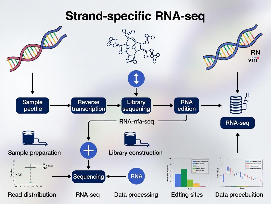
This article provides a comprehensive guide for researchers and drug development professionals on applying strand-specific RNA sequencing to detect and quantify viral RNA editing.
Leveraging Strand-Specific RNA-Seq for Precise Viral RNA Editing Detection
Abstract
This article provides a comprehensive guide for researchers and drug development professionals on applying strand-specific RNA sequencing to detect and quantify viral RNA editing. It covers foundational principles, including the critical advantage of resolving ambiguous reads from overlapping viral and host transcripts. The content details robust methodological pipelines, such as the dUTP protocol, for accurate application in virology. It further addresses common troubleshooting and optimization challenges and explores advanced validation techniques and comparative analyses against non-stranded approaches. By synthesizing these core intents, this resource empowers scientists to implement this powerful methodology, enhancing the accuracy of discoveries in viral pathogenesis and host-pathogen interactions.
The Critical Role of Strand-Specificity in Resolving Viral and Host Transcriptomes
RNA sequencing (RNA-seq) has emerged as a cornerstone technology in modern biology and clinical science, enabling comprehensive analysis of gene expression, transcript architecture, and functional genomics [1]. Within this field, the distinction between strand-specific (directional) and non-stranded (conventional) library preparation methods represents a critical methodological choice with profound implications for data accuracy and biological interpretation. Strand-specific RNA-seq deliberately preserves information about which genomic strand the original RNA transcript originated from, while non-stranded protocols lose this directional information during cDNA library construction [2] [3]. This preservation of strand information is not merely a technical detail but a fundamental requirement for accurate transcriptome analysis, particularly in complex genomes where overlapping transcripts, antisense regulation, and complex transcriptional architectures are common [4] [5].
The significance of strand-specific protocols extends to specialized research applications, including viral RNA editing detection research where precise mapping of viral transcripts and their editing patterns is essential. For researchers investigating viral RNA editing, strand-specific approaches enable unambiguous identification of RNA editing sites and accurate quantification of viral transcript isoforms without confusion from antisense transcripts or overlapping genes [6]. As the field progresses toward more sophisticated transcriptomic analyses, understanding and implementing strand-specific methodologies becomes increasingly vital for generating biologically meaningful results that accurately reflect the complexity of transcriptional regulation in both host and viral genomes.
Fundamental Concepts: How Strand-Specific RNA-Seq Works
The Technical Basis of Strand Information Loss and Preservation
In conventional non-stranded RNA-seq protocols, the process of converting single-stranded RNA into double-stranded cDNA for sequencing results in the complete loss of information regarding the original transcriptional strand. This occurs because random primers are used for both first- and second-strand cDNA synthesis, and the resulting sequencing products from sense and antisense transcripts become indistinguishable [3]. As illustrated in Figure 1, when two antisense transcripts from the same genomic locus undergo non-stranded library preparation, the final sequencing products are identical, making it impossible to determine the directionality of the original transcript directly from the sequencing data.
Figure 1: Comparison of stranded and non-stranded library preparation protocols
Principal Strand-Specific Methodologies
Several technical approaches have been developed to preserve strand information during RNA-seq library preparation, with the dUTP second strand marking method emerging as one of the most widely adopted and effective protocols [4] [5]. This method incorporates deoxyuridine triphosphate (dUTP) instead of deoxythymidine triphosphate (dTTP) during second-strand cDNA synthesis, effectively "labeling" the second strand. Prior to PCR amplification, the uracil-containing second strand is selectively degraded using Uracil-DNA Glycosylase (UDG), ensuring that only the first strand (complementary to the original RNA transcript) is amplified [3] [5]. This elegant biochemical approach preserves the strand orientation of the original RNA molecule throughout the sequencing process.
Alternative strand-specific methods include ligation-based approaches that attach asymmetric adapters to the 5' and 3' ends of RNA fragments before cDNA synthesis, directly preserving orientation information [4]. Another class of methods employs chemical modification of the RNA template itself, such as bisulfite treatment, to distinguish between the original strands [4]. Comparative evaluations have consistently identified the dUTP method as superior in terms of simplicity, strand specificity, data quality, and compatibility with downstream applications like paired-end sequencing [4] [5]. The robustness of the dUTP method is further demonstrated by its adoption in numerous commercial strand-specific library preparation kits, making it accessible to researchers across diverse biological disciplines.
Quantitative Comparison: Performance Metrics of Stranded vs Non-Stranded Approaches
Experimental Performance Benchmarks
Rigorous comparative analyses have quantified the performance differences between stranded and non-stranded RNA-seq approaches across multiple metrics. These comparisons reveal substantial advantages for strand-specific protocols in accurately capturing the true complexity of transcriptomes. Table 1 summarizes key performance characteristics derived from experimental comparisons.
Table 1: Performance comparison between stranded and non-stranded RNA-seq protocols
| Performance Metric | Non-Stranded Protocol | Stranded Protocol | Impact on Data Quality |
|---|---|---|---|
| Strand Specificity | 0% (inherently unstranded) | 97.4% [5] | Stranded enables correct strand assignment |
| Ambiguous Read Mapping | 6-30% of reads become ambiguous [2] | <3% ambiguous mapping | Drastic reduction in misassignment |
| Antisense Detection | Compromised or impossible | 1.5% of gene-mapping reads [7] | Enables comprehensive regulatory analysis |
| Ribosomal RNA Retention | ~7% with polyA selection [7] | Varies with depletion method | Method-dependent, not strandedness-dependent |
| Library Complexity | 88% unique paired-reads (control) [4] | 84% unique paired-reads (dUTP) [4] | Comparable performance |
| Differential Expression Accuracy | Higher false positives (>10%) and false negatives (>6%) [2] | Significant reduction in errors | More reliable differential expression calls |
Impact on Read Assignment and Transcript Quantification
The quantitative differences between stranded and non-stranded protocols have direct implications for transcript quantification and differential expression analysis. In non-stranded protocols, 28% of reads that were ambiguously mapped in unstranded workflows can be correctly reassigned to their proper transcriptional strand using strand-specific methods [2]. This dramatic improvement in mapping accuracy directly translates to more reliable gene expression estimates, particularly for genes with overlapping transcripts, antisense regulation, or complex genomic contexts.
The ability to accurately detect and quantify antisense transcription represents another significant advantage of strand-specific protocols. In comparative studies, stranded libraries identified approximately 20% more genes expressing antisense signal despite having lower read depth and higher ribosomal RNA retention compared to non-stranded approaches [7]. This enhanced sensitivity to antisense transcription is particularly valuable for viral RNA editing research, where comprehensive profiling of all viral transcripts is essential for understanding editing mechanisms and their functional consequences.
Practical Implementation: Strand-Specific Protocol for Viral RNA Editing Research
Detailed dUTP Protocol for Strand-Specific Library Construction
The following protocol outlines the key steps for implementing the dUTP-based strand-specific RNA-seq method, with particular considerations for viral RNA editing detection research:
RNA Isolation and Quality Control: Extract total RNA from infected cells or clinical samples using appropriate isolation methods. Assess RNA quality using capillary electrophoresis (e.g., Bioanalyzer RNA Integrity Number). Minimum requirement: RIN > 8.0 for optimal library construction.
Ribosomal RNA Depletion: Treat 100-1000 ng of total RNA with ribosomal depletion reagents (e.g., RiboZero Gold) rather than polyA selection to capture both polyadenylated and non-polyadenylated viral transcripts. Note: RiboZero demonstrates superior ribosomal depletion (<2.5% rRNA retention) compared to alternative methods (~65% retention) [5].
RNA Fragmentation: Fragment purified RNA to 200-300 nucleotides using metal-induced hydrolysis (e.g., magnesium buffer at 94°C for 5-15 minutes). Optimal fragmentation prevents bias in transcript coverage.
First-Strand cDNA Synthesis: Synthesize first-strand cDNA using random hexamers and reverse transcriptase with addition of Actinomycin D to prevent spurious DNA-dependent synthesis. Include purification steps to remove reagents before proceeding.
Second-Strand Synthesis with dUTP Incorporation: Perform second-strand synthesis using DNA Polymerase I with dUTP substituted for dTTP in the nucleotide mix. This creates the strand-specific marking essential for downstream directional information.
End Repair, A-Tailing, and Adapter Ligation: Process double-stranded cDNA using standard library preparation techniques, ensuring compatible adapters for your sequencing platform.
Uracil Digestion and Strand Selection: Treat with Uracil-DNA Glycosylase (UDG) to selectively degrade the dUTP-marked second strand, preserving only the original strand-complementary cDNA.
Library Amplification and Quality Control: Amplify the strand-selected library with 10-15 PCR cycles using proofreading polymerases. Validate library quality by capillary electrophoresis and quantify using fluorometric methods.
Special Considerations for Viral RNA Editing Studies
For research specifically focused on viral RNA editing detection, several modifications to the standard strand-specific protocol enhance sensitivity and accuracy:
Selective Enrichment for Viral Transcripts: Implement sequence-specific capture probes to enrich for viral RNAs, which often represent a small fraction of the total transcriptome in infected cells.
Controls for Editing Validation: Include synthetic RNA standards with known editing patterns to quantify detection sensitivity and specificity.
Duplicate Management: Monitor PCR duplication rates carefully, as these can be elevated in low-input protocols (approximately 20% in some kits) [7]. Utilize unique molecular identifiers (UMIs) to distinguish biological variants from technical artifacts.
Computational Pipeline Selection: Employ specialized bioinformatics tools like CADRES (Calibrated Differential RNA Editing Scanner) that combine sophisticated DNA/RNA variant calling with statistical analysis of editing depth to distinguish genuine RNA editing events from sequencing artifacts and DNA mutations [6].
Essential Reagents and Research Solutions
Table 2: Essential research reagents for strand-specific RNA-seq
| Reagent/Category | Specific Examples | Function in Protocol | Considerations for Viral RNA Research |
|---|---|---|---|
| Library Prep Kits | Illumina TruSeq Stranded mRNA, Takara Bio SMARTer Stranded Total RNA-Seq | Provides complete reagent systems | Select based on input requirements: TruSeq (100ng-1μg), SMARTer (1ng-10ng) [7] |
| Ribosomal Depletion Kits | RiboZero Gold, RiboMinus | Removes ribosomal RNA without polyA bias | RiboZero more effective (2.24% rRNA vs 65.7%) [5]; essential for non-polyadenylated viral RNAs |
| Strand-Specific Enzymes | Uracil-DNA Glycosylase (UDG) | Digests dUTP-marked second strand | Critical for strand selection; quality affects specificity |
| Reverse Transcriptase | SuperScript IV, Maxima H- | Synthesizes first-strand cDNA | High processivity improves coverage of structured viral RNA regions |
| RNA QC Instruments | Agilent Bioanalyzer, Fragment Analyzer | Assesses RNA integrity | Essential for input quality control; RIN >8.0 recommended |
| Unique Molecular Identifiers | UMIs (various vendors) | Tags individual molecules pre-amplification | Critical for distinguishing true biological variants from artifacts in viral populations |
Computational Analysis Considerations for Strand-Specific Data
Specialized Bioinformatics Approaches
The analysis of strand-specific RNA-seq data requires appropriate computational tools and parameters to fully leverage the preserved directional information. Modern aligners like STAR (Spliced Transcripts Alignment to a Reference) demonstrate superior performance for stranded data, mapping >94% of quality-trimmed reads with significantly faster processing times compared to alternatives like TopHat2 [5]. When using such tools, researchers must specify the correct library type parameter (e.g., "--outSAMstrandField intronMotif" for STAR) to ensure proper interpretation of strand information.
For viral RNA editing detection, specialized variant calling approaches are essential to distinguish genuine RNA editing events from DNA mutations and technical artifacts. Pipelines like CADRES implement sophisticated statistical frameworks that combine DNA and RNA sequencing data to identify differential RNA editing sites with high specificity [6]. These tools utilize a two-phase approach: first comparing genomic DNA and cDNA sequences to filter DNA variants, then applying statistical tests (e.g., Generalized Linear Mixed Models) to identify sites showing significant differences in editing levels across experimental conditions.
Data Quality Assessment Metrics
Quality control for strand-specific libraries should include verification of strand specificity through metrics that quantify the percentage of reads aligning to the expected transcriptional strand. High-quality dUTP libraries typically achieve >97% strand specificity [5]. Additional QC measures should include:
- Library complexity assessment to identify potential amplification biases
- Gene body coverage uniformity evaluation
- Ribosomal RNA contamination quantification (<5% ideal)
- Transcript assembly completeness using benchmarking tools
Strand-specific RNA-seq represents a fundamental methodological advancement over non-stranded approaches, providing critical transcriptional strand information that dramatically improves the accuracy of transcript quantification, annotation, and discovery. For viral RNA editing research, these protocols enable unambiguous identification of viral transcripts, precise mapping of editing sites, and comprehensive profiling of antisense transcription that may play important regulatory roles in the viral life cycle.
The implementation of robust strand-specific methods, particularly the dUTP-based protocol, combined with appropriate computational approaches like CADRES for editing detection, provides a powerful framework for investigating the complex landscape of viral RNA modifications. As the field continues to evolve, strand-specific RNA-seq will remain an essential tool for unraveling the mechanistic basis of RNA editing in viral pathogenesis and developing novel therapeutic strategies targeting these processes.
In virology, accurately interpreting the complex life cycles and regulatory mechanisms of viruses depends on obtaining complete and unambiguous transcriptomic data. A fundamental technical aspect, strand-specific RNA sequencing (RNA-seq), is non-negotiable for distinguishing the true nature of viral transcription, especially when investigating overlapping genes and pervasive antisense transcription. Non-stranded, or unstranded, RNA-seq protocols discard the information about which genomic strand an RNA molecule originated from, leading to significant ambiguities in data interpretation [8] [2].
This ambiguity is particularly problematic for RNA viruses, where the distinction between genomic and antigenomic strands is critical, and for DNA viruses with overlapping gene architectures. Preserving strand information is essential for accurately identifying authentic RNA editing events, such as Adenosine-to-Inosine (A-to-I) editing, which appears as A-to-G changes in sequencing reads and requires strand-specific data to distinguish from other mutations like single nucleotide polymorphisms (SNPs) or replication errors [8]. This application note details the methodologies and experimental protocols that leverage strand-specific RNA-seq to drive precise viral discovery.
Quantitative Impact of Strand-Specific Protocols
The choice between stranded and unstranded library preparation has a direct and measurable impact on data quality and biological interpretation. The table below summarizes key quantitative comparisons.
Table 1: Quantitative Impact of Strand-Specific RNA-Seq in Transcriptomic Analysis
| Metric | Unstranded Protocol | Strand-Specific Protocol | Biological Implication |
|---|---|---|---|
| Ambiguous Read Mapping | 6–30% of reads become ambiguous [2] | Ambiguity reduced by ~50% or more [2] | Drastically improved accuracy of transcript assignment and quantification. |
| False Positives in Differential Expression | Can inflate false positives by >10% [2] | Significantly reduced false positive rates [2] | More reliable identification of truly regulated genes and transcripts. |
| False Negatives in Differential Expression | Can inflate false negatives by >6% [2] | Significantly reduced false negative rates [2] | Enhanced sensitivity to detect subtle but biologically relevant expression changes. |
| Detection of Antisense Transcription | Often hidden or misinterpreted as sense signal [9] [2] | Enables clear identification and quantification [9] [2] | Unlocks the study of a crucial layer of viral and host gene regulation. |
| Identification of RNA Editing (e.g., A-to-I) | Compromised; cannot distinguish from SNP/ replication errors [8] | Enabled; A-to-G variation is specific to one strand [8] | Allows for validation of true RNA editing events in host-virus interactions. |
Core Methodologies and Experimental Protocols
Strand-Specific Library Preparation: The dUTP Second Strand Marking Method
The dUTP/Uracil DNA Glycosylase (UDG) method is a widely adopted and robust protocol for creating strand-specific RNA-seq libraries [10] [2].
Workflow Overview:
- RNA Fragmentation and First-Strand cDNA Synthesis: Isolated total or mRNA is fragmented and reverse-transcribed using random primers. This produces the first-strand cDNA, which is complementary to the original RNA template.
- Second-Strand Synthesis with dUTP Incorporation: The RNA template is degraded, and the second DNA strand is synthesized. The reaction mixture substitutes dTTP with dUTP. This results in a double-stranded cDNA molecule where the second strand contains uracil.
- Adapter Ligation and UDG Digestion: Double-stranded adapters are ligated to the cDNA ends. The library is then treated with Uracil DNA Glycosylase (UDG), which specifically digests the uracil-containing second strand.
- PCR Amplification: Only the original first strand remains as a template for PCR amplification, ensuring that every sequenced read retains the strand-of-origin information.
Diagram Title: Strand-Specific RNA-seq Library Prep (dUTP Method)
Validating RNA Editing in Viral Transcriptomes
Distinguishing true RNA editing from SNPs or sequencing errors is a major challenge. Strand-specific sequencing is a foundational step in a multi-tiered validation workflow [8] [11].
Recommended Validation Workflow:
In Silico Analysis:
- Prerequisite: Begin with strand-specific RNA-seq data to ensure A-to-G variations are genuine and not an artifact of symmetrical T-to-C variations from non-stranded data [8].
- Linkage Analysis: Analyze the linkage between variations in RNA-seq reads. True RNA editing events may show partial linkage due to the properties of the editing enzyme (ADAR), whereas SNPs are strongly linked, and sequencing errors are random [8].
- Hyperediting Pipeline: Use specialized bioinformatic pipelines (e.g., "hyperediting" tools) to detect clusters of editing events within single reads, which are strong indicators of authentic ADAR activity [8].
Orthogonal Experimental Validation:
- Mass Spectrometry (MS): Use MS to detect peptides with amino acid changes corresponding to the RNA-level edit (e.g., a lysine to arginine change for an A-to-I edit in a coding region). This provides direct protein-level validation [8].
- Use of ADAR-Deficient Cells: Repeat infection experiments in host cells where ADAR enzymes have been knocked out or knocked down. A significant reduction in A-to-G variations in the viral transcriptome provides functional evidence that the changes were ADAR-mediated editing events [8].
Diagram Title: RNA Editing Validation Workflow
The Scientist's Toolkit: Essential Research Reagents and Materials
Successful execution of strand-specific virology studies requires a suite of trusted reagents and computational tools.
Table 2: Essential Research Reagent Solutions for Strand-Specific Viral Transcriptomics
| Item/Category | Function/Description | Example Applications |
|---|---|---|
| Stranded RNA Library Prep Kits | Commercial kits implementing dUTP or ligation-based methods for strand preservation. | Illumina Stranded mRNA Prep, NEBNext Ultra II Directional RNA Library Prep Kit. Foundation for all downstream analysis. |
| Ribodepletion Reagents | Kits to remove ribosomal RNA (rRNA), crucial for viruses lacking poly-A tails or for studying non-coding RNAs. | Ribo-Zero Plus (Illumina), NEBNext rRNA Depletion Kit. Analysis of total viral RNA content. |
| ADAR-Deficient Cell Lines | Genetically engineered host cells (e.g., CRISPR/Cas9 KO) lacking RNA editing enzymes. | Functional validation of A-to-I editing events in viral RNAs [8]. |
| Reverse Genetics Systems | Platforms for generating recombinant viruses from cDNA. | To study the functional impact of specific RNA editing sites or disrupted antisense transcripts by introducing mutations into viral genomes. |
| Computational Tools: Hyperediting Pipelines | Specialized software to detect clusters of RNA editing events within single reads. | Validation of authentic ADAR activity in viral sequence data [8]. |
| Computational Tools: Graph-based Visualizers | Tools like Graphia Professional for visualizing complex RNA-seq assembly graphs and splice variants. | Resolving complex viral transcript architectures and overlapping units [12]. |
Visualizing Complex Viral Transcription Architectures
Overlapping genes are a common feature in viral genomes, used to maximize the coding capacity of a compact genome. Resolving these structures requires precise strand-of-origin data.
Diagram Title: Resolving Overlapping Viral Genes with Strand-Specific Data
Concluding Recommendations
For the virology research community, adopting strand-specific RNA-seq is a critical best practice. It is no longer a specialized option but a fundamental requirement for studies aiming to:
- Accurately quantify gene expression from viral and host genomes.
- Discover and characterize overlapping genes and antisense transcripts, which are widespread in viruses [13].
- Validate authentic RNA editing events and distinguish them from replication errors [8].
- Build robust and reproducible models of viral transcription and host-virus interactions.
The slight increase in protocol complexity and cost is vastly outweighed by the dramatic gain in data clarity, accuracy, and biological insight. For any investigative study of viral transcriptomics, strand information is non-negotiable.
In standard RNA sequencing (RNA-seq), the process of creating cDNA libraries loses a critical piece of information: which original genomic strand transcribed the RNA. This occurs because synthesis of randomly primed double-stranded cDNA, followed by adapter addition for next-generation sequencing, does not preserve the strand of origin [4] [14]. Strand-specific RNA-seq protocols solve this fundamental problem by deliberately preserving the orientation information of the original RNA transcript throughout the library preparation and sequencing process [2].
For researchers investigating viral RNA editing, this capability is not merely a technical refinement but a necessity for accurate biological interpretation. Preserving strand orientation allows scientists to correctly assign reads to sense or antisense transcripts, resolve overlapping genes, and accurately quantify gene expression—all essential for understanding viral pathogenesis and host-response mechanisms [2] [14]. Without strand information, distinguishing viral RNA editing events from transcriptional artifacts or antisense interference becomes significantly challenging, potentially leading to incorrect biological conclusions [2] [11].
Core Methodological Principles
Strand-specific RNA-seq methods employ distinct biochemical strategies to mark the original transcript strand, with two primary classes emerging as the most prevalent and reliable.
The dUTP Second-Strand Marking Method
The dUTP method has been extensively validated and identified as a leading protocol due to its robust performance and simplicity [4] [14]. This approach incorporates deoxyuridine triphosphate (dUTP) during second-strand cDNA synthesis, followed by enzymatic degradation of the uracil-containing strand before PCR amplification [14] [15]. The step-by-step mechanism operates as follows:
- First-Strand cDNA Synthesis: Reverse transcriptase generates the first cDNA strand complementary to the original RNA template using random hexamers or oligo(dT) primers.
- Second-Strand Synthesis with dUTP: DNA polymerase I synthesizes the second strand using deoxyadenosine, deoxyguanosine, deoxycytidine, and deoxyuridine triphosphates (dATP, dGTP, dCTP, dUTP) instead of thymidine triphosphate (dTTP) [15].
- Uracil-Containing Strand Degradation: Prior to PCR amplification, the enzyme Uracil-N-Glycosylase (UNG) recognizes and selectively degrades the second strand containing uracil bases [15].
- Library Amplification: Only the first strand (complementary to the original RNA) serves as a template for PCR amplification, ensuring all sequenced fragments maintain the correct orientation relative to the transcript of origin.
Table 1: Key Steps and Rationale of the dUTP Strand-Specific Method
| Step | Key Components | Biochemical Function | Strand Preservation Outcome |
|---|---|---|---|
| First-Strand Synthesis | Reverse transcriptase, dNTPs | Creates cDNA complement to RNA | Establishes complementary copy of original transcript |
| Second-Strand Synthesis | DNA polymerase I, dATP/dGTP/dCTP/dUTP | Replaces dTTP with dUTP in new strand | Labels newly synthesized strand for subsequent removal |
| Strand Degradation | Uracil-N-Glycosylase (UNG) | Enzymatically cleaves uracil-containing DNA | Eliminates second strand; only original complement remains |
| Library Amplification | DNA polymerase, PCR primers | Amplifies remaining first strand | Ensures all sequenced fragments maintain correct orientation |
The Directional Ligation Method
An alternative strategy relies on asymmetric adapter ligation to preserve strand information. This class of methods attaches distinct adapters to the 5' and 3' ends of cDNA fragments in a known orientation relative to the original RNA transcript [4]. Commercial implementations like the Swift and Swift Rapid kits employ "Adaptase" technology to directly ligate truncated adapters to single-stranded cDNA, eliminating the need for second-strand synthesis altogether [16]. The sequential logic of this approach includes:
- First-Strand Synthesis: Generation of cDNA from RNA templates.
- Asymmetric Adapter Ligation: Specific, non-interchangeable adapters are ligated to the 5' and 3' ends of the cDNA, maintaining the transcriptional orientation.
- Library Amplification: PCR amplification using primers complementary to the distinct adapters preserves the strand-of-origin information throughout sequencing.
Quantitative Impact on Data Accuracy
The choice between stranded and non-stranded protocols substantially influences downstream analytical outcomes, with stranded protocols providing demonstrably superior accuracy.
Resolution of Ambiguous Mappings
In complex transcriptomes where genes overlap on opposite strands, non-stranded RNA-seq cannot determine the transcriptional origin of reads, leading to ambiguous mappings. Research demonstrates that in the human genome, approximately 19% (about 11,000 genes) overlap with other genes transcribed from the opposite strand [14]. Empirical RNA-seq data reveals that stranded protocols reduce ambiguous read assignments by approximately 3.1% compared to non-stranded approaches [14]. This reduction directly corresponds to the resolution of gene overlap from opposite strands, enabling more precise quantification.
Table 2: Quantitative Comparison of Stranded vs. Non-Stranded RNA-seq
| Metric | Non-Stranded RNA-seq | Stranded RNA-seq | Experimental Basis |
|---|---|---|---|
| Ambiguous Reads | ~6.1% | ~2.94% | Whole blood mRNA-seq analysis [14] |
| Opposite Strand Overlap | Cannot be resolved | Fully resolved | Theoretical & empirical analysis [14] |
| Gene Expression Accuracy | Compromised for ~28% of ambiguous reads | Significantly improved | Human fibroblast benchmark [2] |
| Antisense Transcription Detection | Limited or impossible | Enabled | Evaluation of regulatory RNAs [2] [14] |
Enhanced Detection Capabilities
Strand-specific protocols unlock critical biological insights by enabling accurate detection of antisense transcription, which plays important regulatory roles in both cellular and viral systems [2] [14]. Studies have demonstrated that without strand information, antisense long non-coding RNAs can be misinterpreted as increased sense transcription or remain entirely undetected [2]. In viral research, this capability is particularly valuable for identifying antisense transcripts that may regulate viral persistence, latency, or reactivation.
Application in Viral RNA Editing Research
For viral RNA editing detection, strand-specific protocols provide essential experimental safeguards against misinterpretation.
The CADRES pipeline, designed for precise identification of RNA editing sites, emphasizes that comparing DNA and RNA sequences while assessing differential editing levels across conditions is crucial for distinguishing true RNA editing events from DNA mutations or sequencing artifacts [6]. Strand-specific sequencing enhances this discrimination by correctly identifying the transcribed strand, reducing false positives in editing detection.
Guidelines for RNA editing studies specifically recommend careful consideration of strand specificity in experimental design to avoid misinterpreting sequencing artifacts as genuine editing events [11]. This is particularly relevant for detecting C-to-U editing mediated by APOBEC enzymes, which can play significant roles in host-viral interactions [6].
Experimental Protocol: dUTP Method for Strand-Specific RNA-seq
Sample Preparation and RNA Isolation
- Starting Material: Extract total RNA using appropriate isolation methods (e.g., Trizol for tissues, column-based methods for cells). For viral studies, include appropriate biosafety precautions.
- RNA Quality Assessment: Determine RNA Integrity Number (RIN) using Agilent Bioanalyzer or similar platform. Use only high-quality RNA (RIN > 8.0) for library construction.
- PolyA+ RNA Selection: Isolate polyadenylated RNA using oligo(dT) magnetic beads according to manufacturer protocols. This enriches for mRNA while depleting ribosomal RNA [15].
Strand-Specific Library Construction
First-Strand cDNA Synthesis:
- Combine 0.5-1μg polyA+ RNA with random hexamer and oligo(dT) primers in reverse transcription buffer containing dNTPs and DTT [15].
- Add actinomycin D to inhibit spurious DNA-dependent synthesis [4].
- Incorporate SuperScript III reverse transcriptase and perform first-strand synthesis with temperature cycling (25°C for 10 min, 42°C for 45 min, 50°C for 25 min) [15].
Second-Strand Synthesis with dUTP Incorporation:
- Purify first-strand cDNA using gel filtration spin columns to remove dNTPs.
- Perform second-strand synthesis using E. coli DNA polymerase I and RNase H in buffer containing dATP, dGTP, dCTP, and dUTP (replacing dTTP) [15].
- Incubate at 16°C for 2 hours, then purify double-stranded cDNA using QIAquick columns.
Library Preparation and Uracil Strand Degradation:
- Fragment dsDNA by sonication or enzymatic methods to ~200-300bp.
- Repair DNA ends and add 'A' bases to 3' ends following standard Illumina library protocols.
- Critical Step: Before PCR amplification, treat with Uracil-N-Glycosylase (UNG) at 37°C for 15 minutes to degrade the dUTP-marked second strand [15].
- Perform PCR amplification with Illumina indexing primers to create the final sequencing library.
Quality Control and Sequencing
- Library QC: Assess library quality and concentration using Bioanalyzer and qPCR methods.
- Sequencing: Utilize Illumina platforms with recommended read lengths (2×100bp or 2×150bp) and appropriate sequencing depth (typically 20-40 million reads per sample for viral transcriptomes).
The Scientist's Toolkit: Essential Research Reagents
Table 3: Key Reagents for Strand-Specific RNA-seq Library Construction
| Reagent/Category | Specific Examples | Function in Protocol |
|---|---|---|
| Reverse Transcriptase | SuperScript III, SuperScript IV | Synthesizes first-strand cDNA from RNA templates with high fidelity and processivity |
| Nucleotide Mixes | dATP, dGTP, dCTP, dUTP | dUTP substitutes for dTTP in second-strand synthesis to enable strand marking |
| Strand-Degrading Enzymes | Uracil-N-Glycosylase (UNG) | Recognizes and cleaves uracil-containing DNA strands for selective removal |
| Second-Strand Synthesis Enzymes | DNA Polymerase I, RNase H | Synthesizes the second cDNA strand while degrading the RNA template |
| Library Preparation Kits | Illumina TruSeq Stranded mRNA, SMARTer Stranded Total RNA | Commercial kits that incorporate dUTP or ligation-based strand marking |
| RNA Selection Beads | Oligo(dT) Magnetic Beads | Enriches polyadenylated mRNA from total RNA inputs |
| Library Amplification | Illumina Indexing Primers, PCR Master Mix | Amplifies final strand-specific libraries with unique sample indexes |
Workflow Visualization
The following diagram illustrates the complete dUTP-based strand-specific RNA-seq workflow:
Strand-specific RNA-seq protocols, particularly the dUTP method, provide an essential foundation for accurate transcriptome characterization by preserving the strand origin of sequenced fragments. For viral RNA editing research, this capability is indispensable for correctly identifying editing events, resolving complex transcriptional overlaps, and detecting regulatory antisense transcripts. The methodological rigor afforded by strand-specific approaches significantly enhances data interpretation reliability, enabling more confident conclusions about viral pathogenesis and host-response mechanisms. As transcriptomic analyses continue to advance, adopting strand-specific protocols as a standard practice ensures maximal biological insight from RNA-seq experiments.
Strand-specific RNA sequencing (RNA-Seq) is a powerful advancement in transcriptome analysis that preserves the original orientation of RNA transcripts. Among various methods, the dUTP second-strand marking technique has emerged as a leading protocol, particularly for applications requiring high accuracy in transcript annotation and detection of antisense transcription. This is especially critical in viral RNA editing research, where distinguishing the true strand origin of RNA molecules is essential for accurately identifying host-driven RNA editing events, such as adenosine-to-inosine (A-to-I) deamination, and for resolving complex viral transcriptomes. The dUTP method, recognized for its robust performance and compatibility with standard Illumina sequencing platforms, provides the strand specificity necessary to overcome the fundamental limitations of non-stranded approaches [17] [4] [14].
Principles of the dUTP Strand-Specific RNA-Seq
The core principle of the dUTP method lies in the biochemical labeling and subsequent selective degradation of the second cDNA strand, thereby preserving only the first strand that is complementary to the original RNA template for sequencing. This process ensures that the resulting sequence reads can be unambiguously mapped to their strand of origin.
In a standard non-stranded RNA-Seq protocol, double-stranded cDNA is synthesized from RNA templates, and both strands are sequenced without retaining information about which strand was originally transcribed. This leads to a significant challenge: when a genomic locus has genes on both strands, it becomes impossible to determine from which strand a particular read originated [14] [3]. The dUTP method solves this by incorporating deoxyuridine triphosphate (dUTP) instead of deoxythymidine triphosphate (dTTP) during the synthesis of the second cDNA strand. This incorporation "marks" the second strand. Prior to the final PCR amplification, the enzyme Uracil-DNA-Glycosylase (UDG) is used to specifically degrade the uracil-containing second strand. Consequently, only the first strand is amplified and sequenced, preserving the strand information of the original mRNA throughout the entire process [17] [18].
Comparative Performance of Strand-Specific Protocols
A comprehensive comparative analysis of seven strand-specific RNA-Seq protocols identified the dUTP method as one of the top-performing approaches. The evaluation used the well-annotated S. cerevisiae transcriptome as a benchmark and assessed methods based on critical quality metrics [4].
Table 1: Performance Comparison of Leading Strand-Specific RNA-Seq Methods
| Performance Metric | dUTP Method | Illumina RNA Ligation Method | Standard Non-Stranded Method (Control) |
|---|---|---|---|
| Strand Specificity | High (Exact values provided in [4]) | High | Not Applicable |
| Library Complexity (Paired-end) | 84% unique paired-reads | Not detailed for paired-end | 88% unique paired-reads |
| Evenness of Coverage | High agreement with known annotations | High agreement with known annotations | Baseline |
| Quantitative Accuracy | Accurate for expression profiling | Accurate for expression profiling | Baseline |
| Ease of Use | Relatively simple protocol | Requires specialized RNA adaptors | Simplest protocol |
This analysis concluded that the dUTP method and the Illumina RNA ligation method were the leading protocols. The dUTP method was particularly favored because it benefits from the availability of paired-end sequencing, which provides more accurate library complexity measurements and better resolution of transcript isoforms [4].
Step-by-Step Experimental Protocol
The following section details a modified dUTP protocol compatible with the Illumina TruSeq kit, enabling robust strand-specific library construction within two days [17] [18].
RNA Isolation and mRNA Enrichment
Begin with high-quality total RNA (0.1–4 μg). Isate the polyadenylated (polyA) mRNA fraction using oligo(dT) magnetic beads. This enrichment step is typical for standard RNA-Seq and focuses the sequencing on protein-coding transcripts [17] [19].
RNA Fragmentation and First-Strand Synthesis
Chemically fragment the purified mRNA to the desired size distribution (e.g., 200-300 bp). Use random hexamer primers and reverse transcriptase to synthesize the first-strand cDNA. This first strand is complementary to the original RNA template [17].
Second-Strand Synthesis with dUTP Incorporation
Synthesize the second strand of cDNA using a reaction mix where dTTP is replaced with dUTP. This creates a double-stranded cDNA product where the second strand is biochemically marked with uracil, while the first strand contains thymine [17] [18].
End-Repair, A-Tailing, and Adapter Ligation
Process the double-stranded cDNA fragments following a standard Illumina library preparation workflow:
- End-Repair: Convert the fragmented cDNA ends to blunt ends.
- A-Tailing: Add a single 'A' nucleotide to the 3' ends to facilitate ligation to the 'T'-overhang of Illumina adapters.
- Adapter Ligation: Ligate indexed sequencing adapters to the cDNA fragments [17].
Strand Selection via UDG Digestion and Size Selection
This is the critical, strand-defining step. Incubate the adapter-ligated library with Uracil-DNA-Glycosylase (UDG). This enzyme selectively degrades the second cDNA strand that contains uracil. The result is a library consisting only of the first-strand cDNA molecules [17] [18]. Finally, perform size selection (e.g., using gel electrophoresis or SPRI beads) to isolate cDNA fragments of the desired length for sequencing [17] [4].
Library Amplification and Quantification
Perform a limited-cycle PCR to amplify the remaining strand-specific library. Purify the final library and quantify it using methods such as qPCR or bioanalyzer before sequencing on an Illumina platform [17].
Diagram 1: dUTP Strand-Specific RNA-Seq Workflow.
The Scientist's Toolkit: Essential Research Reagents
Table 2: Key Reagents for dUTP Strand-Specific RNA-Seq Library Construction
| Reagent / Kit | Function in the Protocol |
|---|---|
| Oligo(dT) Magnetic Beads | Enriches for polyadenylated mRNA from total RNA. |
| Reverse Transcriptase & Random Hexamers | Synthesizes the first-strand cDNA from fragmented mRNA. |
| dNTP/dUTP Mix (dATP, dCTP, dGTP, dUTP) | Incorporates dUTP instead of dTTP during second-strand synthesis to mark the strand. |
| Uracil-DNA-Glycosylase (UDG) | The key enzyme for strand selection; degrades the dUTP-marked second cDNA strand. |
| Illumina TruSeq or Compatible Kit | Provides reagents for end-repair, A-tailing, adapter ligation, and PCR amplification. |
| Size Selection Beads (e.g., SPRI beads) or Gel Matrix | Purifies and selects cDNA fragments of the desired size range for sequencing. |
Application in Viral RNA Editing Detection Research
The dUTP method's high strand specificity is not just a technical improvement; it is a critical requirement for accurately detecting and validating RNA editing in viruses like SARS-CoV-2.
In non-stranded RNA-Seq, an A-to-I editing event (recorded as A-to-G in the RNA) can manifest as both A-to-G variations in the sense strand and T-to-C variations in the antisense strand. This "symmetry problem" makes it impossible to distinguish true A-to-I editing from replication errors or single nucleotide polymorphisms (SNPs) that are incorporated into the viral genome, as both can produce T-to-C variations [8]. Strand-specific RNA-Seq directly resolves this ambiguity. Because the protocol preserves the original RNA orientation, a true A-to-I editing event will appear exclusively as an A-to-G variation in reads originating from the sense strand. This eliminates the confounding T-to-C signal from the antisense strand, thereby significantly improving the signal-to-noise ratio in the search for authentic RNA editing sites [8].
Diagram 2: Strand-Specific RNA-Seq Resolves Ambiguity in Viral RNA Editing.
Furthermore, stranded data is essential for advanced in silico validation approaches, such as linkage analysis, which examines the co-occurrence of multiple editing events on the same RNA molecule. This analysis depends on knowing the precise strand orientation of reads to correctly establish linkage between variations [8].
Quantitative Impact on Data Accuracy
The practical advantages of stranded RNA-Seq, as enabled by the dUTP method, translate into direct, measurable improvements in data quality and interpretation.
Table 3: Impact of Stranded vs. Non-Stranded RNA-Seq on Read Assignment
| Data Attribute | Stranded RNA-Seq (dUTP) | Non-Stranded RNA-Seq |
|---|---|---|
| Percentage of Ambiguous Reads | ~2.94% (arising only from same-strand overlaps) | ~6.1% (arising from same-strand and opposite-strand overlaps) |
| Confidence in Antisense RNA Quantification | High. Enables accurate identification and quantification. | Low. Difficult to distinguish from sense transcription. |
| Accuracy for Overlapping Opposite Strand Genes | High. Correctly assigns reads to the transcribed strand. | Low. Reads are ambiguously assigned to both gene models. |
Research has demonstrated that a significant portion (approximately 19% or 11,000 genes in the Gencode annotation) of the genome consists of genes that overlap on opposite strands [14]. In non-stranded RNA-Seq, reads falling within these overlapping regions cannot be assigned confidently, leading to a higher rate of "ambiguous" reads (~6.1%) compared to stranded RNA-Seq (~2.94%). This ~3.1% drop in ambiguity directly corresponds to the resolution of overlaps from opposite strands, leading to more accurate quantification of gene expression for thousands of genes [14]. This precision is fundamental in viral research, where overlapping genes are common, and accurately determining their individual expression levels is critical for understanding viral replication and pathogenesis.
Implementing Robust Strand-Specific RNA-Seq Workflows for Viral RNA Editing
This application note provides a detailed, practical protocol for implementing a strand-specific RNA sequencing (RNA-Seq) pipeline optimized for the detection and analysis of viral RNAs. Within the broader context of viral RNA editing research, maintaining strand-of-origin information is crucial for accurately distinguishing viral genomic RNA from complementary transcripts and antisense RNA species that play key regulatory roles in viral replication. The workflow encompasses every stage from initial sample preparation through computational viral read mapping, with special emphasis on experimental design considerations that enhance sensitivity for viral detection in complex biological samples. This comprehensive guide serves researchers, scientists, and drug development professionals seeking to implement robust, strand-aware sequencing approaches for virology and antiviral therapeutic development.
RNA sequencing has revolutionized transcriptome analysis, providing unprecedented resolution for studying gene expression and RNA biology. Strand-specific RNA-Seq represents a critical advancement over conventional protocols by preserving the directional origin of each transcript [20] [14]. This capability is particularly valuable in viral research, where many viruses produce overlapping and antisense transcripts that regulate infection cycles [20]. Without strand information, it is impossible to distinguish whether a read originated from the positive-sense viral genome or from a complementary negative-sense transcript, potentially leading to misinterpretation of viral gene expression and regulatory mechanisms.
The dUTP second-strand marking method has emerged as a leading protocol for stranded library preparation due to its superior performance in strand specificity and data quality [17] [14]. This technique incorporates deoxyuridine triphosphates during second-strand cDNA synthesis, enabling enzymatic degradation of this strand before amplification and ensuring that only the original RNA orientation is sequenced. For viral detection studies, this approach provides the critical advantage of unequivocally identifying the transcriptional strand origin of viral reads, which is essential for understanding viral replication dynamics and host-pathogen interactions.
Strand-Specific Library Preparation Protocol
RNA Extraction and Quality Control
The initial RNA quality fundamentally impacts downstream sequencing success and viral detection sensitivity. For studies focusing on viral transcripts, consider that some viral RNAs may be non-polyadenylated or contain unusual structural features.
- Input Material: The protocol typically requires 25 ng - 1 µg of total RNA as starting material [21]. When working with viral samples, the input amount may need optimization based on expected viral RNA abundance.
- RNA Integrity: Assess RNA quality using the RNA Integrity Number (RIN), with values >7.0 generally recommended for high-quality sequencing [21]. However, for samples with expected high degradation (such as clinical specimens), ribosomal RNA depletion may be preferable to polyA selection.
- Quality Metrics: Evaluate 260/280 and 260/230 ratios to ensure minimal protein or chemical contamination. Visual inspection of electropherograms from Bioanalyzer or TapeStation systems should show distinct ribosomal peaks.
For viral detection in biologics, efficient nucleic acid extraction is critical, particularly for breaking down viral envelopes and capsids to release viral RNA [22] [23]. The extraction method should be validated for the specific virus targets of interest, as recovery efficiency varies significantly among viruses with different physicochemical properties.
rRNA Depletion and PolyA Selection Considerations
The choice between ribosomal RNA depletion and polyA enrichment significantly impacts viral detection capability:
Table 1: Comparison of RNA Selection Methods for Viral Detection
| Method | Advantages for Viral Studies | Limitations | Recommended Applications |
|---|---|---|---|
| PolyA Selection | Simplifies library complexity; reduces sequencing costs; enriches for eukaryotic mRNA | May miss non-polyadenylated viral RNAs; requires intact RNA | Studies focused on host response to infection; viruses with polyA tails |
| rRNA Depletion | Retains non-polyadenylated transcripts; works with degraded RNA | Higher background; requires more sequencing depth | Discovery-oriented viral detection; surveillance of unknown viruses |
| Total RNA | Maximizes detection of all RNA species | High ribosomal content; requires extensive depletion | Comprehensive virome studies; detection of diverse viral families |
Ribosomal RNA constitutes approximately 80% of cellular RNA [21], and its removal is essential for efficient viral transcript detection. Probe-based depletion methods (e.g., RNase H-mediated degradation) show greater reproducibility compared to bead-based subtraction approaches [21]. Note that some depletion methods may inadvertently remove viral RNAs with similarity to host rRNA sequences.
Strand-Specific cDNA Library Construction
The core strand-specific protocol follows these key steps [17]:
- RNA Fragmentation: Fragment purified mRNA to 200-300 nucleotides using divalent cations under elevated temperature.
- First-Strand cDNA Synthesis: Use random hexamers and reverse transcriptase to synthesize cDNA from fragmented RNA.
- Second-Strand Synthesis with dUTP Incorporation: Incorporate dUTP instead of dTTP during second-strand synthesis, creating a strand-specific mark.
- End Repair and A-Tailing: Polish fragment ends and add single adenosine overhangs for adapter ligation.
- Adapter Ligation: Ligate platform-specific sequencing adapters to both ends of cDNA fragments.
- dUTP Strand Degradation: Treat with Uracil-DNA-Glycosylase (UDG) to selectively degrade the second strand containing uracils.
- Library Amplification: Perform limited-cycle PCR to enrich for adapter-ligated fragments.
The dUTP marking method effectively preserves strand information, with one study demonstrating a 3.1% reduction in ambiguous mappings compared to non-stranded approaches [14]. This is particularly valuable for identifying antisense viral transcripts that may regulate gene expression.
Sequencing Considerations for Viral Detection
Read Length and Depth Optimization
Sequencing parameters must balance cost with sufficient sensitivity for viral detection, particularly for low-abundance viral transcripts:
Table 2: RNA-Seq Sequencing Recommendations for Viral Detection
| Application | Recommended Read Length | Recommended Depth | Rationale |
|---|---|---|---|
| Viral Gene Expression Profiling | 50-75 bp single-end | 30-60 million reads | Sufficient for quantification of moderate to high abundance viral transcripts |
| Viral Transcriptome Assembly | 75-100 bp paired-end | 100-200 million reads | Longer reads facilitate assembly of novel viral transcripts and isoforms |
| Low Abundance Viral Detection | 75-100 bp paired-end | 60-100 million reads | Increased depth enhances detection sensitivity for rare viral transcripts |
| Multiplexed Screening | 50-75 bp single-end | 5-25 million reads per sample | Cost-effective for large sample numbers when targeting high-titer viruses |
For reference, a multi-laboratory study demonstrated that 10^4 genome copies/mL of spiked viruses could be reliably detected using short-read sequencing technologies [23]. Some optimized workflows achieved detection limits as low as 10^2 genome copies/mL for certain viruses, highlighting the importance of protocol optimization.
Experimental Design and Replication
Appropriate experimental design is crucial for meaningful viral detection studies:
- Biological Replicates: A minimum of three replicates per condition is standard, though more may be needed for samples with high biological variability [24].
- Controls: Include positive controls (samples with known viral content) and negative controls (virus-free samples) to establish detection limits and specificity.
- Spike-in Controls: Consider using exogenous RNA spike-ins at known concentrations to monitor technical performance and quantitative accuracy.
The high titer of production viruses in some biological samples can create background challenges; one study successfully detected spiked adventitious viruses in backgrounds of 1-5 × 10^9 genome copies/mL of adenovirus 5 [23].
Bioinformatics Pipeline for Viral Read Mapping
Preprocessing and Quality Control
Raw sequencing data requires thorough quality assessment and cleaning before alignment:
- Quality Control: Use FastQC or multiQC to evaluate base quality scores, sequence duplication rates, adapter contamination, and overall read quality [24].
- Adapter Trimming: Remove adapter sequences and low-quality bases using tools such as Trimmomatic, Cutadapt, or fastp [24].
- Quality Filtering: Discard reads with excessive ambiguous bases or overall poor quality.
Post-trimming quality assessment ensures data meets minimum standards for downstream analysis. Over-trimming should be avoided as it reduces data quantity and can impact mapping sensitivity for divergent viruses.
Read Alignment Strategies
Two principal alignment approaches are available for viral detection:
Host Subtraction Approach: First align reads to the host reference genome (e.g., human GRCh38) using splice-aware aligners like STAR or HISAT2 [24]. Unmapped reads are then extracted and aligned to viral reference databases. This approach efficiently reduces host background but may miss viral reads with similarity to host sequences.
Direct Composite Alignment: Create a combined reference containing both host and viral genomes, then align all reads simultaneously. This approach prevents loss of viral reads that might have weak similarity to host sequences but requires more computational resources.
For strand-specific data, ensure that alignment software is configured to recognize library strandedness, typically using the "fr-firststrand" parameter in common aligners.
Viral Read Identification and Quantification
After alignment, specialized approaches are needed for viral detection:
- Reference-Based Mapping: Align to comprehensive viral databases (RVDB, RefSeq Viruses) using tools like BWA, Bowtie2, or STAR [23].
- Abundance Estimation: Use featureCounts or HTSeq-count to quantify reads mapping to viral features [24]. For strand-specific libraries, ensure counting is performed with appropriate strandness parameters.
- Threshold Determination: Establish minimum read thresholds for viral detection based on negative controls and statistical significance. The multi-laboratory study used both targeted and non-targeted bioinformatic analyses, with targeted analysis showing greater sensitivity for expected viruses [23].
Advanced approaches include de novo assembly of unmapped reads followed by BLAST comparison to viral databases, which can detect novel or highly divergent viruses not present in reference databases.
Research Reagent Solutions
Table 3: Essential Research Reagents for Strand-Specific Viral RNA-Seq
| Reagent/Category | Function | Examples/Options |
|---|---|---|
| RNA Extraction Kits | Isolation of high-quality RNA from various matrices | Column-based methods; magnetic bead systems |
| rRNA Depletion Kits | Removal of abundant ribosomal RNA | Probe-based subtraction; RNase H-mediated degradation |
| Stranded Library Prep Kits | Construction of strand-specific cDNA libraries | dUTP-based methods; ligation-based methods |
| Sequence Adapters | Platform-specific sequences for cluster generation | Illumina TruSeq; IDT for Illumina |
| Quality Control Assays | Assessment of RNA and library quality | Bioanalyzer; TapeStation; qPCR quantification |
| Alignment Software | Mapping reads to reference sequences | STAR; HISAT2; BWA; Bowtie2 |
| Viral Reference Databases | Reference sequences for viral identification | RVDB; RefSeq Viruses; NCBI Viral Genome Database |
The implementation of a robust strand-specific RNA-Seq pipeline for viral detection requires careful consideration at each step, from sample preparation through bioinformatic analysis. The dUTP second-strand marking method provides high-quality strand-specific libraries that enable unambiguous identification of viral transcript orientation, which is crucial for understanding viral gene expression and regulation. By following the detailed protocols and considerations outlined in this application note, researchers can establish a sensitive and specific workflow for viral detection and characterization that supports both basic virology research and applied drug development efforts. As sequencing technologies continue to advance, the integration of strand-specific information will remain essential for unraveling the complex interactions between viruses and their hosts.
Library Construction Showdown: dUTP vs. RNA Ligation Methods for Viral Samples
Strand-specific RNA sequencing (RNA-seq) is a powerful tool that preserves the original orientation of transcripts, enabling precise mapping of viral RNA molecules to their genomic strand of origin. This capability is critical for detecting RNA editing events, characterizing antisense transcription, and accurately quantifying gene expression in overlapping genomic regions—common features in viral genomes. Among the various strategies for constructing strand-specific libraries, the dUTP second-strand marking and RNA ligation methods have emerged as leading protocols. This application note provides a detailed, evidence-based comparison of these two methods, focusing on their application in viral RNA editing detection research. We summarize quantitative performance data, provide detailed experimental protocols, and outline key reagent solutions to guide researchers and drug development professionals in selecting the optimal library construction method for their virology studies.
Performance Comparison: dUTP vs. RNA Ligation Methods
Comprehensive comparative analyses have evaluated multiple strand-specific RNA-seq protocols across critical performance metrics. The table below synthesizes key findings from these studies to facilitate direct comparison between dUTP and RNA ligation methods.
Table 1: Performance comparison between dUTP and RNA ligation methods for strand-specific RNA-seq
| Performance Metric | dUTP Method | RNA Ligation Method | Experimental Context |
|---|---|---|---|
| Strand Specificity | >90% [16] | >97% [25] | Universal Human Reference RNA (UHRR); human embryonic stem cells |
| Library Complexity (Unique Paired Reads) | 84% [4] | Not reported for paired-end | S. cerevisiae polyA+ RNA |
| Compatibility with Paired-End Sequencing | Yes (benefits significantly) [4] [14] | Limited (primarily single-end) [4] | Protocol design evaluation |
| Coverage Uniformity | Even coverage across gene body [16] | 5' bias observed [25] | Human transcriptome coverage |
| Sensitivity to Long Transcripts | Accurate quantification [26] | Underestimates long transcripts [26] | Comparison of TruSeq, SMARTer, and TeloPrime |
| Detection of Antisense/Overlapping Genes | Accurate [14] | Accurate [4] | Evaluation with overlapping genomic loci |
Experimental Protocols for Viral RNA Studies
dUTP Second-Strand Marking Protocol
The dUTP method incorporates uracil during second-strand synthesis, enabling selective degradation of this strand before amplification to preserve strand information.
Table 2: Key research reagents for the dUTP method
| Reagent | Function | Example Product |
|---|---|---|
| Oligo(dT) or Gene-Specific Primers | Reverse transcription priming | Thermo Scientific SuperScript Reverse Transcriptase |
| dUTP Nucleotides | Second-strand labeling | Illumina TruSeq Stranded mRNA Kit |
| Uracil-DNA Glycosylase (UDG) | Degradation of second strand | New England Biolabs UDG |
| DNA Polymerase (dUTP-Compatible) | cDNA amplification | Phusion High-Fidelity DNA Polymerase |
Detailed Workflow:
RNA Fragmentation and Priming: Fragment viral RNA and prime with oligo(dT) or sequence-specific primers targeting viral genomic or antigenomic strands.
First-Strand cDNA Synthesis: Synthesize first-strand cDNA using reverse transcriptase with dNTPs (including dTTP, not dUTP at this stage).
Second-Strand Synthesis: Incorporate dUTP instead of dTTP during second-strand synthesis, creating a uracil-labeled complementary strand.
End Repair and A-Tailing: Repair ends of double-stranded cDNA and add adenine nucleotide overhangs for adapter ligation.
Adapter Ligation: Ligate platform-specific sequencing adapters to cDNA fragments.
UDG Treatment: Treat with Uracil-DNA Glycosylase (UDG) to selectively degrade the dUTP-labeled second strand, preserving only the original first strand.
Library Amplification: Amplify the strand-specific library using PCR with indexed primers for multiplexing.
RNA Ligation Protocol
The RNA ligation method preserves strand information by directly ligating adapters to RNA fragments before cDNA synthesis, maintaining the original transcript orientation throughout library construction.
Table 3: Key research reagents for the RNA ligation method
| Reagent | Function | Example Product |
|---|---|---|
| Fragmentation Buffer | Controlled RNA fragmentation | Illumina Fragmentation Reagent |
| T4 RNA Ligase | Adapter ligation to RNA | New England Biolabs T4 RNA Ligase |
| 3' Di-deoxycytosine Adapters | Prevents self-ligation | IDT Swift RNA Kit |
| RNase Inhibitor | Prevents RNA degradation | Thermo Scientific RNaseOUT |
Detailed Workflow:
RNA Fragmentation: Fragment viral RNA using heat or enzymatic methods to optimal size for sequencing.
Adapter Ligation (RNA Level): Directly ligate specific adapters to the 3' end of fragmented RNA using T4 RNA ligase. Some protocols also ligate 5' adapters at this stage.
Reverse Transcription: Synthesize first-strand cDNA using reverse transcriptase with primers complementary to the 3' adapter.
cDNA Purification: Remove excess adapters and reagents to prevent interference with downstream steps.
Second-Strand Synthesis: Synthesize second-strand cDNA using DNA polymerase.
Library Amplification: Amplify the full library using PCR with indexed primers to add complete adapter sequences and indexes for multiplexing.
Method Selection Guide for Viral RNA Editing Research
Choosing between dUTP and RNA ligation methods requires careful consideration of research goals, viral genome characteristics, and practical laboratory constraints.
Table 4: Method selection guide based on research applications
| Research Application | Recommended Method | Rationale |
|---|---|---|
| Detection of C>U or A>I RNA Editing Sites | dUTP [6] | Paired-end sequencing enhances accuracy for identifying differential RNA variants |
| Antisense Transcription Profiling | dUTP [14] [3] | Superior strand specificity resolves overlapping transcripts from opposite strands |
| Viral Genome Annotation | RNA Ligation [4] [3] | High strand specificity supports accurate transcript boundary mapping |
| Expression Quantification of Overlapping Genes | dUTP [14] | Resolves ambiguity in gene assignment for dense viral genomes |
| Studies with Limited RNA Input | RNA Ligation (LM-Seq) [25] | Effective with as little as 10 ng total RNA |
| High-Throughput Screening | dUTP (Swift/IDT kits) [16] | Compatible with automation and multiplexing |
Both dUTP and RNA ligation methods provide high-quality strand-specific RNA-seq data suitable for viral RNA editing detection research, yet they offer distinct advantages. The dUTP method excels in applications requiring paired-end sequencing, provides higher library complexity, and enables more accurate quantification of overlapping transcripts—critical for characterizing complex viral transcriptomes [4] [14]. The RNA ligation method demonstrates exceptional strand specificity and can be more suitable for low-input samples [25].
For viral RNA editing studies specifically, the dUTP method's compatibility with paired-end sequencing provides significant advantages for detecting and validating RNA editing sites, as paired-end reads offer more comprehensive coverage of viral transcripts. Furthermore, the dUTP protocol's robustness across varying input amounts makes it suitable for diverse sample types, including clinical viral isolates with limited material [16].
When investigating cytidine deaminase activity (e.g., APOBEC-mediated C>U editing) in viral genomes, strand-specific information is essential to distinguish true RNA editing events from DNA-level mutations or sequencing artifacts [6] [27]. Both methods facilitate this discrimination, though the dUTP approach provides greater flexibility in sequencing strategies.
Researchers should select their library construction method based on priority applications: the dUTP method for maximum data quality and analytical flexibility, and RNA ligation for specific applications requiring direct RNA manipulation or when working with extremely limited viral RNA samples. As viral RNA editing research advances, both methods will continue to play crucial roles in unraveling the complex interactions between viral pathogens and host editing mechanisms.
RNA editing, particularly Adenosine-to-Inosine (A-to-I) conversion, represents a critical post-transcriptional process that increases transcriptome diversity. In virology, distinguishing these true RNA editing events from underlying genetic variants in the host or virus is essential for understanding host-virus interactions and viral evolution [28] [11]. This application note details a robust bioinformatics workflow that integrates DNA-Seq and RNA-Seq data to accurately identify bona fide RNA editing sites, with specific considerations for strand-specific RNA-seq protocols used in viral RNA editing detection research.
The core challenge stems from the fact that both single nucleotide variants (SNVs) in the genome and RNA editing events appear as mismatches when RNA-seq reads are aligned to a reference genome [29]. Without DNA-seq data from the same sample, one cannot confidently segregate these two types of sequence variations. This is particularly pertinent in viral research, where accurate identification of RNA edits can illuminate mechanisms of viral persistence, latency, and immune evasion.
Background and Key Concepts
RNA Editing and its Detection via Sequencing
A-to-I RNA editing, catalyzed by ADAR (Adenosine Deaminases Acting on RNA) enzymes, is the most prevalent RNA editing type in animals [28]. As inosine (I) is base-called as guanosine (G) during reverse transcription and sequencing, A-to-I editing is detected as A-to-G mismatches in aligned RNA-seq data [28] [30]. A less frequent but equally important type is Cytidine-to-Uridine (C-to-U) editing, mediated by APOBEC enzymes, which appears as C-to-T changes [31].
The Critical Role of Strand-Specific RNA-Seq
In the context of viral transcriptomics, strand-specific RNA-seq protocols are invaluable. They preserve the information about which genomic strand the RNA originated from, allowing researchers to unambiguously determine the direction of transcription [2]. This is crucial for:
- Accurately assigning edits to viral genes, especially in complex viral transcription units.
- Resolving overlapping transcripts from antisense or complementary viral strands.
- Eliminating a significant source of false positives caused by misassignment of reads from the opposite strand [32] [2]. Non-stranded protocols can misattribute antisense transcription to sense strands, inflating apparent mismatch counts and confounding downstream analysis.
Computational Strategies and Tool Selection
Two primary computational strategies exist for identifying RNA editing events, with the integrated DNA+RNA approach being the gold standard for minimizing false positives.
Table 1: Comparison of Computational Strategies for RNA Editing Detection
| Strategy | Description | Advantages | Limitations | Key Tools |
|---|---|---|---|---|
| Integrated DNA+RNA Analysis | Directly compares matched DNA-Seq and RNA-Seq from the same sample to filter genomic variants. | Highest accuracy; effectively removes false positives from private SNPs and somatic mutations. | Requires additional DNA sequencing; computationally intensive. | CADRES [31], JACUSA2 [33], GATK Best Practices [31] |
| RNA-Seq Only Analysis | Relies on RNA-Seq data alone, using filters (e.g., known SNPs, splice regions) and features (e.g., editing type) to predict sites. | Cost-effective; usable when DNA-Seq is unavailable. | Higher false positive rate; cannot filter novel or sample-specific genetic variants. | REDItools [34], SPRINT [33], L-GIREMI (for long-read data) [30] |
The "integrated" strategy, as implemented in the CADRES pipeline, operates in two phases: the RNA–DNA Difference (RDD) phase to remove genomic variants, and the RNA-RNA Difference (RRD) phase to identify sites with statistically significant differences in editing levels across conditions [31]. This is crucial for identifying condition-specific editing events, such as those induced during viral infection.
Benchmarking of Detection Tools
A benchmark study evaluating several RNA editing detection tools using ADAR1-knockout HEK293T cell data provides critical performance insights [33]. The study measured runtime, CPU usage, and maximum memory (RAM), offering practical guidance for tool selection based on available computational resources. Tools like JACUSA2 and SPRINT demonstrated robust performance, balancing accuracy and computational efficiency [33].
Detailed Experimental Protocol
This protocol outlines the steps for identifying RNA editing sites using matched DNA-Seq and strand-specific RNA-Seq data.
Sample Preparation and Sequencing
- Nucleic Acid Extraction: Co-extract high-quality genomic DNA and total RNA from the same biological sample (e.g., virus-infected cells). Ensure RNA integrity (RIN > 8) for reliable transcriptome analysis.
- Library Preparation:
- For DNA-Seq, prepare a standard whole-genome or whole-exome sequencing library.
- For RNA-Seq, prepare a strand-specific library (e.g., using dUTP-based methods) [2]. This preserves strand information, which is critical for accurate editing detection and resolving overlapping viral transcripts.
Data Preprocessing and Alignment
- Quality Control: Use FastQC to assess raw read quality from both DNA and RNA sequencing.
- Trimming and Adapter Removal: Employ tools like Trimmomatic or Cutadapt to remove low-quality bases and adapter sequences.
- Read Alignment:
- DNA-Seq Reads: Align to the host and viral reference genome using a splice-unaware aligner like BWA-MEM [35] [33].
- RNA-Seq Reads: Align to the reference using a splice-aware aligner (e.g., STAR or HISAT2) [33]. For tools requiring it, aligners like BWA can be used with a reference that includes exon-exon junction sequences [33].
- Post-Alignment Processing: Sort and index BAM files. Mark PCR duplicates using tools like Picard. For RNA-Seq, it is crucial to specify the correct strandedness during downstream analysis if your tool requires it.
Variant Calling and RNA Editing Identification with CADRES
The following diagram illustrates the core workflow of the CADRES pipeline for precise differential RNA editing site detection.
Workflow Title: CADRES Pipeline for Differential RNA Editing Detection
The CADRES pipeline ensures precise identification of Differential Variants on RNA (DVRs) through a two-phase process [31]:
RNA–DNA Difference (RDD) Phase:
- Perform joint variant calling on the DNA and RNA BAM files using GATK4 MuTect2 to create an initial set of variants [31].
- This step is critical for generating a sample-specific "denoised" dataset by removing variants present in the genome.
Recalibration and Final Calling:
- Use the variants identified in the RDD phase, augmented with known RNA editing sites from databases like REDIportal [34], as known sites for Base Quality Score Recalibration (BQSR).
- Perform a final, comprehensive mutation calling on the recalibrated RNA-Seq BAM file, applying stringent filters to remove artefacts and isolate bona fide RNA editing sites [31].
RNA–RNA Difference (RRD) Phase:
- To identify condition-specific editing (e.g., infected vs. mock-treated), utilize the Generalised Linear Mixed Model (GLMM) within statistical frameworks like rMATS.
- This model analyzes the depth of reference and alternative alleles across replicates of different conditions [31].
- Sites that show statistically significant alterations in editing levels are classified as high-confidence DVRs.
The Scientist's Toolkit
Table 2: Essential Research Reagents and Computational Tools
| Category | Item/Software | Specific Function in Protocol |
|---|---|---|
| Wet-Lab Reagents | Strand-Specific RNA Library Prep Kit (e.g., dUTP-based) | Preserves transcriptional strand orientation during cDNA library construction [2]. |
| High-Fidelity Reverse Transcriptase | Minimizes introduction of errors during cDNA synthesis from viral and host RNA [29]. | |
| DNA & RNA Extraction Kits | Co-isolation of genomic DNA and total RNA from the same sample ensures variant comparability. | |
| Computational Tools & Databases | CADRES Pipeline | Integrates DNA-Seq and RNA-Seq for precise DVR detection; uses GATK and GLMM [31]. |
| REDIportal | Curated database of known RNA editing sites; used for annotation and filtering [34]. | |
| JACUSA2 | A comprehensive software for RNA editing detection that can compare DNA and RNA samples, handling replicate data [33]. | |
| dbSNP Database | Public repository of human genetic variants; filters common polymorphisms [32]. | |
| STAR Aligner | Splice-aware aligner for accurate mapping of RNA-seq reads across exon junctions [33]. |
Analysis of Results and Validation
Key Metrics and Filtering Criteria
After running an editing detection pipeline, the results must be rigorously filtered. The following table summarizes common filters and quality metrics used to achieve a high-confidence set of RNA editing sites.
Table 3: Key Filters and Metrics for High-Confidence RNA Editing Sites
| Filtering Step | Rationale and Implementation | Expected Outcome |
|---|---|---|
| Remove Known SNPs | Exclude sites overlapping with dbSNP and sample-specific DNA variants [35] [32]. | Eliminates most common genetic variants. |
| Editing Type Enrichment | Authentic A-to-I editing should lead to a strong enrichment of A-to-G mismatches among all variant types [35] [32]. | In human cells, >80% of high-confidence filtered sites are A-to-G [32]. |
| Strand Bias Filter | Remove variants where the alternative allele is not evenly represented on both genomic strands, indicating mapping artifacts [35]. | Reduces false positives from inaccurate read alignment. |
| Proximity to Splice Sites | Exclude variants very close (e.g., ≤ 4 bp) to exon-intron boundaries, as these are prone to splice mis-mapping [35] [32]. | Eliminates a major class of alignment artefacts. |
| Editing Level | Filter out sites with very low (<10%) or very high (100%) editing levels, which may represent sequencing errors or mapping to homologous regions, respectively [35]. | Balances sensitivity and specificity. |
Validation of Candidate Sites
Computational predictions require experimental validation:
- Amplicon Sequencing: Design primers flanking the candidate editing site and perform Sanger or deep sequencing of both genomic DNA and cDNA. A true RNA editing site will show the variant only in the cDNA [28] [11].
- Orthogonal Validation with Enzyme-Assisted Methods: Utilize chemically-assisted or enzyme-assisted methods (e.g., using endonuclease V, which cleaves at inosines) to independently confirm the presence of inosine [28].
The integration of DNA-Seq data is a non-negotiable step for the precise identification of RNA editing events in strand-specific transcriptomic studies of viral infection. The CADRES pipeline [31] exemplifies a robust framework that combines stringent variant filtering with differential expression analysis to reveal condition-specific editing. By adhering to this detailed protocol—utilizing strand-specific libraries, matched DNA/RNA sequencing, and a rigorous bioinformatic workflow—researchers can confidently decipher the RNA editome, paving the way for novel discoveries in viral pathogenesis and host antiviral mechanisms.
RNA editing is a crucial post-transcriptional modification process that enables cells to make changes to RNA molecules after their synthesis, significantly enhancing proteomic diversity and fine-tuning gene expression [36] [6]. The two predominant types of RNA editing in mammals are adenosine-to-inosine (A-to-I) editing, catalyzed by ADAR (Adenosine Deaminases Acting on RNA) enzymes, and cytidine-to-uridine (C-to-U) editing, mediated by APOBEC (Apolipoprotein B mRNA Editing Enzyme) family members [6] [37]. In next-generation sequencing data, these biochemical changes are detected as A-to-G and C-to-T mismatches when comparing RNA sequences to their original DNA templates [6].
The detection of RNA editing sites (RES) presents substantial computational challenges due to interference from sequencing errors, alignment artifacts, and genetic variants such as single nucleotide polymorphisms (SNPs) [36] [6]. This is particularly relevant in viral RNA research, where strand-specific RNA-seq provides critical information about the origin of transcripts, helping resolve overlapping genes and antisense transcription events common in viral genomes [38] [2]. Within this context, computational tools like CADRES and RED-ML have been developed to address these challenges, enabling precise identification of authentic RNA editing events from high-throughput sequencing data.
Key Computational Tools
Table 1: Comparison of RNA Editing Detection Tools
| Tool Name | Primary Methodology | Editing Types Detected | Key Features | Input Requirements |
|---|---|---|---|---|
| CADRES (Calibrated Differential RNA Editing Scanner) | Statistical analysis with DNA/RNA variant calling [36] | C>U (primary), A>I [6] | Identifies differential RNA editing sites across conditions; filters DNA mutations [36] | Matched DNA-seq and RNA-seq data [6] |
| RED-ML (RNA Editing Detection based on Machine Learning) | Machine learning [39] [40] | A>I (primary) [39] | User-friendly; predicts novel sites without curated databases [39] [40] | Single BAM file (optional matched DNA variants) [39] |
| SPRINT | Heuristic filtering [37] | A>I [37] | Optimized for high-performance computing; handles repetitive regions [37] | RNA-seq BAM files [37] |
| REDItools2 | Statistical methods [33] | A>I, C>U [33] | Serial and parallel analysis modes; comprehensive annotation [33] | RNA-seq BAM files [33] |
| JACUSA2 | Statistical testing [33] | A>I, C>U [33] | Call-by-call approach; detects editing in multiple conditions [33] | RNA-seq BAM files (requires replicates) [33] |
Performance Benchmarking Insights
Recent benchmarking studies evaluating RNA editing detection tools have provided critical insights for tool selection. These evaluations typically measure precision (ability to avoid false positives), recall (ability to detect true positives), computational efficiency, and usability [33].
Notably, a comprehensive benchmark using RNA-seq data from ADAR1-knockout HEK 293T cells revealed that tool performance varies significantly based on the genomic context [33]. For instance, the fraction of true RNA editing events depends on both the analytical method used and genomic location, with most predicted sites in protein-coding exons often representing false positives, while authentic editing events are frequently located in non-coding transcripts [37]. This highlights the critical importance of validation, particularly for studies focusing on recoding events.
The CADRES Pipeline: Application Notes
Pipeline Architecture and Workflow
CADRES (Calibrated Differential RNA Editing Scanner) is specifically designed to address the challenging problem of identifying C>U RNA editing sites, which has been particularly difficult due to the dual DNA and RNA editing activities of APOBEC enzymes [36] [6]. The pipeline employs a sophisticated two-phase approach that combines DNA/RNA variant calling with detailed statistical analysis of editing depth.
CADRES Two-Phase Analysis Workflow: The pipeline processes matched DNA and RNA sequencing data through RNA-DNA Difference (RDD) and RNA-RNA Difference (RRD) phases to identify high-confidence differential RNA editing sites.
The RDD (RNA-DNA Difference) phase systematically compares genomic DNA sequences from Whole Genome Sequencing (WGS) or Whole Exome Sequencing (WES) against complementary DNA (cDNA) sequences from RNA-seq to filter out single nucleotide variants (SNVs) that could masquerade as RNA editing sites [6]. This is particularly crucial for C>U editing detection since APOBEC enzymes can target both DNA and RNA, making it essential to distinguish true RNA editing events from DNA mutations.
The subsequent RRD (RNA-RNA Difference) phase identifies Differential Variants on RNA (DVRs) by assessing statistical differences in editing levels across multiple biological conditions and replicates [6]. This phase employs a Generalized Linear Mixed Model (GLMM) within the rMATS statistical framework to sample the depth of reference and alternative alleles, ensuring only sites with significant alterations in editing levels are classified as genuine DVRs.
CADRES Experimental Protocol
Sample Preparation and Sequencing
Cell Culture and Treatment: Establish biological replicates for each experimental condition (e.g., induced vs. non-induced for APOBEC3B expression). Maintain consistent cell numbers and culture conditions across replicates [6].
Nucleic Acid Extraction:
- Extract genomic DNA using standard phenol-chloroform protocol or commercial kits
- Extract total RNA ensuring RNA Integrity Number (RIN) > 8.0
- Treat RNA samples with DNase I to remove contaminating genomic DNA
Library Preparation and Sequencing:
- For DNA: Prepare sequencing libraries for WGS or WES following standard protocols
- For RNA: Prepare strand-specific RNA-seq libraries using dUTP/UDG method or directional ligation approaches [2]
- Sequence DNA libraries with 2×150 nt reads to achieve minimum 30× coverage
- Sequence RNA libraries with 2×100 nt strand-specific reads to achieve 16-20 million reads per sample [6]
Data Analysis Protocol
Read Preprocessing:
Read Alignment:
CADRES Execution:
RED-ML: Application Notes
Machine Learning Framework
RED-ML (RNA Editing Detection based on Machine Learning) employs a sophisticated machine learning framework to distinguish true RNA editing sites from sequencing artifacts and genetic variants [39] [40]. Unlike methods that rely heavily on curated databases of known editing sites, RED-ML can accurately predict novel RNA editing events, making it particularly valuable for discovering previously uncharacterized editing sites [39].
The tool utilizes a classification approach that integrates multiple sequence and alignment features, including:
- Base quality scores
- Mapping quality scores
- Sequence context information
- Read depth and supporting read counts
- Neighborhood sequence patterns
RED-ML outputs not only the identified RNA editing sites but also a confidence score for each site, facilitating downstream filtering and prioritization based on the specific requirements of the research project [39] [40].
RED-ML Experimental Protocol
Input Data Preparation
RNA-seq Data Requirements:
- Obtain strand-specific RNA-seq data for superior accuracy in identifying editing sites in antisense transcripts and overlapping genomic regions [2]
- Ensure minimum sequencing depth of 30 million reads per sample for reliable detection
- Include biological replicates (minimum n=3) for robust statistical analysis
Reference Genome and Annotations:
- Download appropriate reference genome (GRCh37 or GRCh38) from GENCODE
- Obtain gene transfer format (GTF) file with comprehensive gene annotations
- Download dbSNP database release for known SNP filtering
- Acquire repetitive element annotations (e.g., Alu sequences) for improved specificity
RED-ML Execution Protocol
Data Preprocessing:
RED-ML Execution:
Post-processing and Filtering:
Research Reagent Solutions
Table 2: Essential Research Reagents for RNA Editing Studies
| Reagent Category | Specific Products/Tools | Application Purpose | Key Considerations |
|---|---|---|---|
| Strand-Specific RNA-seq Kits | Illumina Stranded mRNA Prep | Library preparation preserving strand information | dUTP/UDG method provides robust strand specificity [2] |
| RNA Extraction Kits | Qiagen RNeasy, TRIzol | High-quality RNA isolation | Ensure RNA Integrity Number (RIN) > 8.0 for optimal results |
| Reference Genomes | GENCODE GRCh37/GRCh38 | Alignment and variant calling | Use consistent version across all analyses |
| Variant Databases | dbSNP, REDIportal | Filtering known polymorphisms | Critical for reducing false positives [6] |
| Alignment Tools | STAR, HISAT2, BWA | Read mapping to reference genome | Splice-aware aligners essential for RNA-seq data [33] |
Viral RNA Editing Research Applications
The investigation of RNA editing in viral RNAs represents a particularly promising application for these computational tools. Research on SARS-CoV-2 has demonstrated that the viral genome forms complex RNA structures, including ultra-long-range RNA-RNA interactions that can recruit host ADAR1 enzymes to edit viral RNA [41]. These editing events may significantly impact viral fitness and infectivity.
In viral studies, strand-specific RNA-seq is particularly valuable as it enables precise mapping of transcription events in compact viral genomes where genes frequently overlap and antisense transcription is common [38] [2]. The CADRES pipeline's ability to differentiate true RNA editing from DNA variants is especially relevant for viruses like SARS-CoV-2, where RNA secondary structures spanning thousands of nucleotides have been found to interact with ADAR1 [41].
When applying these tools to viral research, consider these specific modifications to the standard protocol:
- Include viral genome sequence as an additional reference during alignment
- Account for high mutation rates in viral populations by adjusting variant frequency thresholds
- Consider time-course experiments to capture dynamic editing patterns during infection
- Utilize structure-probing data (e.g., SHAPE-MaP) to investigate relationships between RNA structure and editing prevalence
Computational detection of RNA editing sites has evolved significantly with tools like CADRES and RED-ML addressing critical challenges in distinguishing authentic editing events from technical artifacts. The integration of strand-specific RNA-seq protocols substantially enhances detection accuracy, particularly for viral RNA editing research where transcript origin is crucial for biological interpretation. As the field advances, these tools will continue to be refined, potentially incorporating deep learning approaches and multi-omics integration to further improve detection sensitivity and specificity. Researchers should select tools based on their specific editing type of interest, experimental design, and available genomic resources, while always including appropriate validation steps to confirm high-confidence editing sites.
Solving Common Challenges and Enhancing Specificity in Viral Editing Studies
In strand-specific RNA sequencing (RNA-seq) for viral RNA editing detection, the accuracy of your results is paramount. False positives arising from sequencing artifacts and DNA contamination can severely compromise data integrity, leading to incorrect biological conclusions and hindering drug development efforts. These artifacts can originate from multiple sources, including sample handling, library preparation, and bioinformatic analysis. This application note provides detailed, actionable strategies and protocols to help researchers identify, mitigate, and filter these false positives, ensuring the reliability of your data in sensitive applications like adenosine-to-inosine (A-to-I) viral RNA editing research.
False positives in sequencing data are classified based on their origin. Understanding these sources is the first step in developing an effective mitigation strategy.
Sequencing Artifacts are errors introduced during the laboratory processing of samples. A major source is PCR duplication, which occurs during library amplification. When the input RNA mass is low or too many PCR cycles are used, the diversity of the original sample is not captured, leading to the over-amplification of a subset of molecules. One study found that for RNA inputs below 125 ng, 34–96% of reads could be PCR duplicates, with the percentage increasing as input decreases. This reduces the effective sequencing depth and can skew quantitative expression estimates [42].
DNA Contamination can be introduced from several sources:
- Environmental Contamination: Reagents, laboratory kits, and the research environment itself can be sources of contaminating DNA. Common bacterial contaminants include Cutibacterium acnes, Burkholderia, and Pseudomonas [43] [44].
- Within-Species Contamination: The accidental introduction of DNA from another human sample is a particularly dangerous form of contamination. It has been estimated that 1.5% human DNA contamination can generate approximately 0.2 erroneous somatic mutation calls per megabase, a significant burden in variant calling [45].
- Sample-Intrinsic DNA: In RNA-seq experiments, genomic DNA that has not been completely removed can be sequenced and misidentified as transcriptional signal.
The impact of these contaminants is most severe in low-biomass samples, where the signal of interest can be easily overwhelmed by background noise [43] [44]. In the context of detecting viral RNA editing, which relies on identifying A-to-G changes in sequenced reads, these false positives can be misconstrued as genuine editing events, derailing subsequent validation and functional studies.
Wet-Lab Strategies for Contamination Prevention
Preventing the introduction of contaminants during the experimental phase is more effective than attempting to filter them out bioinformatically later.
Protocol: Implementing a Contamination-Aware RNA-seq Workflow
This protocol is designed to minimize the introduction of artifacts and contamination during sample and library preparation for strand-specific RNA-seq.
I. Reagent and Kit Quality Control
- Action: Use the same batch of DNA extraction or RNA library prep kits throughout a project to minimize batch-specific contaminant variation [44].
- Action: Include negative controls (e.g., "blank" samples with no template) during both DNA/RNA extraction and PCR amplification to profile the background contaminant species specific to your lab and reagents [44].
II. Strand-Specific Library Construction with UMI Integration
- Rationale: Strand-specific protocols preserve the information of which genomic strand the RNA originated from. This is crucial for accurately assigning reads from overlapping transcripts and for identifying antisense viral RNA, which could otherwise be misinterpreted [2]. Non-stranded protocols can lead to misassignment of ~6–30% of reads, inflating false positives by over 10% [2].
- Action: Incorporate Unique Molecular Identifiers (UMIs) during first-strand cDNA synthesis. UMIs are short random oligonucleotide tags that are added to each original RNA molecule before amplification. This allows for precise tracking and collapse of PCR duplicates during data analysis, distinguishing them from biologically distinct but identical molecules [42].
III. Optimized PCR Amplification
- Action: Use the lowest possible number of PCR cycles during library amplification that still yields sufficient material for sequencing. For input amounts above 10 ng but below 125 ng, a strong positive correlation exists between the number of PCR cycles and the proportion of PCR duplicates [42].
- Action: For very low-input samples (e.g., < 15 ng), follow kit recommendations strictly and avoid unnecessary additional amplification cycles, as this can drastically increase duplication rates and introduce sequence errors [42].
Advanced Technique: SIFT-seq for Metagenomic Contamination Removal
For applications involving direct sequencing of viral RNA (e.g., from patient samples), the SIFT-seq (Sample-Intrinsic microbial DNA Found by Tagging and sequencing) method provides a robust wet-lab solution.
Principle: Sample-intrinsic DNA is chemically tagged directly in the original sample (e.g., plasma, urine) before DNA isolation. Any DNA introduced after this step (i.e., contaminants) lacks the tag and can be bioinformatically identified and removed [43].
Workflow:
- Tagging: Treat the sample with bisulfite, which converts unmethylated cytosines to uracils. This conversion pattern serves as the sample-intrinsic tag.
- Library Preparation: Proceed with standard DNA isolation and library preparation.
- Sequencing and Filtering: After sequencing, reads that do not show the expected bisulfite conversion pattern (i.e., that contain too many unconverted cytosines) are flagged as contaminants and filtered out [43].
Application: This method has been shown to reduce contaminant molecules by up to three orders of magnitude and can completely remove common contaminants like C. acnes from many samples [43].
Diagram 1: SIFT-seq workflow for intrinsic DNA tagging and contamination removal.
Bioinformatic Strategies for False Positive Filtering
Even with meticulous wet-lab practices, bioinformatic cleaning is an essential step.
Protocol: A Bioinformatics Pipeline for Taxonomic Classification
This protocol is adapted from metagenomic pathogen detection and is highly relevant for distinguishing true viral reads from false positives caused by cross-mapping to closely related sequences or contaminants.
I. Sensitive but Non-Specific Classification
- Tool: Use Kraken2 with a broad database (e.g., standard Kraken2 database) and a low confidence threshold (e.g., default=0). This step maximizes sensitivity and captures most true positives but also introduces many false positives, often classifying reads as closely related genera (e.g., misclassifying Salmonella as Escherichia or Citrobacter) [46].
II. Specificity Filtering via Confidence Thresholds
- Action: Re-process the Kraken2 outputs using progressively higher confidence thresholds (e.g., 0.25, 0.5, 1). This reduces false positives but may push some true positive reads to higher taxonomic levels (e.g., from "Salmonella" to "Enterobacteriaceae") [46].
- Quantitative Data: The table below summarizes the performance of Kraken2 with different confidence thresholds and databases on simulated datasets containing Salmonella.
Table 1: Impact of Kraken2 Parameters on Classification Accuracy
| Confidence Threshold | Database | Sensitivity | Specificity/Precision | Effect on False Positives |
|---|---|---|---|---|
| 0 (Default) | Standard | High | Low | Many false positives from related genera [46] |
| 0.25 | Standard | Moderate | High | Near-complete removal of false positives in benchmark [46] |
| 0.25 | Custom (kr2bac) | High | High | Near-perfect precision and high recall [46] |
| 1 | Any | Low | Very High | Most reads classified at higher taxonomic levels [46] |
III. Confirmatory Mapping to Specific Markers
- Action: To retain high sensitivity while ensuring specificity, take reads putatively classified as your target (e.g., a specific virus) and map them against a set of species-specific regions (SSRs) or marker genes. This is analogous to the SNIPE pipeline [46].
- Implementation: For a virus, these markers could be unique, conserved genomic regions. Only reads that map to these confirmatory markers are retained for final analysis. This step has been shown to substantially reduce false positives, even at low confidence thresholds [46].
Diagram 2: Bioinformatic pipeline for false positive removal in taxonomic classification.
Tool: MAP2B for High-Precision Metagenomic Profiling
An alternative to the Kraken2/SSR pipeline is MAP2B (MetAgenomic Profiler based on type IIB restriction sites).
- Principle: Instead of using whole microbial genomes or universal marker genes, MAP2B uses species-specific Type IIB restriction endonuclease digestion sites as references. These sites are abundant and randomly distributed across microbial genomes, providing excellent coverage and naturally avoiding the multi-alignment problem of short reads [47].
- Advantage: MAP2B uses a machine learning model with features like genome coverage uniformity, sequence count, and taxonomic count to distinguish true positives from false positives, moving beyond simple relative abundance thresholds. It has demonstrated superior precision in species identification compared to other profilers like MetaPhlAn and mOTUs [47].
The Scientist's Toolkit: Essential Reagents and Tools
Table 2: Key Research Reagent Solutions for Mitigating False Positives
| Reagent / Tool | Function | Role in False Positive Mitigation |
|---|---|---|
| Stranded RNA Library Prep Kit | Library construction that preserves transcript strand-of-origin. | Prevents misassignment of reads from overlapping transcripts on opposite strands, reducing false positive gene/transcript calls [2]. |
| UMI Adapters | Oligonucleotides containing random molecular barcodes. | Enables precise computational collapse of PCR duplicates, removing amplification artifacts and improving quantification accuracy [42]. |
| Bisulfite Conversion Kit | Chemical treatment for cytosine deamination. | Core reagent for SIFT-seq; tags sample-intrinsic DNA for subsequent bioinformatic removal of contaminants [43]. |
| Consistent Kit Batches | Using the same lot of extraction/library prep kits. | Minimizes variation in background contamination profile across experiments, improving reproducibility [44]. |
| Bioinformatic Tools (Kraken2) | k-mer based taxonomic classifier. | Provides initial, highly sensitive classification of sequencing reads, forming the basis for downstream filtering [46]. |
| Bioinformatic Tools (MAP2B) | Taxonomic profiler using Type IIB restriction sites. | Offers high-precision species identification by leveraging a unique reference database and a false-positive recognition model [47]. |
Accurate genomic characterization of viral populations, particularly those with low viral load, presents significant challenges for researchers investigating viral RNA editing. The integrity of downstream biological interpretation—including variant calling, editing event detection, and evolutionary analysis—heavily depends on appropriate experimental design addressing two interconnected factors: library complexity and sequencing depth [48] [49]. Library complexity, defined as the expected number of distinct molecules sequenced in a given set of reads, determines the representativeness of your data, while sequencing depth affects the statistical power to detect rare variants and authentic editing events [50] [48]. When working with limited viral RNA, both factors are compromised, requiring specialized approaches to avoid distorted representations of viral population diversity and false conclusions in RNA editing research [49].
Within the context of strand-specific RNA-seq for viral RNA editing detection, these considerations become paramount. Strand-specific protocols preserve the orientation of original RNA transcripts, enabling precise mapping of transcripts to their genomic strand of origin [2]. This is crucial for accurately identifying RNA editing events, as non-stranded protocols can misassign 6-30% of reads, potentially leading to false positives or negatives in editing detection [2]. This application note provides detailed methodologies and data-driven recommendations for optimizing library preparation and sequencing parameters to successfully overcome the challenges of low viral load samples in viral RNA editing studies.
Understanding and Quantifying Library Complexity
The Fundamental Importance of Molecular Diversity
Library complexity serves as a key quality metric in sequencing experiments, especially when working with limited viral RNA. Low-complexity libraries, containing excessive duplicates from a small number of original molecules, yield redundant data that wastes sequencing resources and introduces biases in downstream analyses [50]. In viral RNA editing studies, this can manifest as distorted variant frequency estimates and failure to detect authentic editing events that are present in the viral population but lost during library preparation [49].
The mathematical definition of complexity is the expected number of distinct molecules sequenced in a given set of reads produced in a sequencing experiment [50]. This function, called the complexity curve, efficiently summarizes new information to be gained from additional sequencing and is generally robust to variation between sequencing runs. Understanding this curve enables researchers to make informed decisions about whether to sequence more deeply from an existing library or generate another library when sequencing depth appears insufficient [50].
Empirical Bayesian Method for Complexity Prediction
An empirical Bayesian method has been developed to implicitly model any source of bias and accurately characterize the molecular complexity of a DNA sample or library in almost any sequencing application [50]. This approach borrows methodology from capture-recapture statistics, which deals with analogous statistical questions of estimating the sizes of animal populations or the diversity of animal species [50]. The specific model employed is the classic Poisson non-parametric empirical Bayes model, which uses frequency counts of unique observations to estimate the expected number of molecules that would be observed once, twice, and so on, in an experiment of the same size from the same library [50].
Table 1: Comparison of Library Complexity Estimation Methods
| Method | Key Principle | Extrapolation Range | Relative Error | Best Application |
|---|---|---|---|---|
| Rational Function Approximation (RF) | Combines Good-Turing power series with rational function approximations | Up to 60x initial sample size | <5% error | Viral populations with unknown diversity |
| Euler's Transform (ET) | Traditional method for improving convergence of Good-Turing series | <2x initial sample size | Diverges beyond 40M reads | Shallow surveys only |
| Zero-truncated Negative Binomial (ZTNB) | Models count data with overdispersion | Variable | >35% downward bias | Not recommended for complex viral populations |
To overcome technical limitations in extrapolation, researchers have combined the Good-Turing power series with rational function approximation, an approach commonly used in theoretical physics [50]. Rational functions are ratios of polynomials and when used to approximate a power series, they often have a vastly increased radius of convergence. This hybrid approach enables accurate prediction of library complexity several orders of magnitude larger than the initial "shallow" sequencing run, making it particularly valuable for estimating requirements for deep sequencing of low viral load samples [50].
Practical Implications for Viral RNA Studies
In practice, library complexity estimation provides crucial guidance for resource allocation in viral RNA editing studies. For example, complexity curves can reveal unexpected behaviors where libraries with initially lower complexity trajectories ultimately yield greater distinct observations after deeper sequencing [50]. This phenomenon underscores the danger of making sequencing decisions based solely on shallow surveys and highlights the value of accurate complexity prediction methods.
For viral RNA editing detection, where distinguishing true editing events from artifacts requires sufficient coverage of authentic viral molecules, optimizing library complexity is not merely a cost-saving measure but a fundamental requirement for biological accuracy. Without adequate complexity, even extensive sequencing depth will only provide redundant information while missing critical aspects of viral population diversity [50] [49].
Sequencing Depth Considerations for Viral RNA Applications
Determining Optimal Depth for Variant Detection
Sequencing depth requirements for viral RNA studies vary significantly based on research objectives, viral load, and desired sensitivity for variant detection. While deeper sequencing generally improves detection of rare variants, there are diminishing returns and practical limits that must be considered, especially when working with low viral load samples [48].
For gene expression profiling of highly expressed viral genes, 5-25 million reads per sample may be sufficient, while a more global view of viral gene expression and alternative splicing typically requires 30-60 million reads per sample [51]. However, for comprehensive characterization of viral transcriptomes, particularly when seeking to identify rare RNA editing events or assemble novel transcripts, 100-200 million reads may be necessary [51]. Targeted RNA approaches require fewer reads, with some panels requiring only 3 million reads per sample [51].
Table 2: Recommended Sequencing Depth for Viral RNA Studies
| Research Objective | Recommended Depth | Key Considerations | Applications in Viral Research |
|---|---|---|---|
| Viral gene expression profiling | 5-25 million reads | Quick snapshot of highly expressed genes | Viral load estimation, gene expression dynamics |
| Global viral transcriptome view | 30-60 million reads | Balance of cost and information content | Alternative splicing, basic variant calling |
| Comprehensive viral diversity | 100-200 million reads | Resource-intensive but most comprehensive | Rare variant detection, RNA editing identification |
| Targeted viral sequencing | 1-5 million reads | High sensitivity for specific targets | Known variant screening, diagnostic applications |
The "Effective Depth" Concept in Viral Sequencing
A critical concept when working with low viral load samples is the distinction between raw read depth and "effective depth" [49]. Effective depth accounts for the fact that neither high read depth nor high template number in a sample guarantee the precision of a dataset for viral population studies [49]. Distortion of the population composition by the experimental procedure or genuine within-host diversity between samples may each affect results independently of raw sequencing metrics.
The effective depth statistic compares allele frequencies between replicate datasets to calculate the depth of an idealised sequencing process that would give an amount of variance equal to that observed in the actual data [49]. This approach recognizes that noise in genome sequence data may arise from multiple sources, including unrepresentative sampling of material from a host or technical processing of this material [49]. For viral RNA editing studies, this means that simply increasing sequencing depth cannot compensate for fundamental issues in sample collection or library preparation that limit effective depth.
Minimum Depth Requirements for Reliable Detection
Research has demonstrated that a minimum of 20 million reads was sufficient to elicit key toxicity functions and pathways in toxicogenomics studies using three replicates [48]. The identification of differentially expressed genes was positively associated with sequencing depth to a certain extent, with diminishing returns observed beyond certain thresholds [48]. For viral RNA editing detection, where the goal is often to identify rare editing events, more conservative depth requirements are warranted.
Studies have shown that library preparation methodology significantly impacts the reproducibility of biological interpretation [48]. Using consistent library preparation methods across samples is crucial for obtaining comparable results, particularly when investigating subtle phenomena like RNA editing. Furthermore, the use of unique molecular identifiers (UMIs) can help distinguish authentic biological variants from technical artifacts introduced during amplification and sequencing [50].
Experimental Protocols for Low Viral Load Samples
RAPIDprep Protocol for Viral Metagenomics
The RAPIDprep assay provides a streamlined RNA-metagenomic next-generation sequencing (RNA-mNGS) method capable of detecting pathogen RNA from sample collection to sequencing and analysis in less than 24 hours [52]. This approach is particularly valuable for low viral load samples where rapid processing minimizes degradation and maximizes recovery of intact viral RNA.
Procedure:
- gDNA Removal: Combine 8 µL of sample extract with 1 µL each of Invitrogen 10X ezDNase Buffer and enzyme. Incubate 10 min at 37°C, then transfer to ice [52].
- rRNA Depletion: Add 1 µL of Qiagen FastSelect Mix to the previous reaction. Perform step-wise incubation from 75°C to 25°C, holding for 2 min at each step, then transfer to ice [52].
- First Strand cDNA Synthesis: Add 4 µL of SuperScript IV VILO Master Mix (5X) and 5 µL of water to the previous reaction. Incubate at 25°C for 10 min, 50°C for 20 min, and 85°C for 5 min, then transfer to ice [52].
- Second Strand cDNA Synthesis: Add 8 µL of Sequenase reaction buffer (5X), 1 µL diluted Sequenase enzyme, and 11 µL of water to the previous reaction. Incubate starting at 4°C with a slow ramp (0.1°C/s) to 37°C for 10 min, then 95°C for 2 min. Add a further 1 µL of diluted Sequenase enzyme before incubating at 37°C for 30 min [52].
- Purification: Purify double-stranded cDNA using Omega Bio-tek Mag-Bind Total Pure NGS cleanup beads with a 0.8X bead to sample ratio. Elute with 22 µL of Qiagen EB [52].
- Library Preparation: Use 5 µL of purified ds-cDNA with the Nextera XT DNA Library Preparation Kit with IDT for Illumina-Nextera DNA unique dual indexing kit. Use 16 cycles for library amplification followed by purification with a 0.8X bead to sample ratio [52].
- Quality Control and Sequencing: Perform library QC using Agilent 2200 TapeStation system with fragment gating between 200-700 bp. Dilute to 0.1 nM for sequencing on Illumina iSeq with paired-end 150 bp sequencing [52].
Optimized Whole-Genome Sequencing for Influenza A Virus
For targeted viral sequencing, an optimized multisegment RT-PCR (mRT-PCR) protocol enhances amplification of all eight influenza A virus segments using modified RT and PCR conditions [53]. This approach introduces a dual-barcoding approach for the Oxford Nanopore platform, enabling high-throughput multiplexing without compromising sensitivity—particularly valuable for low viral load samples.
Procedure:
- Reverse Transcription: Use LunaScript RT Master Mix Kit with MBTuni-12 and MBTuni-12.4 primers in a 1:4 ratio at a final molarity of 0.5 μM, and 7.5 μL of RNA eluate as input. Perform cDNA synthesis at 25°C for 2 min and 55°C for 30 min, followed by heat-inactivation at 95°C for 1 min [53].
- PCR Amplification: Use 2.5 µL of cDNA as template for a 25 µL PCR reaction with 0.02 U/μL of Q5 Hot Start High-Fidelity DNA Polymerase and 200 μM dNTP mix, with 0.5 μM of each primer MBTuni-13 and MBTuni-12.4R. Use PCR cycling: initial denaturation at 98°C for 30 s, followed by 35 cycles of denaturation (98°C for 10 s), annealing (64°C for 20 s), and elongation (72°C for 105 s), with final elongation at 72°C for 5 min [53].
- Size Selection: Subject amplicons to size selection using AMPure XP Bead-Based Reagent at a 0.5× ratio to remove PCR amplicons smaller than 500 bp [53].
- Library Preparation and Sequencing: Prepare libraries using the appropriate kit for your sequencing platform (Oxford Nanopore or Illumina) following manufacturer's instructions with optimized cycling conditions [53].
Special Considerations for Viral RNA Editing Detection
Distinguishing RNA Editing from Sequence Variants
RNA editing detection in viral transcriptomes requires specialized approaches distinct from traditional single-nucleotide variant (SNV) identification [54]. The mismatches between RNA-Seq reads and reference genome come from multiple sources, with RNA editing events and replication errors (SNPs) being two major biological sources [54]. These two mutation sources are distinguishable based on unique features, and several bioinformatic tools have been developed specifically to faithfully identify RNA editing sites using pipelines different from traditional SNP-calling [54].
When applying strand-specific RNA-seq to viral RNA editing detection, researchers must implement additional analytical steps beyond basic variant calling:
- Clustering-based approaches: Tools like SPRINT utilize the concept that nearby adenosine sites are often targeted by ADAR enzymes simultaneously, causing A-to-I editing sites to appear in clusters, unlike randomly distributed SNPs [54].
- Linkage analysis: Methods like GIREMI use mutual information to identify that SNPs show tight linkage in RNA-sequencing data, while RNA editing events do not exhibit such strong linkage [54].
- Hyper-editing detection: Approaches that identify reads containing multiple RNA editing events through special alignment strategies, as these heavily edited reads may not map to the reference genome using standard methods [54].
Strand-Specific Protocols for Editing Detection Accuracy
Stranded RNA-seq protocols are particularly important for viral RNA editing studies because they preserve information about which genomic strand the original RNA came from [2]. This directional information enables precise mapping of transcripts to their genomic strand of origin, which is crucial for accurately distinguishing viral RNA editing events from other sources of sequence variation [2].
The dUTP/UDG method incorporates deoxyUTP during second strand synthesis and then removes that strand with uracil DNA glycosylase (UDG), ensuring only the first strand cDNA complementary to the original RNA is amplified [2]. Directional ligation approaches attach asymmetric adapters to the 5′ and 3′ ends before amplification, preserving read orientation throughout library construction [2]. Both methods significantly reduce read misassignment compared to non-stranded protocols, with studies showing stranded protocols reassign approximately 28% of reads that had been ambiguously mapped by unstranded workflows [2].
Implementation Tools and Reagents
Essential Research Reagent Solutions
Table 3: Key Reagents for Viral RNA Library Preparation
| Reagent/Kits | Primary Function | Application Notes | Compatible Samples |
|---|---|---|---|
| Illumina TruSeq RNA Sample Preparation Kit | RNA library preparation | Standardized workflow, good for higher input | Cell culture, high titer clinical samples |
| SuperScript IV VILO Master Mix | cDNA synthesis | High efficiency reverse transcription | Low viral load samples, degraded RNA |
| Qiagen FastSelect rRNA Depletion Kits | Host and microbial rRNA removal | Reduces background, increases viral signal | Clinical samples with high host background |
| Nextera XT DNA Library Prep Kit | Tagment-based library prep | Fast processing, minimal hands-on time | Metagenomic samples, low input applications |
| Q5 Hot Start High-Fidelity DNA Polymerase | PCR amplification | High fidelity amplification crucial for variant calling | All viral RNA applications |
| Omega Bio-tek Mag-Bind Total Pure NGS Beads | Cleanup and size selection | Magnetic bead-based purification | All sample types, adaptable to automation |
Workflow Visualization
Diagram 1: Comprehensive workflow for viral RNA editing detection from low viral load samples, highlighting critical optimization points for library complexity and sequencing depth.
Successful viral RNA editing detection from low viral load samples requires integrated optimization of both library complexity and sequencing depth. By implementing strand-specific protocols, accurately estimating complexity requirements, applying appropriate sequencing depth, and utilizing specialized bioinformatics tools for editing detection, researchers can overcome the significant challenges presented by limited viral RNA material. The protocols and recommendations presented here provide a framework for generating reliable, reproducible results in viral RNA editing studies, ultimately supporting advances in understanding viral evolution, host-pathogen interactions, and potential therapeutic interventions.
In viral RNA editing detection research, strand-specific RNA sequencing is indispensable for precisely determining the origin and abundance of viral RNA strands. A significant technical challenge in this sensitive workflow is the presence of PCR duplicates—artificially inflated copies of original RNA molecules generated during library amplification. These duplicates can severely skew quantitative measurements, leading to inaccurate estimates of viral transcript abundance and misrepresentation of editing frequencies. Unlike standard RNA-seq, strand-specific protocols preserve the orientation of transcripts, enabling researchers to distinguish between sense and antisense viral RNAs—a critical capability for understanding viral replication dynamics where both genomic and antigenomic strands play distinct biological roles [3] [2].
The conventional method of identifying PCR duplicates based solely on their genomic mapping coordinates is particularly problematic for strand-specific viral RNA studies. This approach fails to distinguish between true technical replicates (PCR duplicates) and biologically meaningful reads originating from different RNA molecules that happen to share start and end positions due to uniform fragmentation patterns [55] [56]. In the context of viral genomics, where identical RNA sequences may be produced at high frequencies from compact genomes, coordinate-based deduplication can aggressively remove valid biological data, thereby introducing substantial bias into expression quantification and editing detection analyses [55] [57]. Consequently, establishing best practices for accurate duplicate removal is fundamental to data integrity in viral research.
The Critical Role of Strand-Specificity in Viral RNA Research
Technical Foundations of Strand-Specific Library Construction
Strand-specific RNA-seq, also known as directional RNA-seq, preserves the original orientation of RNA transcripts during library preparation, allowing researchers to determine unambiguously which genomic strand produced each sequenced read. The most widely adopted method for achieving strand specificity is the dUTP second-strand marking technique [14] [2]. This approach incorporates deoxyuridine triphosphates (dUTP) instead of deoxythymidine triphosphates (dTTP) during second-strand cDNA synthesis. Prior to PCR amplification, the uracil-containing second strand is selectively degraded using uracil-DNA glycosylase (UDG), ensuring that only the first strand—complementary to the original RNA template—is amplified. The resulting sequencing reads are reverse complements to the originating mRNA transcripts, thereby preserving strand information throughout the sequencing process [14].
Alternative approaches include directional ligation methods that attach asymmetric adapters to the 5' and 3' ends of cDNA fragments before amplification [2]. Regardless of the specific protocol, stranded library construction introduces additional procedural steps compared to non-stranded approaches, but provides invaluable transcriptional orientation data that is essential for accurate interpretation of viral transcriptomes.
Application to Viral RNA Quantification and Editing Detection
The preservation of strand information is particularly crucial in virology research, where many RNA viruses produce both genomic and antigenomic strands during replication. For positive-strand RNA viruses like alphaviruses (e.g., chikungunya and o'nyong-nyong viruses), the synthesis of full-length complementary minus strands is a hallmark of active replication [58] [59]. Accurate strand-specific quantification enables researchers to distinguish these replication intermediates from abundant genomic strands, providing critical insights into viral replication dynamics and mechanisms of persistence in host organisms [58].
Furthermore, strand-specific protocols significantly enhance the accuracy of viral RNA editing detection. Without strand information, reads derived from overlapping genomic features or antisense transcription events cannot be confidently assigned to their correct transcriptional units. This ambiguity can lead to misinterpretation of editing sites, particularly when adenosine-to-inosine (A-to-I) editing occurs in regions with complementary viral transcripts. Studies have demonstrated that approximately 3.1% of reads in non-stranded RNA-seq become ambiguous due to overlapping genes on opposite strands [14], and stranded protocols can reassign up to 28% of reads that were previously ambiguously mapped in unstranded workflows [2]. For viral RNAs that may integrate into host transcripts or generate antisense regulators, this precision is indispensable for valid biological conclusions.
Challenges in PCR Duplicate Identification and Removal
Limitations of Coordinate-Based Deduplication
The conventional approach to PCR duplicate removal relies on identifying reads that map to identical genomic coordinates, operating under the assumption that these represent amplified copies of a single original molecule. However, this method presents significant limitations for RNA-seq applications. Fragmentation bias during library preparation and the presence of highly expressed short transcripts can naturally generate multiple RNA fragments with identical start and end positions, which are biologically valid rather than technical artifacts [56] [57]. This is particularly problematic for viral RNAs, which often originate from compact genomic regions and may include highly abundant transcripts.
Research has demonstrated that coordinate-based deduplication can be overly aggressive, potentially eliminating legitimate biological duplicates and introducing substantial bias into gene expression measurements [55]. One study found that computational removal of PCR duplicates based only on mapping coordinates introduced substantial bias into data analysis, disproportionately affecting shorter transcripts and highly expressed genes [55]. For viral RNA quantification, where accurate measurement of transcript abundance is essential for understanding replication dynamics and identifying editing events, such biases can compromise experimental conclusions.
Impact on Viral RNA Quantification
The table below summarizes key limitations of coordinate-based deduplication specifically for viral RNA studies:
Table 1: Limitations of Coordinate-Based PCR Duplicate Removal in Viral RNA Studies
| Limitation | Impact on Viral RNA Quantification |
|---|---|
| Inability to distinguish biological duplicates | Legitimate viral RNA fragments with identical coordinates from different molecules are incorrectly removed [55] [56] |
| Systematic bias against short transcripts | Shorter viral transcripts are more likely to generate fragments with identical coordinates, leading to under-representation [55] |
| Loss of sensitivity for highly expressed genes | Highly abundant viral RNAs naturally produce more duplicates, resulting in disproportionate removal [56] [57] |
| Misassignment of overlapping transcripts | Inability to distinguish viral sense/antisense transcripts sharing genomic coordinates [14] |
The limitations of coordinate-based approaches are exacerbated in strand-specific libraries, where the preserved orientation information enables more precise transcript mapping but does not resolve the fundamental challenge of distinguishing PCR artifacts from biological duplicates. Consequently, more sophisticated methods are required for accurate duplicate management in viral RNA studies.
UMI-Based Duplicate Removal: A Superior Approach
Principles of Unique Molecular Identifiers
Unique Molecular Identifiers represent a transformative approach for accurate PCR duplicate identification in RNA-seq applications. UMIs are short random nucleotide sequences (typically 4-12 bases in length) that are incorporated into library adapters, providing each original RNA molecule with a unique barcode before any amplification occurs [55]. Following sequencing, reads sharing both identical genomic coordinates and the same UMI are confidently identified as PCR duplicates derived from a single molecule, whereas reads with identical coordinates but different UMIs represent distinct biological molecules [55].
The implementation of UMIs in RNA-seq protocols addresses the fundamental limitation of coordinate-based methods by enabling true molecular resolution. This approach recognizes that fragmentation and sequencing library construction are not random processes, and that identical fragment boundaries can occur naturally from different RNA molecules, particularly for highly expressed genes or short transcripts [55] [56]. By tagging each molecule before amplification, UMIs provide an unambiguous molecular fingerprint that persists through the amplification process, allowing for precise duplicate identification without sacrificing legitimate biological data.
UMI Implementation in Strand-Specific Protocols
Incorporating UMIs into strand-specific RNA-seq libraries requires careful adapter design to maintain both strand information and molecular barcoding. One effective strategy inserts a five-nucleotide random UMI at each end of the cDNA fragment, creating 1,024 possible unique barcodes per end (45 combinations) for a theoretical maximum of 1,048,576 unique combinations [55]. To ensure accurate UMI identification despite potential sequencing errors, protocols often include a "UMI locator"—a predefined trinucleotide sequence adjacent to the UMI that serves as an anchor for unambiguous UMI identification [55].
For strand-specific viral RNA studies, UMI integration provides particular value by enabling precise quantification of both genomic and antigenomic strands, even when they share identical sequences or mapping coordinates. This is especially important for detecting rare editing events or quantifying replication intermediates present at low frequencies amid abundant genomic strands. Research has demonstrated that UMI-based duplicate removal significantly increases the reproducibility of RNA-seq data while minimizing technical artifacts [55].
Best Practices for Experimental Design and Implementation
Strategic Experimental Planning
The decision to implement UMI-based duplicate removal in strand-specific viral RNA studies should be guided by several experimental considerations. UMIs are particularly recommended in two key scenarios: (1) studies involving very low input RNA samples, where amplification bias is more pronounced; and (2) projects requiring very deep sequencing (>80 million reads per sample) to detect rare events such as low-frequency RNA editing [56]. For viral RNA editing detection research, both scenarios frequently apply, as samples may be limited and editing events can occur at low frequencies.
When designing strand-specific UMI protocols for viral applications, researchers must ensure that the number of possible UMI combinations sufficiently exceeds the diversity of RNA molecules in the starting sample. For complex viral transcriptomes or studies aiming to detect rare RNA species, longer UMIs (e.g., 10 nucleotides providing 410 = 1,048,576 combinations) may be necessary to minimize the probability of different molecules receiving identical UMIs (i.e., "UMI collisions") [55]. Additionally, UMI placement should be optimized to maintain sequence diversity during initial sequencing cycles, as low diversity can impair base calling accuracy on Illumina platforms [55].
Research Reagent Solutions
The table below outlines essential reagents and their functions for implementing UMI-based strand-specific RNA-seq:
Table 2: Essential Research Reagents for UMI-Based Strand-Specific RNA-Seq
| Reagent/Chemical | Function in Protocol |
|---|---|
| dUTP Nucleotides | Incorporates uracil bases during second-strand cDNA synthesis, enabling strand-specificity through subsequent enzymatic degradation [14] [2] |
| Uracil-DNA Glycosylase (UDG) | Selectively degrades uracil-containing second cDNA strand, preserving only the original strand for amplification [14] [2] |
| UMI Adapters | Double-stranded DNA oligonucleotides containing random nucleotide sequences for molecular barcoding [55] |
| RNase H | Efficiently removes ribosomal RNA from total RNA samples, particularly beneficial for low-quality or fragmented samples [60] |
| Strand-Specific Primers | Reverse transcription primers designed with specific tag sequences to preserve strand orientation during cDNA synthesis [58] [59] |
| Exonuclease I | Removes unincorporated primers after reverse transcription to reduce background and improve specificity [58] [59] |
Workflow Comparison: Standard vs. UMI-Stranded RNA-seq
The following diagram illustrates the key procedural differences between standard and UMI-integrated strand-specific RNA-seq workflows:
Analytical Framework for UMI-Enhanced Data
Computational Processing of UMI Data
The analysis of UMI-based strand-specific RNA-seq data requires specialized computational approaches that differ from conventional RNA-seq pipelines. Following sequencing, the initial processing step involves UMI extraction and integration into read identifiers, typically accomplished using tools such as UMI-tools or similar utilities [56]. This step transfers the UMI sequence from the read body to the read header while preserving the strand orientation information encoded during library preparation.
The subsequent alignment and deduplication process must account for both strand specificity and UMI information. Following alignment to a reference genome (viral and/or host), duplicate identification considers three factors: (1) genomic coordinates, (2) UMI sequences, and (3) strand orientation. This tripartite approach enables precise differentiation between technical duplicates and biological molecules, even for overlapping transcripts derived from opposite strands. Critical considerations during computational analysis include handling UMI sequencing errors through clustering algorithms that account for single-nucleotide discrepancies, and strand-specific counting to ensure reads are assigned to the correct transcriptional unit [55] [56].
Quality Assessment and Validation Metrics
Robust quality control is essential for validating UMI-based strand-specific libraries. Key metrics include:
- UMI complexity and distribution: Assess whether UMIs are evenly distributed without evidence of sequence-specific bias
- Strand specificity: Calculate the percentage of reads mapping to the expected genomic strand, with effective protocols typically achieving >90% strand specificity [14] [2]
- Duplicate frequency: Compare the rate of PCR duplicates identified by UMI-based versus coordinate-based methods
- Coverage uniformity: Evaluate evenness of transcript coverage, as UMI-based protocols typically demonstrate superior uniformity compared to coordinate-based deduplication [55]
For viral RNA editing studies, additional validation should include spike-in controls of synthetic viral RNAs with known editing frequencies to quantify the accuracy and sensitivity of variant detection with and without UMI-based duplicate removal.
Concluding Recommendations for Viral RNA Editing Research
The integration of UMI technology with strand-specific library construction represents the current gold standard for accurate viral RNA quantification and editing detection. This combined approach addresses the fundamental limitations of coordinate-based deduplication while preserving the transcriptional orientation information essential for interpreting viral replication mechanisms. For researchers investigating viral RNA editing, this methodology provides the precision necessary to distinguish true editing events from technical artifacts, particularly for low-frequency modifications or overlapping transcriptional units.
Implementation requires careful experimental design, including selection of appropriate UMI lengths, adapter design strategies, and computational pipelines capable of processing both strand and UMI information. However, the substantial improvements in data accuracy and reproducibility justify these additional considerations. As viral RNA research continues to advance toward increasingly sensitive applications—including single-cell analysis, rare variant detection, and comprehensive characterization of viral reservoirs—UMI-enhanced strand-specific protocols will remain indispensable for generating biologically meaningful results.
Navigating Off-Target Effects and Improving Signal-to-Noise Ratio
This application note provides a detailed guide for researchers investigating viral RNA editing using strand-specific RNA sequencing. A significant challenge in this field is the accurate distinction of true editing events from two major confounding factors: sequence-based off-target effects and experimental noise. We outline the mechanistic origins of these challenges, present robust computational and experimental protocols for their mitigation, and provide a toolkit of reagents and bioinformatic pipelines designed to enhance the specificity and reliability of your data.
Detecting RNA editing in viral transcripts presents unique challenges. The viral life cycle often involves double-stranded RNA intermediates, which are prime substrates for host adenosine deaminases acting on RNA (ADARs), leading to A-to-I editing [28]. However, when using RNA-seq to study these events, two pervasive issues can compromise data integrity:
Sequence-Dependent Off-Target Effects: These occur when experimental tools, such as siRNAs or antisense oligonucleotides (ASOs) used to perturb the system, bind to unintended RNA targets due to partial sequence complementarity. A common mechanism is the "seed" region-mediated effect, where nucleotides 2-8 of the siRNA guide strand bind to the 3' untranslated regions (UTRs) of off-target mRNAs, causing miRNA-like repression [61]. For ASOs, even a single mismatch with an off-target sequence has been shown to cause significant gene suppression [62].
Low Signal-to-Noise Ratio (SNR): This problem is acute in experiments with low replication, high biological heterogeneity, or when targeting rare editing events. Noise stems from various sources, including sequencing artifacts, base-calling errors, and the inherent difficulty of distinguishing true RNA variants from background genetic variation or DNA-level mutations [63] [31].
The following sections provide actionable protocols to navigate these challenges.
Quantifying the Problem: Off-Target Binding and Noise Thresholds
Understanding the quantitative relationship between sequence complementarity and off-target effects is crucial for experimental design. Furthermore, establishing noise thresholds is key to credible variant calling.
Table 1: Impact of Oligonucleotide Mismatches on Off-Target Gene Suppression
| Number of Mismatches | Effect on Off-Target Gene Expression | Study Context |
|---|---|---|
| 0 Mismatches | Strong, on-target knockdown | Antisense Oligonucleotides (ASOs) [62] |
| 1 Mismatch | Significant downregulation observed | Antisense Oligonucleotides (ASOs) [62] |
| ≥ 2 Mismatches | Dramatically reduced off-target potential | Antisense Oligonucleotides (ASOs) [62] |
Table 2: SNR and Statistical Benchmarks for Reliable Variant Calling
| Metric | Recommended Threshold / Value | Method / Context |
|---|---|---|
| Signal-to-Noise Ratio (SNR) | > 1 (for a gene to be considered reliably detected) | LSTNR Method [63] |
| Alignment Signal-to-Noise | ~45 (Ratio of 4sU-induced conversions to error-based conversions) | NASC-seq2 (New RNA Detection) [64] |
| Statistical Test for DVRs | GLMM (Generalized Linear Mixed Model) | CADRES Pipeline [31] |
Protocols for Mitigating Off-Target Effects
Protocol: Computational Prediction of siRNA Off-Targets with SeedMatchR
This protocol uses the SeedMatchR R package to identify transcripts susceptible to seed-mediated off-target effects prior to experimentation [61].
Key Reagents & Resources:
- Software: R environment, SeedMatchR (available on CRAN).
- Input Files: siRNA guide sequence, species-specific reference genome (e.g., FASTA), and gene annotation file (e.g., GTF).
Procedure:
- Installation: Install SeedMatchR in R using the command
install.packages("SeedMatchR"). - Load Dependencies: Load the required libraries:
library(SeedMatchR); library(Biostrings). - Define Seed Sequence: Use the
get_seed()function with your siRNA guide sequence to extract the canonical seed region (nucleotides 2-8). Example:my_seed <- get_seed("YourSiRNASequence"). - Prepare Annotation Objects: Generate a TxDb object from your GTF file and a DNAStringSet object from the reference genome using built-in Bioconductor functions.
- Run Seed Match Analysis: Execute the primary
SeedMatchR()function, providing your differential expression results (or a placeholder gene list), the annotation objects, and the siRNA guide sequence. - Statistical Analysis & Visualization: Use the
de_fc_ecdf()andecdf_stat_test()functions to test for a significant leftward shift (downregulation) in the fold-change distribution of genes containing seed matches, compared to a background set of genes without matches. Generate plots withplot_seeds().
Troubleshooting Tip: If the ECDF plot shows a significant shift for your siRNA, consider redesigning it with a modified seed sequence or incorporating chemical modifications to the seed region (e.g., GNA at position g7) to mitigate off-target binding [61].
Protocol: Experimental Validation of Off-Target Effects via RNA-seq
After transfecting cells with your siRNA or ASO, this protocol uses RNA-seq to empirically measure off-target transcriptomic changes.
Key Reagents & Resources:
- Cell Lines: Relevant human cell lines for viral research (e.g., HEK293T, A549, Huh-7).
- Reagents: siRNA or ASO, transfection reagent, RNA extraction kit, RNA-seq library prep kit.
Procedure:
- Experimental Design: Include at least three biological replicates for each condition: test group (siRNA/ASO), negative control (non-targeting scramble oligonucleotide), and mock control (transfection reagent only).
- Treatment & Harvest: Transfert cells, incubate for an appropriate duration (e.g., 48-72 hours), and harvest total RNA.
- Library Preparation & Sequencing: Prepare strand-specific RNA-seq libraries and sequence on an Illumina platform to a sufficient depth (e.g., 30-40 million reads per sample).
- Differential Expression Analysis: Process raw FASTQ files through a standardized pipeline (e.g., HISAT2 for alignment, featureCounts for quantification, DESeq2 for differential expression) [65]. The negative control group is critical for identifying sequence-specific effects.
- Off-Target Identification: Cross-reference the list of significantly downregulated genes from the test group (vs. controls) with the list of genes predicted to have seed matches (from Protocol 3.1). Overlapping genes are high-confidence off-targets.
Diagram 1: Integrated workflow for predicting and validating oligonucleotide off-target effects.
Protocols for Enhancing Signal-to-Noise Ratio
Protocol: The LSTNR Method for Noisy, Low-Replication Data
The Leveraged Signal-to-Noise Ratio (LSTNR) method uses generalized linear modeling (GLM) to define a dynamic detection threshold, prioritizing genes with better sequencing resolution [63].
Key Reagents & Resources:
- Software: JMP statistical software or an R implementation of the LSTNR principles.
- Input Data: Raw RNA-seq count data.
Procedure:
- Data Normalization: Calculate expression as Reads Per Million (RPM) for each gene.
- Define Dynamic Range: Use an agnostic independent filtering strategy to define the reliable detection limit for aggregate read counts per gene across all samples.
- Statistical Modeling: Apply a weighed two-way ANOVA model (gene × group blocks) to the log2-transformed fold changes (Log2FC). The weights are based on the cumulative hazard of significance scores from a GLM, which assigns higher priority to genes further from the noise floor.
- Gene Stratification: Identify Significant Genes (SGs) with an FDR-adjusted p-value < 0.05. From these, define Differentially Expressed Genes (DEGs) as those with a practical effect size (δLog2FC) greater than a defined threshold (δEffect) and a post-hoc pairwise-significant Log2FC between groups.
- Signal-to-Noise Benchmark: As a final benchmark, ensure that at least one experimental group exhibits an average Log2FC signal greater than the transcriptome-wide measurement noise (SNR > 1), where noise is defined as the 95% Tolerance Interval.
Protocol: Precise RNA Editing Detection with the CADRES Pipeline
This pipeline is specifically designed to detect Differential Variants on RNA (DVRs), such as C>U and A>I edits, with high precision by filtering DNA mutations and sequencing artifacts [31].
Key Reagents & Resources:
- Software: CADRES pipeline (GATK, Picard, rMATS), JACUSA2.
- Input Data: Paired DNA-seq (WGS/WES) and RNA-seq data from the same sample, under multiple conditions.
Procedure:
- Read Mapping & Alignment: Prepare high-quality alignment files (BAM) for both DNA and RNA data using standard aligners (e.g., STAR for RNA) and Picard tools.
- Boost Recalibration (RDD Phase): Perform joint DNA-RNA variant calling with GATK4 MuTect2. Use the initial calls to create a "known site" reference of high-confidence RNA editing sites, combining them with databases like REDIportal. Recalibrate the RNA-seq BAM files using Base Quality Score Recalibration (BQSR) with this curated list to prevent downgrading of true RNA variants.
- Variant Calling on Recalibrated Data: Re-run mutation calling on the recalibrated BAM files. Apply stringent filters to remove sequencing artifacts and DNA-based variants. The remaining sites are bona fide RNA editing sites.
- Identify Differential Editing (RRD Phase): Use the rMATS statistical framework (GLMM) to compare the depth of reference and alternative alleles across conditions (e.g., infected vs. uninfected). Sites with statistically significant differences in editing levels are classified as DVRs.
Diagram 2: The CADRES pipeline workflow for precise differential RNA editing detection.
The Scientist's Toolkit: Essential Research Reagents & Software
Table 3: Key Resources for Off-Target and SNR Analysis
| Resource Name | Type | Primary Function | Application Context |
|---|---|---|---|
| SeedMatchR | R Package | Predicts & visualizes siRNA seed-mediated off-target effects from RNA-seq data [61]. | In silico off-target screening. |
| CADRES Pipeline | Bioinformatic Pipeline | Identifies differential RNA editing sites (DVRs) by integrating DNA and RNA-seq data [31]. | Differentiating true RNA edits from DNA mutations. |
| LSTNR Method | Statistical Algorithm | Improves DEG detection in noisy, low-replication RNA-seq data by leveraging SNR [63]. | Analyzing experiments with high biological noise or low N. |
| NASC-seq2 | Wet-lab / Computational Method | Profiles newly transcribed RNA in single cells via 4sU labeling, enhancing kinetic inference [64]. | Studying transcriptional bursting and dynamics. |
| HypaCas9, evoCas9 | Protein Reagent | High-fidelity Cas9 variants engineered to reduce CRISPR off-target cleavage [66]. | For CRISPR-based studies in viral systems. |
| JACUSA2 | Software / Statistical Framework | Detects RNA modifications from sequencing data by comparing variant calls across conditions [31]. | Complementary validation of RNA editing sites. |
Benchmarking Performance and Validating Viral RNA Editing Discoveries
The choice between stranded and non-stranded RNA sequencing (RNA-Seq) is a critical methodological decision, especially in the context of viral RNA editing research. This choice directly impacts the accuracy with which researchers can discern genuine post-transcriptional modifications from other sources of variation. Stranded RNA-Seq preserves the original orientation of transcripts during library preparation, enabling unambiguous determination of whether a read originated from the sense or antisense strand. In contrast, non-stranded RNA-Seq loses this directional information during cDNA synthesis, resulting in a pool of sequencing reads where the strand of origin cannot be directly determined [3] [67].
The fundamental technical difference lies in the library preparation protocol. In stranded RNA-Seq, methods such as dUTP second-strand marking are employed to preserve strand information. This approach incorporates dUTP instead of dTTP during second-strand cDNA synthesis, followed by enzymatic degradation of the uracil-containing strand before amplification. This ensures that only the first strand is amplified, maintaining the transcriptional directionality throughout the sequencing process [14] [3]. Non-stranded protocols omit these specific steps, utilizing random priming for both first and second-strand synthesis without distinguishing between them, thus losing strand information in the final sequencing library [67].
Table 1: Core Characteristics of Stranded and Non-Stranded RNA-Seq
| Feature | Stranded RNA-Seq | Non-Stranded RNA-Seq |
|---|---|---|
| Library Prep Complexity | Higher (additional strand-preservation steps) [67] | Lower (simpler, more direct protocol) [67] |
| Cost | Generally higher [67] | More cost-effective [67] |
| Strand Information | Preserved | Lost |
| Key Differentiating Method | dUTP labeling, strand-specific adapters [14] [3] | Standard cDNA synthesis with random primers [3] |
| Ideal Application | Transcriptome annotation, antisense transcription, RNA editing, overlapping genes [67] [68] | Gene expression profiling in well-annotated genomes [67] |
Application in Viral RNA Editing Detection
The distinction between library types becomes paramount when investigating RNA editing in viruses, such as SARS-CoV-2. RNA editing, particularly Adenosine-to-Inosine (A-to-I) deamination catalyzed by host ADAR enzymes, is a key host-virus interaction point. As inosines are read as guanosines by the cellular machinery and sequencing platforms, these events manifest as A-to-G mismatches in sequenced reads when compared to the reference genome [8] [69].
A significant challenge in identifying these true RNA editing sites lies in distinguishing them from single nucleotide polymorphisms (SNPs) and replication errors introduced by the virus's own RNA-dependent RNA polymerase. Non-stranded RNA-Seq data presents an inherent ambiguity in this differentiation. During the double-stranded replication stage of an RNA virus, an A-to-I edit on the positive-sense strand will ultimately appear as an A-to-G change. However, in a non-stranded library, the same original editing event can also yield a T-to-C variation in the complementary strand [8]. This "symmetry problem" makes it impossible to determine the origin of observed variations from the sequencing data alone, severely compromising the signal-to-noise ratio for editing detection [8].
Stranded RNA-Seq directly resolves this issue. Because the strand of origin is known for every read, a true A-to-I editing event will consistently manifest as an A-to-G change in reads originating from the sense strand. This allows for the definitive assignment of variation origin and the enrichment of genuine RNA editing signals, making it an indispensable tool for this field of research [8]. Studies investigating A-to-I editing in SARS-CoV-2 have therefore relied on strand-specific sequencing data to validate the authenticity of detected editing sites, employing specialized bioinformatic workflows that leverage the preserved strand information to filter out false positives [69].
Experimental Protocols and Workflows
Protocol for Stranded RNA-Seq Library Preparation
The following protocol, based on the dUTP method, is recommended for studies focused on viral RNA editing:
- RNA Fragmentation and Isolation: Extract total RNA from the infected model system (e.g., cell culture, animal model). Fragment the RNA to the desired size for sequencing, typically using metal-induced hydrolysis. For mRNA-Seq, poly(A)+ RNA is then selected using oligo(dT) beads [14] [67].
- First-Strand cDNA Synthesis: Synthesize the first strand of cDNA using reverse transcriptase and random hexamer primers. This first strand is complementary to the original RNA template [67].
- Second-Strand cDNA Synthesis with dUTP Marking: Synthesize the second strand of cDNA. The reaction mix uses dATP, dCTP, dGTP, and dUTP instead of dTTP. This creates a second strand that is labeled with uracil and is complementary to the first strand [14] [3].
- End Repair, A-Tailing, and Adapter Ligation: Prepare the double-stranded cDNA for sequencing by repairing the ends, adding an 'A' overhang, and ligating platform-specific sequencing adapters.
- dUTP Strand Digestion: Treat the library with Uracil-Specific Excision Reagent (USER) enzyme or Uracil-DNA Glycosylase (UDG), which specifically degrades the uracil-labeled second strand [14] [3].
- Library Amplification: Perform a limited-cycle PCR to amplify the remaining first-strand cDNA. Since the second strand was destroyed, only the original first strand (which carries the correct orientation) is amplified [3].
- Sequencing: The resulting library is sequenced on an NGS platform. The reads are derived directly from the first strand of cDNA, preserving the information that Read 1 is reverse-complementary to the original RNA transcript [14].
Protocol for Non-Stranded RNA-Seq Library Preparation
- RNA Fragmentation and Isolation: As with the stranded protocol, begin with RNA extraction, fragmentation, and poly(A) selection if required.
- First-Strand cDNA Synthesis: Synthesize the first strand of cDNA using reverse transcriptase and random primers.
- Second-Strand cDNA Synthesis: Synthesize the second strand using a standard nucleotide mix (dATP, dCTP, dGTP, dTTP) and DNA polymerase I, typically with RNase H activity. No strand marking is performed [3].
- End Repair, A-Tailing, and Adapter Ligation: Prepare the double-stranded cDNA library as in the stranded protocol.
- Library Amplification: Amplify the final library via PCR. Both strands of the cDNA are amplified equally, and the original strand information is lost.
- Sequencing: The library is sequenced. A given read could have originated from either the original sense or antisense transcript, and this cannot be determined from the data alone [3].
Bioinformatic Analysis Workflow for RNA Editing Detection
A representative workflow for detecting RNA editing from stranded RNA-Seq data, incorporating steps from recent studies, is as follows [8] [69]:
- Quality Control and Trimming: Process raw sequencing reads with tools like FASTP to remove low-quality bases and adapter sequences [69].
- Alignment to a Composite Reference: Map the cleaned reads to a comprehensive reference genome that includes both the host (e.g., human, Chlorocebus sabaeus for Vero cells) and the viral genome (e.g., SARS-CoV-2 NC_045512.2) using a splice-aware aligner like STAR or GSNAP [69].
- Strandness Verification: Use a tool like
infer_experiment.pyfrom the RSeQC package to confirm the strand-specificity of the aligned reads [69]. - Variant Calling: Perform initial variant calling on the viral reads using specialized software like REDItools. It is critical to inform the variant caller that the data is from a stranded library [69].
- Filtering and Validation:
- Remove Known Genomic Variants: Filter out sites that match known viral SNPs or variants from databases.
- Leverage Strand Information: In stranded data, authentic A-to-I (A-to-G) edits should appear predominantly or exclusively on the sense strand. This is a key filter to eliminate false positives arising from the symmetry problem [8].
- Hyper-Editing Detection: Use custom scripts or pipelines (e.g., SubstitutionsPerSequence.py) to detect reads with clusters of A-to-G changes, a hallmark of ADAR activity [69].
- Linkage Analysis: Examine the linkage between A-to-G variations within single reads; RNA editing events often show partial linkage due to the processivity of the editing enzyme, whereas SNPs are completely linked [8].
- Orthogonal Validation: Where possible, validate sites using orthogonal methods such as mass spectrometry, or by analyzing data from ADAR-deficient host cells [8].
The following workflow diagram illustrates the comparative paths for stranded and non-stranded data in RNA editing analysis:
The Scientist's Toolkit: Essential Research Reagents and Materials
Successful execution of a comparative study on viral RNA editing requires specific reagents and tools. The following table details key solutions and their functions.
Table 2: Essential Research Reagents and Materials for Strand-Specific Viral RNA-Editing Studies
| Category | Item / Reagent | Function / Application |
|---|---|---|
| Library Preparation | Stranded RNA-Seq Kit (e.g., dUTP-based) [14] | Prepares strand-specific cDNA libraries, preserving transcript orientation. |
| Poly(A) Selection Beads (e.g., Oligo(dT)) [14] | Enriches for polyadenylated mRNA, reducing ribosomal RNA contamination. | |
| Ribosomal Depletion Kits | Alternative to poly(A) selection; removes abundant ribosomal RNAs. | |
| Enzymes | Reverse Transcriptase | Synthesizes first-strand cDNA from RNA templates. |
| Uracil-DNA Glycosylase (UDG) [3] | Digests the dUTP-marked second strand in stranded protocols. | |
| Bioinformatic Tools | Quality Control Tools (FASTP) [69] | Performs initial read trimming and quality assessment. |
| Aligners (STAR, GSNAP, BWA) [69] | Maps sequencing reads to a composite host+virus reference genome. | |
| Strandness Checker (RSeQC) [69] | Verifies the strand-specificity of the sequenced library. | |
| Variant Callers (REDItools, GATK) [31] [69] | Identifies nucleotide variations from RNA-Seq data. | |
| RNA Editing Pipelines (CADRES) [31] | Specialized pipelines for differential RNA editing site detection. | |
| Reference Materials | Host and Viral Reference Genomes | Provides sequence for read alignment and variant calling. |
| RNA Editing Databases (REDIportal) [31] | Curated database of known RNA editing sites for validation. |
Data Interpretation and Quantitative Comparison
The impact of choosing a stranded versus non-stranded approach is quantifiable and significant. Empirical data shows that a substantial fraction of genes in complex genomes are transcribed from both strands or have overlapping regions. In the human genome, approximately 19% (about 11,000 genes) overlap with another gene transcribed from the opposite strand [14]. This genomic architecture directly impacts RNA-Seq data interpretation.
When reads are mapped, non-stranded libraries exhibit a much higher rate of ambiguous reads. Analysis of whole blood RNA-Seq data revealed that an average of 6.1% of mapped reads in non-stranded libraries were ambiguous—meaning they could map equally well to multiple genes on opposite strands. In contrast, stranded RNA-Seq data reduced this ambiguity to 2.94%, effectively resolving the 3.1% of reads that were ambiguous due to overlap from opposite strands [14]. This direct quantitative evidence underscores the superior accuracy of stranded RNA-Seq for gene expression quantification in complex genomic contexts, which is directly analogous to the challenge of resolving viral RNA editing signals from background noise.
Table 3: Quantitative Performance Comparison Based on Empirical Data
| Metric | Stranded RNA-Seq | Non-Stranded RNA-Seq |
|---|---|---|
| Average Read Ambiguity | ~2.94% [14] | ~6.1% [14] |
| Opposite-Strand Ambiguity | ~0% (Resolved) [14] | ~3.1% (Unresolved) [14] |
| Impact on RNA Editing | Enables filtering based on strand-specificity (e.g., A-to-G on sense strand only), drastically reducing false positives [8]. | Cannot resolve origin of T-to-C variations, leading to a mixed signal of true editing and replication errors/SNPs [8]. |
| Differential Expression Calls | 1751 genes were identified as differentially expressed when comparing stranded to non-stranded data from the same sample, with antisense and pseudogenes significantly enriched [14]. | Standard non-stranded analysis, potentially misattributing expression counts for overlapping genes. |
For viral RNA editing studies, this translates into a critical analytical advantage. The ability to filter for A-to-G changes occurring only on the positive-sense viral strand allows for a precise isolation of the true RNA editing signal. In a non-stranded dataset, the concurrent T-to-C variations from the same underlying editing event inflate the background noise and complicate the bioinformatic separation of editing from other types of mutations [8]. Therefore, while non-stranded RNA-Seq may be sufficient for basic gene expression profiling in well-annotated genomes without extensive antisense transcription, stranded RNA-Seq is the unequivocally recommended approach for rigorous investigation of viral RNA editing and other strand-specific transcriptional phenomena.
Within viral RNA editing research, the accuracy of next-generation sequencing data is paramount. Strand-specific RNA sequencing has emerged as a critical methodology, enabling researchers to accurately determine the origin of viral transcripts and precisely identify post-transcriptional modifications like adenosine-to-inosine (A-to-I) editing. Unlike traditional non-stranded approaches, strand-specific protocols preserve the directional information of RNA transcripts, which is particularly crucial for RNA viruses like SARS-CoV-2 that utilize both genomic and antigenomic strands during replication. This application note details standardized protocols for quantifying three essential quality metrics—strand specificity, library complexity, and editing site accuracy—to ensure data integrity in viral RNA editing studies.
Quantifying Strand Specificity
Background and Importance
Strand specificity refers to the ability of an RNA-seq library preparation method to retain information about the original transcriptional strand of origin. In viral transcriptomics, this is essential for correctly assigning reads to the correct viral genomic strand, which is critical for identifying the source of RNA editing events and accurately quantifying gene expression for overlapping transcriptional units. Non-stranded protocols lose this information, leading to significant ambiguities; studies show that 6–30% of reads can become misassigned when strandedness is ignored, increasing both false positives (>10%) and false negatives (>6%) in differential expression analysis [2] [14]. For RNA viruses, strand-specific sequencing is particularly vital as it directly reflects the sequence of the RNA and helps distinguish genuine RNA editing events from replication errors or other artifacts [8].
Measurement Protocol
The following procedure enables precise calculation of strand specificity rate:
Step 1: Alignment and Read Assignment
- Align RNA-seq reads to the reference genome using a splice-aware aligner (e.g., STAR or HISAT2) with appropriate parameters for stranded libraries [70].
- For viral studies, include both the viral genome and host reference to identify potential host-pathogen interactions.
Step 2: Strand-Specific Read Counting
- Using featureCounts or a similar tool, count reads assigned to genes/features with strand-specific parameters [14] [70].
- Set the
-sparameter in featureCounts to 1 (reverse-stranded) or 2 (forward-stranded) according to your library preparation kit.
Step 3: Calculation
- Strand Specificity Rate = (Number of reads mapping to the expected strand / Total number of strand-mapped reads) × 100
- Most stranded RNA-seq methods achieve >90% strand specificity [16].
Table 1: Expected Strand Specificity Performance Based on Library Preparation Method
| Library Method | Chemistry | Expected Strand Specificity | Key Characteristics |
|---|---|---|---|
| dUTP Second Strand | UDG digestion | >90% [16] | Most widely validated; high reproducibility |
| Illumina TruSeq | dUTP labeling | >90% [16] | De facto standard for bulk transcriptomics |
| Swift RNA | Adaptase technology | >90% [16] | Faster workflow (4.5 hours); low input (10 ng) |
| Swift Rapid RNA | Adaptase technology | >90% [16] | Fastest workflow (3.5 hours) |
Interpretation Guidelines
- Minimum Threshold: Libraries with <85% strand specificity should be considered for resequencing or exclusion from viral editing analysis.
- Optimal Performance: >90% strand specificity indicates high-quality stranded data suitable for detecting antisense transcription and strand-specific editing events [16].
- Troubleshooting: Low strand specificity may indicate issues during library preparation, particularly in the strand degradation or ligation steps.
Assessing Library Complexity
Background and Importance
Library complexity reflects the diversity of unique DNA fragments in a sequencing library before amplification. High complexity ensures that the library adequately represents the original transcriptome diversity, which is crucial for detecting rare viral transcripts and low-frequency RNA editing events. In viral research, where input material is often limited, assessing complexity prevents misinterpretation of artifacts from PCR duplication as biological signals.
Measurement Protocol
Step 1: Sequence Alignment and Duplicate Marking
- Align reads to the reference transcriptome using STAR or HISAT2 [70].
- Sort the BAM file by coordinate and mark duplicates using Picard Tools MarkDuplicates.
Step 2: Complexity Calculation
- Extract duplicate metrics from Picard output.
- Calculate complexity metrics:
- PCR Bottleneck Coefficient 1 (PBC1) = (Number of genomic locations with exactly one unique read) / (Number of genomic locations with at least one unique read)
- PBC2 = (Number of genomic locations with exactly one unique read) / (Number of distinct unique reads)
- Non-Redundant Fraction (NRF) = (Number of unique reads) / (Total reads)
Step 3: Interpretation
- Compare calculated metrics against established standards (Table 2).
Table 2: Library Complexity Standards and Interpretation
| Complexity Metric | Low Complexity (Concern) | Moderate Complexity | High Complexity (Ideal) |
|---|---|---|---|
| PBC1 | <0.5 | 0.5-0.8 | >0.8 |
| NRF | <0.5 | 0.5-0.7 | >0.7 |
| Duplicate Rate | >50% | 20-50% | <20% |
Optimization Strategies
- Input RNA Quality: Use high-quality RNA with RIN >8 for optimal results [16].
- Library Protocol Selection: For low-input viral samples (10-100 ng), consider specialized kits like Swift RNA that maintain complexity with minimal input [16].
- PCR Cycles: Minimize PCR amplification cycles (generally 10-15 cycles) to reduce duplicate rates while maintaining adequate library yield.
Validating RNA Editing Sites
Background and Importance
Accurately identifying RNA editing sites in viral transcriptomes presents unique challenges due to the absence of genomic DNA controls and the need to distinguish true editing from sequencing errors, reverse transcription artifacts, and viral replication errors. Strand-specific RNA-seq is particularly valuable for this application as it preserves the directional information needed to confirm authentic RNA-level modifications [8].
Bioinformatics Validation Pipeline
The following workflow, specifically optimized for viral RNA editing detection, incorporates multiple validation strategies:
Figure 1: Comprehensive RNA Editing Validation Workflow for Viral Transcriptomes
Step 1: Initial Variant Calling with Strand-Specific Data
- Use strand-specific RNA-seq data as input for variant calling [8].
- For A-to-I editing detection, focus initially on A-to-G mismatches.
- For non-stranded data, expect symmetric distribution of A-to-G and T-to-C variants; asymmetric distribution suggests strand-specific editing [8].
Step 2: In Silico Validation Methods
- Strand Specificity Confirmation: Confirm that putative editing sites show the expected strand-specific pattern. In genuine A-to-I editing with strand-specific protocols, A-to-G variations should appear predominantly in reads from one strand [8].
- Hyperediting Detection: Implement a hyperediting pipeline (e.g., from the Levanon group) to detect reads with multiple editing events that might be missed by standard aligners [8] [54].
- Linkage Analysis: Apply mutual information methodologies (e.g., as in GIREMI tool) to distinguish true editing sites from SNPs based on their linkage patterns in sequencing reads [8] [54].
- Orthology Check: When possible, compare identified sites with known RNA editing databases (e.g., REDIportal) or related viruses to prioritize conserved sites for validation [8].
Step 3: Experimental Validation Approaches
- Mass Spectrometry (MS) and HPLC: Direct biochemical analysis of viral RNA to confirm nucleotide modifications [8].
- ADAR-Deficient Cells: Repeat infection experiments in ADAR-knockout host cells to demonstrate enzyme-dependent editing [8].
- Orthogonal Sequencing: Confirm editing sites using different library preparation methods or sequencing platforms.
Quality Metrics for Editing Sites
Table 3: Acceptance Criteria for Validated RNA Editing Sites
| Metric | Threshold | Purpose |
|---|---|---|
| Editing Level | ≥5% | Ensures biological relevance |
| Coverage Depth | ≥20 reads per site | Provides statistical power |
| Strand Bias | <10% in opposite strand | Confirms strand specificity |
| Database Support | Presence in REDIportal or similar | Increases confidence |
| Replicate Consistency | Detected in ≥80% of replicates | Ensures reproducibility |
Research Reagent Solutions
Table 4: Essential Reagents for Strand-Specific Viral RNA Editing Studies
| Reagent/Category | Specific Examples | Function in Workflow |
|---|---|---|
| Stranded RNA Library Kits | Illumina TruSeq Stranded mRNA, Swift RNA Library Prep, Swift Rapid RNA Library Prep | Maintains strand information during cDNA library preparation |
| RNA Extraction Kits | Qiagen miRNeasy, Zymo Research Quick-RNA Viral Kit | Isols high-quality total RNA including small RNAs from viral samples |
| rRNA Depletion Kits | Illumina Ribozero, NEBNext rRNA Depletion | Removes ribosomal RNA to enrich for viral transcripts |
| Variant Callers | GATK, REDItools, SPRINT, GIREMI | Identifies potential RNA editing sites from sequencing data |
| Editing Databases | REDIportal, DARNED | Provides reference of known editing sites for comparison |
| Specialized Cell Lines | ADAR-knockout host cells | Validates ADAR-dependent editing events experimentally |
Rigorous quality assessment of strand specificity, library complexity, and editing site validation forms the foundation of reliable viral RNA editing research. The standardized protocols and metrics outlined in this application note provide researchers with a comprehensive framework for ensuring data quality and biological validity. Implementation of these practices will enhance reproducibility, enable more accurate distinction between true editing events and technical artifacts, and ultimately advance our understanding of RNA editing in viral pathogenesis and host-pathogen interactions.
In the field of viral RNA biology, the accurate identification of post-transcriptional modifications, particularly Adenosine-to-Inosine (A-to-I) RNA editing, is crucial for understanding viral pathogenesis and host immune responses. A-to-I editing, catalyzed by adenosine deaminase acting on RNA (ADAR) enzymes, is a widespread post-transcriptional modification that can alter coding potential, splicing patterns, and RNA structure, significantly impacting viral replication cycles [71]. However, detection of these events is often confounded by technical artifacts such as single nucleotide polymorphisms, reverse transcription errors, and sequencing miscalls. Orthogonal validation—the practice of using independent methodological approaches to verify experimental findings—provides an essential framework for confirming genuine RNA editing events while minimizing false positives [72] [73].
The principle of orthogonal validation is particularly critical in RNA editing research, where findings can have substantial implications for understanding viral evolution and developing antiviral strategies. Clarence Mills, R&D senior scientist at Horizon Discovery, emphasizes that "ideally, the orthogonal method should alleviate any potential concerns about the intrinsic limitations of the primary methodology" [72]. This approach is especially valuable in viral RNA editing studies, where technical artifacts can easily mimic true editing events. By implementing complementary detection strategies, researchers can distinguish authentic editing from noise with greater confidence, strengthening subsequent functional analyses and their potential therapeutic applications [71].
Strand-Specific RNA-Seq as a Foundation for Editing Detection
Strand-specific RNA sequencing (RNA-Seq) provides a critical technological foundation for accurate RNA editing detection in viral systems. Unlike non-stranded protocols that lose transcriptional orientation information, strand-specific methods preserve the directionality of RNA molecules, enabling precise mapping of RNA editing events to their correct genomic strands [4] [20] [3].
Methodological Approaches to Strand Specificity
- dUTP Second-Strand Marking: This method incorporates dUTP during second-strand cDNA synthesis, effectively labeling this strand. Prior to amplification, uracil-DNA glycosylase degrades the dUTP-marked strand, ensuring that only the first strand is amplified and sequenced. This approach preserves strand orientation information and is compatible with paired-end sequencing [4].
- Illumina RNA Ligation Method: This technique utilizes distinct adapters ligated to the 5' and 3' ends of RNA fragments in a known orientation before reverse transcription. The resulting cDNA library maintains strand information through these directional adapters [4].
Advantages for Viral RNA Editing Studies
The implementation of strand-specific RNA-Seq offers particular advantages for viral RNA editing research. First, it enables accurate discrimination of overlapping transcripts from antisense promoters, which are common in viral genomes [20]. Second, it allows precise mapping of editing events in regions with bidirectional transcription or convergent genes. Third, it reduces misannotation of editing sites that might otherwise be assigned to the wrong strand in non-stranded approaches [3]. A comprehensive comparative analysis determined that the dUTP method provides excellent strand specificity, library complexity, and coverage uniformity—all critical parameters for confident editing detection [4].
Table 1: Comparison of Strand-Specific RNA-Seq Methods for RNA Editing Studies
| Method | Strand Specificity | Library Complexity | Compatibility with Viral Applications | Key Advantages |
|---|---|---|---|---|
| dUTP Marking | High (>90%) [4] | High (84% unique paired-reads) [4] | Excellent for diverse viral genomes | Compatible with paired-end sequencing; robust performance across metrics |
| Illumina RNA Ligation | High [4] | Moderate to High [4] | Good, with protocol optimization | Established protocol; reliable strand specificity |
| Bisulfite Treatment | Variable [4] | Lower than dUTP method [4] | Limited due to RNA degradation | Direct RNA sequencing; no cDNA synthesis artifacts |
| SMRT Sequencing | Inherently stranded | High for full-length transcripts | Excellent for novel viral variants | Long reads enable phased editing detection; direct RNA sequencing |
Figure 1: Workflow Impact of Strand-Specific vs. Non-Stranded RNA-Seq on RNA Editing Detection. Strand-specific methods (dUTP) preserve transcriptional orientation, enabling precise mapping and confident editing calls, while non-stranded approaches lose strand information, leading to ambiguous assignments, particularly in complex genomic regions.
Orthogonal Methodologies for RNA Editing Validation
Chemically-Assisted Detection Methods
Chemical modification approaches leverage specific reactions with inosine residues to distinguish them from adenosine, providing an independent validation mechanism beyond sequencing-based inference.
- Inosine Chemical Labelling: This method utilizes cyanoethylation or acrylonitrile treatment to selectively modify inosine residues, creating structural signatures detectable through reverse transcription stops or mobility shifts. These chemical modifications create adducts that can be detected through mass spectrometry or capillary electrophoresis, offering direct biochemical evidence of editing independent of sequencing artifacts [71].
- Bisulfite Treatment for RNA Editing Detection: Adapted from DNA methylation analysis, bisulfite treatment of RNA converts unedited adenosines but leaves inosines unaffected, creating sequence signatures detectable by conventional sequencing. This chemical conversion provides a complementary approach to enzyme-based methods, though it requires careful optimization to prevent RNA degradation [71].
Enzyme-Assisted Detection Approaches
Enzyme-based methods harness the specificity of natural RNA editing enzymes or engineered counterparts to validate editing events.
- ADAR Enzyme Assays: In vitro reactions with recombinant ADAR enzymes can confirm editing susceptibility at specific sites. Substrates containing suspected editing sites are incubated with purified ADAR proteins, and editing efficiency is quantified through reverse transcription-PCR or sequencing. This approach directly tests the enzymatic editability of putative sites, providing functional validation alongside observational detection [71].
- Endonuclease V-Based Detection: Endonuclease V cleaves specifically at inosine residues in RNA, creating detectable fragmentation patterns. Treatment of RNA samples with this enzyme generates cleavage products at edited positions, which can be visualized through gel electrophoresis or quantified through PCR-based methods. This biochemical approach complements sequencing-based findings with enzymatic specificity [71].
Table 2: Orthogonal Validation Methods for A-to-I RNA Editing Detection
| Method Category | Specific Techniques | Detection Principle | Advantages | Limitations |
|---|---|---|---|---|
| Chemically-Assisted | Inosine cyanoethylation [71] | Chemical modification of inosine | Direct biochemical evidence; minimal equipment requirements | Limited throughput; optimization challenges |
| Enzyme-Assisted | ADAR in vitro editing [71] | Recombinant enzyme specificity | Functional validation; controlled reaction conditions | May not reflect cellular context; protein purification needed |
| Sequencing-Based | RNA-seq with strand specificity [4] [20] | Multiple independent library preps | Genome-wide coverage; quantitative assessment | Computational complexity; higher cost for replication |
| PCR-Based | Restriction fragment length polymorphism | Introduction of cleavage sites by editing | High sensitivity; cost-effective for few sites | Limited to editing that creates restriction sites |
Integration of Multiple Orthogonal Approaches
The most robust validation strategy combines methods from different categories to leverage their complementary strengths. For example, a suspected editing site identified through strand-specific RNA-Seq can be validated through in vitro ADAR assays (enzyme-assisted) followed by mass spectrometric detection of inosine-containing peptides (chemically-assisted). This multi-layered approach addresses the limitations of any single method, providing converging evidence for authentic editing events. As noted in gene editing research, "using an orthogonal method not well-suited to the experiment could introduce complexity and uncertainty, whereas a well-designed orthogonal experiment conducted with the appropriate gene editing or gene modulation reagents will enhance the study" [72].
Figure 2: Orthogonal Validation Workflow for Viral RNA Editing Studies. The primary discovery using strand-specific RNA-Seq generates candidate editing sites that are validated through independent chemical, enzymatic, and sequencing approaches before investigation of functional consequences and therapeutic applications.
Experimental Protocols for Orthogonal Validation
Protocol 1: Strand-Specific RNA-Seq Library Preparation with dUTP Method
This protocol provides a robust foundation for initial detection of RNA editing events in viral samples.
Materials:
- Viral RNA sample (≥100 ng)
- Oligo(dT) or random hexamer primers
- SuperScript II Reverse Transcriptase
- dNTP mix (including dUTP for second strand)
- RNase H
- DNA Polymerase I
- Uracil-DNA Glycosylase (UDG)
- Illumina sequencing adapters
Procedure:
- RNA Fragmentation: Fragment viral RNA to 200-300 bp using divalent cations at elevated temperature (94°C for 5-15 minutes).
- First-Strand cDNA Synthesis: Use random hexamers or oligo(dT) primers with SuperScript II Reverse Transcriptase at 42°C for 50 minutes.
- Second-Strand Synthesis: Synthesize second strand using DNA Polymerase I and RNase H in dNTP mix containing dUTP instead of dTTP.
- End Repair and A-Tailing: Blunt ends and add 3'A-overhangs using appropriate enzymes.
- Adapter Ligation: Ligate Illumina sequencing adapters to cDNA fragments.
- UDG Treatment: Incubate with Uracil-DNA Glycosylase to degrade dUTP-containing second strand.
- Library Amplification: Perform PCR amplification (12-15 cycles) with Illumina PCR primers.
- Quality Control and Sequencing: Validate library quality using Bioanalyzer and sequence on appropriate Illumina platform.
Troubleshooting Tips:
- If library yield is low, increase RNA input or PCR cycle number
- If strand specificity is compromised, verify UDG activity and optimize incubation time
- For viral RNAs with low abundance, include ribosomal RNA depletion step
Protocol 2: Inosine Cyanoethylation Validation
This orthogonal protocol provides biochemical validation of A-to-I editing sites identified through RNA-Seq.
Materials:
- Viral RNA sample (≥1 μg)
- Acrylonitrile (freshly prepared)
- Dimethyl sulfoxide (DMSO)
- Tris-EDTA buffer
- Ethanol (molecular biology grade)
- Reverse transcription reagents
- PCR reagents
Procedure:
- RNA Denaturation: Denature 1 μg viral RNA at 90°C for 2 minutes, then immediately place on ice.
- Cyanoethylation Reaction:
- Prepare reaction mix: 78% DMSO, 20% acrylonitrile, 2% Tris-EDTA buffer
- Incubate RNA with reaction mix at 37°C for 30 minutes
- RNA Precipitation: Add 2.5 volumes ethanol and 0.1 volume sodium acetate to precipitate RNA
- Reverse Transcription: Use gene-specific primers for sites of interest
- PCR Amplification and Analysis: Amplify cyanoethylated regions and analyze for reverse transcription stops at edited positions
Validation:
- Compare amplification patterns between treated and untreated samples
- Sites with editing will show truncated products in treated samples
- Quantify editing frequency by band intensity or sequencing peak heights
Research Reagent Solutions for Orthogonal Validation
Table 3: Essential Research Reagents for RNA Editing Detection and Validation
| Reagent Category | Specific Examples | Vendor Examples | Application in RNA Editing Research |
|---|---|---|---|
| Strand-Specific Library Prep Kits | dUTP-based stranded RNA-Seq kits | Illumina, Thermo Fisher | Preserve transcript directionality for accurate editing mapping [4] |
| ADAR Enzymes | Recombinant human ADAR1, ADAR2 | Sigma-Aldrich, Novus Biologicals | In vitro validation of editing susceptibility [71] |
| Chemical Modifiers | Acrylonitrile, bisulfite reagents | Sigma-Aldrich, Thermo Fisher | Biochemical validation through inosine-specific modifications [71] |
| Nucleases | Endonuclease V, RNase T1 | New England Biolabs | Specific cleavage at inosine residues for detection |
| Reverse Transcriptases | SuperScript IV, PrimeScript | Thermo Fisher, Takara | High-fidelity cDNA synthesis minimizing artifacts |
| PCR Reagents | High-fidelity polymerases, dNTPs | KAPA Biosystems, NEB | Accurate amplification of editing sites |
| Viral RNA Isolation Kits | QIAamp Viral RNA Mini Kit | Qiagen | High-quality RNA extraction from viral samples |
Implementation Framework and Best Practices
Successful implementation of orthogonal validation for RNA editing studies requires strategic planning and quality control measures throughout the experimental workflow.
Quality Control Metrics
Establish rigorous quality control checkpoints at each stage of the validation pipeline:
- RNA Quality: RIN ≥8.0 for strand-specific RNA-Seq
- Library Complexity: ≥80% unique reads for confident variant calling
- Strand Specificity: ≥90% reads mapping to correct strand
- Validation Concordance: ≥85% agreement between primary and orthogonal methods
Experimental Design Considerations
- Biological Replication: Include minimum of three biological replicates per condition to distinguish technical artifacts from true biological variation
- Positive Controls: Spike-in synthetic RNA standards with known editing rates to assess detection sensitivity and specificity
- Negative Controls: Include RNA samples from ADAR-knockdown systems or editing-deficient mutants to establish background signals
- Methodological Independence: Ensure orthogonal methods employ different biochemical principles rather than technical replicates of the same approach
Data Integration and Interpretation
Develop a systematic framework for integrating results across orthogonal methods:
- Weighted Evidence Scoring: Assign confidence scores based on concordance between methods, with higher weights for technically distinct approaches
- Contextual Analysis: Consider genomic context (e.g., overlapping transcripts, repetitive elements) when interpreting validation results
- Functional Prioritization: Focus validation efforts on editing events in coding regions, splice sites, or other functionally consequential locations
The implementation of orthogonal validation follows the principle that "using complementary approaches, researchers can minimize the likelihood that one technique's shortcomings lead to a false finding" [74]. This is particularly critical in viral RNA editing research, where accurately identified editing events may inform therapeutic strategies or illuminate mechanisms of viral persistence and pathogenesis.
This case study details the application of a strand-specific RNA sequencing (RNA-Seq) protocol to identify APOBEC-mediated cytidine-to-uridine (C>U) editing in viral genomes. The methodology leverages a Safe Sequencing System (SSS) to overcome high error rates of standard next-generation sequencing (NGS) and reliably distinguish true RNA editing events from sequencing artifacts and genomic mutations [75]. The experimental and bioinformatics workflow was validated in an investigation of SARS-CoV-2, successfully identifying host APOBEC3A-driven C>U mutations in the viral RNA [75]. This application note provides a detailed protocol for researchers aiming to study the role of APOBEC enzymes in viral evolution and host-pathogen interactions.
APOBEC (Apolipoprotein B mRNA Editing Catalytic Polypeptide-like) enzymes are a family of cytidine deaminases that function as part of the innate immune system. While their ability to introduce C>U mutations into single-stranded DNA (ssDNA) of retroviruses is well-established, growing evidence confirms they also edit RNA substrates, including viral RNAs [76] [77]. Several APOBEC family members, including APOBEC1, APOBEC3A (A3A), and APOBEC3G (A3G), have demonstrated RNA editing activity [75] [76].
When editing viral RNA, APOBEC enzymes preferentially deaminate cytidines within specific sequence motifs, leaving a distinctive mutational signature in the viral genome. For instance, APOBEC3A favors a UC context, while APOBEC3G prefers a CC context [76] [77]. Analysis of SARS-CoV-2 sequence variants from patients revealed a significant overrepresentation of C>U transitions, consistent with the mutational signature of APOBEC activity [75] [76]. This host-driven editing can shape viral evolution, potentially influencing viral fitness, replication, and immune evasion [75].
Detecting these events requires sophisticated sequencing approaches because C>U changes in RNA-Seq data are indistinguishable from C>T single nucleotide variants (SNVs) in the DNA template or sequencing errors. Strand-specific RNA-Seq is critical as it preserves the information about which DNA strand was transcribed, allowing for the accurate assignment of the edited RNA strand.
Experimental Design and Workflow
The overarching goal is to capture viral RNAs and identify C>U edits with high confidence by comparing sequences to the reference viral genome and filtering out false positives. The core strategy involves using a strand-specific RNA-Seq protocol coupled with a Safe Sequencing System (SSS) that utilizes Unique Molecular Identifiers (UMIs) [75].
The diagram below illustrates the complete end-to-end workflow, from sample preparation through final variant annotation.
Key Research Reagent Solutions
The following table catalogues the essential reagents and tools required to implement this protocol successfully.
Table 1: Essential Research Reagents and Tools for APOBEC-mediated Viral RNA Editing Detection
| Item | Function/Description | Example/Source |
|---|---|---|
| Strand-Specific RNA Library Prep Kit | Preserves the strand-of-origin information during cDNA library construction, crucial for accurate strand assignment of C>U edits. | KAPA Stranded mRNA-Seq Kit [78] |
| Safe Sequencing System (SSS) | A protocol using Unique Identifiers (UIDs) to tag original RNA molecules, enabling computational correction of sequencing errors and artifacts [75]. | Adapted from [75] |
| High-Fidelity Reverse Transcriptase | Minimizes errors introduced during cDNA synthesis, reducing background noise in variant calling. | AccuScript Reverse Transcriptase [75] |
| Reference Viral Genome | A curated genomic sequence of the virus under study, used as a reference for read alignment and variant calling. | NCBI Virus Database |
| STAR Aligner | Spliced Transcripts Alignment to a Reference; accurately aligns RNA-Seq reads to the genome, handling splice junctions [78]. | [78] |
| REDItools2 | A specialized computational package for the systematic discovery and quantification of RNA editing events from high-throughput sequencing data [78]. | [78] |
| CADRES Pipeline | An analytical pipeline that combines DNA/RNA variant calling with statistical analysis to precisely identify differential C>U RNA editing sites [31]. | [31] |
Detailed Protocol
Sample Preparation and Strand-Specific RNA Library Construction
Cell Infection and RNA Extraction:
- Culture permissive cells (e.g., Caco-2 for SARS-CoV-2) and infect with the virus of interest at a suitable multiplicity of infection (MOI) [75].
- Harvest cells at the desired time point post-infection.
- Extract total RNA using a commercial kit like TRIzol, ensuring RNA Integrity Number (RIN) > 8.0 for high-quality libraries.
Strand-Specific Library Construction with UIDs:
- Follow a strand-specific mRNA-Seq library preparation protocol, such as the KAPA Stranded mRNA-Seq Kit [78].
- Critical Step: Incorporate the Safe Sequencing System (SSS) during the initial PCR cycles by using primers containing a string of 15 randomized nucleotides, which serve as Unique Identifiers (UIDs) for each original RNA molecule [75].
- Amplify the final library and perform quality control (e.g., Bioanalyzer) before sequencing. Use an Illumina platform to generate paired-end 150 bp reads.
Bioinformatics Analysis for C>U Editing Detection
The computational workflow involves sequential steps to transform raw sequencing data into a high-confidence list of APOBEC-mediated editing sites.
- Read Alignment:
- Use the STAR aligner to map the processed reads to the reference viral genome [78].
- Command example:
- Variant Calling with REDItools2:
- Use REDItools2 to scan the aligned BAM files for base substitutions, specifically searching for C-to-T (positive strand) and G-to-A (negative strand) mismatches indicative of C>U editing [78].
- Command example:
- Identification of APOBEC-specific Sites:
- Sequence Motif Filtering: Filter the initial list of C>U sites to retain only those occurring in known APOBEC preference motifs, primarily UC for APOBEC3A or CC for APOBEC3G [75] [76] [77].
- Strand-Specificity Confirmation: Confirm that the candidate sites align with the strand-specific information from the library prep. A true C>U edit on the genomic viral RNA will manifest as a C>T change on the positive strand.
- Statistical Analysis: Use a pipeline like CADRES to perform statistical testing (e.g., using a Generalized Linear Mixed Model (GLMM)) to identify sites with significant differences in editing levels between experimental conditions, further strengthening confidence in the results [31].
Data Interpretation and Analysis
After executing the pipeline, the final output is a list of high-confidence APOBEC-mediated RNA editing sites. The analysis should focus on characterizing the patterns and potential functional impacts of these edits.
Table 2: Key Quantitative and Qualitative Metrics for Analysis of Detected C>U Sites
| Analysis Dimension | Metric/Approach | Biological Significance |
|---|---|---|
| Editing Efficiency | Percentage of C>U conversion at each site. Calculated as (Number of T-containing reads / Total reads covering the position) * 100. | Reveals the efficiency and heterogeneity of APOBEC editing on the viral population. |
| Sequence Context | Frequency of C>U edits within specific dinucleotide motifs (e.g., UC, AC, CC). | Helps identify the specific APOBEC enzyme responsible (e.g., A3A vs A3G) [75] [76]. |
| Genomic Distribution | Location of edits across viral genes (e.g., spike protein, RNA-dependent RNA polymerase). | Identifies genomic "hotspots" and suggests which viral proteins and functions are most affected by host editing. |
| Functional Impact | Annotation of edits as synonymous (silent) or nonsynonymous (amino acid change). | Nonsynonymous edits are more likely to alter protein function and impact viral fitness or antigenicity [75] [76]. |
| Validation | Independent validation of top candidate sites using methods like Sanger sequencing. | Confirms the reliability of the RNA-Seq findings. |
Troubleshooting and Technical Notes
- Low Coverage in Viral RNA: Ensure high viral titer during infection and use ribosomal RNA depletion instead of poly-A selection to capture non-polyadenylated viral RNAs.
- High Background Noise: Strictly adhere to the UMI-based error correction steps of the SSS protocol. This is critical for suppressing false positives arising from sequencing errors [75].
- Distinguishing RNA Editing from DNA Mutation: When working with non-integrating viruses, this is less of a concern. However, for viruses that integrate or if working from patient samples, comparing RNA-Seq data with whole-genome sequencing data from the same sample is essential to rule out genomic variants [31] [29].
- Attribution to Specific APOBEC Enzyme: The sequence motif is suggestive but not definitive. Correlative analysis with APOBEC expression data (from RNA-Seq) or functional validation using APOBEC-knockout cell models is required for definitive attribution.
Conclusion
Strand-specific RNA-seq is an indispensable tool that moves beyond conventional transcriptome profiling by accurately assigning the origin of sequencing reads. This capability is paramount in virology for distinguishing viral sense and antisense RNAs and for the precise detection of RNA editing sites, such as those mediated by host APOBEC enzymes on viral genomes. The foundational principles, optimized methodologies, and rigorous validation frameworks outlined provide a reliable path for researchers to uncover novel regulatory mechanisms in viral infection and host immune responses. Future directions will involve the integration of these protocols with single-cell and spatial transcriptomics to map RNA editing dynamics at cellular resolution, ultimately accelerating the development of novel antiviral therapeutics and diagnostic markers.
References
- 1. RNA-seq data science: From raw data to effective ... [https://www.frontiersin.org/journals/genetics/articles/10.3389/fgene.2023.997383/full]
- 2. Strand-Specific RNA-Seq: Accurate Transcriptomics [https://www.revvity.com/blog/strand-specific-rna-seq-accurate-transcriptomics]
- 3. Stranded vs Non-Stranded RNA-Seq [https://blog.genewiz.com/stranded-vs-non-stranded-rna-seq]
- 4. Comprehensive comparative analysis of strand-specific ... [https://pmc.ncbi.nlm.nih.gov/articles/PMC3005310/]
- 5. Analysis of stranded information using an automated ... [https://bmcgenomics.biomedcentral.com/articles/10.1186/1471-2164-15-631]
- 6. Precise detection of differential RNA editing sites across ... [https://pmc.ncbi.nlm.nih.gov/articles/PMC12137548/]
- 7. Comparative evaluation of RNA-Seq library preparation ... [https://www.nature.com/articles/s41598-019-49889-1]
- 8. In silico and experimental approaches for validating RNA ... [https://pmc.ncbi.nlm.nih.gov/articles/PMC11591476/]
- 9. Natural Antisense Transcripts at the Interface between Host ... [https://www.frontiersin.org/journals/microbiology/articles/10.3389/fmicb.2017.02292/full]
- 10. A survey of best practices for RNA-seq data analysis [https://genomebiology.biomedcentral.com/articles/10.1186/s13059-016-0881-8]
- 11. Recommendations for detection, validation, and evaluation ... [https://www.sciencedirect.com/science/article/pii/S2162253123003037]
- 12. Visualization and analysis of RNA-Seq assembly graphs [https://pmc.ncbi.nlm.nih.gov/articles/PMC6698738/]
- 13. Overlapping genes in natural and engineered genomes [https://pmc.ncbi.nlm.nih.gov/articles/PMC8490965/]
- 14. Comparison of stranded and non-stranded RNA-seq ... [https://bmcgenomics.biomedcentral.com/articles/10.1186/s12864-015-1876-7]
- 15. Transcriptome analysis by strand-specific sequencing of ... [https://pmc.ncbi.nlm.nih.gov/articles/PMC2764448/]
- 16. Systematic comparative analysis of strand-specific RNA ... [https://www.nature.com/articles/s41598-021-04583-z]
- 17. A strand-specific library preparation protocol for RNA ... [https://pubmed.ncbi.nlm.nih.gov/21943893/]
- 18. A simple strand-specific RNA-Seq library preparation ... [https://www.sciencedirect.com/science/article/pii/S0006291X12009205]
- 19. A Beginner's Guide to Analysis of RNA Sequencing Data [https://pmc.ncbi.nlm.nih.gov/articles/PMC6096346/]
- 20. How do strand sequencing specific work? protocols [https://www.ecseq.com/support/ngs/how-do-strand-specific-sequencing-protocols-work]
- 21. Commentary: a review of technical considerations for planning ... [https://bmcgenomics.biomedcentral.com/articles/10.1186/s12864-025-12094-8]
- 22. Evaluation of RNA extraction library preparation methods ... [https://www.rna-seqblog.com/evaluation-of-rna-extraction-library-preparation-methods-to-detect-viral-rna-from-different-sample-matrices/]
- 23. Virus detection by short read high throughput sequencing ... [https://www.nature.com/articles/s41541-025-01104-1]
- 24. a practical guide to RNA sequencing data analysis [https://pmc.ncbi.nlm.nih.gov/articles/PMC12599993/]
- 25. A cost-effective RNA sequencing protocol for large-scale ... [https://www.nature.com/articles/srep09570]
- 26. A comparison of mRNA sequencing (RNA-Seq) library ... [https://pmc.ncbi.nlm.nih.gov/articles/PMC9008973/]
- 27. Epitranscriptome-wide profiling identifies RNA editing ... [https://www.frontiersin.org/journals/genetics/articles/10.3389/fgene.2025.1663827/full]
- 28. Advances in Detection Methods for A‐to‐I RNA Editing - PMC [https://pmc.ncbi.nlm.nih.gov/articles/PMC11995373/]
- 29. RNA-Seq Variant Calling: Key Challenges and Emerging ... [https://www.cd-genomics.com/resource-variant-calling-rna-seq-challenges-solutions.html]
- 30. L-GIREMI uncovers RNA editing sites in long-read RNA-seq [https://genomebiology.biomedcentral.com/articles/10.1186/s13059-023-03012-w]
- 31. Precise detection of differential RNA editing sites across ... [https://www.nature.com/articles/s41598-025-04957-7]
- 32. RNA editing in the human ENCODE RNA-seq data - PMC [https://pmc.ncbi.nlm.nih.gov/articles/PMC3431480/]
- 33. Benchmarking RNA Editing Detection Tools [https://www.mdpi.com/2673-6284/12/3/56]
- 34. Investigating RNA editing in deep transcriptome datasets ... [https://www.rna-seqblog.com/investigating-rna-editing-in-deep-transcriptome-datasets-with-reditools-and-rediportal/]
- 35. Analysis and design of RNA sequencing experiments for ... [https://pmc.ncbi.nlm.nih.gov/articles/PMC3683905/]
- 36. CADRES – precise detection of differential RNA editing sites ... [https://www.rna-seqblog.com/cadres-precise-detection-of-differential-rna-editing-sites-across-varied-biological-conditions/]
- 37. Lessons from discovery of true ADAR RNA editing sites in a ... [https://bmcbiol.biomedcentral.com/articles/10.1186/s12915-023-01651-w]
- 38. Strand-Specific RNA-Seq Provides Greater Resolution of ... [https://pmc.ncbi.nlm.nih.gov/articles/PMC3664467/]
- 39. RED-ML: a novel, effective RNA editing detection method ... [https://pubmed.ncbi.nlm.nih.gov/28328004/]
- 40. RED-ML: a novel, effective RNA editing detection method based ... [https://www.omicsdi.org/dataset/biostudies-literature/S-EPMC5467039]
- 41. A conserved long-range RNA interaction in SARS-CoV-2 ... [https://www.nature.com/articles/s41467-025-63297-2]
- 42. The impact of PCR duplication on RNAseq data generated ... [https://genomebiology.biomedcentral.com/articles/10.1186/s13059-025-03613-7]
- 43. A metagenomic DNA sequencing assay that is robust ... [https://www.nature.com/articles/s41467-022-31654-0]
- 44. Strategies to Mitigate Common DNA Contamination in ... [https://www.cd-genomics.com/resource-dna-contamination-in-microbial-sequencing.html]
- 45. DNA Contamination - an overview [https://www.sciencedirect.com/topics/biochemistry-genetics-and-molecular-biology/dna-contamination]
- 46. Managing false positives during detection of pathogen ... [https://pmc.ncbi.nlm.nih.gov/articles/PMC11613480/]
- 47. Removal of false positives in metagenomics-based ... [https://www.nature.com/articles/s41467-023-41099-8]
- 48. Impact of Sequencing Depth and Library Preparation on ... [https://pmc.ncbi.nlm.nih.gov/articles/PMC7887111/]
- 49. On the effective depth of viral sequence data - PMC [https://pmc.ncbi.nlm.nih.gov/articles/PMC5724399/]
- 50. Predicting the molecular complexity of sequencing libraries - PMC [https://pmc.ncbi.nlm.nih.gov/articles/PMC3612374/]
- 51. Considerations for RNA Seq read length and coverage [https://knowledge.illumina.com/library-preparation/rna-library-prep/library-preparation-rna-library-prep-reference_material-list/000001243]
- 52. RAPIDprep: A Simple, Fast Protocol for RNA Metagenomic ... [https://pmc.ncbi.nlm.nih.gov/articles/PMC10146689/]
- 53. Optimized high-throughput whole-genome sequencing workflow for surveillance of influenza A virus | Genome Medicine | Full Text [https://genomemedicine.biomedcentral.com/articles/10.1186/s13073-025-01512-x]
- 54. RNA editing detection in SARS-CoV-2 transcriptome ... [https://link.springer.com/article/10.1007/s13353-022-00706-y]
- 55. Elimination of PCR duplicates in RNA-seq and small RNA-seq ... [https://bmcgenomics.biomedcentral.com/articles/10.1186/s12864-018-4933-1]
- 56. Should I remove PCR duplicates from my RNA-seq data? [https://dnatech.ucdavis.edu/faqs/should-i-remove-pcr-duplicates-from-my-rna-seq-data]
- 57. Removing PCR duplicates in RNA-seq Analysis [https://bioinformatics.stackexchange.com/questions/2282/removing-pcr-duplicates-in-rna-seq-analysis]
- 58. Accurate Strand-Specific Quantification of Viral RNA | PLOS One [https://journals.plos.org/plosone/article?id=10.1371/journal.pone.0007468]
- 59. Accurate Strand-Specific Quantification of Viral RNA [https://pmc.ncbi.nlm.nih.gov/articles/PMC2760750/]
- 60. Comprehensive comparative analysis of RNA sequencing ... [https://pmc.ncbi.nlm.nih.gov/articles/PMC3821180/]
- 61. identify off-target effects mediated by siRNA seed regions ... [https://pmc.ncbi.nlm.nih.gov/articles/PMC10799297/]
- 62. Using RNA-seq to Assess Off-Target Effects of Antisense ... [https://pubmed.ncbi.nlm.nih.gov/33314011/]
- 63. A Leveraged Signal-to-Noise Ratio (LSTNR) Method to Extract ... [https://pmc.ncbi.nlm.nih.gov/articles/PMC5964166/]
- 64. Single-cell new RNA sequencing reveals principles of ... [https://www.nature.com/articles/s41556-024-01486-9]
- 65. A Guide to Basic RNA Sequencing Data Processing and ... [https://en.bio-protocol.org/en/bpdetail?id=5295&type=0]
- 66. CRISPR 101: Off-Target Effects [https://blog.addgene.org/crispr-101-off-target-effects]
- 67. Comparison of Stranded and Non-Stranded RNA ... [https://rna.cd-genomics.com/resource/stranded-and-non-stranded-rna-sequencing.html]
- 68. Why is strand-specific library preparation important? [https://lcsciences.com/why-is-strand-specific-library-preparation-important/]
- 69. Detection of A-to-I RNA Editing in SARS-COV-2 [https://www.mdpi.com/2073-4425/13/1/41]
- 70. RNASequencing Data Analysis : A Comprehensive Guide [https://medium.com/@biodiversityfirst/rnasequencing-data-analysis-a-comprehensive-guide-cacda8068ef1]
- 71. Advances in Detection Methods for A-to-I RNA Editing [https://pubmed.ncbi.nlm.nih.gov/40223708/]
- 72. Orthogonal Validation in Genome Editing Discovery Work [https://www.genengnews.com/resources/orthogonal-validation-in-genome-editing-discovery-work/]
- 73. Orthogonal Validation: A Means To Strengthen Gene Editing and Gene Modulation Research | Technology Networks [https://www.technologynetworks.com/genomics/articles/orthogonal-validation-a-means-to-strengthen-gene-editing-and-gene-modulation-research-377821]
- 74. Parallel lines: why orthogonal validation strengthens gene-modulation research [https://www.nature.com/articles/d42473-021-00353-7]
- 75. The roles of APOBEC-mediated RNA editing in SARS-CoV ... [https://www.nature.com/articles/s41598-022-19067-x]
- 76. Potential APOBEC-mediated RNA editing of the genomes of ... [https://pmc.ncbi.nlm.nih.gov/articles/PMC7831814/]
- 77. Defining APOBEC-induced mutation signatures and ... [https://pmc.ncbi.nlm.nih.gov/articles/PMC12324072/]
- 78. Effective in vivo RNA base editing via engineered cytidine ... [https://github.com/intell-biol-lab/2025-nat-commun-proapobec-rna-editing]