Mastering CLIP-seq QC: Essential Metrics for Robust RNA-Protein Interaction Data in Biomedical Research
This comprehensive guide details the critical quality control (QC) metrics for Cross-Linking and Immunoprecipitation followed by sequencing (CLIP-seq) experiments.
Mastering CLIP-seq QC: Essential Metrics for Robust RNA-Protein Interaction Data in Biomedical Research
Abstract
This comprehensive guide details the critical quality control (QC) metrics for Cross-Linking and Immunoprecipitation followed by sequencing (CLIP-seq) experiments. Aimed at researchers and drug development professionals, it covers the foundational principles of CLIP-seq QC, methodological steps for application and calculation, systematic troubleshooting for common data quality issues, and comparative frameworks for validating results against established benchmarks and alternative methods. The article empowers scientists to produce high-confidence, reproducible interaction data crucial for understanding post-transcriptional regulation and identifying therapeutic targets.
Understanding CLIP-seq QC: Why Metrics Are the Foundation of Reliable RNA-Protein Interaction Data
Technical Support Center: CLIP-seq Troubleshooting
Troubleshooting Guides
Guide 1: Low RNA Yield After Immunoprecipitation
- Problem: Insufficient RNA recovered after the crosslinking, immunoprecipitation (IP), and purification steps.
- Potential Causes & Solutions:
- Cause: Inefficient crosslinking.
- Solution: Optimize UV crosslinking time and intensity. Perform a calibration experiment using a range of 0.1-0.4 J/cm² at 254 nm.
- Cause: Poor antibody efficacy for IP.
- Solution: Validate antibody using western blot or knockout/knockdown controls. Use antibodies with proven CLIP-seq applications. Increase antibody amount or incubation time.
- Cause: RNase contamination.
- Solution: Use RNase-free reagents and consumables. Add RNase inhibitors to all appropriate buffers.
- Cause: Inefficient RNA isolation from beads.
- Solution: Ensure proteinase K digestion is complete (incubate at 55°C for 30-60 min). Use acid-phenol:chloroform extraction for maximal recovery.
- Cause: Inefficient crosslinking.
Guide 2: High Background in Sequencing Libraries
- Problem: Excessive non-specific reads mapping outside of known binding sites.
- Potential Causes & Solutions:
- Cause: Incomplete removal of free adapters after ligation.
- Solution: Perform stringent size selection using gel electrophoresis or bead-based cleanups (e.g., double-sided SPRI). Optimize adapter concentration.
- Cause: Non-specific RNA binding during IP.
- Solution: Increase stringency of wash buffers (e.g., increase salt concentration, add detergent like 0.1% SDS). Include pre-clearing steps with beads alone.
- Cause: RNA degradation leading to spurious ligation events.
- Solution: Maintain RNA integrity by working quickly on ice and using fresh RNase inhibitors.
- Cause: Incomplete removal of free adapters after ligation.
Frequently Asked Questions (FAQs)
Q1: What are the most critical quality control (QC) checkpoints in a CLIP-seq experiment, and what metrics should I assess at each stage? A1: The success of a CLIP-seq experiment hinges on rigorous QC at multiple stages, as outlined in the table below. This structured approach is central to producing reliable data for functional genomics and downstream thesis research on RBP binding.
Table 1: Essential QC Checkpoints and Metrics in CLIP-seq Workflow
| Experiment Stage | QC Method | Key Metric(s) | Target/Passing Criteria |
|---|---|---|---|
| Post-IP RNA | Bioanalyzer (Pico) / qPCR | RNA Concentration, Fragment Size | >1 ng total RNA; smear ~70-200 nt |
| Post-Library | Bioanalyzer (High Sensitivity) | Library Size Distribution | Sharp peak at expected size (~200-300 bp) |
| Sequencing | FASTQ QC (e.g., FastQC) | Read Quality (Phred), Adapter Content | Q30 > 70%, Adapter content < 10% |
| Post-Mapping | Dedicated CLIP-seq QC Tools | Unique Mapping Rate, PCR Bottlenecking Coefficient (PBC) | >50% uniquely mapped; PBC > 0.7 |
| Peak Calling | Irreproducible Discovery Rate (IDR) | Number of High-Confidence Peaks | IDR < 0.05 for replicates |
Q2: My replicates show poor correlation. What could be the issue? A2: Poor correlation between biological replicates often stems from technical variability or insufficient sequencing depth.
- Solution 1: Ensure consistent cell culture, crosslinking, and IP conditions. Normalize input material by cell number, not just total protein/RNA.
- Solution 2: Check if sequencing depth is adequate. For most RBPs, aim for 10-20 million uniquely mapped reads per replicate.
- Solution 3: Use the Irreproducible Discovery Rate (IDR) framework to identify consistent peaks across replicates rather than relying solely on correlation of read counts.
Q3: How do I choose the right crosslinking method (UV-C at 254 nm vs. iCLIP's 365 nm)? A3: The choice impacts crosslinking efficiency and mutation signatures for analysis.
- UV-C (254 nm): Standard for protein-RNA crosslinking. Creates covalent bonds primarily via pyrimidine bases. Use for most RNA-binding proteins (RBPs).
- UV-A (365 nm): Used in iCLIP or PAR-CLIP. Requires a photoactivatable ribonucleoside (e.g., 4-SU) incorporated into RNA. Induces T-to-C transitions in sequencing, providing nucleotide-resolution crosslink sites. Choose for high-resolution mapping.
Experimental Protocol: Standard CLIP-seq (eCLIP protocol adapted)
Title: Detailed Protocol for Enhanced CLIP (eCLIP) Sequencing Library Preparation. Principle: Crosslink RBP to RNA in vivo, immunoprecipitate, and prepare a sequencing library to identify binding sites. Materials: See "Research Reagent Solutions" table. Procedure:
- In vivo Crosslinking: Wash cells with cold PBS. Irradiate cells in a Stratagene Stratalinker 2400 with 0.15 J/cm² of 254 nm UV-C light (on ice). For iCLIP, incorporate 4-SU into RNA prior to 365 nm irradiation (0.15 J/cm²).
- Cell Lysis & RNA Fragmentation: Lyse cells in strong RIPA buffer with RNase inhibitors. Partially digest RNA with 0.5 U/µl RNase I for 3 min at 37°C to generate ~70-200 nt fragments.
- Immunoprecipitation: Pre-clear lysate with protein G beads. Incubate with specific antibody (2 µg) for 2 hrs at 4°C. Add beads and incubate for 1 hr. Wash stringently with high-salt buffer (5x).
- RNA Processing: Dephosphorylate RNA ends with PNK. Ligate a pre-adenylated 3' adapter. Radiolabel 5' ends with [γ-³²P]ATP and PNK for visualization. Run samples on a 4-12% Bis-Tris NuPAGE gel.
- Membrane Transfer & Isolation: Transfer to a nitrocellulose membrane. Expose membrane to a phosphor screen, excise the region corresponding to the RBP's size, and digest with proteinase K.
- RNA Extraction & Library Prep: Recover RNA by acid-phenol:chloroform extraction. Reverse transcribe with Superscript III. Circulate cDNA with Circligase. Amplify with 12-18 PCR cycles using indexed primers.
- Library QC & Sequencing: Purify library with double-sided SPRI bead selection. Validate on a Bioanalyzer. Sequence on an appropriate platform (e.g., Illumina NextSeq, 75 bp single-end).
The Scientist's Toolkit: Research Reagent Solutions
Table 2: Essential Reagents for a Robust CLIP-seq Experiment
| Reagent / Material | Function | Example Product / Note |
|---|---|---|
| UV Crosslinker | Induces covalent bonds between RBP and bound RNA. | Stratagene Stratalinker 2400 (254 nm). For iCLIP, ensure 365 nm capability. |
| RIPAbuffer + RNase Inhibitors | Maintains RNA integrity and protein-RNA complexes during lysis. | Use SUPERase•In RNase Inhibitor. Add DTT and protease inhibitors to lysis buffer. |
| High-Quality Antibody | Specifically immunoprecipitates the target RBP. | Validated for CLIP/IP (e.g., from EMBL, Sigma, or in-house validated). |
| Protein G Magnetic Beads | Capture antibody-RBP-RNA complexes. | Facilitate stringent washing. |
| RNase I | Partially digests RNA to produce manageable fragments. | Use an RNase-free, quality-controlled enzyme (e.g., Ambion). |
| Pre-adenylated 3' Adapter | Ligates to RNA 3' ends without ATP to prevent adapter concatenation. | Essential for preventing background. |
| T4 PNK (Polynucleotide Kinase) | Dephosphorylates RNA 5' ends and radiolabels for size selection. | Use for 5' end labeling with [γ-³²P]ATP. |
| Proteinase K | Digests proteins to release crosslinked RNA after membrane transfer. | Must be molecular biology grade. |
| Reverse Transcriptase (Robust) | Synthesizes cDNA from highly modified, crosslinked RNA fragments. | Superscript III or IV for challenging templates. |
| High-Fidelity PCR Mix | Amplifies cDNA library with minimal bias. | KAPA HiFi HotStart ReadyMix. |
| Size Selection Beads | Removes unligated adapters and selects correct library size. | SPRIselect beads for double-sided selection. |
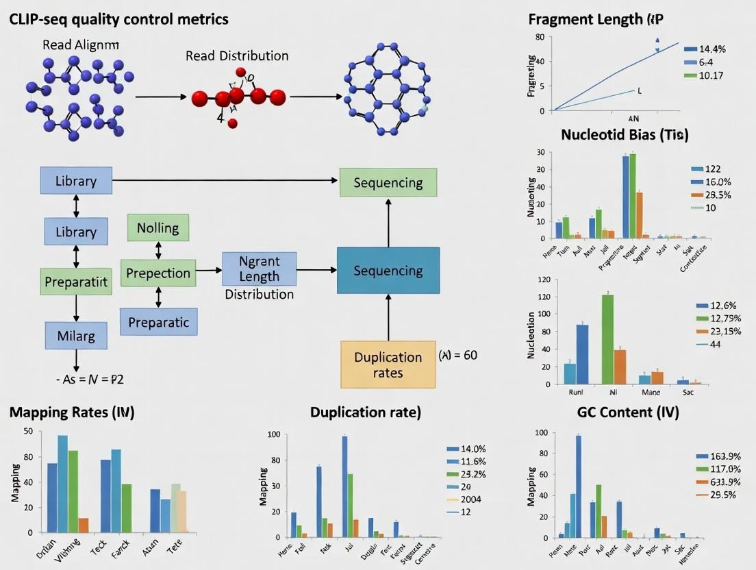
Visualization: CLIP-seq Workflow and QC Pipeline
Diagram Title: CLIP-seq Experimental and Quality Control Workflow
Diagram Title: CLIP-seq Data Analysis and QC Metrics Pipeline
Welcome to the Technical Support Center for CLIP-seq research. This resource, framed within our broader thesis on CLIP-seq quality control metrics, provides targeted troubleshooting guides and FAQs for researchers, scientists, and drug development professionals. Ensuring rigorous QC at each experimental stage is paramount for generating robust, reproducible data.
Troubleshooting Guides & FAQs
Stage 1: Cell Culture & Crosslinking
Q1: My UV crosslinking efficiency seems low. How can I troubleshoot this? A: Low crosslinking efficiency leads to weak signal. Key checks:
- UV Calibration: Verify the UV irradiance (e.g., 254 nm, 0.15-0.4 J/cm² for standard PAR-CLIP) with a radiometer. Old bulbs lose intensity.
- Cell Monolayer Density: Ensure cells are 70-90% confluent as a dense monolayer scatters UV light.
- Buffer Transparency: Use PBS without phenol red. Remove all media and wash cells thoroughly before crosslinking.
- 4-Thiouridine (4SU) Incorporation (for PAR-CLIP): Confirm 4SU concentration (typically 100-500 µM) and incubation time (6-16 hours) are optimal for your cell type.
Q2: I observe high cell death after 4SU treatment. What should I do? A: 4SU can be cytotoxic. Titrate the concentration (start at 100 µM) and reduce incubation time. Use a fresh stock solution prepared in DMSO or medium.
Stage 2: Lysis & Immunoprecipitation (IP)
Q3: My RNA-protein complexes are degrading during lysis. How do I prevent this? A: Degradation compromises complex integrity.
- RNase Inhibition: Ensure lysis buffer contains potent RNase inhibitors (e.g., 40 U/µL RNasin, 1 U/µL SUPERase•In) and is supplemented fresh.
- Protease Inhibition: Use complete EDTA-free protease inhibitor cocktails.
- Temperature: Perform all lysis and subsequent steps at 4°C or on ice.
- Lysis Buffer pH: Verify pH is neutral (~7.5).
Q4: I get high background in my IP. What are the primary causes? A: High background obscures specific signals.
- Antibody Specificity: Pre-clear lysate with beads alone. Use a validated, high-specificity antibody for your target protein. Include an IgG isotype control.
- Wash Stringency: Increase the number and rigor of washes. Use high-salt wash buffers (e.g., containing 500 mM NaCl) to reduce non-specific binding.
- Bead Blocking: Ensure magnetic/protein A/G beads are adequately blocked with BSA or yeast tRNA.
Stage 3: RNA Processing & Library Prep
Q5: My adapter ligation efficiency is poor. What factors should I check? A: Poor ligation leads to low library diversity.
- RNA Integrity: Check RNA fragment size post-phosphatase/kinase treatment on a Bioanalyzer. Ideal range is 50-200 nt.
- Adapter Concentration: Use a 5-10x molar excess of adapter to RNA fragments. Avoid over-diluting adapters.
- Enzyme Activity: Use a high-activity, thermostable T4 RNA ligase and fresh ATP. Ensure the correct reaction temperature.
- Inhibitors: Purify RNA fragments after enzymatic steps to remove salts or enzymes that inhibit ligation.
Q6: I detect primer dimer peaks in my final library QC. How can I mitigate this? A: Primer dimers compete for sequencing cycles.
- Size Selection: Perform stringent gel or bead-based size selection after cDNA synthesis/PCR to remove fragments <100 bp.
- PCR Cycle Number: Minimize PCR cycles (often 10-15 cycles). Use a polymerase with low bias.
- Dual-Size Selection: Implement a dual-SPRI bead cleanup (e.g., 0.7x and 1.2x ratios) to exclude small fragments.
Stage 4: Sequencing & Bioinformatic QC
Q7: My sequence data shows low complexity or overrepresented sequences. What went wrong? A: This indicates issues in early wet-lab stages.
- PCR Over-amplification: Reduce PCR cycles. Use unique molecular identifiers (UMIs) to deduplicate reads.
- Contamination: Check for adapter-adapter ligation artifacts or ribosomal RNA contamination. Use ribo-depletion during library prep.
- Insufficient Input: Starting with too little RNA-protein complex material forces excessive PCR, amplifying bias.
Q8: What are the key bioinformatic QC metrics I must check post-sequencing? A: Critical metrics for our thesis on CLIP-seq QC are summarized below.
Table 1: Essential Post-Sequencing QC Metrics for CLIP-seq
| Metric | Target Value/Range | Indication of Problem | Common Cause |
|---|---|---|---|
| Total Reads | >20 million per sample | Low statistical power | Inefficient library prep or sequencing depth |
| Mapping Rate | >70% to genome | Poor library quality or wrong reference | Adapter contamination, degraded RNA |
| Duplicate Rate | <50% (lower with UMIs) | PCR over-amplification, low complexity | Insufficient starting material |
| Insert Size | Peak ~50-200 nt | Improper fragmentation or size selection | RNase over-digestion, poor gel cut |
| Mutation Rate (PAR-CLIP) | 2-10% at T-to-C transitions | Low crosslinking efficiency | Suboptimal 4SU concentration or UV dose |
| Peak Distribution | Enriched in exons, 3' UTRs | Non-specific background | Poor antibody specificity or wash stringency |
Experimental Protocols
Protocol 1: Optimized UV Crosslinking for PAR-CLIP
- Grow cells in medium supplemented with 100-500 µM 4-thiouridine (4SU) for 12-16 hours.
- Aspirate medium, wash cells twice with 10 mL room-temperature PBS.
- Aspirate PBS completely. Place culture dish on ice.
- Irradiate cells in a Stratalinker 2400 (or equivalent) at 365 nm (for 4SU) with 0.15 J/cm². Perform irradiation on ice for heat dissipation.
- Immediately scrape cells in ice-cold PBS and pellet by centrifugation (500 x g, 5 min, 4°C). Proceed to lysis or flash-freeze pellet.
Protocol 2: Stringent RNA Immunoprecipitation (RIP) Wash
After antibody-bound bead complexes have formed and been captured:
- Low Salt Wash: Wash twice with 1 mL of IP Wash Buffer 1 (50 mM HEPES pH 7.5, 300 mM NaCl, 0.1% SDS, 0.5% NP-40, 0.5% Sodium Deoxycholate). Incubate on rotator for 2 minutes at 4°C each time.
- High Salt Wash: Wash once with 1 mL of IP Wash Buffer 2 (50 mM HEPES pH 7.5, 500 mM NaCl, 0.1% SDS, 0.5% NP-40, 0.5% Sodium Deoxycholate). Incubate for 5 minutes.
- LiCl Wash: Wash once with 1 mL of IP Wash Buffer 3 (50 mM HEPES pH 7.5, 250 mM LiCl, 0.5% NP-40, 0.5% Sodium Deoxycholate).
- Final TE Wash: Wash twice with 1 mL of TE buffer (10 mM Tris pH 7.5, 1 mM EDTA) + 50 mM NaCl.
- Proceed to Proteinase K digestion and RNA extraction.
Visualizations
Diagram Title: CLIP-seq Workflow with Critical Quality Control Checkpoints
Diagram Title: CLIP-seq Bioinformatics Pipeline with Failure Analysis Points
The Scientist's Toolkit: CLIP-seq Research Reagent Solutions
Table 2: Essential Materials for a CLIP-seq Experiment
| Item | Function | Example/Notes |
|---|---|---|
| 4-Thiouridine (4SU) | Photosensitive nucleoside analog for PAR-CLIP. Incorporated into RNA, enabling efficient crosslinking at 365 nm and inducing T-to-C mutations. | MilliporeSigma, #T4509. Prepare fresh stock in DMSO. |
| UV Crosslinker | Provides calibrated UV irradiation at specific wavelengths (254 nm for standard CLIP, 365 nm for PAR-CLIP). | Stratagene Stratalinker 2400. Critical: Annual radiometer calibration. |
| RNase Inhibitor | Protects RNA from degradation during cell lysis and immunoprecipitation steps. | Promega RNasin Ribonuclease Inhibitor or Thermo Fisher SUPERase•In. |
| Magnetic Beads (Protein A/G) | Solid support for antibody-mediated capture of RNA-protein complexes. Enable stringent washing. | Dynabeads Protein A/G, Novex Magnetic beads. |
| High-Specificity Antibody | Enriches for the target RNA-binding protein (RBP). The single most critical reagent for signal-to-noise ratio. | Validated for IP/CLIP. Use knockout cell line controls if possible. |
| T4 RNA Ligase 1/2, truncated | Ligates pre-adenylated DNA adapters to RNA fragments during library preparation. Lowers adapter dimer formation. | NEB, #M0437M (truncated). |
| SUPERscript IV Reverse Transcriptase | Reverse transcribes crosslinked, fragmented RNA into cDNA with high efficiency and processivity. | Thermo Fisher, #18090050. |
| Unique Molecular Identifiers (UMIs) | Short random nucleotide sequences ligated to RNA fragments pre-amplification. Enables bioinformatic removal of PCR duplicates. | Integrated into 5' or 3' adapters. |
| High-Fidelity PCR Mix | Amplifies final cDNA library with minimal bias for sequencing. | KAPA HiFi HotStart ReadyMix, NEB Next Ultra II Q5. |
| Bioanalyzer/TapeStation | Provides precise size distribution and quantification of RNA fragments and final sequencing libraries. | Agilent 2100 Bioanalyzer with High Sensitivity DNA/RNA chips. |
Troubleshooting Guides & FAQs
Q1: My CLIP-seq experiment shows high background noise in non-expressed genomic regions. Which QC metrics should I check, and how can I improve specificity? A: High background noise directly impacts Specificity. This often indicates non-specific antibody binding or insufficient RNase digestion. First, check the Signal-to-Noise Ratio calculated from your negative control regions (e.g., intronic or intergenic regions known to be devoid of binding). A ratio below 5 suggests a specificity issue. Improve specificity by:
- Titrate RNase I: Perform an RNase titration (e.g., 0.1, 0.5, 1.0 U/µg) to find the optimal condition that leaves crosslinked protein-RNA complexes intact but digests unprotected RNA.
- Increase Wash Stringency: Use high-salt wash buffers (e.g., 500-1000 mM NaCl) in your immunoprecipitation protocol.
- Validate Antibody: Use a knock-out/knock-down cell line as a control to confirm antibody specificity.
Q2: I suspect my CLIP-seq is missing genuine binding sites (false negatives). How do I assess and enhance Sensitivity? A: Low Sensitivity means true binding events are not detected. Quantify this using a Recovery Rate of known positive control binding sites (from validated literature). If recovery is <70%, consider these steps:
- Crosslinking Optimization: UV crosslinking efficiency is critical. Ensure cells are in a monolayer, washed with PBS to remove media, and use 254 nm UV light at 400 mJ/cm². For iCLIP or eCLIP, use 365 nm at 0.15 J/cm².
- Improve Library Complexity: A high PCR duplication rate (>80%) reduces sensitivity. Use unique molecular identifiers (UMIs) during adapter ligation to correct for PCR bias and increase the detectable unique molecule count.
- Increase Input Material: If working with low-abundance RBPs, scale up cell numbers (10-20 million per IP) and correspondingly increase reagent volumes.
Q3: My replicates show inconsistent peaks. How do I troubleshoot Reproducibility in CLIP-seq? A: Poor Reproducibility is measured by metrics like the Irreproducible Discovery Rate (IDR). An IDR score > 0.1 indicates low consistency between replicates. To improve reproducibility:
- Standardize Cell Culture: Maintain consistent cell passage numbers, confluence (aim for 80%), and handling conditions across replicates.
- Control RNA Integrity: Use an RNA Integrity Number (RIN) > 9.0 for all samples. Degraded RNA increases technical variation.
- Quantify IP Efficiency: Perform a parallel western blot for the target RBP on 2% of your IP eluate and input. Calculate the percentage of protein immunoprecipitated. Aim for >10% efficiency with <20% variation between replicates.
Q4: How do I differentiate between low Complexity and poor Sensitivity in my sequencing data? A: Complexity refers to the diversity of unique RNA fragments in your library, distinct from Sensitivity. Use these diagnostic tables:
Table 1: Diagnosing Data Quality Issues from Sequencing Metrics
| Metric | Formula | Good Value | Indicates Problem With |
|---|---|---|---|
| PCR Duplication Rate | (Duplicated Reads / Total Reads) x 100 | < 50% (with UMIs) | Library Complexity |
| Fraction of Reads in Peaks (FRiP) | (Reads in called peaks / Total mapped reads) x 100 | > 5-15% (varies by RBP) | Signal Strength / Sensitivity |
| Non-Redundant Fraction (NRF) | (Deduplicated reads / Total mapped reads) | > 0.8 | Library Complexity |
| IDR Score | Score from comparing peak lists of two replicates | < 0.1 | Reproducibility |
Table 2: Actionable Steps Based on Diagnosis
| Primary Issue | Supporting Evidence | Corrective Action |
|---|---|---|
| Low Complexity | High PCR duplication rate, Low NRF | 1. Use UMIs in adapters.2. Increase amount of starting RNA.3. Reduce PCR cycle number (aim for 8-12 cycles). |
| Poor Sensitivity | Low FRiP, Low recovery of known sites | 1. Optimize crosslinking (see A2).2. Increase IP efficiency (see A3).3. Sequence deeper (increase read depth). |
Essential Methodologies for CLIP-seq QC
Protocol 1: RNase I Titration for Optimal Specificity
- Crosslink 5 million cells per condition (in triplicate).
- Lysate cells in stringent lysis buffer (50 mM Tris-HCl pH 7.4, 100 mM NaCl, 1% NP-40, 0.1% SDS, 0.5% sodium deoxycholate).
- Partial RNase Digestion: Split lysate into 4 aliquots. Add RNase I (ThermoFisher) to final concentrations of 0.1, 0.5, 1.0, and 2.0 U/µg of RNA. Incubate at 37°C for 3 minutes. Include a no-RNase control.
- Immunoprecipitate your target RBP.
- Run the purified RNA on a 10% Urea-PAGE gel. The optimal condition shows a smear centered around 50-70 nt after crosslink reversal and RNA isolation. The no-RNase control should show a high molecular weight smear.
Protocol 2: Calculating Irreproducible Discovery Rate (IDR) Between Replicates
- Peak Calling: Call peaks on each biological replicate independently using a caller like
CLIPperorPyPeak. - Rank Peaks: Sort peaks for each replicate by p-value or fold-enrichment.
- Run IDR Analysis: Use the IDR pipeline (
idrpackage on GitHub). Command example:
- Interpretation: Peaks passing an IDR threshold of 0.05 or 0.1 are considered highly reproducible.
Visualizations
Title: CLIP-seq Workflow with Integrated QC Checkpoints
Title: Troubleshooting CLIP-seq Data Quality Problems
The Scientist's Toolkit: CLIP-seq QC Research Reagent Solutions
| Reagent / Material | Function in QC Context | Example Product / Specification |
|---|---|---|
| High-Specificity Antibody | Critical for Specificity and Sensitivity. Determines IP efficiency and background noise. | Validated CLIP-grade antibody (e.g., from Cell Signaling, Abcam). Always use with matched knockout control. |
| RNase I (Ultrapure) | Digests unprotected RNA to define binding site resolution. Titration is key for Specificity. | ThermoFisher EN0601; ensure it is protease and DNase-free. |
| Unique Molecular Identifiers (UMIs) | Short random nucleotide sequences in adapters to tag unique RNA fragments. Essential for measuring true Complexity and correcting PCR duplicates. | TruSeq Small RNA Kit (Illumina) or custom-synthesized adapters. |
| Magnetic Protein A/G Beads | For immunoprecipitation. Consistent bead size and binding capacity affect Reproducibility between replicates. | Dynabeads Protein A/G (ThermoFisher). |
| Size Selection Cassettes | Precise isolation of ~50-70 nt RNA-protein complexes post-RNase digestion. Affects Specificity and background. | Pippin Prep (Sage Science) with 3% agarose cassettes. |
| High-Fidelity PCR Mix | Used during library amplification. Reduces PCR errors and maintains sequence diversity for accurate Complexity assessment. | KAPA HiFi HotStart ReadyMix (Roche). |
| Spike-in Control RNAs | Synthetic RNA sequences added before IP. Used to normalize between samples and assess technical variation in Reproducibility. | ERCC RNA Spike-In Mix (ThermoFisher). |
Technical Support Center
FAQs and Troubleshooting Guides
Q1: During CLIP-seq data alignment, my mapping rates to the genome are consistently below 50%, far from the ENCODE benchmark of 70-90%. What could be the issue?
A: Low mapping rates often stem from poor RNA quality or adapter contamination. First, run a Bioanalyzer trace to ensure your input RNA has an RIN > 8.0. Second, verify your adapter trimming. Use the ENCODE-recommended cutadapt parameters: -a AGATCGGAAGAGC -q 20 -m 15. Re-align with STAR using genome indices that include splice junctions. If the problem persists, your UV cross-linking efficiency may be too high, causing excessive protein-RNA fragmentation.
Q2: How do I interpret the "PCR bottleneck coefficient" (PBC) in my CLIP-seq library QC, and what is the ENCODE standard? A: The PBC measures library complexity. It is the ratio of genomic locations with exactly one unique read (ND) to locations with at least one (NR). ENCODE standards for ChIP-seq (often applied to CLIP) are: PBC > 0.9 is optimal, 0.5-0.9 is moderate, and < 0.5 indicates severe bottlenecking requiring library re-preparation. For CLIP-seq, aim for PBC > 0.8. Low values suggest insufficient starting material or over-amplification.
Q3: My CLIP-seq experiment shows high background in the non-crosslinked control (no-UV control). What steps should I take? A: High background in the no-UV control indicates non-specific RNA binding or carryover. Follow this troubleshooting protocol:
- Increase RNase Concentration: Titrate RNase I (e.g., from 1:1000 to 1:500 dilution) during fragmentation to reduce non-specific RNA fragments.
- Enhance Wash Stringency: Add high-salt (e.g., 1M NaCl) or low-concentration SDS (0.1%) washes to your bead immunoprecipitation buffer.
- Verify Antibody Specificity: Run a western blot from the IP eluate. Consider using a knockout cell line control if available.
- Implement Size Selection: Use gel purification to strictly select RNA-protein adducts (~50-70 nt), excluding larger non-specific RNAs.
Q4: Which consensus guidelines should I follow for CLIP-seq replicates and statistical thresholds? A: Adhere to a combination of ENCODE (general NGS) and CLIP-specific (e.g., IRCLIP consortium) guidelines:
- Replicates: Perform at least two biological replicates. The ENCODE standard requires an Irreproducible Discovery Rate (IDR) < 0.05 for peak calling concordance between replicates.
- Peak Calling: Use multiple callers (e.g., PEAKachu, CLIPper). A true peak must have a fold-enrichment > 8 over the no-UV control (IRCLIP guideline).
- False Discovery Rate (FDR): Apply a stringent FDR cutoff of ≤ 0.001 for high-confidence peaks.
Table 1: Key CLIP-seq QC Metrics & Consortium Benchmarks
| Metric | Calculation / Definition | ENCODE Optimal Guideline (ChIP-seq) | CLIP-specific (e.g., IRCLIP) Guideline | Common Troubleshooting Target |
|---|---|---|---|---|
| Mapping Rate | (Reads aligned to genome / Total reads) * 100 | ≥ 70% | ≥ 60% (lower due to crosslink-induced mutations) | Adapter trimming, RNA quality, crosslinking optimization |
| Non-Redundant Fraction (NRF) | (Unique mapping reads) / (Total mapping reads) | ≥ 0.8 | ≥ 0.7 | Library complexity, PCR duplication |
| PCR Bottleneck Coeff. (PBC) | ND (distinct loci with 1 read) / NR (distinct loci with ≥1 read) | PBC1 (Optimal): > 0.9 | Aim for > 0.8 | Starting material quantity, PCR cycle number |
| Reads in Peaks (RIP) | (Reads falling in called peaks / Total reads) * 100 | Not directly specified | > 10-15% (varies by target) | Antibody efficiency, background in control |
| IDR (Replicate Concordance) | Rank consistency of peaks between two replicates | IDR < 0.05 (for two reps) | IDR < 0.05 recommended | Biological variability, experimental consistency |
Experimental Protocol: CLIP-seq with Rigorous ENCODE-Compliant QC
Protocol: RNA-Protein Crosslinking, Immunoprecipitation, and Library Prep for CLIP-seq
Materials:
- Cells of interest
- UV-Crosslinker (254 nm)
- RNase I (Thermo Fisher, AM2295)
- Protein G Dynabeads (Invitrogen, 10004D)
- T4 PNK (NEB, M0201S)
- PNK Buffer (with and without ATP)
- Critical: 5’ App DNA/RNA Adapter (IDT, 5’/rApp/NNNN…/3SpC3/)
- Superscript IV Reverse Transcriptase (Thermo Fisher, 18090050)
- Circligase II (Lucigen, CL9025K)
Methodology:
- In Vivo Crosslinking: Grow cells to 80% confluency. Wash once with cold PBS. Irradiate with 254 nm UV light at 0.15 J/cm². Immediately lyse in stringent RIPA buffer + RNase inhibitors.
- Partial RNase Digestion: Treat lysate with RNase I (1:1000 dilution) for 3 min at 22°C. Quench on ice.
- Immunoprecipitation: Pre-clear lysate. Incubate with validated antibody-bound Protein G beads for 2h at 4°C. Wash 5x with high-salt RIPA (1M NaCl final in one wash).
- Phosphatase & Kinase Treatment:
- Wash beads in PNK buffer (no ATP). Treat with T4 PNK (removes 3' phosphates) for 20 min at 37°C.
- Wash, then add PNK buffer with ATP and T4 PNK to exchange 5' phosphate for radioactive ATP (for visualization) or for subsequent 5' adapter ligation.
- Ligation of 3' Adapter: Wash beads. Use T4 RNA Ligase 2, truncated (NEB, M0242S) to ligate a pre-adenylated 3' DNA adapter to the RNA 3' end in a reaction without ATP overnight at 16°C.
- On-Bead Reverse Transcription: After ligation wash, perform RT directly on beads using Superscript IV and a primer complementary to the 3' adapter.
- cDNA Circularization & PCR: Elute cDNA. Circulate single-stranded cDNA using Circligase II. Amplify with 10-14 PCR cycles using indexed primers. Size-select (120-200 bp) via gel extraction.
- QC Sequencing: Assess library quality via Bioanalyzer (peak ~160 bp) and qPCR for quantification. Sequence on Illumina platform with ≥ 20 million reads per replicate.
Visualizations
Diagram 1: CLIP-seq Experimental Workflow with QC Checkpoints
Diagram 2: CLIP-seq Data Analysis & ENCODE QC Pipeline
The Scientist's Toolkit: Key Research Reagent Solutions
Table 2: Essential Reagents for ENCODE-Compliant CLIP-seq Experiments
| Reagent | Vendor (Example) | Catalog Number | Critical Function in CLIP-seq Protocol |
|---|---|---|---|
| RNase I | Thermo Fisher Scientific | AM2295 | Partially digests RNA to leave ~50-70 nt crosslinked fragments; concentration is key for signal-to-noise. |
| Protein G Dynabeads | Invitrogen | 10004D | Magnetic beads for efficient antibody-based pulldown of RNA-protein complexes with low nonspecific binding. |
| T4 Polynucleotide Kinase (PNK) | New England Biolabs | M0201S | Removes 3' phosphates left by RNase cleavage and enables 5' end labeling/ligation. |
| 5' App DNA/RNA Adapter | Integrated DNA Technologies (IDT) | Custom Synthesis | Pre-adenylated 3' adapter; essential for ligation to RNA 3' end without ATP (prevents RNA circularization). |
| T4 RNA Ligase 2, Truncated | New England Biolabs | M0242S | Specifically ligates pre-adenylated 3' adapter to RNA 3' OH group. |
| Superscript IV Reverse Transcriptase | Thermo Fisher Scientific | 18090050 | High-temperature RTase for efficient cDNA synthesis from crosslinked, fragmented, and adapter-ligated RNA. |
| Circligase II ssDNA Ligase | Lucigen | CL9025K | Circularizes single-stranded cDNA post-RT, enabling small RNA library prep and reducing concatemer formation. |
| Anti-FLAG M2 Antibody | Sigma-Aldrich | F1804 | Common antibody for tagged RBPs; high specificity and affinity, recommended by ENCODE for validation. |
Common Artifacts and Biases in CLIP-seq Data and How QC Metrics Detect Them
Technical Support Center
Troubleshooting Guides & FAQs
Q1: My CLIP-seq library has an unusually high percentage of ribosomal RNA reads. What artifact does this indicate and how can I diagnose it?
A: This indicates insufficient RNase digestion or incomplete RNase I inactivation. High rRNA suggests the RNase concentration was too low, leaving abundant structured RNAs intact, which then dominate the library.
Diagnosis via QC:
- Primary Metric: Examine the read distribution across genomic features (Table 1). A >20% mapping rate to rRNA loci is a strong indicator.
- Supporting Metric: Check the complexity/deduplication metrics. High rRNA often correlates with low library complexity (high PCR duplication rate).
Experimental Protocol to Prevent This:
- Titrate RNase I: Perform a pilot experiment with a range of RNase I concentrations (e.g., 0.1, 0.5, 1, 2 U/mL) on a small-scale UV-crosslinked sample.
- Inactivation: Post-digestion, add SUPERase•In RNase inhibitor (2 U/µL) before adding proteinase K. Ensure proper purification to remove all RNase activity before reverse transcription.
- QC Check: Sequence a small pilot library (e.g., 1-2M reads) and analyze genomic feature distribution before deep sequencing.
Q2: My data shows a strong bias towards reads starting with adenine (A) at the crosslink site. Is this a technical artifact?
A: Yes, this is a known library preparation bias often referred to as "A-rule" or "adenine bias." It arises during adapter ligation and reverse transcription, where polymerases have a tendency to add an extra A nucleotide opposite the crosslink-induced modification or abasic site, rather than accurately reading the original base.
Diagnosis via QC:
- Primary Metric: Generate a nucleotide composition frequency plot around the crosslink site (typically position -1 to +5 relative to the crosslink peak center). A dominant >50% frequency of adenine at position +1 is indicative of this bias (Table 2).
- Tool: Use tools like
CLIPperorPiranhawhich often include nucleotide frequency analysis in their output.
Experimental Protocol to Mitigate This:
- Use Non-Templated Nucleotide-Supplemented RT: Add a low concentration of dATP (e.g., 0.5 mM) to the reverse transcription mix. This can help saturate the non-templated addition and make it more random.
- Alternative Enzymes: Test different reverse transcriptases (e.g., TGIRT, Superscript IV) which may exhibit different propensities for non-templated nucleotide addition.
- Bioinformatic Normalization: In downstream analysis, use tools that can model and correct for this sequence bias.
Q3: I observe very broad peaks or a high background signal across my CLIP-seq profile. What could be the cause?
A: This suggests over-digestion with RNase or non-specific RNA-protein interactions due to suboptimal washing stringency. Over-digestion creates very short RNA fragments, leading to mapping ambiguity and diffuse peaks.
Diagnosis via QC:
- Primary Metric: Analyze the fragment length distribution of immunoprecipitated RNAs post-library prep. A median length below 20 nt suggests over-digestion.
- Secondary Metric: Evaluate the signal-to-noise ratio (SNR). Calculate the ratio of reads in called peak regions vs. genomic background. An SNR < 5 is often problematic (Table 1).
- Tertiary Metric: Check the PCR bottleneck coefficient (PBC). A low PBC (<0.5) indicates high background and low complexity.
Experimental Protocol to Optimize:
- RNase Titration (Revisited): As in Q1, titrate RNase concentration. Aim for a post-purification RNA fragment distribution centered at 30-60 nt.
- Increase Wash Stringency: Implement more stringent washes. After immunoprecipitation, perform 2-3 high-salt washes (e.g., with 1M urea, 50mM Tris-HCl pH 7.5) followed by a final low-salt wash.
- Use a Size Selection Step: Incorporate a gel-based or bead-based size selection (e.g., selecting for 50-100 nt RNAs) after RNA purification but before adapter ligation.
Q4: My negative control (e.g., no-UV or IgG control) shows significant peaks. How do QC metrics flag this?
A: This indicates non-specific binding/background contamination. QC metrics are critical to objectively assess if your experimental signal is above this background.
Diagnosis via QC:
- Primary Metric: Irreproducible Discovery Rate (IDR) Analysis. Compare replicates of your true IP against each other and against the negative control. A high IDR score when comparing IP to control indicates poor specificity.
- Secondary Metric: Peak Overlap. Calculate the percentage of peaks in your experimental condition that overlap with peaks called in the negative control. >15% overlap is a concern (Table 2).
- Visualization: Create a Venn diagram or upset plot to visualize the overlap.
Experimental Protocol to Improve Specificity:
- Optimize Antibody: Validate the antibody for IP specificity via western blot. Pre-clear the lysate with protein A/G beads.
- Include Rigorous Controls: Always include a matched no-UV crosslinking control and an isotype control (IgG) for antibody-based CLIP.
- Use Crosslinking-Induced Truncation Site (CITS) Analysis: Authentic binding sites show a truncation signature at the crosslink nucleotide. Background peaks lack this signature. Tools like
PureCLIPare designed to detect CITS.
Table 1: Quantitative QC Metrics for CLIP-seq Data Assessment
| QC Metric | Calculation/Description | Optimal Range | Artifact/Bias Detected |
|---|---|---|---|
| Ribosomal RNA % | (Reads mapping to rRNA loci / Total mapped reads) * 100 | < 5% | Insufficient RNase digestion, sample degradation |
| PCR Bottleneck Coefficient (PBC) | PBC = N1 / Ndistinct (N1=genomic locations with 1 read; Ndistinct=total distinct locations) | PBC > 0.5 (High complexity) | Low complexity, over-amplification, high background |
| Signal-to-Noise Ratio (SNR) | SNR = (Reads in peaks) / (Reads in non-peak genomic regions) | SNR > 5 | Over-digestion, non-specific binding |
| Fragment Length Median | Median length of sequenced inserts after mapping | 30 - 60 nucleotides | Over- or under-digestion with RNase |
| Peak Overlap with Control | (% of experimental peaks overlapping negative control peaks) | < 15% | Non-specific antibody binding, background |
Table 2: Sequence-Based Bias Metrics
| QC Metric | Calculation/Description | Interpretation | Artifact/Bias Detected |
|---|---|---|---|
| Adenine Bias at +1 | Frequency of 'A' nucleotide at position +1 from crosslink site | < 40% is acceptable; >50% indicates strong bias | "A-rule" reverse transcription bias |
| Nucleotide Enrichment Motif | Sequence logo generated from regions around crosslink sites | Should resemble known RBP motif (if available) | Technical biases masking true biological signal |
| Crosslinking-induced Mutation/Truncation Rate | Percentage of reads with deletions or mismatches at peak summits | Should be enriched in IP vs control | Confirms true crosslinking sites; low rates suggest background. |
The Scientist's Toolkit: Key Research Reagent Solutions
| Reagent/Material | Function in CLIP-seq Protocol |
|---|---|
| RNase I (Affinity-purified) | Digests unprotected RNA to leave only protein-bound fragments. Critical for resolution. |
| SUPERase•In RNase Inhibitor | Inactivates RNase I after digestion to prevent further RNA degradation during subsequent steps. |
| Phosphatase (e.g., CIP) | Removes 3' phosphates left by RNase cleavage or fragmentation, enabling 3' adapter ligation. |
| T4 PNK (with 3' phosphatase minus mutant) | (1) Phosphorylates 5' ends for ligation. (2) The mutant version is used in iCLIP to mark crosslink sites. |
| Proteinase K | Digests proteins after IP to recover crosslinked RNA fragments. Must be molecular biology grade. |
| Glycogen (or RNase-free carrier) | Precipitates and recovers the very small amounts of RNA fragments after proteinase K treatment. |
| High-Fidelity Reverse Transcriptase (e.g., TGIRT, Superscript IV) | Reverse transcribes crosslinked RNA, which can be chemically modified and challenging to read. Minimizes bias. |
| High-Sensitivity DNA Bioanalyzer/ScreenTape Assay | Accurately sizes and quantifies final cDNA libraries pre-sequencing; essential for quality assessment. |
Experimental Workflow and Quality Control Checkpoints
Title: CLIP-seq Experimental Workflow with QC Checkpoints
CLIP-seq Data Analysis and Artifact Diagnosis Pathway
Title: CLIP-seq Data Analysis and Artifact Diagnosis Pathway
A Step-by-Step Guide to Calculating and Interpreting Essential CLIP-seq QC Metrics
Technical Support Center
Troubleshooting Guides
Issue 1: Poor overall read quality after FASTQ generation.
- Symptoms: Low Per Base Sequence Quality scores in FastQC report, many reads in the red zone.
- Diagnosis: This often indicates problems during sequencing, such as degraded flow cells or issues with polymerase incorporation.
- Solution: First, re-run FastQC to confirm. If confirmed, consult your sequencing facility. For CLIP-seq, do not proceed with low-quality data as it severely impacts crosslink site identification. Consider re-sequencing.
Issue 2: High percentage of reads lost during adapter trimming.
- Symptoms: Trimming tools (e.g., Cutadapt) report >40% of reads being trimmed or discarded.
- Diagnosis: Adapter sequence may be incorrect, or read-through of short RNA fragments is excessive.
- Solution: Verify the exact adapter sequence used in your CLIP-seq library prep protocol. Use the
--info-fileflag in Cutadapt to see which adapters are being matched. Adjust the allowed error rate (-e) parameter cautiously.
Issue 3: Very low alignment rate to the reference genome.
- Symptoms: STAR or HISAT2 alignment rate is below 50-60%.
- Diagnosis: Potential causes: 1) Incorrect or poor-quality reference genome index. 2) High contamination. 3) Major species mismatch. 4) For CLIP-seq, high multimapping reads are expected, but uniquely mapped reads should still be present.
- Solution: 1) Re-check the species and genome assembly version. Rebuild the index if necessary. 2) Run FastQC again to check for overrepresented sequences (potential contamination). 3) For CLIP-seq, ensure the aligner is configured to handle multimapping reads appropriately (e.g., STAR's
--outFilterMultimapNmaxparameter).
Issue 4: PCR duplication levels are critically high (>80%).
- Symptoms: MarkDuplicates (Picard) reports extremely high duplication rates.
- Diagnosis: This is common in CLIP-seq due to the limited starting material and amplification bias. However, rates >80% may indicate over-amplification or insufficient input.
- Solution: Optimize PCR cycle number during library prep. For analysis, use duplicate-marking tools that consider both alignment coordinates and unique molecular identifiers (UMIs) if your protocol included them.
Frequently Asked Questions (FAQs)
Q1: Which FastQC modules are most critical for CLIP-seq data, and what are the acceptable thresholds? A: The most critical modules for CLIP-seq initial QC are:
- Per base sequence quality: Q-score should be mostly >30 for bases used in analysis.
- Adapter Content: Should be <5% for the majority of the read length. Higher levels necessitate aggressive trimming.
- Sequence Duplication Levels: High duplication is expected, but note the level for later steps.
- Per base N content: Should be <5% for all positions.
Q2: Should I trim low-quality bases or entire reads for CLIP-seq data? A: Conservative quality trimming is recommended. Use a sliding-window approach (as in Trimmomatic or Cutadapt quality trimming) to remove low-quality regions rather than whole reads, as CLIP-seq reads are precious. A typical setting is a 4bp window with average Q<20.
Q3: How do I handle the high rate of multimapping reads in CLIP-seq alignment? A: Multimapping is inherent to CLIP-seq due to repetitive RNA elements. Best practices include:
- Using an aligner like STAR that can output all mapping locations.
- In downstream peak calling, using tools specifically designed for CLIP-seq (e.g., PEAKachu, CLIPper) that can incorporate multimapping read information.
- Documenting the percentage of multimapping reads as a standard QC metric for your thesis.
Q4: What is a typical alignment rate distribution for a successful CLIP-seq experiment? A: Expect a distribution similar to the following:
| Alignment Category | Typical Percentage Range | Notes for CLIP-seq |
|---|---|---|
| Uniquely Mapped | 40-70% | Varies by RNA-binding protein and cell type. |
| Multimapped | 20-50% | Expected to be higher than in RNA-seq. |
| Unmapped | 5-15% | Investigate if >20%. |
Q5: Why is duplicate marking different for CLIP-seq, and how should I do it?
A: Standard duplicate marking assumes duplicates are PCR artifacts. In CLIP-seq, identical reads can originate from biologically relevant, highly abundant binding sites. If your protocol includes UMIs, use UMI-aware deduplication tools (e.g., umi_tools dedup). Without UMIs, mark but do not remove duplicates for peak calling, as the tools weight them appropriately.
Experimental Protocols
Protocol 1: Adapter Trimming and Quality Filtering for CLIP-seq FASTQ files
Principle: Remove 3' adapter sequences and low-quality bases while preserving the maximal amount of meaningful sequence data. Steps:
- Tool: Cutadapt (version 4.4+)
- Command:
- Parameters Explained:
-a: Adapter sequence to trim from the 3' end.--minimum-length 18: Discard reads shorter than 18nt after trimming.--max-n 0.1: Discard reads with >10% ambiguous (N) bases.-q 20: Trim low-quality bases from 3' end using a Phred threshold of 20.-j 8: Use 8 CPU cores.
Protocol 2: Alignment of CLIP-seq Reads using STAR
Principle: Map trimmed reads to a reference genome, allowing for multiple mapping positions to capture repetitive element binding. Steps:
- Tool: STAR (version 2.7.10a+)
- Genome Index Generation (one-time):
- Alignment:
Visualizations
Title: CLIP-seq QC Pipeline Workflow
Title: Low Alignment Rate Troubleshooting
The Scientist's Toolkit: Research Reagent Solutions
| Item | Function in CLIP-seq QC Pipeline |
|---|---|
| FastQC | Initial quality control visualization tool. Assesses base quality, adapter content, duplication levels, and more from raw FASTQ files. |
| Cutadapt | Precisely removes adapter sequences and trims low-quality bases from read ends. Critical for clean alignment. |
| Trimmomatic | Alternative to Cutadapt. Performs a variety of trimming tasks using a sliding-window approach. |
| STAR Aligner | Spliced-aware genome aligner. Preferred for its speed and ability to handle a high number of multimapping reads common in CLIP-seq. |
| HISAT2 | A sensitive and fast aligner, another excellent option for mapping CLIP-seq data. |
| SAMtools | Swiss-army knife for manipulating SAM/BAM files. Used for sorting, indexing, filtering, and basic statistics (flagstat). |
| Picard Tools | Provides robust utilities for marking PCR duplicates and collecting alignment metrics. |
| Qualimap | Generates comprehensive quality control metrics from aligned BAM files, including coverage profiles and bias detection. |
| UMI Tools | If UMI barcodes are incorporated in the library protocol, this suite is essential for accurate duplicate removal and error correction. |
Troubleshooting Guides & FAQs
Q1: During CLIP-seq analysis, my library shows extremely high PCR duplication levels (>80%). What are the primary causes and solutions? A: High PCR duplication in CLIP-seq typically indicates insufficient starting material or over-amplification.
- Causes:
- Low RNA input or RNA degradation prior to library prep.
- An excessive number of PCR cycles during library amplification.
- Inefficient ligation of adapters, reducing complexity.
- Solutions:
- Quantify Input: Use fluorescence-based assays (e.g., Qubit) for accurate RNA quantification. Increase input if possible.
- Optimize PCR: Reduce the number of amplification cycles. Perform a qPCR-based pilot to determine the minimum cycles needed for sufficient yield.
- Verify Adapter Ligation: Check adapter efficiency using Bioanalyzer/TapeStation. Ensure fresh T4 RNA ligase and optimal reaction conditions.
Q2: How do I interpret the relationship between "Effective Depth" and "Total Reads" in my CLIP-seq QC report? A: Effective depth (or non-duplicate reads) is the subset of total reads that map to unique genomic locations, representing biologically independent molecules. A large discrepancy suggests a high-duplication, low-complexity library.
| Metric | Description | Ideal Range for CLIP-seq | Implication if Out of Range |
|---|---|---|---|
| Total Reads | Total number of sequencing reads. | Project-dependent (e.g., 20-50M) | Low reads: insufficient statistical power. |
| Effective Depth | Number of unique (non-duplicate) reads. | >70% of Total Reads | Low %: High PCR duplication, poor library complexity. |
| Duplication Rate | Percentage of PCR-derived duplicate reads. | <30% | High rate: Potential bottleneck in library prep. |
Q3: Which computational tool should I use to calculate library complexity metrics, and what's the basic workflow?
A: Picard Tools' MarkDuplicates is the standard. The basic protocol is:
- Align Reads: Align your CLIP-seq FASTQ files to the reference genome using a spliced aligner (e.g., STAR).
- Sort BAM: Sort the BAM file by coordinate using
samtools sort. - Run MarkDuplicates: Execute Picard.
- Extract Metrics: The
metrics.txtfile contains key metrics likePERCENT_DUPLICATIONandESTIMATED_LIBRARY_SIZE.
Q4: My estimated library size seems low compared to my sequencing depth. Does this invalidate my experiment? A: Not necessarily, but it flags a quality issue. A low estimated library size indicates that adding more sequencing reads would yield diminishing returns of new biological information. For CLIP-seq, where binding sites are limited, this may still be acceptable if saturation of major sites is achieved. Cross-validate findings with an orthogonal assay if complexity is very low.
Experimental Protocol: Assessing Library Complexity with Preseq
Objective: To estimate the complexity and future yield of a CLIP-seq library.
Method: Use preseq to project the complexity curve.
- Input: Coordinate-sorted BAM file (after alignment but before duplicate removal).
Run
preseq lc_extrap:Interpret Output: The output file lists total reads sampled vs. expected distinct reads. Plot these values. A curve that plateaus sharply indicates low complexity; a curve that rises linearly with more sampling indicates high complexity.
Visualizations
Title: Causes and Detection of PCR Duplicates in CLIP-seq
Title: Workflow for Library Complexity Analysis with Preseq
The Scientist's Toolkit: Research Reagent Solutions
| Item | Function in CLIP-seq / Complexity Assessment |
|---|---|
| RNase Inhibitor (e.g., RNasin) | Critical for protecting often low-abundance, protein-bound RNA fragments during immunoprecipitation and library construction. |
| High-Sensitivity RNA Assay Dyes (Qubit) | Accurately quantifies picogram amounts of purified RNA-crosslinked material to ensure sufficient input for library prep. |
| T4 RNA Ligase 1/2, truncated (NEB) | Catalyzes adapter ligation to RNA 3' ends. Efficiency directly impacts library complexity; fresh enzyme is crucial. |
| UMI Adapters (Unique Molecular Identifiers) | Short random nucleotide sequences added to each molecule before PCR. Allows bioinformatic correction for PCR duplication, enabling true molecule counting. |
| High-Fidelity PCR Master Mix (e.g., KAPA HiFi) | Reduces PCR errors and minimizes duplicate generation by favoring accurate amplification over fewer cycles. |
| AMPure XP Beads (Beckman Coulter) | Used for size selection and clean-up. Precise bead-to-sample ratios are vital to recover the full complexity of fragment sizes. |
Troubleshooting Guides & FAQs
Q1: During CLIP-seq alignment, an unusually high percentage of multi-mapped reads is observed (>40%). What could be the cause and how can it be resolved? A: This is often caused by repetitive genomic elements or inadequate read length/quality. First, check the raw read quality using FastQC for potential adapter contamination or degraded 3' ends. For troubleshooting:
- Filter short reads: Remove reads below 25 nt post-adapter trimming, as shorter reads map less uniquely.
- Increase alignment stringency: Use
-kparameter in STAR or-min Bowtie2 to report fewer secondary alignments initially. - Employ a splice-aware aligner: For CLIP-seq, use STAR or HISAT2 with
--outFilterMultimapNmax 20 --outSAMmultNmax 1to initially manage multi-mapping. - Post-alignment filtering: Use tools like
samtools viewwith-q(minimum mapping quality) to isolate reads with higher uniqueness probability. Reads from common repeat families (e.g., Alu, LINE) can be filtered using annotation BED files from UCSC.
Q2: How should I decide on the threshold for mapping quality (MAPQ) to filter multi-mapped reads in my CLIP-seq analysis pipeline? A: The optimal MAPQ threshold depends on the aligner. Aligners assign MAPQ scores differently. Use the following table as a guideline:
| Aligner | Typical MAPQ for Unique Alignment | Recommended Minimum MAPQ | Notes for CLIP-seq |
|---|---|---|---|
| STAR | 255 | 10 | STAR uses 255 for uniquely mapped reads. A threshold of 10 filters reads with high multimapping. |
| Bowtie2 | 42 | 10 | Bowtie2 MAPQ=42 is typically unique. A threshold of 10-20 is common. |
| HISAT2 | 60 | 10 | Similar to Bowtie2. Start with MAPQ >= 10. |
| BWA | 60 | 10 | BWA's MAPQ=60 is typically unique. Use MAPQ >= 10 for stringent filtering. |
Experimental Validation Protocol: To empirically determine the threshold, isolate reads from a key positive control RNA (e.g., MALAT1 for NEAT1) and plot the distribution of crosslink sites across MAPQ scores. A sharp drop in site density at lower MAPQ values can indicate an appropriate cutoff.
Q3: What are the best practices for handling multi-mapped reads in CLIP-seq peak calling to avoid false positives? A: The safest practice is to exclude them from initial peak calling but retain them for downstream annotation and visualization with caution.
- Primary Input for Peak Calling: Use only uniquely mapped reads (after applying a MAPQ filter) as the primary BAM file input to peak callers like
PiranhaorCLIPper. - Rescuing Multi-maps for Annotation: After peak calling, multi-mapped reads can be probabilistically redistributed to their potential genomic loci using tools like
RSEMorMMDiffbased on local read density and expression estimates. This provides context for which gene families a peak may belong to. - Visualization: In genome browsers, display multi-mapped reads in a separate, distinct track (e.g., in light gray) to differentiate them from uniquely mapped evidence (in black or blue).
Q4: How does the choice of reference genome (basic vs. inclusive of alternate haplotypes) impact multi-mapping rates in CLIP-seq?
A: Using a reference genome that includes alternate haplotype sequences (e.g., GRCh38 with "alt" contigs) can increase multi-mapping rates, as reads originating from duplicated or highly similar regions will have more perfect matches. For most CLIP-seq QC analyses focused on primary binding sites, it is standard to use the primary assembly only (e.g., GRCh38.primary_assembly.genome.fa). This provides a clearer interpretation of mapping statistics and reduces alignment ambiguity. The alternate contigs should be included only for specific studies of polymorphic or paralogous regions.
The Scientist's Toolkit: CLIP-seq Mapping & QC Essential Reagents & Tools
| Item | Function in CLIP-seq Mapping/Alignment Context |
|---|---|
| High-Fidelity Reverse Transcriptase (e.g., SuperScript IV) | Critical for generating cDNA reads that accurately represent the RNA fragment without introducing errors that cause spurious multi-mapping. |
| UMI (Unique Molecular Identifier) Adapters | Allows bioinformatic correction for PCR duplicates. Essential for accurate quantification, especially when multi-mapped reads are probabilistically redistributed. |
| RNase Inhibitor (e.g., RNasin Plus) | Prevents RNA degradation during library prep, preserving full read length which aids in unique alignment. |
| Size Selection Beads (SPRIselect) | Precise size selection (e.g., 70-90 nt inserts) removes overly short fragments that contribute to multi-mapping. |
| Splice-Aware Aligner (STAR) | Software tool for accurate alignment across splice junctions, reducing misalignment that can be misinterpreted as multi-mapping. |
| SAM/BAM Tools (samtools) | Essential software for filtering, sorting, and indexing alignment files based on MAPQ and other flags. |
| Repeat Masker Annotation File | Genomic coordinate file of repetitive elements used to annotate and filter alignments derived from known repeats. |
Visualizations
Diagram 1: CLIP-seq Read Mapping Classification Workflow
Diagram 2: Decision Logic for Handling Multi-mapped Reads
Troubleshooting & FAQs
Q1: My peak caller (e.g., MACS2) reports thousands of peaks, but visual inspection in a genome browser shows many appear to be in "background" or untagged control regions. What's wrong?
A: This indicates a poor Signal-to-Noise Ratio (SNR). The peak caller's statistical model may be overwhelmed by background. First, verify your input/control library. It must be a proper matched input (e.g., pre-cleared lysate) or IgG control, not a different cell type. Use the --broad flag with caution. Re-run peak calling with a more stringent p-value or q-value cutoff (e.g., -q 0.01 instead of -q 0.05). Crucially, calculate the FRiP (Fraction of Reads in Peaks) score. A FRiP < 1% for a transcription factor or < 5-10% for a histone mark often signifies a failed experiment.
Q2: How do I interpret the "fold enrichment" reported in my peak file? Why do some high-confidence binding sites have surprisingly low fold enrichment? A: Fold enrichment is highly dependent on the size and quality of the control library. A shallow control library can inflate enrichment values artificially. Conversely, genuine binding sites in high-background genomic regions (e.g., open chromatin) may show modest fold enrichment but be statistically robust due to high read counts. Always prioritize the statistical significance (q-value) over fold enrichment alone. Cross-reference with metrics like the Signal-to-Noise Ratio calculated from non-peak genomic regions.
Q3: After CLIP-seq peak calling, my FRiP score is acceptable, but the peaks seem "noisy" and don't correlate well with gene features. What metrics should I check next? A: This is a common issue in CLIP-seq QC. Beyond FRiP, calculate the following signal metrics:
- Crosslinking-induced mutation rate (for PAR-CLIP): Should be significantly higher in peaks vs. background.
- Non-conversion rate (for iCLIP): Should be low in peaks, indicating successful cDNA truncation at crosslink sites.
- Read distribution across gene features: Use a tool like
RSeQCto see if reads are enriched in 3' UTRs (for RBPs) as expected.
Q4: I have replicate experiments. How do I use peak-calling results to quantitatively assess reproducibility, not just visual overlap? A: Do not rely on peak overlap Venn diagrams alone. Use the Irreproducible Discovery Rate (IDR) framework, a robust statistical method for assessing replicate consistency in high-throughput experiments. It ranks peaks by significance (p-value) from two replicates and models the consistency of their rankings. An IDR threshold of 0.05 or 0.01 is standard for identifying high-confidence peaks.
Key Experimental Protocols
Protocol 1: Calculating Critical Signal Metrics for CLIP-seq QC
- Generate Read Count Matrix: Using tools like
featureCountsorbedtools multicov, count reads in called peaks, in a set of negative control genomic regions (e.g., gene deserts, or regions called in the input sample), and in the entire mappable genome. - Calculate FRiP:
FRiP = (Total reads in peaks) / (Total aligned reads in library). - Calculate Signal-to-Noise Ratio (SNR):
SNR = (Median read density in peak regions) / (Median read density in negative control regions). - Calculate Fold Enrichment (FE):
FE = (Read count in peak region from IP) / (Read count in same region from control)normalized by total library size.
Protocol 2: Performing Irreproducible Discovery Rate (IDR) Analysis on Replicates
- Run Peak Calling Per Replicate: Call peaks on each biological replicate independently using the same parameters (e.g.,
MACS2 callpeak -t rep1.bam -c input.bam -n rep1). - Prepare Input Files: Sort the peak files (
_peaks.narrowPeakfor MACS2) by p-value or q-value in descending order. - Execute IDR: Run the IDR tool:
idr --samples rep1_peaks.narrowPeak rep2_peaks.narrowPeak --input-file-type narrowPeak --rank p.value --output-file idr_output.txt --plot. - Extract High-Confidence Peaks: Filter the pooled peaks from both replicates based on the IDR output column (e.g.,
idr_threshold <= 0.05).
Table 1: Benchmarking Signal Metrics for CLIP-seq Experiment QC
| Metric | Calculation | Optimal Range (CLIP-seq) | Interpretation Below Range |
|---|---|---|---|
| FRiP Score | Reads in Peaks / Total Aligned Reads | 5% - 20% (varies by target) | Low specificity; potential antibody or protocol failure. |
| Signal-to-Noise Ratio (SNR) | Median Density(Peaks) / Median Density(Control Regions) | > 5 | High background; poor enrichment over non-specific noise. |
| IDR Rate (at 0.05) | % of Global Peaks passing IDR threshold | > 70% for true replicates | Poor replicate reproducibility; technical or biological inconsistency. |
| Fold Enrichment | Normalized IP Count / Control Count | Often > 10, but context-dependent | Can be misleading if control library is inadequate. |
Table 2: Research Reagent Solutions Toolkit
| Item | Function | Example/Note |
|---|---|---|
| High-Affinity, Validated Antibody | Immunoprecipitation of target protein-RNA complexes. | Critical; use knock-out/knock-down validation if possible. |
| RNase Inhibitor | Prevents degradation of RNA during immunoprecipitation. | Must be added to all buffers post-lysis. |
| Precision Enzymes (e.g., PNK, FastAP) | For RNA end repair and adapter ligation in library prep. | Essential for maintaining library complexity. |
| Magnetic Protein A/G Beads | Solid-phase support for antibody capture and washes. | Allow for stringent washing to reduce background. |
| Size Selection Beads (SPRI) | For cDNA fragment isolation and library clean-up. | Determines final library insert size distribution. |
| High-Fidelity Polymerase | Amplification of cDNA libraries with minimal bias. | Critical for maintaining sequence diversity. |
| Unique Molecular Identifiers (UMIs) | Molecular barcodes to correct for PCR duplicates. | Mandatory for accurate quantification in modern CLIP. |
| Matched Negative Control | Input lysate or IgG immunoprecipitation. | Non-negotiable for accurate peak calling and SNR calculation. |
Visualizations
Diagram Title: CLIP-seq QC & Peak-Calling Workflow
Diagram Title: Signal-to-Noise Ratio Conceptual Model
Troubleshooting Guides and FAQs
Q1: During CLIP-seq library prep, my final yield after PCR is very low or I get no product. What are the common causes? A: Low yield often stems from inefficient RNA adapter ligation or over-truncation during CDS analysis. First, verify UV crosslinking was successful by checking for a shift in the target protein's mobility on a post-crosslinking SDS-PAGE gel. Second, ensure rigorous removal of non-crosslinked RNA during the stringent wash steps; residual RNases can degrade the bound RNA fragments. Third, optimize the RNase concentration and digestion time to avoid over-digestion, which leaves RNA fragments too short for adapter ligation. A control using a known RNA-protein complex is recommended.
Q2: My CDS analysis shows high background noise with many truncation sites in negative control (e.g., no-crosslink) samples. How can I improve specificity? A: High background in controls indicates non-specific RNA precipitation or sequencing artifacts. 1) Increase the stringency of wash buffers (e.g., use high-salt or detergent-containing buffers). 2) Employ more specific purification methods, such as using antibodies with higher affinity or tag-based purification in conjunction with control cell lines. 3) Implement a more robust computational pipeline that requires truncation sites to be significantly enriched over the matched input or no-crosslink control (p-value < 0.01, fold-change > 5). See Table 1 for benchmarked thresholds.
Q3: How do I distinguish a true CDS from a random RNase cleavage site or a sequencing error?
A: Authentic CDS sites are characterized by crosslink-dependent, reproducible truncations at specific nucleotide positions. Validate by: 1) Performing replicate experiments (biological n≥2) and using consensus calling tools like PureCLIP. 2) Checking for a dominant truncation at a single nucleotide, not a broad cluster, which is a hallmark of a precise protein-RNA crosslink. 3) Correlating the site with protein-binding motifs (e.g., via motif discovery analysis on sequences surrounding the CDS).
Q4: What are the critical QC metrics for a successful CDS analysis experiment within a CLIP-seq framework? A: The following quantitative metrics should be calculated and reported for every experiment. Compare your values to the benchmarks in Table 1.
Table 1: CLIP-seq with CDS Analysis - Key Quality Control Metrics and Benchmarks
| Metric | Calculation | Optimal Range | Implication of Low Value |
|---|---|---|---|
| Crosslinking Efficiency | (Signal in crosslinked sample / Signal in non-crosslinked control) | > 10-fold | Inadequate UV exposure; poor specificity. |
| Library Complexity | Non-redundant reads / Total reads | > 0.5 | Over-amplification; insufficient starting material. |
| CDS Reproducibility | Pearson correlation of CDS counts between replicates | R > 0.8 | Technical variability; poor experimental consistency. |
| Signal-to-Noise Ratio | Reads in IP / Reads in size-matched input | > 5 | High background; insufficient washing. |
| Unique CDS Sites | Number of high-confidence (FDR < 0.05) sites per replicate | Experiment-dependent | May indicate failed enrichment or analysis. |
Q5: Can you provide a detailed protocol for the key step of isolating crosslinked RNA-protein complexes for CDS analysis? A: Protocol: Immunoprecipitation and Rigorous Washing of Crosslinked RNP Complexes.
- Cell Lysis: Lyse crosslinked cells in 1 mL of strong lysis buffer (e.g., 50 mM Tris-HCl pH 7.4, 1% NP-40, 0.1% SDS, 0.5% sodium deoxycholate, 1 mM EDTA, 150 mM NaCl, 1x protease inhibitor, 20 U/mL RNase inhibitor) on ice for 15 min. Shear DNA by passing through a 25G needle 5 times.
- RNase Digestion: Add 1 µL of RNase I (1:1000 dilution) and incubate at 22°C for 15 min with gentle agitation. Immediately place on ice.
- Immunoprecipitation: Pre-clear lysate with Protein A/G beads for 30 min. Incubate supernatant with antibody-conjugated beads for 2 hours at 4°C.
- Stringent Washes: Perform sequential washes on a rotating platform at 4°C:
- Wash 1: 1 mL High-Salt Buffer (50 mM Tris-HCl pH 7.4, 1 M NaCl, 1% NP-40, 0.1% SDS, 1 mM EDTA) - 5 min.
- Wash 2: 1 mL Low-Salt Buffer (20 mM Tris-HCl pH 7.4, 250 mM LiCl, 0.5% NP-40, 0.5% sodium deoxycholate) - 5 min.
- Wash 3: 1 mL TBE Buffer (1x Novex TBE) - 5 min, repeated twice.
- Phosphatase Treatment (Optional but recommended for CDS): Wash beads once with 1 mL PNK buffer (without detergent). Resuspend in 50 µL PNK buffer with 1 µL FastAP Thermosensitive Alkaline Phosphatase. Incubate at 37°C for 10 min.
- Proceed to 3' linker ligation and subsequent library preparation steps.
The Scientist's Toolkit: Key Research Reagent Solutions
Table 2: Essential Materials for CDS Analysis in CLIP-seq
| Reagent / Material | Function | Critical Consideration |
|---|---|---|
| RNase I | Partially digests RNA not protected by the crosslinked protein, leaving a fragment for CDS mapping. | Concentration and digestion time must be titrated for each protein to avoid over-digestion. |
| Phosphatase (e.g., FastAP) | Removes 3' phosphates from RNA fragments created by RNase cleavage. Essential for efficient 3' adapter ligation in many protocols. | Must be performed on-bead after stringent washes to prevent dephosphorylation of free adapters. |
| PNK (T4 Polynucleotide Kinase) | In the radioactive labeling QC step, it transfers a γ-32P phosphate to the 5' end of the crosslinked RNA for visualization. | Essential for traditional QC but can be omitted if using modern, high-sensitivity library prep kits. |
| 3' Pre-adenylated Ligation Adapter | Ligates to the 3' end of the crosslinked RNA fragment in a ATP-independent reaction, preventing adapter self-ligation. | Use a truncated, inactive ligase (e.g., T4 RNA Ligase 2, truncated) to ensure specificity for pre-adenylated adapters. |
| UV-Crosslinker (254 nm) | Creates covalent bonds between RNA and interacting proteins at zero-distance. | Calibrate energy output (typically 150-400 mJ/cm²). Over-crosslinking can cause protein degradation. |
| Protein A/G Magnetic Beads | For antibody-mediated capture of the RNA-protein complex. | Magnetic beads allow for more efficient and stringent washing compared to agarose beads. |
Experimental Workflow and Pathway Visualizations
Title: CLIP-seq with CDS Analysis Core Experimental Workflow
Title: Computational Validation Pipeline for Authentic CDS Sites
Troubleshooting & FAQs
Q1: CLIPper fails with the error "No peaks found." What are the likely causes and solutions? A: This typically indicates insufficient signal-to-noise or incorrect parameter settings.
- Cause 1: Poor library quality or low RNA-binding protein (RBP) occupancy. Solution: Check sequencing library QC metrics (e.g., rRNA %, complexity). Increase sequencing depth or verify RBP expression.
- Cause 2: Mismatched adapter sequence specified. Solution: Use
--clip-leftand--clip-rightto correctly trim the specific adapters used in your protocol. - Cause 3: Overly stringent clustering parameters. Solution: Relax the
--thresholdand--binsize. Try default parameters (--bin 25 --threshold 35) first.
Q2: PEAKachu produces an overwhelming number of peaks, many of which appear to be false positives. How can I refine the results? A: This often stems from not adequately controlling for background in CLIP-seq data.
- Cause 1: Using input or size-matched input controls is insufficient for CLIP. Solution: Always use a dedicated background model (e.g.,
--background-modelin PEAKachu) or, ideally, a matched RNase-treated control sample (like in eCLIP). - Cause 2: Incorrect p-value or fold-change cutoff. Solution: Apply stricter thresholds (
-pand-fc). Validate top peaks with known targets or motifs. - Cause 3: Read extension length is inappropriate. Solution: Adjust the
--extendparameter to match the expected fragment length of your library.
Q3: The nf-core/clipseq pipeline fails at the "BAM2BED" process with a memory error. How do I proceed? A: This is a common issue with large BAM files.
- Solution 1: Increase the process memory allocation in your Nextflow configuration (
nextflow.config). Add:process { withName: 'BAM2BED' { memory = '32.GB' } }. - Solution 2: Pre-filter your BAM file to remove unmapped or low-quality reads before running the pipeline.
- Solution 3: Ensure you are using the latest version of the pipeline, as updates often include optimized resource profiles.
Q4: How do I choose between a dedicated peak caller (CLIPper) and an integrated suite (nf-core/clipseq)? A: The choice depends on experimental design and computational expertise.
| Tool/Suite | Best For | Key Consideration | Typical Output |
|---|---|---|---|
| CLIPper | Focused analysis, specific protocol (e.g., HITS-CLIP), full control over parameters. | Requires manual setup of workflow (alignment, deduplication). | BED file of peak regions. |
| PEAKachu | Improved statistical modeling, especially with matched background controls. | Multiple background correction options must be selected appropriately. | BED file with significance scores. |
| nf-core/clipseq | Reproducible, end-to-end analysis from FASTQ to peaks with comprehensive QC. | Higher computational overhead, less parameter flexibility per step. | Standardized outputs: peaks, QC plots, alignment stats. |
Q5: My CLIP-seq data shows high PCR duplication levels (>80%). Should I deduplicate? A: This is a critical QC metric in CLIP-seq thesis research. Do not blindly deduplicate. High duplication is inherent to CLIP due to crosslinking-induced truncation. Deduplication based solely on coordinate will remove genuine signal. Use unique molecular identifiers (UMIs) during library prep and process them in the workflow (as nf-core/clipseq does) to collapse true PCR duplicates accurately.
Experimental Protocol: Standard eCLIP-seq Analysis with nf-core/clipseq
1. Sample Preparation & Sequencing: Perform eCLIP protocol (Van Nostrand et al., 2016) for your RBP and matched input/SMInput control. Generate 75-100bp paired-end reads.
2. Pipeline Execution:
- samplesheet.csv: A comma-separated file specifying sample IDs, conditions, and file paths.
3. Key QC Checkpoints:
- Adapter Content: Verify >90% adapter trimming.
- Peak Distribution: Check for enrichment in genic regions (3' UTR, exons) via pipeline output HTML.
- Signal Reproducibility: Assess IDR (Irreproducible Discovery Rate) for replicate concordance.
Visualizations
Title: CLIP-seq Analysis Core Workflow
Title: Key QC Metrics Impact on Peak Calling
The Scientist's Toolkit: Essential Research Reagent Solutions
| Item | Function | Example/Note |
|---|---|---|
| RNase Inhibitor | Prevents degradation of RNA-protein complexes during immunoprecipitation. | Use a high-concentration, carrier-free formulation. |
| UV Crosslinker | Creates covalent bonds between RNA and closely interacting proteins. | 254 nm wavelength; calibration of energy (e.g., 400 mJ/cm²) is critical. |
| Magnetic Protein A/G Beads | Captures antibody-RBP-RNA complexes for washing and elution. | Bead size and composition affect non-specific binding. |
| PNK Enzyme (T4 Polynucleotide Kinase) | Radioactively labels RNA 5' ends for traditional CLIP; also used in 3' dephosphorylation for modern protocols. | Essential for library preparation steps. |
| UMI-Adapters | Unique Molecular Identifiers ligated to RNA fragments to track PCR duplicates. | Crucial for accurate deduplication in quantitative analysis. |
| High-Sensitivity DNA Assay Kit | Accurate quantification of low-yield CLIP libraries prior to sequencing. | qPCR-based kits provide the most accurate quantification. |
Troubleshooting CLIP-seq QC Failures: Diagnosing Problems and Optimizing Protocols
Within the broader scope of CLIP-seq quality control metrics research, diagnosing low library complexity is a critical step. Low complexity, characterized by an overrepresentation of a small number of unique sequences, can severely compromise the statistical power and biological validity of an experiment. This technical support center provides targeted troubleshooting guides for researchers, scientists, and drug development professionals.
Troubleshooting Guides & FAQs
Q1: What are the primary experimental causes of low library complexity in CLIP-seq? A: The main causes often occur during the early stages of the protocol:
- Insufficient Input Material: Starting with too little RNA or protein-RNA complex leads to over-amplification and bottlenecking.
- Over-Amplification during PCR: Excessive PCR cycles preferentially amplify the most abundant fragments, skewing the library distribution.
- Inefficient cDNA Synthesis: Poor reverse transcription efficiency drastically reduces the number of unique molecules available for amplification.
- Incomplete Adapter Ligation: Low ligation efficiency limits the pool of fragments that can be amplified.
- RNA Degradation: Degraded sample input reduces the diversity of starting molecules.
Q2: What QC metrics specifically indicate low library complexity? A: Key metrics from sequencing data analysis include:
| Metric | Description | Threshold for Concern |
|---|---|---|
| PCR Bottleneck Coefficient (PBC) | Measures library complexity based on unique read locations. | PBC1 < 0.5 (Low complexity) |
| Non-Redundant Fraction (NRF) | Fraction of unique reads over total reads. | NRF < 0.5 indicates high duplication. |
| Sequence Duplication Level | Percentage of reads that are exact duplicates. | > 50% duplication is problematic. |
| Library Complexity Score | Estimated number of unique molecules. | Significantly lower than sequenced read count. |
Q3: How can I adjust my CLIP-seq protocol to improve library complexity? A: Implement the following detailed protocol adjustments:
Protocol: Optimal Input and Amplification
- Quantify Input Rigorously: Use fluorescence-based assays (e.g., Qubit) for accurate RNA-protein complex quantification. Aim for > 1 µg of total RNA as starting material.
- Perform Pilot PCR Amplification:
- Set up multiple, identical PCR reactions.
- Remove tubes after different cycle numbers (e.g., 8, 10, 12, 14 cycles).
- Analyze each product on a Bioanalyzer or TapeStation.
- Select the minimum number of PCR cycles that yields sufficient library for sequencing (typically 100-200 ng).
- Use High-Fidelity Polymerase: Enzymes with lower bias reduce preferential amplification.
- Incorporate Unique Molecular Identifiers (UMIs): Add UMIs during adapter ligation or reverse transcription to bioinformatically correct for PCR duplication.
Protocol: Enhancing Reverse Transcription & Ligation
- Increase RT Reaction Efficiency:
- Use a robust reverse transcriptase (e.g., SuperScript IV).
- Include RNase inhibitors.
- Optimize incubation temperature and time (e.g., 50°C for 50 min).
- Optimize Adapter Ligation:
- Use fresh, high-activity T4 RNA Ligase.
- Ensure a molar excess of adapter to target fragments (e.g., 10:1 ratio).
- Purify ligation products thoroughly to remove unincorporated adapters before PCR.
- Increase RT Reaction Efficiency:
Visualizing the Diagnostic and Adjustment Workflow
Low Library Complexity Troubleshooting Decision Tree
The Scientist's Toolkit: Key Research Reagent Solutions
| Item | Function in CLIP-seq for Complexity |
|---|---|
| Fluorometric RNA Assay (Qubit) | Accurately quantifies low concentrations of RNA or cDNA, critical for determining sufficient input material. |
| High-Fidelity DNA Polymerase | Reduces amplification bias during library PCR, preventing dominance of specific sequences. |
| Unique Molecular Identifiers (UMIs) | Short random nucleotide sequences added to each molecule pre-PCR, enabling bioinformatic removal of PCR duplicates. |
| Robust Reverse Transcriptase | Ensures high-efficiency cDNA synthesis from limited CLIP-ed RNA fragments, maximizing molecule diversity. |
| T4 RNA Ligase 2, truncated | Efficiently ligates adapters to RNA with reduced sequence bias compared to standard T4 RNA Ligase 1. |
| Magnetic Beads (SPRI) | Provides consistent size selection and cleanup between steps, removing enzymes and unincorporated adapters. |
| Bioanalyzer/TapeStation | Assesses library fragment size distribution and quantifies yield pre-sequencing, guiding PCR cycle optimization. |
Technical Support Center: Troubleshooting Guides & FAQs
FAQ 1: Why is my CLIP-seq experiment producing high background noise, making specific signal identification difficult?
High background in CLIP-seq often stems from non-specific RNA binding or inadequate washing stringency. A primary quantitative metric is the fraction of reads in peaks (FRiP). For a successful eCLIP experiment, the FRiP for the target-specific IP should be significantly higher than the matched size-input control. Insufficient RNase digestion can also leave large RNA fragments that non-specifically precipitate.
FAQ 2: What are the critical controls to include in my experimental design to assess and improve IP specificity?
You must include a matched input control and, crucially, a non-specific IgG or knockout/knockdown control IP. Comparing the target IP to these controls allows you to calculate enrichment scores and filter out background binding sites. The following table summarizes key quality control metrics for CLIP-seq data:
| QC Metric | Target Value/Range | Purpose | Calculation Method |
|---|---|---|---|
| Fraction of Reads in Peaks (FRiP) | >5-10% for target IP; <<1% for IgG control | Measures enrichment over background | (Reads in called peaks) / (Total aligned reads) |
| PCR Bottlenecking Coefficient (PBC) | >0.9 (ideal), >0.8 (acceptable) | Assesses library complexity; low values indicate over-amplification | (Unique genomic locations with 1 read) / (Unique genomic locations) |
| Enrichment over Input (Fold-Change) | >10-fold for top peaks | Quantifies signal-to-noise for specific sites | (Read depth in IP peak) / (Read depth in input control region) |
| Crosslink-induced Mutation Rate | ~2-10% at crosslink sites | Validates authentic protein-RNA interaction sites | % of T→C (iCLIP) or deletions (eCLIP) at peak summit |
FAQ 3: My signal-to-noise ratio is low. What protocol adjustments can I make during the immunoprecipitation and wash steps?
Follow this detailed stringent wash protocol after antibody-bead complex incubation with lysate:
- Low Salt Wash: Wash beads 3x with 1 mL of IP Wash Buffer 1 (50 mM Tris-HCl pH 7.4, 300 mM NaCl, 0.1% SDS, 0.5% NP-40, 0.5% Sodium Deoxycholate, 1 mM EDTA). Perform washes quickly on a rotator at 4°C.
- High Salt Wash: Wash beads 2x with 1 mL of stringent IP Wash Buffer 2 (50 mM Tris-HCl pH 7.4, 500 mM NaCl, 0.1% SDS, 0.5% NP-40, 0.5% Sodium Deoxycholate, 1 mM EDTA). This is critical for removing non-specifically bound RNA.
- Final Wash: Wash beads 2x with 1 mL of PNK Buffer (20 mM Tris-HCl pH 7.4, 10 mM MgCl2, 0.2% Tween-20).
- Always keep beads suspended and cold. Remove all supernatant carefully after each wash.
FAQ 4: How can I optimize RNase digestion to improve resolution without losing my specific signal?
Titrate RNase I concentration. A standard starting point is 1:1000 dilution of RNase I (from 100 U/μL stock) per 10^7 cells in 1 mL lysis buffer. Perform a pilot experiment with a range (e.g., 1:500, 1:1000, 1:2000). Assess fragment size distribution on a Bioanalyzer post-RNA isolation. Aim for a modal size of 50-150 nucleotides. Over-digestion reduces library complexity, while under-digestion increases background.
FAQ 5: What bioinformatic filters can I apply post-sequencing to enhance specificity?
Apply these sequential filters during peak calling and analysis:
- Remove peaks present in the matched input control (p-value cutoff: 0.05).
- Subtract peaks called in the non-specific IgG or knockout control IP.
- Require a minimum fold-enrichment (e.g., ≥ 8-fold) of the target IP over both input and control IP.
- For eCLIP or iCLIP, require the presence of a significant crosslink-induced mutation site at the peak summit.
The Scientist's Toolkit: Research Reagent Solutions
| Reagent/Material | Function & Importance for Specificity |
|---|---|
| RNase I (High Specificity Grade) | Fragments RNA at protein-binding sites. Low non-specific nuclease activity is critical to prevent random RNA degradation and background. |
| Magnetic Protein A/G Beads | Uniform size and high binding capacity ensure consistent IP efficiency and reduce non-specific bead-based RNA adherence. |
| UV Crosslinker (254 nm) | Covalently fixes protein-RNA interactions in vivo. Calibrated energy output (e.g., 400 mJ/cm²) ensures consistent crosslinking efficiency. |
| Phosphatase/Kinase Buffers | For 5' dephosphorylation and 3' linker ligation. High-efficiency enzymes are essential for maintaining low-multiplexity library complexity. |
| UMI (Unique Molecular Identifier) Adapters | Allows bioinformatic correction for PCR duplicates, providing an accurate count of unique RNA fragments and improving quantification accuracy. |
| Size-Selection SPRI Beads | Enables precise isolation of optimally digested RNA-protein complexes (~50-150 nt) to exclude long, non-specifically bound RNA. |
Experimental Protocols
Protocol: Enhanced CLIP (eCLIP) with Size-Matched Input Objective: To generate high-specificity CLIP-seq libraries with matched input controls for accurate background subtraction.
- In Vivo Crosslinking & Lysis: Cells are UV-crosslinked (254 nm, 400 mJ/cm²). Lysates are prepared in stringent RIPA buffer with RNase and protease inhibitors.
- Controlled RNase Digestion: Lysate is partially digested with a titrated amount of RNase I (e.g., 1:1000 dilution) for 3 minutes at 37°C.
- Immunoprecipitation: Pre-cleared lysate is incubated with antibody-bound magnetic beads for 2 hours at 4°C.
- Stringent Washes: Beads are washed sequentially with low-salt, high-salt, and PNK buffers as detailed in FAQ 3.
- RNA Processing: RNA is dephosphorylated, a 3' RNA adapter is ligated, and the complex is resolved by SDS-PAGE. The region corresponding to the protein-RNA complex is excised.
- Proteinase K Digestion & RNA Recovery: RNA is eluted from the gel slice via proteinase K digestion, recovered by phenol-chloroform extraction, and ethanol precipitated.
- cDNA Library Construction: RNA is reverse transcribed with a primer containing a random molecular barcode (UMI) and a 5' Illumina adapter sequence. cDNA is circularized, PCR-amplified with indexed primers, and sequenced.
Protocol: Titration of RNase I for Optimal Fragmentation Objective: To determine the RNase I concentration that yields ideal fragment length (50-150 nt) for your specific cell type and protein of interest.
- Prepare 6 identical aliquots of crosslinked cell lysate (from ~10^6 cells each).
- Add a serial dilution of RNase I to each aliquot (e.g., No RNase, 1:5000, 1:2000, 1:1000, 1:500, 1:100).
- Incubate for 3 minutes at 37°C, then immediately place on ice and add SUPERase•In RNase Inhibitor.
- Perform a standard IP with your target antibody.
- After the final wash, elute and recover RNA from each condition.
- Analyze 1 μL of each RNA sample on an Agilent Bioanalyzer using the RNA Nano chip.
- Select the condition where the electropherogram shows a sharp peak in the 50-150 nt range with minimal longer smear.
Visualizations
CLIP-seq Quality Control Decision Workflow
Essential Controls for IP Specificity Analysis
Mitigating RNA Degradation and Adapter Dimer Contamination
Troubleshooting Guides & FAQs
Q1: During library prep for CLIP-seq, I observe a significant smear below the main ribosomal RNA bands on my Bioanalyzer trace. What does this indicate and how can I address it? A1: A low molecular weight smear indicates RNA degradation. This critically compromises CLIP-seq data as it reduces crosslinked RNA-protein fragment recovery. To address:
- Immediate Action: Discard the sample. Degradation is irreversible.
- Prevention Protocol: Use RNase inhibitors (e.g., RNasin Plus, SUPERase•In) in all buffers. Perform all pre-hybridization steps on ice with chilled, RNase-free reagents. Use magnetic bead-based RNA isolation (e.g., RNAClean XP beads) to minimize tube transfers.
- QC Check: Always run an RNA Integrity Number (RIN) check via Bioanalyzer before proceeding with CLIP. Proceed only if RIN > 8.0.
Q2: My final CLIP-seq library shows a prominent peak at ~120-130 bp on the Bioanalyzer, suggesting adapter-dimer contamination. How can I remove this and prevent it in future experiments? A2: Adapter dimers deplete sequencing depth and complicate data analysis. Implement a size-selection protocol.
- Remediation: Perform a double-sided bead-based size selection. For a desired insert size of ~70-100 nt, use the following ratios of sample to beads:
- First Bead Cleanup (Remove Large Fragments): Use a 0.5x bead-to-sample ratio. Keep the supernatant.
- Second Bead Cleanup (Remove Small Dimers): Take the supernatant from step 1 and perform a 0.8x bead-to-sample ratio. Elute the pellet.
- Prevention: Quantify adapters accurately using fluorometry (Qubit). Use diluted or reduced adapter amounts (e.g., 0.5 µM final concentration) and include a no-adapter control in your ligation step to diagnose dimer source. Ensure ligase is inactivated before PCR.
Q3: What are the key QC metrics in a CLIP-seq experiment that specifically signal issues with RNA integrity or adapter dimer contamination? A3: These issues manifest in specific QC checkpoints. The table below summarizes the critical metrics.
Table 1: Key CLIP-seq QC Metrics for RNA Integrity and Adapter Dimers
| QC Checkpoint | Metric | Optimal Value (Healthy) | Problem Indicator | Implied Issue |
|---|---|---|---|---|
| Post-RNA Isolation | RNA Integrity Number (RIN) | RIN > 8.0 | RIN < 7.0 | RNA Degradation |
| Post-Library Prep | Fragment Analyzer/Bioanalyzer Profile | Single peak at expected library size (e.g., ~200-300 bp) | Prominent peak at ~120-130 bp | Adapter Dimer Contamination |
| Post-Library Prep | Molarity (qPCR vs. Bioanalyzer) | qPCR conc. ≈ Bioanalyzer conc. | qPCR conc. >> Bioanalyzer conc. | High adapter dimer fraction inflates qPCR signal |
| Post-Sequencing | % of Reads Aligning to Genome | High (>70-80%) | Very Low (<50%) | High proportion of non-biological adapter-dimer reads |
| Post-Sequencing | Duplication Rate | Low to moderate | Extremely High (>80%) | Low complexity library due to degraded RNA or adapter dimers |
Detailed Experimental Protocols
Protocol 1: Double-Sided Bead Size Selection for Adapter Dimer Removal
- Materials: SPRIselect beads (Beckman Coulter), fresh 80% ethanol, nuclease-free water, magnetic rack.
- Method:
- Bring library volume to 50 µL with nuclease-free water.
- Add 0.5x volume (25 µL) of well-resuspended SPRIselect beads. Mix thoroughly.
- Incubate at RT for 5 min. Place on magnet for 5 min until clear.
- Transfer 70 µL of the supernatant (containing fragments smaller than the cut-off) to a new tube.
- To this supernatant, add 0.8x of the original volume (40 µL) of fresh beads. Mix thoroughly.
- Incubate at RT for 5 min. Place on magnet for 5 min. Discard supernatant.
- Wash beads twice with 150 µL 80% ethanol. Air dry for 5 min.
- Elute in 17-22 µL nuclease-free water.
Protocol 2: Rigorous RNA Handling for CLIP Experiments
- Materials: RNaseZap surfaces decontaminant, dedicated RNase-free pipettes and tips, pre-chilled buffers with 40 U/mL RNase inhibitor, 2-mercaptoethanol (for TRIzol), dry ice or liquid N₂.
- Method:
- Decontaminate: Wipe down all surfaces, pipettes, and tube racks with RNaseZap.
- Work Cold: Pre-chill centrifuges to 4°C. Keep samples on dry ice or in liquid N₂ until ready.
- Lysis: Homogenize tissue/cells in TRIzol containing 1% 2-mercaptoethanol. Process immediately or flash-freeze in liquid N₂.
- Isolation: Use phase-separation for TRIzol, followed by silica membrane column purification (e.g., miRNeasy Kit) which includes a DNase digest step. Elute in nuclease-free water.
- Storage: Aliquot RNA, assess RIN on one aliquot, and store at -80°C. Avoid freeze-thaw cycles.
Visualizations
Diagram Title: CLIP-seq Workflow with Critical QC Checkpoints
Diagram Title: RNA Degradation Sources and Mitigation Pathway
The Scientist's Toolkit: Key Research Reagent Solutions
Table 2: Essential Reagents for Mitigating RNA and Adapter Issues in CLIP-seq
| Reagent/Material | Function & Rationale | Example Product |
|---|---|---|
| RNase Inhibitors | Inactivate RNases introduced during sample handling. Critical for preserving RNA integrity post-lysis. | SUPERase•In, RNasin Plus |
| RNase Decontaminant | Eliminates RNases from work surfaces, pipettes, and equipment to prevent sample degradation. | RNaseZap / RNase AWAY |
| Silica-Membrane Columns | Provide high-purity RNA isolation, often including an on-column DNase digest step to remove genomic DNA. | miRNeasy Kit (Qiagen), Zymo-Spin II Columns |
| Magnetic Beads (Size Selective) | Enable precise size selection of nucleic acid fragments to remove adapter dimers and select optimal insert sizes. | SPRIselect / AMPure XP Beads |
| Fluorometric Quantitation Dye | Accurately quantifies adapters and final libraries. Prevents adapter overuse (a cause of dimers). | Qubit dsDNA HS / RNA HS Assay |
| High-Fidelity, Hot-Start Polymerase | Reduces non-specific amplification during library PCR, minimizing background and dimer amplification. | KAPA HiFi HotStart, Q5 Hot Start |
| Low-Range Molecular Weight Ladder | Essential for accurate sizing of small RNA fragments and adapter dimers on gel or capillary electrophoresis. | Bioanalyzer High Sensitivity DNA Kit |
Optimizing Crosslinking and RNase Digestion Conditions Based on QC Feedback
Technical Support Center: Troubleshooting & FAQs
Q1: Our CLIP-seq libraries show very low yields after adapter ligation. What are the primary causes related to crosslinking and RNase digestion? A: Low library yields often stem from over-crosslinking or over-digestion. Excessive UV crosslinking (e.g., >400 mJ/cm² at 254 nm) can create protein-RNA adducts that are difficult to reverse, impeding reverse transcription. Over-digestion with RNase III or RNase A/T1 can leave RNA fragments too short (<15 nt) for efficient adapter ligation.
- Troubleshooting Steps:
- Perform a crosslinking titration (150, 250, 400 mJ/cm²) and monitor RNA fragment size post-digestion via Bioanalyzer.
- Titrate RNase concentration (e.g., 1:50, 1:100, 1:200 dilution of RNase I) and use a denaturing urea PAGE to assess fragment distribution before library prep.
- Ensure thorough proteinase K digestion and PCI (phenol:chloroform:isoamyl alcohol) extraction to remove crosslinked protein.
Q2: Our Bioanalyzer profiles show a broad smear of RNA fragments after RNase digestion instead of a defined peak. How can we optimize digestion? A: A broad smear indicates inconsistent digestion. This is frequently due to suboptimal RNase activity caused by residual components from the lysis or wash buffers, or an incorrect digestion temperature.
- Troubleshooting Steps:
- Verify the salt and pH conditions of your digestion buffer match the optimal range for your specific RNase (see Table 1).
- Increase the stringency of wash buffers post-immunoprecipitation (IP) to remove contaminants. Include a high-salt wash (e.g., 1M NaCl).
- Perform digestion in a thermomixer with consistent agitation to ensure uniform enzyme accessibility.
- Pre-clear your lysate to reduce non-specific background.
Q3: The PCR duplication rate in our final sequencing data is extremely high (>80%). Could crosslinking efficiency be a factor? A: Yes. Insufficient crosslinking leads to RNA dissociation from the RBP during IP washes, resulting in the loss of authentic binding sites. The few remaining, truly crosslinked fragments are then over-amplified during PCR, causing high duplication rates.
- Troubleshooting Steps:
- Increase crosslinking energy incrementally. For cells, consider adding a chemical crosslinker like formaldehyde (short incubation) prior to UV, but validate with QC metrics.
- Use a non-reversible crosslinker (like 4-thiouridine (4-SU) combined with 365 nm UV) for more efficient crosslinking in live cells.
- Implement Unique Molecular Identifiers (UMIs) in your adapters to accurately assess and correct for PCR duplication.
Q4: What are the key QC checkpoints after crosslinking and RNase digestion, and what metrics should we target? A: The following checkpoints are critical within the thesis framework on CLIP-seq QC metrics:
- Post-Digestion RNA Fragment Size: Analyze on a Bioanalyzer High Sensitivity RNA chip. Target a peak between 50-100 nucleotides.
- Crosslinking Efficiency: Assess via a radioactive 5' ligation assay or by comparing the amount of co-purifying RNA with the target RBP in crosslinked vs. non-crosslinked samples. Efficiency should be >5-10%.
- Library Complexity: Estimate from sequencing data using preseq or from the number of unique reads pre-deduplication. High-quality CLIP libraries typically have millions of unique starting molecules.
Summarized Quantitative Data
Table 1: Optimization Parameters for Crosslinking and RNase Digestion
| Condition | Typical Range | Optimal Starting Point | Key Metric to Monitor | Impact on QC (Thesis Context) |
|---|---|---|---|---|
| UV 254 nm Crosslink | 150 - 400 mJ/cm² | 250 mJ/cm² | Post-reverse transcription yield | Efficiency: Low yield indicates over-crosslinking. Specificity: Measured by signal-to-noise in peak calling. |
| 4-SU + 365 nm Crosslink | 0.1 - 0.4 J/cm² | 0.2 J/cm² | cDNA library diversity | Complexity: High PCR duplicates indicate under-crosslinking. |
| RNase I Dilution | 1:50 - 1:1000 | 1:200 (commercial kits) | RNA fragment peak (Bioanalyzer) | Precision: Sharp peak ~70nt indicates optimal digestion for single-nucleotide mapping. |
| Digestion Time | 3 - 15 minutes | 5-7 minutes @ 37°C | Fragment size distribution | Resolution: Broad smear reduces mapping precision and binding site resolution. |
| Post-IP Wash Stringency | 0.1% - 1% SDS, 150mM - 1M NaCl | Medium Salt (300-500mM NaCl) | Background RNA in control IP | Specificity: High background in control IP necessitates stricter washes. |
Detailed Experimental Protocols
Protocol 1: Titration of UV Crosslinking Energy for Adherent Cells
- Culture cells in 10-cm dishes to 70-80% confluency.
- Place dishes on ice, aspirate media, and wash 3x with cold PBS.
- Remove PBS and irradiate plates at 254 nm in a UV crosslinker (e.g., Stratagene Stratalinker) at energies of 150, 250, and 400 mJ/cm².
- Lyse cells immediately in 1 mL of stringent lysis buffer (e.g., 50 mM Tris-HCl pH 7.5, 1% SDS, 150 mM NaCl, 1 mM DTT, protease inhibitors, RNasin).
- Shear chromatin by passing lysate 10x through a 25-gauge needle.
- Centrifuge at 16,000 x g for 15 min at 4°C. Proceed with IP and RNA isolation for each condition.
- Analyze RNA yield and fragment size post-RNase digestion as per Protocol 2.
Protocol 2: Optimization of RNase Digestion Conditions
- After IP and bead washing, split the beads into 3-4 aliquots.
- Prepare a master mix of 1X RNase buffer (e.g., 50 mM Tris-HCl pH 7.5, 150 mM NaCl, 1 mM MgCl2). Add RNase I to create serial dilutions (e.g., 1:50, 1:200, 1:1000).
- Resuspend each bead aliquot in 50 µL of the different RNase mixes.
- Incubate in a thermomixer at 37°C for 5 minutes with 900 rpm agitation.
- Immediately stop digestion by adding 150 µL of Proteinase K buffer and incubating at 55°C for 30 min.
- Extract RNA with acid phenol:chloroform, precipitate, and resuspend.
- Analyze 1 µL on an Agilent Bioanalyzer High Sensitivity RNA chip to visualize the fragment size distribution.
Visualizations
Diagram Title: CLIP-seq Experimental Workflow with QC Checkpoints
Diagram Title: Key Factors Influencing CLIP-seq QC Metrics
The Scientist's Toolkit: Research Reagent Solutions
Table 2: Essential Materials for Crosslinking & Digestion Optimization
| Item | Function in CLIP-seq | Key Consideration |
|---|---|---|
| UV Crosslinker (254 nm) | Induces covalent bonds between RBPs and bound RNA in close proximity. | Calibrate energy output regularly. Use models with uniform chamber irradiation. |
| 4-Thiouridine (4-SU) | Photoactivatable nucleoside for enhanced crosslinking efficiency at 365 nm. | Incorporate into RNA during cell growth; optimize concentration to avoid cytotoxicity. |
| RNase I (Commercial Grade) | Endoribonuclease that cleaves single-stranded RNA; preferred for eCLIP. | Purchase high-purity, carrier-free enzyme. Titrate carefully for optimal fragment length. |
| RNase A/T1 Mix | Commonly used for traditional CLIP. RNase A cuts at pyrimidines, T1 at guanosines. | Can create sequence bias in fragmentation. Use for RBPs with known sequence preference. |
| Magnetic Protein A/G Beads | Solid support for antibody-based immunoprecipitation of the RBP-RNA complex. | Pre-clear beads with yeast tRNA/BSA to reduce non-specific RNA binding. |
| Stringent Wash Buffers | Remove non-specifically bound RNA after IP (e.g., high-salt, detergent buffers). | Critical for reducing background. Include 0.1% SDS and 300-500mM NaCl in main wash. |
| Agilent Bioanalyzer/ TapeStation | Microfluidics-based system for precise analysis of RNA fragment size pre- and post-digestion. | Essential QC tool. Use High Sensitivity RNA assays for low-concentration samples. |
| Unique Molecular Identifiers (UMIs) | Random nucleotide barcodes in adapters to tag individual RNA molecules. | Allows computational correction for PCR duplicates, providing true measure of library complexity. |
Technical Support Center: CLIP-seq Troubleshooting Hub
Troubleshooting Guides & FAQs
FAQ 1: My CLIP-seq library has a low unique mapping rate (<40%). What does this indicate?
- Answer: A low unique mapping rate often indicates excessive PCR duplication, high adapter contamination, or poor RNA fragmentation. This metric, derived from alignment software like STAR or Bowtie2, suggests that your effective library complexity is low, leading to unreliable binding site identification. First, check your pre-alignment QC with FastQC for overrepresented sequences (adapters). Consider increasing the amount of starting material, optimizing fragmentation conditions (e.g., RNase III concentration/time), or using UMIs (Unique Molecular Identifiers) in your protocol to correct for PCR bias.
FAQ 2: The peak distribution from my CLIP-seq experiment shows an unexpected bias towards 3' UTRs, contrary to the known protein's function. How should I interpret this?
- Answer: This pattern frequently indicates RNA degradation or suboptimal RNase conditions. If the RNA is degraded, the 5' ends of transcripts are lost, and the remaining 3' fragments are immunoprecipitated. Alternatively, if RNase concentration is too high, it may over-digest binding sites, leaving only protected fragments in generally stable regions like 3' UTRs. Review your RNA integrity number (RIN) from the Bioanalyzer before CLIP and titrate your RNase (e.g., RNase I, RNase T1) concentration in a pilot experiment.
FAQ 3: My negative control (e.g., IgG or no-UV crosslink) shows high read counts, resembling my experimental sample. What is the likely cause and rescue strategy?
- Answer: High background in the control suggests non-specific RNA binding or inadequate washing stringency. The issue often lies in the bead-based immunoprecipitation step. Increase the salt concentration (e.g., use high-salt wash buffers with 500mM LiCl) and incorporate more stringent washes (e.g., with 0.1% SDS or 1% sodium deoxycholate). Ensure you are using a sufficiently specific antibody and that the UV crosslinking step was performed correctly to create covalent protein-RNA bonds.
FAQ 4: The crosslinking-induced mutation (CIMS/CITS) analysis yielded very few significant sites. What parameters should I re-examine?
- Answer: A low yield of crosslink sites is typically related to sequencing depth or crosslinking efficiency. First, verify that your sequencing depth is sufficient (>20 million uniquely mapping reads for standard CLIP). Second, optimize UV crosslinking energy (commonly 254 nm at 400 mJ/cm² for cells). For in vivo studies, ensure rapid tissue dissection and processing. The analysis pipeline (e.g., using tools like
PyicocliporCLIPper) requires careful tuning of mutation detection thresholds; adjusting p-value cutoffs and requiring replicate concordance can improve specificity.
Table 1: Common CLIP-seq QC Metrics and Failure Thresholds
| QC Metric | Target Range | Warning Zone | Failure Threshold | Primary Implication |
|---|---|---|---|---|
| Unique Mapping Rate | 60-85% | 40-60% | <40% | High duplication, adapter contamination |
| PCR Bottlenecking Coefficient | >0.8 | 0.5-0.8 | <0.5 | Severe loss of library complexity |
| Reads in Peaks (RIP) | >5% | 1-5% | <1% | Poor signal-to-noise, weak enrichment |
| Non-Ribosomal RNA % | >70% | 50-70% | <50% | Insfficient rRNA depletion |
| Fragment Size (Post-Adapter Trim) | 20-60 nt | 15-20 nt or 60-100 nt | <15 nt or >100 nt | Suboptimal RNase digestion |
Table 2: Rescue Experiment Design for Common Failures
| Failed QC Metric | Likely Root Cause | Proposed Rescue Experiment | Key Parameter to Titrate |
|---|---|---|---|
| Low Unique Mapping Rate | PCR over-amplification | Re-run library prep with UMI | Cycle number (reduce by 4-6 cycles) |
| High Background (Control) | Non-specific antibody binding | Perform a more stringent IP | Wash buffer stringency (LiCl: 250mM -> 500mM) |
| Few/No Peaks Called | Low crosslinking efficiency | Optimize UV crosslinking | UV energy (e.g., 200 to 400 mJ/cm²) |
| Bias towards 3' UTRs | RNA degradation | Assess RNA integrity pre-CLIP | RNase inhibitor concentration & handling speed |
| Low Mutation Count | Insufficient sequencing depth | Sequence deeper or use biological replicates | Sequencing depth (aim for >30M reads) |
Detailed Experimental Protocols
Protocol 1: RNase Titration for Optimal Fragment Size
- Prepare RNase Master Mixes: Serially dilute RNase I (or RNase T1) in 1x PBS + 0.1% BSA across 6 tubes (e.g., from 1:100 to 1:10,000 of stock).
- Aliquot Crosslinked Lysate: Divide the clarified lysate from UV-crosslinked cells into 6 equal volumes.
- Digestion: Add each RNase dilution to one lysate aliquot. Incubate at 22°C for 3 minutes.
- Stop Reaction: Immediately place tubes on ice and add SUPERase•In RNase Inhibitor.
- Proceed with IP: Continue with the standard immunoprecipitation protocol for each condition.
- Analysis: Run a small aliquot of the final purified RNA on a Bioanalyzer High Sensitivity RNA chip to visualize the fragment size distribution. Select the condition yielding a majority of fragments between 30-70 nucleotides.
Protocol 2: High-Stringency Immunoprecipitation Wash Following the initial bead-antibody-target complex formation and low-stringency washes, perform these sequential washes on a magnetic rack:
- High Salt Wash: Wash 3x with 1 mL of IP Wash Buffer (50 mM HEPES pH 7.5, 500 mM LiCl, 1 mM EDTA, 1% NP-40, 0.5% Sodium Deoxycholate). Incubate with rotation for 2 minutes per wash.
- Denaturing Wash: Wash 1x with 1 mL of Urea Wash Buffer (50 mM HEPES pH 7.5, 1 M Urea, 250 mM LiCl, 1% NP-40, 0.5% Sodium Deoxycholate).
- Final Wash: Wash 2x with 1 mL of 1x T4 PNK Buffer (50 mM Tris-HCl pH 7.5, 50 mM NaCl, 10 mM MgCl₂).
- Proceed to on-bead dephosphorylation and linker ligation as per standard protocol.
Mandatory Visualizations
(Title: CLIP-seq Experimental Workflow with Critical QC Checkpoints)
(Title: From Failed QC to Rescue Experiment Decision Pathway)
The Scientist's Toolkit: CLIP-seq Research Reagent Solutions
Table 3: Essential Reagents for CLIP-seq and Rescue Experiments
| Reagent/Material | Function in CLIP-seq | Key Consideration for Rescue |
|---|---|---|
| RNase I (or RNase T1) | Fragments RNA post-lysis to release protein-bound regions. | Critical for titration. Over-digestion causes 3' bias; under-digestion yields long fragments. |
| Magnetic Protein A/G Beads | Captures antibody-protein-RNA complexes during immunoprecipitation. | Use beads with low RNA binding background. Increase bead blocking time with yeast tRNA/BSA if background is high. |
| High-Salt Wash Buffer (e.g., with 500mM LiCl) | Removes non-specifically bound RNA after IP. | Primary rescue reagent for high background. Systematically increase salt concentration and number of washes. |
| T4 PNK (Polynucleotide Kinase) | Dephosphorylates RNA 3' ends for linker ligation; also used in mutation analysis. | Ensure fresh DTT is added to reaction buffer for optimal activity. |
| UMI (Unique Molecular Identifier) Adapters | Short random nucleotide sequences added to cDNA to tag unique molecules, correcting PCR duplication. | Rescue for low complexity libraries. Use in library prep to computationally remove PCR duplicates. |
| SUPERase•In RNase Inhibitor | Inactivates RNases during lysate preparation and after digestion. | Vital for preventing degradation. Use fresh aliquots and include in all lysis/wash buffers if degradation is suspected. |
| Crosslinking Optimizer (e.g., Stratlinker) | Delivers calibrated UV energy (254 nm) for consistent covalent crosslinking. | Rescue for low efficiency. Calibrate device and test a range of energies (e.g., 150-400 mJ/cm²). |
Beyond Internal QC: Validating CLIP-seq Data with Orthogonal Methods and Comparative Analysis
Technical Support Center: Troubleshooting CLIP-seq Validation
Welcome to the technical support center for CLIP-seq quality control and validation. This guide, framed within our thesis research on CLIP-seq QC metrics, provides solutions for integrating RIP-qPCR and functional assays to robustly validate your findings.
FAQs & Troubleshooting Guides
Q1: My RIP-qPCR validation shows no enrichment for my top CLIP-seq target, despite strong peaks. What could be wrong? A: This discrepancy often originates in the CLIP-seq data or RIP conditions.
- Troubleshooting Steps:
- Check CLIP-seq Peak Quality: Re-examine the genomic context of the peak. Is it in a repetitive region? Use the CLIP-seq crosslink-induced mutation sites (CIMS) or truncation sites (CITS) as higher-confidence markers than peak height alone.
- Verify Antibody Specificity: The antibody for RIP must match the protein studied in CLIP-seq. Perform a western blot on the RIP input and eluate to confirm immunoprecipitation of the correct protein.
- Optimize RIP Lysis Buffer Stringency: Your CLIP-seq used stringent conditions. If your RIP buffer is too mild, it may co-precipitate non-specific RNA. Increase salt concentration (e.g., to 500 mM NaCl) and include RNAse inhibitors.
Q2: How do I choose between a Luciferase Reporter Assay and an MS2-tagging/RNA FISH assay for functional validation of an RBP binding event? A: The choice depends on the hypothesized function and required resolution.
| Assay | Best For Validating... | Key Advantage | Throughput |
|---|---|---|---|
| Luciferase Reporter | Direct transcriptional or post-transcriptional regulation (e.g., splicing, stability) via a defined sequence. | Quantitative, easily standardized, suitable for mutating binding sites. | High (96-well plate) |
| MS2-tagging/FISH | Subcellular localization, co-localization with other RBPs or organelles, and single-molecule visualization. | Spatial context at single-cell resolution. | Low (imaging-based) |
Q3: My functional assay (e.g., splicing reporter) shows an effect, but my RIP-qPCR for the same condition is inconsistent. How should I proceed? A: Functional assays can be more sensitive to subtle changes. Focus on rigorous RIP-qPCR controls.
- Solution: Implement the following controls in every RIP-qPCR experiment to standardize results:
- Negative Control IgG: Assess non-specific RNA background.
- Positive Control RNA: A known target of the RBP (from literature or your CLIP-seq).
- Negative Control RNA: A transcript not bound by the RBP (e.g., from a different pathway).
- Input Sample: Represents total RNA before IP; critical for calculating % input enrichment.
Table 1: Standard RIP-qPCR Control Panel & Expected Outcomes
| Control Type | Example Target | Purpose | Expected Result (vs. IgG IP) |
|---|---|---|---|
| Negative IP | Non-specific IgG | Baseline background | ≤ 1-fold enrichment |
| Positive Target | Known high-affinity site | Assay validity | High enrichment (>10-fold common) |
| Negative Target | GAPDH, ACTB (if not bound) | Specificity check | Low enrichment (~1-2 fold) |
| Test Target | Your CLIP-seq candidate | Experimental result | Significant enrichment |
Detailed Experimental Protocols
Protocol 1: Stringent RIP-qPCR for CLIP-seq Validation This protocol uses high-stringency buffers to mirror CLIP-seq conditions.
- Lyse Cells: Harvest 5-10 x 10^6 cells per IP. Lyse in 1 mL of high-stringency RIPA buffer (50 mM Tris-HCl pH 7.5, 500 mM NaCl, 1% NP-40, 0.5% Na-deoxycholate, 0.1% SDS, 1 mM DTT, RNase inhibitor, protease inhibitors) on ice for 10 min.
- Pre-clear & Immunoprecipitate: Clear lysate with protein A/G beads for 30 min. Incubate supernatant with 2-5 µg of specific antibody or control IgG for 2 hrs at 4°C. Add beads and incubate for 1 hr.
- Stringent Washes: Wash beads 5x with 1 mL of lysis buffer.
- RNA Elution & Purification: Resuspend beads in Proteinase K buffer and digest at 55°C for 30 min. Extract RNA with acid phenol:chloroform and precipitate with ethanol.
- qPCR Analysis: Reverse transcribe and run qPCR. Express data as % of Input using the ΔΔCt method: % Input = 100 * 2^(Ct[Input] - Ct[IP]).
Protocol 2: Dual-Luciferase Splicing Reporter Assay For validating RBP binding that affects alternative splicing.
- Reporter Construction: Clone the genomic region of interest (containing the putative RBP binding site(s) and alternative exon) into a splicing reporter vector (e.g., pSpliceExpress).
- Mutagenesis: Generate a control reporter with mutations in the RBP binding motif.
- Transfection: Co-transfect HEK293T cells with the reporter plasmid and either an RBP overexpression plasmid or siRNA for knockdown. Include a Renilla luciferase plasmid for normalization.
- Analysis: Harvest cells 48h post-transfection. Measure Firefly and Renilla luciferase activity. Splicing efficiency is calculated as the ratio of the exon-included luciferase activity to the total (included + excluded) activity, normalized to the Renilla control.
The Scientist's Toolkit: Research Reagent Solutions
| Reagent / Material | Function in Validation | Key Consideration |
|---|---|---|
| High-stringency RIPA Lysis Buffer | Mimics CLIP-seq conditions during RIP to reduce non-specific RNA-protein interactions. | Adjust NaCl to 300-500 mM; include RNase inhibitors. |
| Sequence-Specific RBP Antibody | Essential for specific immunoprecipitation in RIP-qPCR. | Validate for IP; do not rely on western blot data alone. |
| Control IgG (e.g., Rabbit/Mouse) | Critical for determining non-specific RNA background in RIP. | Match the host species and isotype of your primary antibody. |
| Acid Phenol:Chloroform (pH 4.5) | Effectively separates RNA from protein after Proteinase K digestion in RIP. | Use low-pH phenol for RNA isolation, not neutral phenol. |
| Dual-Luciferase Reporter Assay System | Quantifies changes in gene expression, splicing, or stability driven by RBP binding. | Choose the right reporter backbone (e.g., minimal promoter for splicing). |
| MS2 Stem-Loop Plasmid System | Tags endogenous RNA for live-cell imaging or FISH to assess localization. | Requires engineering the target gene locus or expressing a tagged transcript. |
Visualizations
This technical support center is developed as part of a broader thesis research project on CLIP-seq quality control (QC) metrics. It provides troubleshooting guidance for researchers conducting eCLIP, iCLIP, and PAR-CLIP experiments, focusing on method-specific benchmarks for critical QC parameters.
Troubleshooting Guides & FAQs
FAQ 1: Library Complexity and PCR Over-Amplification
Q: My CLIP library yields low unique read counts despite high total reads. What are the method-specific benchmarks and solutions? A: This indicates poor library complexity, often from PCR over-amplification. Method-specific benchmarks for post-deduplication unique molecular identifier (UMI)-based complexity are:
- eCLIP: Target >50% of reads as unique after UMI collapse. Over-amplification often occurs if pre-adapter >20 PCR cycles are needed.
- iCLIP: Aim for >40% unique reads. High amplification (>22 cycles) of low-input material is a common cause.
- PAR-CLIP: Expect >60% complexity due to higher starting RNA. Issues arise from inefficient 4-thiouridine incorporation or RNase over-digestion. Troubleshooting: Reduce RNase concentration to increase RNA fragment recovery, optimize adapter ligation efficiency, and use qPCR to stop amplification at the linear phase (typically 12-18 cycles).
FAQ 2: Crosslinking Efficiency and Background Noise
Q: How do I interpret my non-crosslinked background control, and what are acceptable signal-to-noise ratios for each method? A: The background control (no UV crosslinking) is critical for identifying non-specific RNA-protein interactions.
- Benchmark: For all methods, >90% of peaks in the experimental sample should be absent in the matched background control. Specific metrics:
- eCLIP: ENCODE guidelines define a successful experiment as having an Irreproducible Discovery Rate (IDR) of <0.1 between replicates and a PCR bottleneck coefficient (PBC) >0.8.
- iCLIP: Signal in the cDNA start site (a hallmark of true crosslink sites) should be significantly enriched over background (typically >5-fold).
- PAR-CLIP: >70% of clusters should contain T-to-C transitions (the mutation signature), distinguishing them from background. Troubleshooting: If background is high, increase wash stringency (e.g., use high-salt or SDS washes), titrate RNase to generate longer fragments, and ensure complete RNA hydrolysis post-immunoprecipitation.
FAQ 3: Read Distribution and Mutation Rates
Q: What are the expected read distribution patterns and mutation rates for each CLIP variant? A: These are key method-specific fingerprints.
- iCLIP: Reads should pile up at crosslink sites, with a sharp truncation at the cDNA start due to RT stop. No specific nucleotide mutation is expected.
- PAR-CLIP: Requires a high T-to-C mutation rate (>5-15% of reads in clusters) from 4-thiouridine incorporation.
- eCLIP: Reads form broader peaks around protein binding sites. No mutation signature is used; identification relies on paired background subtraction. Troubleshooting (PAR-CLIP specific): If T-to-C mutation rate is low, increase 4-thiouridine concentration (typically to 100-400 µM) and ensure 365 nm UV crosslinking is used. Check RNA digestion efficiency, as over-digestion can destroy the mutation site.
FAQ 4: Antibody and IP Efficiency
Q: How much protein/RNA recovery is sufficient after immunoprecipitation for each method? A: Recovery is highly antibody-dependent, but general benchmarks exist.
- Input Material: Typically 1-10 million cells or 100-500 µg of nuclear extract.
- IP Efficiency: Aim to recover >1% of the target protein. For RNA, successful experiments often yield 1-10 pg of cDNA library for sequencing.
- Method Notes: eCLIP includes a size-matched input (SMInput) control, which requires careful normalization. iCLIP's stringent washes make yield lower, so high-sensitivity library prep is critical. Troubleshooting: Perform a western blot on the IP eluate to confirm target protein pull-down. Pre-clear lysate with beads to reduce non-specific binding. For low yield, test multiple antibody clones or consider a tagged protein approach.
Table 1: Key Experimental Benchmarks
| QC Metric | eCLIP | iCLIP | PAR-CLIP | Measurement Method |
|---|---|---|---|---|
| Library Complexity | >50% unique reads | >40% unique reads | >60% unique reads | UMI-based deduplication |
| Peak Reproducibility | IDR < 0.1 | Correlation > 0.8 (Pearson) | Correlation > 0.8 (Pearson) | IDR or correlation between replicates |
| Signal vs. Background | Peaks absent in SMInput | cDNA start site enrichment >5x | T-to-C mutation in >70% of clusters | Fold-enrichment / mutation analysis |
| Crosslinking Signature | None (background subtract) | cDNA truncation site | T-to-C transitions (>5-15% in clusters) | Read start/mutation analysis |
| Typical PCR Cycles | 12-18 | 14-22 | 12-16 | qPCR monitoring |
Table 2: Common Failure Points & Solutions
| Problem | Likely Cause (by Method) | Primary Solution |
|---|---|---|
| No library | Failed adapter ligation (all), inefficient cDNA circularization (iCLIP) | Check RNA adapter concentration, use fresh ligase, optimize circ-ligase (iCLIP) |
| High background reads | Incomplete washing (all), over-digested RNA (iCLIP, PAR-CLIP) | Increase wash stringency, titrate RNase concentration |
| Low mutation rate | N/A | PAR-CLIP Specific: Increase 4-thiouridine, verify 365 nm UV lamp |
| Poor peak concordance | Variable IP efficiency (all), inconsistent RNase digestion | Normalize to input, use controlled RNase titration |
Detailed Experimental Protocols
Protocol 1: Standard QC Workflow for CLIP-seq Data
- Raw Read Processing:
- Trim adapters (e.g., using cutadapt) with method-specific parameters.
- For iCLIP/eCLIP: Identify and collapse UMIs (e.g., with umi_tools).
- For PAR-CLIP: Extract reads with T-to-C mutations.
- Alignment:
- Map reads to the genome (e.g., STAR or HISAT2) allowing for multimapping as appropriate for repetitive RNA.
- Peak Calling:
- eCLIP: Use CLIPper or similar, comparing experimental to size-matched input control.
- iCLIP: Call peaks from cDNA start sites (e.g., with PureCLIP).
- PAR-CLIP: Identify significant mutation clusters (e.g., with PARalyzer).
- QC Metric Calculation:
- Calculate library complexity (unique vs. total reads).
- Assess reproducibility (IDR or inter-replicate correlation).
- Compute signal-to-noise ratio (enrichment over background control).
Protocol 2: In-lab Crosslinking Efficiency Check (PAR-CLIP Focus)
- Pulse-label cells with 100-400 µM 4-thiouridine for 16 hours.
- Crosslink using 365 nm UV light at 0.15 J/cm². Include a no-UV control.
- Extract RNA and digest with RNase T1.
- Analyze by HPLC-MS/MS to quantify the ratio of 4-thiouridine to unmodified uridine. A successful incorporation typically shows a 1-5% replacement.
- Alternative check: After library prep, before sequencing, assess the percentage of reads containing T-to-C mutations in a subset of data.
Visualizations
Diagram 1: CLIP-seq QC Decision Pathway
Diagram 2: Method-Specific Crosslinking Signatures
The Scientist's Toolkit: Research Reagent Solutions
| Item | Function & Method Specificity | Example Product/Note |
|---|---|---|
| RNase I / RNase T1 | Generates RNA-protein crosslink fragments. Titration is critical for all methods. | Thermo Fisher RNase I (Ambion). Use low concentration (e.g., 0.01-0.5 U/µl). |
| 4-Thiouridine (4sU) | Nucleoside analog for PAR-CLIP. Incorporated into RNA to induce T-to-C mutations. | Sigma-Aldrich T4509. Use at 100-400 µM in cell culture. |
| UV Crosslinker | For RNA-protein crosslinking. iCLIP/eCLIP use 254 nm; PAR-CLIP requires 365 nm. | Spectrolinker XL-1500 (365 nm bulb essential for PAR-CLIP). |
| Phosphatase/Kinase | Prepares RNA ends for adapter ligation. Essential for iCLIP/eCLIP workflows. | T4 PNK (NEB). Used for dephosphorylation and 5' phosphorylation. |
| UMI Adapters | Unique Molecular Identifiers to label RNA fragments pre-amplification for accurate deduplication. | IDT TruSeq Small RNA Kit adapters with UMIs, or custom synthesis. |
| Protein A/G Magnetic Beads | For immunoprecipitation of RNA-protein complexes. Choice depends on antibody host species. | Pierce Magnetic Beads. Ensure high binding capacity and low RNA background. |
| High-Sensitivity DNA Assay | Quantifies tiny yields of final cDNA library prior to sequencing (often in pg/µl range). | Qubit dsDNA HS Assay Kit (Thermo Fisher). Essential for accurate pooling. |
Troubleshooting & FAQ Guide
Q1: What does a high IDR score (e.g., > 0.05) in my CLIP-seq replicates indicate, and how should I proceed? A: A high IDR score suggests poor reproducibility between your replicates. This is a critical quality control metric in CLIP-seq analysis. First, check the quality of your input data (raw sequencing reads) using FastQC. Common culprits include low library complexity, high PCR duplication rates, or technical artifacts. Re-process your data from the alignment step, ensuring consistent parameters. If the issue persists, the biological reproducibility may be low, indicating a need to repeat the experiment.
Q2: After running IDR, I have very few peaks passing the threshold. Is my experiment a failure? A: Not necessarily. While a low number of high-confidence peaks requires scrutiny, it may reflect biology. First, verify your IDR analysis parameters. The standard cutoff is an IDR score ≤ 0.05 (or 5%). Ensure you used the correct input (e.g., narrowPeak files from MACS2 for transcription factor CLIP, broadPeak for histone marks). Compare the Irreproducible Discovery Rate to the reproducibility of your negative control (e.g., mock IP). If controls also show low peaks, the issue is likely experimental. If controls are normal, your protein of interest may genuinely have few very high-confidence binding sites.
Q3: How many replicates are absolutely required for a valid IDR analysis in a CLIP-seq thesis project? A: IDR is designed for two replicates. It models the rank-order consistency of peaks between them. For a robust CLIP-seq QC pipeline, a minimum of two biological replicates is considered essential. A third replicate is highly recommended for validation. IDR can be run on pairs (Rep1 vs Rep2, Rep1 vs Rep3, Rep2 vs Rep3), and the consensus high-confidence peaks can be merged for final analysis.
Q4: My IDR output files (*-overlapped-peaks.txt) are confusing. How do I interpret the columns to get my final peak list?
A: The key columns for filtering are global_idr_value and rank. The standard protocol is to take peaks that meet the IDR threshold (default ≤ 0.05) and are within the top N peaks ranked by signal value, where N is the minimum number of peaks passing a p-value threshold in each replicate. See the protocol below for a stepwise guide.
Q5: Can I use IDR for eCLIP or iCLIP data, which often have many, overlapping peaks? A: Yes, but with caution. The IDR framework was initially developed for ChIP-seq of punctate transcription factors. For CLIP variants with broader peaks (like some eCLIP targets), ensure you use relaxed peak-calling parameters to call initial peaks, but be aware that IDR's assumption of a one-to-one correspondence between peaks may be violated. An alternative is to use the IDR on narrower "summits" rather than full peak regions.
Detailed Experimental Protocol: IDR Analysis for CLIP-seq Replicates
Objective: To derive a high-confidence set of reproducible binding sites from two CLIP-seq replicates using the Irreproducible Discovery Rate (IDR) framework.
Materials: Sorted BAM files for two biological replicates (Rep1, Rep2) and corresponding input or background control BAM files.
Software: MACS2, IDR package (≥ 2.0.3), BedTools, Unix command-line tools.
Method:
- Peak Calling (Per Replicate):
Call peaks independently for each replicate against its matched control.
Sorting Peak Files: Sort peaks by -log10(p-value) in descending order.
Running IDR: Execute the IDR analysis using the sorted files.
Filtering for High-Confidence Peaks: Extract peaks passing the IDR threshold of 0.05.
This file contains your final, high-confidence, reproducible peak set.
Table 1: IDR Output Interpretation Guide
| Column Name (in output) | Description | Key for Filtering |
|---|---|---|
chr |
Chromosome | - |
start |
Peak start coordinate | - |
end |
Peak end coordinate | - |
name |
Peak identifier | - |
score |
Score from initial peak caller | - |
strand |
Strand | - |
signalValue |
Measurement of enrichment | - |
p-value |
-log10(p-value) from peak caller | - |
q-value |
-log10(q-value) from peak caller | - |
summit |
Summit offset | - |
localIDR |
IDR value for the peak | - |
globalIDR |
IDR value after fitting the model | Use this. Filter for ≤ 0.05 |
Table 2: Common IDR Results and Recommended Actions
| Scenario | Rep1 Peaks | Rep2 Peaks | Peaks Passing IDR (≤0.05) | Implication | Recommended Action |
|---|---|---|---|---|---|
| Ideal | 15,000 | 18,000 | 12,500 | High reproducibility. | Proceed with downstream analysis. |
| Low Overlap | 40,000 | 5,000 | 800 | Poor reproducibility. | Check library quality, alignment rates, and peak-calling thresholds. Repeat experiment. |
| High Background | 50,000 | 55,000 | 48,000 | Very low stringency. | Re-call peaks with stricter p-value (e.g., 0.01) or use a better matched control. |
Visualizations
Title: CLIP-seq IDR Analysis Workflow
Title: IDR Result Quality Control Decision Tree
The Scientist's Toolkit: Research Reagent Solutions
Table 3: Essential Materials for Reproducible CLIP-seq & IDR Analysis
| Item | Function in CLIP-seq/IDR Analysis |
|---|---|
| High-Quality Antibody | For specific immunoprecipitation of the RBP-complex. Critical for signal-to-noise ratio. |
| RNase Inhibitors | Prevent degradation of RNA-protein complexes during cell lysis and IP. |
| Ultrapure Agarose | For size selection of protein-RNA complexes post-crosslinking, crucial for resolution. |
| Proteinase K | Digests protein after IP to release crosslinked RNA for library preparation. |
| Magnetic Beads (Protein A/G) | For efficient and clean immunoprecipitation. |
| High-Fidelity PCR Mix | For limited-cycle library amplification to minimize duplicate reads. |
| Bioanalyzer/TapeStation | Quality control of library fragment size distribution before sequencing. |
| IDR Software (v2.0.3+) | The core computational tool for quantifying reproducibility between replicates. |
| MACS2 Peak Caller | Standard tool for initial identification of enriched regions from aligned reads. |
| GENCODE Annotations | Reference transcriptome for aligning reads and annotating final high-confidence peaks. |
Troubleshooting Guides & FAQs
Q1: After integrating CLIP-seq peaks with RNA-seq data from an RBP knockdown, I observe no significant correlation between RBP binding and mRNA expression changes. What could be the cause? A: This is a common issue. First, verify the efficacy of your knockdown via western blot or qPCR. A partial knockdown may not yield strong phenotypic effects. Second, consider the RBP's primary function; many RBPs regulate splicing or localization with minimal direct impact on steady-state mRNA levels. Re-analyze your RNA-seq data for differential exon usage (e.g., using rMATS or DEXSeq) instead of just gene-level expression. Third, ensure your CLIP-seq peaks are high-confidence by applying strict quality control metrics (e.g., from your thesis work on CLIP-seq QC). Finally, biological replicates are crucial—low replicate numbers lack statistical power to detect subtle correlations.
Q2: In a splicing minigene assay, my CLIP-seq-identified mutant binding site does not show altered splicing compared to the wild-type sequence. How should I troubleshoot? A: Begin by confirming the in vivo binding specificity. Re-visit your CLIP-seq data: Was the peak reproducible across replicates? Was it significant after controlling for crosslinking artifacts and background? Use tools like CLIPper or PEAKachu. Next, check your minigene design. The genomic context of the exonic/intronic sequence must be sufficiently long to include all necessary regulatory elements. Consider testing both genomic and cDNA-based reporters. Validate that the RBP is expressed in your transfection cell line. Include a positive control minigene with a known RBP-responsive element. Lastly, the RBP may function cooperatively; the single point mutation might be insufficient, requiring cluster mutation.
Q3: When correlating eCLIP peaks with public RNA-seq datasets from RBP knockdowns (e.g., from ENCODE), how do I handle differences in cell lines, conditions, and processing pipelines? A: This introduces batch effects. Always use data processed through a uniform pipeline when possible (ENCODE provides these). For correlation analysis, focus on RBP targets that are consistently identified across multiple independent studies or cell lines as high-confidence targets. Use rank-based correlation methods (Spearman) rather than Pearson. Perform stringent normalization of the RNA-seq counts (e.g., DESeq2's median of ratios). Create a consensus target list from your CLIP-seq by intersecting peaks from at least two independent experiments or using an irreproducible discovery rate (IDR) framework. Confine your primary analysis to the cell line most biologically relevant to your thesis question.
Q4: My CLIP-seq shows binding in introns, but RBP knockdown RNA-seq reveals no splicing changes. Is this contradictory? A: Not necessarily. Intronic binding can serve functions beyond splicing regulation, such as in transcription, RNA editing, or chromatin organization. The RBP might bind precursor mRNA (pre-mRNA) without affecting the splicing outcome. Re-examine your splicing analysis parameters: ensure you are using a junction-aware aligner and have sufficient sequencing depth for splicing analysis. Look for changes in specific splicing event types (cassette exons, retained introns, etc.). Consider performing additional functional assays like cellular fractionation followed by qPCR to test if the RBP regulates RNA nuclear export instead.
Table 1: Common Correlation Coefficients Between CLIP-seq Signal and Functional Genomics Perturbation Outcomes
| Functional Assay | Typical Correlation Metric | Expected Range (Strong Effect) | Common Tools for Analysis | |
|---|---|---|---|---|
| RNA-seq (Knockdown) | Spearman's ρ (gene expression) | -0.4 to -0.7 / 0.4 to 0.7 | DESeq2, edgeR | |
| Splicing (ΔPSI) | Pearson's r (exon inclusion) | -0.6 to -0.9 / 0.6 to 0.9 | rMATS, DEXSeq, MAJIQ | |
| RBP Occupancy vs. mRNA Half-life | Pearson's r | -0.5 to 0.5 | GRAND-SLAM, INSPECT |
Table 2: Recommended Sequencing Depths for Integration Studies
| Experiment Type | Minimum Recommended Depth | Optimal Depth for Correlation |
|---|---|---|
| CLIP-seq (eCLIP) | 10-20 million usable reads | 20-40 million usable reads |
| RNA-seq (Knockdown) | 30 million paired-end reads | 40-60 million paired-end reads |
| Long-read RNA-seq (Isoform) | 5-10 million reads | 10-20 million reads |
Experimental Protocols
Protocol 1: Validating RBP Binding Sites via Splicing Reporter Minigene Assay
- Amplify Genomic Region: PCR-amplify a 500-1000 bp genomic fragment encompassing the exon of interest and its flanking introns from wild-type and mutant (CLIP-motif disrupted) genomic DNA.
- Clone into Reporter Vector: Ligate the fragment into a splicing reporter vector (e.g., pSpliceExpress or pMINI) between two constitutive exons, using restriction sites (e.g., BamHI/XhoI).
- Transfect Cells: Co-transfect 500 ng of reporter plasmid and 50 ng of RBP expression plasmid (or siRNA for knockdown) into HEK293T cells in a 24-well plate using a transfection reagent like Lipofectamine 3000.
- RNA Isolation & RT-PCR: 48 hours post-transfection, isolate total RNA with TRIzol. Perform reverse transcription with random hexamers. Amplify the spliced product using primers in the vector's constitutive exons.
- Gel Analysis: Resolve PCR products on a 2-3% agarose gel. Quantify band intensities (included vs. skipped isoform) using ImageJ to calculate Percent Spliced In (PSI).
Protocol 2: Integrated Analysis of CLIP-seq and RBP Knockdown RNA-seq
- CLIP-seq Peak Calling: Process CLIP-seq fastq files with a dedicated pipeline (e.g., CLIPtoolkit). Align to the genome (STAR). Call peaks using CLIPper or PEAKachu with a matched input control. Apply QC filters (IDR, enrichment score).
- RNA-seq Differential Analysis: Process knockdown/control RNA-seq with STAR alignment and featureCounts quantification. Perform differential expression and splicing analysis with DESeq2 (gene) and rMATS (splicing), using a threshold of FDR < 0.05 and |log2FC| > 0.5 or |ΔPSI| > 0.1.
- Integration & Correlation: Annotate high-confidence CLIP-seq peaks to genes. For each gene with a peak, extract its expression log2 fold change from the RNA-seq results. Perform a non-parametric rank correlation (Spearman) test. Visualize with a scatter plot.
- Motif & Pathway Enrichment: Perform de novo motif discovery (HOMER) on binding sites associated with differentially expressed/spliced targets. Conduct pathway analysis (g:Profiler) on these high-confidence target genes.
Visualization Diagrams
Title: Workflow for Correlating CLIP-seq with Knockdown Data
Title: RBP Binding Leads to Diverse Functional Outcomes
The Scientist's Toolkit: Research Reagent Solutions
Table 3: Essential Reagents & Tools for Integrated RBP Studies
| Item | Function & Application | Example Product/Kit |
|---|---|---|
| CLIP-seq Grade Anti-RBP Antibody | Specific immunoprecipitation of crosslinked RBP-RNA complexes. Critical for signal-to-noise ratio. | Validated antibodies from companies like Abcam, Sigma, or custom. |
| UV Crosslinker (254 nm) | Creates covalent bonds between RBPs and their bound RNA in vivo. | Spectrolinker XL-1000. |
| RNase Inhibitors (RNAsin Plus) | Prevents RNA degradation during all CLIP and RNA extraction steps. | Promega RNasin. |
| 3'-Biotinylated RNA Probes for Pull-down | For validating specific RBP-RNA interactions in vitro (RNA EMSA or pulldown). | IDT DNA Ultramer or custom synthesis. |
| Splicing Reporter Vector | Backbone for cloning genomic regions to assay splicing changes (minigene assay). | pSpliceExpress, pMINI. |
| Ionizable Lipids for siRNA/mRNA Delivery | Efficient knockdown (siRNA) or overexpression (mRNA) of RBPs in hard-to-transfect cells. | Lipofectamine RNAiMAX, TransIT-mRNA. |
| Long-Read Sequencing Kit (Isoform Sequencing) | Directly sequence full-length RNA isoforms to detect splicing changes from RBP perturbation. | Oxford Nanopore PCR-cDNA or PacBio Iso-Seq kit. |
| Single-Cell Multiome Kit (ATAC + Gene Expression) | Profiles chromatin accessibility and transcriptome simultaneously to link RBP binding to regulatory changes. | 10x Genomics Multiome ATAC + Gene Exp. |
Technical Support Center: Troubleshooting Guides and FAQs
This support center addresses common issues encountered when using public data repositories like GEO and ENCODE for benchmarking CLIP-seq experiments within a QC metrics research framework.
FAQs & Troubleshooting
Q1: My lab’s CLIP-seq data shows consistently lower read counts in crosslinked regions compared to ENCODE benchmark datasets. What are the potential causes? A: This discrepancy often stems from UV crosslinking efficiency or RNA fragmentation. First, verify your crosslinking protocol's energy output (typically 254nm, 0.15-0.4 J/cm²). Second, calibrate RNA fragmentation time. Use the ENCODE consortium's recommended 5-minute baseline in alkaline fragmentation buffer. A control spike-in of in vitro transcribed, crosslinked RNA from a known organism (e.g., yeast) can help isolate the issue.
Q2: When using GEO datasets as controls, the gene body coverage profile is skewed towards the 3’ end compared to my data. How do I reconcile this for QC?
A: This usually indicates differences in ribosomal RNA depletion or poly-A selection protocols. ENCODE standardizes on Ribo-Zero Gold for total RNA-seq. Check the GEO dataset's metadata (library_selection field in SRA). If they used poly-A selection and your protocol is total RNA, you must filter your alignment to mRNA features before comparison or seek a total RNA-seq control dataset.
Q3: The mapping rates from my CLIP-seq pipeline are >20% lower than those reported for comparable ENCODE eCLIP experiments. How should I troubleshoot? A: Systematically check your pipeline against the ENCODE eCLIP processing pipeline.
- Adapter Trimming: Ensure you are using the exact adapter sequences. ENCODE eCLIP uses defined 5’ and 3’ adapters. Mismatches here cause massive read loss.
- Reference Genome: Confirm you are using the same primary assembly (e.g., GRCh38/hg38 without alt contigs) and transcript annotation (e.g., GENCODE v41) as the ENCODE benchmark.
- Duplicate Removal: ENCODE allows for a higher threshold of duplicate reads due to crosslinking. Verify if your duplicate removal (e.g., UMI-based) is overly stringent.
Q4: How do I handle batch effect correction when integrating my lab’s CLIP-seq data with public repository data for composite QC analysis? A: Direct merging of raw counts is not advised. Use a two-step approach:
- Within-lab normalization: Normalize your replicates using standard methods (e.g., DESeq2's median of ratios).
- Between-dataset comparison: Use normalized metrics like TIN (Transcript Integrity Number) scores or 5’ to 3’ coverage bias calculated independently for your dataset and the public set. Compare the distributions of these metrics, not the raw merged data.
Q5: The IDR (Irreproducible Discovery Rate) scores between my replicates are poor when assessed against ENCODE’s IDR thresholds. What experimental steps should I revisit? A: Poor IDR indicates low reproducibility between replicates. Focus on pre-sequencing variables:
- Cell Line/Animal Health: Ensure identical passage number and viability.
- Antibody Specificity: Perform a western blot or immunofluorescence validation for your target protein. Titrate the antibody for immunoprecipitation.
- PCR Amplification: Limit PCR cycles to ≤18 and use high-fidelity polymerase. Consider incorporating UMIs to account for PCR duplicates.
The following table summarizes key QC metric thresholds derived from the ENCODE eCLIP pipeline, which serve as a gold standard for CLIP-seq QC research.
Table 1: ENCODE eCLIP v1.0 QC Metric Thresholds for Human Data
| QC Metric | Minimum Threshold | Optimal Range | Calculation Source |
|---|---|---|---|
| Mapped Reads (Pass1) | ≥ 10 million | 15-30 million | Uniquely mapping, non-duplicate reads. |
| PCR Bottleneck Coefficient (PBC) | ≥ 0.5 | ≥ 0.8 | (Non-duplicate reads) / (Unique genomic locations). |
| Unique Read Percent | ≥ 50% | ≥ 70% | (Deduplicated reads) / (Mapped reads). |
| Reads in Peaks (RIP) | ≥ 1% | 5-15% | (Reads overlapping called peaks) / (Mapped reads). |
| IDR (Irreproducible Discovery Rate) | ≤ 0.05 | ≤ 0.01 | Rank consistency of peaks between two replicates. |
Table 2: Common GEO CLIP-seq Data Issues & Resolutions
| Issue Frequency in GEO | Problem | Recommended Filter for QC Benchmarking |
|---|---|---|
| High (~30% of datasets) | Incomplete metadata (lack of adapter info) | Exclude from automated pipelines; use only for manual method comparison. |
| Medium (~20%) | Different genome build (e.g., hg19) | Liftover coordinates to current build (hg38) using UCSC tools. |
| Medium (~15%) | No raw sequencing files (only peaks) | Use for peak characteristics analysis only, not for read-level QC. |
| Low (<5%) | Contamination or mislabelled samples | Cross-check metadata with original publication; perform species-mapping check. |
Experimental Protocols for Cited Key Experiments
Protocol 1: Generating an ENCODE-Compliant CLIP-seq Library for Direct Benchmarking Objective: Produce CLIP-seq data that can be directly compared to ENCODE eCLIP reference datasets. Materials: See "Research Reagent Solutions" table. Method:
- Crosslinking: Culture 10-20 million cells per IP. Wash with PBS. Irradiate on ice with 254 nm UV-C light at 0.15 J/cm² in a Stratalinker.
- Lysis & Immunoprecipitation: Lyse cells in 1 mL NP-40 lysis buffer with RNase Inhibitor and protease inhibitors. Shear DNA by brief sonication (Bioruptor, 3x 30 sec pulses). Pre-clear lysate with Protein A/G beads. Incubate with 5-10 µg of validated antibody overnight at 4°C. Capture with beads, wash stringently (High Salt Wash Buffer: 50 mM Tris-HCl, 1M NaCl, 1% NP-40, 1% Sodium Deoxycholate, 0.1% SDS).
- On-Bead RNA Processing: Dephosphorylate with FastAP. Ligate 3’ RNA adapter. Radiolabel 5’ ends with PNK-γ-32P for visualization. Run samples on a 4-12% Bis-Tris NuPAGE gel. Transfer to nitrocellulose, expose, and excise protein-RNA complex above the IgG heavy chain (~70 kDa region).
- RNA Elution & Purification: Digest protein with Proteinase K. Extract RNA with acid phenol:chloroform. Precipitate with glycogen.
- Library Construction: Ligate 5’ adapter. Reverse transcribe with Superscript III. Amplify cDNA with 12-18 PCR cycles using indexed primers. Purify with AMPure XP beads.
- QC & Sequencing: Assess library profile on Bioanalyzer (expect ~200-500 bp). Sequence on Illumina platform with 75bp single-end reads.
Protocol 2: Cross-Platform QC Metric Extraction from GEO Datasets Objective: Systematically extract and normalize QC metrics from diverse GEO CLIP-seq entries for meta-analysis. Method:
- Dataset Curation: Search GEO with query
"CLIP"[Title] AND "Homo sapiens"[Organism]. Filter by"Series Type"equal to"Expression profiling by high throughput sequencing". DownloadSRA Run Infotable. - Raw Data Acquisition: Use
prefetchandfasterq-dumpfrom the SRA Toolkit to download.fastqfiles. Note adapter sequences fromlibrary_constructionmetadata. - Uniform Processing: Process all
.fastqfiles through a standardized pipeline (e.g.,fastpfor adapter/quality trimming,STARfor alignment to hg38,SAMtoolsfor statistics). - Metric Calculation: Compute key metrics:
- Mapping Rate:
(STAR Log.final.out: Uniquely mapped reads number) / (Total reads) - Duplication Rate: From
picard MarkDuplicatesmetrics. - 5' Bias: Compute using
RSeQC'sgeneBody_coverage.pyon a subset of housekeeping genes.
- Mapping Rate:
Diagrams
The Scientist's Toolkit: Research Reagent Solutions
Table 3: Essential Reagents for CLIP-seq QC Benchmarking Studies
| Item | Function in QC Context | Example Product/Catalog # |
|---|---|---|
| UV Crosslinker (254 nm) | Standardizes crosslinking energy for comparison to ENCODE protocols. Critical for RIP metric. | Spectrolinker XL-1000 |
| Validated Antibody | Ensures specific IP. Primary source of irreproducibility. Must be benchmarked against ENCODE-used antibodies. | Sigma-Aldrich Anti-RBFOX2 (MABE568) |
| RNase Inhibitor | Preserves RNA integrity during lysis and IP. Affects RNA fragment size distribution. | Protector RNase Inhibitor (3335402001) |
| 3' & 5' RNA Adapters | Exact sequences determine adapter trimming efficiency, impacting mapping rate. | ENCODE eCLIP Adapters (5’: /5rApp/AGATCGGAAG... , 3’: /5Phos/...GAUCG) |
| UMI (Unique Molecular Identifier) Adapters | Enables precise duplicate removal, critical for calculating PBC and library complexity. | TruSeq Small RNA Kit (20020496) |
| High-Fidelity PCR Mix | Limits PCR bias and over-amplification, which skews peak calling and IDR scores. | KAPA HiFi HotStart ReadyMix (KK2602) |
| RNA Spike-in Control Mix | External RNA controls consortium (ERCC) or SIRV spike-ins for normalization across batches and platforms. | SIRV Set 3 (050.0003) |
| Bioanalyzer DNA High Sensitivity Kit | QC of final library size distribution prior to sequencing. Essential for detecting adapter dimers. | Agilent 5067-4626 |
Conclusion
Robust quality control is non-negotiable for deriving biologically meaningful insights from CLIP-seq experiments. A meticulous approach to foundational metrics ensures data integrity, while systematic application and troubleshooting prevent costly experimental repeats. Ultimately, validation through orthogonal methods and comparative analysis against public benchmarks transforms raw sequencing data into a high-confidence map of RNA-protein interactions. As CLIP-seq evolves towards single-cell and clinical applications, standardized, stringent QC frameworks will be paramount for identifying novel drug targets and understanding disease mechanisms at the RNA regulatory layer. Future directions include the integration of machine learning for automated QC assessment and the development of unified metrics for cross-platform and cross-study comparisons, further solidifying CLIP-seq's role in translational biomedicine.