Mastering RNA-seq: A Practical Guide to Optimizing Sequencing Depth and Coverage for Robust Results
This guide provides researchers, scientists, and drug development professionals with a comprehensive framework for managing sequencing depth and coverage in RNA-seq experiments.
Mastering RNA-seq: A Practical Guide to Optimizing Sequencing Depth and Coverage for Robust Results
Abstract
This guide provides researchers, scientists, and drug development professionals with a comprehensive framework for managing sequencing depth and coverage in RNA-seq experiments. It covers foundational principles, from distinguishing between depth and coverage to calculating requirements. It then delves into methodological best practices for experimental design and data analysis, followed by strategies for troubleshooting common issues like uneven coverage and batch effects. Finally, it addresses the validation of results, comparing analysis tools and discussing when orthogonal verification is necessary. The goal is to empower researchers to design cost-effective and powerful RNA-seq studies that yield accurate, reliable, and biologically meaningful data.
Sequencing Depth vs. Coverage: Demystifying the Core Concepts of RNA-seq
Frequently Asked Questions
What is the difference between sequencing depth and coverage? Sequencing depth (or read depth) refers to the number of times a specific nucleotide is read during sequencing. It is often expressed as an average, e.g., 30x depth, and indicates the confidence in base calling [1] [2]. Coverage (or coverage breadth) refers to the percentage of the target genome or transcriptome that has been sequenced at least once [1] [2]. Depth is about how many times you sequence a base, while coverage is about how much of the total area you sequence.
How do I calculate the average depth of sequencing for a genome? The average depth of coverage can be theoretically calculated using the formula: (L × N) / G, where L is the read length, N is the number of reads, and G is the haploid genome length [3].
My experiment has low sequencing depth. How will this affect my results? Low sequencing depth reduces the statistical power of your experiment. It can lead to an inability to detect rare variants, accurately quantify lowly expressed genes, or identify differential expression with confidence [4] [3]. This increases the likelihood of both false positives and false negatives.
I have achieved high sequencing depth, but my coverage breadth is low. What could be the cause? High depth but low breadth indicates that the sequenced reads are not evenly distributed across the target region. Common causes include:
Should I prioritize higher sequencing depth or more biological replicates for my RNA-seq experiment? For differential gene expression analysis, increasing the number of biological replicates often provides a greater boost in statistical power than increasing sequencing depth beyond a certain point [4] [5]. A study found that increasing replicates from 2 to 6 at 10 million reads led to a higher increase in gene detection and power than increasing reads from 10 million to 30 million with only 2 replicates [4].
What is a good minimum sequencing depth for a standard bulk RNA-Seq differential gene expression experiment? For a standard differential gene expression analysis in humans, 5 million mapped reads is often considered a bare minimum to get a snapshot of highly expressed genes [4]. Many published experiments use 20-50 million reads per sample to achieve a more global view of gene expression and enable some analysis of features like alternative splicing [4] [6]. The exact requirement depends on the organism's complexity and project aims [6].
How does read length (e.g., single-end vs. paired-end) impact my experiment? Single-end reads are often sufficient for simple gene expression profiling and are less expensive [6]. Paired-end reads provide more information and are beneficial for applications like novel transcriptome assembly, identifying novel splice variants, and detecting insertions or deletions, as they sequence both ends of a fragment [7] [6] [5].
Sequencing Depth Recommendations for RNA-Seq Experiments
The optimal sequencing depth varies significantly with the goals of your study. The following table summarizes recommended depths for different RNA-seq applications.
| Experiment Goal | Recommended Read Depth (Mapped Reads per Sample) | Key Considerations |
|---|---|---|
| Gene Expression Profiling (Snapshot) | 5 - 25 million [4] [6] | Sufficient for highly expressed genes; allows for high multiplexing [4]. |
| Standard Differential Expression | 20 - 50 million [4] | A common range for a global view of gene expression in published studies. |
| Alternative Splicing Analysis / Global Transcriptome View | 30 - 60 million [6] | Provides enough information to investigate different transcript isoforms. |
| Novel Transcript Discovery/Assembly | 100 - 200 million [6] | Very deep sequencing is needed for de novo assembly and to detect rare transcripts. |
| Targeted RNA Sequencing | ~3 million [6] | Fewer reads are required as the analysis is focused on a specific panel of genes. |
| miRNA or Small RNA Analysis | 1 - 5 million [6] | Requirements vary by tissue type; the short length of the targets means fewer reads are needed. |
The Scientist's Toolkit: Essential Research Reagents and Materials
| Item | Function |
|---|---|
| Illumina TruSeq RNA Sample Preparation Kit | A widely used kit for constructing sequencing libraries from RNA samples [8]. |
| DNA 1000 Kit (Agilent Bioanalyzer) | Used to assess the quality and size distribution of the prepared sequencing libraries before sequencing [8]. |
| PhiX Control | A standard control (often added at 1%) used to improve base calling on Illumina sequencing runs, especially for low-complexity libraries [7]. |
| Unique Barcodes/Indexes | Short DNA sequences added to each sample's library during preparation, allowing multiple libraries to be pooled and sequenced together (multiplexed) and later bioinformatically separated [7]. |
| RNase-free DNase | Used to treat RNA samples to remove genomic DNA contamination, which is a critical step in ensuring pure RNA sequencing data [7]. |
Troubleshooting Common Experimental Issues
Problem: Inconsistent results between technical replicates or unexpected library complexity.
- Potential Cause: Bias introduced during the PCR amplification step of library preparation [5].
- Solution: Optimize the number of PCR cycles to minimize over-amplification. Use PCR enzymes and protocols designed to reduce bias. Monitor library quality with an Agilent Bioanalyzer or similar instrument [8].
Problem: Low overall alignment rate of sequenced reads to the reference.
- Potential Causes:
- Sample Quality: Degraded RNA or DNA contamination can lead to poor-quality libraries [7].
- Contamination: Presence of ribosomal RNA or other non-target nucleic acids.
- Incorrect Reference: Using an incorrect or poor-quality reference genome/transcriptome.
- Solution: Always check RNA quality (e.g., RIN score) before library prep. Use rRNA depletion kits if needed. Ensure you are using the correct and well-annotated reference for your organism [7].
- Potential Causes:
Problem: Failure to detect known differentially expressed genes or genetic variants.
- Potential Cause: Insufficient statistical power due to low sequencing depth or too few biological replicates [4] [8] [3].
- Solution: Re-evaluate your experimental design. For differential expression, a study found that a minimum of 20 million reads was sufficient to elicit key toxicity pathways in a model with three biological replicates [8]. When possible, increase the number of biological replicates.
Experimental Protocol: Determining Minimum Sequencing Depth
This methodology outlines a wet-lab and computational approach to empirically determine the minimum sequencing depth required for your specific RNA-seq study, based on subsampling existing data [8].
1. Principle: Existing high-depth sequencing data from a pilot or previous experiment is computationally subsampled to lower depths. Key outcomes (e.g., number of detected genes, differential expression results) are re-calculated at each depth to find the point of saturation, beyond more depth yields diminishing returns.
2. Materials:
- High-depth RNA-seq data set (BAM file format) from a representative sample [8].
- Bioinformatics tools for subsampling (e.g., Picard DownsampleSam) and read counting [8].
- Differential expression analysis software (e.g., DESeq2, edgeR).
3. Procedure:
1. Subsampling: Use a tool like the Picard DownsampleSam module to create subsets of your original BAM file at a series of lower depths (e.g., 10M, 20M, 40M, 60M reads) [8].
2. Gene Quantification: For each subsampled BAM file, generate a raw read count for each gene using a tool like featureCounts or HTSeq.
3. Differential Expression Analysis: Perform a standard differential expression analysis between your experimental conditions at each sequencing depth level.
4. Saturation Analysis: Plot the number of detected genes or the number of significantly differentially expressed genes against the sequencing depth. The "elbow" of the curve, where the gains level off, indicates a sufficient minimum depth.
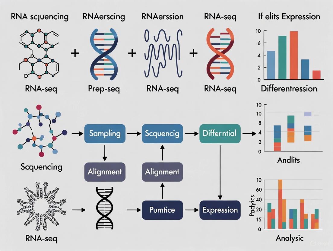
The logical workflow for this protocol and the relationship between key metrics and experimental goals can be visualized in the following diagrams:
Defining the Core Concepts
What are Sequencing Depth and Coverage?
In RNA-Seq experiments, sequencing depth (or read depth) and coverage are two fundamental yet distinct metrics that are crucial for data quality.
- Sequencing Depth refers to the average number of times a specific nucleotide in the genome is read during the sequencing process. It is expressed as an average multiple, such as 30x or 100x. A higher depth means more data points for each base, which increases confidence in base calling and helps mitigate sequencing errors and technical noise [1].
- Coverage describes the percentage of the target genome or transcriptome that has been sequenced at least once. It is typically expressed as a percentage (e.g., 95% coverage). High coverage ensures that the entire region of interest is represented in the data, leaving no gaps [1].
| Metric | Definition | What It Measures | Why It Matters |
|---|---|---|---|
| Sequencing Depth | The average number of times a base is sequenced [1]. | The redundancy of sequencing for a given location. | Higher depth increases confidence in variant calls, especially for low-abundance variants or heterogeneous samples [1]. |
| Coverage | The proportion of the target region sequenced at least once [1]. | The completeness of the sequenced data. | High coverage ensures no regions are missed, preventing gaps in the data that could lead to missed discoveries [1]. |
The relationship between them is synergistic: increasing sequencing depth generally also improves coverage, as more reads have a higher likelihood of covering more regions. However, due to biases in library preparation or sequencing, certain regions may still be underrepresented or missed entirely [1].
Troubleshooting Guide: Common Scenarios and Solutions
FAQ 1: "I am not detecting known low-frequency variants in my cancer RNA-Seq data. What should I optimize?"
- Likely Cause: Insufficient sequencing depth. The transcripts containing the variants may be expressed at low levels, and a shallow sequence depth fails to generate enough reads to distinguish a true variant from background noise [9].
- Solution: Increase your total sequencing depth. For somatic variant calling in cancer RNA-Seq, studies have shown that between 30 million and 40 million 100 bp paired-end reads are often needed to recover 90-95% of single nucleotide variants (SNVs) in recurrently mutated genes [9]. Sensitivity drops significantly (to around 80%) with only 20 million fragments [9].
FAQ 2: "My transcriptome assembly has many gaps, missing known exons and genes. What is the issue?"
- Likely Cause: Inadequate transcriptome coverage. The sequencing depth may be too low to capture the full diversity of transcripts, particularly those that are lowly expressed or transient [10] [11].
- Solution: Increase the total number of sequenced reads to improve coverage. Research indicates that for a complex transcriptome, a depth of about 10 million 75 bp reads can detect approximately 80% of annotated genes, while over 30 million reads may be required to detect nearly all annotated genes [10]. Note that increasing depth beyond a certain point (e.g., 2-8 Gbp) yields diminishing returns for recovering annotated exonic regions and instead recovers a large number of unannotated, single-exon transcripts [11].
FAQ 3: "I am getting high genotyping error rates for SNPs in my population study. How can I improve accuracy?"
- Likely Cause: Insufficient read depth at the variant site. Low coverage means the genotype call is based on very few observations, which is highly susceptible to random sequencing errors [12].
- Solution: Ensure a high minimum depth at each locus. For SNP genotyping using restriction-enzyme-based methods, error rates are highly sensitive to coverage. One study found that while a coverage of ≥5x yielded a median genotyping error rate of 0.03, increasing the minimum coverage to ≥30x reduced the median error rate to ≤0.01 in reference-aligned datasets [12].
FAQ 4: "For single-cell RNA-Seq, should I sequence more cells shallowly or fewer cells deeply?"
- Likely Cause: A suboptimal balance between the number of cells (n_cells) and the sequencing depth per cell (n_reads) for a fixed budget [13].
- Solution: For estimating population-level gene properties (like gene expression distributions), the optimal strategy is often to maximize the number of cells while ensuring an average sequencing depth of around one read per cell per gene. This approach provides a better overview of biological heterogeneity. Sequencing much deeper (e.g., 10x more reads per cell) without increasing cell number can be less efficient, potentially leading to a twofold higher estimation error for the same total budget [13].
The following diagram illustrates the core trade-off and relationship between these key parameters in an RNA-Seq experiment.
Experimental Protocols: Determining Optimal Depth
Methodology 1: A Downsampling Approach to Determine Sufficient Depth for Variant Calling
This protocol, adapted from a study on acute myeloid leukemia, uses computational downsampling to determine the minimal depth needed for sensitive variant detection [9].
- Deep Sequencing: Begin by sequencing a subset of pilot RNA samples (e.g., 3-5 samples) to a very high depth (e.g., >100 million paired-end reads).
- Variant Calling: Call variants on this deep dataset using your chosen pipeline (e.g., a combination of VarDict, MuTect, or VarScan) to establish a "truth set" of high-confidence variants [9].
- Computational Downsampling: Use a script (e.g., in Perl or Python) to randomly sample subsets of reads from the original deep dataset to simulate lower sequencing depths (e.g., 80M, 50M, 40M, 30M, and 20M fragments) [10] [9].
- Variant Recall: Re-call variants at each downsampled depth.
- Sensitivity Calculation: Calculate the sensitivity (percentage of variants from the "truth set" recovered) at each depth.
- Determine Optimal Depth: Identify the depth where sensitivity plateaus at an acceptable level (e.g., >90%). The study found that sensitivity dropped markedly below 30M fragments [9].
Methodology 2: Random Sampling to Determine Depth for Transcriptome Coverage
This method, used in a chicken transcriptome study, assesses how sequencing depth affects gene detection [10].
- Generate High-Depth Data: Sequence a cDNA library to a high depth (e.g., 30 million 75 bp reads).
- Random Sampling: Use a custom program to randomly draw without replacement a fixed number of reads (e.g., 10M, 15M, 20M) from the full dataset. Repeat this process multiple times (e.g., 4 replicates) to ensure statistical robustness [10].
- Gene Detection Analysis: Map each sub-sampled dataset to the reference genome and count the number of annotated genes detected.
- Plot and Analyze: Plot the number of detected genes against sequencing depth. The point where the curve begins to plateau indicates a sufficient depth for transcriptome coverage. The study showed that 10M reads detected ~80% of genes, with minimal gains beyond 20M-30M reads for many applications [10].
The table below summarizes key recommendations from various studies.
| Application | Recommended Sequencing Depth | Key Findings and Rationale |
|---|---|---|
| Variant Calling (Cancer RNA-Seq) | 30M - 40M fragments (100bp PE) [9] | Recovers 90-95% of initial SNVs. Sensitivity drops significantly below 30M fragments [9]. |
| Whole Transcriptome Profiling | 10M - 30M reads (75bp) [10] | 10M reads detects ~80% of annotated genes; 30M reads detects >90% of genes. Serves as a replacement for microarrays [10]. |
| De Novo Transcriptome Assembly | 2 - 8 Gbp total [11] | The amount of exomic sequence assembled typically plateaus in this range. Deeper sequencing mainly recovers unannotated single-exon transcripts [11]. |
| Single-Cell RNA-Seq (Gene Property Estimation) | ~1 UMI/read per cell per gene [13] | For a fixed budget, maximizing cells with shallow depth per cell is optimal for estimating gene expression distributions [13]. |
| SNP Genotyping (ddRAD) | ≥30x coverage [12] | Median genotyping error rates decline to ≤0.01 at coverage ≥30x, compared to ≥0.03 at ≥5x coverage [12]. |
The Scientist's Toolkit: Essential Research Reagents and Materials
| Item | Function in RNA-Seq Workflow |
|---|---|
| Oligo(dT) Beads | To enrich for polyadenylated mRNA from total RNA by hybridization, reducing ribosomal RNA background [10]. |
| RNA Sequencing Sample Preparation Kit (e.g., Illumina) | Provides the necessary reagents for cDNA library construction, including fragmentation, end-repair, adapter ligation, and PCR amplification [10]. |
| DNase I | Digests and removes genomic DNA contamination from RNA samples post-isolation, ensuring a pure RNA template [10]. |
| SPIA Amplification Kit (e.g., NuGEN) | Uses single primer isothermal amplification for linear amplification of cDNA, which can be critical for low-input samples [5]. |
| Universal Human Reference RNA (UHRR) | A standardized reference RNA sample used as a control to compare the performance of different sequencing technologies or library prep protocols [11]. |
| CD34+ Cells | Can be used to create a "Panel of Normals" (PON) for variant filtering in cancer studies, helping to identify and remove common technical artifacts and germline variants [9]. |
Troubleshooting Guides
Guide 1: Addressing Inadequate Sequencing Depth
Problem: My RNA-seq experiment failed to detect differentially expressed genes, especially those expressed at low levels.
Diagnosis and Solution:
- Symptoms: Inability to detect known low-abundance transcripts; high variability in gene expression measurements between replicates.
- Root Cause: Insufficient sequencing depth, leading to undersampling of the transcriptome.
- Recommended Actions:
- Recalculate Required Depth: For mammalian transcriptomes, aim for 20-40 million reads per sample for mRNA libraries, or 40-80 million reads for total transcriptome libraries that include non-coding RNAs [14].
- Increase Biological Replicates: When budget-constrained, prioritize more biological replicates over deeper sequencing, as this provides greater statistical power for detecting differential expression [14].
- Optimize Library Complexity: Ensure high RNA quality (RIN >7) and use appropriate depletion methods to maximize informative reads rather than ribosomal RNA [15].
Guide 2: Managing Excessive Sequencing Costs
Problem: My sequencing costs are exceeding budget without proportional scientific benefit.
Diagnosis and Solution:
- Symptoms: Diminishing returns where additional sequencing depth yields minimal new biological insights; budget depletion limiting sample size.
- Root Cause: Oversequencing individual samples beyond what is necessary for the research question.
- Recommended Actions:
- Right-Size Your Depth: For many applications, including eQTL studies, sequencing more samples at lower coverage (e.g., ~6 million reads/sample) provides better statistical power than fewer samples at high coverage [16].
- Choose Appropriate Sequencing Mode: Use single-end sequencing instead of paired-end when your primary goal is differential expression analysis rather than alternative splicing detection [14].
- Implement rRNA Depletion: Reduce wasteful sequencing of ribosomal RNA (comprising ~80% of cellular RNA) through ribosomal depletion methods to enhance cost-effectiveness [15].
Frequently Asked Questions
Experimental Design FAQs
How do I determine the optimal sequencing depth for my RNA-seq experiment? The ideal depth depends on your transcriptome size and research goals. Use the following table as a guideline:
Table 1: Recommended RNA-seq Sequencing Depth Guidelines
| Application | Recommended Depth | Key Considerations |
|---|---|---|
| Mammalian mRNA-seq | 20-40 million reads/sample | Sufficient for most differential expression studies [14] |
| Total transcriptome (including non-coding RNAs) | 40-80 million reads/sample | Required for adequate coverage of diverse RNA species [14] |
| eQTL discovery studies | ~6 million reads/sample | More samples at lower depth increases power [16] |
| Bacterial transcriptomes | 5-10 million reads/sample | Smaller genomes require less depth [17] |
| De novo transcriptome assembly | 100 million reads/sample | Comprehensive coverage needed for reconstruction [17] |
Should I use single-end or paired-end sequencing for my experiment? Choose based on your research priorities and budget:
- Single-end: Recommended for most gene expression studies; much cheaper with minimal information loss for differential expression analysis once reads exceed 50 bp [14].
- Paired-end: Essential for alternative splicing analysis, novel transcript detection, or when working with poor-quality reference genomes [14].
How many biological replicates do I need? The optimal number depends on your experimental system:
- In vitro studies with homogeneous cell lines: Fewer replicates may suffice (e.g., 3-4).
- Primary cells from human subjects: More replicates are essential due to greater biological variability.
- General rule: Prioritize more replicates over deeper sequencing, as this significantly boosts statistical power for detecting differentially expressed genes [14].
Technical Optimization FAQs
When should I use rRNA depletion versus poly-A selection? The choice depends on your RNA quality and research focus:
Table 2: RNA Selection Method Comparison
| Method | Best For | RNA Quality Requirements | Key Limitations |
|---|---|---|---|
| Poly-A selection | mRNA enrichment in eukaryotes | High-quality RNA (RIN ≥8) with intact polyA tails [18] | Unsuitable for degraded samples or non-polyadenylated RNAs |
| rRNA depletion | Degraded samples (FFPE), non-coding RNA, bacterial RNA | Compatible with low-quality RNA (RIN 2-3) [17] [18] | Additional cost and processing step; potential off-target effects [15] |
| Globin depletion (blood samples) | Improving detection of low-expression transcripts in blood | Standard blood RNA quality | Removes globin transcripts, which may be biologically relevant in some studies [15] [17] |
What are the key considerations for working with challenging sample types?
- FFPE samples: Use rRNA depletion or targeted RNA exome approaches due to expected RNA degradation [17]. The SMARTer Universal Low Input RNA Kit is specifically validated for degraded RNA (RIN 2-3) [18].
- Blood samples: Implement both rRNA and globin depletion to significantly improve detection of low-expression transcripts [17].
- Ultra-low input samples: Utilize specialized kits like SMARTer Ultra Low or SMART-Seq v4 designed for 10 pg-10 ng total RNA or 1-1,000 cells [18].
When should I consider using UMIs (Unique Molecular Identifiers)? Incorporate UMIs in these scenarios:
- Deep sequencing (>50 million reads/sample) to correct PCR amplification biases [17].
- Low-input library preparation where amplification bias is a significant concern [17].
- Absolute transcript quantification requirements, as UMIs enable accurate molecular counting [17].
Experimental Workflow: Balancing Comprehensiveness and Cost
The following diagram illustrates the key decision points in designing a cost-efficient RNA-seq experiment:
Diagram 1: RNA-seq experimental design workflow for cost efficiency.
The Scientist's Toolkit: Essential Research Reagents
Table 3: Key Reagent Solutions for RNA-seq Experiments
| Reagent/Kit | Primary Function | Optimal Use Cases | Input Requirements |
|---|---|---|---|
| SMART-Seq v4 Ultra Low Input Kit | Full-length cDNA synthesis from ultra-low input | 1-1,000 cells or 10 pg-10 ng total RNA; requires high-quality RNA (RIN ≥8) [18] | Oligo(dT) priming |
| SMARTer Stranded RNA-Seq Kit | Strand-specific library prep | 100 pg-100 ng of full-length or degraded RNA; maintains strand information >99% [18] | Requires rRNA depletion or poly-A enrichment |
| SMARTer Universal Low Input RNA Kit | Library prep from degraded samples | 200 pg-10 ng degraded RNA (RIN 2-3); compatible with FFPE samples [18] | Random priming; requires rRNA depletion |
| RiboGone - Mammalian Kit | Ribosomal RNA depletion | 10-100 ng samples of mammalian total RNA; improves cost-efficiency [18] | Works with various RNA qualities |
| ERCC Spike-in Mix | RNA quantification standardization | 92 synthetic transcripts for sensitivity assessment; not recommended for low-concentration samples [17] | Added before library prep |
FAQs on Estimating Sequencing Depth and Coverage
1. What is the difference between sequencing depth and coverage? While often used interchangeably, sequencing depth and coverage are distinct metrics. Sequencing depth (or read depth) refers to the average number of times a specific nucleotide base is read during sequencing. It is expressed as a multiple (e.g., 30x) and is crucial for the accuracy of base calling and variant detection [19] [1]. Coverage refers to the proportion of the target genome or transcriptome that has been sequenced at least once. It is typically expressed as a percentage (e.g., 95% coverage) and indicates the comprehensiveness of the sequencing data [19] [1].
2. What is the recommended sequencing depth for a standard RNA-Seq experiment? For standard RNA-Seq differential gene expression analysis, a sequencing depth of 10 to 50 million reads per sample is often sufficient [19] [20]. This typically translates to a coverage of approximately 10x to 30x [19]. The exact requirement depends on the goals of your study; detecting rare or lowly-expressed transcripts generally requires greater depth [20] [21].
3. How do I calculate the required sequencing depth for my experiment? You can estimate the required sequencing depth using a variation of the Lander/Waterman equation for coverage [21]: C = (L * N) / G Where:
- C = Coverage
- L = Read length
- N = Number of reads
- G = Haploid genome or transcriptome length
To solve for the number of reads (N) needed to achieve a desired coverage (C), you can rearrange the formula: N = (C * G) / L [21].
4. My data has uneven coverage. What are the common causes and solutions? Uneven coverage is a common issue in RNA-Seq and can be caused by:
- Technical biases from library preparation, such as PCR amplification bias [19].
- Biological factors, including regions with high GC content or repetitive sequences that are difficult to sequence [19] [1].
- Transcript length bias, where longer transcripts naturally accumulate more reads in whole transcriptome protocols [22]. Solutions include optimizing library preparation protocols, using unique molecular identifiers (UMIs) to account for PCR duplicates, and ensuring you use appropriate normalization methods (like TMM or median-of-ratios) in your downstream analysis to correct for these biases [19] [20].
5. How does the choice between Whole Transcriptome Sequencing and 3' mRNA-Seq affect my depth and coverage needs? The choice of RNA-Seq methodology significantly impacts your experimental design:
| Methodology | Recommended Depth | Key Considerations |
|---|---|---|
| Whole Transcriptome (WTS) | Higher depth required; often 20-50 million reads or more [20]. | Reads are distributed across the entire transcript. Essential for detecting splice variants, fusion genes, and novel isoforms [22]. |
| 3' mRNA-Seq | Lower depth sufficient; often 1-5 million reads [22]. | Reads are localized to the 3' end of transcripts. Ideal for high-throughput, cost-effective gene expression quantification, especially for large sample numbers [22]. |
Troubleshooting Guide: Common Issues with Depth and Coverage
Problem: Inability to detect differentially expressed genes, especially low-abundance transcripts.
- Potential Cause: Insufficient sequencing depth.
- Solution: Increase the sequencing depth per sample. For projects focused on rare transcripts or low-fold-change differences, consider sequencing up to 50-100 million reads or more. Use power analysis tools on pilot data to determine the optimal depth [20].
Problem: Large portions of the transcriptome are missing from the data.
- Potential Cause: Inadequate coverage, often due to poor RNA quality, inefficient library preparation, or an insufficient number of total sequenced reads.
- Solution: Check RNA integrity (e.g., RIN score) before library prep. Optimize or troubleshoot the cDNA synthesis and library construction steps. Ensure you are generating a sufficient volume of raw sequencing data to cover your target transcriptome [19] [1].
Problem: High variability in read counts between biological replicates.
- Potential Cause: An insufficient number of biological replicates, which reduces the statistical power to detect true differences.
- Solution: Increase the number of biological replicates. While three is often a minimum, more replicates are required when biological variability is high. More replicates generally provide greater statistical power than simply increasing sequencing depth alone [20].
The Scientist's Toolkit: Research Reagent Solutions
| Item | Function |
|---|---|
| Poly(A) Selection Beads | Isolates messenger RNA (mRNA) from total RNA by binding to the poly(A) tail, enriching for coding transcripts and reducing ribosomal RNA (rRNA) contamination. |
| Ribosomal Depletion Probes | Selectively removes abundant ribosomal RNA (rRNA) sequences from total RNA, allowing for the sequencing of both coding and non-coding RNA species. |
| Reverse Transcriptase Enzyme | Synthesizes complementary DNA (cDNA) from the RNA template, creating a stable copy for downstream library construction and amplification. |
| Oligo(dT) Primers | Primers that bind to the poly(A) tail of mRNA to initiate cDNA synthesis; a key component of 3' mRNA-Seq protocols [22]. |
| Random Hexamer Primers | Primers that bind randomly to RNA fragments, used in whole transcriptome protocols to generate coverage across the entire length of the transcript [22]. |
| Fragmentation Enzymes/Buffers | Physically or enzymatically shears cDNA or RNA into appropriately sized fragments for optimal sequencing on NGS platforms. |
Workflow for Determining Sequencing Needs
The following diagram outlines the key decision points for planning your RNA-seq experiment to ensure adequate depth and coverage.
This guide is based on the latest best practices in the field [19] [20] [22]. For further details on specific protocols and statistical methods, please refer to the cited literature.
From Theory to Bench: Designing and Executing Your RNA-seq Experiment
Frequently Asked Questions
What is the difference between sequencing depth and coverage? These terms are often used interchangeably but have distinct meanings [1].
- Sequencing Depth (or Read Depth): Refers to the number of times a specific nucleotide is read during sequencing. It is expressed as an average (e.g., 100x depth) and is crucial for confident variant calling [1].
- Coverage: Describes the percentage of the entire genome or target region that has been sequenced at least once. It ensures the completeness of the data (e.g., 95% coverage) [1].
How does experimental goal influence sequencing depth? Your study's objective is the primary driver for determining the appropriate sequencing depth [1].
- Gene Expression Profiling of highly expressed genes requires less depth.
- Detection of Rare Transcripts or Splicing Variants requires greater depth for sufficient sensitivity.
- Single-Cell RNA-Seq involves a trade-off between sequencing many cells shallowly or fewer cells more deeply [13].
My differential gene expression analysis lacks power. Could sequencing depth be the issue? Yes, insufficient sequencing depth is a common cause. For standard bulk RNA-Seq DGE analysis, a minimum of 20–30 million reads per sample is often sufficient [20]. However, the required depth increases if your study focuses on lowly expressed genes. Using too few replicates also reduces power; three replicates per condition is a typical minimum, but more are needed when biological variability is high [20].
Sequencing Depth Recommendations by Application
Table 1: Recommended sequencing depth and key considerations for various RNA-Seq applications.
| Application | Recommended Depth (per sample) | Key Considerations & Goals |
|---|---|---|
| Gene Expression Profiling | 5 - 25 million reads [6] | Quick snapshot of highly expressed genes; allows for high multiplexing. |
| Standard DGE Analysis | 30 - 60 million reads [6] | Global view of expression; some information on alternative splicing. |
| In-depth Transcriptome | 100 - 200 million reads [6] | Novel transcript assembly, comprehensive splicing analysis. |
| Targeted RNA Panels | ~3 million reads [6] | Targeted approaches (e.g., TruSight RNA Pan Cancer) require fewer reads. |
| miRNA / Small RNA Seq | 1 - 5 million reads [6] | Varies significantly by tissue type. |
| Single-Cell RNA-Seq | Varies by cell number | Balance between number of cells and depth. A mathematical framework suggests an optimal allocation may be shallow sequencing (e.g., ~1 read per cell per gene) of many cells [13]. |
Single-Cell RNA-Seq Experimental Design
For single-cell RNA-seq (scRNA-seq), the experimental design question revolves around how to allocate a fixed sequencing budget: should you sequence a few cells deeply or many cells shallowly? [13]
A mathematical framework suggests that for estimating many important gene properties, the optimal allocation is to sequence at a depth of around one read per cell per gene. Interestingly, this often means maximizing the number of cells sequenced while ensuring that at least ~1 UMI per cell is observed on average for biologically critical genes [13]. One analysis demonstrated that sequencing 10 times more cells at 10 times shallower depth could reduce the estimation error by twofold [13].
The following workflow outlines the key steps and considerations for designing your sequencing experiment:
The Scientist's Toolkit
Table 2: Essential reagents, tools, and software for RNA-Seq experiments and data analysis.
| Item | Function / Purpose |
|---|---|
| NEBNext Poly(A) mRNA Magnetic Isolation Kit | Isolates mRNA from total RNA for library preparation [23]. |
| NEBNext Ultra DNA Library Prep Kit for Illumina | Prepares sequencing libraries from cDNA [23]. |
| Cell Ranger | Standardized pipeline for processing raw data from 10x Genomics scRNA-seq platforms [24]. |
| Trimmomatic / Cutadapt | Tools for read trimming to remove adapter sequences and low-quality bases [20]. |
| STAR / HISAT2 | Aligns (maps) sequencing reads to a reference genome [20]. |
| Kallisto / Salmon | Performs pseudo-alignment for fast transcript abundance estimation [20]. |
| featureCounts / HTSeq | Counts the number of reads mapped to each gene [20]. |
| DESeq2 / edgeR | Software packages for differential gene expression analysis [20]. |
| Seurat | A comprehensive R package for the analysis of single-cell RNA-seq data [24]. |
| FastQC / MultiQC | Performs initial quality control on raw sequenced data and generates reports [20]. |
FAQs on Biological Replicates in RNA-Seq
Why are biological replicates more important than sequencing depth for most genes?
Multiple independent studies have concluded that for the majority of genes, increasing the number of biological replicates has a larger impact on the statistical power of differential expression analysis than increasing sequencing depth [25] [26] [27]. Biological replicates capture the natural random variation that occurs between different biological subjects (e.g., different mice, different batches of cells), allowing you to determine if an observed effect is consistent and generalizable [28]. While deeper sequencing helps detect lowly expressed genes, beyond a certain point (often ~20-30 million reads per sample), it yields diminishing returns. Power, however, continues to increase significantly with more replicates [20] [26].
What is the fundamental difference between a biological and a technical replicate?
Understanding this distinction is critical for proper experimental design.
- Biological Replicates are measurements taken from biologically distinct samples. They capture the biological variation in a population. Examples include:
- Technical Replicates are repeated measurements of the same biological sample. They assess the variability of your assay or measurement technique. Examples include:
Technical replicates tell you about the precision of your lab work, while biological replicates tell you whether your findings are reproducible across a population [30] [29] [28].
How many biological replicates are needed for a robust RNA-seq experiment?
There is no universal number, as it depends on the desired power, effect size, and biological variability of your system. However, evidence-based guidelines provide a strong starting point.
- Absolute Minimum: 3 biological replicates per condition. However, with only three, most statistical tools will detect only 20-40% of all differentially expressed (DE) genes identified with high replicate numbers (e.g., 42). Power is sufficient primarily for genes with very large fold changes (>4-fold) [32].
- Recommended Minimum: 6 biological replicates per condition. This offers a much better balance for identifying DE genes across a range of fold changes [32].
- For Robust Detection: 12 or more biological replicates. To detect >85% of all DE genes, including those with more subtle fold changes, more than 20 replicates may be necessary [32]. For specific contexts like toxicology dose-response studies, at least 4 replicates are recommended [27].
The following table summarizes key quantitative findings from the literature:
| Recommendation / Finding | Minimum Replicates | Context / Key Outcome | Source |
|---|---|---|---|
| General Guideline | 4 | Tomato research; ensures detection of ~1000 DE genes with 20M reads/sample. | [25] |
| Practical Minimum | 6 | Superior true/false positive performance with tools like DESeq2 and edgeR. | [32] |
| For All Fold Changes | 12 | Needed to detect >85% of SDE genes, regardless of effect size. | [32] |
| Power vs. Depth | >20 | Replicate number has a larger impact on power than sequencing depth. | [25] [26] |
| Toxicology Context | 4 | Reliable benchmark dose (BMD) pathways in dose-response studies. | [27] |
Which statistical tools are best for differential expression analysis with low replicate numbers?
For experiments with fewer than 12 replicates, DESeq2 and edgeR provide a superior combination of true positive detection and false positive control [32]. These tools use the negative binomial distribution to model RNA-Seq count data, which accurately accounts for the biological variation measured by your replicates [26] [32].
How can I formally estimate the number of replicates needed for my specific experiment?
You should use a power analysis tool before conducting your experiment. These tools use parameters from previous, similar datasets to estimate the sample size required to achieve your desired statistical power.
- RnaSeqSampleSize is a Bioconductor R package and web tool that uses real data distributions (e.g., from TCGA) to provide realistic sample size estimates, taking into account the varying expression levels and dispersions across thousands of genes [33].
- Performing a pilot experiment and analyzing the resulting data is another highly effective way to estimate parameters for a full-scale study [25].
Experimental Protocol: Power and Sample Size Estimation Using RnaSeqSampleSize
Objective: To determine the optimal number of biological replicates required for a robust RNA-seq experiment by performing a power analysis based on a reference dataset.
Materials:
- Computer with R installed.
- RnaSeqSampleSize package from Bioconductor.
- Reference RNA-seq dataset (e.g., from a public repository like TCGA or a pilot experiment).
Methodology:
- Install and Load Package: Install the
RnaSeqSampleSizepackage from Bioconductor and load it into your R session [33]. - Define Analysis Parameters: Set the key statistical parameters for your planned experiment:
- False Discovery Rate (FDR): Typically set to 0.05.
- Desired Statistical Power: Typically set to 0.8 or 0.9.
- Fold Change: The minimum effect size (e.g., 2-fold) you are interested in detecting.
- Gene Signature (Optional): If your interest is in a specific pathway, provide a list of relevant genes or a KEGG pathway ID [33].
- Input Reference Data: Provide a reference dataset that closely resembles your expected experimental system. This allows the tool to use realistic distributions of gene expression and dispersion for its calculations [33].
- Perform Power Calculation: Execute the package's functions to estimate either:
- The power achievable with a given sample size, or
- The sample size required to achieve a given power.
- Visualize and Interpret: Use the package's built-in plotting functions to visualize power curves, which show the relationship between sample size and statistical power for your parameters [33].
This workflow for determining the optimal number of replicates can be summarized in the following decision pathway:
The Scientist's Toolkit: Research Reagent Solutions
| Item / Resource | Function / Application | Context in Replicate Design |
|---|---|---|
| DESeq2 | A statistical software package for differential analysis of RNA-seq count data. | Recommended tool for DE analysis, especially with lower replicate numbers (n<12) [32]. |
| edgeR | A statistical software package for differential expression analysis of RNA-seq data. | Recommended tool for DE analysis, especially with lower replicate numbers (n<12) [32]. |
| RnaSeqSampleSize | An R/Bioconductor package for sample size and power estimation. | Uses real data distributions to calculate necessary biological replicates before a full-scale experiment [33]. |
| TCGA (The Cancer Genome Atlas) | A public repository containing a vast array of RNA-seq datasets. | Serves as an ideal source of reference data for power analysis in human cancer studies [33]. |
| Biological Samples (e.g., Cell Cultures, Model Organisms) | The fundamental units of study from which RNA is extracted. | Must be processed as independent, biologically distinct entities to qualify as true biological replicates [29] [28]. |
Sequencing depth and coverage are foundational concepts in designing a robust RNA-seq experiment. Within the context of this thesis, managing these parameters is critical for generating biologically meaningful results. Sequencing depth (or read depth) refers to the number of times a specific nucleotide is read during sequencing, directly influencing confidence in base calling and variant detection [1]. Coverage describes the percentage of the target genome or transcriptome that has been sequenced at least once, ensuring comprehensive representation [1]. For RNA-seq, the required read depth varies significantly based on experimental goals, ranging from 5 million reads per sample for a quick snapshot of highly expressed genes to 100-200 million reads for novel transcript assembly and in-depth analysis [6]. Balancing sufficient depth and coverage against available resources is a central challenge in experimental design, impacting everything from initial read quality to the final list of differentially expressed genes.
The Standard RNA-seq Analysis Workflow
The following diagram illustrates the complete pathway for RNA-seq data analysis, from raw sequencing data to biological interpretation, highlighting key quality control checkpoints.
Workflow Stages and Key Considerations
- Raw FASTQ to Quality Control: Process begins with raw sequencing data, assessing initial quality metrics like per-base sequence quality and adapter content [34] [35].
- Trimming and Post-Trimming QC: Remove low-quality bases and adapter sequences, then verify improvements [35].
- Alignment and Quantification: Map reads to a reference genome or transcriptome, then count reads associated with genes [34] [35].
- Differential Expression Analysis: Normalize count data to account for confounding factors and identify statistically significant expression changes [36] [37].
- Visualization and Interpretation: Explore results and derive biological meaning.
The Scientist's Toolkit: Essential Research Reagent Solutions
Table 1: Key Software Tools and Resources for RNA-seq Analysis
| Tool Category | Specific Tool(s) | Primary Function | Key Considerations |
|---|---|---|---|
| Quality Control | FastQC [34] [38] [35], MultiQC [34], fastp [34] [39], Trimmomatic [35], Trim_Galore [39] | Assesses raw and trimmed read quality; trims adapters and low-quality bases. | FastQC provides visual reports; MultiQC aggregates multiple reports; fastp is fast and integrated; Trimmomatic is highly cited but complex. |
| Alignment | STAR [34] [35], TopHat2 [38] | Aligns RNA-seq reads to a reference genome. | STAR is splice-aware and widely used; requires genome indexing. |
| Quantification | FeatureCounts [35], HTSeq [38], Salmon [34] | Generates count data for each gene by counting reads overlapping genomic features. | Can be performed on aligned BAM files (FeatureCounts) or via pseudoalignment (Salmon). |
| Differential Expression | DESeq2 [34] [36] [38], edgeR [36] | Identifies statistically significant differentially expressed genes. | Both use negative binomial models; DESeq2 is known for stringent normalization. |
| Normalization Methods | DESeq2's Median of Ratios [37], edgeR's TMM [36] [37] | Scales raw counts to make samples comparable. | Essential for correcting for library size and RNA composition. TMM assumes most genes are not DE. |
Troubleshooting Guide: Common RNA-seq Pipeline Issues
How Do I Determine the Appropriate Sequencing Depth for My RNA-seq Experiment?
The required sequencing depth depends heavily on your experimental objectives and organism complexity [6]. The ENCODE project provides excellent guidelines, but you should also consult primary literature specific to your field and organism [6].
Table 2: Recommended Sequencing Depth for Different RNA-seq Goals
| Experimental Goal | Recommended Reads Per Sample | Rationale |
|---|---|---|
| Quick Snapshot / Targeted Expression | 5 - 25 million [6] | Sufficient for profiling highly expressed genes. Allows for high multiplexing of samples. |
| Standard Gene Expression Profiling | 30 - 60 million [6] | Encompasses most published mRNA-seq experiments. Provides a global view of expression. |
| Alternative Splicing Analysis | 30 - 60 million [6] | Paired-end reads are recommended to capture splice junctions. |
| Novel Transcript Discovery/Assembly | 100 - 200 million [6] | Deeper sequencing helps assemble complete transcripts and identify rare isoforms. |
| Small RNA Analysis (e.g., miRNA) | 1 - 5 million [6] | Due to their short length and lower complexity, fewer reads are required. |
My Alignment Rate is Low. What Are the Potential Causes and Solutions?
A low uniquely mapped read rate (generally below 60-70% [35]) indicates problems.
- Cause 1: Poor Read Quality or Adapter Contamination. Solution: Re-inspect the FastQC report, particularly the "Per base sequence quality" and "Adapter content" modules [35]. Re-trim your reads using fastp or Trimmomatic with appropriate adapter sequences [34] [35].
- Cause 2: Reference Genome Mismatch. Solution: Ensure the reference genome and annotation file are compatible (e.g., same naming conventions like "chr1" vs "1") and from the same source/version [34]. Use the most up-to-date files available.
- Cause 3: High Levels of RNA Degradation. Solution: Check the RNA Integrity Number (RIN) of your samples prior to sequencing. Degraded samples will have a biased representation of transcripts and map poorly. Ensure all reagents and equipment are RNase-free to prevent degradation during extraction [40].
How Can I Account for Unwanted Variation in My Dataset During Differential Expression Analysis?
Sample-level quality control is essential to identify major sources of variation before performing differential expression testing [37].
- Technique 1: Principal Component Analysis (PCA). Plot the samples using the first few principal components. Ideally, replicates should cluster together, and the condition of interest should be the primary source of variation. If other factors (e.g., batch, sex, library preparation date) are driving the variation, they can be included in the DESeq2 design formula to regress out their effect [37].
- Technique 2: Hierarchical Clustering. This heatmap displays correlation between all sample pairs. Samples generally show high correlations (>0.80). Samples with low correlation to their group may be outliers and warrant further investigation [37].
I Suspect RNA Degradation During Extraction. How Can I Confirm and Prevent This?
RNA degradation is a common issue that compromises data quality.
- Causes: RNase contamination, improper sample storage, repeated freezing and thawing, or long storage times [40].
- Confirmation: Check the FastQC report. A sharp drop in sequence quality or per-base sequence content at the 5' end can indicate degradation. Bioanalyzer traces (e.g., low RIN) from the original sample are a definitive check.
- Prevention:
What is the Difference Between Normalization Methods, and Which One Should I Use?
Normalization is critical for accurate gene expression comparisons. Different methods account for different "uninteresting" factors.
Table 3: Common RNA-seq Normalization Methods
| Method | Accounted Factors | Recommended Use | Not Recommended For |
|---|---|---|---|
| CPM (Counts Per Million) | Sequencing depth | Gene count comparisons between replicates of the same sample group. | Within-sample comparisons or DE analysis [37]. |
| TPM (Transcripts Per Million) | Sequencing depth, Gene length | Gene count comparisons within a sample or between samples of the same group [37]. | DE analysis [37]. |
| RPKM/FPKM | Sequencing depth, Gene length | Gene count comparisons between genes within a sample [37]. | Between-sample comparisons or DE analysis (values are not comparable between samples) [37]. |
| DESeq2's Median of Ratios | Sequencing depth, RNA composition | Gene count comparisons between samples and for DE analysis [37]. | Within-sample comparisons [37]. |
| edgeR's TMM (Trimmed Mean of M-values) | Sequencing depth, RNA composition | Gene count comparisons between samples and for DE analysis [37]. | Within-sample comparisons [37]. |
For differential expression analysis with tools like DESeq2 or edgeR, you should use the built-in normalization method (Median of Ratios or TMM, respectively). These methods are robust to library size and RNA composition biases, which is essential for accurate between-sample comparisons [36] [37].
A fundamental research problem in many RNA-seq studies is the identification of differentially expressed genes (DEGs) between distinct sample groups. The choice of computational tools for this task is critical, as it can markedly affect the outcome of the data analysis [41]. Numerous statistical methods have been developed, each with unique statistical approaches and assumptions. Understanding the differences between popular tools like edgeR, DESeq2, and limma-voom will help you select the most appropriate method for your experimental context, ensuring robust and reliable biological conclusions [42].
Understanding the Core Statistical Approaches
Differential expression analysis tools primarily use parametric or non-parametric approaches to model RNA-seq count data and test for significant changes.
- Parametric Methods: These assume that the count data follows a specific probability distribution. Tools like DESeq2 and edgeR use a negative binomial (NB) distribution to model counts, as this distribution effectively accounts for biological variability and overdispersion (where the variance between replicated measurements is higher than the mean) [41] [43]. They then employ empirical Bayes techniques to "shrink" or moderate the estimates of gene-wise dispersion towards a common trend, improving stability for experiments with small numbers of replicates [41] [43].
- Transformation-Based Methods: The limma-voom pipeline uses a hybrid approach. It applies a voom transformation to convert normalized count data into continuous log2-counts per million (log-CPM) values. Subsequently, it uses precision weights to account for the mean-variance relationship in the data, enabling the application of powerful linear modeling and empirical Bayes methods originally developed for microarray data [41] [42].
- Non-Parametric Methods: Tools like NOISeq and SAMseq make fewer assumptions about the underlying data distribution. They are often based on resampling techniques or model the null distribution of noise directly from the data, which can be advantageous when parametric assumptions are violated [41] [43].
Comparative Performance of Differential Expression Methods
Independent evaluations have benchmarked the performance of various methods across different experimental conditions. Key performance metrics include the ability to control the False Discovery Rate (FDR)—the expected proportion of false positives among all detections—and statistical power, the probability of correctly detecting a truly differentially expressed gene [41] [43].
Performance Based on Sample Size
The table below summarizes findings from a 2022 evaluation of eight popular methods, highlighting how performance varies with sample size when data follows a negative binomial distribution [43].
| Sample Size (per group) | Recommended Method(s) | Key Performance Notes |
|---|---|---|
| 3 | EBSeq | Better FDR control, power, and stability compared to other methods with very small sample sizes [43]. |
| 6 or 12 | DESeq2 | Performs slightly better than other methods in terms of FDR control and power as sample size increases [43]. |
| Very Small (e.g., 2) | edgeR | Designed to be efficient with small sample sizes; exact tests can work with as few as 2 replicates [42] [43]. |
| Large (e.g., >20) | Wilcoxon rank-sum test | In population-level studies with large samples, parametric methods (DESeq2, edgeR) may fail to control FDR; non-parametric Wilcoxon test is more robust to outliers and provides better FDR control [44]. |
General Comparisons Between Widely-Used Tools
The following table provides a direct comparison of the three most widely-used tools—DESeq2, edgeR, and limma-voom—based on their core characteristics [42] [45].
| Aspect | DESeq2 | edgeR | limma-voom |
|---|---|---|---|
| Core Statistical Approach | Negative binomial GLM with empirical Bayes shrinkage [42]. | Negative binomial model with empirical Bayes moderation [42]. | Linear modeling with empirical Bayes moderation on voom-transformed counts [42]. |
| Default Normalization | Median-of-ratios method (corrects for library composition) [20]. | TMM (Trimmed Mean of M-values; corrects for library composition) [41] [20]. | TMM normalization, followed by voom transformation [42]. |
| Ideal Sample Size | ≥3 replicates, performs well with more [42] [43]. | ≥2 replicates, efficient with small samples [42] [43]. | ≥3 replicates per condition [42]. |
| Best Use Cases | Moderate to large sample sizes, high biological variability, subtle expression changes [42]. | Very small sample sizes, large datasets, technical replicates [42]. | Small sample sizes, multi-factor experiments, time-series data, integration with other omics [42]. |
| Computational Efficiency | Can be computationally intensive for large datasets [42]. | Highly efficient, fast processing [42]. | Very efficient, scales well with large-scale datasets [42]. |
| Key Limitations | Can be conservative in fold change estimates; FDR control can be exaggerated in large population studies [42] [44]. | Requires careful parameter tuning; common dispersion may miss gene-specific patterns [42]. | Requires careful QC of the voom transformation; may not handle extreme overdispersion well [42]. |
Frequently Asked Questions (FAQs) and Troubleshooting
Q1: My RNA-seq experiment has only 2 replicates per condition. Is differential analysis even possible, and which tool should I use?
While technically possible, analysis with only two replicates greatly reduces the ability to estimate biological variability and control false discovery rates [20]. If you must proceed, edgeR is specifically developed for experiments with very small numbers of replicates and is generally considered the safest choice in this scenario [42] [43]. Its empirical Bayes procedure moderates the degree of overdispersion by borrowing information between genes, which is crucial when per-group sample sizes are minimal [41]. However, you should interpret the results with caution and consider any findings as preliminary until validated.
Q2: I am analyzing data from a population-level study with over 100 samples per group. My colleague warned me that DESeq2/edgeR might have high false discovery rates. Is this true?
Yes, this is a significant and recently highlighted concern. When analyzing human population RNA-seq samples with large sample sizes (dozens to thousands), parametric methods like DESeq2 and edgeR have been shown to have exaggerated false positives, with actual FDRs sometimes exceeding 20% when the target FDR is 5% [44]. This is often due to violations of the negative binomial model assumptions, potentially caused by outliers in the data. In such cases, a non-parametric method like the Wilcoxon rank-sum test is recommended, as it is more robust to outliers and provides better FDR control for large-sample studies [44].
Q3: I keep getting an error that a condition or group is "not found" when I try to run DESeq2 or make contrasts in limma. What is wrong?
This error typically indicates a problem with your sample metadata (colData) or the design formula. The software cannot find the factor level you specified in the model. To troubleshoot:
- Check Factor Levels: Ensure that the condition names in your sample metadata table exactly match those used in your design formula. A common mistake is a typo or incorrect case (e.g., "Control" vs. "control").
- Set the Reference Level: By default, R uses the alphabetically first factor level as the reference (base) group. It is good practice to explicitly set the reference level to your control condition. For example, in R: This ensures "Untreated" is the baseline for comparison [46].
- Verify Data Match: Confirm that the order of samples in your count matrix columns exactly matches the order of rows in your sample metadata. DESeq2 and other packages will not alert you if they are mismatched, leading to incorrect analyses [46] [47].
Q4: How does sequencing depth impact my differential expression analysis, and what is a sufficient depth?
Sequencing depth directly impacts the sensitivity of your analysis. Deeper sequencing captures more reads per gene, increasing your ability to detect lowly expressed transcripts [20]. A depth of 20–30 million reads per sample is often sufficient for standard differential gene expression analysis in many organisms [20] [10]. However, the sufficient depth depends on the complexity of the transcriptome and your specific goals. One study found that while 10 million 75-bp reads detected about 80% of annotated genes in chicken, 30 million reads were required to detect over 90% of genes [10]. If your goal is to detect rare transcripts or splice variants, you may need greater depth. Tools like Scotty can help model power and estimate depth requirements during experimental design [20].
Essential Research Reagent Solutions
The table below lists key computational tools and their roles in a standard RNA-seq differential expression workflow.
| Tool / Resource | Function in Workflow | Brief Explanation |
|---|---|---|
| FastQC / MultiQC | Quality Control | Assesses raw sequence data for technical errors, adapter contamination, and overall quality [20]. |
| Trimmomatic / Cutadapt | Read Trimming | Removes adapter sequences and low-quality bases from reads to improve mapping accuracy [20]. |
| STAR / HISAT2 | Read Alignment | Maps (aligns) cleaned sequencing reads to a reference genome [20]. |
| featureCounts / HTSeq | Read Quantification | Counts the number of reads mapped to each gene, generating a raw count matrix [20]. |
| DESeq2 / edgeR / limma | Differential Expression | Statistical analysis of the count matrix to identify genes expressed at different levels between conditions [42] [45]. |
| Salmon / Kallisto | Pseudo-alignment & Quantification | An alternative, faster workflow that estimates transcript abundances without full base-by-base alignment [20] [46]. |
Experimental Protocol: A Standard DESeq2 Workflow
The following is a detailed protocol for performing differential expression analysis using the DESeq2 package in R, from data input to generating a results table [46] [42].
Step 1: Load Packages and Data
- Install and load the required R packages (
DESeq2,tidyverse). - Load the raw count matrix and the sample information table (metadata). The count matrix should have genes as rows and samples as columns. The metadata must contain the experimental conditions for each sample.
Step 2: Verify and Prepare Data
- Crucially, ensure that the order of samples in the count matrix columns matches the order of rows in the metadata. Use
all(colnames(count_matrix) == metadata$SampleName)to check [46]. - Define the experimental design using a formula, e.g.,
design <- ~ condition. Set the reference level of your factor to the control group usingfactor()andrelevel()[46].
Step 3: Create DESeqDataSet and Filter Genes
- Create a
DESeqDataSetobject from the count matrix, metadata, and design formula. - Filter out genes with very low counts across all samples, as they are uninformative. A common filter is to keep genes with at least a few counts (e.g., >5) in a minimum number of samples [46].
Step 4: Run the Core DESeq2 Analysis
- The main analysis can be performed in a single step using the
DESeq()function. This wrapper executes three steps internally [46]:- Estimation of size factors (for normalization).
- Estimation of dispersion for each gene.
- Fitting of a negative binomial generalized linear model (GLM) and performing Wald tests.
- Alternatively, you can run these steps individually for more control over parameters.
Step 5: Extract and Interpret Results
- Use the
results()function to extract a table of results, including log2 fold changes, p-values, and adjusted p-values (FDR). You can specify significance thresholds (e.g.,alpha=0.05) and fold change thresholds here [46]. - The results table can be sorted by adjusted p-value and exported for further analysis and visualization.
Visual Workflow and Decision Guide
The following diagram illustrates a standard RNA-seq data analysis workflow, from raw data to differential expression results.
Diagram 1: Standard RNA-seq Differential Expression Analysis Workflow.
The decision of which differential expression tool to use depends heavily on your experimental design. The following logic can guide your selection.
Diagram 2: A Decision Guide for Selecting a Differential Expression Method.
Solving Common Pitfalls: Strategies for Optimizing Depth and Coverage
Frequently Asked Questions
What are the main sources of technical noise in RNA-seq? Technical noise in RNA-seq arises from multiple sources in the experimental pipeline. It is commonly categorized into three areas:
- Molecular noise: Stemming from upfront processes like cell lysis, reverse transcription, and cDNA amplification. This includes pipetting errors, technician variability, and amplification bias.
- Machine noise: Introduced by the sequencing process itself, such as cluster generation on the flow cell and lane-to-lane variability.
- Analysis noise: Generated during bioinformatic processing through steps like quality trimming, alignment parameters, and data normalization [48].
How does technical noise differ from biological noise? Biological noise refers to the natural, cell-to-cell variability in gene expression within an isogenic population, predominantly attributed to stochastic fluctuations in transcription [49]. Technical noise is non-biological variability injected by the experimental and computational process. One study estimated that in a well-optimized RNA-seq pipeline, process noise (a component of technical noise) can introduce approximately 24-30% variability in the data. In contrast, biological noise is often 5 to 10 times greater than this process noise [48].
Why is it crucial to account for technical noise in single-cell RNA-seq (scRNA-seq)? scRNA-seq is particularly prone to technical biases like dropout events (where a transcript is expressed but not detected) and amplification bias due to the minute starting amount of RNA [50] [51]. These technical effects vary from cell to cell and, if not properly corrected, can confound downstream analyses like differential expression, leading to false positives or negatives [51].
Troubleshooting Guides
Issue: Low RNA Input and Yield
This issue is common when working with rare cell populations or limited clinical samples and leads to low sequencing coverage and high technical noise [50].
- Potential Causes and Solutions
| Cause | Solution |
|---|---|
| Incomplete homogenization or lysis | Optimize homogenization conditions to ensure complete cell disruption and RNA release [40]. |
| RNA degradation | Ensure all tubes, tips, and solutions are RNase-free. Store samples at -65°C to -85°C and avoid repeated freeze-thaw cycles [52] [40]. |
| Low RNA precipitation efficiency | For small tissue or cell quantities, reduce the volume of lysis reagent (e.g., TRIzol) proportionally to prevent excessive dilution. Use glycogen as a carrier to aid precipitation [40]. |
| General low extraction rate | Increase sample lysis time to over 5 minutes at room temperature. Adjust sample input to ensure it is not excessive for the reagent volume [40]. |
- Experimental Protocol: Improving RNA Quality from Suboptimal Samples
- Quality Control: Assess RNA integrity using an Agilent Bioanalyzer. A high-quality sample should have an RNA Integrity Number (RIN) > 6 [52].
- Library Prep Selection: For degraded samples (e.g., FFPE tissues), use specialized single-cell combinatorial indexing (SCI) or random priming protocols that perform better with fragmented RNA [50].
- Pre-amplification: Incorporate a pre-amplification step during cDNA synthesis to increase the amount of material before library construction [50].
Issue: Amplification Bias
Amplification bias causes skewed representation of transcripts, overestimating highly expressed genes and underestimating low-abundance ones [50].
- Potential Causes and Solutions
| Cause | Solution |
|---|---|
| Stochastic variation in PCR amplification | Use Unique Molecular Identifiers (UMIs). UMIs are short random sequences that tag individual mRNA molecules before amplification, allowing bioinformatic correction for duplicate reads [50] [51]. |
| Non-linear amplification | Use spike-in controls. These are synthetic RNA molecules added at known concentrations to the sample, providing an internal standard to model and correct for amplification efficiency and technical variation [51]. |
| Library preparation protocol | Standardize library preparation protocols and optimize the number of amplification cycles to minimize bias [50]. |
- Experimental Protocol: Using Spike-in Controls
- Selection: Choose a spike-in kit (e.g., ERCC spike-ins) that covers a wide range of concentrations [51].
- Addition: Add a fixed volume of spike-in solution to the cell lysis buffer at the very beginning of the workflow [51].
- Analysis: Use statistical frameworks (e.g., TASC) that leverage the known concentrations of spike-ins to model and subtract cell-specific technical noise, including amplification bias and dropout rates [51].
Issue: Dropout Events
Dropouts are false negatives where a transcript expressed in a cell fails to be captured or amplified, which is especially problematic for detecting lowly expressed genes and rare cell populations [50].
- Potential Causes and Solutions
| Cause | Solution |
|---|---|
| Low capture efficiency of reverse transcription | Use specialized protocols like SMART-seq, which have higher sensitivity and are better at detecting low-abundance transcripts [50]. |
| Stochastic sampling of lowly expressed transcripts | Increase sequencing depth. Deeper sequencing provides a higher chance of capturing rare transcripts [53]. For diagnostic-level detection, ultra-deep sequencing (up to 1 billion reads) may be necessary to saturate gene detection [53]. |
| Inefficient primer binding | Computational imputation methods can be applied. These methods use statistical models and machine learning to predict the expression levels of missing genes based on patterns in the data from other cells and genes [50]. |
- Experimental Protocol: A Scalable Approach to Mitigate Dropouts
- Design: Plan sequencing depth based on study goals. While 30-60 million reads per sample is standard, aim for 100-200 million reads for an in-depth view of the transcriptome [6].
- Validation: For key findings, especially involving low-expression genes, validate results using an orthogonal method like single-molecule RNA FISH (smFISH), which is considered a gold standard for mRNA quantification due to its high sensitivity [49].
- Normalization: Apply scRNA-seq-specific normalization algorithms (e.g., SCTransform, scran, BASiCS) that are designed to account for varying sequencing depths and dropout events across cells [49] [50].
Managing Sequencing Depth and Coverage to Control Noise
The following diagram illustrates the strategic relationship between sequencing depth, technical noise, and the solutions discussed in this guide.
Strategic Flow for Noise Management
Key Recommendations:
- Define Study Objectives: The required depth depends on the goal. Gene expression profiling may need 5-25 million reads per sample, while novel transcript assembly or detecting rare variants may require 100-200 million reads or more [6] [53].
- Understand the Difference:
- Sequencing Depth (Read Depth): The average number of times a specific nucleotide is read. Higher depth increases confidence in base calling and is crucial for detecting rare variants [1].
- Coverage: The percentage of the target genome or transcriptome that has been sequenced at least once. High coverage ensures no genomic regions are missed [1].
- Balance Depth and Cost: Ultra-deep sequencing (e.g., 1 billion reads) can approach saturation for gene detection but may not be cost-effective for all studies. A balance must be struck based on the specific research question [53].
The Scientist's Toolkit: Key Research Reagent Solutions
| Item | Function in Managing Technical Noise |
|---|---|
| Unique Molecular Identifiers (UMIs) | Short random barcodes that label individual mRNA molecules before amplification, allowing for accurate digital counting and correction for amplification bias and PCR duplicates [50] [51]. |
| Spike-in Controls (e.g., ERCC) | Synthetic RNA controls added at known concentrations. They enable precise modeling of technical variation, including amplification efficiency and dropout rates, for per-cell normalization [51]. |
| Stranded Library Prep Kits | Library preparation protocols that preserve the strand information of transcripts. This is critical for accurate transcriptome assembly, distinguishing overlapping genes on opposite strands, and reducing misidentification errors [52]. |
| Ribosomal RNA Depletion Kits | Kits that remove abundant ribosomal RNA (rRNA), which can constitute over 95% of total RNA. This greatly increases the sequencing coverage of mRNA and non-coding RNA of interest [52]. |
| Poly-A Selection Kits | Kits that enrich for messenger RNA (mRNA) by targeting the poly-A tail. This simplifies the transcriptome by focusing on protein-coding genes, but may miss non-polyadenylated RNAs [52]. |
In RNA sequencing (RNA-seq), achieving uniform coverage is fundamental for accurate transcript quantification and detection. However, two persistent technical challenges routinely compromise data integrity: the under-representation of GC-rich regions (sequences with high guanine and cytosine content) and 3' bias (the preferential sequencing of the 3' end of transcripts) [54] [55]. These biases are not merely nuisances; they directly impact the reliability of your measurements for differential expression analysis and novel transcript detection. Effectively managing them is a critical component of optimizing sequencing depth and coverage, ensuring that your data is both comprehensive and representative of the true biological state [19] [11]. This guide provides targeted troubleshooting strategies to overcome these challenges.
Frequently Asked Questions (FAQs)
Q1: Why are GC-rich regions problematic in sequencing? GC-rich sequences (typically defined as ≥60% GC content) are challenging due to their biochemical properties. The three hydrogen bonds in G-C base pairs confer higher thermal stability than the two bonds in A-T pairs, making these regions resistant to denaturation during PCR cycling [56] [57]. This stability promotes the formation of stable secondary structures, such as hairpin loops, which can block the progression of the polymerase enzyme during cDNA synthesis or amplification, leading to dropouts or low coverage in these areas [56] [54].
Q2: What are the primary causes of 3' bias in RNA-seq libraries? 3' bias, also known as positional bias, often arises from the library preparation method [55]. When RNA is degraded or fragmented, it often breaks from the 5' end, leading to a surplus of 3' fragments [54]. Furthermore, protocols that use oligo-dT primers for reverse transcription are inherently designed to capture the 3' end of polyadenylated transcripts. Even with random hexamer priming, inefficiencies can lead to an under-representation of the 5' ends [54] [58].
Q3: How do GC bias and 3' bias affect the interpretation of my sequencing data? These biases distort the true representation of transcript abundance. GC bias can lead to the false absence of variants or under-expression of genes located in GC-rich regions, which is particularly critical in studies of gene promoters, often found in GC-rich areas [57]. 3' bias prevents a full-length view of transcripts, complicating isoform-level analysis and can lead to inaccurate gene-level counts if the bias is not consistent across all samples [54] [55]. Both biases can introduce systematic errors that confound differential expression analysis.
Q4: Can increasing sequencing depth compensate for these biases? While increasing depth can help recover signals from underrepresented transcripts, it is an inefficient and costly solution to a technical problem [19] [11]. Deeper sequencing will proportionally amplify both the true signal and the bias. A more effective strategy is to first optimize the wet-lab protocol to minimize bias during library construction and then use computational tools to correct for any residual bias, ensuring a more accurate and cost-effective outcome [19] [59].
Troubleshooting Guides
Troubleshooting Guide for GC-Rich Regions
GC-rich regions are a common hurdle in sequencing. The following workflow outlines a systematic approach to diagnose and resolve issues related to amplifying and sequencing these difficult areas.
Diagram 1: A systematic troubleshooting workflow for GC-rich region amplification.
Problem: Poor or failed amplification of GC-rich templates, resulting in blank gels, smeared bands, or low/no coverage in sequencing data [56] [57].
Primary Solutions:
- Polymerase and Buffer Selection: Standard polymerases often struggle with GC-rich structures. Switch to a polymerase specifically engineered for high GC content, such as OneTaq or Q5 High-Fidelity DNA Polymerase [56] [57]. These are often supplied with a specialized GC Buffer and a GC Enhancer additive, which help denature stable secondary structures and increase primer stringency.
- Thermal Cycling Optimization: Increase the denaturation temperature to 95-98°C to better melt apart GC-rich duplexes. For the first few cycles, using a higher annealing temperature can improve specificity. Additionally, employing a slow ramp rate between the denaturing and annealing steps can facilitate better primer binding to these challenging templates [56].
- Mg²⁺ Concentration Titration: Magnesium is a critical cofactor for polymerase activity. However, excessive Mg²⁺ can promote non-specific amplification. Perform a gradient PCR testing MgCl₂ concentrations from 1.0 mM to 4.0 mM in 0.5 mM increments to find the optimal concentration that maximizes yield without introducing spurious bands [56] [57].
- Use of Additives: Include additives that reduce secondary structure formation or increase primer annealing stringency. Common examples include:
Table 1: Reagent Solutions for GC-Rich Amplification
| Reagent | Function | Example Product |
|---|---|---|
| High-GC Polymerase | Engineered to process through stable secondary structures | OneTaq DNA Polymerase, Q5 High-Fidelity DNA Polymerase [57] |
| GC Buffer | Specialized buffer formulation that enhances denaturation | OneTaq GC Buffer, Q5 GC Enhancer [56] [57] |
| Betaine | Additive that equalizes DNA melting temperatures | PCR Enhancer, 5M Betaine Solution [57] [60] |
| DMSO | Additive that disrupts DNA secondary structures | Molecular Biology Grade DMSO [56] [57] |
Troubleshooting Guide for 3' Bias
3' bias compromises the completeness of transcript coverage. The following workflow guides you through key steps to achieve more uniform coverage across the entire transcript length.
Diagram 2: A troubleshooting workflow for mitigating 3' bias in RNA-seq libraries.
Problem: Sequencing reads are disproportionately mapped to the 3' ends of transcripts, leading to poor or no coverage of the 5' ends [54] [55].
Primary Solutions:
- RNA Quality Control: RNA degradation is a major contributor to 3' bias. Always use high-quality, intact RNA. Assess RNA integrity using an instrument like the Bioanalyzer and ensure a RNA Integrity Number (RIN) > 8.0 for optimal results. For degraded samples (e.g., FFPE), use a protocol designed for low-input/degraded RNA, which often involves random priming and higher input [54].
- Fragmentation Method: The timing of RNA fragmentation is crucial. Post-fragmentation (fragmenting the RNA before reverse transcription) is generally preferred over fragmenting the cDNA, as it produces more uniform coverage. Using chemical (e.g., zinc-based) rather than enzymatic fragmentation can also reduce sequence-specific bias [54].
- Priming Strategy: The choice of reverse transcription primer determines the starting point of cDNA synthesis.
- Oligo-dT Priming: Strongly biases sequencing towards the 3' end. Avoid for full-transcript coverage goals.
- Random Hexamer Priming: Provides a more uniform starting point across the transcript length and is the standard for most RNA-seq protocols aiming for whole-transcript coverage [54].
- PCR Amplification Control: Over-amplification during library PCR can exacerbate 3' bias, as shorter fragments (often from the 3' end) amplify more efficiently. Use the minimum number of PCR cycles necessary to obtain sufficient library yield. For high-quality input, consider PCR-free library preparation protocols to eliminate this bias entirely [54] [61].
The Scientist's Toolkit: Research Reagent Solutions
Table 2: Essential Reagents for Managing Sequencing Bias
| Category | Reagent/Kit | Specific Function in Bias Mitigation |
|---|---|---|
| Polymerases | OneTaq DNA Polymerase with GC Buffer | Optimized for amplification of GC-rich templates [57] |
| Q5 High-Fidelity DNA Polymerase | High-fidelity enzyme suitable for long or GC-rich amplicons [57] | |
| Library Prep | VAHTS Universal V8 RNA-seq Library Prep Kit | Standardized protocol for cDNA library construction [59] |
| Ribo-off rRNA Depletion Kit | Removes ribosomal RNA, enriching for mRNA and improving library complexity [59] | |
| RNA Extraction & QC | RNAiso Plus Kit | Total RNA isolation for high-quality input [59] |
| mirVana miRNA Isolation Kit | An alternative protocol noted for producing high-yield, high-quality RNA [54] | |
| Additives | DMSO, Betaine, GC Enhancers | Chemical agents that help denature secondary structures in GC-rich regions [56] [57] [60] |
Advanced Strategies: Computational Mitigation of Bias
Even with optimized wet-lab protocols, some biases may persist. Computational tools can be used post-sequencing to recognize and correct these patterns, leading to more accurate gene expression estimates.
- GC Bias Correction: Tools like Salmon and DESeq2 incorporate algorithms (
--gcbiasin Salmon) that model and correct for the relationship between read coverage and GC content. The Gaussian Self-Benchmarking (GSB) framework is a novel method that uses the theoretical Gaussian distribution of GC content in natural transcripts to correct for multiple co-existing biases simultaneously [59] [55]. - Positional Bias Correction: The
--seqbiasflag in the Salmon aligner is designed to correct for biases related to the position of the read within the transcript, which directly addresses 3' bias [55]. - Normalization Methods: For differential expression analysis, using specialized normalization methods in software like DESeq2 or edgeR is crucial. These methods, such as the variance-stabilizing transformation (VST) in DESeq2, are robust to the remaining technical biases and differences in library size, ensuring reliable statistical comparisons [55].
In RNA-seq research, managing sequencing depth and coverage is crucial for generating biologically meaningful data. However, technical variations known as batch effects often confound these measurements, introducing non-biological differences that can compromise data reliability and lead to misleading conclusions [62] [63]. This guide provides actionable strategies for researchers to address batch effects through robust experimental design and computational correction, ensuring the integrity of transcriptomic analyses.
Frequently Asked Questions (FAQs)
1. What exactly are batch effects in RNA-seq data? Batch effects are systematic technical variations introduced during experimental processing that are unrelated to the biological questions being studied. They can arise from differences in reagent lots, personnel, sequencing runs, sample preparation protocols, or equipment used across different batches of samples [63] [64]. These effects can be on a similar scale or even larger than the biological differences of interest, potentially obscuring true signals and reducing statistical power for detecting differentially expressed genes [62].
2. How do batch effects impact RNA-seq analysis? Batch effects can dilute biological signals, reduce statistical power, and introduce noise that leads to misleading conclusions [63]. In severe cases, they can cause false positives in differential expression analysis or mask true biological differences, ultimately compromising research reproducibility and validity [65] [63]. One clinical example noted that batch effects from a change in RNA-extraction solution led to incorrect classification outcomes for 162 patients, 28 of whom received incorrect or unnecessary chemotherapy [63].
3. Can proper experimental design prevent batch effects? While complete prevention is challenging, strategic experimental design significantly minimizes batch effect impact. Key strategies include processing all samples simultaneously when possible, using the same reagent lots, randomizing sample processing order, and ensuring that biological groups are distributed evenly across batches [63] [64]. For sequencing, multiplexing libraries across flow cells helps distribute technical variation [64].
4. What is the relationship between sequencing depth, coverage, and batch effects? Sequencing depth refers to the number of times a specific nucleotide is read, while coverage pertains to the proportion of the genome or transcriptome sequenced at least once [1]. Higher depth increases confidence in variant calling and expression quantification, but variations in depth across batches can introduce batch effects if not properly controlled [1] [13]. In single-cell RNA-seq, the tradeoff between sequencing more cells versus deeper sequencing per cell must be carefully balanced within the total sequencing budget [13].
5. How do I know if my data has batch effects? Batch effects can be detected through quality control metrics and exploratory data analysis. Techniques include Principal Component Analysis (PCA) to check for batch clustering, examining quality score differences between batches, and using machine-learning-based quality assessment tools that can automatically detect quality differences correlated with batches [65]. Significant differences in quality scores between batches often indicate the presence of batch effects [65].
Troubleshooting Common Batch Effect Issues
Problem: RNA Degradation and Quality Issues Between Batches
Causes:
- Presence of RNase contamination in certain batches
- Improper sample storage or varying storage times
- Repeated freezing and thawing of samples in some batches
- Differences in electrophoresis conditions [40]
Solutions:
- Ensure all centrifuge tubes, tips, and solutions are RNase-free for all batches
- Process samples in a clean area with personnel wearing appropriate protective equipment
- Store samples at -85°C to -65°C and avoid repeated freeze-thaw cycles
- Pre-treat electrophoresis tanks with 3% hydrogen peroxide or RNase removers
- Use fresh electrophoresis buffer prepared with RNase-free water [40]
Problem: Genomic DNA Contamination Affecting Batch Comparisons
Causes:
- High sample input leading to incomplete separation
- Inefficient DNA removal during RNA extraction
- Variations in extraction efficiency across batches [40]
Solutions:
- Reduce starting sample volume and increase volume of lysis reagent
- Add appropriate amount of HAc during sample lysis
- Use reverse transcription reagents with genome removal modules
- Design trans-intron primers to avoid genomic DNA amplification [40]
Problem: Incomplete Homogenization and Low RNA Yield
Causes:
- Excessive sample amounts leading to incomplete homogenization
- Inadequate TRIzol volume resulting in neutral pH and DNA dissolution
- Short lysis time (less than 5 minutes) [40]
Solutions:
- Adjust sample amounts to ensure effective homogenization
- Ensure sufficient TRIzol volume for proper acidic phenol extraction
- Extend sample lysis time to over 5 minutes at room temperature
- Optimize homogenization conditions, especially for tough tissues [40]
Computational Correction Methods
Several computational approaches have been developed to correct batch effects in RNA-seq data. The table below summarizes key methods and their applications:
Table 1: Computational Batch Effect Correction Methods
| Method | Primary Approach | Data Type | Key Features |
|---|---|---|---|
| ComBat-ref [62] | Negative binomial model with reference batch | Bulk RNA-seq count data | Selects batch with smallest dispersion as reference; preserves count data for reference batch |
| ComBat-seq [62] | Empirical Bayes with negative binomial model | Bulk RNA-seq count data | Preserves integer count data; suitable for downstream DE analysis with edgeR/DESeq2 |
| Machine Learning Quality-Based [65] | Quality-aware correction using predicted sample quality | Bulk and single-cell RNA-seq | Uses automatically derived quality scores without prior batch knowledge |
| Harmony [64] | Integration using soft k-means clustering | Single-cell RNA-seq | Iterative process that removes batch effects while preserving biological variation |
| Mutual Nearest Neighbors (MNN) [64] | Nearest-neighbor matching between batches | Single-cell RNA-seq | Identifies mutual nearest neighbors across batches for correction |
| Seurat Integration [64] | Anchor-based integration | Single-cell RNA-seq | Identifies "anchors" between datasets to correct technical differences |
Experimental Design Workflow
The following diagram illustrates a comprehensive workflow for designing RNA-seq experiments to mitigate batch effects:
Batch Effect Correction Decision Framework
When batch effects are detected in your data, follow this decision framework to select the appropriate correction strategy:
Table 2: Batch Effect Correction Strategy Selection
| Scenario | Recommended Approach | Considerations |
|---|---|---|
| Bulk RNA-seq with known batches | ComBat-ref or ComBat-seq [62] | ComBat-ref preferred when batches have different dispersions; preserves count data structure |
| Single-cell RNA-seq data integration | Harmony, MNN, or Seurat [64] | Choose based on dataset size and complexity; Seurat works well for diverse cell types |
| Batches unknown or poorly documented | Machine-learning quality-based correction [65] | Uses predicted sample quality for correction without prior batch knowledge |
| Minor batch effects with clear biological signal | Include batch as covariate in DESeq2/edgeR [62] | Simple approach for mild batch effects; maintains model interpretability |
| Severe batch effects with quality concerns | Combined correction with outlier removal [65] | Remove severe outliers before correction; improves performance of most methods |
Table 3: Key Research Reagent Solutions for Batch Effect Mitigation
| Reagent/Resource | Function | Batch Effect Consideration |
|---|---|---|
| RNase-free consumables | Prevent RNA degradation during processing | Use same manufacturer and lot across all samples |
| Standardized RNA extraction kits | Consistent RNA isolation | Maintain single lot number for entire study |
| UMI (Unique Molecular Identifier) adapters [66] | Accurate transcript counting | Reduces PCR amplification biases between batches |
| Polymerase chain reaction reagents | Library amplification | Use same enzyme lots to maintain consistent efficiency |
| Sequencing control RNAs | Process monitoring | Spike-in controls detect technical variations |
| Quality assessment tools [65] | Sample quality evaluation | Machine-learning approaches detect batch-related quality differences |
Effective management of batch effects requires both preventive experimental design and strategic computational correction. By implementing the guidelines and troubleshooting approaches outlined in this technical guide, researchers can significantly improve the reliability, reproducibility, and biological validity of their RNA-seq studies. As sequencing technologies continue to evolve, maintaining vigilance against batch effects remains essential for generating high-quality transcriptomic data that accurately reflects underlying biology.
FAQs: Single-Cell RNA-Sequencing
Q1: What are the key advantages of single-cell RNA-seq over bulk RNA-seq? Single-cell RNA-seq enables the resolution of complex tissues and systems, such as cancer microenvironments, stem cell niches, and organoids, at the individual cell level. This allows researchers to identify rare cell types, characterize cellular heterogeneity, and trace developmental trajectories, which are often obscured in bulk sequencing [67].
Q2: My single-cell experiment did not capture enough cells. What are common causes? Low cell capture rates can often be traced to sample preparation. Ensure that your starting material consists of a high-viability, single-cell suspension. Clogged or damaged microfluidic chips in droplet-based systems can also be a culprit. Always perform cell counting and viability assessment immediately before loading the instrument.
Q3: I suspect ambient RNA is contaminating my data. How can I mitigate this? Ambient RNA, free-floating in the solution, can be taken up by cells during droplet formation. To reduce this, wash your cells thoroughly after dissociation and use cell viability enhancers. In your data analysis, employ bioinformatics tools like SoupX or DecontX to estimate and subtract the background ambient RNA signal.
Q4: What sequencing depth is recommended for a standard single-cell RNA-seq experiment? While requirements vary by biological question, a common target is 20,000 to 50,000 reads per cell. This depth is typically sufficient to detect a high proportion of expressed genes per cell. However, for detecting low-abundance transcripts or for more complex analyses like splicing, deeper sequencing may be beneficial [20].
Troubleshooting Guide: Single-Cell RNA-seq
Table: Common Single-Cell RNA-seq Issues and Solutions
| Problem | Possible Cause | Recommended Solution |
|---|---|---|
| Low Cell Viability | Over-digestion during tissue dissociation; Apoptosis. | Optimize dissociation protocol; Use fresh cell culture; Incorporate a viability dye during cell sorting. |
| High Doublet Rate | Overloading the chip with too many cells. | Accurately count cells and load the recommended number to minimize co-encapsulation of multiple cells. |
| Low Gene Detection per Cell | Insufficient sequencing depth; Poor cDNA amplification; Low mRNA content. | Increase sequencing depth per cell; Check reverse transcription and amplification reagent quality; Use cells with healthy RNA. |
| High Technical Variation | Inefficient reverse transcription or library prep; PCR artifacts. | Use unique molecular identifiers (UMIs) to correct for amplification bias; Ensure reagent freshness and protocol consistency. |
| Batch Effects | Processing samples on different days or with different reagent lots. | Randomize sample processing across batches; Use technical replicates; Apply batch correction algorithms (e.g., Harmony, ComBat). |
FAQs: Long-Read RNA-Sequencing
Q1: When should I choose long-read RNA-seq over short-read? Long-read sequencing is particularly advantageous for applications that require the full-length context of RNA molecules. This includes the discovery and quantification of full-length splice isoforms, the detection of gene fusions, the characterization of non-coding RNAs, and direct RNA sequencing to detect base modifications like methylation [68] [69].
Q2: What is the main limitation of long-read sequencing technologies? The primary limitations have historically been higher error rates and cost per base compared to short-read Illumina sequencing. However, the accuracy of PacBio HiFi reads has improved significantly. Other challenges include the requirement for high molecular weight DNA/RNA and less mature bioinformatics pipelines compared to short-read technologies [69].
Q3: Can I combine long-read and short-read data? Yes, this is a powerful approach. Short-read data can provide high base-level accuracy for variant calling, while long-read data can resolve complex regions, phase haplotypes, and scaffold genomes. A hybrid assembly strategy leverages the strengths of both technologies [68].
Q4: How does sequencing depth for diagnostic long-read RNA-seq compare to standard short-read? Clinical RNA-seq for Mendelian disorders often uses 50-150 million reads with short-read tech. Emerging research on ultra-deep long-read RNA-seq suggests that depths of 200 million to over one billion reads can reveal pathogenic splicing abnormalities and low-abundance transcripts that are missed at standard depths, offering significant potential for improving diagnostic yields [70].
Troubleshooting Guide: Long-Read RNA-seq
Table: Common Long-Read RNA-seq Issues and Solutions
| Problem | Possible Cause | Recommended Solution |
|---|---|---|
| Short Read Lengths | RNA degradation; Shearing during extraction; Nuclease contamination. | Use RNA integrity number (RIN) >8.5; Employ gentle pipetting and high molecular weight extraction kits; Use RNase inhibitors. |
| Low Sequencing Yield | Degraded RNA template; Damaged flow cells (Nanopore) or SMRT cells (PacBio); Suboptimal library concentration. | Quality control input RNA with a Bioanalyzer; Check instrument performance and storage conditions; Accurately quantify the final library. |
| High Adapter Content | Inefficient library purification step; Too much adapter in the ligation reaction. | Perform size selection (e.g., with BluePippin or beads) to remove unligated adapters; Optimize adapter-to-sample ratio. |
| Poor Base Calling Quality | Pore clogging (Nanopore); Damaged SMRT cells (PacBio); Old sequencing chemistry. | Follow sample cleanup protocols rigorously; Use fresh, approved chemistry kits; Monitor instrument performance metrics. |
| Difficulty with Data Analysis | Complex data formats; Lack of established pipelines for novel applications. | Utilize platforms' recommended software (e.g., PacBio SMRT Link, Oxford Nanopore's MinKNOW/Guppy); Seek community-developed tools on GitHub. |
Experimental Protocols
Protocol 1: Standard Bulk RNA-seq Workflow for Differential Expression
Methodology Summary (as cited in foundational reviews):
- RNA Isolation & QC: Isolate total RNA using a column-based or TRIzol method. Assess RNA integrity using an Agilent Bioanalyzer; an RNA Integrity Number (RIN) >8 is typically required.
- Library Preparation: Deplete ribosomal RNA or enrich poly-A containing mRNA. Synthesize cDNA, fragment it, and add platform-specific sequencing adapters. Protocols from Illumina (TruSeq) are widely used [20].
- Sequencing: Sequence the library on an Illumina platform to a depth of 20-30 million paired-end reads per sample for standard differential expression analysis [20].
- Bioinformatics Analysis:
- Quality Control: Use FastQC to assess read quality.
- Trimming & Adapter Removal: Use Trimmomatic or Cutadapt.
- Alignment: Map reads to a reference genome using STAR or HISAT2.
- Quantification: Generate a count matrix using featureCounts or HTSeq-count.
- Differential Expression: Perform statistical analysis with tools like DESeq2 or edgeR, which use normalization methods (e.g., median-of-ratios, TMM) to correct for library composition and depth [20].
Protocol 2: Ultra-Deep RNA-seq for Splice Variant Detection
Methodology Summary (based on clinical research applications):
- Sample Selection: Use Clinically Accessible Tissues (CATs) such as patient fibroblasts, blood, or lymphoblastoid cell lines (LCLs).
- Library Preparation and Sequencing: Follow a standard stranded RNA-seq library prep protocol. Sequence on a platform capable of cost-effective ultra-deep sequencing (e.g., Ultima Genomics or Illumina NovaSeq) to achieve depths of 200 million to over 1 billion reads per sample to saturate gene and isoform detection [70].
- Bioinformatics Analysis for Splicing:
- Perform standard QC, trimming, and alignment (as in Protocol 1).
- Use splice-aware aligners like STAR.
- For long-read data from PacBio or Nanopore, use tools specific to the platform for isoform-level clustering and quantification (e.g., Iso-Seq analysis pipeline for PacBio).
- Detect splicing events and aberrant junctions using tools like LeafCutter or rMATS. Compare against deep RNA-seq reference databases (e.g., MRSD-deep) to identify rare pathogenic events [70].
Workflow Diagrams
scRNA-seq Cell Capture
Long-Read Isoform Detection
RNA-seq Depth vs. Detection
Research Reagent Solutions
Table: Essential Materials for Advanced RNA-seq Applications
| Item | Function | Example Application |
|---|---|---|
| 10x Genomics Chromium Controller | Partitions single cells into nanoliter-scale droplets for barcoding. | High-throughput single-cell RNA-seq (3' or 5' gene expression). |
| PacBio SMRTbell Prep Kit | Prepares DNA libraries for long-read sequencing on PacBio systems. | Full-length isoform sequencing (Iso-Seq) for splice variant discovery. |
| Oxford Nanopore Ligation Sequencing Kit | Prepares DNA libraries for sequencing through nanopores. | Direct RNA sequencing or cDNA-based long-read transcriptomics. |
| UMIs (Unique Molecular Identifiers) | Short random barcodes added to each molecule during library prep to correct for PCR amplification bias. | Accurate digital counting of transcripts in both single-cell and bulk RNA-seq. |
| High Molecular Weight (HMW) DNA/RNA Extraction Kits | Gently isolates long, intact nucleic acids, minimizing fragmentation. | Critical input material for long-read sequencing to maximize read lengths. |
| Ribosomal RNA Depletion Kits | Removes abundant ribosomal RNA to increase sequencing coverage of mRNA and non-coding RNA. | Essential for bulk RNA-seq of degraded samples (e.g., FFPE) or bacterial RNA-seq. |
Ensuring Data Integrity: Validation, Tool Comparison, and Best Practices
Frequently Asked Questions (FAQs)
Q1: What are the key metrics for evaluating differential expression (DE) tools? The primary metrics for evaluating DE tools are sensitivity (the ability to correctly identify true differentially expressed genes), specificity (the ability to correctly avoid false positives), and the False Discovery Rate (FDR) (the proportion of falsely identified genes among all genes called significant). Robust tools maintain a balance of high sensitivity and high specificity, effectively controlling the FDR at the stated level [71].
Q2: How does sequencing depth impact the choice and performance of a DE tool? Sequencing depth directly influences statistical power. At lower depths (e.g., below 20 million reads per sample), detection of low-abundance transcripts is limited, which can reduce the sensitivity of all DE tools. Sufficient sequencing depth (often 20-30 million reads per sample for standard analyses) is required to ensure that gene counts are high enough for statistical models to reliably detect differences. Some tools, particularly those designed for data with high sparsity or individual-level variability, may perform better at different depths [72] [73].
Q3: My RNA-seq data has many biological replicates. Which tools are best suited? With a sufficient number of biological replicates (typically >5 per condition), most established DE tools perform well. Benchmarking studies suggest that with larger sample sizes, tools like edgeR and voom + limma show robust performance. Furthermore, newer methods like DiSC are specifically designed for multi-individual studies and can be computationally more efficient, being up to 100 times faster than other state-of-the-art methods while effectively controlling the FDR [72] [71].
Q4: I am working with data that has low replicate numbers. What are my options? Low replicate numbers (n<3) greatly reduce the power to estimate biological variance and control the FDR. While generally discouraged, if unavoidable, a non-parametric method like NOISeq has been shown in some studies to be more robust in these scenarios compared to parametric methods [71].
Q5: Are there DE tools that can handle both RNA-seq and other sequencing data types like 16S rRNA? Yes. The ALDEx2 package uses a compositional data analysis approach (log-ratio transformations) instead of count-based normalization. This makes it applicable for identifying differential abundance in data from multiple sequencing modalities, including RNA-seq and 16S rRNA data, while maintaining high precision (few false positives) [74].
Troubleshooting Guides
Problem 1: High False Discovery Rate in Results
Symptoms
- An unexpectedly large number of genes are called differentially expressed.
- The list of significant genes is dominated by genes with low fold-changes and no clear biological relevance.
- Validation (e.g., by qPCR) fails for many top hits.
Diagnosis and Solutions
| Potential Cause | Diagnostic Checks | Corrective Actions |
|---|---|---|
| Insufficient Replicates | Check number of biological replicates per condition. | Increase biological replicates to improve variance estimation. A minimum of 3-5 is recommended [73]. |
| Inappropriate Tool Selection | Review tool assumptions (e.g., negative binomial vs. non-parametric). | Switch to a tool known for high precision/FDR control. Benchmarking suggests NOISeq and ALDEx2 can have very high precision [71] [74]. |
| Poor Data Quality/Low Depth | Check total read counts and alignment rates per sample. | Re-sequence low-quality samples. Consider deeper sequencing if sensitivity for low-expression genes is required [73]. |
| Inadequate FDR Adjustment | Verify the multiple testing correction method used (e.g., Benjamini-Hochberg). | Ensure your analysis pipeline correctly implements FDR adjustment. |
Problem 2: Low Sensitivity (Missing Known True Positives)
Symptoms
- Biologically well-established differentially expressed genes are not detected.
- Very few genes pass the significance threshold.
Diagnosis and Solutions
| Potential Cause | Diagnostic Checks | Corrective Actions |
|---|---|---|
| Low Sequencing Depth | Check average read counts per gene; many genes may have 0 or very low counts. | Increase sequencing depth in future experiments. For current data, consider a tool that pools information across genes, like DESeq2 or edgeR. |
| Overly Stringent Thresholds | Review adjusted p-value and fold-change cutoffs. | Slightly relax significance thresholds (e.g., use FDR<0.1 instead of FDR<0.05) if the experimental context allows. |
| High Biological Variability | Check PCA plots for high dispersion within condition groups. | Increase the number of biological replicates to overcome high variability. Use a tool robust to variable data, such as edgeR or voom [71]. |
Quantitative Benchmarking of Differential Expression Tools
The following tables summarize key findings from benchmarking studies, comparing the performance of popular DE tools. These results should be used as a guide, as performance can vary based on specific dataset characteristics.
Table 1: Relative Robustness and Performance of DE Tools on Gene-Level Data
| Tool | Underlying Model | Relative Robustness Ranking* | Key Strengths / Characteristics |
|---|---|---|---|
| NOISeq | Non-parametric | 1 (Most Robust) | High robustness to sample size and library size changes; good for data that doesn't fit standard distributions [71]. |
| edgeR | Negative Binomial | 2 | High sensitivity and good FDR control with sufficient replicates; widely used and trusted [74] [71]. |
| voom + limma | Linear Modeling | 3 | Good performance, especially for complex experimental designs; applies robust linear modeling to log-CPMs [71]. |
| EBSeq | Bayesian | 4 | Useful for multi-condition and isoform-level analysis [71]. |
| DESeq2 | Negative Binomial | 5 | High sensitivity but can be less robust with smaller sample sizes or high variability; excellent for experiments with low read counts [71]. |
| ALDEx2 | Compositional (Log-Ratio) | N/A | Very high precision (few false positives); applicable to multiple data types (RNA-seq, 16S) [74]. |
| DiSC | Omnibus Permutation | N/A | Designed for individual-level scRNA-seq; very fast (100x faster than some methods); good FDR control [72]. |
*Ranking based on a controlled analysis of robustness to sequencing alterations and sample size, as reported in [71].
Table 2: Tool Recommendations Based on Experimental Context
| Experimental Context | Recommended Tools | Rationale |
|---|---|---|
| Standard Bulk RNA-seq (Adequate Replicates) | edgeR, DESeq2, voom+limma | Well-validated methods with high sensitivity and good FDR control under standard conditions [74] [71]. |
| Low Number of Replicates (n<5) | NOISeq | Non-parametric nature provides greater robustness when variance cannot be reliably estimated [71]. |
| Single-cell RNA-seq (Multi-individual) | DiSC, MAST, muscat | Designed to account for nested variability (cells within individuals); DiSC offers high speed [72]. |
| High Precision / Low FPR Required | ALDEx2, NOISeq | These tools are benchmarked to produce fewer false positives, though sometimes at the cost of sensitivity [74] [71]. |
| Data from Multiple Sequencing Modalities | ALDEx2 | Its compositional data approach is agnostic to the specific sequencing technology [74]. |
Experimental Protocols for Benchmarking
Protocol: In Silico Benchmarking of Differential Expression Tools
This protocol outlines a method for comparing the performance of different DE tools on a dataset where the "true positive" genes are known, either through a validated gold standard or a spike-in control.
1. Objective To empirically evaluate the sensitivity, specificity, and FDR control of candidate DE tools using a controlled dataset.
2. Materials and Reagents
- A count matrix from an RNA-seq experiment with multiple biological replicates across at least two conditions.
- A list of validated differentially expressed genes or spike-in control information to serve as a ground truth.
3. Procedure
- Step 1: Data Preparation. Obtain or generate a normalized count matrix. If using spike-ins, ensure they are included in the count matrix but excluded from the tool's normalization step if necessary.
- Step 2: Tool Execution. Run each DE tool (e.g., DESeq2, edgeR, limma-voom, ALDEx2) on the count matrix using the same contrast (e.g., Condition A vs. Condition B). Use consistent parameters and significance thresholds (e.g., FDR < 0.05).
- Step 3: Result Compilation. For each tool, compile a list of genes called significantly differentially expressed.
- Step 4: Performance Calculation. Compare the tool's output to the ground truth list to calculate:
- Sensitivity (Recall): True Positives / (True Positives + False Negatives)
- Precision: True Positives / (True Positives + False Positives)
- F1-Score: 2 * (Precision * Sensitivity) / (Precision + Sensitivity)
4. Analysis Create summary tables and plots (e.g., ROC curves, precision-recall curves) to visually compare the performance of all tools. The tool with the best balance of high sensitivity and high precision (F1-score) for your specific data type is often the optimal choice.
Research Reagent Solutions
Table 3: Key Computational Tools and Resources for Differential Expression Analysis
| Item | Function in Analysis | Example Tools / Packages |
|---|---|---|
| Differential Expression Tools | Statistical testing to identify genes with significant expression changes between conditions. | DESeq2, edgeR, limma-voom, NOISeq, ALDEx2, DiSC [72] [74] [71]. |
| Alignment & Quantification | Map sequencing reads to a reference genome/transcriptome and generate count matrices. | STAR, HISAT2, Kallisto, Salmon [73]. |
| Quality Control | Assess raw sequence data and aligned reads for technical artifacts and biases. | FastQC, MultiQC, Qualimap, Picard [73]. |
| Normalization Methods | Adjust raw counts to remove technical biases (e.g., sequencing depth) to enable cross-sample comparison. | TMM (edgeR), Median-of-Ratios (DESeq2), Counts Per Million (CPM) [74] [73]. |
| Visualization Packages | Create plots to explore and present results (e.g., PCA, heatmaps, volcano plots). | ggplot2, pheatmap, EnhancedVolcano (R/Bioconductor) [73]. |
Analysis Workflow and Tool Relationships
The following diagram illustrates the typical RNA-seq differential expression analysis workflow and the stage at which key tools and decisions are applied.
RNA-seq DE Analysis Workflow
Tool Selection Decision Pathway
This diagram provides a logical framework for selecting an appropriate differential expression tool based on the characteristics of your data and experimental design.
DE Tool Selection Guide
To Validate or Not to Validate? Assessing the Need for Orthogonal Verification (e.g., qPCR)
Frequently Asked Questions (FAQs)
Q1: Is qPCR validation always required after RNA-seq? No, qPCR is not always required. When an RNA-seq experiment is performed with a sufficient number of biological replicates and follows state-of-the-art protocols, the data is generally considered reliable on its own [75]. The need for validation depends on the biological question and how the data will be used.
Q2: In what specific scenarios should I consider using qPCR? Orthogonal validation with qPCR is recommended in these key situations [75]:
- Critical Gene Reliance: When your entire scientific story hinges on the differential expression of just a few genes.
- Low Expression or Small Changes: When the genes of interest have low expression levels or show only small fold-changes (typically less than 1.5 to 2) [75].
- Extended Experimental Confirmation: When you want to confirm the expression of a key gene in additional strains, conditions, or patient samples beyond the original RNA-seq study.
Q3: What are the limitations of using qPCR for validation? Using qPCR as a validation method has its own challenges [75] [76]:
- Not a True "Gold Standard": qPCR is itself a technical measurement and is not a perfect gold standard. Discrepancies can arise from differences in how the two techniques measure expression (e.g., qPCR is often designed for a specific transcript region, while RNA-seq can profile the entire transcript).
- Workload and Cost: Performing qPCR for many genes is laborious and costly, somewhat defeating the high-throughput advantage of RNA-seq.
- Selection Bias: Randomly selecting a few genes for qPCR confirmation does not guarantee that all other genes identified by RNA-seq are correct.
Q4: How does sequencing depth impact the need for validation? Adequate sequencing depth increases confidence in your RNA-seq results, thereby reducing the need for validation. If depth is insufficient, especially for lowly expressed genes, expression estimates may be inaccurate, increasing the potential for false positives and the need for confirmation [77] [53]. The table below summarizes general recommendations for sequencing depth.
Table 1: Recommended Sequencing Depth for RNA-seq Experiments
| Research Goal | Recommended Depth (Mapped Reads) | Key Rationale |
|---|---|---|
| Standard Gene Detection | 20-30 million reads [20] | A balance of cost and data quality for detecting most expressed genes. |
| Standard Differential Expression | 30-50 million reads [77] [53] | Provides sufficient power to detect expression changes for a majority of genes. |
| Detection of Low-Abundance Transcripts | 80 million reads or more [53] | Increases the likelihood of capturing reads from rarely expressed genes. |
| Diagnostic/Splicing Analysis | 50-150 million reads [53] | Enables confident detection of aberrant splicing events and low-level expression relevant to disease. |
Q5: Can I use RNA-seq data itself to improve my qPCR experiments? Yes. One of the powerful applications of RNA-seq is to identify new and better reference genes (housekeeping genes) for qPCR experiments. By analyzing your RNA-seq data, you can find genes that are consistently and stably expressed across all your specific experimental conditions, leading to more accurate qPCR normalization [78].
Troubleshooting Guide
Problem: Inconsistent results between RNA-seq and qPCR.
When your qPCR validation does not confirm your RNA-seq findings, consider the following troubleshooting steps.
Step 1: Investigate Gene-Specific Factors
- Check Expression Level and Fold-Change: Genes with low expression levels or small fold-changes (less than 2) are the most common source of discordance [75]. Focus validation efforts on genes with larger, more reliable changes.
- Examine Transcript Complexity: RNA-seq and your qPCR assay may be measuring different transcript isoforms. Ensure your qPCR primers are targeting the same transcript region that was quantified in your RNA-seq analysis.
Step 2: Audit Your qPCR Experiment
- Confirm Primer Specificity: Verify that your qPCR primers produce a single, specific amplification product without primer-dimers.
- Calculate Amplification Efficiency: Determine the amplification efficiency for each primer pair. Efficiencies should be close to 100% (90-110% is often acceptable), and results should be normalized using a standard curve or a method that accounts for efficiency differences [78].
- Select Optimal Reference Genes: Do not use traditional reference genes (e.g., GAPDH, ACTB) without verifying their stability in your specific experimental system. Use your RNA-seq data or tools like geNorm or NormFinder to identify the most stable reference genes [78].
Step 3: Review Your RNA-seq Analysis
- Check for Mapping Bias: For highly polymorphic genes (like HLA genes in humans), standard RNA-seq alignment tools may misalign reads. Consider using specialized pipelines designed for such regions [76].
- Confirm Normalization Method: Ensure that the normalization method used in your RNA-seq analysis (e.g., DESeq2's median-of-ratios, edgeR's TMM) is appropriate for your experimental design [20].
Experimental Protocols
Protocol 1: An Orthogonal qPCR Validation Workflow
This protocol provides a general guide for validating RNA-seq results using qPCR.
1. Candidate Gene Selection:
- Select 5-10 target genes from your RNA-seq analysis for validation.
- Prioritize genes critical to your study's conclusions.
- Include genes with a range of expression levels and fold-changes.
2. RNA Sample Preparation:
- Use the same RNA samples that were used for RNA-seq, or prepare new samples under identical conditions.
- Treat samples with DNase I to remove genomic DNA contamination [76].
- Assess RNA purity and integrity (e.g., using a NanoDrop spectrophotometer and/or BioAnalyzer) [77].
3. cDNA Synthesis:
- Convert 1 µg of total RNA to cDNA using a reverse transcription kit with random hexamers and/or oligo-dT primers.
4. qPCR Assay Design and Optimization:
- Primer Design: Design primers to amplify 80-150 bp products. Place primers to span an exon-exon junction if possible to avoid genomic DNA amplification.
- Specificity Check: Perform a melt curve analysis at the end of the qPCR run to confirm a single amplification product [78].
- Efficiency Calculation: Run a dilution series of a pooled cDNA sample to create a standard curve and calculate primer amplification efficiency (E). The slope of the standard curve should be between -3.1 and -3.6, corresponding to an efficiency of 90-110% [78].
5. qPCR Run and Data Analysis:
- Run reactions in technical triplicates.
- Include no-template controls (NTCs) for each primer pair.
- Use a stable, experimentally validated reference gene (or a geometric mean of multiple genes) for normalization.
- Calculate relative expression using a method like the 2^(-ΔΔCq) method, accounting for primer efficiencies if necessary.
The following diagram illustrates this validation workflow and its relationship with RNA-seq.
Protocol 2: Using RNA-seq Data to Find Superior Reference Genes for qPCR
This modern approach uses your RNA-seq data to improve the accuracy of future qPCR assays.
1. Data Extraction:
- From your RNA-seq analysis, extract the read counts or normalized expression values (e.g., FPKM, TPM) for all genes across all your samples and replicates.
2. Stability Analysis:
- Calculate a stability metric for each gene. A common method is the Variation Coefficient (VC), which is the standard deviation of expression divided by the mean expression across all samples. A lower VC indicates more stable expression [78].
- Alternatively, use the RNA-seq data as input for specialized algorithms like geNorm or NormFinder to rank genes by stability.
3. Candidate Gene Selection:
- Select the top 3-5 genes with the lowest VC (highest stability) as your new candidate reference genes.
- Avoid traditional housekeeping genes unless they rank highly in your specific dataset.
4. Experimental Validation:
- Design and optimize qPCR assays for these new candidate genes as described in Protocol 1.
- Test their stability in a new set of samples via qPCR to confirm their performance.
The diagram below contrasts the traditional and RNA-seq-informed approaches to qPCR.
The Scientist's Toolkit: Research Reagent Solutions
Table 2: Essential Materials for RNA-seq and qPCR Experiments
| Item | Function / Application | Key Considerations |
|---|---|---|
| DNase I | Enzymatic degradation of genomic DNA during RNA preparation. | Prevents false positives in qPCR by removing contaminating DNA [76]. |
| Oligo(dT) Beads / Magnetic Beads | Enrichment for polyadenylated mRNA from total RNA. | Used in library prep for RNA-seq to focus on protein-coding genes [77]. |
| Reverse Transcription Kit | Synthesis of complementary DNA (cDNA) from RNA templates. | Essential for both RNA-seq library prep and qPCR [20]. |
| qPCR Master Mix | Contains enzymes, dNTPs, buffer, and fluorescent dye for real-time PCR. | SYBR Green is common; probe-based mixes (TaqMan) offer higher specificity. |
| Stable Reference Genes | Internal controls for normalizing qPCR data. | Must be empirically validated for each experimental system (e.g., ARD2/VIN3 in tomato-Pseudomonas pathosystem) [78]. |
| External RNA Control Consortium (ERCC) Spike-Ins | Synthetic RNA controls added to samples before library prep. | Used to monitor technical performance, assess accuracy, and normalize RNA-seq data [53]. |
Sample pooling is a strategy employed in genomics and diagnostic testing to enhance efficiency and reduce costs, particularly in large-scale screening projects. In RNA sequencing (RNA-seq) experiments, this involves mixing RNA from several biological samples before library preparation and sequencing [79]. While this approach can be cost-effective under specific conditions, particularly when biological variability is high, it introduces significant risks, including an increased rate of false positives in differential gene expression (DGE) analysis [79] [80]. Understanding these pitfalls is crucial for researchers, scientists, and drug development professionals who must balance cost constraints with the integrity of their data and conclusions. This guide outlines the core problems, provides evidence-backed troubleshooting, and clarifies when to avoid pooling to ensure reliable research outcomes.
Key Concepts and Definitions
To fully grasp the pitfalls of sample pooling, it is essential to understand its relationship with core sequencing metrics:
- Sequencing Depth: Also called read depth, this refers to the average number of times a specific nucleotide in the genome is read during sequencing. A higher depth increases confidence in variant calling and helps mitigate sequencing errors [1] [3].
- Coverage: This pertains to the percentage of the target genome or transcriptome that has been sequenced at least once. It ensures the completeness of the data and helps identify gaps in the sequenced regions [1] [3].
- Sample Pooling: The process of combining multiple individual RNA samples into a single pool before conducting an RNA-seq experiment. This is often considered to reduce the number of biological replicates and lower costs [79].
FAQs & Troubleshooting Guides
How does sample pooling lead to increased false positives in RNA-seq experiments?
Pooling RNA samples can distort the statistical foundations of DGE analysis, leading to erroneously long lists of genes identified as differentially expressed.
- Evidence: A key study comparing pooled and individual RNA samples found that pooled samples fail to accurately represent the biological variation present in the population. This results in within-group variances that are significantly less than the true variances of the individual samples. This underestimation of variance causes statistical tests to be overly sensitive, inflating the number of false positives and yielding a low positive predictive value for the resulting gene list [80].
- Underlying Mechanism: In a typical RNA-seq experiment without pooling, a library represents a single biological sample, allowing for direct estimation of biological variability. In a pooled experiment, a library represents a pool of
qbiological samples. The data-generating model shows that while pooling can reduce the variability of gene expression measurements, it simultaneously masks the true biological variance between individuals. This loss of information on sample-level variance is a primary driver of inaccurate statistical inferences [79].
When should I absolutely avoid using a sample pooling strategy?
Avoid sample pooling in the following scenarios, as the risks significantly outweigh the benefits.
- When Accurate Estimation of Biological Variance is Critical: If your study aims to understand the natural variation in gene expression within a population or condition, pooling is detrimental. It removes your ability to measure this variance directly [79] [80].
- When the Number of Biological Replicates is Already Low: Pooling further reduces the effective number of replicates (
mpools instead ofnindividual samples, wherem < n). This drastically reduces the statistical power of your experiment and the ability to generalize findings [80]. - When Investigating Sample-Level Confounding Factors: Pooling makes it impossible to account for confounding factors at the level of individual samples, such as age, sex, or specific environmental exposures [79].
- In Diagnostic Settings with High Prevalence: While from a different field, the principle is universal. The UK's NHS SOP for COVID-19 pooling explicitly states that pooling is inefficient and should not be used when the positivity rate exceeds 10%, as the need for widespread retesting negates any efficiency gains [81].
Are there any situations where sample pooling is acceptable?
Yes, but only under carefully controlled conditions and with a clear understanding of the trade-offs.
- When Biological Variability is Very High: For populations with extremely high within-group gene expression variability, creating small RNA sample pools can be effective in reducing this variability and compensating for the loss of replicates, provided the pool size and sequencing depth are optimized [79].
- When Constrained by Input Material or Budget: Pooling may be considered when the starting RNA material is insufficient for individual library preparations or under strict budget limitations. However, this should be accompanied by stringent false discovery corrections and plans for high-throughput validation of identified differentially expressed genes [79] [80].
- In Large-Scale Screening of Low-Prevalence Events: The theory of group testing, which includes pooling, is well-established for efficiently screening large populations for rare events, such as pathogen surveillance in low-prevalence populations [82] [83]. This logic can be carefully extended to transcriptomics for initial screening phases.
What are the key parameters to optimize if I must use pooling?
If pooling is deemed necessary after evaluating the risks, the following parameters must be strategically defined to minimize pitfalls [79]:
- Number of Pools (
m): The number of pooled replicates per condition. A highermis always better for statistical power. - Pool Size (
q): The number of individual biological samples combined into a single pool. Smaller pool sizes are generally preferred to minimize dilution effects and variance distortion. - Sequencing Depth: The number of reads per pool. A higher sequencing depth can help maintain the power to detect expression changes, especially for low-abundance genes.
The table below summarizes the effect of adjusting these parameters:
| Parameter | Increase | Decrease |
|---|---|---|
Number of Pools (m) |
↑ Statistical Power↑ Ability to estimate variance | ↓ Statistical Power↑ Risk of False Discoveries |
Pool Size (q) |
↑ Dilution effect↓ Measured variability | ↓ Dilution effect↓ Cost savings |
| Sequencing Depth | ↑ Detection of low-expression genes↑ Data accuracy | ↓ Power for rare transcripts↑ Potential for false negatives |
What is the most effective alternative to sample pooling for cost savings?
The evidence strongly points towards a different strategy: increasing the number of individual biological replicates while potentially using a moderate sequencing depth.
- Evidence: Research indicates that increasing the number of biological replicates is more effective at improving the power to detect true differentially expressed genes than increasing sequencing depth beyond a baseline (e.g., 10 million reads per sample). Limiting sequencing depth to this baseline can reduce costs, allowing researchers to sequence more individual replicates, which in turn provides a more robust and accurate estimate of biological variation and enhances statistical power [80].
Experimental Protocols & Validation
Protocol for Comparing Pooled vs. Individual Sample Analysis
To empirically validate the impact of pooling in your specific experimental context, follow this methodology adapted from published research [79] [80].
- Sample Collection & RNA Extraction: Collect tissue or cells from your biological population of interest. Extract high-quality, intact RNA from each individual sample using an RNase-free protocol [40].
- Experimental Design:
- Individual Sample Group: Process
nindividual RNA samples for RNA-seq library preparation and sequencing. - Pooled Sample Group: Randomly assign the same
nbiological samples intompools, each containingqsamples (wherem * q = n). Physically mix the RNA samples in equitable proportions (e.g., equal mass or volume) before library preparation [79].
- Individual Sample Group: Process
- Library Preparation and Sequencing: Prepare sequencing libraries for all
nindividual samples andmpooled samples. Sequence all libraries under the same conditions, ensuring consistent sequencing depth and platform. - Data Analysis:
- Differential Expression Analysis: Perform DGE analysis separately on the individual sample dataset and the pooled sample dataset using the same statistical pipeline (e.g.,
edgeRorDESeq2). - Comparison Metrics:
- Calculate the number and overlap of differentially expressed genes (DEGs) identified by each method.
- Compare the within-group variances estimated from the pooled data versus the individual data.
- Correlate the logarithmic fold changes (LFC) for genes identified in both analyses.
- Validate a random subset of DEGs from the pooled analysis using an orthogonal method (e.g., qPCR) to estimate the false discovery rate (FDR) [80].
- Differential Expression Analysis: Perform DGE analysis separately on the individual sample dataset and the pooled sample dataset using the same statistical pipeline (e.g.,
Workflow Diagram: Pooling Experiment and Decision Pathway
The following diagram illustrates the experimental protocol for validating pooling effects and a logical decision path for its use.
The Scientist's Toolkit: Research Reagent Solutions
The table below lists key materials and considerations for designing RNA-seq experiments where pooling is a consideration.
| Item | Function & Rationale |
|---|---|
| RNase-free Reagents & Consumables | Prevents degradation of RNA during extraction and library prep, ensuring that observed variances are biological and not technical [40]. |
| High-Quality RNA Input | Intact, pure RNA is crucial. Degradation or impurities can exacerbate dilution effects in pools and lead to inaccurate expression measurements [40]. |
| Unique Molecular Indexes (UMIs) | While not a direct fix for pooling pitfalls, UMIs can help account for PCR duplication biases, which is a separate but important factor in accurate transcript quantification. |
| Validated Library Prep Kits | Using robust, stranded RNA-seq library preparation kits ensures high conversion efficiency of RNA to sequenceable cDNA, improving coverage and accuracy [6]. |
| External RNA Controls Consortium (ERCC) Spikes | Adding known concentrations of synthetic RNA transcripts to each sample (or pool) can help monitor technical performance and assess the dynamic range of expression measurements. |
The following table consolidates key quantitative findings from the literature regarding the pitfalls and parameters of sample pooling.
| Finding / Parameter | Quantitative Evidence | Source |
|---|---|---|
| Recommended RNA-seq Reads | 5M-200M reads per sample, depending on goal (e.g., 30M-60M for standard gene expression) | [6] |
| Pooling & False Discoveries | Pooled samples produce "erroneously long DEG lists with low positive predictive values". | [80] |
| Effective Alternative | "Increasing the number of replicates is more effective to improve the power... than increasing sequencing depth above 10 million reads per sample." | [80] |
| Optimal Pooling Condition | Effective when "the number of pools, pool size and sequencing depth are optimally defined" for high variability scenarios. | [79] |
| Pool Size Consideration | Should be limited (e.g., ≤12 for COVID-19 PCR) to minimize false negatives from the dilution effect. | [81] |
Sample pooling in RNA-seq is a double-edged sword. While it offers a seemingly attractive path to cost savings, the evidence clearly shows it introduces a significant risk of increased false positives by distorting the estimation of biological variance. Researchers should prioritize increasing biological replicates over pooling as the primary cost-saving strategy. If pooling cannot be avoided, it must be deployed with careful optimization of pool size, number of pools, and sequencing depth, accompanied by rigorous validation to mitigate the inherent risks to data integrity.
Frequently Asked Questions (FAQs)
What are MIQE and MINSEQE, and why should I follow them?
The MIQE (Minimum Information for Publication of Quantitative Real-Time PCR Experiments) and MINSEQE (Minimum Information about a high-throughput Nucleotide SEQuencing Experiment) guidelines are sets of rules that describe the minimum information required from your experiment to enable its unambiguous understanding and reproduction by others [84] [85].
Adhering to these guidelines is crucial because it ensures the reliability, transparency, and reproducibility of your data. This is especially important in drug development, where decisions based on flawed data can have significant consequences. Furthermore, most high-impact scientific journals now require proof of MIQE/MINSEQE compliance for manuscript publication, and the deposition of sequencing data in a public repository like the Gene Expression Omnibus (GEO) is often a mandatory part of this process [86] [85].
How do sequencing depth and coverage relate to MINSEQE compliance?
Sequencing depth (or coverage depth) and coverage are fundamental technical metrics that underpin the quality of your sequencing data, and reporting them is implicit in MINSEQE's requirement for a complete experimental description [1] [21].
- Sequencing Depth: The average number of times a specific nucleotide is read during sequencing (e.g., 30x depth). Higher depth increases confidence in base calls and variant detection [1] [87].
- Coverage: The percentage of the target genome or transcriptome that is sequenced at least once. High coverage ensures you are not missing key genomic regions [1] [21].
For RNA-Seq, depth is often discussed in terms of total reads per sample. The table below summarizes general recommendations.
| Application | Recommended Sequencing Depth | Key Considerations |
|---|---|---|
| RNA-Seq (Gene Expression) | 10-20 million paired-end reads per sample for coding mRNA; 25-60 million for total RNA [88]. | Detecting lowly expressed or rare transcripts requires greater depth [20] [21]. |
| Whole Genome Sequencing (WGS) | 30x to 50x for human genomes [21]. | Required depth depends on the application (e.g., variant calling, de novo assembly) and technology [87]. |
| Whole-Exome Sequencing (WES) | 100x mean target depth [21]. | Ensures sufficient reads over the exonic regions. |
| ChIP-Seq | 10-15 million reads for transcription factors; ~30 million reads for histone marks [88]. | Broader binding patterns require more sequencing depth. |
What is the minimum number of replicates required for a robust RNA-seq experiment?
While it is technically possible to run an analysis with fewer, a minimum of 3 biological replicates per condition is the widely accepted standard for RNA-seq experiments to ensure statistical rigor [20] [88]. Including 4 or more replicates is considered the optimum minimum and greatly improves the power to detect true differential expression, especially when biological variability is high [20] [89].
- Biological Replicates: These are independent biological samples (e.g., cells from different cultures, animals from different litters) and are essential for capturing natural biological variation [89].
- Technical Replicates: Multiple sequencing runs of the same biological sample are less critical than biological replicates and are primarily used to assess technical noise in the laboratory workflow [89].
My data is under review; can I keep my GEO submission private?
Yes. When you submit your data to GEO, you can specify a release date. The records will remain private, and you will receive an accession number that you can cite in your manuscript for the review process. You can provide reviewers with a confidential "token" to access the private records [86]. It is critical to ensure your data is made public as soon as the associated manuscript or preprint is published [86].
What are the most common pitfalls in qPCR that MIQE aims to correct?
The revised MIQE 2.0 guidelines address persistent issues in the literature, including [85]:
- Poor Nucleic Acid Quality: RNA/DNA integrity is not properly assessed.
- Unvalidated Assays: PCR efficiency is assumed, not measured.
- Inappropriate Normalization: Use of reference genes that are neither stable nor validated.
- Overinterpretation of Data: Reporting small fold-changes (e.g., 1.2-fold) as biologically meaningful without assessing measurement uncertainty.
Troubleshooting Guides
Problem: High Technical Variation in RNA-seq Data
Symptoms: Poor correlation between technical replicates; principal component analysis (PCA) plots show samples clustering by processing date rather than experimental group.
Solutions:
- Minimize Batch Effects: Process all RNA extractions and library preparations at the same time. If processing in batches is unavoidable, ensure that each batch contains replicates from all experimental conditions so the effect can be measured and removed bioinformatically [88].
- Standardize Protocols: Limit the number of different users performing the experimental work or establish rigorous inter-user reproducibility in advance [23].
- Use Spike-In Controls: Artificial RNA spike-in controls (e.g., SIRVs) can be added to each sample. They serve as an internal standard to measure technical variability, assay performance, and aid in normalization [89].
Problem: Inconsistent or Failed qPCR Results
Symptoms: High replicate variability, irregular amplification curves, or failure to detect a signal.
Solutions:
- Validate Nucleic Acid Quality: Use an automated electrophoresis system (e.g., Bioanalyzer, TapeStation) to determine the RNA Integrity Number (RIN). Only use samples with high-quality RNA (e.g., RIN > 7 or 8) [23] [85].
- Check PCR Efficiency: For each assay, run a standard curve with a dilution series to determine the actual amplification efficiency. Do not assume it is 100% [85].
- Validate Reference Genes: Test candidate reference genes across all your experimental conditions to confirm their expression stability. Do not use a single housekeeping gene without validation [85].
Problem: Low Sequencing Coverage in Specific Genomic Regions
Symptoms: Gaps in the sequenced data, leading to missed variants or incomplete transcript information.
Solutions:
- Increase Sequencing Depth: The most direct solution is to sequence deeper, which increases the likelihood of covering hard-to-sequence regions [21].
- Review Library Prep: Some regions (e.g., high GC-content, repetitive elements) are notoriously difficult to sequence. Consider using library preparation kits designed to mitigate these biases [1].
- Check Sample Quality: Low-quality or degraded starting material can lead to uneven coverage. Always use high-quality DNA/RNA [1].
The Scientist's Toolkit: Research Reagent Solutions
| Item | Function |
|---|---|
| RNA Spike-In Controls (e.g., SIRV) | A mixture of synthetic RNA molecules added to each sample before library prep. Used to monitor technical performance, quantify absolute RNA abundance, and normalize data [89]. |
| High-Quality Antibodies (ChIP-seq grade) | For ChIP-seq experiments, using validated, high-specificity antibodies is critical for successful and reproducible target immunoprecipitation [88]. |
| RNA Isolation Kit (with DNase treatment) | For purifying high-integrity RNA from your sample type (e.g., cells, tissue, FFPE). Must effectively remove genomic DNA contamination [23] [89]. |
| Library Prep Kit with rRNA Depletion | For whole transcriptome analysis where non-coding RNA or strand-specific information is required, this method removes abundant ribosomal RNA instead of enriching for poly-A tails [89]. |
| Nucleic Acid Quality Assessment Kits | Reagents for systems like Agilent Bioanalyzer or TapeStation that provide an RNA Integrity Number (RIN), a crucial quality metric for both RNA-seq and qPCR [23] [85]. |
Experimental Workflow Diagrams
MINSEQE-Compliant RNA-Seq Workflow
qPCR Workflow with MIQE Checkpoints
Conclusion
Effective management of sequencing depth and coverage is not a one-size-fits-all formula but a critical, deliberate process that underpins the success of any RNA-seq study. By understanding the foundational concepts, applying methodological best practices in experimental design, proactively troubleshooting technical challenges, and rigorously validating results, researchers can maximize the return on their sequencing investment. As the field advances, these principles will remain central to harnessing emerging technologies—from single-cell to long-read sequencing—enabling the discovery of robust biomarkers, the identification of novel therapeutic targets, and the ultimate translation of genomic insights into clinical applications.
References
- 1. Sequencing Depth vs. Coverage: Key Metrics in NGS ... [https://3billion.io/blog/sequencing-depth-vs-coverage]
- 2. What is the difference between coverage and read depth? [https://support.basepairtech.com/support/solutions/articles/14000124252-what-is-the-difference-between-coverage-and-read-depth-how-many-reads-do-you-need-for-atac-seq-data-]
- 3. Sequencing depth and coverage: key considerations in ... [https://www.nature.com/articles/nrg3642]
- 4. What is a good sequencing depth for bulk RNA-Seq? [https://www.ecseq.com/support/ngs/what-is-a-good-sequencing-depth-for-bulk-rna-seq]
- 5. RNA-seq Data: Challenges in and Recommendations for ... [https://pmc.ncbi.nlm.nih.gov/articles/PMC4230301/]
- 6. Considerations for RNA Seq read length and coverage [https://knowledge.illumina.com/library-preparation/rna-library-prep/library-preparation-rna-library-prep-reference_material-list/000001243]
- 7. FAQs | High-Throughput Sequencing Facility (HTSF) [https://www.med.unc.edu/genomics/about/faqs/]
- 8. Impact of Sequencing Depth and Library Preparation on ... [https://pmc.ncbi.nlm.nih.gov/articles/PMC7887111/]
- 9. Finding a suitable library size to call variants in RNA-Seq [https://pmc.ncbi.nlm.nih.gov/articles/PMC7708150/]
- 10. Evaluation of the coverage and depth of transcriptome by RNA ... [https://pmc.ncbi.nlm.nih.gov/articles/PMC3236848/]
- 11. Impact of sequencing depth and technology on de novo RNA ... [https://bmcgenomics.biomedcentral.com/articles/10.1186/s12864-019-5965-x]
- 12. the impact of coverage and sequence quality on single ... [https://pubmed.ncbi.nlm.nih.gov/26946083/]
- 13. Determining sequencing depth in a single-cell RNA-seq ... [https://www.nature.com/articles/s41467-020-14482-y]
- 14. RNAseq FAQs | CHMI services [https://chmi-sops.github.io/mydoc_RNAseq_FAQs.html]
- 15. Commentary: a review of technical considerations for ... [https://pmc.ncbi.nlm.nih.gov/articles/PMC12522984/]
- 16. Blood transcriptomics: sequence deeper or ... [https://alitheagenomics.com/blog/post/blood-transcriptomics-sequence-deeper-or-sequence-more-samples]
- 17. RNA-Seq Frequently Asked Questions [https://web.genewiz.com/faqs/rna-seq]
- 18. RNA-seq FAQs [https://www.takarabio.com/learning-centers/next-generation-sequencing/rna-seq/rna-seq-faqs?srsltid=AfmBOopxZi9-eyLBoLQ0X2VDTAXUhnN8YsJa2TGPRQ1dcmge-3N0TjMy]
- 19. Decoding Sequencing Depth and Coverage [https://www.cd-genomics.com/resource-sequencing-depth-and-coverage.html]
- 20. a practical guide to RNA sequencing data analysis [https://pmc.ncbi.nlm.nih.gov/articles/PMC12599993/]
- 21. Sequencing Coverage for NGS Experiments [https://www.illumina.com/science/technology/next-generation-sequencing/plan-experiments/coverage.html]
- 22. Whole Transcriptome vs 3' mRNA-Seq: How to Choose ... [https://www.lexogen.com/blog/choosing-between-whole-transcriptome-total-rna-seq-and-3-mrna-seq/]
- 23. A Beginner's Guide to Analysis of RNA Sequencing Data [https://pmc.ncbi.nlm.nih.gov/articles/PMC6096346/]
- 24. Data analysis guidelines for single-cell RNA-seq in ... [https://mmrjournal.biomedcentral.com/articles/10.1186/s40779-022-00434-8]
- 25. About optimal replicate number for RNA-Seq experiments [https://www.rna-seqblog.com/about-optimal-replicate-number-for-rna-seq-experiments/]
- 26. Power analysis and sample size estimation for RNA-Seq ... [https://pmc.ncbi.nlm.nih.gov/articles/PMC4201821/]
- 27. Evaluation of Replicate Number and Sequencing Depth in ... [https://www.sciencedirect.com/science/article/pii/S2468111324000094]
- 28. Types of Replicates: Technical vs. Biological [https://www.licorbio.com/blog/technical-and-biological-replicates]
- 29. Replicates and repeats—what is the difference and is it ... [https://pmc.ncbi.nlm.nih.gov/articles/PMC3321166/]
- 30. Replication (statistics) [https://en.wikipedia.org/wiki/Replication_(statistics)]
- 31. Replicates and repeats in designed experiments - Minitab [https://support.minitab.com/en-us/minitab/help-and-how-to/statistical-modeling/doe/supporting-topics/basics/replicates-and-repeats-in-designed-experiments/]
- 32. How many biological replicates are needed in an RNA-seq ... [https://pubmed.ncbi.nlm.nih.gov/27022035/]
- 33. RnaSeqSampleSize: real data based sample size estimation ... [https://bmcbioinformatics.biomedcentral.com/articles/10.1186/s12859-018-2191-5]
- 34. RNA-seq: a step-by-step analysis pipeline. [https://github.com/CebolaLab/RNA-seq]
- 35. A Quick Start Guide to RNA-Seq Data Analysis [https://blog.genewiz.com/a-quick-start-guide-to-rna-seq-data-analysis]
- 36. Differential gene expression analysis pipelines and ... [https://pmc.ncbi.nlm.nih.gov/articles/PMC10951429/]
- 37. Differential gene expression (DGE) analysis | Training-modules [https://hbctraining.github.io/Training-modules/planning_successful_rnaseq/lessons/sample_level_QC.html]
- 38. Data analysis pipeline for RNA-seq experiments [https://pmc.ncbi.nlm.nih.gov/articles/PMC6373869/]
- 39. A comprehensive workflow for optimizing RNA-seq data analysis [https://bmcgenomics.biomedcentral.com/articles/10.1186/s12864-024-10414-y]
- 40. Troubleshooting Guide: RNA Extraction for Sequencing [https://rna.cd-genomics.com/resource/troubleshooting-rna-extraction.html]
- 41. Comparison of software packages for detecting differential ... [https://pmc.ncbi.nlm.nih.gov/articles/PMC4293378/]
- 42. How to Analyze RNAseq Data for Absolute Beginners Part 20 [https://ngs101.com/how-to-analyze-rnaseq-data-for-absolute-beginners-part-20-comparing-limma-deseq2-and-edger-in-differential-expression-analysis/]
- 43. An evaluation of RNA-seq differential analysis methods [https://journals.plos.org/plosone/article?id=10.1371/journal.pone.0264246]
- 44. Exaggerated false positives by popular differential expression ... [https://genomebiology.biomedcentral.com/articles/10.1186/s13059-022-02648-4]
- 45. RNA-seq differential expression analysis with DEseq2, edgeR ... [https://montilab.github.io/BS831/articles/docs/DiffanalysisRNAseqComparison.html]
- 46. the DESeq2 session - RNA-seq analysis in R [https://bioinformatics-core-shared-training.github.io/Bulk_RNAseq_Course_2021/Markdowns/10_DE_analysis_with_DESeq2.html]
- 47. rnaseq count files not being recognized by Limma, deseq2, ... [https://help.galaxyproject.org/t/rnaseq-count-files-not-being-recognized-by-limma-deseq2-edger/12054]
- 48. An in-depth look at process noise. - In RNA-Seq [https://cofactorgenomics.com/process-noise-rna-seq/]
- 49. Single-cell RNA sequencing algorithms underestimate ... [https://pmc.ncbi.nlm.nih.gov/articles/PMC11704610/]
- 50. Challenges in Single-Cell RNA Seq Data Analysis & ... [https://www.elucidata.io/blog/challenges-and-solutions-in-single-cell-rna-seq-data-analysis]
- 51. Accounting for technical noise in differential expression ... [https://pmc.ncbi.nlm.nih.gov/articles/PMC5737676/]
- 52. RNA Sequencing and Analysis - PMC - PubMed Central - NIH [https://pmc.ncbi.nlm.nih.gov/articles/PMC4863231/]
- 53. The utility of ultra-deep RNA sequencing in Mendelian ... [https://www.sciencedirect.com/science/article/pii/S0002929725003696]
- 54. Bias in RNA-seq Library Preparation: Current Challenges ... [https://pmc.ncbi.nlm.nih.gov/articles/PMC8079181/]
- 55. Introduction to RNA-seq [https://alexslemonade.github.io/refinebio-examples/03-rnaseq/00-intro-to-rnaseq.html]
- 56. 5 easy tips to address problems amplifying GC-rich regions [https://bitesizebio.com/24002/problems-amplifying-gc-rich-regions-problem-solved/]
- 57. Having trouble when amplifying GC-rich sequences? [https://bionordika.no/science-hub/blog-articles/having-trouble-when-amplifying-gc-rich-sequences-here-are-four-tips-on-how-to-troubleshoot]
- 58. Reducing bias in RNA sequencing data: a novel approach to ... [https://bmcbioinformatics.biomedcentral.com/articles/10.1186/1471-2105-15-S1-S7]
- 59. Enhancing RNA - seq mitigation with the Gaussian... bias [https://pmc.ncbi.nlm.nih.gov/articles/PMC11441123/]
- 60. Summarizing and correcting the GC content bias in high- ... [https://pmc.ncbi.nlm.nih.gov/articles/PMC3378858/]
- 61. How to Troubleshoot Sequencing Preparation Errors (NGS ... [https://www.cd-genomics.com/resource-sequencing-preparation-troubleshooting.html]
- 62. Highly effective batch effect correction method for RNA-seq ... [https://pmc.ncbi.nlm.nih.gov/articles/PMC11718288/]
- 63. Assessing and mitigating batch effects in large-scale omics studies | Genome Biology | Full Text [https://genomebiology.biomedcentral.com/articles/10.1186/s13059-024-03401-9]
- 64. Batch Effect Correction [https://www.10xgenomics.com/analysis-guides/introduction-batch-effect-correction]
- 65. Batch effect detection and correction in RNA-seq data using ... [https://bmcbioinformatics.biomedcentral.com/articles/10.1186/s12859-022-04775-y]
- 66. Optimizing RNA-Seq Strategies for Transcriptomic Analysis ... [https://pmc.ncbi.nlm.nih.gov/articles/PMC6288342/]
- 67. New Single cell Methods Guide 2025 [https://www.rna-seqblog.com/new-single-cell-methods-guide-2025/]
- 68. Long-Read Sequencing Technology | For challenging ... [https://www.illumina.com/science/technology/next-generation-sequencing/long-read-sequencing.html]
- 69. Long-read sequencing — Knowledge Hub [https://www.genomicseducation.hee.nhs.uk/genotes/knowledge-hub/long-read-sequencing/]
- 70. The Utility of Ultra-Deep RNA sequencing in Mendelian ... [https://www.medrxiv.org/content/10.1101/2025.01.28.25321295v1.full-text]
- 71. Robustness of differential gene expression analysis ... [https://www.sciencedirect.com/science/article/pii/S200103702100221X]
- 72. DiSC: a statistical tool for fast differential expression analysis ... [https://pmc.ncbi.nlm.nih.gov/articles/PMC12203077/]
- 73. From bench to bytes: a practical guide to RNA sequencing ... [https://www.frontiersin.org/journals/genetics/articles/10.3389/fgene.2025.1697922/full]
- 74. Benchmarking differential expression analysis tools for RNA ... [https://bmcbioinformatics.biomedcentral.com/articles/10.1186/s12859-018-2261-8]
- 75. Do results obtained with RNA-sequencing require ... [https://pmc.ncbi.nlm.nih.gov/articles/PMC7823214/]
- 76. Comparison between qPCR and RNA-seq reveals challenges ... [https://pmc.ncbi.nlm.nih.gov/articles/PMC9883133/]
- 77. Evaluation of the coverage and depth of transcriptome by RNA ... [https://bmcbioinformatics.biomedcentral.com/articles/10.1186/1471-2105-12-S10-S5]
- 78. Use of RNA-seq data to identify and validate RT-qPCR ... [https://www.nature.com/articles/srep44905]
- 79. On the utility of RNA sample pooling to optimize cost and ... [https://pmc.ncbi.nlm.nih.gov/articles/PMC7168886/]
- 80. Researchers discourage sample pooling in RNA-Seq ... [https://www.rna-seqblog.com/researchers-discourage-sample-pooling-in-rna-seq-experiments/]
- 81. Pooling of asymptomatic SARS COV-2 COVID-19 samples ... [https://www.england.nhs.uk/coronavirus/documents/pooling-of-asymptomatic-sars-cov-2-covid-19-samples-for-pcr-or-other-testing/]
- 82. Sample pooling: burden or solution? - PMC - PubMed Central [https://pmc.ncbi.nlm.nih.gov/articles/PMC9477502/]
- 83. Sample pooling methods for efficient pathogen screening [https://journals.plos.org/plosone/article?id=10.1371/journal.pone.0236849]
- 84. MINSEQE [https://www.fged.org/projects/minseqe/]
- 85. MIQE 2.0 and the Urgent Need to Rethink qPCR Standards [https://www.mdpi.com/1422-0067/26/11/4975]
- 86. Frequently Asked Questions - GEO - NCBI - NIH [https://www.ncbi.nlm.nih.gov/geo/info/faq.html]
- 87. Sequencing 101: Sequencing coverage [https://www.pacb.com/blog/sequencing-101-sequencing-coverage/]
- 88. Experimental Design: Best Practices - Bioinformatics [https://bioinformatics.ccr.cancer.gov/ccbr/project-support/experimental-design-best-practices/]
- 89. RNA-Seq Experimental Design Guide for Drug Discovery [https://www.lexogen.com/blog/planning-for-success-a-strategic-design-guide-for-rna-seq-experiments-in-drug-discovery/]