Navigating Bulk RNA-seq Data Normalization: Challenges, Solutions, and Best Practices for Reliable Transcriptomic Analysis
Bulk RNA-seq is a cornerstone of modern transcriptomics, yet its accuracy hinges on appropriate data normalization to overcome significant technical biases.
Navigating Bulk RNA-seq Data Normalization: Challenges, Solutions, and Best Practices for Reliable Transcriptomic Analysis
Abstract
Bulk RNA-seq is a cornerstone of modern transcriptomics, yet its accuracy hinges on appropriate data normalization to overcome significant technical biases. This article provides a comprehensive guide for researchers and drug development professionals, detailing the core challenges of normalization—from managing technical variability and selecting methods to integrating data across studies and validating results. We systematically explore foundational concepts, compare widely used methodologies like TPM, FPKM, TMM, and RLE, and offer troubleshooting strategies for complex scenarios, including covariate adjustment and batch effects. By synthesizing recent benchmarking studies and practical considerations, this review empowers scientists to make informed decisions that enhance the reliability of their downstream analyses and biological conclusions.
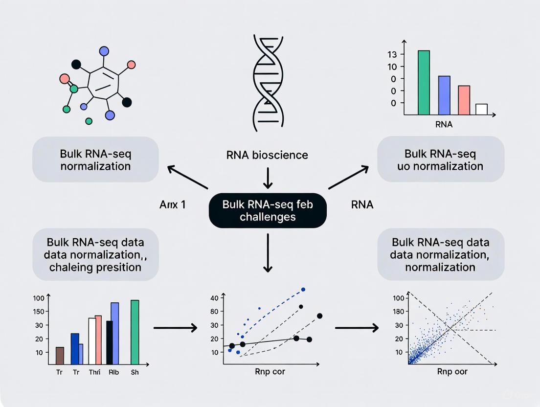
Why Normalization is Non-Negotiable: Unpacking the Core Challenges in Bulk RNA-seq
Bulk RNA-seq data is affected by multiple technical factors that, if uncorrected, can obscure the true biological signal. These include library size, gene length, and sample composition biases. The core goal of normalization is to adjust the raw count data to eliminate these non-biological sources of variation, enabling valid comparisons of gene expression levels between samples.
This guide addresses the most frequent challenges researchers face during this critical step of the analysis.
Frequently Asked Questions (FAQs) and Troubleshooting
1. What is the fundamental difference between within-sample and between-sample normalization methods?
Within-sample methods, like FPKM and TPM, normalize counts based on features of the individual sample, such as its total count and gene lengths. In contrast, between-sample methods, such as RLE (used by DESeq2) and TMM (used by edgeR), use information across all samples in the experiment to compute scaling factors, under the assumption that most genes are not differentially expressed [1]. Between-sample methods are generally recommended for differential expression analysis.
2. I have an outlier sample that looks very different from my other replicates. Should I remove it before normalization?
Proceed with caution. First, investigate the cause. Use quality control tools like FastQC, MultiQC, or RNA-SeQC to check for technical issues like adapter contamination, low sequencing quality, or high rRNA content [2]. If the outlier is due to a technical failure, removal may be justified. However, if it is a valid biological replicate, normalization methods like TMM and RLE, which are robust to a small number of DE genes, can often handle the situation without removing the sample [1].
3. Can I use quantile normalization, a method from microarrays, on my RNA-seq data?
This is generally not recommended. Using microarray-specific normalization methods like normalizeBetweenArrays() from limma with its "quantile" method on RNA-seq data is not standard practice and can lead to incorrect results [3]. RNA-seq data has different statistical properties, and it is crucial to follow well-tested RNA-seq-specific tutorials and use methods designed for count data, such as those in edgeR or DESeq2 [3].
4. How does normalization affect my downstream analysis beyond differential expression?
The choice of normalization method can significantly impact other types of analyses. For instance, when mapping RNA-seq data onto genome-scale metabolic models (GEMs), using between-sample methods (RLE, TMM, GeTMM) resulted in metabolic models with lower variability and more accurate capture of disease-associated genes compared to within-sample methods (TPM, FPKM) [1].
5. My data has known covariates like age, gender, or batch. How should I handle them?
Covariates can be adjusted for during the normalization process. A benchmark study on Alzheimer's disease and lung cancer data demonstrated that applying covariate adjustment to normalized data (for factors like age, gender, and post-mortem interval) improved the accuracy of condition-specific metabolic models derived from the transcriptome [1]. This can often be integrated into the statistical model in tools like DESeq2 or limma.
Normalization Method Comparison
The table below summarizes the key characteristics and recommended use cases for common normalization methods.
Table 1: Comparison of Common Bulk RNA-seq Normalization Methods
| Normalization Method | Type | Key Principle | Commonly Used In | Best Use Cases |
|---|---|---|---|---|
| TPM (Transcripts Per Million) [1] | Within-sample | Normalizes for sequencing depth and gene length. | General expression reporting, e.g., ENCODE pipeline [4]. | Comparing expression levels of different genes within a single sample. |
| FPKM (Fragments Per Kilobase Million) [1] | Within-sample | Similar to TPM but with a different order of operations. | General expression reporting. | (Largely superseded by TPM for within-sample comparisons). |
| TMM (Trimmed Mean of M-values) [1] | Between-sample | Assumes most genes are not DE; uses a weighted trim mean of log expression ratios. | edgeR package. |
Differential expression analysis, especially with balanced DE genes. |
| RLE (Relative Log Expression) [1] | Between-sample | Assumes most genes are not DE; median of ratios to a reference sample. | DESeq2 package. |
Differential expression analysis, robust to large numbers of DE genes. |
| GeTMM (Gene length corrected TMM) [1] | Combined | Combines gene-length correction of TPM with between-sample normalization of TMM. | - | Tasks requiring both within-and between-sample comparisons. |
Experimental Protocols for Key Normalization Analyses
Protocol 1: Performing Differential Expression Analysis with DESeq2 (RLE Normalization)
This protocol uses the RLE normalization method inherent to the DESeq2 package to identify differentially expressed genes.
- Load Required Libraries: Install and load the
DESeq2andtximport(if using transcript-level quantifiers like Salmon) packages in R. - Prepare Input Data: Create a data frame (
colData) with sample metadata (e.g., condition, batch). Prepare a table of raw counts or point to the output files from quantification tools like Salmon. - Create DESeq2 Dataset: Use the
DESeqDataSetFromMatrix()orDESeqDataSetFromTximport()function, specifying the count data, sample information, and the experimental design (e.g.,~ condition). - Run Differential Expression Analysis: Execute the core analysis steps—estimation of size factors (RLE normalization), dispersion, and fitting of the negative binomial model—with a single call to the
DESeq()function. - Extract Results: Use the
results()function to extract a table of log2 fold changes, p-values, and adjusted p-values for all genes.
Protocol 2: Generating Condition-Specific Metabolic Models from Normalized Data
This protocol, based on benchmarks from npj Systems Biology and Applications, outlines how to create metabolic models using normalized RNA-seq data and the iMAT algorithm [1].
- Normalize RNA-seq Data: Begin with raw count data. Normalize it using one of the five methods (e.g., RLE, TMM, TPM) as described in the comparison table above.
- Covariate Adjustment (Optional but Recommended): For datasets with covariates like age or gender, perform covariate adjustment on the normalized expression values to remove their influence.
- Map Data to a Metabolic Model: Use the Integrative Metabolic Analysis Tool (iMAT) algorithm. Input the normalized (and optionally adjusted) expression data and a generic human genome-scale metabolic model (GEM).
- Generate Personalized Models: Run iMAT for each sample in your dataset to reconstruct a condition-specific, personalized metabolic model.
- Analyze Model Content: Compare the resulting models (e.g., control vs. disease) to identify significantly affected metabolic reactions and pathways based on the chosen normalization method.
Workflow and Decision Diagrams
The following diagram illustrates the logical workflow for approaching bulk RNA-seq data normalization, from data preparation to method selection.
The Scientist's Toolkit: Essential Research Reagents and Software
Table 2: Key Tools and Reagents for Bulk RNA-seq Analysis
| Item Name | Category | Function/Brief Explanation |
|---|---|---|
| STAR [5] [4] | Alignment Software | A splice-aware aligner that accurately maps RNA-seq reads to a reference genome. |
| Salmon [5] | Quantification Software | A fast tool for transcript-level quantification that uses "pseudo-alignment," handling uncertainty in read assignment. |
| RSEM [5] [4] | Quantification Software | Estimates gene and isoform abundance levels from RNA-seq data, often used with STAR alignments. |
| DESeq2 [1] | R Package for DE Analysis | Performs differential expression analysis using its built-in RLE normalization and a negative binomial model. |
| edgeR [1] | R Package for DE Analysis | Performs differential expression analysis using its built-in TMM normalization and statistical methods. |
| ERCC Spike-In Controls [4] | Research Reagent | Exogenous RNA controls added to samples before library preparation to provide a standard for absolute transcript quantification and quality assessment. |
| FastQC [2] | Quality Control Tool | Provides an initial quality overview of raw sequence data, informing on potential issues like adapter contamination or low-quality bases. |
| MultiQC [2] | Quality Control Tool | Aggregates results from multiple tools (FastQC, STAR, etc.) across all samples into a single, interactive HTML report. |
| RSeQC [2] | Quality Control Tool | Analyzes various aspects of RNA-seq data, including read distribution, coverage uniformity, and strand specificity. |
| nf-core/rnaseq [5] | Workflow Manager | A portable, community-maintained Nextflow pipeline that automates the entire analysis from raw FASTQ files to counts and QC. |
Troubleshooting Guide: Identifying and Mitigating RNA-Seq Biases
This guide addresses common technical issues related to sequencing depth, gene length, and compositional effects in bulk RNA-Seq experiments, providing practical solutions for researchers.
Q1: How do I determine the optimal sequencing depth for my bulk RNA-Seq experiment?
A: Sequencing depth requirements vary significantly based on your experimental goals and sample quality. The ENCODE standards recommend ≥30 million mapped reads as a baseline for typical poly(A)-selected RNA-Seq, but recent multi-center benchmarking shows real-world applications can require ~40–420 million total reads per library [6]. Use the following guidelines tailored to your specific research objectives [6]:
| Research Objective | Recommended Depth | Read Length | Key Considerations |
|---|---|---|---|
| Differential Gene Expression | 25-40 million reads | 2×75 bp | Sufficient for high-quality RNA (RIN ≥8); stabilizes fold-change estimates [6] |
| Isoform Detection & Splicing | ≥100 million reads | 2×75 bp or 2×100 bp | Required to capture a comprehensive view of splice events [6] |
| Fusion Detection | 60-100 million reads | 2×75 bp (2×100 bp preferred) | Paired-end libraries essential for anchoring breakpoints [6] |
| Allele-Specific Expression | ≥100 million reads | Paired-end | Essential for accurate variant allele frequency estimation [6] |
For degraded RNA samples (e.g., FFPE), increase sequencing depth by 25-50% for DV200 of 30-50%, and use 75-100 million reads for DV200 <30% [6]. When working with limited input RNA (≤10 ng), incorporate UMIs to accurately collapse PCR duplicates during deep sequencing [6] [7].
Q2: How does gene length bias affect my analysis, and should I normalize for it?
A: Gene length significantly impacts count data. Longer genes generate more sequencing fragments, resulting in higher counts regardless of actual expression levels [8]. However, normalization strategies depend on your analytical goals:
- For differential expression analysis (comparing the same gene across samples): Do NOT normalize for gene length. Tools like DESeq2 and edgeR are designed to compare counts of the same gene across samples, where length is constant and normalization would reduce statistical power for longer genes [9].
- For within-sample comparisons (comparing different genes within the same sample): Normalize for gene length using measures like TPM (Transcripts Per Million). This enables meaningful comparison of expression levels between different genes within the same sample [9].
The decision flow below outlines this logic:
Q3: What is compositional bias, and how can I correct for it in my data?
A: Compositional bias is a fundamental artifact where sequencers measure relative, not absolute, abundances [10]. When a few highly abundant features change between conditions, the relative proportions of all other features appear to change artificially, even if their absolute abundances remain constant [10].
Detection: Suspect compositional bias when you observe strong inverse correlations in log-fold changes between highly and lowly expressed features [10].
Correction Strategies:
- Gold Standard: Use spike-in controls (e.g., ERCC RNA Spike-In Mix) added during library preparation. These provide an external reference for absolute quantification [7] [11].
- Computational Correction: Apply scaling normalization methods (e.g., TMM in edgeR, DESeq2's median of ratios) that assume most features are not differentially abundant [10] [12]. For sparse data (e.g., metagenomic 16S surveys), consider empirical Bayes approaches like Wrench [10].
Experimental Workflow: The following diagram illustrates where key biases are introduced during a standard bulk RNA-Seq workflow and where corrections can be applied.
| Tool/Reagent | Function/Purpose | Application Context |
|---|---|---|
| ERCC Spike-In Mix [7] [11] | External RNA controls with known concentrations to standardize quantification and assess technical variation. | Determining sensitivity, dynamic range, and accuracy; particularly valuable for identifying compositional bias. |
| Unique Molecular Identifiers (UMIs) [6] [7] | Short random sequences that tag individual mRNA molecules prior to PCR amplification. | Correcting PCR amplification bias and accurately counting molecules in low-input or deep sequencing (>50 million reads) applications. |
| Ribosomal RNA Depletion Kits | Remove abundant ribosomal RNA to increase sequencing coverage of informative transcripts. | Essential for prokaryotic RNA-Seq, non-polyA transcripts (e.g., lncRNAs), and degraded samples (FFPE) where polyA selection is inefficient [6] [7]. |
| DV200 Metric | Measures the percentage of RNA fragments >200 nucleotides; alternative to RIN for degraded RNA. | RNA quality assessment for FFPE and other compromised samples; guides protocol selection and sequencing depth [6]. |
| Poly(A) Selection Beads | Enrich for polyadenylated mRNA molecules from total RNA. | Standard approach for eukaryotic mRNA sequencing when RNA quality is high (RIN ≥8, DV200 >70%) [6] [7]. |
Frequently Asked Questions
1. What is the fundamental goal of RNA-seq normalization? The primary goal is to adjust raw transcriptomic data to account for technical factors that may mask actual biological effects and lead to incorrect conclusions. Normalization ensures that differences in read counts reflect true biological variation rather than technical artifacts like sequencing depth, transcript length, or batch effects [13] [14].
2. Can I use within-sample normalized data (like TPM) for direct between-sample comparisons? No, this is a common misunderstanding. Within-sample normalization methods like TPM and FPKM are suitable for comparing the relative expression of different genes within the same sample. However, for comparing the expression of the same gene between different samples, between-sample normalization methods (like TMM or RLE) are required, as they account for additional technical variation such as differences in sequencing depth and RNA composition [13] [15].
3. What happens if I skip normalization or use an inappropriate method? Errors in normalization can have a significant impact on downstream analysis. It can lead to a high rate of false positives or false negatives in differential expression analysis, mask true biological differences, or create the false appearance of differential expression where none exists [13] [14] [1].
4. Is there a single "best" normalization method for all experiments? There is no consensus on a single best-performing normalization method for all scenarios [11] [16]. The choice depends on the specific features of your dataset and the biological question. It is good practice to assess the performance of different normalization methods using data-driven metrics relevant to your downstream analysis [11] [1].
5. My data comes from multiple studies. What is the most important normalization step? When integrating data from multiple independent studies, normalization across datasets (batch-effect correction) is critical. Batch effects, often introduced by different sequencing times, methods, or facilities, can be the greatest source of variation and can mask true biological differences if not corrected [13] [17].
Troubleshooting Guides
Problem 1: Inconsistent Differential Expression Results After Normalization
Symptoms: Your list of differentially expressed (DE) genes changes drastically when you switch normalization methods, or you get counter-intuitive results (e.g., globally down-regulated genes when one condition is known to be highly active).
Diagnosis: This often indicates that the assumptions of your chosen normalization method are being violated. For example:
- Global-scaling methods (TMM, RLE) assume that most genes are not differentially expressed [14] [1]. If a large fraction of genes are truly DE in your experiment (e.g., a global shift in expression), these methods can produce misleading results [14].
- Composition bias may be present, where a few highly expressed genes in one condition consume a large share of the sequencing reads, making non-DE genes in that condition appear down-regulated [14].
Solutions:
- Assumption Check: Investigate the biology of your experiment. If a global shift in expression is expected, be cautious with methods that assume a stable transcriptome background.
- Method Comparison: Run your differential expression pipeline with multiple between-sample normalization methods (e.g., TMM, RLE). Compare the results and investigate genes that are consistently identified.
- Spike-in Controls: If available, use spike-in RNA controls to create an absolute standard for normalization, which can be more robust in these situations [11] [18].
Problem 2: High Variability in Model Outputs When Mapping to Genome-Scale Metabolic Models (GEMs)
Symptoms: When using normalized RNA-seq data to create condition-specific metabolic models (e.g., with iMAT or INIT algorithms), the number of active reactions varies widely between samples, leading to unstable predictions.
Diagnosis: This is frequently caused by using within-sample normalization methods (TPM, FPKM) for this purpose. These methods do not adequately control for technical variability between samples, which propagates into the metabolic models [1].
Solutions:
- Switch Normalization Method: Use between-sample normalization methods like RLE (from DESeq2), TMM (from edgeR), or GeTMM. Benchmarking studies show these methods produce metabolic models with lower variability and can more accurately capture disease-associated genes [1].
- Covariate Adjustment: Account for known covariates like age, gender, or post-mortem interval during the normalization process, as this can further improve the consistency and accuracy of the generated models [1].
Problem 3: Poor Integration of Multiple Datasets
Symptoms: When you combine data from different batches or studies, samples cluster strongly by batch or study origin instead of by biological condition in dimensionality reduction plots (e.g., PCA).
Diagnosis: Strong batch effects are confounding your analysis. Within-dataset normalization is not sufficient to remove these systematic technical differences [13] [17].
Solutions:
- Apply Batch-Effect Correction: Use specialized tools like ComBat (from the
svapackage) or Limma'sremoveBatchEffectfunction after performing within-dataset normalization [13]. - Experimental Design: For future experiments, use a blocking design and multiplex samples from all conditions across sequencing lanes and batches to minimize confounding [17].
Normalization Methodologies at a Glance
The following table summarizes the core methodologies for the three tiers of normalization.
Table 1: Summary of RNA-seq Normalization Tiers
| Normalization Tier | Primary Goal | Common Methods | Key Considerations |
|---|---|---|---|
| Within-Sample [13] | Enable comparison of expression between different genes within the same sample. Corrects for gene length and sequencing depth. | TPM, FPKM/RPKM | Not suitable for between-sample comparisons. TPM is generally preferred over FPKM as the sum of all TPMs is constant across samples [13] [15]. |
| Between-Sample [13] [14] | Enable comparison of expression for the same gene between different samples. Corrects for library size and RNA composition. | TMM [1], RLE (used in DESeq2) [1], GeTMM [1] | Often assume most genes are not differentially expressed. Performance can be poor if this assumption is violated [14]. |
| Across Datasets [13] | Remove batch effects when integrating data from different studies, sequencing runs, or facilities. | ComBat [13], Limma [13] | Requires knowledge of the batch variables. Should be applied after within-dataset normalization. |
Experimental Protocols for Benchmarking Normalization Methods
Given that there is no single best method, a robust analysis pipeline should include an evaluation of different normalization approaches. Below is a general protocol for benchmarking normalization methods in a differential expression analysis context.
Protocol: Evaluating Normalization Method Performance
- Data Preparation: Begin with a raw count matrix from your bulk RNA-seq experiment.
- Apply Multiple Normalizations: Normalize the dataset using several between-sample methods. Common choices include:
- TMM: Implemented in the
edgeRpackage with thecalcNormFactorsfunction. - RLE: Used by default in the
DESeq2package (via theestimateSizeFactorsfunction). - GeTMM: A method that combines gene-length correction with TMM-like between-sample normalization [1].
- TMM: Implemented in the
- Downstream Analysis: Perform differential expression analysis on each normalized dataset using the corresponding statistical framework (e.g.,
edgeRfor TMM,DESeq2for RLE). - Performance Assessment: Evaluate the results using data-driven metrics. Common approaches include:
- Clustering and Visualization: Use PCA plots to see if normalization effectively separates samples by biological condition rather than technical artifacts.
- Evaluation Metrics: Assess the performance using metrics like the Silhouette Width (for cluster separation) and the K-nearest neighbor batch-effect test (to check for residual batch effects) [11].
- Biological Validation: Compare the lists of differentially expressed genes to known biological pathways or prior knowledge to assess biological relevance [1].
The Scientist's Toolkit: Essential Reagents & Materials
Table 2: Key Research Reagents and Tools for RNA-seq Normalization
| Reagent / Tool | Function in Normalization | Key Considerations |
|---|---|---|
| Spike-in RNA Controls (e.g., ERCCs) [11] | Exogenous RNA molecules added in known quantities to each sample. Provide an absolute standard to distinguish technical from biological variation and to normalize data, especially in complex scenarios. | Must be added during library preparation. Not feasible for all sequencing platforms [11]. |
| Unique Molecular Identifiers (UMIs) [11] | Random barcodes ligated to each mRNA molecule during reverse transcription. Allow for accurate counting of original mRNA molecules by correcting for PCR amplification biases. | Commonly used in droplet-based scRNA-seq protocols (e.g., 10X Genomics). Less common in bulk RNA-seq but powerful for molecular counting [11] [16]. |
| Library Quantification Kits (e.g., qPCR) [17] | Precisely measure the concentration of sequencing libraries before pooling. Helps minimize technical variation by ensuring libraries are diluted to the same concentration, reducing lane-to-lane and flow-cell effects. | Critical for a balanced multiplexed sequencing run [17]. |
Logical Workflow for Normalization
The following diagram illustrates the logical decision process for applying the three tiers of normalization in an RNA-seq analysis pipeline.
Diagram 1: RNA-seq Normalization Workflow
Relationship Between Normalization Types and Analysis Goals
Different downstream analyses have different requirements for which normalization tier is most critical. This diagram maps the primary focus for common analytical goals.
Diagram 2: Normalization Focus for Analysis Goals
By understanding these three distinct tiers of normalization—within-sample, between-sample, and across datasets—researchers can make informed decisions that are critical for generating accurate, reliable, and biologically meaningful results from their RNA-seq experiments.
In bulk RNA-seq analysis, normalization is not merely a preliminary step but a foundational computational process that corrects for technical variations to enable meaningful biological comparisons. The choice of normalization method directly and measurably influences every subsequent analysis, from differential expression to pathway mapping. This guide addresses common challenges and provides troubleshooting advice for researchers navigating the impact of normalization decisions within their thesis projects.
Frequently Asked Questions (FAQs)
1. Why is normalization essential in bulk RNA-seq analysis? Normalization adjusts raw read counts to account for technical variability, such as differences in sequencing depth (total number of reads per sample) and RNA composition (the relative abundance of different RNA molecules in a sample) [15]. Without correct normalization, observed differences in gene expression can be technically driven rather than biological, leading to false conclusions in downstream analysis [14].
2. My PCA plot shows poor separation between experimental conditions. Could normalization be the cause? Yes. The choice of normalization method can significantly impact the outcome of exploratory analyses like Principal Component Analysis (PCA) [19]. Different normalization techniques alter the correlation structure of the data, which in turn affects the principal components and the clustering of samples in the reduced-dimensional space. If the assumed data characteristics by the normalization method do not hold (e.g., the assumption that most genes are not differentially expressed), the resulting PCA may fail to reveal true biological separation [14] [19].
3. How does normalization affect differential expression analysis? Normalization has the most substantial impact on differential expression analysis, even more than the choice of subsequent test statistic [14]. Methods like DESeq2's RLE and edgeR's TMM make specific statistical assumptions about the data. If these assumptions are violated—for instance, if a global shift in expression occurs—the error propagates, inflating false positives or negatives [14]. One study found that normalization was the largest statistically significant source of variation in gene expression estimation accuracy [20].
4. I am mapping expression data to genome-scale metabolic models (GEMs). Does normalization matter? Absolutely. Benchmarking studies show that the normalization method directly affects the content and predictive accuracy of condition-specific GEMs generated by algorithms like iMAT and INIT [1]. Between-sample methods (RLE, TMM) produce models with lower variability in the number of active reactions and more accurately capture disease-associated genes compared to within-sample methods (TPM, FPKM) [1].
5. When should I use spike-in controls for normalization? Spike-in controls are synthetic RNA molecules added to your sample in known quantities. They can be valuable when the assumption that "most genes are not differentially expressed" is violated, such as in experiments with global transcriptional shifts [21]. However, their use requires caution. Technical variability can affect spike-in reads, and they must be affected by technical factors in the same way as endogenous genes to be reliable [21]. Methods like Remove Unwanted Variation (RUV) that use spike-ins as negative controls can help account for more complex nuisance technical effects [21].
Troubleshooting Common Normalization Problems
Problem 1: Inflated False Positive Rates in Differential Expression
Symptoms: An unexpectedly high number of differentially expressed genes (DEGs), many of which are lowly expressed and lack biological plausibility.
Diagnosis: This often results from a violation of the normalization method's core assumptions. For example, TMM and DESeq2 assume that the majority of genes are not differentially expressed [14] [15]. In experiments where this is not true (e.g., extreme treatments, global shifts), these methods can perform poorly.
Solutions:
- Use Spike-in Controls: If available, employ a control-based method like RUVg [21].
- Explore Alternative Methods: Consider a non-parametric method like quantile normalization, which makes different assumptions about the data distribution [13] [15].
- Filter Genes: Prior to normalization, filter out lowly expressed genes that contribute disproportionately to noise using a function like
filterByExprfrom edgeR [22].
Problem 2: Batch Effects Masking Biological Signal
Symptoms: Samples cluster by batch (e.g., sequencing date, library preparation kit) instead of by experimental condition in a PCA plot.
Diagnosis: Standard global scaling normalization methods (e.g., TMM, RLE) primarily correct for sequencing depth but may not account for more complex technical artifacts introduced by batch processing [21].
Solutions:
- Apply Batch Correction: After between-sample normalization, use a batch-effect correction tool like ComBat (from the
svapackage) or removeBatchEffect (from thelimmapackage) to account for known batches [13] [21]. - Use RUV Methods: The RUV family of methods can simultaneously estimate and adjust for unknown sources of unwanted variation using control genes or samples [21].
Problem 3: Inconsistent Results in Pathway and Metabolic Analysis
Symptoms: Pathway enrichment analysis or metabolic model predictions are unstable and vary drastically with different normalization inputs.
Diagnosis: Within-sample normalization methods like FPKM and TPM can introduce high variability in the scaled counts across samples, which propagates into functional analyses [1].
Solutions:
- Switch to Between-Sample Methods: For downstream tasks like building metabolic models with iMAT or INIT, use RLE, TMM, or GeTMM, which demonstrate higher consistency and accuracy [1].
- Validate Findings: Cross-validate key pathway results using a different between-sample normalization method to ensure robustness.
Comparative Analysis of Normalization Methods
Table 1: Characteristics and Best Practices for Common Normalization Methods
| Method | Class | Key Principle | Best For | Major Assumption | Downstream Impact |
|---|---|---|---|---|---|
| TPM/FPKM [13] [15] | Within-Sample | Normalizes for gene length and sequencing depth. | Comparing gene expression within a single sample. | Total RNA output is comparable between samples. | Not recommended for DE; high variability in metabolic model reactions [1]. |
| TMM [13] [15] | Between-Sample | Uses a trimmed mean of log-expression ratios (M-values) against a reference sample. | Datasets with different RNA compositions or a few highly expressed genes. | The majority of genes are not differentially expressed. | Robust performance in DE and metabolic modeling [1]. |
| RLE (DESeq2) [1] [15] | Between-Sample | Calculates a size factor as the median of ratios of counts to a geometric mean. | Most standard experiments, especially with small sample sizes. | The majority of genes are not differentially expressed. | Robust performance in DE and metabolic modeling; low model variability [1]. |
| Quantile [13] [15] | Between-Sample | Forces the distribution of gene expression to be identical across all samples. | Complex experimental designs with many samples. | Observed distribution differences are technical, not biological. | Reduces technical variation for multivariate analysis. |
| RUV [21] | Between-Sample | Uses factor analysis on control genes/samples to estimate and remove unwanted variation. | Experiments with global shifts, known/unknown batch effects, or spike-in controls. | Technical effects impact control genes and regular genes similarly. | Improves DE accuracy and sample clustering in complex scenarios. |
Table 2: Impact of Normalization on Downstream Applications (Based on Benchmark Studies)
| Application | Key Performance Metric | Top-Performing Methods | Poorly Performing Methods | Evidence Source |
|---|---|---|---|---|
| Differential Expression | Accuracy/Precision of fold-change, FDR control | RLE (DESeq2), TMM (edgeR) | FPKM, TPM (for DE analysis) | [20] [15] |
| Metabolic Model (iMAT/INIT) | Accuracy in capturing disease genes, model stability | RLE, TMM, GeTMM | FPKM, TPM | [1] |
| Principal Component Analysis | Biological interpretability, cluster separation | Method-dependent; no single best | Varies by dataset | [19] |
Essential Experimental Protocols for Validation
Protocol 1: Evaluating Normalization Performance Using SeQC Benchmark Data
This protocol is adapted from large-scale consortium studies to quantitatively evaluate a normalization method's impact on gene expression estimation [20].
Research Reagent Solutions:
- SEQC Benchmark Dataset: Publicly available data (e.g., from Sequence Read Archive under project SRP010938) consisting of reference RNA samples with validated properties [20].
- qPCR Gold-Standard: A subset of genes with validated expression levels via qPCR.
- Software: R/Bioconductor with packages like
DESeq2,edgeR, andlimma.
Methodology:
- Data Acquisition: Download the SEQC benchmark dataset (Samples A and B).
- Pipeline Application: Process the raw reads through your chosen alignment and quantification pipeline.
- Normalization: Apply the normalization methods you wish to evaluate (e.g., TMM, RLE, FPKM).
- Metric Calculation:
- Accuracy: Calculate the deviation of RNA-seq-derived log ratios (e.g., A vs. B) from the corresponding qPCR-based log ratios. A smaller deviation indicates higher accuracy.
- Precision: Compute the coefficient of variation (CoV) of gene expression across technical replicate libraries. A smaller CoV indicates higher precision.
- Downstream Correlation: Correlate the accuracy and precision metrics with the performance of a downstream task, such as predicting clinical outcomes. Pipelines with better metrics typically yield better downstream results [20].
Protocol 2: Assessing Count-Depth Relationship in Your Dataset
A key assumption of global scaling methods is a consistent relationship between gene counts and sequencing depth. This protocol checks for violations.
Methodology:
- Plot Raw Data: Before normalization, create a scatter plot of log (non-zero counts +1) for each gene against the log sequencing depth for each sample.
- Check for Trends: Visually inspect if the relationship between expression and depth is consistent across all genes or if there are systematic patterns where lowly and highly expressed genes show different trends.
- Post-Normalization Check: Apply your chosen normalization method and plot the normalized data in the same way. A successful normalization should remove the dependence of expression counts on sequencing depth. If systematic trends persist, consider a method like SCnorm that groups genes with similar count-depth relationships for normalization [23].
The following workflow diagram summarizes the logical process for diagnosing and addressing common normalization-related issues in an RNA-seq analysis.
Troubleshooting Workflow for RNA-seq Normalization
Table 3: Essential Research Reagent Solutions and Software Tools
| Item / Resource | Function / Description | Use Case in Normalization Context |
|---|---|---|
| ERCC Spike-in Controls [21] | A set of 92 synthetic RNA transcripts at known concentrations. | Serves as negative controls for methods like RUV to estimate unwanted variation. |
| SEQC Benchmark Dataset [20] | A well-characterized RNA-seq dataset with qPCR validation. | A gold standard for benchmarking normalization accuracy and precision. |
| DESeq2 (R package) [15] | A package for differential expression analysis. | Implements the RLE normalization method for between-sample comparison. |
| edgeR (R package) [15] | A package for differential expression analysis. | Implements the TMM normalization method. |
| sva (R package) [13] [21] | A package for removing batch effects and other unwanted variation. | Contains ComBat and surrogate variable analysis (sva) for post-normalization adjustment. |
| MultiQC [22] | A tool to aggregate results from multiple QC tools. | Assesses overall sample quality and identifies outliers before normalization. |
A Practical Guide to Bulk RNA-seq Normalization Methods: From TPM to TMM
Frequently Asked Questions (FAQs)
Q1: What are TPM and FPKM, and how do they differ?
A1: TPM (Transcripts Per Million) and FPKM (Fragments Per Kilobase of transcript per Million fragments mapped) are both within-sample normalization methods designed to make gene expression counts comparable within a single sample by accounting for two key technical biases: sequencing depth (the total number of sequenced fragments) and gene length (the length of the transcript) [24] [25] [26].
The core difference lies in the order of operations during calculation [25]:
- FPKM first normalizes for sequencing depth (to get "Fragments Per Million") and then normalizes for gene length.
- TPM first normalizes for gene length (to get "Transcripts Per Kiloby") and then normalizes for sequencing depth.
This subtle difference has a profound consequence: the sum of all TPM values in a sample is always constant (1 million), allowing you to directly compare the proportion of transcripts a gene constitutes across different samples. With FPKM, the sum of normalized values can vary from sample to sample, making such proportional comparisons difficult [25].
Q2: When is it appropriate to use TPM or FPKM?
A2: The consensus from multiple comparative studies is that TPM and FPKM are primarily suited for within-sample comparisons [26]. The table below outlines their appropriate and inappropriate use cases.
Table: Appropriate Use Cases for TPM and FPKM
| Application | Recommendation | Rationale |
|---|---|---|
| Comparing expression of different genes within the same sample | Appropriate [26] | Controls for gene length and sequencing depth, allowing you to ask "Which gene is more highly expressed in this specific sample?" |
| Comparing expression of the same gene across different samples | Not Recommended [27] [26] | Values are relative and can be skewed by the composition of the total RNA population in each sample, leading to inaccurate comparisons. |
Q3: What are the common pitfalls of misusing TPM/FPKM for cross-sample comparison?
A3: Using TPM or FPKM for cross-sample comparison is a common misconception, as these values are relative abundances and not absolute measurements [27]. The primary pitfall is that the expression value for a gene depends on the expression levels of all other genes in that sample.
For example, if a very few genes are extremely highly expressed in one sample, they "use up" a large portion of the "per million" count. This artificially deflates the TPM/FPKM values of all other genes in that sample, making it appear as if they are less expressed compared to another sample with a more balanced transcript repertoire [27]. This is particularly problematic when comparing data from different sample preparation protocols, such as poly(A)+ selection versus rRNA depletion, which capture vastly different RNA populations [27].
Q4: If not TPM/FPKM, what should I use to compare gene expression across samples?
A4: For cross-sample comparisons and differential expression analysis, between-sample normalization methods applied to raw count data are strongly recommended [24] [1]. These methods are specifically designed to deal with library composition differences between samples. The most widely used and recommended methods include:
- Normalized Counts from tools like DESeq2 (which uses the RLE, or Relative Log Expression method) [24] [1] [28].
- Normalized Counts from tools like edgeR (which uses the TMM, or Trimmed Mean of M-values, method) [24] [1].
These methods have been shown to provide superior reproducibility, lower variation between biological replicates, and more accurate results in downstream analyses like differential expression and metabolic model building compared to TPM and FPKM [24] [1] [28].
Troubleshooting Common Problems
Problem: High variability between replicate samples after normalization with TPM.
Solution:
- Diagnosis: This is a known issue. A 2021 study on patient-derived xenograft models demonstrated that TPM and FPKM have higher median coefficients of variation (CV) and lower intraclass correlation (ICC) across replicates compared to normalized counts from DESeq2 or edgeR [24] [28].
- Action: Re-analyze your raw count data using a between-sample normalization method like those provided by DESeq2 (RLE) or edgeR (TMM). The same study found that hierarchical clustering of replicate samples was most accurate using these normalized counts [24].
Problem: Inconsistent results when integrating my TPM data with a public dataset.
Solution:
- Diagnosis: Differences in sample preparation protocols (e.g., poly(A)+ selection vs. rRNA depletion) drastically alter the RNA repertoire. TPM values from these different protocols are not directly comparable because the "per million" scaling factor is applied to different underlying populations [27].
- Action: If possible, obtain the raw sequencing data (FASTQ files) for both datasets and re-process them through the same bioinformatics pipeline (alignment and quantification) to generate normalized counts. If this is not feasible, be extremely cautious in your interpretation and acknowledge the technical bias as a major limitation.
Experimental Protocols & Data Analysis
Benchmarking Protocol: Comparing Normalization Methods
The following methodology is adapted from a 2021 study comparing quantification measures [24] [28].
1. Sample Preparation and Data Acquisition:
- Samples: Use replicate samples from the same biological source (e.g., 61 human tumor xenografts from 20 distinct models) [24].
- RNA-seq: Perform standard RNA-seq library preparation and sequencing. Download raw data (FASTQ files) and associated quantification files.
2. Data Processing and Quantification:
- Process all samples uniformly. The cited study mapped reads to a reference transcriptome using Bowtie2, generated BAM files, and performed gene-level quantification with RSEM to obtain TPM, FPKM, and expected counts [24].
- For cross-sample methods, generate normalized counts using DESeq2 (RLE) and edgeR (TMM) from the expected counts matrix [1].
3. Performance Evaluation Metrics:
- Coefficient of Variation (CV): Calculate the median CV for the same gene across all replicate samples. A lower CV indicates higher reproducibility [24].
- Intraclass Correlation Coefficient (ICC): Calculate ICC across replicate samples from the same model. A higher ICC indicates better agreement between replicates [24].
- Cluster Analysis: Perform hierarchical clustering on the different normalized datasets. The method that most accurately groups replicate samples from the same model together is superior [24].
Table: Typical Results from a Benchmarking Study (based on [24])
| Quantification Measure | Median Coefficient of Variation (CV) | Intraclass Correlation (ICC) | Cluster Accuracy |
|---|---|---|---|
| Normalized Counts (e.g., DESeq2) | Lowest | Highest | Most Accurate |
| TPM | Intermediate | Intermediate | Less Accurate |
| FPKM | Highest | Lowest | Least Accurate |
Workflow Diagram: From Raw Data to Normalized Quantities
The diagram below illustrates the key steps in calculating FPKM and TPM, highlighting the difference in the order of operations.
Table: Essential Computational Tools for RNA-seq Quantification
| Tool / Resource | Function | Key Outputs |
|---|---|---|
| RSEM (RNA-Seq by Expectation-Maximization) | A software package for estimating gene and isoform abundances from RNA-Seq data. | Expected counts, TPM, FPKM [24] |
| Salmon / Kallisto | Ultra-fast, alignment-free tools for transcript-level quantification. Ideal for large datasets. | Estimated counts, TPM [27] |
| DESeq2 | A widely used R/Bioconductor package for differential expression analysis. | Normalized counts (using RLE normalization) [24] [1] |
| edgeR | Another widely used R/Bioconductor package for differential expression analysis. | Normalized counts (using TMM normalization) [24] [1] |
| NCI PDMR (Patient-Derived Model Repository) | A public resource providing annotated RNA-seq data from patient-derived models, useful for benchmarking. | FASTQ files, TPM, FPKM, and count data [24] |
Decision Pathway: Selecting a Normalization Method
Use the following decision diagram to guide your choice of normalization method based on your experimental goal.
Troubleshooting Guides
Guide 1: Addressing High Variability in Model Size or Differential Expression Results
Problem: When creating condition-specific metabolic models or performing differential expression analysis, the number of active reactions or identified genes shows high variability between samples.
Explanation: This is a common issue when using within-sample normalization methods like TPM or FPKM for between-sample comparative analyses. These methods do not adequately account for differences in RNA composition between samples. A few highly expressed genes in one sample can consume a large portion of the sequencing depth, distorting the expression proportions of all other genes [29] [30].
Solution: Switch to a between-sample normalization method.
- Recommended Action: Re-normalize your RNA-seq data using TMM, RLE, or GeTMM.
- Expected Outcome: These methods produce models and results with considerably lower variability in the number of active reactions across samples [29]. They are designed to be robust against RNA composition effects, which is particularly important for tissues with different transcriptional architectures [30].
- Verification: After re-normalization, check the distribution of your model sizes (e.g., number of active reactions) or the number of significant hits. The inter-sample variability should be substantially reduced.
Guide 2: Choosing a Normalization Method for Both Inter- and Intra-Sample Analysis
Problem: Your analysis requires comparing expression between samples (e.g., disease vs. control) AND comparing the expression levels of different genes within the same sample (e.g., for gene signature validation).
Explanation: Standard between-sample methods like TMM and RLE do not correct for gene length. Therefore, a longer gene will have more reads than a shorter gene even if their true expression levels are identical, making them unsuitable for within-sample gene comparisons [31]. Conversely, TPM corrects for gene length but is sensitive to highly expressed outliers for between-sample comparisons [29] [32].
Solution: Use the GeTMM (Gene length corrected Trimmed Mean of M-values) method.
- Recommended Action: Implement the GeTMM normalization procedure.
- Rationale: GeTMM combines the robust between-sample correction of TMM with gene length normalization. This allows the same normalized dataset to be used reliably for both inter-sample and intra-sample analyses [31].
- Expected Outcome: You will obtain a dataset that performs equivalently to TMM and RLE in differential expression analysis while also enabling valid within-sample gene-level comparisons, much like TPM [29] [31].
Frequently Asked Questions (FAQs)
FAQ 1: What is the core assumption behind TMM and RLE normalization methods?
Both TMM (Trimmed Mean of M-values) and RLE (Relative Log Expression) operate on the fundamental assumption that the majority of genes in an experiment are not differentially expressed [29] [30] [33]. They use this assumption to robustly estimate scaling factors that correct for technical variations, such as differences in library size and RNA composition, between samples.
FAQ 2: How does RNA composition bias affect normalization, and how do TMM/RLE correct for it?
RNA composition bias occurs when one sample has a small subset of genes that are extremely highly expressed. These genes consume a large fraction of the sequencing reads, thereby proportionally reducing the read counts for all other genes in that sample. If not corrected, this can make most other genes appear under-expressed in that sample [30].
- TMM corrects for this by calculating a weighted trimmed mean of the log expression ratios (M-values) between the sample and a reference, excluding highly expressed genes and genes with high variance [30].
- RLE uses the median of the ratios of each gene's count to its geometric mean across all samples as the scaling factor, which is also robust to outliers [29] [31].
FAQ 3: My dataset has known covariates like age, gender, or batch effects. Should I adjust for these before or after normalization?
Evidence suggests that covariate adjustment applied to normalized data can improve the accuracy of downstream analysis. A benchmark study on Alzheimer's and lung cancer data showed an increase in the accuracy of capturing disease-associated genes when covariate adjustment was applied to data normalized with RLE, TMM, or GeTMM [29]. The typical workflow is to first normalize the raw count data using a method like TMM or RLE, and then adjust the normalized values for known technical or biological covariates using an appropriate linear model.
FAQ 4: Why might my results be difficult to replicate, even when using proper normalization?
While correct normalization is crucial, the number of biological replicates is a critical factor for replicability. Studies have shown that results from RNA-seq experiments with small cohort sizes (e.g., fewer than 5-6 replicates) can be difficult to replicate due to high biological heterogeneity and low statistical power [34] [35]. It is recommended to use at least 6-12 biological replicates per condition for robust and replicable detection of differentially expressed genes [34] [35].
Data Presentation: Method Comparison
Table 1: Key Characteristics and Applications of TMM, RLE, and GeTMM Normalization Methods
| Feature | TMM | RLE | GeTMM |
|---|---|---|---|
| Full Name | Trimmed Mean of M-values | Relative Log Expression | Gene length corrected TMM |
| Primary Package | edgeR [30] |
DESeq2 [29] |
Custom implementation [31] |
| Corrects Gene Length? | No | No | Yes |
| Robust to RNA Composition? | Yes [30] | Yes [29] | Yes [31] |
| Ideal Use Case | Differential expression between samples [29] | Differential expression between samples [29] | Both inter- and intra-sample analysis [31] |
| Reported Performance | Lower variability in model results; high accuracy (~0.80) for disease genes [29] | Similar to TMM in performance for DE analysis [29] [36] | Equivalent to TMM/RLE for DE; lower false positives; enables intra-sample use [29] [31] |
Table 2: Quantitative Benchmarking of Normalization Methods on Disease Datasets (using iMAT models) This table summarizes results from a benchmark study that generated personalized metabolic models for Alzheimer's Disease (AD) and Lung Adenocarcinoma (LUAD) [29].
| Metric | TPM / FPKM | TMM | RLE | GeTMM |
|---|---|---|---|---|
| Variability in # of Active Reactions | High | Low | Low | Low |
| Number of Significantly Affected Reactions | Highest | Medium | Medium | Medium |
| Accuracy for AD-Associated Genes | Lower | ~0.80 | ~0.80 | ~0.80 |
| Accuracy for LUAD-Associated Genes | Lower | ~0.67 | ~0.67 | ~0.67 |
| Impact of Covariate Adjustment | Increase in accuracy | Increase in accuracy | Increase in accuracy | Increase in accuracy |
Experimental Protocols
Protocol 1: Implementing GeTMM Normalization
GeTMM reconciles within-sample and between-sample normalization by adding gene length correction to the TMM method [31]. Below is a detailed methodology.
1. Input Data Requirements
- A raw count matrix (genes x samples).
- A vector of gene lengths (e.g., the total exon length in base pairs for each gene).
2. Step-by-Step Procedure
- Step 1: Calculate Reads Per Kilobase (RPK). For each gene ( g ) in sample ( j ), compute RPK: ( \text{RPK}{gj} = \frac{\text{raw count}{gj}}{\text{gene length in kb}} ).
- Step 2: Apply TMM Normalization to the RPK matrix. Use the standard TMM algorithm from the
edgeRpackage on the RPK values to calculate a scaling factor for each sample [30] [31].- The TMM method selects a reference sample and then for each test sample, it computes ( M )-values (log fold changes) and ( A )-values (average log expression).
- It trims the extremes (default is 30% of ( M )-values and 5% of ( A )-values) and calculates a weighted average of the remaining ( M )-values to get the log scaling factor.
- Step 3: Generate normalized GeTMM values. Multiply the RPK values by the TMM scaling factors to obtain the final GeTMM values [31].
3. Key Considerations
- Software: While not a standard function in
edgeR, the process can be implemented by calculating RPK and then using thecalcNormFactorsfunction inedgeRon the RPK matrix. - Validation: The performance of GeTMM has been validated on real data (e.g., 263 colon cancer samples), showing it allows for both inter- and intra-sample analysis with the same dataset [31].
Mandatory Visualization
GeTMM Normalization Workflow
Normalization Method Selection Logic
The Scientist's Toolkit
Table 3: Essential Research Reagents and Computational Tools
| Item / Resource | Function / Description | Relevance to Normalization |
|---|---|---|
edgeR (R Package) |
A comprehensive package for differential expression analysis of digital gene expression data. | Provides the implementation for the TMM normalization method [30]. |
DESeq2 (R Package) |
A popular package for analyzing RNA-seq data and determining differential expression. | Provides the implementation for the RLE normalization method [29]. |
| Gene Length Annotations | A data file (e.g., from GENCODE or Ensembl) containing the length of each gene's transcript. | Essential for GeTMM and TPM. Used to correct for transcript length bias [31]. |
| High-Quality RNA Samples | Total RNA with high integrity (RIN > 8) to minimize technical bias from degradation. | All normalization methods assume data is free from major technical artifacts. Poor RNA quality can skew results. |
| Adequate Biological Replicates | Multiple independent biological samples per condition (recommended n ≥ 6) [34]. | Critical for statistical power. No normalization method can compensate for a complete lack of replication. |
FAQ: Bulk RNA-Seq Data Normalization
What is the primary goal of normalization in bulk RNA-seq analysis?
Normalization is a critical step that removes non-biological technical variations to make gene counts comparable within and between samples. This process accounts for technical biases like sequencing depth (variation in the total number of reads generated per sample), library composition (differences in RNA population composition between samples), and gene length, allowing for meaningful biological comparisons [37] [11].
How does the choice of normalization method impact my downstream analysis?
The normalization method you select has a profound effect on all subsequent analyses. Benchmark studies demonstrate that method choice affects the ability to accurately capture biologically significant results. For instance, when mapping transcriptome data to genome-scale metabolic models (GEMs), between-sample methods like RLE and TMM produced models with lower variability and more accurately identified disease-associated genes compared to within-sample methods like TPM and FPKM [1]. An inappropriate choice can increase false positive predictions or cause true biological signals to be missed.
When should I use within-sample versus between-sample normalization methods?
- Within-sample methods (e.g., TPM, FPKM) normalize counts based on features of individual samples. They are useful for comparing expression levels across different genes within the same sample because they account for gene length. However, they can introduce variability when comparing the same gene across different samples [1].
- Between-sample methods (e.g., TMM, RLE) normalize by considering the distribution of counts across all samples. They are generally preferred for identifying differentially expressed genes between conditions as they more effectively account for library size differences and composition biases [1] [37].
What are the key statistical assumptions made by different normalization methods?
Most normalization methods rely on core statistical assumptions about the data:
- TMM and RLE assume that the majority of genes are not differentially expressed across the samples being compared. They operate by estimating size factors to adjust library sizes [1].
- Methods relying on the negative binomial distribution (used in DESeq2 and edgeR) assume that RNA-seq count data exhibits overdispersion (variance greater than the mean), which is commonly observed in sequencing data [37].
- Global scaling methods like CPM assume that the total library size is the primary source of technical variation.
Comparative Analysis of Bulk RNA-Seq Normalization Methods
Table 1: Key characteristics, assumptions, and Bioconductor implementations of major bulk RNA-seq normalization methods.
| Method | Full Name | Core Mathematical Principle | Key Assumptions | Primary Use Case | Bioconductor Package |
|---|---|---|---|---|---|
| TMM | Trimmed Mean of M-values | Scales library sizes based on a weighted trimmed mean of log expression ratios (M-values) after excluding extreme genes [1]. | Most genes are not differentially expressed; the expression values of the remaining genes are similarly high across samples [1]. | Between-sample comparisons; differential expression analysis [1]. | edgeR |
| RLE (Median of Ratios) | Relative Log Expression | Calculates a size factor for each sample as the median of the ratios of its counts to the geometric mean across all samples [1] [37]. | Most genes are not differentially expressed [1]. | Between-sample comparisons; differential expression analysis (default in DESeq2) [1] [37]. | DESeq2 |
| TPM | Transcripts Per Million | Normalizes for both sequencing depth and gene length by first dividing counts by gene length (in kb) and then by the sum of these length-normalized counts per sample (in millions) [1]. | Appropriate for comparing relative expression of different genes within a single sample [1]. | Within-sample gene comparison; often used for visualizations and export. | Calculated from raw counts, often by packages like tximport/tximeta [38]. |
| FPKM | Fragments Per Kilobase Million | Similar to TPM but the order of operations differs—normalizes for sequencing depth first, then gene length. This makes it non-comparable across samples for a given gene [1]. | Appropriate for within-sample comparisons. Largely superseded by TPM for cross-sample comparability [1]. | Within-sample gene comparison (legacy use). | Calculated from raw counts. |
| GeTMM | Gene-length corrected TMM | Combines the gene-length correction of TPM/FPKM with the between-sample normalization of TMM, performing both steps simultaneously [1]. | Reconciles the need for both within-sample and between-sample comparisons [1]. | Studies requiring both cross-gene and cross-sample comparability. | - |
| DegNorm | Degradation Normalization | Uses core degradation groups to model and remove mRNA degradation bias, which is a common issue in clinical samples with varying RNA quality [39]. | mRNA degradation affects genes in a systematic way that can be estimated and corrected. | Data with suspected mRNA degradation bias (e.g., clinical samples with varying RNA integrity). | DegNorm |
Essential Experimental Protocols for Normalization
Protocol 1: Standard Differential Expression Analysis Workflow with DESeq2
This protocol outlines the standard workflow for normalization and differential expression analysis using the DESeq2 package, which employs the RLE (median of ratios) method [37] [38].
- Data Preparation: Load a gene-count matrix into R, where rows represent genes and columns represent samples. The count matrix should be generated from tools like
Salmon(viatximport/tximeta) orfeatureCounts[38]. - Create DESeqDataSet: Construct a DESeqDataSet object from the count matrix and a sample information (colData) data frame that describes the experimental conditions.
- Normalization and Modeling: Execute the core DESeq2 analysis with a single command:
dds <- DESeq(dds). This function performs the following steps internally [37]:- Estimation of size factors (RLE normalization) to control for sequencing depth.
- Estimation of dispersion for each gene.
- Fitting of a generalized linear model and execution of the Wald test or likelihood ratio test for differential expression.
- Extract Results: Use the
results()function to extract a table of differential expression results, including log2 fold changes, p-values, and adjusted p-values.
Protocol 2: Benchmarking Normalization Methods for Metabolic Modeling
This protocol is derived from a published benchmark study that evaluated normalization methods for building condition-specific metabolic models [1].
- Data Input: Obtain raw RNA-seq count data for the cohorts of interest (e.g., disease vs. control).
- Normalization and Covariate Adjustment: Apply the five normalization methods (RLE, TMM, GeTMM, TPM, FPKM) to the raw count data. Generate a second set of normalized data where covariates like age, gender, and post-mortem interval are statistically adjusted for.
- Model Reconstruction: Map each normalized dataset onto a generic human genome-scale metabolic model (GEM) using algorithms like iMAT or INIT to generate personalized, condition-specific metabolic models.
- Performance Evaluation: Compare the resulting models based on:
- Variability in the number of active reactions across samples.
- The number of significantly affected reactions and pathways.
- Accuracy in capturing known disease-associated genes, validated against external datasets like metabolome data.
Workflow Visualization
Diagram 1: Normalization method selection workflow.
Table 2: Key software tools and packages essential for bulk RNA-seq normalization and analysis.
| Resource Name | Type | Primary Function in Normalization & Analysis |
|---|---|---|
| DESeq2 | Bioconductor Package | Performs differential expression analysis using its built-in RLE (median of ratios) normalization. It models count data using a negative binomial distribution [37] [38]. |
| edgeR | Bioconductor Package | Performs differential expression analysis using the TMM normalization method. Also uses a negative binomial model to account for overdispersion in count data [1] [37]. |
| tximport / tximeta | Bioconductor Package | Facilitates the import of transcript-level abundance estimates from quantification tools (e.g., Salmon, kallisto) into R, and can be used to generate gene-level count matrices for input to DESeq2 or edgeR [38]. |
| Salmon | Quantification Tool | A fast and accurate alignment-free tool for quantifying transcript abundances. It can be run in alignment-based mode or pseudoalignment mode and provides output that can be summarized to the gene level [5] [38]. |
| STAR | Read Aligner | A splice-aware aligner used to map RNA-seq reads to a reference genome. Alignment files (BAM) can be used as input for quantification tools like Salmon or counted directly using featureCounts [5] [40]. |
| DegNorm | Bioconductor Package | A specialized package designed to correct for mRNA degradation bias in RNA-seq data, which is not addressed by standard normalization methods [39]. |
| FastQC | Quality Control Tool | Generates quality control metrics for raw sequencing reads, which is a critical first step before normalization and analysis to identify potential technical issues [40]. |
Frequently Asked Questions (FAQs)
What are spike-ins and why are they crucial for RNA-seq normalization?
Spike-ins are synthetic RNA molecules of known sequence and quantity that are added to an RNA sample before library preparation [41]. They serve as an external standard to control for technical variation. Unlike relying on the assumption that most endogenous genes are not differentially expressed, spike-ins provide a fixed baseline, allowing for a more direct measurement and removal of cell-specific or sample-specific technical biases, such as differences in capture efficiency and library generation [42]. This is particularly vital in experiments where global transcriptional changes are expected, as this violates the core assumption of many non-spike-in normalization methods [43].
My lab is new to using spike-ins. What are the primary options available?
The two most common sets of spike-in RNAs are the External RNA Controls Consortium (ERCC) set and the Spike-in RNA Variants (SIRV) set [42] [44]. The ERCC spike-ins are derived from synthetic DNA sequences or sequences from bacterial genomes and are designed to have minimal homology with eukaryotic transcripts [41]. The SIRV set is designed to comprehensively cover transcription and splicing events, allowing for quality control and validation on the transcript level [44]. Your choice may depend on your experimental needs, such as whether you require isoform-level resolution.
I've heard spike-in addition can be inconsistent. Does this invalidate the method?
This is a common concern, but experimental evidence suggests it is not a major practical issue. A 2017 study specifically designed mixture experiments to quantify the variance in the amount of spike-in RNA added to each well in a plate-based protocol. The research demonstrated that variance in spike-in addition is a quantitatively negligible contributor to the total technical variance and has only minor effects on downstream analyses like detection of highly variable genes [42]. The study concluded that spike-in normalization is reliable enough for routine use in single-cell RNA-seq, and this reliability extends to bulk RNA-seq applications.
What are some common hidden quality issues that can skew my analysis?
Beyond obvious problems like low read quality, "quality imbalances" between sample groups are a significant but often overlooked threat. A recent analysis of 40 clinically relevant RNA-seq datasets found that 35% exhibited significant quality imbalances [45]. These imbalances can inflate the number of differentially expressed genes, leading to false positives or negatives, because the analysis can become driven by data quality rather than true biological differences. It is crucial to proactively check for these imbalances using quality control tools, similar to how batch effects are managed [45].
When is spike-in normalization absolutely necessary?
Spike-in normalization is highly recommended in the following scenarios:
- Bulk RNA-seq experiments where the experimental condition is expected to cause large-scale changes in total RNA content or the overall transcriptome profile [43]. For example, in a study of sorghum under chilling stress, the use of spike-ins prevented misinterpretation of time-of-day-specific gene expression patterns [43].
- Single-cell RNA-seq (scRNA-seq) analyses, where technical cell-to-cell variation is profound and the assumption of a stable non-DE gene set is often untenable due to extreme biological heterogeneity [42] [11].
- Any situation where you need to control for variability in sample input, RNA degradation, or capture efficiency across your experiment.
Troubleshooting Guide
Problem: Even after normalization, my results don't match biological expectations.
Potential Cause: Hidden quality imbalances between your sample groups may be biasing the analysis.
Solution:
- Systematic QC Assessment: Use a comprehensive quality control pipeline like
RNA-QC-chainorFastQCto generate a wide array of metrics, including sequencing quality scores, adapter contamination, ribosomal RNA residue, and alignment statistics [46] [22]. - Check for Group-wise Imbalances: Aggregate QC metrics from all samples and use Principal Component Analysis (PCA) or hierarchical clustering on these metrics to identify if samples cluster more by quality than by biological group. Tools like
MultiQCare excellent for consolidating this data [22]. - Employ Specialized Tools: Use machine learning-based tools like
seqQscorerthat are specifically designed to statistically characterize NGS quality features and automatically detect quality imbalances that might not be obvious from individual metrics [45]. - Account for Imbalances: If imbalances are detected, you must account for them in your differential expression model, similar to how you would correct for a batch effect.
Problem: My spike-in coverage is too low or inconsistent across samples.
Potential Cause: Improper handling or dilution of the spike-in reagent, or adding an inappropriate amount relative to the endogenous RNA.
Solution:
- Pre-mix Spike-in Reagents: To minimize well-to-well variability, create a large, master mix of your spike-in solution that can be added equally to all samples [42].
- Accurate Quantification: Precisely quantify your endogenous RNA sample to determine the appropriate amount of spike-in RNA to add. The goal is to have sufficient spike-in reads for a robust signal without wasting sequencing depth.
- Validate Spike-in Performance: Monitor the log-ratio of different spike-in sets if multiple are used, or check the correlation of spike-in counts against their known input concentrations to ensure they are behaving as expected [42] [41].
Problem: I am not sure which normalization method to use for my bulk RNA-seq data.
Potential Cause: The choice of normalization method depends on the data characteristics and the experimental question. Using a method that makes incorrect assumptions will lead to biased results.
Solution: Refer to the following table to select an appropriate method based on your experimental conditions. Note that spike-in normalization is often the most reliable when its core assumptions are met, as it does not rely on assumptions about the endogenous biology [42] [43].
| Normalization Method | Principle | Best For | Limitations |
|---|---|---|---|
| Spike-in (e.g., ERCC, SIRV) | Scales counts based on coverage of known, externally added RNA transcripts [42] [41]. | Experiments with global transcriptional changes; when technical variation in RNA capture is a major concern [43]. | Requires precise addition; spike-ins must be processed in parallel with endogenous RNA [42]. |
| TMM (Trimmed Mean of M-values) | Assumes most genes are not differentially expressed. Calculates scaling factors after removing extreme fold-changes and expression levels [13]. | Standard bulk RNA-seq where the majority of genes are not expected to change between conditions [13]. | Can be biased if a large fraction of genes are differentially expressed [13]. |
| TPM (Transcripts Per Million) | Normalizes for both sequencing depth and gene length, making expression comparable within a sample [13]. | Comparing the relative abundance of different transcripts within a single sample. | Not sufficient for between-sample comparisons on its own; requires an additional between-sample method [13]. |
| Quantile | Makes the distribution of gene expression values the same across all samples [13]. | Making sample distributions comparable, often as a pre-processing step for cross-dataset analysis. | Assumes the overall expression distribution should be identical, which may not be biologically true. |
The Scientist's Toolkit: Research Reagent Solutions
| Reagent / Tool | Function | Example Use Case |
|---|---|---|
| ERCC Spike-in Mix | A set of synthetic RNAs used as external controls for normalization, allowing for precise quantification and removal of technical variation [41]. | Added to sorghum RNA samples to accurately identify chilling stress-responsive genes that were misinterpreted by standard normalization [43]. |
| SIRV Spike-in Mix | A set of spike-in RNAs designed to mimic complex splicing events, used for quality control and validation of RNA-seq pipelines on the transcript level [44]. | Used in mixture experiments to quantify the variance in spike-in addition and validate the reliability of spike-in normalization [42]. |
| UMIs (Unique Molecular Identifiers) | Short random nucleotide sequences that tag individual mRNA molecules before PCR amplification, allowing for the accurate counting of original transcripts and correction of PCR duplicates [11]. | Integrated into full-length scRNA-seq protocols like Smart-Seq3 to improve transcript quantification accuracy [11]. |
| rRNA Depletion Kits | Kits designed to remove ribosomal RNA from the total RNA sample, thereby enriching for mRNA and increasing the informative sequencing depth [22]. | Used in library preparation to remove >95% of rRNA, preventing it from dominating the sequencing library [22]. |
Experimental Protocol: Validating Spike-in Normalization with a Mixture Experiment
This protocol is adapted from a study that quantified the variance and reliability of spike-in normalization [42].
Objective: To empirically estimate the well-to-well variance in spike-in RNA addition and its impact on normalization accuracy.
Key Materials:
- Two distinct spike-in sets (e.g., ERCC and SIRV)
- 96-well microtiter plate
- Single-cell suspension (e.g., mouse 416B cells)
- Reagents for a plate-based scRNA-seq protocol (e.g., Smart-seq2)
Methodology:
- Separate Addition Experiment:
- Lysate a single cell into each well of a 96-well plate.
- Add an equal volume of the ERCC spike-in set separately to each well.
- Add an equal volume of the SIRV spike-in set separately to each well. The order of addition can be reversed for a subset of wells to test for effects.
- Process all wells to generate cDNA libraries and sequence.
Premixed Addition Experiment (Control):
- On the same plate, include wells where the two spike-in sets are first pooled at a 1:1 ratio to create a "premixed" spike-in solution.
- Add the same volume of this premixed solution to the cell lysate in these control wells.
- Process and sequence alongside the separate-addition wells.
Computational Analysis:
- For each library (well), map reads and compute the total count for each spike-in set.
- Calculate the log2-ratio of the totals (ERCC/SIRV) for each well.
- Calculate the variance of this log-ratio across all wells from the premixed-addition experiment. This establishes the baseline technical variance.
- Calculate the variance of the log-ratio across all wells from the separate-addition experiment.
- Estimate the variance of volume addition as the difference between the variance from the separate-addition and the premixed-addition experiments. A small value indicates that volume addition is not a major source of noise.
Workflow and Relationship Diagrams
Spike-in Normalization Workflow
Decision Guide for Normalization Methods
Advanced Troubleshooting: Overcoming Common Pitfalls and Optimizing Your Normalization Strategy
In bulk RNA-seq experiments, technical and biological covariates are unavoidable sources of non-biological variation that can compromise data integrity if not properly addressed. Batch effects, systematic technical variations introduced by different sequencing runs, reagents, or personnel, and biological covariates like age and gender, can obscure true biological signals and lead to misleading conclusions [47] [48]. The profound impact of unaddressed batch effects ranges from reduced statistical power to irreproducible findings and even retracted studies [47]. Within the broader context of RNA-seq normalization challenges, effective covariate adjustment is not merely an optional refinement but a fundamental requirement for ensuring biologically valid results.
This guide provides a practical framework for identifying, addressing, and troubleshooting common issues related to covariate adjustment, empowering researchers to produce more reliable and interpretable transcriptomic data.
Frequently Asked Questions (FAQs) on Covariate Adjustment
What exactly are batch effects and why are they so problematic?
Batch effects are technical variations introduced during experimental processing that are unrelated to the biological questions under investigation. They arise from multiple sources throughout the RNA-seq workflow [47] [48]:
- Different sequencing runs, instruments, or lanes
- Variations in reagent lots or manufacturing batches
- Changes in sample preparation protocols over time
- Differences in personnel handling the samples
These technical variations create systematic artifacts that can be mistakenly interpreted as biological signals. The consequences include [47] [48]:
- Differential expression analysis may identify genes that differ between batches rather than biological conditions
- Clustering algorithms might group samples by batch instead of true biological similarity
- Pathway enrichment analysis could highlight technical artifacts rather than meaningful biology
- Meta-analyses combining data from multiple sources become particularly vulnerable to batch effects
When should I adjust for biological covariates like age and gender?
Biological covariates should be adjusted for when they are confounded with your primary variable of interest or when they explain a significant portion of expression variability. For diseases like Alzheimer's and lung adenocarcinoma, factors such as age and gender are known to significantly influence gene expression patterns [1]. Including these covariates in your model increases the precision to detect true treatment effects by accounting for unwanted variation. However, careful variable selection is crucial, as adjusting for irrelevant covariates can unnecessarily reduce statistical power [49].
What's the difference between including covariates in the statistical model versus pre-correction methods?
There are two primary philosophical approaches to handling covariates [48]:
Direct Model Inclusion: Batch or covariate information is included as terms in the statistical model during differential expression analysis (e.g., in DESeq2, edgeR, or limma). This approach is generally preferred for known batch effects as it directly models the sources of variation.
Pre-correction Methods: The data is transformed to remove batch-related variation before downstream analysis using methods like ComBat-seq or removeBatchEffect. These approaches are particularly useful when incorporating batch information directly into the model is computationally challenging or when dealing with complex batch structures.
For biological covariates like age and gender, direct inclusion in the statistical model is typically recommended, as these represent genuine biological variables rather than technical noise.
Troubleshooting Guides
Problem 1: Suspected Batch Effects in Data
Symptoms:
- Samples cluster by processing date rather than biological group in PCA plots
- High variance in positive control genes across batches
- Poor replicate concordance across different sequencing runs
Diagnostic Steps:
- Visualize with PCA: Generate a PCA plot colored by potential batch variables (processing date, sequencing lane, etc.)
- Check Controls: Examine expression of housekeeping genes across suspected batches
- Statistical Testing: Use the K-nearest neighbor batch-effect test (BET) or similar metrics to quantitatively assess batch effects [11]
Solutions:
- For known batches: Apply ComBat-seq for raw count data or include batch as a covariate in DESeq2/edgeR models [48]
- For unknown batches: Implement surrogate variable analysis (SVA) to estimate and adjust for hidden batch effects [49]
- Critical Consideration: When batches are completely confounded with biological conditions (e.g., all controls in one batch and all treatments in another), statistical correction becomes extremely challenging, highlighting the importance of proper experimental design
Problem 2: Over-Correction Removing Biological Signal
Symptoms:
- Loss of expected biological differentiation (e.g., known marker genes no longer differential)
- Reduced effect sizes for validated treatment responses
- Biological groups becoming indistinguishable after correction
Solutions:
- Use methods that specifically disentangle biological from technical variation, such as scDisInFact (adapted for bulk RNA-seq) [50]
- For batch correction, apply condition-aware methods that preserve biological variation
- Validate correction by examining positive controls and known biological signatures
Problem 3: Choosing Relevant Biological Covariates
Symptoms:
- High unexplained variance in model residuals
- Inconsistent differential expression results across analyses
- Known biological confounders not accounted for in the model
Diagnostic Steps:
- Exploratory Analysis: Check correlations between potential covariates and primary variables of interest
- Variance Partitioning: Use tools like variancePartition to quantify contribution of each covariate to expression variance
- Model Selection: Implement false selection rate (FSR) methods to identify relevant covariates [49]
Solutions:
- Use integrated strategies like FSRsva or SVAallFSR that jointly address covariate selection and hidden factors [49]
- Prioritize covariates based on biological knowledge and empirical evidence from the data
- Avoid adjusting for covariates that are mediators of the treatment effect rather than confounders
Experimental Protocols for Covariate Adjustment
Protocol 1: Comprehensive Batch Effect Correction Using ComBat-seq
Purpose: Remove technical batch effects from raw count data while preserving biological signals.
Materials:
- Raw count matrix from RNA-seq experiment
- Batch information for each sample
- Biological group information
Procedure:
- Filter low-expressed genes: Remove genes with counts <10 in more than 80% of samples [48]
- Set up batch and model information:
- Apply ComBat-seq:
- Validate correction: Generate PCA plots before and after correction to visualize batch effect removal
Troubleshooting Notes:
- If biological signal is diminished, check for complete confounding between batch and group
- For small sample sizes, consider using the empirical Bayes parameter for more stable results
Protocol 2: Covariate Adjustment in Differential Expression Analysis with DESeq2
Purpose: Account for multiple covariates during differential expression analysis.
Materials:
- Raw count matrix
- Sample metadata including primary variable of interest and covariates
Procedure:
- Create DESeq2 object with full design:
- Perform standard DESeq2 analysis:
- Check results:
Troubleshooting Notes:
- If convergence warnings appear, simplify the model by removing covariates with small contributions
- For complex designs with multiple interacting terms, use likelihood ratio tests instead of Wald tests
Comparative Analysis of Approaches
Table 1: Comparison of RNA-seq Normalization Methods for Covariate Adjustment
| Method | Type | Handles Batch Effects | Handles Biological Covariates | Best Use Cases |
|---|---|---|---|---|
| TPM/FPKM | Within-sample | No | No | Gene expression comparisons within samples [15] |
| TMM | Between-sample | Partial | No | Datasets with different RNA compositions [1] [15] |
| RLE (DESeq2) | Between-sample | Partial | No | Datasets with substantial library size differences [1] [15] |
| ComBat-seq | Batch correction | Yes | No | Known batch effects in count data [48] |
| DESeq2/edgeR with covariates | Statistical modeling | Yes | Yes | Controlled experiments with known covariates [51] [49] |
| SVA + FSR | Hidden factor adjustment | Yes | Yes | Complex studies with unknown confounders [49] |
Table 2: Impact of Normalization Methods on Downstream Analysis Accuracy
| Normalization Method | Reaction Coverage Accuracy (Alzheimer's) | Reaction Coverage Accuracy (Lung Adenocarcinoma) | Effect of Covariate Adjustment |
|---|---|---|---|
| RLE | ~80% | ~67% | Increased accuracy [1] |
| TMM | ~80% | ~67% | Increased accuracy [1] |
| GeTMM | ~80% | ~67% | Increased accuracy [1] |
| TPM | Lower accuracy, high variability | Lower accuracy, high variability | Moderate improvement [1] |
| FPKM | Lower accuracy, high variability | Lower accuracy, high variability | Moderate improvement [1] |
Workflow Visualization
Covariate Adjustment Workflow for RNA-seq Data
Table 3: Key Computational Tools for Covariate Adjustment
| Tool/Package | Primary Function | Application Context | Key Reference |
|---|---|---|---|
| DESeq2 | Differential expression analysis with covariate adjustment | Bulk RNA-seq with known covariates | [51] [1] |
| edgeR | Differential expression analysis with TMM normalization | Bulk RNA-seq with complex designs | [1] [15] |
| sva | Surrogate variable analysis for hidden batch effects | Studies with unknown technical artifacts | [49] |
| ComBat-seq | Batch effect adjustment for count data | Removing known batch effects | [48] |
| limma | Linear models with removeBatchEffect function | Pre-correction of batch effects | [48] |
| csrnaseq | False selection rate controlled variable selection | Choosing relevant covariates | [49] |
Effective covariate adjustment is an essential component of rigorous RNA-seq analysis, particularly within the challenging landscape of normalization methodologies. By implementing the strategies outlined in this guide—thoughtful experimental design, appropriate normalization method selection, and careful application of covariate adjustment techniques—researchers can significantly enhance the reliability and biological validity of their transcriptomic findings. The integration of established practices with emerging methodologies for disentangling technical and biological variations promises to further advance our capacity to extract meaningful insights from complex RNA-seq datasets.
Troubleshooting Guides and FAQs
Why is my differential expression analysis failing or producing unreliable results?
Answer: Widespread differential expression can violate the core assumptions of many standard normalization and analysis methods. Bulk RNA-seq tools often assume that only a small subset of genes is differentially expressed between conditions. When this assumption breaks down—such as in experiments involving global transcriptional regulators, strong treatments, or different cell types—you may encounter:
- Normalization biases: Global scaling methods (e.g., TMM, RPKM) can produce skewed results if most genes are changing in one direction.
- Inaccurate dispersion estimates: DESeq2 and similar tools may miscalculate gene-wise dispersion when the majority of genes are DE.
- Convergence issues: Model fitting algorithms may fail to converge or produce unstable results [11].
How can I identify if my dataset has widespread differential expression?
Answer: Before formal analysis, perform these diagnostic checks:
- P-value distribution: A "p-value histogram" should show a high bar at
0 - 0.05and a roughly uniform tail. Deviations from this pattern can indicate problems with the model assumptions [52]. - PCA plots: If samples separate extremely well along the first principal component, this may indicate global transcriptional changes.
- MA plots pre-normalization: Visualize unnormalized data to assess the proportion of genes with large log-fold changes.
Table: Diagnostic Checks for Widespread DE
| Check | Normal Pattern | Widespread DE Indicator |
|---|---|---|
| P-value histogram [52] | High bar at 0-0.05, uniform tail | Strong peak at very low p-values, U-shaped distribution |
| Principal Component 1 variance | Explains moderate variance (e.g., 20-40%) | Explains very high variance (e.g., >60%) |
| MA plot distribution | Points centered around y=0 | Majority of points shifted above or below y=0 |
What normalization strategies are robust to widespread DE?
Answer: Consider moving beyond standard global scaling methods.
- Within-sample normalization: Methods that operate on individual samples without assuming stable expression across conditions can be more robust. These include Transcripts Per Million (TPM) and methods utilizing spike-in controls or housekeeping genes [11].
- Between-sample algorithms with robust features: Some methods are designed to be less sensitive to symmetrical or asymmetrical expression changes. The Variance Stabilizing Transformation (VST) in DESeq2 can help manage heteroscedasticity when many genes are DE [53].
- Advanced and specialized tools: For extreme cases, consider tools explicitly designed for global shifts, such as ReDeconv, a novel method developed for normalization when transcriptome size varies significantly [54].
Table: Normalization Methods for Challenging Datasets
| Method Category | Examples | Best For | Key Assumption/Limitation |
|---|---|---|---|
| Global Scaling [11] | TMM, RPKM | Standard experiments with few DE genes | Most genes are not differentially expressed |
| Within-Sample [11] | TPM, Spike-ins (ERCC) | Experiments with global transcriptome changes | Requires reliable controls or accurate gene length data |
| Mixed Methods [11] | VST (DESeq2) [53] | Managing variance in count data | Complex datasets where variance depends on the mean |
| Machine Learning-Based [11] | ReDeconv [54] | scRNA-seq and complex bulk deconvolution | Computationally intensive; may require tuning |
How should I configure DESeq2 for analyzing data with strong responses?
Answer: Proper experimental design and tool configuration are critical.
- Biological Replicates: They are "absolutely essential for accurate results" to estimate biological variance reliably. The number of replicates per condition directly impacts the power to detect true differences, even when they are widespread [53].
- Factor Levels: Ensure the
designformula and factor levels are correctly specified. The first level is treated as the reference state. A positive log2 fold change means the gene is upregulated in the condition of the second level compared to the first [55]. - Explicit Contrasts: When extracting results, explicitly state the comparison using the
contrastargument in theresults()function to avoid errors from automatic level ordering [55].
My counts matrix has different numbers of rows for replicates. What went wrong?
Answer: This indicates a fundamental upstream problem in the data processing pipeline. A count matrix must have the same genes (rows) in the same order for all samples (columns). This issue arises if:
- Different reference annotations were used for counting reads for different samples [56].
- Gene manipulation or filtering was performed inconsistently after the counting step [56].
Solution: Re-run the read counting step (e.g., using featureCounts or HTSeq) using the exact same reference annotation file (GTF/GFF) for all samples. Do not perform any gene-level manipulation between counting and DESeq2 analysis [56].
Experimental Protocols
Protocol: Robust Differential Expression Analysis with DESeq2
This protocol is adapted for experiments where widespread differential expression is suspected [53] [55] [52].
Data Preparation and Diagnostics
- Input: Start with a raw counts matrix (integer counts). Do not use pre-normalized or scaled counts (e.g., RPKM, FPKM) [55].
- Inspection: Verify that all count files have the same number of rows (genes). Different row counts indicate an upstream error that must be fixed before proceeding [56].
- Diagnostic Visualization: Generate a pre-analysis PCA plot and p-value histogram after a preliminary DESeq2 run to assess the scale of transcriptional changes [52].
DESeq2 Object Creation and Design
- Use
DESeqDataSetFromMatrix()to create theDESeqDataSetobject (dds) [55]. - Critical Step - Design Formula: Specify the
designformula based on the experimental metadata (e.g.,~ condition + batch). Ensure thecolDataaccurately reflects your experimental groups [55]. - Check Factor Levels: Verify the order of factor levels. The first level is the reference. Use
dds$conditionto check and re-level if necessary [55].
- Use
Model Fitting and Results Extraction
- Run the core analysis with
dds <- DESeq(dds). This function performs estimation of size factors, dispersion, and fits the model [55]. - Extract Results: Use the
results()function. To avoid ambiguity, explicitly state the comparison using thecontrastargument [55]. - Log Fold Change Shrinking: For visualization and ranking, consider using
lfcShrink()to generate shrunken log2 fold changes, which are more robust and accurate, especially for low-count genes [52].
- Run the core analysis with
Annotation and Visualization
- Add Annotation: Add gene names, descriptions, and other identifiers using resources like
AnnotationHub,biomaRt, or organism-specific databases (e.g.,Mus_musculusEnsDb) [52]. - Output Results: Write the final annotated results table to a file for downstream analysis.
- Key Visualizations:
- Volcano Plot: To visualize the relationship between log2 fold change and statistical significance.
- Heatmap: To cluster samples and genes based on expression patterns.
- MA Plot: To assess the distribution of log2 fold changes versus average expression.
- Add Annotation: Add gene names, descriptions, and other identifiers using resources like
DESeq2 Analysis Workflow for Challenging Data
The Scientist's Toolkit
Table: Essential Research Reagents and Resources
| Item | Function/Application |
|---|---|
| ERCC Spike-in Controls [11] | Exogenous RNA controls added to each sample to create a standard baseline for normalization, especially useful when global gene expression changes are expected. |
| Reference Annotation File (GTF/GFF) [56] [53] | A consistent genome annotation file used for all read counting steps to ensure the same genes are quantified across all samples. |
| DESeq2 [53] [55] | An R package for differential analysis of count data based on a model using the negative binomial distribution. It performs normalization, dispersion estimation, and hypothesis testing. |
| AnnotationHub / biomaRt [52] | Bioconductor resources to retrieve the latest gene annotations (symbols, descriptions, Entrez IDs) to add biological context to differential expression results. |
| Mus musculus EnsDb (e.g., release 102) [52] | An organism-specific annotation database for mouse, ensuring compatibility between gene IDs from the count matrix and the added annotation. |
| Raw Counts Matrix [55] | The primary input for DESeq2, containing integer counts of reads mapping to each gene for each sample, as generated by tools like featureCounts or HTSeq. |
Frequently Asked Questions
FAQ 1: Should I use
removeBatchEffect(limma) prior to differential expression analysis?- Answer: No. The
removeBatchEffectfunction is intended for data visualization (e.g., PCA, heatmaps) and should not be used on data prior to differential expression testing. For differential analysis, it is statistically better to include the batch factor directly in your linear model using the design matrix in packages like limma, edgeR, or DESeq2 [57] [58] [48].
- Answer: No. The
FAQ 2: What is the core philosophical difference between using ComBat and including batch in a linear model?
- Answer: Methods like ComBat directly modify your data to eliminate batch effects, which can sometimes result in negative expression values and may inadvertently remove biological signal. In contrast, including batch as a covariate in a linear model estimates the effect size of the batch and accounts for it during statistical inference without altering the original data, which is often the preferred approach [57].
FAQ 3: My design matrix has one fewer batch column than my batch factor levels. Is this an error?
- Answer: No, this is standard. In a linear model without an intercept (
~ 0 + ...), all group levels are represented. When an intercept is included, one level of each factor (including batch) is absorbed into the intercept as a reference. This is normal and your analysis correctly accounts for all batches [59].
- Answer: No, this is standard. In a linear model without an intercept (
FAQ 4: How can I visually assess if my batch correction was successful or if I over-corrected the data?
- Answer: Use PCA plots. Successful correction is indicated when samples cluster by biological group rather than batch. Over-correction is a risk and is indicated by distinct cell types or conditions clustering together when they shouldn't, or a complete overlap of samples from very different biological conditions [60].
FAQ 5: When should I consider using ComBat-seq or ComBat-ref over other methods?
- Answer: Consider these methods when working directly with RNA-seq count data and when simpler model-based covariate adjustment is insufficient. ComBat-seq preserves integer counts, making it suitable for downstream DE tools. ComBat-ref, a newer refinement, selects the batch with the smallest dispersion as a reference and can offer superior performance, especially when batch dispersions vary significantly [61].
Troubleshooting Guides
Problem 1: Batches Still Visible After Correction in PCA
- Symptoms: After applying a batch correction method, samples in a PCA plot still cluster by batch instead of by biological condition.
- Potential Causes and Solutions:
- Cause: Strong batch effect overwhelming the biological signal.
- Solution: Ensure the experimental design matrix (
design) inremoveBatchEffector ComBat correctly specifies the biological conditions to preserve. Verify that the batch effect is not confounded with your biological groups [58] [48]. - Cause: Incorrect method or parameters.
- Solution: For complex, non-linear batch effects, consider more advanced integration tools like Harmony or investigate if sample imbalance (different cell type proportions per batch) is affecting the correction [60].
Problem 2: Loss of Biological Signal After Correction (Over-correction)
- Symptoms: Biologically distinct groups (e.g., different cell types or treatments) are no longer separable in a PCA plot after correction.
- Potential Causes and Solutions:
- Cause: The correction method was too aggressive.
- Solution: This is a known risk of direct data adjustment methods. Try a less aggressive method. The best practice is often to avoid pre-correction and instead include batch as a covariate in your differential expression model, as this models the effect without distorting the underlying biological data [57] [60].
Problem 3: Errors When Creating a Design Matrix with Batch
- Symptoms: R returns errors about collinearity or a singular matrix when fitting a model with
lmFitafter including batch in the design. - Potential Causes and Solutions:
- Cause: The batch variable is confounded or perfectly aligned with another factor in the model (e.g., one biological group was processed entirely in one batch).
- Solution: This is primarily an experimental design issue that is difficult to fix computationally. Re-check your experimental design to avoid confounding. If confounding is minimal, surrogate variable analysis (SVA) with the
svapackage can attempt to estimate and adjust for unknown batch effects [59] [62].
The following table summarizes key methods discussed within the context of bulk RNA-seq data normalization challenges.
Table 1: Comparison of Batch Effect Adjustment Methods for Bulk RNA-seq
| Method | Core Principle | Input Data Type | Key Strengths | Key Limitations |
|---|---|---|---|---|
Model Covariate (e.g., in limma, edgeR, DESeq2) [57] |
Includes batch as a factor in the linear model during statistical testing. | Raw or normalized counts | Does not alter raw data; considered a statistically sound and conservative approach. | Cannot produce a "corrected" dataset for visualization or clustering. |
removeBatchEffect (limma) [58] [48] |
Fits a linear model and removes the batch effect component. | Normalized log-expression values (e.g., log-CPM) | Simple and fast; useful for preparing data for exploratory analysis and visualization. | Not recommended for use prior to differential expression analysis. |
| ComBat / ComBat-seq [57] [61] | Empirical Bayes framework to adjust for additive and multiplicative batch effects. | ComBat: Normalized dataComBat-seq: Raw count data | Powerful for known batches; ComBat-seq preserves integer counts for downstream DE tools. | Risk of over-correction; directly modifies data, which may introduce artifacts. |
| ComBat-ref [61] | An improved ComBat-seq that adjusts batches towards a low-dispersion reference batch. | Raw count data | Can achieve high statistical power comparable to data without batch effects. | A newer method; may be less tested across a wide range of public datasets. |
| Surrogate Variable Analysis (SVA) [62] | Estimates unknown sources of variation (surrogate variables) from the data. | Normalized data | Corrects for unknown batch effects and other unwanted variation without prior knowledge. | Risk of estimating and removing biological signal of interest if not carefully applied. |
Experimental Protocols
Protocol 1: Batch Correction withlimmafor Visualization
This protocol uses removeBatchEffect to prepare data for PCA plots, which is a common step in a bulk RNA-seq normalization workflow.
- Normalize Data: Create a DGEList object and perform TMM normalization, followed by a voom transformation to obtain log2-CPM values with precision weights [48].
- Remove Batch Effects: Use the
removeBatchEffectfunction on the voom-transformed data. - Visualize: Perform PCA on the corrected matrix to check if batch effects are reduced.
Protocol 2: Integrating Batch in a Linear Model for Differential Expression
This is the recommended method for differential expression analysis as part of a bulk RNA-seq thesis.
- Construct Design Matrix: Create a design matrix that includes both the biological condition of interest and the batch factor.
- Fit Linear Model: Use the standard
limmapipeline with this design. - Extract Results: Obtain the list of differentially expressed genes, which are now adjusted for batch effects.
Protocol 3: Batch Correction for Count Data using ComBat-seq
This protocol is useful when you need a batch-corrected count matrix for tools that require one.
- Install and Load Package: Ensure the
svapackage is installed and loaded. - Run ComBat-seq: Apply the function directly to the raw count matrix.
- Proceed with Analysis: The
corrected_countsobject is an integer count matrix that can be used for downstream differential expression analysis withedgeRorDESeq2[61] [48].
Workflow and Decision Diagrams
Batch Correction Strategy Selection
Bulk RNA-seq Analysis with Batch Correction
The Scientist's Toolkit
Table 2: Essential Research Reagent Solutions for Computational Batch Correction
| Item | Function in Analysis | Example / Note |
|---|---|---|
| R / Bioconductor | The computational environment for statistical analysis and executing batch correction methods. | Ensure latest versions of limma, sva, edgeR [48]. |
| Normalized Count Matrix | The input for many correction methods; represents expression levels comparable across samples. | Typically log2-CPM or variance-stabilized counts from voom or DESeq2 [48]. |
| Design Matrix | A mathematical matrix defining the experimental design, specifying biological groups and batches. | Created via model.matrix(~ group + batch) in R [59] [48]. |
| Batch Covariate | A factor variable encoding the batch membership (e.g., sequencing run) for each sample. | Critical known information that must be recorded during wet-lab experimentation [57]. |
| PCA Plot | A visualization tool to assess the presence of batch effects and the success of correction. | The primary diagnostic plot. Look for clustering by batch before correction [60] [48]. |
This technical support document addresses a critical, yet often underestimated, step in the analysis of bulk RNA-seq data for metabolic modeling: data normalization. The process of normalizing raw transcriptomic data is essential to account for technical variations, such as differences in sequencing depth and gene length, which can otherwise mask true biological signals and lead to incorrect conclusions in downstream analyses [13]. This case study is framed within a broader thesis on bulk RNA-seq data normalization challenges, focusing specifically on how the choice of normalization method directly impacts the performance of algorithms that build condition-specific Genome-Scale Metabolic Models (GEMs). GEMs provide a comprehensive network of metabolic reactions in an organism, and tailoring them to specific conditions (e.g., a disease state) using transcriptome data is a powerful approach for understanding metabolic dysfunctions [1].
A recent benchmark study systematically investigated this issue by comparing five common RNA-seq normalization methods and their effect on two popular GEM reconstruction algorithms: the Integrative Metabolic Analysis Tool (iMAT) and Integrative Network Inference for Tissues (INIT) [1]. The experiments utilized RNA-seq data from two major human diseases: Alzheimer's disease (AD) and lung adenocarcinoma (LUAD). The core workflow involved mapping normalized gene expression data onto a human GEM to generate personalized, condition-specific metabolic models for each sample. The performance of normalization methods was then evaluated based on the content and predictive accuracy of the resulting metabolic models [1].
Experimental Protocols & Workflows
Key Experiment: Benchmarking Normalization for Metabolic Modeling
Objective: To compare the effects of five RNA-seq normalization methods on the reconstruction of condition-specific genome-scale metabolic models (GEMs) using iMAT and INIT algorithms.
Methodology Summary:
Data Acquisition: The study used two primary datasets:
- ROSMAP Data: RNA-seq data related to Alzheimer's disease.
- TCGA Data: RNA-seq data related to lung adenocarcinoma (LUAD). Relevant covariates were considered: age and gender for both datasets, plus the post-mortem interval (PMI) for the brain tissue in the AD dataset [1].
Data Normalization: The raw RNA-seq count data from each dataset was processed using five different normalization methods:
- Within-sample methods: TPM and FPKM.
- Between-sample methods: TMM and RLE.
- Hybrid method: GeTMM, which combines gene-length correction with between-sample normalization. Furthermore, covariate-adjusted versions of each normalized dataset were created to account for the influence of age, gender, and PMI [1].
Model Reconstruction: For each sample in the datasets, a personalized, condition-specific GEM was reconstructed using the iMAT and INIT algorithms. This resulted in a separate metabolic model for each sample-normalization method combination [1].
Performance Evaluation: The generated models were compared based on:
- The number and variability of active reactions in the personalized models.
- The number of significantly affected metabolic reactions between disease and control states.
- The accuracy in capturing known disease-associated genes.
- The consistency of identified affected pathways across methods [1].
The following workflow diagram illustrates this experimental process:
Detailed Methodology: Common Normalization Procedures
The following are generalized protocols for the key normalization methods evaluated in the case study.
Protocol: Calculating TPM (Transcripts Per Million) TPM is a two-step within-sample normalization method that accounts for both sequencing depth and gene length [13].
- Normalize by gene length: Divide the read counts mapped to each gene by the length of the gene (in kilobases). This gives Reads Per Kilobase (RPK).
- Normalize by sequencing depth: Sum all the RPK values in the sample and divide this total by 1,000,000 to get a "per million" scaling factor. Divide each gene's RPK value by this scaling factor to obtain TPM.
Protocol: TMM (Trimmed Mean of M-values) Normalization using edgeR TMM is a between-sample normalization method that assumes most genes are not differentially expressed [13] [1].
- Select a reference sample: Typically, one sample from the dataset is chosen as the reference.
- Calculate fold changes (M-values) and absolute expression levels (A-values) for each gene between the test sample and the reference sample.
- Trim the data: Remove a predefined percentage (e.g., 30%) of the genes with the most extreme M-values and 5% of the genes with the most extreme A-values.
- Compute the scaling factor: Calculate the weighted mean of the remaining M-values. This mean is the TMM scaling factor for that sample relative to the reference.
- Apply the scaling factor: Use these factors to adjust the library sizes of all samples before differential expression analysis.
Results & Data Presentation
The benchmark study yielded clear, quantitative differences in the performance of normalization methods. The table below summarizes the key findings when using the iMAT algorithm.
Table 1: Impact of Normalization Methods on Personalized GEM Reconstruction with iMAT [1]
| Normalization Method | Type | Variability in Model Size (Number of Active Reactions) | Number of Significantly Affected Reactions Identified | Accuracy in Capturing Disease-Associated Genes (AD) | Accuracy in Capturing Disease-Associated Genes (LUAD) |
|---|---|---|---|---|---|
| TPM | Within-sample | High | Highest | Lower | Lower |
| FPKM | Within-sample | High | High | Lower | Lower |
| TMM | Between-sample | Low | Moderate | ~0.80 | ~0.67 |
| RLE | Between-sample | Low | Moderate | ~0.80 | ~0.67 |
| GeTMM | Hybrid | Low | Moderate | ~0.80 | ~0.67 |
Key Observations from the Data:
- Model Stability: Between-sample methods (TMM, RLE) and the hybrid GeTMM method produced metabolic models with low variability in the number of active reactions across samples. In contrast, within-sample methods (TPM, FPKM) resulted in models with high variability [1].
- Specificity vs. Sensitivity: While TPM and FPKM identified the highest number of significantly affected reactions, this likely includes a larger number of false positives. The between-sample methods (TMM, RLE, GeTMM) achieved a better balance, reducing false positives at the expense of missing some true positives, which led to higher accuracy when validated against known disease genes [1].
- Effect of Covariate Adjustment: The application of covariate adjustment (for age, gender, etc.) further improved the accuracy of the metabolic models for all normalization methods, reducing unwanted technical and biological noise [1].
Troubleshooting Guides
Troubleshooting RNA Extraction for Sequencing
RNA extraction is a critical pre-analytical step. Poor RNA quality can introduce severe biases that no normalization method can fully correct.
Table 2: Common RNA Extraction Issues and Solutions [63]
| Observation | Possible Cause | Solution |
|---|---|---|
| RNA Degradation | RNase contamination; improper sample storage; repeated freeze-thaw cycles. | Use RNase-free tubes and reagents; wear gloves; store samples at -85°C to -65°C; avoid repeated freezing/thawing. |
| Low RNA Yield/Purity | Protein, polysaccharide, or fat contamination; incomplete precipitation. | Reduce starting sample volume; increase lysis reagent volume; increase 75% ethanol rinses; avoid aspirating supernatant pellet. |
| Genomic DNA Contamination | High sample input; inefficient DNA removal during extraction. | Reduce sample input; use reverse transcription reagents with a genomic DNA removal module; design trans-intron primers for PCR. |
| No RNA Precipitation | Incomplete homogenization; excessive dilution in lysis reagent. | Optimize homogenization to break up genomic DNA; for small tissue/cell quantities, proportionally reduce TRIzol volume to prevent dilution. |
Troubleshooting Library Preparation and Normalization
Issue: Broad library size distribution on the Bioanalyzer.
- Possible Cause: Under-fragmentation of the RNA [64].
- Solution: Increase the RNA fragmentation time [64].
Issue: Presence of a large adaptor-dimer peak (~127 bp) on the Bioanalyzer.
- Possible Causes: Addition of non-diluted adaptor; RNA input was too low; inefficient ligation [64].
- Solution: Dilute the adaptor before setting up the ligation reaction; clean up the PCR reaction again with a bead-based purification system [64].
Issue: High variability in metabolic model content.
- Possible Cause: Use of a within-sample normalization method (TPM/FPKM) that does not adequately control for between-sample technical variation [1].
- Solution: Re-normalize data using a between-sample method (e.g., TMM, RLE) or a hybrid method (GeTMM), and apply covariate adjustment if necessary.
Frequently Asked Questions (FAQs)
Q1: Why is normalization of RNA-seq data necessary for metabolic modeling? A: Normalization removes technical biases such as differences in sequencing depth (library size) and gene length. If unaccounted for, these biases can be misinterpreted by metabolic mapping algorithms as biological differences, leading to inaccurate condition-specific models with poor predictive power [13] [1].
Q2: What is the fundamental difference between TPM/FPKM and TMM/RLE? A: TPM and FPKM are within-sample normalization methods. They primarily make expression values comparable within a single sample by accounting for gene length and sequencing depth. TMM and RLE are between-sample normalization methods. Their primary goal is to make expression values comparable across different samples by assuming that the majority of genes are not differentially expressed and using this to calculate scaling factors [13] [1].
Q3: Based on the case study, which normalization method should I choose for building metabolic models? A: The benchmark study strongly suggests that between-sample normalization methods (TMM or RLE) or the hybrid GeTMM method are superior for this task. They generated more stable and accurate metabolic models for both Alzheimer's disease and lung cancer datasets compared to within-sample methods like TPM and FPKM [1].
Q4: How do covariates like age and gender affect the analysis, and how should I handle them? A: Covariates can be a significant source of variation in transcriptome data, potentially confounding the biological signal of interest (e.g., the disease state). The case study showed that adjusting for covariates after normalization increased the accuracy of the resulting metabolic models for all normalization methods tested. Tools like Limma or ComBat can be used for this covariate adjustment step [1].
Q5: My single-cell RNA-seq data is very sparse with many zero counts. Do the same normalization recommendations apply? A: While the core principles of normalization apply, single-cell data has unique features like an excess of zeros (dropouts) and high cell-to-cell variability. Specific normalization methods have been developed for scRNA-seq data to address these challenges, and the choice of method can differ from bulk RNA-seq best practices [11].
The Scientist's Toolkit: Research Reagent Solutions
Table 3: Key Research Reagents and Tools for RNA-seq Normalization & Analysis
| Item | Function in Research | Example / Note |
|---|---|---|
| edgeR | A Bioconductor package in R used for differential expression analysis of digital gene expression data. It implements the TMM normalization method. | Essential for applying the TMM between-sample normalization [1]. |
| DESeq2 | A Bioconductor package for analyzing RNA-seq data. It uses the RLE (Relative Log Expression) method for normalization. | Commonly used as an alternative to edgeR for normalization and differential expression [1]. |
| Limma | An R package for the analysis of gene expression data, especially the use of linear models for differential expression. | Highly effective for adjusting for known batch effects and covariates after normalization [1]. |
| SPRIselect Beads | Magnetic beads used for the size-selective purification and clean-up of DNA and RNA, such as in library preparation. | Used to clean up PCR reactions and remove primer dimers [64]. |
| ERCC Spike-In Controls | A set of synthetic, exogenous RNA transcripts used as controls. | Added to samples before library prep to create a standard baseline for counting and normalization, helping to distinguish technical from biological variation [11]. |
| UMIs (Unique Molecular Identifiers) | Short random nucleotide sequences added to each molecule during library preparation. | Allows for accurate counting of original mRNA molecules and correction of PCR amplification biases [11]. |
Benchmarking Normalization Performance: Validation Metrics and Comparative Analysis
Frequently Asked Questions (FAQs)
FAQ 1: What is the core difference between "within-sample" and "between-sample" normalization methods, and why does it matter for metabolic modeling?
Within-sample normalization methods, such as TPM and FPKM, are designed to make gene expression levels comparable within a single sample. They primarily correct for technical biases like gene length and sequencing depth. In contrast, between-sample normalization methods, including RLE and TMM, are specifically designed to make expression levels comparable across different samples. They correct for additional technical variations like library size and RNA composition that can differ between samples [65].
This distinction is critical for metabolic modeling because these models are built by integrating data from multiple samples (e.g., diseased vs. healthy). Using within-sample normalized data (TPM, FPKM) for this across-sample task introduces high variability and more false-positive predictions in the resulting metabolic models. Between-sample methods (RLE, TMM, GeTMM) produce more consistent and reliable models by properly accounting for inter-sample variations [29].
FAQ 2: My metabolic models show high variability in the number of active reactions between samples. Could my normalization method be the cause?
Yes. A key benchmark finding is that using within-sample normalization methods like TPM and FPKM leads to personalized metabolic models with high variability in the number of active reactions across samples. This high variability can obscure true biological signals.
Switching to between-sample normalization methods such as RLE, TMM, or GeTMM significantly reduces this variability, leading to more consistent and robust models [29]. If you observe high model variability, re-normalizing your raw count data with a between-sample method is a recommended troubleshooting step.
FAQ 3: How does the choice of normalization method directly impact the false discovery rate in my analysis of disease-associated metabolic genes?
The normalization method directly influences the balance between finding true positive genes and avoiding false positives. Benchmarking studies on Alzheimer's disease and lung adenocarcinoma data reveal that:
- Between-sample methods (RLE, TMM, GeTMM) more accurately capture disease-associated genes. They achieve this by reducing false positive predictions, albeit at the potential cost of missing a small number of true positives [29].
- Within-sample methods (TPM, FPKM) identify a higher number of affected reactions and pathways, but this comes with an increased risk of false discoveries [29].
The table below summarizes the quantitative performance of different methods from a benchmark study:
Table 1: Performance of Normalization Methods in Capturing Disease-Associated Genes [29]
| Normalization Method | Type | Accuracy for Alzheimer's Disease | Accuracy for Lung Adenocarcinoma | Model Variability |
|---|---|---|---|---|
| RLE | Between-Sample | ~0.80 | ~0.67 | Low |
| TMM | Between-Sample | ~0.80 | ~0.67 | Low |
| GeTMM | Between-Sample | ~0.80 | ~0.67 | Low |
| TPM | Within-Sample | Lower than between-sample | Lower than between-sample | High |
| FPKM | Within-Sample | Lower than between-sample | Lower than between-sample | High |
FAQ 4: Should I adjust for covariates like age and gender in my RNA-seq data before building condition-specific metabolic models?
Yes, covariate adjustment is highly recommended. Benchmark studies show that adjusting for covariates such as age, gender, and post-mortem interval (where relevant) increases the accuracy of all normalization methods in identifying disease-associated metabolic genes [29].
Covariates are a source of biological variation that can confound the analysis. By estimating and removing their effect from the normalized data before applying metabolic mapping algorithms like iMAT or INIT, you can achieve a clearer and more accurate view of the metabolic changes specifically attributable to the disease state [29].
Troubleshooting Guides
Problem: Inflated false positives in differential expression analysis when using population-level RNA-seq data.
Explanation: In large population-level studies with dozens to hundreds of samples, popular parametric methods like DESeq2 (which uses RLE) and edgeR (which uses TMM) can sometimes fail to control the false discovery rate (FDR). One study found that when the target FDR was 5%, the actual FDR for these methods could exceed 20% [66]. This is often due to violations of the underlying statistical models (e.g., the negative binomial distribution) caused by outliers or other complex variations in population data [66].
Solution:
- Consider a Non-Parametric Method: For population-level RNA-seq studies with large sample sizes (e.g., >8 per condition), the Wilcoxon rank-sum test has been shown to consistently control the FDR while maintaining comparable power [66].
- Use Pseudobulk Approaches for Single-Cell Data: If you are working with single-cell RNA-seq data and performing differential expression, avoid methods that compare individual cells. Instead, aggregate cells within each biological replicate to form "pseudobulk" samples, and then apply standard bulk RNA-seq tools like edgeR or DESeq2. This approach accounts for variation between biological replicates and avoids a systematic bias towards highly expressed genes that plagues many single-cell-specific methods [67].
Problem: My single-cell RNA-seq normalized data is leading to inaccurate deconvolution of bulk RNA-seq data.
Explanation: A common practice is to use single-cell RNA-seq data as a reference to deconvolute the cellular composition of bulk RNA-seq samples. The standard normalization for scRNA-seq data, CP10K (Counts Per 10,000), assumes all cells have the same transcriptome size (total mRNA content). However, different cell types have intrinsically different transcriptome sizes [68]. CP10K normalization removes this biological variation, creating a scaling effect that distorts the true expression levels across cell types and leads to biased deconvolution results, particularly for rare cell types [68].
Solution:
- Use Advanced Tools: Employ a deconvolution toolkit like ReDeconv that is specifically designed to handle transcriptome size variation.
- Specialized Normalization: Within such tools, use a normalization method like CLTS (Counts based on Linearized Transcriptome Size), which preserves the biological variation in transcriptome size across cell types, leading to more accurate deconvolution [68].
The Scientist's Toolkit: Key Research Reagents & Materials
Table 2: Essential Computational Tools and Their Functions in RNA-seq Normalization and Metabolic Modeling
| Tool / Resource | Function / Description | Relevant Normalization Methods |
|---|---|---|
| DESeq2 | An R/Bioconductor package for differential expression analysis. | Implements the RLE (Relative Log Expression) normalization method. |
| edgeR | An R/Bioconductor package for differential expression analysis. | Implements the TMM (Trimmed Mean of M-values) normalization method. |
| iMAT Algorithm | Integrative Metabolic Analysis Tool; a algorithm for building condition-specific genome-scale metabolic models (GEMs) from transcriptomic data. | Used to map normalized expression data onto metabolic networks. |
| INIT Algorithm | Integrative Network Inference for Tissues; another algorithm for constructing condition-specific GEMs. | Used to map normalized expression data onto metabolic networks. |
| Human1 GEM | A comprehensive, consensus genome-scale metabolic model of human metabolism. | Serves as the template model for constructing context-specific models. |
| ReDeconv | A toolkit for scRNA-seq normalization and bulk RNA-seq deconvolution that accounts for transcriptome size. | Implements the CLTS normalization method. |
Experimental Workflows & Visualization
Workflow Diagram: Benchmarking RNA-seq Normalization for Metabolic Modeling
The following diagram illustrates the key steps in a benchmark study that evaluates how normalization methods impact the creation of condition-specific metabolic models.
Diagram 1: Workflow for benchmarking RNA-seq normalization in metabolic modeling.
Conceptual Diagram: How Normalization Type Influences Model Outcomes
This diagram summarizes the core findings on how within-sample versus between-sample normalization affects the properties of the resulting metabolic models.
Diagram 2: Impact of normalization type on metabolic model quality.
Frequently Asked Questions
Q1: What are the most informative QC metrics for identifying low-quality samples in a bulk RNA-seq pipeline? Research indicates that no single metric is sufficient; an integrated approach is necessary. However, some pipeline-derived metrics are highly correlated with sample quality [69]:
- % and # Uniquely Aligned Reads: Low values indicate poor alignment efficiency.
- % rRNA reads: High levels suggest ribosomal RNA contamination.
- # Detected Genes: Low counts can signal low library complexity.
- Area Under the Gene Body Coverage Curve (AUC-GBC): A novel metric for assessing RNA integrity; a skewed 3’->5’ coverage can indicate degraded RNA [69]. "Wet lab" metrics like RNA Integrity Number (RIN) are not always predictive, especially when dealing with precious patient samples or publicly available data where such information is often missing [69].
Q2: After normalization, my data still shows batch effects. What can I do? Batch effects from technical variations like sequencing date or facility can be a major source of differential expression, masking true biological signals [13]. To address this:
- First, apply within-dataset normalization (e.g., TMM, RLE) to make gene expression values comparable between samples [13].
- Then, use batch correction algorithms like
LimmaorComBat[13]. These methods use empirical Bayes statistics to estimate and remove the effects of known batch variables (e.g., sequencing center), even with small sample sizes, by "borrowing" information across genes [13].
Q3: My negative control sample shows high gene detection. Is this normal? No, this is a red flag. A high number of detected genes in a negative control suggests significant sample or reagent contamination. You should [70]:
- Review your lab protocol to identify potential contamination sources.
- Include and check negative controls in every experiment to monitor for background contamination.
- Re-purify reagents and ensure all work is conducted in a clean environment to prevent recurrence [70].
Q4: Are biological replicates necessary for bulk RNA-seq even if I have thousands of individual cell measurements? Yes, biological replicates are absolutely essential. In bulk RNA-seq, the entire sample (a pool of cells) is treated as a single biological unit. Analyzing individual cells as replicates constitutes "pseudoreplication" and confounds within-sample and between-sample variation, leading to a greatly increased false positive rate in differential expression testing [71]. Statistical tests must account for variation between biological replicates, not technical variation between cells within a sample [71].
Troubleshooting Guides
Problem 1: High Variability in Model Outputs After Mapping RNA-seq Data to Metabolic Networks
- Symptoms: When using RNA-seq data to create condition-specific genome-scale metabolic models (GEMs) with algorithms like iMAT or INIT, the resulting models show high sample-to-sample variability in the number of active reactions [1].
- Root Cause: The issue likely stems from using within-sample normalization methods like TPM or FPKM for this specific task. These methods can introduce high variability that is propagated to the metabolic models [1].
- Solution: Switch to a between-sample normalization method.
- Recommended Methods: RLE (used by DESeq2), TMM (used by edgeR), or GeTMM [1].
- Protocol: Normalize your raw count data using one of these methods before inputting it into the GEM reconstruction algorithm. Benchmarking studies show that these methods produce models with lower variability and can more accurately capture disease-associated genes [1].
Problem 2: Consistently Low Final Library Yield
- Symptoms: The final library concentration is unexpectedly low across multiple samples, falling well below expectations (e.g., <10-20% of predicted) [70].
- Diagnostic Steps & Solutions:
| Cause Category | Specific Cause & Mechanism | Corrective Action |
|---|---|---|
| Sample Input & Quality | Inhibitors: Residual salts, phenol, or EDTA inhibit enzymes. | Re-purify input sample; ensure 260/230 > 1.8 and 260/280 ~1.8 [70]. |
| Quantification Error: UV absorbance (NanoDrop) overestimates usable material. | Use fluorometric quantification (e.g., Qubit, PicoGreen) for accurate template measurement [70]. | |
| Fragmentation & Ligation | Suboptimal Ligation: Poor ligase performance or incorrect adapter-to-insert ratio. | Titrate adapter:insert molar ratios; use fresh ligase and buffer; optimize incubation [70]. |
| Purification & Cleanup | Overly Aggressive Size Selection: Desired fragments are accidentally excluded. | Re-optimize bead-based cleanup ratios to minimize loss of target fragments [70]. |
Problem 3: Ineffective Normalization of High-Abundance Genes
- Symptoms: Even after standard normalization (e.g., log-normalization with a scale factor of 10,000), high-abundance genes show disproportionately high variance, and their expression levels remain correlated with a cell's sequencing depth, potentially obscuring biological signals [16].
- Root Cause: Simple scaling normalization assumes that all genes are affected by technical factors in the same way, which is often not the case [16].
- Solution: Use advanced normalization methods that model gene-specific technical biases.
- Recommended Method: SCTransform [16].
- Experimental Protocol: SCtransform uses a regularized negative binomial generalized linear model. It fits each gene's expression against a covariate like sequencing depth, then regularizes the parameters to avoid overfitting. The output is a set of Pearson residuals that are effectively independent of technical variation and can be used directly for downstream analysis like clustering and differential expression [16].
Experimental Protocols for Key Methodologies
Protocol 1: Implementing a Comprehensive QC Workflow with QC-DR
This protocol uses the Quality Control Diagnostic Renderer (QC-DR) software to integrate multiple QC metrics [69].
- Input Preparation: Generate a query table of QC metrics from your RNA-seq pipeline, including # Sequenced Reads, % Post-trim Reads, % Uniquely Aligned Reads, % Mapped to Exons, % rRNA reads, and sequence contamination metrics [69].
- Optional Inputs: For advanced metrics, generate a table of gene expression distributions (for a multi-sample histogram to visualize library complexity) and a folder of BAM files (for calculating the Area Under the Gene Body Coverage Curve) [69].
- Software Execution: Run QC-DR (available on GitHub) with your query table and an optional reference dataset for comparison. The tool will produce a comprehensive report for each sample [69].
- Interpretation: Examine the report to flag samples with aberrant values across the multiple visualized metrics, using them for further inspection or exclusion [69].
Protocol 2: A Hybrid Quantification Workflow for Robust Expression Estimates
This protocol combines the strengths of sequence alignment and pseudo-alignment for optimal QC and quantification [5].
- Alignment: Use a splice-aware aligner like STAR to align raw sequencing reads (FASTQ) to the reference genome. This generates a BAM file essential for generating detailed QC metrics [5].
- Quantification: Use Salmon in its alignment-based mode, using the BAM file generated by STAR as input. Salmon will use its statistical model to handle uncertainty in read assignment and produce accurate, sample-level expression estimates [5].
- Generate Count Matrix: Use a workflow manager like the nf-core RNA-seq pipeline to automate the above steps and aggregate sample-level outputs into a final gene-level count matrix ready for differential expression analysis [5].
Research Reagent Solutions
| Reagent / Tool | Function in Analysis |
|---|---|
| STAR Aligner | A splice-aware aligner for mapping RNA-seq reads to a reference genome, producing BAM files crucial for generating alignment-based QC metrics [5]. |
| Salmon | A tool for transcript quantification that uses a statistical model to handle uncertainty in read assignment, providing accurate expression estimates [5]. |
| DESeq2 (RLE) | A Bioconductor package for differential expression analysis that uses the Relative Log Expression (RLE) method for between-sample normalization [1]. |
| edgeR (TMM) | A Bioconductor package for differential expression analysis that uses the Trimmed Mean of M-values (TMM) method for between-sample normalization [1]. |
| QC-DR | Open-source software that integrates and visualizes a comprehensive panel of QC metrics from an RNA-seq pipeline to help identify low-quality samples [69]. |
| Limma / ComBat | Tools for removing known batch effects from normalized data, using empirical Bayes methods to adjust for technical variations like sequencing date or center [13]. |
| SCTransform | A normalization method for single-cell RNA-seq data that uses regularized negative binomial regression to model and remove technical variation, particularly effective for high-abundance genes [16]. |
Workflow and Relationship Diagrams
The diagram below outlines the key steps for evaluating RNA-seq data performance, from quality control to biological validation.
RNA-seq Performance Evaluation Workflow
The following diagram categorizes common normalization methods based on their primary application and key assumptions.
RNA-seq Normalization Method Classification
Frequently Asked Questions (FAQs)
Q1: What is the most critical step in experimental design for reliable differential expression analysis? A1: The inclusion of sufficient biological replicates is paramount. While three replicates per condition is often considered a minimum, higher numbers are required when biological variability is high. A single replicate does not allow for robust statistical inference, and with only two replicates, the ability to estimate variability and control false discovery rates is greatly reduced. [72] [73]
Q2: Which normalization methods are best for creating condition-specific metabolic models from RNA-seq data? A2: Between-sample normalization methods like RLE (used by DESeq2), TMM (used by edgeR), and GeTMM are recommended. Benchmarking studies on Alzheimer's disease and lung adenocarcinoma data show these methods produce metabolic models with lower variability and more accurately capture disease-associated genes compared to within-sample methods like TPM and FPKM. [1]
Q3: My RNA-seq analysis yielded unexpected results. What are the most common pitfalls? A3: Common pitfalls include:
- Insufficient Sequencing Depth: Leads to underrepresentation of low-abundance transcripts.
- Poor Quality Control: Failing to remove adapter sequences, contaminants, or low-quality reads.
- Incorrect Reference Genome: Using an outdated or inappropriate reference for read alignment.
- Ignoring Batch Effects: Technical variations between sequencing runs that confound results.
- Over-interpreting Functional Analysis: Drawing conclusions without proper biological context or validation. [74]
Q4: Should I use alignment-based or pseudo-alignment-based quantification tools? A4: The choice depends on your goals. Alignment-based tools (e.g., STAR) generate detailed alignment files useful for in-depth quality control but are computationally intensive. Pseudo-alignment tools (e.g., Salmon, Kallisto) are much faster and are an excellent choice for large-scale studies, though they may provide less information for certain QC metrics. A hybrid approach (e.g., the nf-core "STAR-salmon" pipeline) is often recommended to get the benefits of both. [5]
Troubleshooting Guides
Issue 1: High Variability in Downstream Analysis Results
Problem: Results from differential expression or metabolic model reconstruction show unusually high variability across samples, making it difficult to identify consistent biological signals.
Potential Causes and Solutions:
- Cause: Use of within-sample normalization methods (TPM, FPKM) on data with complex covariates (e.g., age, gender).
- Solution: Switch to a between-sample normalization method like RLE (DESeq2) or TMM (edgeR). These methods are designed to correct for library composition differences between samples. [1]
- Cause: Presence of unaccounted technical covariates (e.g., batch effects, post-mortem interval in brain tissue) or biological covariates (e.g., patient age, gender).
- Solution: Perform covariate adjustment during the normalization or statistical modeling phase. Studies on Alzheimer's and lung cancer data have shown that adjusting for covariates like age and gender increases the accuracy of downstream metabolic models. [1]
- Cause: Inadequate sequencing depth or low number of biological replicates.
Issue 2: Poor Performance of Condition-Specific Metabolic Models
Problem: Genome-scale metabolic models (GEMs) built from your RNA-seq data fail to accurately capture known disease-associated metabolic pathways.
Diagnosis and Resolution:
- Diagnosis: Benchmark your normalization method. A systematic benchmark using Alzheimer's disease (ROSMAP) and lung adenocarcinoma (TCGA) data has shown that the choice of normalization significantly impacts model quality. [1]
- Resolution: Consult the following performance table from the benchmark study. The data clearly shows that RLE, TMM, and GeTMM normalization enables the generation of more accurate and less variable metabolic models for both diseases using the iMAT algorithm. [1]
Table 1: Performance of Normalization Methods on Metabolic Model Accuracy
| Normalization Method | Normalization Type | Average Accuracy for AD (ROSMAP) | Average Accuracy for LUAD (TCGA) | Variability in Model Size (No. of Reactions) |
|---|---|---|---|---|
| RLE | Between-sample | ~0.80 | ~0.67 | Low |
| TMM | Between-sample | ~0.80 | ~0.67 | Low |
| GeTMM | Between-sample (and within) | ~0.80 | ~0.67 | Low |
| TPM | Within-sample | Lower than RLE/TMM | Lower than RLE/TMM | High |
| FPKM | Within-sample | Lower than RLE/TMM | Lower than RLE/TMM | High |
Issue 3: Low-Quality Reads and Adapter Contamination
Problem: Initial quality control reports from tools like FastQC indicate the presence of adapter sequences, low-quality bases, or other technical artifacts.
Step-by-Step Resolution:
- Inspect the Report: Carefully review the HTML report from FastQC or MultiQC to identify the specific issues, such as per-base sequence quality or adapter content. [72] [73]
- Trim and Clean: Use a trimming tool like Trimmomatic, Cutadapt, or fastp to remove adapter sequences and trim low-quality regions from the reads.
- Avoid Over-trimming: Be cautious not to cut too many good reads, as this reduces data volume and can weaken the statistical power of subsequent analyses. [72] [73]
- Re-run QC: After trimming, always run the quality control tools again to confirm that the issues have been resolved.
Experimental Protocols & Workflows
Protocol 1: End-to-End Bulk RNA-seq Differential Expression Analysis
This protocol outlines a best-practice workflow for moving from raw sequencing data to a list of differentially expressed genes, suitable for analysis in both Alzheimer's disease and cancer contexts. [72] [5] [73]
Workflow Diagram: Bulk RNA-seq Analysis
Step-by-Step Methodology:
- Quality Control (QC) on Raw Reads:
- Read Trimming and Cleaning:
- Read Alignment to Reference:
- Post-Alignment QC and Quantification:
- Normalization and Differential Expression:
Protocol 2: Building Condition-Specific Metabolic Models from RNA-seq Data
This protocol details how to create personalized metabolic models, a key step in understanding metabolic dysregulation in diseases like Alzheimer's and lung cancer. [1]
Workflow Diagram: Metabolic Model Generation
Step-by-Step Methodology:
- Input Data Preparation:
- Start with a raw count matrix from your RNA-seq analysis pipeline.
- Data Normalization and Covariate Adjustment:
- Action: Normalize the data using a between-sample method like RLE, TMM, or GeTMM.
- Advanced Step: For diseases where covariates like age, gender, or post-mortem interval are influential, perform covariate adjustment on the normalized data to remove these effects. [1]
- Model Reconstruction with Mapping Algorithm:
- Tool: Use an algorithm like iMAT (Integrative Metabolic Analysis Tool) or INIT.
- Action: Map the normalized (and adjusted) gene expression data onto a generic human genome-scale metabolic model (GEM). This algorithm creates a condition-specific model by removing reactions associated with lowly expressed genes. [1]
- Analysis of Personalized Models:
- Action: Generate a separate model for each sample (control and disease). Statistically compare the active reactions across the two groups to identify metabolic pathways that are significantly perturbed in the disease state. [1]
The Scientist's Toolkit: Essential Research Reagents & Materials
Table 2: Key Reagents and Computational Tools for Bulk RNA-seq Studies
| Item | Function / Application | Notes |
|---|---|---|
| STAR Aligner | Splice-aware alignment of RNA-seq reads to a reference genome. | Produces standard BAM alignment files useful for many QC metrics. Recommended for a balance of accuracy and QC capability. [5] [73] |
| Salmon | Fast transcript-level quantification of gene abundance via pseudo-alignment. | Ideal for large datasets. Can be run in alignment-based mode using BAM files from STAR for improved quantification. [5] |
| DESeq2 | Differential expression analysis using the RLE normalization method. | A robust, widely-used R/Bioconductor package for DGE analysis. Assumes data are compositional. [72] [1] |
| edgeR | Differential expression analysis using the TMM normalization method. | Another robust, widely-used R/Bioconductor package for DGE analysis. Also uses a between-sample normalization approach. [72] [1] |
| iMAT Algorithm | Reconstruction of condition-specific genome-scale metabolic models (GEMs) from transcriptomic data. | Does not require a predefined biological objective function, making it suitable for studying complex human diseases. [1] |
| Human GEM (e.g., Recon3D) | A generic, global model of human metabolism. | Serves as the template for building context-specific models using algorithms like iMAT and transcriptomic data. [1] |
Frequently Asked Questions (FAQs)
Q1: Why does standard normalization like CP10K or CPM cause problems for bulk RNA-seq deconvolution?
Standard normalization methods, such as Count Per 10 Thousand (CP10K) or Counts Per Million (CPM), operate on a core assumption that the total RNA content (transcriptome size) is equivalent across all cells [75] [76]. This is biologically inaccurate, as different cell types express vastly different amounts of RNA. For example, a red blood cell expresses essentially one gene (hemoglobin), while a stem cell may express over 10,000 genes [77].
When these methods force-normalize all cells to the same total, they introduce a scaling effect [75] [76]:
- The expression profiles of cell types with naturally small transcriptome sizes (like astrocytes) are artificially amplified.
- The expression profiles of cell types with naturally large transcriptome sizes (like neuronal cells) are artificially suppressed. When this skewed scRNA-seq data is used as a reference for deconvoluting bulk RNA-seq data, it leads to inaccurate estimation of cell type proportions, particularly for rare cell types whose signals can be overwhelmed [75].
Q2: My bulk and single-cell RNA-seq data were generated using different protocols. What specific technical effects must I correct for before deconvolution?
Integrating data from different protocols requires addressing at least three critical technical issues [75]:
- Type-I Issue (Transcriptome Size Scaling): As described in Q1, this is the distortion caused by normalizing scRNA-seq data with methods that ignore biological differences in total RNA content per cell.
- Type-II Issue (Gene Length Effect): Bulk RNA-seq data from total RNA protocols produces raw counts that are correlated with gene length. In contrast, UMI-based scRNA-seq data is not affected by gene length [75]. Applying the same normalization (e.g., TPM) to both datasets does not resolve this fundamental difference in their data generation.
- Type-III Issue (Expression Variance): The expression of each gene in each cell type follows a distribution. Most deconvolution methods use only the mean expression value from the scRNA-seq reference and fail to account for the variance of that gene's expression, discarding information that could improve accuracy [75].
Q3: I use Seurat for my single-cell analysis. How can I incorporate transcriptome size-aware normalization into my workflow?
The Seurat package defaults to CP10K normalization, which has the scaling issues described above. However, you can integrate improved methods without abandoning the powerful clustering and annotation features of Seurat [76]. Here are two recommended approaches:
- Traditional Seurat with CLTS Post-Normalization: Perform clustering and cell type annotation using Seurat's standard CP10K workflow. Afterwards, re-normalize the raw count data using a method like Count based on Linearized Transcriptome Size (CLTS). Use this CLTS-normalized data for all downstream analyses, such as identifying highly expressed genes or preparing a reference for deconvolution [76].
- CLTS Normalization using Seurat Clusters: After Seurat performs cell clustering, treat each cluster as a putative cell type. Use the cluster information and the raw count data to perform CLTS normalization. Then, use this normalized data for the final cell type annotation of the clusters and subsequent analysis [76].
Q4: After deconvolution, my bulk sample expression heatmaps show unusual highs/lows. Could this be a normalization issue?
Yes. The transcriptome size of a bulk sample should logically be related to its cell type composition. A sample with 90% of a high-RNA cell type should have a larger overall transcriptome size than a sample with 10% of that same type. Standard bulk normalization methods like TPM do not account for this compositional effect [76].
You can investigate this by calculating an expected transcriptome size for each bulk sample based on its deconvolved cell type proportions and the average transcriptome sizes of those pure cell types from a properly normalized scRNA-seq dataset (e.g., normalized with CLTS). Comparing these expected sizes to the actual observed TPM values can reveal if a sample is an true outlier or is simply a victim of misleading normalization [76].
Troubleshooting Guides
Issue: Inaccurate Cell Type Proportion Estimates from Bulk Deconvolution
Problem: Deconvolution of bulk RNA-seq data using a single-cell reference returns proportions that are known to be biologically implausible or that do not match orthogonal measurements (e.g., flow cytometry).
Solution: systematically address the three types of issues in your reference and bulk data.
| Step | Action | Rationale & Methodology |
|---|---|---|
| 1 | Normalize scRNA-seq reference with a transcriptome-size-aware method. | Rationale: Corrects Type-I issues by preserving biological RNA content variation. [75]Protocol: Use the CLTS (Count based on Linearized Transcriptome Size) method. [75] The core idea is to leverage the strong linear correlation of the average transcriptome sizes of different cell types across samples. The model uses this correlation to normalize data in a way that makes the average transcriptome size of any given cell type similar across all samples without introducing the scaling artifacts of CP10K. [76] |
| 2 | Apply correct length normalization to bulk RNA-seq data. | Rationale: Corrects Type-II issues (gene length effect). [75]Protocol: Apply TPM (Transcripts Per Million) or FPKM/RPKM normalization to the bulk RNA-seq data only. Do not apply these to the U-based scRNA-seq reference. This ensures the bulk data is corrected for both sequencing depth and gene length, matching the scale of the adjusted single-cell reference. [75] |
| 3 | Use a deconvolution tool that models expression variance. | Rationale: Corrects Type-III issues by using more of the information in the scRNA-seq reference. [75]Protocol: Employ a deconvolution algorithm like ReDeconv that integrates gene expression variance information into its model. It selects signature genes that are not only differentially expressed but also stably expressed within a cell type for more robust proportion estimation. [75] |
Issue: Identifying False Positive Differentially Expressed Genes in Single-Cell Data
Problem: Downstream analysis of scRNA-seq data identifies genes that appear to be highly expressed in a cell type with a small transcriptome size (e.g., astrocytes), but orthogonal validation suggests they are more highly expressed in a different cell type with a large transcriptome size (e.g., L5 neurons).
Solution: Re-normalize your raw count data using a transcriptome-size-aware method.
| Step | Action | Details |
|---|---|---|
| 1 | Obtain raw counts. | Start from the raw UMI count matrix for your dataset, not a pre-normalized expression matrix. |
| 2 | Apply CLTS normalization. | Use the CLTS normalization method, which requires cell type or cluster labels for the cells. These can be derived from an initial Seurat clustering run. [76] |
| 3 | Re-run DEG analysis. | Perform differential expression or highly expressed gene identification on the CLTS-normalized data. This will correct the scaling effects that made genes in low-transcriptome-size cells appear disproportionately highly expressed. [76] |
Experimental Protocols & Data
Protocol: Implementing the CLTS Normalization Method
Purpose: To normalize a multi-sample scRNA-seq dataset for accurate cross-sample comparison and downstream bulk deconvolution, while preserving biological variation in transcriptome size.
Input: A raw UMI count matrix from a multi-sample scRNA-seq experiment, and cell type or cluster labels for each cell.
Methodology:
- Calculate Sample Scaling Factor: For each sample, the total sum of counts from all cells is calculated.
- Compute Cell Type Transcriptome Sizes: The average transcriptome size (total UMI count per cell) is calculated for each cell type within each sample.
- Establish Linear Correlation: The average transcriptome sizes of matching cell types between pairs of samples are plotted to establish and validate the strong linear correlation that underpins the CLTS model [75] [76].
- Linearize Transcriptome Sizes: A linear model is fit to the data. This model is used to adjust the measured transcriptome sizes across samples, effectively removing the technical variation between samples while preserving the biological variation between cell types.
- Normalize Cell Counts: Each cell's raw counts are normalized based on its sample's scaling factor and the linearized transcriptome size for its cell type, producing the final CLTS-normalized expression matrix [75].
Output: A normalized expression matrix where the expression profiles of the same cell type are comparable across different samples, without the scaling artifacts of CP10K.
Table 1: Summary of ReDeconv's performance improvements over existing methods as evaluated in the original study. [75]
| Evaluation Metric | Performance Context | Key Result |
|---|---|---|
| DEG Identification | Orthogonal validation on scRNA-seq data | CLTS normalization corrected differentially expressed genes typically misidentified by standard CP10K. [75] |
| Deconvolution Precision | Evaluation with synthetic and real datasets | ReDeconv "surpasses existing methods in precision," particularly for rare cell types. [75] |
| Error Rate Reduction | Model incorporating biological parameters | A "significant" reduction in error rate was achieved by incorporating transcriptome size, gene length effects, and expression variances. [77] |
Table 2: Comparison of Common RNA-seq Normalization Methods and Their Properties. [75] [13] [1]
| Normalization Method | Scope | Corrects Sequencing Depth | Corrects Gene Length | Preserves Bio. Transcriptome Size | Primary Use Case |
|---|---|---|---|---|---|
| CP10K / CPM | Within-sample | Yes | No | No | Standard scRNA-seq clustering (e.g., Seurat default) |
| TPM / FPKM | Within-sample | Yes | Yes | No | Bulk RNA-seq; within-sample gene expression comparison |
| TMM / RLE | Between-sample | Yes | No (Yes for GeTMM) | No | Differential expression analysis for bulk RNA-seq |
| CLTS | Multi-sample | Yes | Not required (for UMI) | Yes | scRNA-seq for cross-sample comparison & bulk deconvolution |
The Scientist's Toolkit
Table 3: Essential Computational Tools and Resources for Advanced Deconvolution.
| Tool / Resource | Function | Key Feature |
|---|---|---|
| ReDeconv | scRNA-seq normalization & bulk deconvolution | Integrates transcriptome size (CLTS), corrects gene length effects, and models expression variance. [75] [77] |
| SQUID | Bulk RNA-seq deconvolution | Combines RNA-seq transformation and dampened weighted least-squares; shown to improve accuracy in predicting cell mixtures and tissue sample composition. [78] |
| ScRNA-seq Data (10X Genomics) | Reference matrix generation | Provides UMI-based, cell-type-specific transcriptomes. A common data source for building deconvolution references. [78] |
| STAR | Spliced alignment of RNA-seq reads | Aligns bulk or single-cell reads to a genome, facilitating quality checks and generation of files for quantification. [5] |
| Salmon | Alignment-based quantification | Quantifies transcript abundance from aligned reads (BAM files), handling uncertainty in read assignments. [5] |
Conceptual Diagrams
Three Critical Issues in Deconvolution
CLTS Normalization Workflow
Conclusion
Normalization is a critical, non-trivial step that profoundly influences all subsequent interpretations of bulk RNA-seq data. The choice between methods like TPM/FPKM and TMM/RLE/GeTMM is not merely technical but biological, as evidenced by benchmarking studies showing that between-sample methods produce more stable and accurate models for downstream applications like metabolic network mapping. Successful normalization requires a strategic approach that includes covariate adjustment and batch correction to mitigate confounding factors. As the field advances, new considerations such as transcriptome size and integration with single-cell data for deconvolution will further refine normalization practices. Ultimately, a thoughtful, context-driven normalization strategy is paramount for transforming raw sequencing data into robust, biologically meaningful insights that can reliably inform drug discovery and clinical research.
References
- 1. A benchmark of RNA-seq data normalization methods for ... [https://www.nature.com/articles/s41540-024-00448-z]
- 2. List of RNA-Seq bioinformatics tools [https://en.wikipedia.org/wiki/List_of_RNA-Seq_bioinformatics_tools]
- 3. How to normalize my rna seq data? [https://www.biostars.org/p/400102/]
- 4. Bulk RNA-seq Data Standards and Processing Pipeline [https://www.encodeproject.org/data-standards/rna-seq/long-rnas/]
- 5. [Tutorial] Bulk RNA-seq DE analysis [https://informatics.fas.harvard.edu/resources/tutorials/differential-expression-analysis/]
- 6. Optimal selection of read depth and length for bulk RNA-Seq [https://www.revvity.com/blog/optimal-selection-read-depth-and-length-bulk-rna-seq-updated-perspective]
- 7. RNA-Seq Frequently Asked Questions [https://web.genewiz.com/faqs/rna-seq]
- 8. Gene length and detection bias in single cell RNA ... [https://pmc.ncbi.nlm.nih.gov/articles/PMC5428526/]
- 9. Why you shouldn't normalize count data using gene-length ... [https://www.biostars.org/p/406242/]
- 10. Analysis and correction of compositional bias in sparse ... [https://bmcgenomics.biomedcentral.com/articles/10.1186/s12864-018-5160-5]
- 11. Data normalization for addressing the challenges in the ... [https://bmcgenomics.biomedcentral.com/articles/10.1186/s12864-024-10364-5]
- 12. Bulk RNAseq Methodology and Analysis Guide [https://www.cores.emory.edu/eicc/_includes/documents/sections/resources/RNAseq_Methodology.html]
- 13. RNA-Seq Normalization: Methods and Stages [https://bigomics.ch/blog/why-how-normalize-rna-seq-data/]
- 14. Selecting between-sample RNA-Seq normalization ... [https://pmc.ncbi.nlm.nih.gov/articles/PMC6171491/]
- 15. Navigating RNA-Seq Data: A Comprehensive Guide... [https://pluto.bio/resources/Learning%20Series/navigating-rna-seq-data-a-guide-to-normalization-methods]
- 16. Single-cell RNA-seq Data Normalization [https://www.10xgenomics.com/analysis-guides/single-cell-rna-seq-data-normalization]
- 17. RNA-seq Data: Challenges in and Recommendations for ... [https://pmc.ncbi.nlm.nih.gov/articles/PMC4230301/]
- 18. Normalizing single-cell RNA sequencing data - PubMed Central [https://pmc.ncbi.nlm.nih.gov/articles/PMC5549838/]
- 19. Normalization of gene counts affects principal components ... [https://www.sciencedirect.com/science/article/pii/S1874939924000543]
- 20. Impact of RNA-seq data analysis algorithms on gene ... [https://www.nature.com/articles/s41598-020-74567-y]
- 21. Normalization of RNA-seq data using factor analysis ... [https://pmc.ncbi.nlm.nih.gov/articles/PMC4404308/]
- 22. 10 Best Practices for Effective RNA-Seq Data Analysis - Focal [https://www.getfocal.co/post/10-best-practices-for-effective-rna-seq-data-analysis]
- 23. SCnorm: robust normalization of single-cell RNA-seq data [https://pmc.ncbi.nlm.nih.gov/articles/PMC5473255/]
- 24. TPM, FPKM, or Normalized Counts? A Comparative Study of ... [https://translational-medicine.biomedcentral.com/articles/10.1186/s12967-021-02936-w]
- 25. RPKM, FPKM and TPM, clearly explained [https://www.rna-seqblog.com/rpkm-fpkm-and-tpm-clearly-explained/]
- 26. How to choose normalization methods (TPM/RPKM/FPKM) ... [https://www.rna-seqblog.com/how-to-choose-normalization-methods-tpm-rpkm-fpkm-for-mrna-expression/]
- 27. Misuse of RPKM or TPM normalization when comparing ... [https://pmc.ncbi.nlm.nih.gov/articles/PMC7373998/]
- 28. TPM, FPKM, or Normalized Counts? A Comparative Study of ... [https://pubmed.ncbi.nlm.nih.gov/34158060/]
- 29. A benchmark of RNA-seq data normalization methods for ... [https://pmc.ncbi.nlm.nih.gov/articles/PMC11502818/]
- 30. A scaling normalization method for differential expression ... [https://genomebiology.biomedcentral.com/articles/10.1186/gb-2010-11-3-r25]
- 31. Gene length corrected trimmed mean of M-values (GeTMM ... [https://pmc.ncbi.nlm.nih.gov/articles/PMC6013957/]
- 32. [Research] Normalization of RNA-seq: Addressing TPM vs ... [https://keun-hong.github.io/exploration/rna-seq-normalization/]
- 33. The Impact of Normalization Methods on RNA-Seq Data ... [https://pmc.ncbi.nlm.nih.gov/articles/PMC4484837/]
- 34. Replicability of bulk RNA-Seq differential expression and ... [https://journals.plos.org/ploscompbiol/article?id=10.1371/journal.pcbi.1011630]
- 35. Replicability of bulk RNA-Seq differential expression and ... [https://pmc.ncbi.nlm.nih.gov/articles/PMC12077797/]
- 36. Comparison of TMM (edgeR), RLE (DESeq2), and MRN ... [https://www.rna-seqblog.com/comparison-of-tmm-edger-rle-deseq2-and-mrn-normalization-methods/]
- 37. Differential Expression Analysis for Bulk RNA Sequencing [https://bioinformatics.ccr.cancer.gov/docs/bioinformatics-for-beginners-2025/Module2_RNA_Sequencing/Lesson14/]
- 38. gene-level exploratory analysis and differential expression [https://csama2025.bioconductor.eu/lab/4_bulk-rnaseq/rnaseqGene_CSAMA2025.html]
- 39. DegNorm (development version) [https://bioconductor.org/packages/devel/bioc/html/DegNorm.html]
- 40. Statistical and Bioinformatics Analysis of Data from Bulk and ... [https://pmc.ncbi.nlm.nih.gov/articles/PMC7771369/]
- 41. Synthetic spike-in standards for RNA-seq experiments - PMC [https://pmc.ncbi.nlm.nih.gov/articles/PMC3166838/]
- 42. Assessing the reliability of spike-in normalization for analyses ... [https://pmc.ncbi.nlm.nih.gov/articles/PMC5668938/]
- 43. Enhancing Accuracy in Plant RNA-Seq Analysis Through ... [https://sorghumbase.org/post/enhancing-accuracy-in-plant-rna-seq-analysis-through-spike-in-normalization]
- 44. Control RNA-Seq Experiments with Spike-in RNA Variants ... [https://www.lexogen.com/blog/controlling-rna-seq-experiments-using-spike-in-rna-variants/]
- 45. Beware of Hidden Quality Imbalances in RNA-seq Data [https://www.rna-seqblog.com/beware-of-hidden-quality-imbalances-in-rna-seq-data-a-silent-threat-to-your-analysis/]
- 46. RNA-QC-chain: comprehensive and fast quality control for ... [https://bmcgenomics.biomedcentral.com/articles/10.1186/s12864-018-4503-6]
- 47. Assessing and mitigating batch effects in large-scale omics ... [https://pmc.ncbi.nlm.nih.gov/articles/PMC11447944/]
- 48. How to Analyze RNAseq Data for Absolute Beginners 21 [https://ngs101.com/how-to-analyze-rnaseq-data-for-absolute-beginners-21-a-comprehensive-guide-to-batch-effects-covariates-adjustment/]
- 49. Covariate Selection for RNA-Seq Differential Expression ... [https://www.mdpi.com/2227-7390/13/18/3047]
- 50. scDisInFact: disentangled learning for integration and ... [https://www.nature.com/articles/s41467-024-45227-w]
- 51. How to perform covariate correction for RNA-seq data and ... [https://www.rosalind.bio/en/knowledge/how-do-i-perform-a-covariate-correction-rna-seq]
- 52. Introduction to Bulk RNAseq data analysis [https://bioinformatics-core-shared-training.github.io/Bulk_RNAseq_Course_2021_June/Markdowns/11_Annotation_and_Visualisation.html]
- 53. RNA-Seq data analysis, clustering and visualisation tutorial [https://training.galaxyproject.org/training-material/topics/transcriptomics/tutorials/rna-seq-analysis-clustering-viz/tutorial.html]
- 54. Tackling Critical Issues in scRNA-seq Data Normalization and ... [https://labs.sciety.org/articles/by?article_doi=10.20944/preprints202504.0142.v2]
- 55. 06 Differential expression analysis – Introduction to RNA-seq [https://scienceparkstudygroup.github.io/rna-seq-lesson/06-differential-analysis/index.html]
- 56. Troubleshooting problems with DESeq2 [https://help.galaxyproject.org/t/troubleshooting-problems-with-deseq2/6290]
- 57. Batch effects : ComBat or removebatcheffects (limma ... [https://www.biostars.org/p/266507/]
- 58. removeBatchEffect: Remove Batch Effect in limma [https://rdrr.io/bioc/limma/man/removeBatchEffect.html]
- 59. Limma for batch effect correction [https://www.biostars.org/p/9554216/]
- 60. 4 Handy Tips For Correcting Batch Effects | ScRNA-seq ... [https://www.pythiabio.com/post/4-handy-tips-for-correcting-batch-effects-scrna-seq-analysis-101]
- 61. Highly effective batch effect correction method for RNA-seq ... [https://pmc.ncbi.nlm.nih.gov/articles/PMC11718288/]
- 62. Chapter 3 Batch effect adjustment [https://evayiwenwang.github.io/Managing_batch_effects/adjust.html]
- 63. Troubleshooting Guide: RNA Extraction for Sequencing [https://rna.cd-genomics.com/resource/troubleshooting-rna-extraction.html]
- 64. NEBNext® RNA Library Prep Kit Troubleshooting Guide ... [https://www.neb.com/en-us/tools-and-resources/troubleshooting-guides/nebnext-ultratm-directional-rna-kit-troubleshooting-guide?srsltid=AfmBOopRfJsmBfIr07X6epjjeynHNrwf5q2FZ46NcWiRUemwUPgsFAN5]
- 65. Normalizing Behaviors and RNASeq: A Tale of Good and ... [https://medium.com/@decodebox/normalizing-behaviors-and-rnaseq-a-tale-of-good-and-bad-practices-72cb2a122cc9]
- 66. Exaggerated false positives by popular differential expression ... [https://genomebiology.biomedcentral.com/articles/10.1186/s13059-022-02648-4]
- 67. Confronting false discoveries in single-cell differential ... [https://pmc.ncbi.nlm.nih.gov/articles/PMC8479118/]
- 68. Transcriptome size matters for single-cell RNA-seq ... [https://pmc.ncbi.nlm.nih.gov/articles/PMC11787294/]
- 69. Integration of Bulk RNA-seq Pipeline Metrics for Assessing ... [https://pmc.ncbi.nlm.nih.gov/articles/PMC12236924/]
- 70. How to Troubleshoot Sequencing Preparation Errors (NGS ... [https://www.cd-genomics.com/resource-sequencing-preparation-troubleshooting.html]
- 71. Guide to Getting Started with 10X Genomics Single Cell RNA ... [https://biotech.wisc.edu/guide-to-getting-started-with-10x-genomics-single-cell-rna-seq/]
- 72. a practical guide to RNA sequencing data analysis [https://pmc.ncbi.nlm.nih.gov/articles/PMC12599993/]
- 73. From bench to bytes: a practical guide to RNA sequencing ... [https://www.frontiersin.org/journals/genetics/articles/10.3389/fgene.2025.1697922/full]
- 74. What are common pitfalls in RNA-seq data analysis? [https://synapse.patsnap.com/article/what-are-common-pitfalls-in-rna-seq-data-analysis]
- 75. Transcriptome size matters for single-cell RNA-seq ... [https://www.nature.com/articles/s41467-025-56623-1]
- 76. Tackling Critical Issues in scRNA-seq Data Normalization ... [https://www.preprints.org/manuscript/202504.0142/v1]
- 77. St. Jude researchers create tool incorporating ... [https://www.stjude.org/media-resources/news-releases/2025-medicine-science-news/st-jude-researchers-create-tool-incorporating-transcriptome-size-to-improve-rna-seq-analysis.html]
- 78. Effective methods for bulk RNA-seq deconvolution using ... [https://genomebiology.biomedcentral.com/articles/10.1186/s13059-023-03016-6]