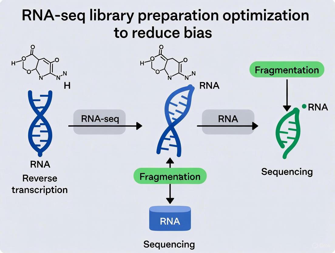
Optimizing RNA-seq Library Preparation: A Comprehensive Guide to Reducing Bias and Enhancing Data Fidelity
This article provides a systematic guide for researchers and drug development professionals on minimizing bias in RNA-seq library preparation.
Optimizing RNA-seq Library Preparation: A Comprehensive Guide to Reducing Bias and Enhancing Data Fidelity
Abstract
This article provides a systematic guide for researchers and drug development professionals on minimizing bias in RNA-seq library preparation. Covering the entire workflow from sample handling to data validation, it details the foundational sources of technical bias, strategic methodological choices for different sample types, practical troubleshooting and optimization protocols, and rigorous frameworks for experimental validation. By synthesizing current best practices and emerging technologies, this resource empowers scientists to produce more reliable and reproducible transcriptome data, thereby strengthening downstream analyses and biological conclusions.
Understanding the Sources of Bias in RNA-seq Workflows
FAQs: Understanding and Mitigating Bias in RNA-seq
Q1: What are the most common sources of technical bias in RNA-seq data? Technical biases can arise at multiple points in the RNA-seq workflow. A frequent and impactful source is sample-specific gene length bias, where sets of particularly short or long genes repeatedly show changes in expression level that are not biologically real but technical artifacts. This bias can lead to the false identification of specific biological functions, such as ribosome-related functions (often encoded by short genes) or extracellular matrix functions (often encoded by long genes), as being significantly altered in an experiment [1]. Other common sources include RNA degradation and contamination during extraction, inadequate experimental design leading to batch effects, and bioinformatic missteps in data normalization and analysis [2] [3] [4].
Q2: How can I tell if my RNA-seq data is affected by gene length bias? You can identify this bias by analyzing the relationship between gene length and apparent differential expression. If you observe a pattern where gene sets of consistently short or long length appear to be differentially expressed when comparing replicate samples from the same biological condition, this strongly indicates a technical length bias. Tools like conditional quantile normalization (cqn) can be applied to correct this sample-specific length effect [1].
Q3: My downstream applications (e.g., PCR) are failing after RNA extraction. What could be wrong? This is often a symptom of low purity RNA. Contaminants carried over from the extraction process can inhibit enzymatic reactions. Common causes and solutions include:
- Protein contamination (low A260/280): Ensure complete sample digestion and consider using a Proteinase K step [2].
- Residual salts or guanidine (low A260/230): Ensure wash steps are performed thoroughly. After the final wash, centrifuge the column for an additional 2 minutes and take care not to contact the flow-through when handling the column [2].
- Polysaccharide or fat contamination: Decrease the starting sample volume and increase the number of 75% ethanol rinses [5].
Q4: How does library preparation choice influence bias in my RNA-seq experiment? The choice between full-length and 3' mRNA-seq methods involves a trade-off between breadth of information and throughput/sensitivity. Full-length methods are essential for discovering novel transcripts, alternative splicing, and isoform-level changes, but they are more susceptible to biases from RNA degradation and can be less efficient for high-throughput screens [3] [6]. In contrast, 3' mRNA-seq methods (like DRUG-seq or BRB-seq) are highly multiplexed, more robust for degraded samples (e.g., RIN < 8), and require fewer reads per sample, making them excellent for large-scale drug screens. However, they provide no information on splicing or transcript variants [3].
Q5: How can I design my RNA-seq experiment to minimize bias from the start? A robust experimental design is your primary defense against bias. Key considerations include:
- Biological Replicates: A minimum of three biological replicates per condition is generally advised, with four to eight being ideal for capturing true biological variability [3].
- Controls: Include appropriate untreated or vehicle controls. Use synthetic spike-in RNAs (e.g., SIRVs, ERCC) to monitor technical performance and enable consistent quantification across samples [3].
- Batch Effects: Plan your plate layout and processing schedule to minimize confounding technical variation with biological conditions. A well-designed layout also facilitates computational correction of any remaining batch effects [3].
- Pilot Testing: Run a small-scale pilot experiment to optimize protocols and validate conditions before committing to a large, costly study [3].
Troubleshooting Guides
RNA Extraction & Purification
| Problem | Cause | Solution |
|---|---|---|
| Low Yield | Incomplete homogenization or lysis [5]. | Increase homogenization time; centrifuge to pellet debris after lysis and use only the supernatant [2]. |
| Too much starting material [2]. | Reduce input amount to match kit specifications; this prevents column overloading and ensures sufficient buffer action. | |
| RNA is degraded [2]. | Flash-freeze samples or use DNA/RNA protection reagent; ensure a RNase-free work environment. | |
| RNA Degradation | RNase contamination [5]. | Use RNase-free tips, tubes, and reagents; wear gloves; use a dedicated clean area. |
| Improper sample storage or repeated freeze-thaw cycles [2]. | Store samples at -80°C in single-use aliquots. | |
| DNA Contamination | Genomic DNA not effectively removed [2]. | Perform an on-column or in-tube DNase I digestion step during extraction. |
| Low Purity (Inhibitors) | Residual protein or salts [2]. | Ensure complete protein digestion and thorough wash steps; re-spin the column after final wash. |
Library Prep & Bioinformatics
| Problem | Cause | Solution |
|---|---|---|
| Gene Length Bias | Technical bias in data generation and flawed statistical analysis [1]. | Apply bias-correction algorithms like conditional quantile normalization (cqn) to decouple gene length from differential expression signals. |
| Poor Sequencing Library | Input RNA is degraded or impure [5]. | Always check RNA quality (e.g., RIN) before library prep; re-extract if necessary. |
| Inefficient cDNA synthesis or adapter ligation, especially for short RNAs [7]. | Use advanced methods like Ordered Two-Template Relay (OTTR), which minimizes bias and improves end-precision for capturing short or fragmented RNAs. | |
| False Positive DEGs | Inadequate normalization or failure to account for batch effects [4]. | Use robust normalization methods (e.g., TMM for bulk RNA-seq); include batch as a covariate in your statistical model. |
| Small sample sizes leading to underpowered statistics [4]. | Use differential analysis methods robust for small samples (e.g., dearseq); increase biological replicates. |
Key Experimental Protocols for Bias Reduction
Protocol: Correcting for Gene Length Bias
Objective: To remove technical bias coupled to gene length that can lead to false positive results in Gene Set Enrichment Analysis (GSEA) [1].
- Data Input: Start with a gene expression matrix (raw counts or normalized values) and a corresponding file of gene lengths.
- Bias Assessment: Generate a scatter plot of gene expression change (e.g., log2 fold change) versus gene length. A non-random pattern (e.g., a lowess line that is not flat) indicates length bias.
- Apply Correction: Use the conditional quantile normalization (cqn) package in R. The function models the effect of gene length and GC content on expression and removes this technical effect.
- Validation: Re-run the differential expression and GSEA analysis on the cqn-corrected data. A successful correction will show that previously enriched gene sets tied to short or long genes are no longer significant, preserving only the biologically genuine signals [1].
Protocol: A Robust RNA-seq Data Analysis Pipeline
Objective: To outline a standardized bioinformatics workflow that ensures reliable identification of differentially expressed genes (DEGs) from raw sequencing data [4].
- Quality Control (QC): Use FastQC to assess the quality of raw sequencing reads and identify potential issues.
- Trimming & Filtering: Use Trimmomatic to remove low-quality bases and adapter sequences.
- Quantification: Use an alignment-free tool like Salmon to estimate transcript abundance. This step is fast and accurate.
- Normalization: Apply the Trimmed Mean of M-values (TMM) method (from the
edgeRpackage) to correct for compositional differences between samples. - Batch Effect Correction: Examine for and correct any batch effects using methods like ComBat if necessary.
- Differential Expression Analysis: Choose a robust statistical method suited to your experimental design. Benchmarking suggests
dearseqfor complex designs or small samples, andvoom-limma,edgeR, orDESeq2for standard bulk RNA-seq [4].
The workflow for this pipeline is summarized in the following diagram:
The Scientist's Toolkit: Research Reagent Solutions
| Item | Function/Benefit |
|---|---|
| Monarch DNA/RNA Protection Reagent | Maintains RNA integrity in samples during storage, preventing degradation before extraction [2]. |
| On-column DNase I | Digests and removes genomic DNA contamination during RNA purification, ensuring pure RNA for downstream applications [2]. |
| SIRV/ERCC Spike-in RNA Controls | Synthetic RNA mixes added to samples before library prep. They act as internal standards for normalization, sensitivity assessment, and technical performance monitoring [3]. |
| Proteinase K | An enzyme used to digest proteins and nucleases during cell lysis, improving RNA yield and purity by breaking down cellular structures and inactivating RNases [2]. |
| Ordered Two-Template Relay (OTTR) | A reverse transcription method that provides improved precision and minimized bias for sequencing short or fragmented RNAs (e.g., miRNA, tRNA fragments) [7]. |
| CapTrap | A technology used in long-read RNA-seq to enrich for full-length, capped mRNA molecules, enabling more accurate transcriptome annotation [6]. |
Optimizing mRNA Design: A Data-Driven Approach
While not a direct source of bias in a standard RNA-seq pipeline, the principles of data-driven optimization are crucial for related fields like mRNA therapeutic development. The RiboDecode framework demonstrates a paradigm shift from rule-based to learning-based sequence design.
RiboDecode integrates a translation prediction model (trained on large-scale Ribo-seq data), an mRNA stability (Minimum Free Energy - MFE) model, and a codon optimizer. It uses gradient ascent to explore a vast sequence space and generate mRNA codon sequences that maximize translation and/or stability for enhanced therapeutic efficacy [8]. This approach has shown substantial improvements in protein expression in vitro and much stronger immune responses or dose-efficiency in vivo compared to previous methods [8]. The following diagram illustrates this generative optimization process.
Frequently Asked Questions (FAQs)
1. What is the single most critical factor for successful RNA preservation? The immediate stabilization of RNA at the point of sample collection is paramount. RNA molecules are naturally susceptible to rapid degradation by ribonucleases (RNases), and transcriptional processes can continue post-collection, altering the true transcriptional landscape. Effective preservation must immediately inhibit both degradative processes and ongoing transcription to maintain accurate gene expression profiles [9].
2. My RNA yields from plant tissues are consistently low. What could be the cause and solution? Low RNA yields from plant tissues are often due to high levels of interfering compounds like polysaccharides, polyphenols, and secondary metabolites. These compounds can bind to or co-precipitate with RNA. Incorporating a sorbitol pre-wash step can significantly improve outcomes. For grape berry skins, this step increased RNA yield from 3.3 ng/µL to 20.8 ng/µL when using a commercial kit and improved the RNA Integrity Number (RIN) from 1.2 to 7.2 [10]. Similarly, for challenging banana tissues (Musa spp.), a modified SDS-based RNA extraction buffer effectively recovered high-quality RNA (2.92 to 6.30 µg/100 mg fresh weight) with high RNA integrity (RNA IQ 7.8–9.9) [11].
3. How do I choose between snap-freezing and chemical preservatives like RNAlater? The choice involves balancing logistical constraints and required RNA quality. RNAlater has demonstrated superior performance in direct comparisons. For human dental pulp tissue, RNAlater storage provided an 11.5-fold higher RNA yield compared to snap-freezing in liquid nitrogen and achieved optimal RNA quality in 75% of cases versus only 33% for snap-frozen samples [9]. RNAlater is often more practical in clinical settings where immediate access to liquid nitrogen is limited.
4. My RNA-seq data shows high ribosomal RNA (rRNA) contamination. How can I improve mRNA enrichment? rRNA contamination is a common issue, as it can constitute over 80% of total RNA. For polyadenylated transcript enrichment, standard one-round of oligo(dT) purification may be insufficient, potentially leaving ~50% rRNA content. Optimization is key:
- Increase the beads-to-RNA ratio: Raising the ratio from 13.3:1 to 50:1 can reduce rRNA content to about 20% [12].
- Perform two rounds of enrichment: This can further reduce rRNA content to less than 10% [12].
- Verify the efficiency of enrichment methods (e.g., capillary electrophoresis) before proceeding to costly sequencing [12].
5. Are commercial RNA extraction kits reliable for all sample types? Commercial kits provide convenience but their performance varies significantly depending on the sample type. For standard cell lines, many kits perform well [13]. However, for recalcitrant tissues (e.g., grape berry skins, woody plants, FFPE samples), they often require protocol modifications or may be ineffective [10]. Systematic comparisons of seven FFPE RNA extraction kits showed notable differences in the quantity and quality of recovered RNA, with some kits consistently outperforming others [14]. Always validate kit performance for your specific sample.
Troubleshooting Guides
Problem: Poor RNA Integrity and Low RIN/RQS Values
Potential Causes and Solutions:
- Cause: Delayed or Inefficient Preservation.
- Solution: Minimize the time between sample dissection and preservation. For tissues high in RNases like dental pulp, complete preservation steps within 90 seconds [9]. Validate that your preservation method (snap-freezing or chemical) immediately halts RNase activity.
- Cause: Incomplete Homogenization or Lysis.
- Solution: For fibrous tissues (e.g., dental pulp, plant matter), ensure thorough homogenization. Use optimized, tissue-specific lysis buffers. The modified SDS-based buffer for banana tissues included LiCl precipitation to enhance yield and purity [11]. For microlepidopterans, protocol optimizations like extended, agitated incubation during protein digestion were crucial [15].
- Cause: Co-purification of Contaminants.
- Solution: Incorporate additional washing steps. The sorbitol pre-wash selectively removes polyphenols and polysaccharides from grape skins without precipitating RNA [10]. For DNA contamination, an additional DNase treatment can be incorporated, which has been shown to significantly reduce genomic DNA levels and intergenic read alignment in RNA-seq [16].
Problem: Low RNA Yield
Potential Causes and Solutions:
- Cause: Suboptimal Preservation Method.
- Solution: Re-evaluate your preservation choice. If using snap-freezing, ensure tissue pieces are small enough for rapid freezing. Consider switching to a chemical preservative if yield is consistently low. See [9] for a quantitative comparison.
- Cause: Inefficient Elution or Precipitation.
- Solution: For column-based kits, ensure elution buffer is applied to the center of the membrane and incubate at room temperature as per optimized protocols [15]. For precipitation methods, ensure correct salt and alcohol concentrations and sufficient precipitation time.
Problem: Inconsistent RNA-seq Results (High Bias/Background)
Potential Causes and Solutions:
- Cause: rRNA Contamination.
- Solution: Optimize your mRNA enrichment as described in FAQ #4. Consider using rRNA depletion kits instead of poly(A) selection, especially if studying non-polyadenylated RNAs [12].
- Cause: gDNA Contamination.
- Solution: Implement a robust DNase digestion step. Studies have shown that an additional DNase treatment can be necessary to reduce intergenic read alignment in sensitive RNA-seq applications [16].
- Cause: Pervasive Sequencing Biases.
- Solution: Employ advanced bioinformatic tools for bias mitigation. The Gaussian Self-Benchmarking (GSB) framework leverages the natural GC-content distribution of transcripts to correct for multiple co-existing biases (GC bias, fragmentation bias, library prep bias) simultaneously, leading to more reliable data [13].
Experimental Protocols & Data
Protocol 1: RNA Preservation with RNAlater for Human Tissues
Methodology (as used for dental pulp tissue) [9]:
- Immediately after dissection, transfer tissue to a sterile Petri dish.
- Wash briefly (10-15 seconds) in sterile DMEM solution using RNase-free vessels.
- Transfer to a new Petri dish containing RNAlater solution.
- Rapidly section tissue into fine fragments (<3 mm) within 90 seconds to prevent degradation.
- Store samples in RNAlater at the recommended temperature.
Protocol 2: Sorbitol Pre-Wash for RNA Extraction from Polyphenol-Rich Tissues
Methodology (as used for grape berry skins) [10]:
- Grind frozen tissue to a fine powder in liquid nitrogen.
- Add 10 mL of pre-chilled Sorbitol Wash Buffer (100 mM Tris-HCl pH 8.0, 100 mM LiCl, 50 mM EDTA, 2% SDS, 1% PVP-40, 0.5% sorbitol) per gram of tissue.
- Vortex vigorously and incubate on ice for 15 minutes.
- Centrifuge at 13,000g for 15 minutes at 4°C.
- Discard the supernatant, which contains the solubilized contaminants.
- Proceed with your standard RNA extraction protocol (e.g., commercial kit or phenol-chloroform) on the resulting pellet.
Protocol 3: Modified SDS-Based RNA Extraction for Challenging Plant Tissues
Methodology (as used for Musa spp.) [11]:
- Extraction Buffer: 2% SDS, 100 mM Tris-HCl (pH 8.0), 50 mM EDTA, 500 mM NaCl, 2% PVP.
- Procedure:
- Homogenize 100 mg tissue in 1 mL of pre-warmed (65°C) extraction buffer.
- Incubate at 65°C for 20 minutes with occasional mixing.
- Add 0.2 volumes of chloroform, mix, and centrifuge.
- Precipitate the RNA from the aqueous phase with an equal volume of 4M LiCl at -20°C for 30+ minutes.
- Centrifuge, wash the pellet with 70% ethanol, and resuspend in RNase-free water.
Comparative Performance of Preservation Methods
Table 1: Quantitative comparison of RNA preservation methods from human dental pulp tissue (n=36). Data adapted from [9].
| Preservation Method | Average Yield (ng/µL) | Average RIN | Samples with Optimal Quality |
|---|---|---|---|
| RNAlater Storage | 4,425.92 ± 2,299.78 | 6.0 ± 2.07 | 75% |
| RNAiso Plus | Not explicitly stated (1.8x lower than RNAlater) | Not explicitly stated | Not explicitly stated |
| Snap Freezing | 384.25 ± 160.82 | 3.34 ± 2.87 | 33% |
Performance of Commercial Kits for FFPE RNA Extraction
Table 2: Summary of findings from a systematic comparison of seven commercial FFPE RNA extraction kits across three tissue types (Tonsil, Appendix, Lymph Node). Data adapted from [14].
| Kit Performance Group | Key Finding | Notable Example |
|---|---|---|
| Higher Quantity | One kit provided the maximum RNA recovery for 7 out of 9 samples. | ReliaPrep FFPE Total RNA Miniprep (Promega) |
| Better Quality | Three kits performed better in terms of RQS and DV200 values. | Roche Kit |
| Best Overall Ratio | One kit yielded the best combination of both quantity and quality. | ReliaPrep FFPE Total RNA Miniprep (Promega) |
The Scientist's Toolkit
Table 3: Essential reagents and kits for RNA preservation and extraction, with specific examples from recent studies.
| Reagent / Kit | Primary Function | Application Context | Key Reference |
|---|---|---|---|
| RNAlater Stabilization Solution | Chemical preservation of RNA in tissues; inhibits RNases. | Optimal for human dental pulp and other clinical tissues. | [9] |
| RNAiso Plus / TRIzol | Monophasic lysis reagent for simultaneous dissociation of cells and inactivation of RNases. | Standard for cell lines (HEK293T); base for plant protocol modifications. | [11] [13] |
| Sorbitol Wash Buffer | Pre-wash to remove polysaccharides and polyphenols without precipitating RNA. | Critical for high-quality RNA from grape berry skins and other polyphenol-rich plants. | [10] |
| Oligo(dT)25 Magnetic Beads | Selection and enrichment of polyadenylated mRNA from total RNA. | Requires optimization of beads-to-RNA ratio for effective rRNA depletion in yeast. | [12] |
| Ribo-off rRNA Depletion Kit | Removal of ribosomal RNA (rRNA) from total RNA using probes. | Used for profiling non-rRNA molecules in human samples. | [13] |
| CTAB Buffer | Lysis buffer effective for disrupting cells with tough walls and removing polysaccharides. | Used in optimized protocols for plants and insects (microlepidopterans). | [11] [15] |
| Poly(A)Purist MAG Kit | Magnetic bead-based selection of polyadenylated RNA. | Compared for mRNA enrichment efficiency in yeast. | [12] |
| VAHTS Universal V8 RNA-seq Kit | Library preparation for next-generation sequencing. | Used for standardized RNA-seq library construction from various samples. | [13] |
Workflow Diagrams
Sample Preservation and RNA Extraction Workflow
Troubleshooting Common RNA Extraction Challenges
In RNA sequencing (RNA-seq), the journey from raw nucleic acids to a sequenced library is a potential minefield of technical bias. Library construction has been identified as a stage where almost every procedural step can introduce significant deviations, compromising data quality and leading to erroneous biological interpretations [17] [18]. A detailed understanding of these biases is crucial for developing robust experiments and accurate data analysis. This guide addresses common biases encountered during RNA-seq library preparation, providing targeted troubleshooting advice and solutions to help researchers mitigate these issues.
Troubleshooting Guides
Problem: Low Library Yield
Low library yield can stall projects and result from issues at multiple preparation stages.
Root Causes and Corrective Actions
| Cause Category | Specific Cause | Corrective Action |
|---|---|---|
| Sample Input/Quality | Degraded RNA or sample contaminants (e.g., phenol, salts) inhibiting enzymes [19]. | Re-purify input sample; ensure 260/230 ratio >1.8 [19]. |
| Quantification errors from absorbance methods (e.g., NanoDrop) overestimating usable material [19]. | Use fluorometric quantification (e.g., Qubit) for accurate measurement [19]. | |
| Fragmentation & Ligation | Suboptimal adapter ligation due to poor ligase performance or incorrect adapter-to-insert molar ratio [19]. | Titrate adapter:insert ratio; ensure fresh ligase and optimal reaction conditions [19]. |
| Amplification/PCR | Enzyme inhibitors present in the reaction [19]. | Use master mixes to reduce pipetting errors and ensure reagent quality [19]. |
| Purification & Cleanup | Overly aggressive purification or size selection, leading to sample loss [19]. | Optimize bead-to-sample ratios and avoid over-drying beads during cleanup [19]. |
Problem: Amplification Bias
PCR amplification can stochastically introduce biases, leading to uneven representation of cDNA molecules and high duplicate rates [18].
Strategies for Mitigation
| Strategy | Methodological Details | Effect on Bias |
|---|---|---|
| Polymerase Selection | Use high-fidelity polymerases (e.g., Kapa HiFi) over others like Phusion for more uniform amplification [18]. | Red preferential amplification of sequences with neutral GC content [18]. |
| Cycle Optimization | Reduce the number of PCR amplification cycles to a minimum [18]. | Minimizes overamplification artifacts and reduces duplicate read rates [19]. |
| PCR Additives | For extreme AT/GC-rich sequences, use additives like TMAC or betaine [18]. | Helps mitigate sequence-dependent amplification bias [18]. |
| Amplification-Free Protocols | For samples with sufficient starting material, use PCR-free library construction methods [18]. | Eliminates PCR amplification bias entirely [18]. |
Problem: Primer and Fragmentation Bias
The initial steps of priming and fragmentation can create non-random representation of the transcriptome.
Sources and Improvements
| Bias Type | Description | Suggestions for Improvement |
|---|---|---|
| Random Hexamer Priming Bias | Inefficient or non-random annealing of hexamer primers during cDNA synthesis, leading to mispriming and uneven 5' coverage [18]. | Use a read count reweighing scheme in bioinformatics analysis to adjust for the bias [18]. For specialized applications, consider direct RNA sequencing without reverse transcription [18]. |
| RNA Fragmentation Bias | Non-random fragmentation using enzymes like RNase III can reduce library complexity [18]. | Use chemical treatment (e.g., zinc) for more random fragmentation [18]. Alternatively, fragment cDNA after reverse transcription using mechanical or enzymatic methods [18]. |
| Adapter Ligation Bias | T4 RNA ligases can have sequence preferences, affecting which fragments are successfully ligated and sequenced [18]. | Use adapters with random nucleotides at the ligation extremities to reduce sequence dependence [18]. |
Frequently Asked Questions (FAQs)
Q1: How much RNA is typically required for a standard RNA-seq library? The quantity of RNA required depends on the sample type and protocol, but a general guideline is 100 nanograms to 1 microgram of total RNA for standard protocols on platforms like Illumina. For low-input or degraded samples, specialized kits are available that can work with significantly less material [20].
Q2: What is "library size" in the context of RNA-seq? Library size refers to the average length of the cDNA fragments in your sequencing library. It is a critical parameter checked by capillary electrophoresis (e.g., Bioanalyzer). For Illumina platforms, the optimal library size typically ranges from 200 to 500 base pairs, which includes the inserted cDNA fragment plus the attached adapters [20].
Q3: How can I reduce bias from my RNA extraction method? RNA extraction methods can introduce bias; for instance, TRIzol can lead to small RNA loss at low concentrations. To improve results:
- Use high concentrations of RNA samples if using TRIzol.
- Consider alternative protocols like the mirVana miRNA isolation kit, which has been reported to produce high-yield and high-quality RNA, especially for non-coding RNAs [18].
Q4: What is an advanced method to minimize bias for short RNAs? The Ordered Two-Template Relay (OTTR) method is a recent (2025) innovation designed for precise end-to-end capture of short or degraded RNAs (e.g., miRNA, tRNA fragments). It minimizes bias inherent to traditional ligation and tailing methods by appending both sequencing adapters in a single reverse transcription step, thereby reducing information loss [7].
Experimental Protocols for Bias Reduction
Library Preparation Using Duplex-Specific Nuclease (DSN) for Normalization
This protocol enriches for low-abundance transcripts by normalizing the cDNA library, substantially decreasing the proportion of reads from highly-expressed RNAs [21].
Key Materials:
- Starting Material: 400 ng polyadenylated RNA from ~20 μg total RNA [21].
- Key Reagent: Crab Duplex-Specific Nuclease (DSN, Evrogen) [21].
- Primers: 19-20 bp primers for initial amplification; full-length Illumina paired-end primers for final amplification [21].
Detailed Methodology:
- Create High-Complexity Library: Generate a standard RNA-seq library through polyA selection, RNA fragmentation, random hexamer-primed reverse transcription, and second-strand synthesis [21].
- Initial Amplification: Amplify the library to generate 500-1200 ng DNA using the short primers [21].
- Denature and Re-anneal: Denature the amplified library and allow it to re-anneal at 68°C. Abundant sequences re-anneal more quickly and become double-stranded, while rare sequences remain single-stranded [21].
- DSN Treatment: Treat the sample with DSN, which preferentially digests the double-stranded (abundant) molecules [21].
- Repeat and Finalize: A second round of amplification and normalization is often performed. The final normalized library is then amplified with the full-length Illumina primers for sequencing [21].
Workflow for Standard RNA-Seq Library Preparation
The following diagram outlines the key steps in a standard RNA-seq library preparation workflow, highlighting stages where specific biases commonly originate.
The Scientist's Toolkit: Key Research Reagent Solutions
| Reagent / Kit | Function in Library Preparation | Consideration for Bias Reduction |
|---|---|---|
| Duplex-Specific Nuclease (DSN) [21] | Normalizes cDNA libraries by digesting double-stranded (abundant) re-annealed molecules, enriching for rare transcripts. | Crucial for reducing dynamic range and improving detection of low-abundance RNAs. |
| High-Fidelity DNA Polymerase (e.g., Kapa HiFi) [18] | Amplifies the adapter-ligated library during PCR. | Provides more uniform amplification across sequences with different GC contents compared to other polymerases. |
| mirVana miRNA Isolation Kit [18] | Extracts total RNA, including small RNAs. | Reduces bias against small non-coding RNAs often encountered with TRIzol extraction. |
| R2 Reverse Transcriptase (in OTTR) [7] | Engineered reverse transcriptase for the OTTR method that enables precise end-to-end capture of RNA molecules. | Minimizes bias from ligation and tailing for short RNAs; improves 3' and 5' end precision. |
| Magnetic Beads (e.g., AMPure, Dynabeads) [21] [19] | Purify and size-select nucleic acids after various enzymatic steps. | Incorrect bead-to-sample ratios can cause size selection bias or sample loss; optimization is key [19]. |
Troubleshooting Guides
FAQ: Managing PCR Duplication in RNA-seq Libraries
Q: What causes high PCR duplication rates in my RNA-seq data, and how can I reduce them?
High PCR duplication rates occur when a small subset of original RNA molecules is over-amplified during library preparation. This skews representation and can mask true biological variation. Common causes and solutions are detailed below.
Cause: Limited Input Material or Low Library Complexity
- Solution: Ensure sufficient RNA input. For low-input or degraded samples (e.g., FFPE), use specialized protocols that require higher RNA input or employ kits designed for low-quality/quantity RNA to maximize the diversity of starting molecules [18].
Cause: Over-Amplification during Library PCR
Cause: Bias from Ligation Steps
- Solution: Consider advanced ligation methods. Standard adaptor ligation using T4 RNA ligases is a known source of bias, leading to over-representation of specific sequences. Using a protocol with randomized splint ligation can significantly reduce this bias and increase the sensitivity for detecting diverse small RNAs [23].
FAQ: Overcoming GC Bias in PCR Amplification
Q: Why are GC-rich templates difficult to amplify, and what strategies can improve success?
GC-rich templates (typically >60% GC content) are challenging due to their high thermostability and tendency to form secondary structures. The following table summarizes the primary challenges and general solution approaches.
| Challenge | Root Cause | Solution Pathway |
|---|---|---|
| High Thermal Stability | Three hydrogen bonds in G-C base pairs require more energy to denature [24] [25]. | Increase denaturation temperature; use specialized polymerases. |
| Formation of Secondary Structures | GC-rich regions readily form stable hairpins and stem-loops that block polymerase progression [24] [25]. | Use additives (e.g., DMSO, betaine); choose high-processivity enzymes. |
| Non-specific Primer Binding | Stable secondary structures in primers and templates promote mispriming [25]. | Optimize Mg²⁺ concentration; increase annealing temperature. |
Detailed solutions for GC bias:
Polymerase and Buffer Selection: Use polymerases specifically engineered for GC-rich templates. Kits often include specialized buffers and GC enhancers. For example:
- OneTaq DNA Polymerase with GC Buffer and Enhancer can amplify templates with up to 80% GC content [24].
- Q5 High-Fidelity DNA Polymerase with its GC Enhancer is ideal for long or difficult amplicons [24].
- AccuPrime GC-Rich DNA Polymerase, derived from Pyrococcus furiosus, retains activity at high temperatures, aiding in denaturation [25].
Optimize Reaction Additives: Additives help denature stable GC structures.
- DMSO: Typically used at 2-10%, but concentrations above 5% can reduce polymerase activity [26].
- Betaine (or TMAC): Used at 0.5-2 M concentrations to equalize the melting temperatures of GC-rich and AT-rich regions [18] [26].
- Glycerol: Can be added at 5-25% to stabilize enzymes and influence DNA melting [26].
Adjust Thermal Cycling Conditions:
- Denaturation: Increase the denaturation temperature (up to 95°C) or extend the denaturation time, especially for the first few cycles [24] [25]. Note that very high temperatures can degrade polymerase over time [25].
- Annealing: Use a temperature gradient to find the optimal annealing temperature. A higher annealing temperature improves specificity but may reduce yield [24] [27] [22].
- Ramp Rates: Employing slower temperature ramp rates ("slow-down PCR") can help polymerase navigate through complex secondary structures [25].
Magnesium Concentration: Optimize Mg²⁺ concentration. While 1.5-2 mM is standard, GC-rich PCR may require fine-tuning. Test a gradient from 1.0 mM to 4.0 mM in 0.5 mM increments to find the ideal concentration for specificity and yield [24] [27].
GC-Rich PCR Troubleshooting Flowchart
Experimental Protocols for Bias Reduction
Protocol: Reducing Ligation Bias with Randomized Splint Ligation
This protocol is adapted from a low-bias small RNA library preparation method [23].
- 3' Adapter Ligation: Ligate a pre-adenylated 3' adapter to the RNA fragments using randomized splint ligation. The splint contains random nucleotides that base-pair with the RNA fragment, reducing sequence-dependent ligation bias.
- Clean-up: Deplete excess adapter using a 5' deadenylase and lambda exonuclease.
- Cleavage: Cleave the degenerate portion of the adapter by treating with USER enzyme to excise deoxyuracil.
- 5' Adapter Ligation: Ligate the 5' adapter using the same randomized splint ligation method.
- Reverse Transcription: Synthesize cDNA using the remaining portion of the 3' adapter splint as a primer.
- PCR Enrichment: Amplify the library using primers complementary to the adapter sequences.
Protocol: Optimized PCR for GC-Rich Targets
This protocol synthesizes recommendations from multiple sources [24] [25] [26].
- Reaction Setup:
- Polymerase: 1-2 units of a GC-optimized polymerase (e.g., OneTaq or Q5).
- Buffer: Use the manufacturer's supplied GC buffer.
- GC Enhancer: If provided, add at the recommended concentration (e.g., 10-20% of the reaction volume).
- Additives: Supplement with DMSO (2-5% final) or betaine (0.5-1 M final).
- Mg²⁺: Start with the manufacturer's recommended concentration (often 1.5-2 mM MgCl₂).
- Template: 10-100 ng of high-quality genomic DNA.
- Thermal Cycling:
- Initial Denaturation: 98°C for 2 minutes.
- Amplification (35-40 cycles):
- Denaturation: 98°C for 20-30 seconds. For very difficult templates, use 95-98°C for 10-20 seconds.
- Annealing: Use a temperature 3-5°C below the primer Tm. For non-specific bands, increase temperature by 2°C increments.
- Extension: 72°C for 1 minute per kb.
- Final Extension: 72°C for 5-10 minutes.
Table 1: Polymerase Systems for GC-Rich and High-Fidelity PCR
| Polymerase System | Key Feature | Ideal GC Content Range | Fidelity (Relative to Taq) | Key Applications |
|---|---|---|---|---|
| OneTaq DNA Polymerase (NEB) | Standard & GC Buffers available | Up to 80% (with GC Enhancer) [24] | 2x [24] | Routine and GC-rich PCR [24] |
| Q5 High-Fidelity DNA Polymerase (NEB) | High Fidelity & GC Enhancer | Up to 80% (with GC Enhancer) [24] | >280x [24] | Long, difficult, and GC-rich amplicons [24] |
| AccuPrime GC-Rich DNA Polymerase (ThermoFisher) | High processivity at high T | High (specific range not stated) | Not specified | GC-rich templates [25] |
| Kapa HiFi DNA Polymerase | Reduced amplification bias | Effective for neutral GC% [28] | High (specific value not stated) | Library amplification for NGS [28] |
Table 2: Optimization of PCR Additives for GC-Rich Templates
| Additive | Recommended Concentration | Mechanism of Action | Key Considerations |
|---|---|---|---|
| DMSO | 2-10% [26] | Disrupts base pairing, reduces secondary structure formation [24] | >5% can inhibit polymerase; may increase error rate [26] |
| Betaine | 0.5 - 2.0 M [26] | Equalizes template melting temps, inhibits secondary structure formation [24] [18] | Can be a component of commercial "GC Enhancer" solutions [24] |
| Glycerol | 5-25% [26] | Stabilizes enzymes, can lower DNA melting temperature [24] | High concentrations may alter enzyme kinetics |
| 7-deaza-dGTP | Partial substitution for dGTP | dGTP analog that weakens base pairing, reducing template stability [24] [25] | Does not stain well with ethidium bromide [24] |
| GC-RICH Resolution Solution (Roche) | 0.5 - 2.5 M (titrate) | Proprietary solution containing detergents and DMSO [26] | Part of a specialized system for GC-rich templates |
The Scientist's Toolkit: Key Reagents for Bias-Reduced PCR
Table 3: Essential Research Reagents for Managing PCR Bias
| Item | Function in Bias Reduction |
|---|---|
| High-Fidelity, GC-Rich Polymerases (e.g., Q5, OneTaq GC) | Engineered for high processivity and affinity to navigate through complex secondary structures in GC-rich templates, providing robust amplification [24] [27]. |
| GC Enhancer / Betaine | Homogenizes the melting behavior of DNA, preventing the stalling of polymerase at stable secondary structures and promoting uniform amplification of regions with varying GC content [24] [18]. |
| Hot-Start DNA Polymerases | Remain inactive until a high-temperature activation step, preventing non-specific priming and primer-dimer formation at lower temperatures, thereby improving specificity and yield [27] [22]. |
| Randomized Splint Adapters | Used in ligation-based library prep to minimize sequence-dependent ligation bias, ensuring a more uniform representation of all RNA fragments in the final library [23]. |
Randomized Splint Ligation Workflow
Sequencing Platform and Contextual Biases
Frequently Asked Questions (FAQs)
What are the main sources of bias in RNA-seq library preparation? Biases can be introduced at virtually every step of the RNA-seq workflow. The primary sources include:
- Sample Preservation: Using formalin-fixed, paraffin-embedded (FFPE) samples can cause RNA fragmentation, cross-linking, and chemical modifications, leading to poor sequencing libraries [18].
- RNA Extraction: Certain extraction methods, like TRIzol, can lead to the loss of small RNAs, especially at low concentrations [18].
- mRNA Enrichment: Poly(A) selection can cause 3'-end capture bias, under-representing the 5' ends of transcripts [18].
- Fragmentation: Enzymatic fragmentation (e.g., using RNase III) is not completely random and can reduce library complexity [18].
- Priming Bias: Random hexamers used in reverse transcription can bind with varying efficiencies across transcripts, creating an uneven representation [18].
- Adapter Ligation: T4 RNA ligases have sequence-dependent preferences, leading to the over-representation of certain fragments [28].
- PCR Amplification: This step stochastically introduces biases, as different molecules are amplified with unequal probabilities. This can lead to under-representation of both AT-rich and GC-rich regions [18] [29].
How does my choice of sequencing platform influence bias? The sequencing platform itself can be a source of bias, often referred to as "platform bias" [18]. Furthermore, the instrument type dictates the required library preparation protocol, which has a major impact. Key specifications like read length and throughput should be matched to your application to minimize interpretive biases [30]. For instance, short-read platforms may struggle with complex genomic regions, while long-read platforms can span repetitive sequences but often have higher per-base error rates [30] [31].
What is the best RNA-seq method for degraded RNA samples, such as those from FFPE tissue? For degraded or low-quality total RNA (e.g., RIN 2-3 from FFPE samples), random-primed library preparation protocols are recommended over oligo(dT)-primed methods [18] [32]. Kits like the SMARTer Stranded or SMARTer Universal Low Input RNA Kit are designed for this purpose, as they do not rely on intact poly-A tails [32]. Prior ribosomal RNA (rRNA) depletion is typically required when using random priming [32].
How can I reduce bias during library amplification?
- Minimize PCR Cycles: Reduce the number of amplification cycles as much as possible to prevent the preferential amplification of certain fragments [18].
- Use High-Fidelity Polymerases: Polymerases like KAPA HiFi are engineered to reduce amplification biases compared to others like Phusion [28].
- Employ UMIs: Unique Molecular Identifiers (UMIs) are short random sequences added to each molecule before amplification. They allow for bioinformatic correction of PCR bias and errors by identifying reads that originated from the same original molecule [33].
- Consider PCR-Free Protocols: For sufficient starting material, amplification-free protocols entirely avoid PCR-induced bias [18].
Troubleshooting Guides
Problem 1: Low Library Yield
Low library yield can halt progress and waste resources. The following table outlines common causes and their solutions.
| Cause | Mechanism of Yield Loss | Corrective Action |
|---|---|---|
| Poor Input Quality / Contaminants | Enzyme inhibition from residual salts, phenol, or EDTA [19]. | Re-purify input sample; ensure high purity (e.g., 260/230 > 1.8); use fresh wash buffers [19]. |
| Inaccurate Quantification | Under-estimating input leads to suboptimal enzyme stoichiometry [19]. | Use fluorometric methods (Qubit) over UV absorbance (NanoDrop); calibrate pipettes [19]. |
| Suboptimal Adapter Ligation | Poor ligase performance or incorrect adapter-to-insert molar ratio [19]. | Titrate adapter:insert ratios; ensure fresh ligase and buffer; optimize incubation time and temperature [19]. |
| Overly Aggressive Purification | Desired library fragments are accidentally removed during clean-up steps [19]. | Optimize bead-based clean-up ratios; avoid over-drying beads to prevent inefficient resuspension [19]. |
Problem 2: Over-representation of Specific Sequences
This bias results in an inaccurate representation of transcript abundance in your data.
Symptoms:
- A few specific sequences have significantly higher read counts than expected.
- Skewed gene expression measurements.
Root Causes and Protocols for Bias Reduction:
Adapter Ligation Bias:
- Cause: T4 RNA ligases have strong sequence and structure preferences. Fragments that co-fold with the adaptor sequence are ligated more efficiently, leading to their over-representation [28].
- Solution - Use Random-Base Adapters: Instead of standard adapters, use adapters with random nucleotides at the extremities to be ligated. This randomizes the ligation junction and can significantly reduce sequence-dependent bias [18].
- Solution - Alternative Ligation Protocol: The CircLig protocol, which uses a single adaptor and circularization, has been shown to reduce over-representation compared to standard duplex adaptor protocols [28].
PCR Amplification Bias:
- Cause: GC-rich or AT-rich regions can amplify less efficiently, leading to their under-representation [18] [29].
- Solution - Polymerase and Additives: Use bias-reducing polymerases like KAPA HiFi. For extremely AT/GC-rich sequences, PCR additives like TMAC or betaine can help, as can lowering extension temperatures [18].
- Solution - Unique Molecular Identifiers (UMIs): Incorporate UMIs during library preparation. During data analysis, UMIs allow you to deduplicate reads, correcting for both PCR bias and errors by counting only unique original molecules [33] [34].
Problem 3: High Ribosomal RNA Read Contamination
A high percentage of reads aligning to rRNA indicates inefficient removal of ribosomal RNA.
Symptoms:
- More than 10-20% of sequencing reads map to ribosomal RNA.
- Lower coverage of your RNA species of interest (e.g., mRNA, lncRNA).
Root Causes and Solutions:
- Incorrect Enrichment Method:
- Inefficient rRNA Depletion Protocol:
- Cause: The depletion kit or protocol is not optimized for your specific sample type or input amount.
- Solution: Follow kit recommendations for input RNA quantity and quality. For example, the RiboGone - Mammalian kit is recommended for 10–100 ng samples of mammalian total RNA [32].
The diagram below maps the standard RNA-seq library preparation workflow and highlights key points where biases are most likely to be introduced.
Research Reagent Solutions
The following table lists key reagents and their roles in reducing specific biases during RNA-seq library preparation.
| Reagent / Kit | Function | Bias-Reducing Application |
|---|---|---|
| RiboGone - Mammalian Kit | Depletes ribosomal RNA from mammalian total RNA samples [32]. | Eliminates the need for poly-A selection, avoiding 3'-end bias. Essential for studying non-polyadenylated RNA (e.g., lncRNA) or degraded samples [32] [33]. |
| SMARTer Stranded RNA-Seq Kit | A random-primed library prep kit that maintains strand-of-origin information [32]. | Ideal for degraded RNA (FFPE) and non-polyadenylated RNA, as it does not rely on an intact 3' poly-A tail [32]. |
| KAPA HiFi DNA Polymerase | A high-fidelity PCR enzyme engineered for robust amplification of GC-rich templates [28]. | Reduces PCR amplification bias, particularly the under-representation of GC-rich and AT-rich regions [18] [28]. |
| Unique Molecular Identifiers (UMIs) | Short random barcodes added to each RNA molecule before any amplification steps [33]. | Allows bioinformatic correction of PCR bias and errors by accurately counting pre-amplification molecules, mitigating over-amplification effects [33] [34]. |
| Random-Base Adapters | Adaptors with degenerate nucleotides at the ligation junctions [18]. | Reduces sequence-specific bias during adapter ligation by randomizing the interaction with T4 RNA ligase [18]. |
| CircLigase ssDNA Ligase | An enzyme used to circularize single-stranded DNA in alternative library prep protocols [28]. | Used in the "CircLig protocol," which has been shown to reduce over-representation of specific sequences compared to standard duplex adaptor protocols [28]. |
Strategic Protocol Selection for Optimal Library Construction
A foundational step in a successful RNA-seq experiment is the selective analysis of messenger RNA (mRNA) against a background where it can constitute as little as 1-5% of total RNA, with ribosomal RNA (rRNA) making up the overwhelming majority (80-98%) [35]. The two primary strategies to overcome this are poly(A) enrichment and rRNA depletion. The choice between them is critical, as it directly influences data quality, coverage, and the accuracy of biological interpretation. Within the broader goal of optimizing RNA-seq library preparation to reduce bias, the integrity of your starting RNA sample is the most decisive factor in selecting the appropriate method. This guide provides troubleshooting and FAQs to help you make an informed choice.
FAQ: Choosing Your Method
What is the core difference between mRNA enrichment and rRNA depletion?
- mRNA Enrichment (Poly(A) Selection): This method uses magnetic beads coated with oligo(dT) sequences to "fish out" RNA molecules that have a poly(A) tail, which is a hallmark of mature eukaryotic mRNAs. It is a targeted enrichment of desired transcripts [35].
- rRNA Depletion (Ribo-Depletion): This method uses species-specific probes (DNA or LNA oligonucleotides) that are complementary to rRNA sequences. These probes hybridize to the rRNA, which is then removed from the sample through magnetic separation or enzymatic digestion. This is a targeted depletion of unwanted transcripts [36] [18].
How does RNA integrity directly affect my choice of method?
The performance of poly(A) enrichment is highly dependent on RNA quality, whereas rRNA depletion is more robust to degradation [35].
- High-Quality RNA (RIN > 8): You can reliably use either method. Poly(A) enrichment is often the default for standard eukaryotic mRNA sequencing due to its cost-efficiency and focus on protein-coding genes [35].
- Degraded or Low-Quality RNA (RIN < 7): You should use rRNA depletion. In degraded samples, RNA fragments may lack the poly(A) tail necessary for capture. Poly(A) enrichment of such samples will lead to a strong 3' bias and significant loss of coverage across the transcript body [35]. rRNA depletion probes target multiple sites along the rRNA molecules, making them effective even on fragmented RNA [37].
Does my organism of study influence the decision?
Absolutely. This is a primary consideration.
- Eukaryotic Samples: You have the flexibility to use either poly(A) enrichment or rRNA depletion.
- Prokaryotic Samples: You must use rRNA depletion. Bacterial mRNAs lack a stable poly(A) tail, making poly(A) enrichment ineffective [35] [37].
What are the specific biases associated with each method?
Understanding inherent biases is key to unbiased data interpretation.
Poly(A) Enrichment Bias:
- 3' Bias: In degraded or low-input samples, the method preferentially captures the 3' ends of transcripts, skewing expression data [35] [18].
- Transcriptome Coverage Bias: It systematically excludes non-polyadenylated RNAs of interest, such as histone genes, many long non-coding RNAs (lncRNAs), and some circular RNAs [35].
rRNA Depletion Bias:
- Probe Design Bias: Depletion efficiency hinges on probe design. Incomplete probe coverage of all rRNA variants can lead to residual rRNA reads (often 5-50%) [36] [38].
- Species Specificity: Probes are often species-specific. Using a kit designed for one organism on a distant relative can result in poor depletion efficiency [35].
- * enzymatic Depletion Bias*: Methods using duplex-specific nucleases (DSN) or RNase H can suffer from off-target activity, digesting non-rRNA transcripts with partial complementarity to the probes [38].
Troubleshooting Guide: Common Scenarios and Solutions
| Scenario | Symptom | Root Cause | Solution |
|---|---|---|---|
| Degraded FFPE or Tissue Sample | Low mapping to mRNA, high 3' bias in coverage plots. | Poly(A) tails are lost or inaccessible due to fragmentation. | Switch to an rRNA depletion protocol. Use high-input RNA amounts to compensate for degradation [18] [39]. |
| High Residual rRNA in Prokaryotic Seq | >50% of reads map to rRNA after "depletion". | Inefficient probe hybridization due to species mismatch or suboptimal protocol. | Use a species-specific depletion kit (e.g., riboPOOLs) or design custom biotinylated probes [38]. Optimize hybridization conditions. |
| Low RNA Input (Bacterial) | Failed library preparation or extremely low complexity. | Standard commercial kits require >100ng total RNA. | Use a specialized low-input method like EMBR-seq, which uses blocking primers and linear amplification for inputs as low as 20pg [37]. |
| Low Gene Detection in Eukaryotic Seq | Fewer genes detected than expected, missing non-coding RNAs. | Poly(A) selection excludes non-polyadenylated transcripts. | If your target includes lncRNAs or other non-poly(A) RNA, switch to rRNA depletion [35] [39]. |
| One Round of Enrichment is Insufficient | rRNA still constitutes ~50% of the sample after one round of poly(A) selection or ribo-depletion. | Standard protocols may not be fully efficient for all sample types. | For poly(A) enrichment, perform two consecutive rounds of selection or optimize the beads-to-RNA ratio to significantly improve purity [36]. |
Decision Workflow
The following diagram outlines the logical decision process for choosing between mRNA enrichment and rRNA depletion.
Research Reagent Solutions
The following table summarizes key commercial solutions and their optimal use cases.
| Reagent / Kit | Method | Primary Application | Key Consideration |
|---|---|---|---|
| Oligo(dT)25 Magnetic Beads [36] | Poly(A) Enrichment | Enrichment of eukaryotic mRNA from high-quality RNA. | Cost-effective; requires user-prepared buffers. Efficiency improves with optimized bead:RNA ratios [36]. |
| RiboMinus Kit [36] | rRNA Depletion | Depletion of rRNA from eukaryotic or prokaryotic RNA. | Targets 18S/25S (eukaryotes) or 16S/23S (prokaryotes). May not cover 5S rRNA, leading to residual contamination [38] [36]. |
| riboPOOLs [38] | rRNA Depletion | High-efficiency, species-specific rRNA depletion. | More effective than pan-prokaryotic kits for specific organisms. An adequate replacement for the discontinued RiboZero [38]. |
| NEBNext Poly(A) mRNA Magnetic Isolation Kit [40] | Poly(A) Enrichment | Standard mRNA sequencing from intact eukaryotic RNA. | Used in published RNA-seq workflows with high-quality input (RIN > 7.0) [40]. |
| EMBR-seq (Protocol) [37] | rRNA Depletion | Sequencing from ultra-low input and degraded bacterial RNA. | Uses blocking primers and in vitro transcription; cost-effective for non-model bacteria [37]. |
| Watchmaker RNA Kit with Polaris Depletion [39] | rRNA Depletion | Sensitive RNA-seq from challenging samples (FFPE, blood). | Includes reagents for rRNA and globin depletion, ideal for clinically derived samples [39]. |
Experimental Protocol: Optimizing Poly(A) Enrichment for Yeast RNA
Background: A single round of poly(A) enrichment may be insufficient, leaving rRNA content as high as 50% [36]. This protocol describes an optimized two-round enrichment to reduce rRNA to below 10%.
Materials:
- Oligo(dT)25 Magnetic Beads (e.g., from New England Biolabs)
- Total RNA from Saccharomyces cerevisiae
- Binding Buffer (e.g., 20 mM Tris-HCl, 1.0 M LiCl, 2 mM EDTA)
- Wash Buffer (e.g., 10 mM Tris-HCl, 0.15 M LiCl, 1 mM EDTA)
- Nuclease-free water
Method:
- Denaturation: Dilute 10 µg of total yeast RNA in 50 µL of nuclease-free water. Heat at 65°C for 5 minutes to disrupt secondary structures, then immediately place on ice.
- First Binding: Add 50 µL of well-resuspended Oligo(dT)25 Magnetic Beads and 100 µL of 2X Binding Buffer to the RNA. Use a beads-to-RNA ratio of 1:1 (w/w). Mix thoroughly and incubate at room temperature for 15 minutes with gentle rotation.
- Washing: Place the tube on a magnetic stand. After the solution clears, discard the supernatant. Wash the beads twice with 200 µL of Wash Buffer.
- Elution: Elute the poly(A) RNA from the beads by adding 20 µL of nuclease-free water and heating at 80°C for 2 minutes. Immediately transfer to the magnetic stand and carefully collect the supernatant containing the enriched mRNA.
- Second Binding (Repeat): To the eluate from the first round, add a fresh 50 µL aliquot of Oligo(dT)25 Magnetic Beads and 100 µL of 2X Binding Buffer. Repeat the binding, washing, and elution steps.
- Quality Control: Assess the yield and purity of the final enriched RNA using capillary electrophoresis (e.g., TapeStation). The 18S and 25S rRNA peaks should be drastically reduced, with rRNA content typically falling below 10% of the total RNA [36].
Note: This two-round protocol is more time-consuming and results in lower final yields but provides a much purer mRNA population for sequencing, reducing costs and improving data quality per sequencing read.
FAQ: Fragmentation Method Selection
Q1: How do I choose between mechanical and enzymatic fragmentation for my RNA-seq library?
The choice depends on your research priorities, including sample input, required uniformity, throughput, and budget.
| Factor | Mechanical Fragmentation | Enzymatic Fragmentation |
|---|---|---|
| Sequence Bias | Minimal sequence bias; closest to ideal molecular randomness [41] | Potential for sequence-specific bias (e.g., motif preference, GC skew) [42] [41] |
| Sample Input | Higher potential for sample loss due to extra handling [42] | Recommended for low-input samples (<100 ng); minimal handling loss [42] [43] |
| Throughput & Automation | Limited parallel processing; less automation-friendly [42] | High-throughput and easily automated; suitable for many samples [42] [43] |
| Equipment Cost | Requires specialized, costly instrumentation (e.g., sonicator) [42] [41] | Lower equipment cost; relies mainly on reagents [42] [43] |
| Uniformity & Coverage | Gold standard for even genome coverage; crucial for variant calling [41] | Modern kits approach mechanical randomness, but may have GC skew [41] |
| Protocol Speed | Slower due to separate shearing and cleanup steps [41] | Faster; can be combined with end-repair and A-tailing in one tube [42] [43] |
For RNA-seq, the fragmentation method is a key determinant of data quality, as more stochastic breakage leads to more even and reliable downstream analysis [41]. If your primary goal is quantitative accuracy with minimal bias, mechanical shearing is superior. For high-throughput studies where speed and cost are paramount, enzymatic methods are more pragmatic [42] [41].
Q2: What are the common signs of fragmentation failure, and how can I troubleshoot them?
Poor fragmentation can manifest in several ways during library QC and sequencing.
| Problem | Observed Failure Signals | Recommended Corrective Actions |
|---|---|---|
| Over-/Under-Fragmentation | Unexpected fragment size distribution; skewed insert sizes [19] [43] | Optimize enzyme concentration/digestion time or sonication energy/duration [42] [19]. Pre-check fragmentation profile [19]. |
| High Adapter-Dimer Peaks | Sharp peak at ~70-90 bp on bioanalyzer electropherogram [19] | Titrate adapter-to-insert molar ratio [19]. Use purification beads with the correct sample-to-bead ratio to remove small fragments [19]. |
| Low Library Yield | Low final concentration; broad or faint peaks during QC [19] | Re-purify input sample to remove enzyme inhibitors. Ensure high purity (260/230 > 1.8) [19]. Optimize ligation conditions [19]. |
| Uneven Coverage/GC Bias | Dropouts in high or low GC regions; uneven read depth [41] | If using enzymatic methods, test PCR-free protocols or use spike-in standards to correct bias [41]. Switch to mechanical shearing for maximal uniformity [41]. |
A general diagnostic flow is to: 1) Check the electropherogram for abnormal peaks or distributions, 2) Cross-validate quantification methods (e.g., Qubit vs. BioAnalyzer), and 3) Trace the problem backwards through the library prep steps to identify the root cause [19].
Q3: How does fragmentation bias impact my RNA-seq results?
Fragmentation bias can severely compromise the integrity and interpretability of your RNA-seq data. Non-random fragmentation generates libraries that are not truly representative of the starting sample, leading to:
- Reduced Library Complexity: This inflates PCR duplicate rates, as fewer unique starting molecules are available for amplification. Downstream software may collapse reads that are in fact derived from different original RNA molecules, leading to a loss of biological information [41].
- Uneven Sequence Coverage: Biased fragmentation creates regions that are over- or under-represented in the sequencing data. This can artificially flatten or inflate expression counts for specific transcripts or genomic regions, making accurate differential expression analysis difficult [41] [13].
- Misleading Variant Calls: Inconsistent coverage can create false positive or false negative variant calls. Dips in coverage can be mistaken for deletions, while highly covered regions may mask the true allele frequency of a single-nucleotide variant (SNV) [41].
- Compromised De Novo Assembly: For transcriptome assembly, non-random breakpoints create "fragile sites" where read clouds terminate abruptly, resulting in shorter contigs and a less complete assembly [41].
Mitigating these biases is therefore critical for obtaining reliable biological conclusions [13].
Experimental Protocols for Key Comparisons
Protocol 1: Benchmarking Fragmentation Bias Using Spike-In Controls
This protocol assesses the sequence-specific bias introduced by different fragmentation methods, which is vital for optimizing RNA-seq library preparation [13].
1. Reagents and Materials
- Purified RNA sample (e.g., from HEK293T cells or human tissue)
- Synthetic spike-in RNA controls (e.g., ERCC ExFold RNA Spike-In Mix)
- Mechanical shearing instrument (e.g., Covaris sonicator)
- Enzymatic fragmentation kit (e.g., using endonuclease or tagmentation chemistry)
- Library preparation kit (e.g., VAHTS Universal V8 RNA-seq Library Prep Kit)
- Magnetic beads for cleanup (e.g., AMPure XP)
- Bioanalyzer or TapeStation for QC
2. Methodology
- Step 1: Sample and Spike-In Setup. Split the total RNA sample into two equal aliquots. To each aliquot, add a known quantity of the ERCC spike-in mix, which contains synthetic RNA transcripts of defined lengths and sequences [33] [13].
- Step 2: Parallel Fragmentation. Fragment one aliquot using the optimized mechanical shearing protocol (e.g., focused-ultrasonication). Fragment the second aliquot using the enzymatic protocol (e.g., with a dsDNA fragmentase or tagmentation enzyme) [41] [13].
- Step 3: Library Preparation and Sequencing. Prepare sequencing libraries from both fragmented samples using the same downstream steps (end-repair, A-tailing, adapter ligation, and PCR amplification) to ensure comparisons are based solely on the fragmentation method [43] [13]. Pool and sequence the libraries on the same flow cell to avoid batch effects.
- Step 4: Data Analysis for Bias Assessment.
- Alignment and Quantification: Map reads to a combined reference genome (e.g., human GRCh38) and the spike-in sequences. Calculate reads per kilobase per million (RPKM) or TPM for each spike-in transcript [13].
- Bias Calculation: For the spike-ins, plot the observed read counts (or RPKM) against the expected molar concentration. The deviation from the expected linear relationship indicates the degree of bias. Calculate correlation coefficients (R²) to quantitatively compare the performance of mechanical vs. enzymatic methods [13].
- GC-Bias Analysis: Analyze the coverage uniformity across transcripts with different GC contents. Mechanical shearing should demonstrate flatter coverage across the GC spectrum compared to enzymatic methods, which may show under-representation of extreme GC-rich or AT-rich regions [41].
Protocol 2: Evaluating Protocol Performance with Low-Quality RNA Samples
This protocol tests the robustness of different fragmentation and library prep methods on degraded RNA, such as that from FFPE tissues, a common challenge in clinical research [3] [33].
1. Reagents and Materials
- High-quality RNA (RIN > 9) and artificially degraded or FFPE-derived RNA (RIN < 5)
- rRNA depletion kit (e.g., Ribo-off rRNA Depletion Kit)
- Both mechanical and enzymatic library prep kits
- Bioanalyzer for RNA and library QC
2. Methodology
- Step 1: Sample Preparation. Obtain or create degraded RNA samples. This can be done by incubating high-quality RNA at elevated temperature for a controlled period. Use the Bioanalyzer to confirm a reduced RNA Integrity Number (RIN) [33].
- Step 2: rRNA Depletion. Treat both high-quality and degraded RNA samples with an rRNA depletion kit. This step is crucial for FFPE/degraded samples as ribosomal RNA fragments can still dominate the sample and poly-A selection fails on fragmented mRNA [33] [13].
- Step 3: Library Preparation with Different Methods. For both RNA quality groups, prepare libraries using:
- Method A: Mechanical shearing (acoustic) followed by a standard stranded RNA-seq protocol.
- Method B: An enzymatic fragmentation protocol (e.g., one utilizing a proprietary nuclease cocktail).
- Method C: A tagmentation-based library prep kit.
- Step 4: QC and Sequencing Analysis.
- Library QC: Assess the final libraries for yield, size distribution, and adapter-dimer content. Enzymatic/tagmentation methods often show better yields from low-input, degraded samples [3].
- Sequencing Metrics: After sequencing, compare the mapping rates, the percentage of reads aligning to exonic regions, the 3' bias (indicative of RNA degradation), and the number of genes detected. Robust methods for low-quality RNA will maintain higher gene detection rates and lower 3' bias [3].
Workflow Diagrams
Fragmentation Method Decision Workflow
RNA-seq Bias Mitigation Strategy
The Scientist's Toolkit: Key Research Reagents
| Reagent / Material | Function in Fragmentation & Library Prep |
|---|---|
| ERCC Spike-In RNA Controls | A set of synthetic RNA transcripts of known concentration used to diagnose technical bias, assess the dynamic range, and normalize samples in RNA-seq experiments [33] [13]. |
| Ribosomal RNA (rRNA) Depletion Kit | Selectively removes abundant ribosomal RNA from the total RNA population, thereby enriching for coding and non-coding RNA of interest and significantly improving sequencing depth for these transcripts. Essential for degraded samples and bacterial RNA [33] [13]. |
| Unique Molecular Identifiers (UMIs) | Short random nucleotide sequences added to each cDNA molecule during library prep. UMIs allow for bioinformatic correction of PCR amplification bias and errors, enabling accurate quantification of the original mRNA molecules [33]. |
| Magnetic Beads (AMPure XP style) | Used for post-fragmentation and post-ligation cleanup to remove enzymes, salts, short fragments, and adapter dimers. The bead-to-sample ratio is critical for effective size selection and yield [19] [43]. |
| High-Fidelity DNA Polymerase | Used during the library amplification (PCR) step. It has a lower error rate than standard Taq polymerase, minimizing the introduction of mutations during amplification, which is crucial for variant detection [43]. |
FAQs on Random Hexamer Bias
What is random hexamer bias and why does it occur in RNA-seq? Random hexamer bias occurs during the reverse transcription step of RNA-seq library preparation. When random hexamer primers (6-base oligonucleotides) are used to initiate cDNA synthesis, they do not bind to the RNA template with equal probability at all locations. This uneven binding efficiency is influenced by the primer's sequence complementarity to the RNA template, the RNA's secondary structure, and the local GC content. Consequently, some regions of the transcriptome are over-represented while others are under-represented in the final sequencing library, distorting true biological expression measurements [44] [45].
How does random hexamer bias specifically affect my gene expression data? This bias introduces significant inaccuracies in transcript quantification. Regions with optimal complementarity to the hexamers will be over-sampled, leading to inflated read counts for corresponding transcripts. Conversely, regions with poor complementarity or those obscured by secondary structures will be under-sampled. This results in:
- Inaccurate measurement of transcript abundance
- Incomplete coverage across transcripts
- Compromised detection of isoforms and allele-specific expression
- Reduced ability to detect novel transcripts or splicing variants [44] [45]
Are some RNA species more affected by this bias than others? Yes, the impact varies significantly across RNA biotypes. Standard poly(A)+ selection methods, often coupled with hexamer priming, actively deplete non-polyadenylated RNAs from your library. This means non-coding RNAs, immature heterogeneous nuclear RNA, and histone mRNAs (which lack polyA tails) are systematically under-represented. Furthermore, degraded RNA samples (common in FFPE or clinically challenging samples) are particularly susceptible because hexamers can only prime from remaining 3' fragments, creating severe 3' bias in coverage [46] [45].
What are the most effective strategies to mitigate random hexamer bias? The most effective approaches involve either sophisticated computational correction or modified experimental priming techniques:
- Computational Correction: The Gaussian Self-Benchmarking (GSB) framework is a novel method that models the expected, unbiased distribution of k-mers based on their GC content. It then corrects the observed sequencing data to fit this theoretical distribution, effectively mitigating multiple co-existing biases, including those introduced by random hexamers [44].
- Experimental Solutions: Using selective random hexamers is a powerful wet-lab approach. This involves computationally designing a primer set from which hexamers complementary to highly abundant, unwanted sequences (like ribosomal RNA) have been removed. This reduces off-target priming and improves coverage uniformity across the transcriptome of interest [45].
Troubleshooting Guides
Problem: Incomplete Transcript Coverage
Symptoms:
- uneven read coverage across transcript bodies, typically with a strong 3' bias.
- failure to detect known isoforms or alternative splicing events.
- low agreement between technical replicates in coverage patterns.
Solutions:
Implement Selective Random Hexamer Priming
- Principle: Redesign the random hexamer pool by excluding primers that bind to highly abundant sequences you are not interested in (e.g., rRNA). This increases the effective priming on your target transcriptome.
- Protocol:
- Identify and computationally subtract all hexamers that are a perfect match to abundant contaminating RNAs (e.g., from rRNA sequence databases).
- Synthesize a custom pool of the remaining "selective" hexamers.
- Use this custom pool in your reverse transcription reaction instead of standard random hexamers. Studies have shown this can reduce rRNA-derived reads from over 10% to more acceptable levels, though it is less efficient than probe-based depletion [45].
Switch to a Strand-Switching Protocol
- Principle: Many modern RNA-seq kits (e.g., Smart-Seq2) use a template-switching oligonucleotide (TSO) and reverse transcriptase with terminal transferase activity. This method is less reliant on uniform random hexamer binding across the entire RNA template for full-length cDNA generation.
- Protocol:
- Reverse transcribe with a defined primer (e.g., oligo-dT or a custom gene-specific primer) instead of random hexamers.
- The reverse transcriptase adds a few non-templated nucleotides to the 3' end of the cDNA.
- A template-switching oligo (TSO) with complementary bases anneals to this overhang, allowing the reverse transcriptase to "switch" templates and continue replicating the TSO sequence.
- This creates a known universal sequence on the end of all cDNAs, enabling efficient amplification with primers targeting the universal sequence, thereby bypassing the need for random priming along the entire fragment [47].
Problem: GC Content Bias
Symptoms:
- a strong correlation between read coverage and the local GC content of transcripts.
- under-representation of transcripts or genomic regions with extremely high or low GC content.
- distorted gene expression measurements, particularly for GC-rich or GC-poor genes.
Solutions:
Apply the Gaussian Self-Benchmarking (GSB) Framework
- Principle: This method corrects for bias by leveraging the natural observation that the GC content of k-mers in a transcriptome follows a Gaussian distribution. It uses this theoretical distribution as a benchmark to correct empirical data.
- Protocol:
- Categorize k-mers: From your sequencing data, categorize all k-mers based on their GC content.
- Aggregate counts: Calculate the total count of k-mers within each GC-content category.
- Model fitting: Fit a Gaussian distribution to the aggregated counts using pre-determined parameters (mean and standard deviation) that reflect the expected, unbiased distribution.
- Correct counts: Adjust the observed counts in each GC category to match the fitted Gaussian model. The corrected counts for each GC category are then averaged over all corresponding k-mers, systematically reducing bias at targeted positions [44].
Optimize Reaction Conditions
- Principle: The efficiency of random hexamer binding is influenced by buffer conditions. Using higher salt concentrations during the priming and reverse transcription steps can promote more stringent binding, reducing off-target priming and mitigating some sequence-specific biases.
- Protocol:
- Perform the reverse transcription reaction in a buffer containing an elevated concentration of salt (e.g., 75-100 mM KCl or NaCl).
- Combine this with a custom selective random hexamer library for a synergistic effect on reducing bias [45].
Table 1: Comparison of Priming Methods for Bias Mitigation
| Method | Key Principle | Advantages | Limitations | Best Suited For |
|---|---|---|---|---|
| Selective Random Hexamers [45] | Removes rRNA-complementary hexamers from the primer pool. | Reduces rRNA contamination; improves coverage of target transcripts. | Less effective than probe-based depletion; requires custom synthesis. | Standard RNA-seq where rRNA depletion is not used. |
| Gaussian Self-Benchmarking (GSB) [44] | Computational correction based on theoretical GC distribution. | Corrects multiple biases simultaneously; does not require protocol changes. | Relies on accurate parameter estimation; a post-sequencing solution. | Researchers with bioinformatics support seeking to improve existing data. |
| Strand-Switching (e.g., Smart-Seq2) [47] | Uses template-switching to generate full-length cDNA. | Excellent for full-length transcript coverage; reduces 3' bias. | Typically lower throughput; higher cost per sample. | Studying isoform diversity, allele-specific expression, or with low-input samples. |
Experimental Workflows
Workflow 1: Gaussian Self-Benchmarking for Bias Correction
Workflow 2: Comparing Standard vs. Selective Hexamer Priming
The Scientist's Toolkit
Table 2: Key Reagents and Kits for Mitigating Priming Bias
| Reagent / Kit | Function | Role in Bias Mitigation |
|---|---|---|
| Custom Selective Hexamers [45] | A synthesized pool of random hexamer primers with sequences complementary to abundant rRNAs removed. | Reduces off-target priming and increases the efficiency of sequencing the target transcriptome. |
| Strand-Switching Kits(e.g., Smart-Seq2) [47] | Kits that utilize template-switching oligonucleotides (TSOs) for cDNA synthesis. | Generates more full-length transcripts, overcoming 3' bias and improving coverage across the entire transcript. |
| RNase H-based Depletion Kits(e.g., Ribo-off) [44] [45] | Kits that use RNAse H to enzymatically degrade rRNA after hybridization with DNA probes. | Actively removes rRNA, reducing the burden of non-informative reads and indirectly improving the effective sequencing depth of mRNA. |
| Gaussian Self-BenchmarkingSoftware/Algorithm [44] | A computational framework for post-sequencing data correction. | Simultaneously corrects for GC bias and other sequence-dependent biases introduced during library prep, including hexamer priming bias. |
Adapter Ligation and the Rise of Tagmentation Technology
In the pursuit of reducing bias in RNA-seq research, library preparation methodology stands as a critical determinant of data quality. The choice between traditional adapter ligation and increasingly popular tagmentation technologies involves balancing multiple factors: input requirements, workflow efficiency, coverage uniformity, and potential introduction of technical artifacts. This technical support center provides researchers, scientists, and drug development professionals with practical guidance for selecting, optimizing, and troubleshooting these fundamental approaches.
Adapter ligation technology has long been recognized for its high coverage uniformity, precise strand information, and reliable performance even with degraded samples [48]. Meanwhile, tagmentation methods utilizing bead-linked transposomes offer integrated fragmentation and adapter incorporation, significantly streamlining workflow [49] [48]. Understanding the strengths, limitations, and implementation nuances of each approach is essential for generating biologically accurate transcriptome data while minimizing technical bias.
Troubleshooting Guides: Addressing Common Experimental Challenges
Low Library Yield: Causes and Solutions
- Problem: Unexpectedly low final library yield, compromising sequencing depth.
- Diagnostic Steps: Compare quantification methods (Qubit vs qPCR vs BioAnalyzer), examine electropherogram traces for broad peaks or adapter dimer dominance, and review reagent logs for anomalies [19].
- Root Causes & Corrective Actions:
| Cause | Mechanism of Yield Loss | Corrective Action |
|---|---|---|
| Poor Input Quality | Enzyme inhibition from contaminants (phenol, salts, EDTA) or degraded nucleic acids. | Re-purify input sample; ensure high purity (260/230 > 1.8, 260/280 ~1.8); use fluorometric quantification instead of UV absorbance [19]. |
| Fragmentation/Tagmentation Inefficiency | Over- or under-fragmentation reduces adapter ligation/incorporation efficiency. | Optimize fragmentation parameters (time, energy, enzyme concentration); verify fragment size distribution before proceeding [19]. |
| Suboptimal Adapter Ligation | Poor ligase performance, incorrect molar ratios, or improper reaction conditions. | Titrate adapter:insert molar ratios; use fresh ligase and buffer; maintain optimal temperature and incubation time [19]. |
| Overly Aggressive Purification | Desired fragments are excluded during size selection or cleanup. | Adjust bead-to-sample ratios; avoid over-drying beads; implement gentle handling to prevent sample loss [19]. |
Adapter Dimer Contamination
- Problem: Sharp peak at ~70-90 bp in electropherograms, indicating adapter-dimers that consume sequencing reads.
- Primary Cause: Excess adapters and inefficient ligation or tagmentation [19].
- Solutions:
- For Ligation Protocols: Precisely calculate and use optimal adapter-to-insert molar ratios to prevent adapter self-ligation [19].
- For Tagmentation Protocols: Ensure the transposase complex is properly loaded with adapters and that reaction conditions (e.g., Mg²⁺ concentration) are optimized to favor simultaneous DNA cutting and adapter insertion [49].
- For All Protocols: Implement rigorous size selection using magnetic beads or gel electrophoresis to remove short fragments containing adapter dimers before sequencing [19].
Sequence Coverage Bias
- Problem: Non-uniform read distribution across transcript bodies, skewing expression quantification.
- Causes in Ligation Protocols: RNA secondary structures, RNA binding proteins, and non-uniform hydrolysis can block cDNA production or cause premature termination, creating local biases [50].
- Causes in Tagmentation Protocols: The Tn5 transposase exhibits sequence preference, and its integration requires ~10 bp on either end, leading to under-representation of fragment ends [50].
- Solutions:
- Utilize thermostable reverse transcriptases to minimize RNA secondary structures during cDNA synthesis [50].
- For tagmentation, fragment longer molecules to reduce end-bias impact and use optimized buffer systems.
- Employ UMI-based quantification to correct for amplification biases and improve quantitative accuracy [48].
Technology Comparison: Ligation vs. Tagmentation
Performance Characteristics in RNA-seq Applications
A 2022 comparative study of mRNA sequencing kits provides quantitative data on the performance of traditional ligation-based methods (TruSeq) versus full-length cDNA methods [51]. The findings are summarized below:
| Performance Metric | TruSeq (Ligation-based) | SMARTer (Full-length cDNA) | TeloPrime (Cap-selected) |
|---|---|---|---|
| Number of Detected Genes | High | Similar to TruSeq | Approximately 50% fewer than TruSeq |
| Expression Pattern Correlation | Benchmark (R=0.883-0.906 vs. SMARTer) | Strong correlation with TruSeq | Lower correlation (R=0.660-0.760) |
| Bias Against Long Transcripts | Minimal | Moderate | Significant |
| Coverage Uniformity | Good | Most uniform across gene body | Poor (strong 5' bias) |
| TSS Enrichment | Standard | Standard | Highest |
| Splicing Events Detected | Highest (~2x more than SMARTer) | Moderate | Lowest (~3x fewer than TruSeq) |
| gDNA Amplification | Low | Higher than others | Low |
Workflow and Practical Considerations
Frequently Asked Questions (FAQs)
Q1: When should I choose adapter ligation over tagmentation for my RNA-seq project?
Choose adapter ligation when your priority is high coverage uniformity, accurate detection of splicing events, and precise strand information, particularly when working with degraded samples like FFPE tissues [48] [51]. Opt for tagmentation when working with precious samples with low input amounts, when workflow speed and efficiency are critical, or when studying transcription start sites (TSS) with specific kits [49] [51].
Q2: How can I minimize GC bias in my libraries during preparation?
GC bias can be introduced during amplification steps. To minimize it: 1) Use polymerases specifically engineered for minimal GC bias, 2) Limit the number of PCR cycles as much as possible, 3) Validate with samples of known GC content, and 4) Consider using unique molecular identifiers (UMIs) for error correction and bias identification [48] [50]. The choice between ligation and tagmentation itself can also influence GC bias profiles.
Q3: What are the most critical quality control checkpoints in library preparation?
The essential QC steps include:
- Input RNA/DNA Quality: Assess RNA Integrity Number (RIN) or DV200 for FFPE samples (samples with DV200 < 30% are often too degraded) [52].
- Post-Preparation Fragment Analysis: Use BioAnalyzer or TapeStation to verify library size distribution and check for adapter dimer contamination (~70-90 bp peaks) [19].
- Accurate Quantification: Use fluorometric methods (Qubit, Picogreen) rather than UV absorbance for template quantification, as the latter can overestimate usable material [19].
Q4: Our lab is getting sporadic library prep failures with high adapter dimer peaks. What should we investigate first?
Sporadic failures often trace back to human operational variation rather than reagent failure. First, review and standardize technique across all personnel, focusing on: 1) Precise pipetting and thorough mixing, 2) Accurate calculation and preparation of adapter dilution factors, 3) Freshness of wash solutions (e.g., ethanol concentrations), and 4) Strict adherence to incubation times and temperatures. Implementing master mixes, using "waste plates" to prevent accidental discarding of pellets, and creating highlighted SOPs for critical steps can dramatically improve consistency [19].
The Scientist's Toolkit: Essential Research Reagents & Materials
| Item | Function & Application | Technical Notes |
|---|---|---|
| Methylated Adapters | Ligation to A-tailed DNA fragments for sequencing. | Universal, methylated adapter designs allow index incorporation at initial ligation, improving workflow efficiency [48]. |
| Immobilized Transposase Complex | Simultaneously fragments DNA and ligates adapters in tagmentation. | Can be pre-loaded with adapters and immobilized on solid supports for simplified purification [49]. |
| Unique Molecular Identifiers (UMIs) | Random nucleotide tags to identify PCR duplicates. | Essential for reducing false-positive variant calls and improving quantification accuracy in both ligation and tagmentation protocols [48]. |
| High-Fidelity Polymerase | Amplifies library post-ligation/tagmentation. | Select enzymes with minimal GC bias and high processivity to maintain library complexity [19]. |
| Magnetic Beads (SPRI) | Size selection and purification of libraries. | Bead-to-sample ratio is critical; incorrect ratios can exclude desired fragments or fail to remove adapter dimers [19]. |
| Ribo-Zero/RiboCop Reagents | Deplete ribosomal RNA from total RNA samples. | Crucial for RNA-seq to increase informational read yield; efficiency varies between kits and sample types [52]. |
Experimental Protocol: Direct Comparison of Library Prep Methods
Sample Preparation and RNA Extraction
- Extract total RNA from biological samples of interest (e.g., PBMCs, cell lines, tissues).
- For FFPE samples, implement pathologist-assisted macrodissection to ensure high tumor content and RNA quality. Assess RNA quality using DV200 metrics (samples with DV200 < 30% are considered too degraded) [52].
- Quantify RNA using fluorometric methods (e.g., Qubit RNA HS Assay) and assess integrity (e.g., BioAnalyzer).
Parallel Library Construction
- Ligation-based Method (e.g., Illumina TruSeq):
- Perform mRNA capture using oligo-dT magnetic beads.
- Fragment mRNA at 94°C for specified duration.
- Synthesize first-strand cDNA with reverse transcriptase and random primers.
- Perform second-strand synthesis.
- Repair ends, A-tail, and ligate methylated adapters.
- Amplify library with index primers for 10-15 cycles [51].
- Tagmentation Method (e.g., Nextera/Xt):
Library QC and Sequencing
- Purify all libraries using magnetic beads with optimized ratios to remove short fragments.
- Quantify final libraries using fluorometry and validate size distribution using BioAnalyzer/TapeStation.
- Pool libraries at equimolar concentrations and sequence on appropriate Illumina platform (e.g., NovaSeq, NextSeq).
- Analyze data for gene detection, expression correlation, coverage uniformity, and splicing event detection as detailed in the comparison table [51].
The choice between adapter ligation and tagmentation technologies is not a matter of identifying a universally superior method, but rather of selecting the optimal tool for specific research contexts. Adapter ligation remains the gold standard for applications demanding high quantitative accuracy, comprehensive splice variant detection, and superior coverage uniformity. Meanwhile, tagmentation offers compelling advantages in workflow efficiency, lower input requirements, and simplified procedures. By understanding the troubleshooting parameters, performance characteristics, and implementation protocols outlined in this guide, researchers can make informed decisions that minimize technical bias and maximize the biological validity of their RNA-seq data.
PCR Cycle Optimization and the Advent of Real-Time Monitoring
Frequently Asked Questions (FAQs)
1. What are the most common causes of no amplification or low yield in my PCR? No amplification or low yield can often be traced to issues with the DNA template, suboptimal reaction conditions, or insufficient enzyme activity. First, confirm the presence, quantity, and quality of your DNA template. Degraded DNA or the presence of inhibitors (such as residual phenol or salts) can prevent amplification [27] [53]. Then, optimize your PCR conditions by adjusting the annealing temperature, Mg²⁺ concentration, and reaction buffer. Increasing the number of PCR cycles (generally up to 40 cycles) can also help when the starting template copy number is low [27] [54].
2. How can I reduce non-specific products and primer-dimer formation? Non-specific amplification and primer-dimer are typically issues of reaction specificity. Using hot-start DNA polymerases is highly effective, as they remain inactive at room temperature, preventing mis-priming during reaction setup [27] [53]. Optimizing your primer design is also crucial; ensure primers are specific to the target and lack complementary sequences, especially at their 3' ends, to prevent self-annealing. Furthermore, increasing the annealing temperature and optimizing primer concentrations (usually 0.1–1 μM) can greatly enhance specificity [27] [54].
3. My target has high GC content or complex secondary structures. How can I improve amplification? GC-rich sequences and complex structures are challenging because they prevent efficient DNA denaturation and primer binding. To address this, use a DNA polymerase with high processivity, which has a stronger affinity for difficult templates [27]. Incorporating PCR additives or co-solvents, such as GC enhancers, DMSO, or betaine, can help denature these stubborn regions [27] [54]. Increasing the denaturation temperature and/or time can also aid in fully separating the DNA strands [27].
4. What steps can I take to minimize bias in PCR during RNA-seq library preparation? Bias in RNA-seq can be introduced during several steps, including PCR amplification. To minimize this, consider using PCR polymerases known for reduced bias, such as KAPA HiFi [18] [28]. Also, keep the number of amplification cycles as low as possible to prevent the over-amplification of certain sequences [18]. For the ligation step, which is another major source of bias, alternative protocols like the CircLigase-based method have been shown to produce more representative libraries than standard duplex adaptor protocols [28].
5. How does real-time PCR (rt-PCR) provide a more reliable alternative to conventional methods? Real-time PCR (rt-PCR) offers superior sensitivity, specificity, and quantitative capabilities. It directly targets DNA, overcoming issues related to microbial viability and colony morphology that plague traditional culture-based methods [55]. Studies have demonstrated that rt-PCR can achieve a 100% detection rate across replicates, matching or surpassing the performance of classical plate methods, while being significantly faster [55]. Its ability to provide real-time, fluorescent monitoring of amplification makes it a powerful tool for diagnostic and quality-control applications.
Troubleshooting Guide
Table: Common PCR Problems, Causes, and Solutions
| Observation | Possible Cause | Recommended Solution |
|---|---|---|
| No Product or Low Yield [27] [54] [53] | Poor template quality/quantity; suboptimal cycling; inhibitors. | Check DNA integrity/purity; increase template amount; optimize Mg²⁺ and annealing temperature; increase cycle number; use high-sensitivity polymerases. |
| Non-Specific Bands / Multiple Bands [27] [54] | Low annealing temperature; excess enzyme/Mg²⁺; primer design. | Increase annealing temperature; use hot-start polymerase; optimize primer design for specificity; reduce primer/enzyme/Mg²⁺ concentration. |
| Primer-Dimer Formation [27] [53] | High primer concentration; primers with 3' complementarity. | Reduce primer concentration; increase annealing temperature; re-design primers to avoid self-complementarity. |
| Smeared Bands on Gel [53] | Degraded template; non-specific contamination; excessive cycles. | Use high-integrity DNA; change primers to avoid accumulated contaminants; reduce number of cycles. |
| Sequence Errors / Low Fidelity [27] [54] | Low-fidelity polymerase; unbalanced dNTPs; excess Mg²⁺; too many cycles. | Use high-fidelity polymerase (e.g., Q5, Phusion); ensure equimolar dNTPs; optimize Mg²⁺ concentration; reduce number of cycles. |
Table: Optimization Parameters for Challenging Targets
| Target Type | Key Challenge | Optimization Strategy | Recommended Reagents |
|---|---|---|---|
| GC-Rich Sequences [27] [54] | Incomplete denaturation; secondary structures. | Use PCR additives (e.g., DMSO, betaine, GC enhancer); increase denaturation temp/time. | Polymerases with high processivity; specialized GC enhancers. |
| Long Amplicons [27] | Inefficient extension; enzyme dissociation. | Use polymerases designed for long PCR; prolong extension time; reduce extension temperature. | Long-range DNA polymerases. |
| Low Abundance Targets [27] | Insensitive detection. | Use high-sensitivity polymerases; increase number of cycles (up to 40); increase template input. | High-sensitivity DNA polymerases. |
Experimental Protocols for Bias Reduction
Protocol 1: Evaluating Bias in RNA-seq Library Preparation Using a Degenerate RNA Pool
This protocol, adapted from a study comparing ligation-based biases, provides a method to assess the performance of different library prep kits and enzymes [28].
- Prepare RNA Fragment Pool: Create a pool of 20 nt RNAs with a known composition. An example is a pool with a fixed 10 nt sequence at the 5' end followed by a fully degenerate 10 nt region (5'– GCAGUUGCCANNNNNNNNNN –3').
- Library Generation: Convert the RNA pool into a sequencing library using the standard protocol you wish to evaluate (e.g., a standard duplex adaptor protocol) and any alternate protocols for comparison (e.g., a CircLigase-based single adaptor protocol).
- Sequencing and Analysis: Sequence the libraries on your chosen platform (e.g., Ion Torrent PGM). Trim the adaptor sequences and the conserved 5' 10 nt from the reads.
- Assess Bias: Analyze the remaining 10 nt degenerate region. Compare the observed distribution of the over one million possible unique sequences to the theoretical equimolar distribution. An over-dispersed distribution indicates significant bias introduced by the library preparation method [28].
Protocol 2: Standardized Real-Time PCR for Pathogen Detection
This protocol outlines the key steps for implementing a robust, ISO-aligned rt-PCR method for quality control, as demonstrated in cosmetic microbiology [55].
- Sample Enrichment: Incocate samples in an enrichment broth (e.g., Eugon broth) at 32.5°C for 20–24 hours. For complex matrices, a longer incubation (e.g., 36 hours) or sample dilution may be required.
- Automated DNA Extraction: Isolate DNA using a commercial kit (e.g., PowerSoil Pro kit) and an automated extractor (e.g., QIAcube Connect). This standardizes the process and minimizes cross-contamination. Include extraction controls (medium, zero, and extraction controls).
- rt-PCR Setup and Run: Use commercial rt-PCR kits validated for your target pathogens and including an internal reaction control. Analyze each DNA extract in duplicate. Include a no-template control (NTC) and a positive control on each plate.
- Data Interpretation: Follow the kit's guidelines for determining positive results based on cycle threshold (Ct) values. The method's verification should confirm 100% detection rates for low levels of target pathogens, demonstrating superiority to traditional plate counts [55].
Workflow Visualization
Diagram 1: A logical flowchart for systematic PCR troubleshooting.
Diagram 2: Key steps in an RNA-seq workflow where bias can be introduced.
The Scientist's Toolkit: Research Reagent Solutions
Table: Essential Reagents for Optimized and Low-Bias PCR
| Reagent / Material | Function / Application | Key Considerations |
|---|---|---|
| High-Fidelity DNA Polymerase (e.g., Q5, Phusion) [54] | Amplification for cloning and sequencing. | Provides superior accuracy by proofreading, drastically reducing mutation rates in the final amplicon. |
| Hot-Start DNA Polymerase [27] [53] | Routine and high-specificity PCR. | Prevents non-specific amplification and primer-dimer formation by remaining inactive until the initial denaturation step. |
| PCR Additives (e.g., DMSO, Betaine, GC Enhancer) [27] [54] | Amplification of difficult templates (GC-rich, secondary structures). | Helps denature stable DNA structures by interfering with hydrogen bonding or lowering DNA melting temperature. |
| Specialized Ligation Enzymes (e.g., CircLigase, trRnl2 K227Q) [28] | RNA-seq library preparation for reduced bias. | Alternative ligation strategies and enzymes can produce more representative libraries than standard T4 RNA ligase protocols. |
| DNA/RNA Stabilization Solutions (e.g., DNA/RNA Shield) [56] | Sample preservation prior to nucleic acid extraction. | Inactivates nucleases immediately upon sample collection, preserving the true RNA profile and preventing degradation-induced bias. |
| RNase Inhibitors & DNase I [56] | Nucleic acid purification. | Protects RNA during handling and removes contaminating genomic DNA, which can cause false positives in rt-PCR and bias in RNA-seq. |
Practical Solutions for Challenging Samples and Common Pitfalls
Troubleshooting Guide: Frequently Asked Questions
What are the key quality metrics for assessing FFPE sample viability?
The most critical metric for assessing FFPE RNA quality is the DV200 value, which represents the percentage of RNA fragments larger than 200 nucleotides. This metric strongly predicts downstream experimental success. A DV200 score ≥30% is generally considered the threshold for proceeding with single-cell RNA-seq experiments, as scores below this level typically yield reduced cell capture efficiency and data quality. For severely degraded samples (DV200 <40%), the DV100 metric (percentage of fragments >100 nucleotides) may provide better assessment sensitivity. [57] [58]
Table: FFPE RNA Quality Assessment Metrics and Interpretation
| Quality Metric | Threshold Value | Interpretation | Recommended Action |
|---|---|---|---|
| DV200 | ≥30% | Good quality | Proceed with standard single-cell protocols |
| DV200 | 20-30% | Moderate degradation | Expect reduced cell capture efficiency; may require protocol optimization |
| DV200 | <20% | Severe degradation | Consider alternative methods or sample replacement |
| DV100 | <40% | Severe degradation | Avoid processing; replace sample if possible |
How much FFPE tissue input is required for single-cell analysis?
The required tissue input varies by tissue type and preservation quality, but general guidelines can be established. For samples with DV200 >30%, input as little as one 25μm curl can yield adequate cells for processing. Recommended cell counts are at least 200,000 cells post-dissociation and 60,000 cells post-hybridization for optimal results. For standard 5μm sections, multiple sections may be needed as cells are often partially cut, reducing yield. [57]
Table: FFPE Tissue Input Recommendations
| Tissue Format | Minimum Recommended Input | Expected Cell Yield | Special Considerations |
|---|---|---|---|
| FFPE Curls | 1 × 25μm curl | Varies by tissue type | Higher inputs improve cell capture for DV200 20-30% |
| FFPE Sections | Multiple 5μm sections | Lower due to cut cells | Scraping from slides can recover cells but yields vary |
| Punch Cores | Dependent on core diameter | Similar to curls | Enables focused regional analysis |
Which library preparation methods work best for degraded FFPE RNA?
Total RNA library preparation methods using random primers outperform poly(A)-selection methods for degraded FFPE samples. The following comparison highlights two effective commercial kits:
Table: Comparison of FFPE-Compatible Library Preparation Kits
| Parameter | Kit A: TaKaRa SMARTer Stranded Total RNA-Seq V2 | Kit B: Illumina Stranded Total RNA Prep Ligation with Ribo-Zero Plus |
|---|---|---|
| Minimum Input | 5ng (20-fold lower requirement) | 100ng |
| Average Fragment Size | 292bp | 295bp |
| rRNA Depletion | Less effective (17.45% rRNA) | Highly effective (0.1% rRNA) |
| Unique Mapping Rate | 58.44% | 90.17% |
| Intronic Mapping | 35.18% | 61.65% |
| Best Use Cases | Limited RNA samples, low input | Higher quality samples, maximum data quality |
For single-cell applications, probe-based technologies like the 10x Genomics Flex assay specifically target short RNA fragments (50bp), making them ideal for FFPE material. These methods detect comparable cell type signatures to conventional assays while being more tolerant of RNA fragmentation. [57] [59] [60]
What are the optimal sequencing parameters for FFPE-derived libraries?
For single-cell experiments with FFPE samples, target 10,000 cells to ensure adequate representation of cell types. Sequencing depth should be a minimum of 10,000 reads per cell, with 20,000 reads per cell recommended for more comprehensive transcript level assessment. Library sizes typically range between 100-500 base pairs, with an average of approximately 300bp. [57]
How can I improve nuclei isolation from FFPE tissues?
Standard density gradient centrifugation (25%-30%-40%) used for fresh/frozen samples often fails to separate nuclei from cellular debris in FFPE samples. An optimized approach uses a finer density gradient with 25%, 36%, and 48% layers. This creates two distinct layers: a top layer (25%-36% interface) containing pure nuclei and a bottom layer (36%-48% interface) containing debris. This distribution differs from fresh samples, where nuclei typically sediment deeper in the gradient. [61]
The snPATHO-seq protocol enhances this process with serial rehydration, enzyme-based tissue dissociation, and optimized nuclei isolation specifically for FFPE samples. This workflow significantly reduces tissue debris and improves RNA integrity compared to protocols without dedicated nuclei isolation steps. [60] [62]
The Scientist's Toolkit: Essential Research Reagents
Table: Key Reagents for FFPE Single-Cell Analysis
| Reagent/Kit | Application | Key Features |
|---|---|---|
| AllPrep DNA/RNA FFPE Kit (Qiagen) | Simultaneous DNA/RNA extraction | Preserves both nucleic acids from same sample |
| NEBNext Ultra II Directional RNA Library Prep Kit | Library preparation | Optimized for degraded RNA |
| NEBNext rRNA Depletion Kit (Human/Mouse/Rat) | rRNA removal | Reduces ribosomal RNA contamination |
| 10x Genomics Chromium Single Cell Gene Expression Flex | Single-cell FFPE RNA-seq | Probe-based design for fragmented RNA |
| Miltenyi FFPE Tissue Dissociation Kit | Tissue dissociation | Automated protocol reduces operator variability |
| Agilent RNA 6000 Nano Kit | RNA quality control | Essential for DV200 calculation |
| IDT xGen cfDNA and FFPE DNA Library Preparation Kit | DNA library prep | 4-hour workflow for degraded samples |
Advanced Methodologies: scFFPE-ATAC for Chromatin Accessibility
For single-cell chromatin accessibility profiling in FFPE samples, the scFFPE-ATAC method integrates several innovative components: an FFPE-adapted Tn5 transposase, ultra-high-throughput DNA barcoding (>56 million barcodes per run), T7 promoter-mediated DNA damage repair, and in vitro transcription. This approach enables epigenetic profiling in archived specimens where conventional scATAC-seq fails due to extensive DNA damage from formalin fixation. [61]
This technology has been successfully applied to human lymph node samples archived for 8-12 years and lung cancer FFPE tissues, revealing distinct regulatory trajectories between tumor center and invasive edge. The method operates robustly across FFPE punch cores and tissue sections, enabling decoding of tumor epigenetic heterogeneity at single-cell resolution. [61]
What is the DV200 metric, and why is it important for RNA-seq?
The DV200 (Percentage of RNA Fragments > 200 Nucleotides) is a quality metric that represents the proportion of RNA fragments in a sample that are longer than 200 nucleotides [63]. It was developed to more accurately assess RNA quality, especially for samples that are partially degraded, such as those extracted from Formalin-Fixed Paraffin-Embedded (FFPE) tissues [64] [63].
Unlike the more traditional RNA Integrity Number (RINe), which can be less informative for degraded samples due to its reliance on the presence of distinct 18S and 28S ribosomal RNA peaks, the DV200 provides a straightforward measurement of the amount of RNA that is likely long enough for successful downstream sequencing library preparation [63]. This is crucial because next-generation sequencing (NGS) results are highly dependent on input RNA quality, and using compromised samples can lead to wasted resources and unreliable data [18] [63]. Implementing the DV200 metric allows researchers to reliably classify degraded RNA by size and make informed decisions about which samples are suitable for NGS, thereby conserving time and costs [64].
How does DV200 compare to RINe for RNA quality assessment?
Recent studies have directly compared DV200 and RINe to determine their effectiveness in predicting success in NGS library preparation. The findings indicate that DV200 is often a more suitable and consistent indicator, particularly for lower-quality RNA samples.
The table below summarizes a key comparative study's findings:
Table 1: Comparison of DV200 and RINe in Predicting NGS Library Preparation Efficiency
| Metric | Correlation with Library Product (R²) | Recommended Cutoff Value | Sensitivity | Specificity | Area Under the Curve (AUC) |
|---|---|---|---|---|---|
| DV200 | 0.8208 | > 66.1% | 92% | 100% | 0.99 |
| RINe | 0.6927 | > 2.3 | 82% | 93% | 0.91 |
Data adapted from a study comparing 71 RNA samples from FFPE, fresh-frozen tissues, and cell lines [63].
This data demonstrates that DV200 shows a stronger correlation with the amount of library product obtained than RINe [63]. Furthermore, the ROC curve analysis reveals that DV200 is a superior marker for predicting efficient library production, offering higher sensitivity and specificity at its optimal cutoff [63]. A significant finding is that some samples with low RINe values (<5) can still have high DV200 values (>70%), suggesting that using DV200 can increase the number of usable samples in a research pipeline [63].
How do I determine the DV200 value for my RNA samples?
The DV200 value is calculated from electropherograms generated by automated electrophoresis systems, such as those from Agilent Technologies. The process involves defining a specific size region (from 200 nucleotides to the upper limit of the assay, e.g., 10,000 nucleotides) and the software calculates the percentage of the total RNA population that falls within this region [64].
The exact protocol depends on the instrument you are using:
Table 2: DV200 Calculation Methods Across Different Agilent Platforms
| Instrument Platform | Required Software | Key Steps for DV200 Calculation |
|---|---|---|
| 2100 Bioanalyzer | 2100 Expert Software (B.02.10 or higher) | Import the specific DV200 assay file (.xsy) and apply it to your data file via the 'Assay Properties' tab [64]. |
| TapeStation | TapeStation Analysis Software (A.02.02 or higher) | Manually define a region with lower limit 200 nt and upper limit (e.g., 10,000 nt). Name the region "DV200". The value appears in the '% of total' column [64]. |
| Fragment Analyzer | ProSize Data Analysis Software | Perform 'Smear Analysis' by setting the start size to 200 nt and the end size to the upper limit. The '% total' column under the smear analysis tab displays the DV200 value [64]. |
What are common issues when measuring DV200 and how can I troubleshoot them?
Common problems often relate to sample quality and concentration, rather than the DV200 calculation itself. Here are some frequent issues and their solutions:
Table 3: Troubleshooting Common DV200 and RNA QC Issues
| Problem | Potential Cause | Recommended Solution |
|---|---|---|
| Blank or very low signal lane | RNA concentration is too low [65]. | Concentrate your sample to bring it within the instrument's detection range [65]. |
| Missing marker or sample peaks | Sample is too concentrated or too dilute [65]. | Check the sample concentration via fluorometry and dilute it if it's above the assay's linear range. If too dilute, concentrate the sample [65]. |
| Low RNA Yield after cleanup | Incorrect reagent handling; high RNA secondary structure. | Ensure buffers and ethanol are mixed thoroughly. For small RNAs (<45 nt), use 2 volumes of ethanol during binding to improve yield [66]. |
| Low A260/230 ratio (purity) | Carry-over of guanidine salts or other contaminants [67]. | Ensure wash steps are performed completely. Avoid contact between the column and the flow-through. Re-centrifuge if needed [66]. |
| Degraded RNA | RNase contamination or improper storage. | Use RNase-free techniques, wear gloves, and use certified tips and tubes. Store purified RNA at -70°C [66]. |
How can I use DV200 to optimize my RNA-seq workflow and reduce bias?
Integrating the DV200 metric into your RNA-seq planning can significantly reduce experimental bias and increase success rates. Here’s a practical workflow:
Based on the DV200 value, you can make specific adjustments to your protocol to mitigate bias:
For FFPE or Low-DV200 Samples (DV200 < 66%):
- mRNA Enrichment: Use rRNA depletion instead of poly-A selection, as the latter requires intact 3' poly-A tails and is biased towards the 3'-end of transcripts in degraded samples [18] [33].
- RNA Input: Use a higher amount of input RNA to compensate for degradation [18].
- Priming Method: Ensure that the reverse transcription step uses random primers rather than oligo-dT primers, which will fail to bind to fragmented mRNA [18].
Library Preparation Biases: Be aware that the ligation step in library construction is a known source of bias, as T4 RNA ligases can over-represent sequences with specific secondary structures [28]. Consider bias-reducing protocols or enzymes where critical.
PCR Amplification Biases: PCR can stochastically introduce biases and unevenly amplify cDNA molecules [18]. To minimize this:
The Scientist's Toolkit: Key Reagents for RNA QC and DV200 Analysis
Table 4: Essential Research Reagents and Kits for RNA QC and DV200 Implementation
| Item | Function/Description | Example Products/Brands |
|---|---|---|
| Automated Electrophoresis System | Instruments that separate RNA by size and generate the electropherograms used for DV200 calculation. | Agilent 2100 Bioanalyzer, TapeStation, Fragment Analyzer [64]. |
| RNA QC Kits | Assay kits designed for use with the specific electrophoresis systems to analyze RNA integrity. | RNA Nano, RNA Pico, HS RNA kits (Agilent) [64]. |
| RNA Cleanup Kits | For purifying RNA samples to remove contaminants like salts, proteins, or enzymes that can inhibit downstream reactions or skew QC results. | Monarch RNA Cleanup Kit (NEB), RNeasy kits (Qiagen) [66] [63]. |
| RNA Extraction Kits (FFPE) | Optimized for recovering fragmented and cross-linked RNA from challenging FFPE tissue samples. | RNeasy FFPE Kit (Qiagen) [63]. |
| rRNA Depletion Kits | Critical for library prep from degraded samples (low DV200) where poly-A selection is inefficient. | TruSeq RNA Access (Illumina) [33]. |
| High-Fidelity Polymerase | Reduces bias introduced during the PCR amplification step of library preparation. | Kapa HiFi [18] [28]. |
| DNase I | Removes genomic DNA contamination from RNA samples, which is necessary for accurate RNA-seq results. | DNase I (NEB #M0303) [66]. |
Optimizing Input RNA Amount and rRNA Depletion Efficiency
FAQs on Input RNA and rRNA Depletion
What are the key quality metrics for input RNA, and why do they matter?
The integrity and purity of your input RNA are foundational to a successful RNA-seq library. Key metrics and their importance are summarized below [68].
| Metric | Description & Ideal Value | Impact on Library Preparation |
|---|---|---|
| RNA Integrity Number (RIN) | Measures RNA degradation. A high RIN (e.g., >8) is ideal. | Degraded RNA (low RIN) biases gene expression measurements, provides uneven gene coverage, and hampers the detection of splice variants [68]. |
| Purity (A260/A280 & A260/A230) | Assesses contaminant levels. Ideal A260/A280 is ~2.0; ideal A260/A230 is 2.0-2.2 [68]. | Contaminants like phenol, salts, or chaotropic salts can inhibit enzymes in downstream library preparation steps [68] [19]. |
| Accurate Quantification | Use fluorometric quantification (e.g., Qubit) over UV absorbance (e.g., NanoDrop) [68]. | Fluorometric methods are more specific for RNA and prevent overestimation of usable material caused by free nucleotides or contaminants [68]. |
How much input RNA is required for different library prep methods?
The required input RNA amount varies significantly depending on the library preparation technology. Adhering to these guidelines is crucial for achieving optimal sequencing output.
| Library Prep Method | Typical Input Range | Key Considerations |
|---|---|---|
| Traditional Full-Length RNA-seq | 25 ng - 1 µg of total RNA [69] | Often requires high-quality RNA (RIN > 8) [3]. |
| Direct RNA Sequencing (Nanopore) | 300 ng poly(A) RNA or 1 µg total RNA [70] | Can be started with lower input, but this will likely yield lower output [70]. |
| High-Throughput 3' mRNA-seq (e.g., BRB-seq, DRUG-seq) | Robust data even with low RIN values (as low as 2) [3] | Designed for high multiplexing and cost-efficiency; requires lower sequencing depth (~3-5M reads/sample) [3]. |
What are the consequences of inefficient rRNA depletion?
Ribosomal RNA (rRNA) constitutes over 80% of the total RNA in a typical cell. Inefficient depletion directly reduces the sequencing coverage of informative transcripts (like mRNA) because the majority of sequencing reads will be "wasted" on rRNA. This leads to increased sequencing costs to achieve sufficient coverage for your targets and can mask the detection of lowly expressed genes [71] [72].
How do I choose an rRNA depletion kit?
The optimal rRNA depletion method can vary by species. A recent evaluation of three commercial kits for a parasitic nematode found significant performance differences [71]. When selecting a kit, consult data for your specific organism. The table below summarizes the findings from this study.
| Depletion Kit | Performance in Strongyloides ratti |
|---|---|
| Zymo-Seq RiboFree | Demonstrated the highest sensitivity and minimal bias in gene expression measurement [71]. |
| riboPOOL | Showed intermediate performance [71]. |
| QIAseq FastSelect | Showed the least rRNA depletion and significant differential expression biases [71]. |
Troubleshooting Guides
Low Library Yield
Low library yield is a common issue that can stem from problems at various stages of the preparation workflow. The following diagram outlines a systematic diagnostic strategy.
Corrective Actions:
- Adapter Dimers/Inefficient Ligation: Titrate the adapter-to-insert molar ratio to find the optimal balance. Ensure fresh ligase and buffer are used, and maintain the correct incubation temperature [19].
- Sample Degradation/Contamination: Re-purify the input sample using clean columns or beads to remove enzyme inhibitors like salts or phenol. Ensure wash buffers are fresh [19].
- Over-aggressive Purification: Re-optimize bead-based cleanup size selection steps. Using an incorrect bead-to-sample ratio can lead to the unintended loss of your target fragments [19].
Inefficient rRNA Depletion
If your sequencing data shows a high percentage of rRNA reads, the depletion reaction itself may be suboptimal.
Diagnosis:
- Check Depletion Efficiency: Use bioinformatics tools to calculate the percentage of reads aligning to rRNA sequences after sequencing. This is the definitive measure of success.
- Review Protocol: Ensure the depletion kit is compatible with your species and that all steps were followed precisely.
Optimization Using Design of Experiments (DOE): Re-optimizing a protocol by trial-and-error is inefficient. Using a framework like Statistical Design of Experiments (DOE) can systematically improve processes by exploring the quantitative relationship between multiple factors and their interactions [72]. The workflow for this approach is shown below.
This method has been successfully applied to identify significant interactions among protocol factors (like reagent volumes and incubation times) and to develop a more efficient and less expensive depletion protocol in only 36 experiments [72].
The Scientist's Toolkit: Research Reagent Solutions
| Item | Function in RNA-seq Library Prep |
|---|---|
| DNA/RNA Shield / TRIzol | Sample preservation solution that immediately deactivates cellular RNases upon sample collection, preserving RNA integrity [68]. |
| DNase I | Enzyme used to treat purified RNA to eliminate contaminating genomic DNA, which can introduce bias in downstream bioinformatic analysis [68]. |
| Agencourt RNAClean XP Beads | Magnetic beads used for the purification and size selection of RNA and cDNA libraries. Critical for cleaning up reactions and removing unwanted fragments like adapter dimers [70]. |
| Murine RNase Inhibitor | Added to reactions to protect RNA templates from degradation by ubiquitous environmental RNases during library construction [70]. |
| Spike-in RNAs (e.g., ERCC, SIRVs) | Synthetic RNA controls added to the sample. They serve as internal standards for normalization, sensitivity assessment, and overall process validation [3]. |
| Induro Reverse Transcriptase | A reverse transcriptase enzyme used in protocols like Direct RNA Sequencing to synthesize a complementary DNA (cDNA) strand from the RNA template, improving sequencing output stability [70]. |
| T4 DNA Ligase | Enzyme critical for ligating adapter sequences to the cDNA or RNA fragments, a key step in preparing the library for sequencing [70]. |
In RNA-seq library preparation, PCR amplification is a critical step to generate sufficient material for sequencing. However, excessive PCR cycles can introduce significant biases that compromise data integrity and lead to incorrect biological conclusions. This guide provides detailed protocols and troubleshooting advice for determining the optimal PCR cycle number to maintain library complexity and ensure accurate gene expression quantification.
FAQs on PCR Cycle Number Determination
What are the primary consequences of PCR over-amplification in RNA-seq?
PCR over-amplification, or overcycling, occurs when library amplification continues after PCR primers or dNTPs become exhausted. This leads to several detrimental effects:
- Generation of PCR Artefacts: When primers are depleted, PCR products can begin to prime themselves, creating longer chimeric sequences and "bubble products" (heteroduplexes formed from partially homologous fragments) [73].
- Compromised Library Quantification: Over-cycled libraries contain products that are heterogeneous in structure and migration, making accurate quantification difficult with standard gel or microfluidics-based methods [73].
- Reduced Data Quality: Overcycling directly increases the rate of PCR duplicates. For low-input RNA amounts (below 125 ng), this can lead to 34–96% of reads being discarded during deduplication, resulting in reduced read diversity, fewer genes detected, and increased noise in expression counts [74].
- Biased Gene Expression Analysis: Reads derived from chimeric PCR products may map incorrectly or not at all, affecting the accuracy of gene expression quantification. Principal Component Analysis (PCA) can clearly separate correctly amplified and over-cycled libraries from the same input, indicating that overcycling introduces significant and variable bias [73].
How do I determine the correct number of PCR cycles for my RNA-seq library?
The most accurate method to determine the correct PCR cycle number is through a qPCR assay. The following table summarizes the core methodology [73]:
Table 1: Determining PCR Cycle Number via qPCR Assay
| Step | Description | Key Parameter |
|---|---|---|
| 1. qPCR Run | Use a small aliquot (e.g., 1.7 µl) of your library cDNA in a qPCR reaction. | Determine the cycle number at which the reaction reaches 50% of its maximum fluorescence (often denoted as Cq or Ct). |
| 2. Cycle Calculation | Subtract a buffer of 2-3 cycles from the qPCR-determined cycle number. | This buffer accounts for the difference in template concentration between the qPCR assay and the larger endpoint PCR reaction, helping to avoid the exponential phase limit. |
| Example | If the qPCR fluorescence midpoint is at 15 cycles, the remaining library should be amplified with 12 cycles in the endpoint PCR. | Endpoint PCR Cycles = qPCR Fluorescence Midpoint Cycle - 3 [73] |
How can I detect if my library is over-cycled?
Overcycling can be visually detected using gel electrophoresis or bioanalyzer traces. The table below contrasts the profiles of a correctly amplified library and an over-cycled one [73]:
Table 2: Detecting Over-cycled Libraries
| Analysis Method | Correctly Amplified Library | Over-cycled Library |
|---|---|---|
| Bioanalyzer/Gel Trace | A single, clean peak corresponding to the desired library insert size. | A smear of longer products beyond the upper marker and/or a distinct second peak migrating slower than the main library peak, indicating "bubble products" or product-priming artefacts [73]. |
| Data Quality Metrics | Low rate of PCR duplicates, high library complexity. | High rate of PCR duplicates, especially with low RNA input; increased noise in gene expression counts [74]. |
My library is over-cycled. Can it be rescued?
A rescue is possible only in certain scenarios:
- "Bubble Products": If the over-cycled library shows a distinct second peak from heteroduplexes, a "reconditioning" PCR with one or very few cycles can be performed to convert these into perfectly double-stranded products [73].
- Product-Priming Artefacts: If the over-cycling has led to PCR products priming themselves (creating chimeric sequences), this fraction of the library cannot be rescued [73].
Troubleshooting Guide
Table 3: Common Problems and Solutions Related to PCR Amplification
| Problem | Potential Cause | Solution |
|---|---|---|
| Low Library Yield | Undercycling (too few PCR cycles) [73]. | Re-amplify the library, but note this increases the overall cycle number. Optimize cycle number via qPCR for future preps. |
| High Duplicate Rate | Overcycling and/or too low RNA input amount, reducing library complexity [74]. | For low inputs, use the minimum number of PCR cycles recommended for the protocol. Incorporate UMIs (Unique Molecular Identifiers) to accurately identify PCR duplicates. |
| Carryover Contamination | Aerosolized amplification products from previous PCRs contaminating new reactions [75]. | Use uracil-N-glycosylase (UNG) carry-over prevention. Incorporate dUTP in place of dTTP during PCR. UNG will degrade any uracil-containing contaminants from prior runs before the new PCR begins [75] [76]. |
| Inaccurate Gene Expression | PCR biases introduced by overcycling, affecting some transcripts more than others [73]. | Use external RNA controls (e.g., spike-in RNAs) to detect and quantify technical biases. Ensure PCR is within the linear, non-saturating range. |
The Scientist's Toolkit: Key Reagents for Optimal PCR Amplification
Table 4: Essential Reagents for PCR Amplification and Bias Control
| Reagent / Tool | Function | Considerations |
|---|---|---|
| qPCR Instrument | Accurately determines the optimal cycle number for endpoint PCR by monitoring amplification in real-time [73]. | The gold-standard method for cycle determination. |
| Bioanalyzer/TapeStation | Provides a high-resolution profile of library size distribution and quality, enabling visual detection of over-cycling artefacts [73]. | Critical for quality control before sequencing. |
| Uracil-N-glycosylase (UNG) | Enzyme that prevents carry-over contamination by degrading PCR products from previous reactions that contain dUTP [75] [76]. | For one-step RT-qPCR, use a thermolabile UNG (e.g., Cod UNG) that inactivates at lower temperatures to avoid degrading newly synthesized cDNA [76]. |
| Unique Molecular Identifiers (UMIs) | Short random nucleotide sequences added to each RNA molecule before amplification. They allow bioinformatic identification and removal of PCR duplicates, correcting for amplification bias [74]. | Essential for accurate quantification in low-input RNA-seq experiments. |
| Spike-in RNA Controls | Known quantities of exogenous RNA added to the sample. They serve as an internal standard to detect and quantify technical biases, including those from PCR amplification [73]. | Helps to normalize data and identify protocol-specific biases. |
Following RNA-seq library construction, rigorous quality control (QC) is not merely a procedural step but a critical safeguard to ensure the success of your sequencing experiment and the validity of your downstream biological conclusions. Inadequate QC can allow biased or technically flawed libraries to proceed to sequencing, wasting resources and potentially leading to erroneous interpretations. This guide provides a detailed framework for diagnosing and troubleshooting common issues encountered after library construction, directly supporting the broader goal of optimizing RNA-seq workflows to reduce experimental bias.
Frequently Asked Questions (FAQs) and Troubleshooting
What are the primary metrics to check after RNA-seq library construction?
After library construction, you must assess several key metrics before sequencing. The table below summarizes the core parameters, their ideal outcomes, and the tools used for measurement.
Table 1: Essential Post-Library Construction QC Metrics
| QC Metric | Description | Ideal Outcome | Common Assessment Tools |
|---|---|---|---|
| Library Concentration | Quantifies the amount of amplifiable library. | Sufficient for sequencing platform; typically nM range. | Qubit (fluorometer), qPCR |
| Size Distribution | Profiles the fragment length of the library. | Sharp peak at expected size (e.g., 200-500bp); no adapter dimer. | Bioanalyzer, TapeStation |
| Molarity | Concentration expressed in nanomolar (nM). | Adequate for clustering on sequencer. | Calculation from concentration and size |
| Adapter Dimer Presence | Detection of short, adapter-only fragments. | Minimal to no peak at ~70-90 bp. | Bioanalyzer, TapeStation |
My library yield is low. What could be the cause and how can I fix it?
Low library yield is a common failure point. A systematic approach to diagnosing the root cause is essential. The following workflow outlines a step-by-step troubleshooting process.
Post-Library QC Troubleshooting Workflow
Based on the diagnostic flow above, the specific causes and corrective actions are detailed in the following table.
Table 2: Troubleshooting Low Library Yield
| Root Cause | Mechanism of Failure | Corrective Actions |
|---|---|---|
| Poor Input Quality / Contaminants | Residual salts, phenol, or EDTA inhibit enzymatic reactions (ligases, polymerases) during library prep [19]. | Re-purify the input sample; ensure 260/230 and 260/280 ratios are within acceptable limits; use fluorometric quantification (Qubit) over absorbance alone [19]. |
| Fragmentation & Ligation Inefficiency | Over- or under-fragmentation produces suboptimal fragment sizes; incorrect adapter-to-insert ratio reduces ligation yield [19]. | Optimize fragmentation time/energy; verify fragment size distribution pre-ligation; titrate adapter concentration to find the optimal molar ratio [19]. |
| Amplification Problems | Too many PCR cycles leads to duplication and bias; carryover inhibitors reduce polymerase efficiency [19]. | Use the minimum number of PCR cycles necessary; ensure fresh, high-fidelity polymerase; repeat amplification from leftover ligation product if needed [19]. |
| Overly Aggressive Purification | Incorrect bead-to-sample ratio or over-drying of beads leads to irreversible sample loss [19]. | Precisely follow bead cleanup protocols; ensure beads are not over-dried (pellet should appear glossy, not cracked) [19]. |
I see a peak at ~70-90 bp on my Bioanalyzer trace. What is it and how do I remove it?
A sharp peak at ~70-90 bp is a classic indicator of adapter dimers, which are artifacts formed by the self-ligation of adapters without a DNA insert [19]. If these dimers constitute a significant portion of your library, they will consume a large fraction of your sequencing reads, drastically reducing the useful data output.
- Cause: The primary cause is an imbalance in the adapter-to-insert ratio, often due to low input DNA/RNA or inefficient ligation, leading to an excess of free adapters that ligate to each other [19].
- Solution: The most effective solution is to re-perform a clean-up with optimized size selection to exclude fragments in the adapter-dimer size range. Using a slightly lower bead-to-sample ratio can help retain your library while excluding smaller dimers. For future preps, ensure you are using adequate input material and accurately titrating your adapters.
My library looks good on the Bioanalyzer, but sequencing showed high duplication rates and low complexity. Why?
This is a common and insidious problem because the library appears technically sound during QC. The issue often stems from problems before or during the early stages of library construction.
Root Causes:
- Degraded Starting RNA: RNA degradation is a major culprit. While ribosomal RNA depletion can work with moderately degraded samples, poly(A) selection requires high-quality RNA (RIN > 7) [77]. Degraded RNA provides fewer unique starting molecules, leading to high PCR duplication.
- Over-amplification: Using too many PCR cycles during library amplification preferentially amplifies the most abundant fragments, effectively "flattening" the library's diversity and increasing duplicate reads [19]. This is a significant source of PCR bias.
- Low Input Material: Extremely low input amounts provide insufficient starting diversity, making the library highly susceptible to over-amplification bias and stochastic effects.
Prevention: Always assess RNA integrity (RIN) prior to library prep. Use the minimum number of PCR cycles required for adequate yield. If possible, consider PCR-free library workflows, though these require higher input DNA [78].
Experimental Protocols for Key QC Methods
Protocol 1: Accurate Quantification of Library Concentration and Molarity
Principle: Distinguish between the concentration of all DNA (including contaminants) and the concentration of amplifiable, adapter-ligated fragments.
Materials:
- Qubit Fluorometer and dsDNA HS Assay Kit
- qPCR instrument and kit designed for library quantification (e.g., Kapa Biosystems)
- Bioanalyzer High Sensitivity DNA kit or TapeStation
Method:
- Fluorometric Quantification (Qubit):
- Use the Qubit dsDNA HS assay to determine the mass concentration (ng/µL) of the library. This method is more specific for double-stranded DNA than UV absorbance and is less affected by contaminants [19].
- Determine Average Fragment Size:
- Run 1 µL of the library on a Bioanalyzer or TapeStation to obtain the average fragment size (in base pairs).
- Calculate Molarity (nM):
- Use the following formula to convert mass concentration to molarity: Molarity (nM) = [Concentration (ng/µL) / (660 g/mol × Average Library Size (bp))] × 10^6
- Note: 660 g/mol is the average mass of one DNA base pair.
- qPCR Quantification (Gold Standard):
- For Illumina platforms, perform qPCR using primers matching the adapter sequences. This method only quantifies fragments that are competent for sequencing cluster generation, providing the most accurate loading concentration.
Protocol 2: Assessing Size Distribution and Purity via Capillary Electrophoresis
Principle: Visually separate DNA fragments by size to verify the correct insert size distribution and identify contaminants like adapter dimers.
Materials:
- Agilent Bioanalyzer 2100 or 4200 / TapeStation
- High Sensitivity DNA kit (for Bioanalyzer) or D1000/High Sensitivity D1000 ScreenTape (for TapeStation)
Method:
- Prepare the Sample: Dilute 1 µL of the library according to the kit's specifications (typically to a final concentration within 0.5-50 ng/µL).
- Load and Run: Follow the manufacturer's protocol for chip or tape priming, loading, and running the instrument.
- Interpret the Results:
- Ideal Profile: A single, sharp peak corresponding to your expected insert size (e.g., 300 bp). The location of the peak will include the insert plus the adapter sequences.
- Adapter Dimer: A distinct peak at ~70-90 bp. If this peak is larger than a minor shoulder, a cleanup is required.
- Broader Distribution: A broad or multi-peaked distribution can indicate uneven fragmentation or contamination with non-specific products.
The Scientist's Toolkit: Research Reagent Solutions
Table 3: Essential Reagents for Post-Library QC
| Reagent / Tool | Function | Key Consideration |
|---|---|---|
| Qubit Fluorometer & Assay Kits | Accurate, dye-based quantification of DNA concentration. | Resistant to common contaminants that affect UV spectrophotometry; essential for pre-seq quantification [19]. |
| Agilent Bioanalyzer/TapeStation | Micro-capillary electrophoresis for analyzing library size distribution and purity. | The "gold standard" for visualizing adapter dimers and verifying insert size; uses minimal sample volume [19]. |
| qPCR Library Quantification Kits | Precisely quantifies amplifiable, adapter-ligated fragments. | Critical for accurate loading on Illumina sequencers; prevents under- or over-clustering [79]. |
| SPRIselect Beads | Magnetic beads for post-library cleanup and size selection. | The bead-to-sample ratio determines the size cutoff; optimization is key to removing adapter dimers [19]. |
| High-Fidelity PCR Master Mix | Amplifies the library after adapter ligation. | Engineered polymerases reduce amplification bias and errors; allows for fewer cycles [19]. |
Ensuring Reliability: From Bioinformatics to Orthogonal Confirmation
The reliability of any RNA-seq experiment, including the benchmarking of differential expression (DE) analysis tools, is fundamentally contingent on the quality and representativeness of the sequencing library. It is well-established that almost all steps of NGS library preparation protocols introduce bias, a challenge that is particularly acute in RNA-seq [17]. These biases, which can arise from fragmentation, adapter ligation, PCR amplification, and other steps, compromise dataset quality and can lead to erroneous biological interpretations [17]. The choice of library preparation method—such as stranded versus non-stranded protocols or the use of ribosomal RNA depletion—directly influences the resulting data and must be considered when evaluating the performance of bioinformatics tools like edgeR, DESeq2, and Cuffdiff2 [77]. For instance, ribosomal depletion, while cost-effective for enriching non-ribosomal reads, can exhibit variability and introduce off-target effects on gene expression measurements [77]. This technical commentary establishes a framework for troubleshooting and optimizing the use of three prominent DE tools within the critical context of a robust, bias-aware RNA-seq workflow.
Tool Comparison: Statistical Approaches and Performance
A clear understanding of the core methodologies and their relative performance is essential for selecting the appropriate differential expression tool.
Statistical Foundations and Normalization Methods
The following table summarizes the key characteristics of edgeR, DESeq2, and Cuffdiff2.
Table 1: Core Methodologies of edgeR, DESeq2, and Cuffdiff2
| Feature | edgeR | DESeq2 | Cuffdiff2 |
|---|---|---|---|
| Primary Data Input | Gene-level counts [80] | Gene-level counts [81] | Transcript-level abundances [80] |
| Count Distribution | Negative Binomial [80] | Negative Binomial [81] | Beta Negative Binomial [80] |
| Key Normalization | TMM (Trimmed Mean of M-values) [80] | Median-of-ratios method [81] | Geometric (DESeq-like) or quartile [80] |
| Dispersion Estimation | Empirical Bayes moderation toward a common or trended value [80] | Empirical Bayes shrinkage toward a trended mean-dispersion relationship [81] | Not Applicable (models transcript abundance) |
| Core Differential Test | Exact test or GLM likelihood ratio test [80] | Wald test or likelihood ratio test on GLM coefficients [81] | t-test [80] |
| Handling of Low Counts | Information sharing across genes via empirical Bayes [80] | Strong shrinkage of LFC estimates for low-count genes [81] | Incorporated in abundance model |
The following diagram illustrates the general statistical workflow shared by count-based models like edgeR and DESeq2.
Diagram 1: Generalized Workflow for Count-Based DE Tools
Benchmarking Performance and Robustness
Independent comparisons of DE tools provide critical insights for selection. A systematic benchmark of five methods, including edgeR and DESeq2, found that their relative robustness was dataset-agnostic given sufficiently large sample sizes [82]. In this study, the non-parametric method NOISeq was the most robust, followed by edgeR, while DESeq2 ranked last among the tested packages [82]. Another comprehensive benchmark analyzing four methods (voom-limma, edgeR, DESeq2, and dearseq) emphasized that a well-structured pipeline—from rigorous quality control and effective normalization to robust batch effect handling—is paramount for ensuring reliable and reproducible results [83].
Table 2: Relative Tool Performance from Selected Benchmarks
| Benchmark Study | Most Robust | Middle Performance | Least Robust |
|---|---|---|---|
| Robustness to sequencing alterations [82] | NOISeq | edgeR, voom (limma) | EBSeq, DESeq2 |
| General performance & widespread use [80] | edgeR, DESeq2 | Cuffdiff2 | Varies by context |
Frequently Asked Questions (FAQs) and Troubleshooting
This section addresses common, specific issues users encounter when running these tools.
Tool Selection and Experimental Design
Q1: Which tool should I choose for my experiment? The choice depends on your experimental design and biological question.
- For standard gene-level DE with simple designs: Both edgeR and DESeq2 are excellent, industry-standard choices. Benchmarks often show subtle differences, with edgeR sometimes demonstrating marginally higher robustness in certain settings [82] [80].
- For experiments with very small sample sizes (n < 5): Tools like NOISeq or dearseq may offer advantages in robustness, though edgeR and DESeq2 are designed for low replication [82] [83].
- For transcript-level analysis and isoform differentiation: Cuffdiff2 is specifically designed for this purpose, moving beyond simple gene-level counts [80].
Q2: What is the minimum number of replicates required?
While edgeR and DESeq can technically run with no replicates, this is strongly discouraged as it prevents reliable estimation of biological variance and leads to poor statistical inference [80]. For reliable results, a minimum of three biological replicates per condition is recommended. With only two samples, you may encounter fatal errors, as edgeR requires at least three columns of data for functions like plotMDS [84].
Error Messages and Technical Issues
Q3: I get an error in edgeR: "No residual df: setting dispersion to NA" and "Only 2 columns of data: need at least 3". What is wrong?
This error occurs when you attempt an analysis with only two samples (e.g., one replicate per condition) [84]. The estimateDisp function cannot estimate biological variability without residual degrees of freedom, and the plotMDS function requires at least three samples to create a multidimensional scaling plot.
- Solution: The best solution is to include more biological replicates. If this is absolutely impossible, you may need to use an older version of edgeR or DESeq2 that supports a no-replicates mode, or consider a tool like Cuffdiff, though results will have very low reliability [84].
Q4: How do I handle low-count genes before analysis? Low-count genes can reduce the power of your differential testing. Most pipelines include a filtering step to remove genes with very few counts across all samples.
- Standard Practice: A common filter is to remove genes that do not have a minimum count (e.g., 5-10 reads) in a minimum number of samples (e.g., at least the size of the smallest replicate group). Both edgeR and DESeq2 provide built-in functions for this filtering (
filterByExprin edgeR).
Experimental Protocols for a Robust Benchmarking Pipeline
To ensure your benchmarking study is sound, follow this detailed workflow.
Sample Preparation and Library Construction
- RNA Extraction and QC: Begin with high-quality RNA. For most applications, an RNA Integrity Number (RIN) greater than 7 is recommended [77]. Assess purity using 260/280 and 260/230 ratios.
- Library Preparation Strategy:
- Choose a Stranded Protocol: Stranded libraries are preferred as they preserve transcript orientation information, which is critical for identifying overlapping genes and long non-coding RNAs [77].
- Consider Ribosomal Depletion: If working with samples where ribosomal RNA (rRNA) constitutes a large fraction of total RNA (e.g., blood), use rRNA depletion instead of poly-A selection. This is more cost-effective for generating meaningful data. Be aware that depletion efficiency can be variable and may have off-target effects [77].
- Use Updated Methods: To minimize bias, consider newer library preparation methods like Ordered Two-Template Relay (OTTR), which is designed for precise end-to-end capture of RNA sequences with minimized bias [7].
Computational Analysis Workflow
The following diagram outlines a complete, robust RNA-seq data analysis pipeline suitable for benchmarking.
Diagram 2: Robust RNA-seq Analysis Workflow
Quality Control and Trimming:
Alignment and Quantification:
Differential Expression Analysis:
- Normalization: Employ robust normalization within your chosen DE tool. The TMM method in edgeR is a rigorous choice that corrects for compositional differences across samples [83].
- Batch Effects: Examine and, if necessary, correct for batch effects using appropriate statistical methods to prevent technical variation from confounding biological signals [83].
- Execution: Run the DE tools (edgeR, DESeq2, Cuffdiff2) on the same count matrix (or alignment files) to ensure a fair comparison, following the standard protocols outlined in their respective documentation.
Table 3: Key Reagents and Software for RNA-seq and DE Analysis
| Item | Function/Description | Example Tools/Products |
|---|---|---|
| RNA Stabilization Reagent | Preserves RNA integrity at sample collection, especially for sensitive tissues like blood. | PAXgene tubes [77] |
| Stranded Library Prep Kit | Creates RNA-seq libraries that retain strand-of-origin information. | Illumina Stranded mRNA Prep |
| rRNA Depletion Kit | Removes ribosomal RNA to enrich for coding and non-coding transcripts of interest. | Illumina Ribo-Zero Plus, QIAseq FastSelect |
| Quality Control Instrument | Assesses RNA integrity (RIN) and sample purity. | Agilent Bioanalyzer or TapeStation [77] |
| Trimming Tool | Removes adapter sequences and low-quality bases from raw sequencing reads. | fastp, Trim Galore (Cutadapt) [85] |
| Alignment Software | Maps sequencing reads to a reference genome/transcriptome. | STAR, HISAT2 |
| Quantification Tool | Generates counts of reads mapped to each gene or transcript. | featureCounts, Salmon [83] [85] |
| DE Analysis Package | Identifies statistically significant differentially expressed genes. | edgeR, DESeq2, Cuffdiff2 [81] [80] |
FAQs on qPCR Validation for RNA-Seq
1. Is qPCR validation always necessary for RNA-Seq results?
No, qPCR validation is not always necessary. When your RNA-Seq experiment is performed with a sufficient number of biological replicates and follows state-of-the-art protocols, the results are generally reliable on their own [86]. Validation is most valuable when your entire biological conclusion rests on the expression changes of just a few genes, particularly if those changes are small or the genes are lowly expressed [87] [86]. It is also crucial if the initial RNA-Seq was performed with few or no replicates, limiting statistical power [87].
2. What are the key performance criteria for a validated qPCR assay?
A robust qPCR assay should be validated for several key performance characteristics before being used to confirm RNA-Seq data [88]. The table below summarizes the essential parameters and their targets.
Table 1: Key Validation Parameters for qPCR Assays
| Parameter | Description | Target Performance |
|---|---|---|
| Linearity & Range | The ability of the assay to produce results directly proportional to the target amount over a specified range. | Demonstrate a linear dynamic range of 6-8 orders of magnitude; R² value > 0.99 is desirable [89] [88]. |
| Limit of Detection (LOD) | The lowest concentration of the target that can be detected. | Empirically determined as the concentration detected in 95% of replicates [89] [88]. |
| Limit of Quantification (LOQ) | The lowest concentration that can be quantified with acceptable accuracy and precision. | The minimum concentration that can be measured with defined accuracy and reproducibility [89]. |
| Specificity | The ability of the assay to accurately measure the target without interference from non-target sequences. | Confirmed via gel electrophoresis (amplicon size), in silico analysis (e.g., BLAST), and testing against non-target samples [89] [88]. |
| Precision | The closeness of agreement between a series of measurements. | Expressed as Relative Standard Deviation (RSD); for example, an RSD of 12.4% to 18.3% may be acceptable depending on the context [90]. |
| Accuracy | The closeness of the measured value to the true value. | Often demonstrated through spike-recovery experiments; recovery rates of 87.7% to 98.5% are examples of good accuracy [90]. |
3. How should I select genes and samples for qPCR validation?
For the most robust validation, perform qPCR on a new set of RNA samples (different from the ones used for RNA-Seq) that maintain proper biological replication. This approach not only validates the technology but also confirms the underlying biological response [87]. When selecting genes, prioritize those central to your study's conclusions. Be cautious with genes showing low expression levels or small fold-changes (e.g., < 1.5), as these are more prone to non-concordant results between RNA-Seq and qPCR [86].
4. What are common causes of failure in sequencing library preparation and how can they be avoided?
Failures in RNA-Seq library prep can introduce bias and undermine the need for qPCR validation. The table below outlines common issues.
Table 2: Common RNA-Seq Library Preparation Issues and Solutions
| Problem Category | Common Failure Signals | Corrective Actions |
|---|---|---|
| Sample Input & Quality | Low library complexity, smeared electrophoretogram, low yield. | Use high-quality RNA (RIN > 7), check purity (260/280 ratio ~2.0), and use fluorometric quantification (e.g., Qubit) over absorbance alone [91] [19]. |
| Fragmentation & Ligation | Unexpected fragment sizes, high adapter-dimer peaks. | Optimize fragmentation time/energy, titrate adapter-to-insert ratio, and ensure fresh ligation reagents [19]. |
| Amplification (PCR) | Over-amplification artifacts, high duplication rates, bias. | Use the minimum number of PCR cycles necessary, ensure polymerase is not inhibited, and optimize primer design [19]. |
| Purification & Cleanup | Incomplete removal of adapter dimers, high sample loss. | Use correct bead-to-sample ratios, avoid over-drying beads, and perform adequate washing steps [19]. |
Troubleshooting Guide: qPCR Assay Development and Validation
Problem: Poor qPCR Assay Specificity or Efficiency
Symptoms: Multiple peaks in melt curve (for SYBR Green), low amplification efficiency, high background noise, or non-specific amplification.
Solutions:
- Primer and Probe Design: Use specialized software (e.g., Primer3, Primer Express) to design at least three candidate primer/probe sets [88].
- Probe-Based Detection: Use hydrolysis probes (e.g., TaqMan) instead of intercalating dyes (e.g., SYBR Green) for higher specificity and the potential for multiplexing [89] [88].
- In Silico Specificity Check: Use tools like NCBI's Primer-BLAST to check for potential off-target binding against the host genome [88].
- Optimize Reaction Conditions: Titrate primer and probe concentrations and optimize annealing temperatures.
Problem: High Variation in qPCR Replicates
Symptoms: High standard deviation or %CV between technical or biological replicates.
Solutions:
- Pipetting Accuracy: Use calibrated pipettes and master mixes to reduce liquid handling errors [19].
- RNA Quality: Re-check RNA integrity (RIN number) and purity (260/230 ratio). Degraded or impure RNA is a major source of variation [91].
- Contamination Control: Perform nucleic acid extraction, template addition, and PCR setup in separate, dedicated areas to prevent cross-contamination [89]. Always include No Template Controls (NTCs).
Problem: Inconsistent Results Between RNA-Seq and qPCR
Symptoms: A gene shows significant differential expression in RNA-Seq but fails to validate by qPCR, or the fold-change magnitude differs.
Solutions:
- Verify RNA-Seq Analysis: Re-inspect the RNA-Seq alignment and quantification data for the gene of interest. Check for mapping errors, especially for genes with paralogs or pseudogenes.
- Confirm qPCR Targets: Ensure the qPCR assay is targeting the exact same transcript variant identified by RNA-Seq.
- Use a Different Sample Set: As a best practice, validate findings on an independent set of biological samples [87]. This confirms the biology, not just the technology.
- Check qPCR Dynamic Range: Ensure the target's expression level falls within the validated linear dynamic range of your qPCR assay. Results for very low-abundance targets may be less reliable [86].
The Scientist's Toolkit: Essential Reagents and Materials
Table 3: Key Research Reagent Solutions for qPCR Validation
| Reagent/Material | Function | Best Practice Considerations |
|---|---|---|
| Nucleic Acid Extraction Kits | To isolate high-quality, contaminant-free RNA from biological samples. | Match the kit to your sample type (e.g., FFPE, cells, tissue). Use kits with DNase treatment to remove genomic DNA contamination [92]. |
| Reverse Transcription Kits | To synthesize cDNA from RNA templates for gene expression analysis. | Use kits with high efficiency and include RNase inhibitors. The use of random hexamers and oligo-dT primers can provide comprehensive coverage [88]. |
| qPCR Master Mix | Provides the necessary enzymes, dNTPs, and buffers for the PCR reaction. | Choose a probe-based master mix for superior specificity. For multiplexing, select a master mix compatible with multiple fluorophores [89] [88]. |
| Primers & Probes | To specifically amplify and detect the target gene of interest. | Design and test multiple candidate sets. Hydrolysis probes (e.g., TaqMan) are recommended for regulated bioanalysis due to their high specificity [88]. |
| Quantified Reference Standards | To create a standard curve for determining target concentration and assessing assay linearity and efficiency. | Use a serial dilution of a known concentration of the target template, spanning 6-8 orders of magnitude, to establish the standard curve [89] [88]. |
Experimental Workflows
qPCR Validation Decision Workflow
qPCR Assay Development and Validation Workflow
Assessing the Impact of Sample Pooling on Detection Accuracy
Frequently Asked Questions
- Does sample pooling effectively reduce costs in RNA-seq experiments? Yes, the primary advantage of sample pooling is significant cost reduction. By processing multiple biological samples as a single library, you save on reagents and sequencing costs. One study demonstrated that pooling three related bacterial organisms before RNA extraction and library preparation reduced total costs by approximately 50% [93].
- What is the main technical risk associated with sample pooling? The most significant risk is the introduction of pooling bias, which can lead to inaccurate gene expression measurements. This bias occurs because pooled samples do not represent the natural biological variation within a population. Consequently, within-group variances are underestimated, which can generate erroneously long lists of differentially expressed genes (DEGs) with low positive predictive value (PPV) [94] [95].
- Can I use pooled RNA-seq data to estimate biological variance? No. Pooling biological replicates before sequencing removes your ability to estimate the true biological variance within a group. Statistical tests for differential expression rely on this variance estimate; without it, the tests are less reliable and lack statistical power [94] [29].
- My study has low biological variability. Is pooling a good option? Research suggests that under conditions of very low biological variance, a pooled design can identify many of the same differentially expressed genes as a design with individual replicates [29]. However, this approach remains risky for genes with high variance or low expression levels.
- How does pool size affect detection accuracy? Larger pool sizes increase the risk of missing true positive signals due to the dilution effect. For instance, in pathogen detection, pooling more samples can dilute viral RNA from a single positive sample, potentially raising the cycle threshold (Ct) value beyond the assay's limit of detection [96]. In RNA-seq, larger pools have been associated with greater pooling bias and poorer correlation with results from individual samples [95].
- Are there computational methods to correct for pooling bias? Yes, methods are being developed. One example is NBGLM-LBC (Negative Binomial Generalized Linear Model - Library Bias Correction), an R-based tool designed to correct for gene-specific and library yield-associated biases in large-scale, multiplexed RNA-seq studies [97]. However, these methods often require a consistent sample layout where the groups to be compared are equally distributed across library pools.
Troubleshooting Guides
Problem 1: High False Positive Rates in Differential Expression Analysis
- Symptoms: Your analysis identifies a very large number of differentially expressed genes, but subsequent validation (e.g., by qPCR) fails to confirm many of them.
- Causes: This is a classic sign of pooling bias. By pooling samples, you reduce the measured within-group variance. Many statistical models interpret this artificially low variance as high confidence, causing them to call more genes as "significantly" differentially expressed, even when they are not [94] [95].
- Solutions:
- Increase Biological Replicates: The most effective solution is to sequence more individual biological replicates instead of pooling. This provides a true estimate of biological variance and increases the power of your statistical tests [94] [95].
- Apply Stringent Correction: If pooling is unavoidable, apply more stringent false discovery rate (FDR) corrections. Additionally, plan to validate as many identified DEGs as possible using an independent method like high-throughput qPCR [94].
- Use Robust DEG Tools: When analyzing data from pooled samples, some differential expression analysis methods may perform better than others. One validation study found that
edgeRshowed relatively higher sensitivity and specificity compared to other common methods when dealing with pooled data [95].
Problem 2: Loss of Sensitivity for Low-Abundance Transcripts
- Symptoms: Known low-expression genes or transcripts from a minor cell population in a heterogeneous sample are not detected in the pooled dataset.
- Causes: Pooling physically dilutes the RNA from each individual sample. For transcripts that are already rare, their concentration in the final pool may fall below the sequencing technology's limit of detection [96] [98].
- Solutions:
- Optimize Pool Size: Reduce the number of samples per pool. While this increases cost, it mitigates the dilution effect and helps preserve the signal for low-abundance targets.
- Increase Sequencing Depth: Sequence the pooled library more deeply to increase the chance of capturing reads from diluted, low-expression transcripts. However, this offsets some of the cost savings from pooling.
- Utilize UMIs: Incorporate Unique Molecular Identifiers (UMIs) during library preparation. UMIs help account for amplification bias and improve the quantitative accuracy of transcript counts, which is particularly valuable for pooled samples [99].
Problem 3: Inaccurate Quantification Due to Library-Specific Biases
- Symptoms: Technical replicates of the same RNA sample, processed in different library pools, cluster separately in a PCA plot, indicating a strong batch effect.
- Causes: In large studies requiring multiple library pools, differences in library preparation (e.g., reagent batches, amplification efficiency) can create systematic biases that are confounded with your experimental groups [97].
- Solutions:
- Balanced Library Design: Ensure that samples from all experimental conditions (e.g., case and control) are equally represented in every library pool you prepare. This design prevents library effects from being confounded with your biological effects [97].
- Computational Correction: Use bias correction algorithms like NBGLM-LBC. This method estimates and corrects for gene-specific biases across different libraries by assuming the average expression levels per library should be equivalent for reference samples [97].
- Include Control Samples: Distribute the same reference RNA control sample across all library pools. This provides a direct measurement of the inter-library bias and can be used for normalization.
Protocol: Validating Pooling Strategies for Differential Expression
This protocol is adapted from experiments that compared pooled versus individual sequencing [100] [95].
- Sample Collection: Collect biological replicates for your experimental and control groups (e.g., 8 wild-type and 8 mutant individuals).
- RNA Isolation: Extract RNA from each individual replicate. Assess RNA quality and quantity using standardized methods (e.g., Bioanalyzer and Qubit).
- Create Pools:
- Individual Group: Process a subset of individual RNA samples (e.g., 3 per group) separately through library preparation and sequencing.
- Pooled Group: For the remaining samples, combine equal amounts of RNA from the same individuals (e.g., pool 3 individual RNA samples into one tube) before library preparation. Alternatively, some protocols pool tissues or cells before RNA extraction [93].
- Library Preparation and Sequencing: Prepare sequencing libraries for all individual and pooled samples using the same kit and protocol. Sequence all libraries on the same platform with the same depth.
- Bioinformatic Analysis: Map reads and perform differential expression analysis comparing the experimental and control groups for both the individual and the pooled datasets.
- Validation: Select a random set of genes identified as DEGs from the pooled data analysis and validate their expression using an independent method (e.g., qPCR) on the original individual RNA samples.
The following diagram illustrates the key decision points and potential issues in a sample pooling workflow:
Quantitative Comparison of Pooling Strategies
The table below summarizes key findings from published studies on RNA sample pooling.
| Study / Context | Pooling Strategy | Key Finding on Detection Accuracy | Quantitative Measure |
|---|---|---|---|
| RNA-seq in C. elegans [100] | Pooling 6-9 biological replicates before sequencing | Effective for identifying upregulated genes compared to individual sequencing. | Genes identified in pooled samples showed strong overlap with those from individual replicates. |
| RNA-seq in Mouse Brain [95] | Pooling 3 vs. 8 biological replicates | Low Positive Predictive Value (PPV) for identifying DEGs. | PPV was 0.36% (3-sample pool) and 2.94% (8-sample pool) compared to individual samples. |
| SARS-CoV-2 Testing [96] | Pooling 6 vs. 9 patient samples for RT-PCR | Reduced sensitivity due to dilution; larger pools perform worse. | Average Ct value shift: +1.33 (6-sample pool, 2.5x RNA loss) vs. +2.58 (9-sample pool, 6x RNA loss). |
| Bacterial RNA-seq [93] | Pooling different organisms before RNA extraction | Effective for cost reduction without major data loss when organisms are distinct. | Cost reduction of ~50% for preparing three related bacterial organisms. |
The Scientist's Toolkit: Essential Reagents & Materials
This table lists key solutions used in RNA-seq library preparation and sample pooling, as referenced in the studies.
| Research Reagent Solution | Function in Experiment |
|---|---|
| Illumina Stranded Total/mRNA Prep Kits [99] | Standardized, commercially available kits for converting purified RNA into sequence-ready libraries. Often used as a benchmark in protocol comparisons. |
| STRT (Single-Cell Tagged Reverse Transcription) Method [97] | A highly multiplexed RNA-seq method used in studies investigating library-to-library bias, allowing many samples to be processed in a single library. |
| Unique Molecular Identifiers (UMIs) [99] | Short random nucleotide tags that are added to each molecule during library prep. They correct for PCR amplification bias and improve quantification accuracy, which is valuable in pooled experiments. |
| Spike-in RNAs (e.g., ERCC) [97] | Synthetic RNA controls added to each sample in known quantities. They are used to monitor technical variation, normalization efficiency, and detect batch effects across different library pools. |
| NBGLM-LBC (R Package) [97] | A computational tool designed to correct for library-specific biases in read counts, using a negative binomial generalized linear model. |
| Qubit Fluorometer & Bioanalyzer [100] [97] | Essential instruments for accurately quantifying RNA concentration and assessing RNA integrity (RIN) before pooling, ensuring that equal amounts of high-quality RNA are combined. |
Normalization Techniques to Correct for Technical Variability
Technical variability is an unavoidable aspect of RNA sequencing (RNA-seq) experiments, introduced at multiple stages from library preparation through sequencing. This variability can obscure true biological signals and lead to inaccurate conclusions in transcriptomic studies. Normalization techniques are therefore essential computational procedures that adjust raw gene count data to account for these non-biological technical effects, ensuring that observed differences more accurately reflect underlying biology. This guide addresses common challenges and solutions for mitigating technical variability during RNA-seq analysis.
Frequently Asked Questions (FAQs)
Technical variability in RNA-seq arises from multiple experimental stages. Library preparation has been identified as the largest contributor to technical variance, significantly impacting differential expression analysis outcomes [101]. Other major sources include sequencing depth (the total number of reads per sample), GC content bias (where sequences with specific GC compositions are over/under-represented), batch effects from processing samples at different times or locations, and protocol-specific biases from reverse transcription, amplification, or fragmentation steps [102] [44] [101].
How does normalization for RNA-seq differ from microarray normalization?
While both technologies require normalization, the fundamental data structures differ substantially. RNA-seq produces discrete count data (reads mapped to genes), whereas microarrays generate continuous fluorescence intensities. RNA-seq normalization must account for library size (total reads per sample), gene length (longer genes naturally accumulate more reads), and compositional effects (where highly expressed genes in one sample can skew counts for other genes in that sample) [103]. These factors require distinct mathematical approaches from microarray normalization methods.
When should I use spike-in RNAs for normalization?
Spike-in RNAs, such as those developed by the External RNA Control Consortium (ERCC), are synthetic RNA molecules added to samples in known quantities. They are particularly valuable for single-cell RNA-seq experiments where total RNA content varies substantially between cells, and for experiments investigating global transcriptional changes where the assumption that most genes are not differentially expressed is violated [104] [105].
However, standard spike-ins may not be reliable enough for traditional global-scaling normalization [104]. They are instead effectively used in factor-based methods like Remove Unwanted Variation (RUV). Importantly, spike-ins are not recommended for low-concentration samples and require careful experimental design, such as adding them in a "checkerboard pattern" across samples [33].
What is the purpose of Unique Molecular Identifiers (UMIs)?
UMIs are short random nucleotide sequences added to each molecule during reverse transcription before amplification. They enable precise correction of PCR amplification biases by distinguishing between original RNA molecules and their PCR duplicates [33] [105]. When sequenced reads sharing the same UMI originate from the same original molecule, they can be collapsed to correct for both amplification bias and errors. UMIs are particularly recommended for deep sequencing (>50 million reads/sample) or low-input library preparations [33].
Table 1: Essential Research Reagent Solutions for Technical Variability Mitigation
| Reagent/Solution | Primary Function | Application Context |
|---|---|---|
| ERCC Spike-in RNA Mix | External controls for normalization; determine sensitivity, dynamic range, and accuracy [33] | Standardizing RNA quantification across experiments; single-cell RNA-seq [104] [105] |
| Unique Molecular Identifiers (UMIs) | Correct PCR amplification bias and errors by tagging original molecules [33] | Low-input protocols; deep sequencing (>50 million reads/sample) [33] |
| RiboGone Kit (Mammalian) | Depletes ribosomal RNA (rRNA) to enrich for coding and non-coding RNAs of interest [106] | Total RNA sequencing (especially mammalian); prevents >90% of reads mapping to rRNA [106] |
| Ribo-off rRNA Depletion Kit | Effectively removes rRNA from total RNA population [44] | Profiling non-rRNA molecules; enhancing sensitivity for low-abundance transcripts [44] |
| SMARTer Stranded RNA-Seq Kit | Maintains strand information with >99% accuracy; works with degraded RNA [106] | FFPE samples; LCM samples; bacterial RNA; noncoding RNA studies [106] |
How do I choose a normalization method for my specific experiment?
The choice of normalization method depends on your experimental design, sample types, and the specific technical factors you need to address. The table below summarizes common normalization approaches and their optimal applications:
Table 2: RNA-seq Normalization Methods: Applications and Considerations
| Normalization Method | Primary Function | Technical Variability Addressed | Best For | Important Considerations |
|---|---|---|---|---|
| CPM (Counts Per Million) | Within-sample comparison [103] | Sequencing depth [103] | Quick assessment of counts; requires subsequent between-sample method [103] | Does not correct for gene length or RNA composition [103] |
| TPM (Transcripts Per Million) | Within-sample comparison [103] | Sequencing depth & transcript length [103] | Comparing expression levels of different genes within the same sample [103] | Sum of TPMs is same across samples, easing comparison [103] |
| TMM (Trimmed Mean of M-values) | Between-sample comparison [103] | Sequencing depth & RNA composition [103] | Most bulk RNA-seq; assumes most genes are not differentially expressed [103] | Performance affected when many genes are differentially expressed [103] |
| RUV (Remove Unwanted Variation) | Complex technical artifacts [104] | Library preparation, multiple complex technical effects [104] | Large collaborative projects; multiple labs/technicians/ platforms [104] | Uses control genes/samples (e.g., ERCC spike-ins) for factor analysis [104] |
| Quantile Normalization | Between-sample comparison & distribution shaping [103] | Makes expression distribution identical across samples [103] | Preparing data for batch-effect correction; making distributions uniform [103] | Assumes global distribution differences are technical [103] |
| GSB (Gaussian Self-Benchmarking) | Multiple coexisting biases [44] | GC bias, fragmentation, library prep, & mapping biases simultaneously [44] | Complex bias challenges; theoretical benchmark preferred over empirical adjustment [44] | Uses natural GC distribution as intrinsic standard [44] |
Troubleshooting Common Normalization Issues
Problem: Poor Normalization Performance in Differential Expression Analysis
Symptoms: Unexpectedly high numbers of differentially expressed genes, poor replicate clustering, or batch effects evident in PCA plots.
Solutions:
- Apply TMM normalization: This method is robust for bulk RNA-seq experiments where most genes are not differentially expressed, as it calculates scaling factors using a trimmed mean of fold changes relative to a reference sample [103].
- Implement RUV normalization: When spike-in controls or stable housekeeping genes are available, use RUV to perform factor analysis on these controls to isolate and remove unwanted technical variation [104].
- Utilize the GSB framework: For datasets with multiple coexisting biases (GC content, fragmentation, etc.), the Gaussian Self-Benchmarking approach simultaneously addresses multiple technical factors using the natural Gaussian distribution of GC content as an intrinsic standard [44].
Problem: Batch Effects from Multiple Sequencing Runs or Library Preparations
Symptoms: Samples clustering by processing date, sequencing lane, or technician rather than biological group.
Solutions:
- Apply batch correction methods: Use empirical Bayes methods like ComBat or Limma after within-dataset normalization to remove known batch effects [103]. These methods work well even with small sample sizes by "borrowing" information across genes.
- Incorplicate surrogate variable analysis (SVA): For unknown or unrecorded batch effects, SVA can identify and estimate these hidden sources of variation for subsequent correction [103].
- Ensure proper experimental design: Whenever possible, process samples from different experimental groups simultaneously and randomize samples across sequencing runs to minimize batch effects.
Problem: Addressing Protocol-Specific Sequence Bias
Symptoms: Uneven coverage along transcript length, sequence-specific under/over-representation.
Solutions:
- Use sequence bias correction tools: Implement methods like those in the
seqbiaspackage that train Bayesian networks on foreground (read start sites) and background (nearby genomic regions) sequences to estimate and correct position-specific sequencing biases [102]. - Consider GC content normalization: For protocols showing strong GC content bias, methods like the GSB framework specifically model and correct for GC-dependent biases using theoretical distribution expectations [44].
Workflow Diagrams
Normalization Method Selection Workflow
Technical Bias Identification and Correction Methods
Frequently Asked Questions
1. Why is the number of biological replicates more critical than sequencing depth for most RNA-seq experiments?
Increasing the number of biological replicates provides greater statistical power for identifying differentially expressed (DE) genes than increasing the number of sequencing reads per sample. This is because more replicates allow for a more robust estimation of the biological variance within each condition. While deeper sequencing can help detect low-abundance transcripts, it does not compensate for high variability between individual biological samples. For the majority of studies aiming to find DE genes, budget is better spent on additional replicates rather than excessive sequencing depth [107] [108] [109].
2. What is a general guideline for the number of biological replicates needed?
The optimal number depends on the desired robustness of your findings, the expected effect size (fold-change) of gene expression differences, and the biological variability of your system. The table below summarizes general recommendations.
Table 1: Recommended Biological Replicate Numbers for RNA-seq Experiments
| Experimental Goal | Minimum Replicates | Recommended Replicates | Rationale and Evidence |
|---|---|---|---|
| Pilot Study / Initial Discovery | 3 | 4-6 | With only 3 replicates, studies show tools identify only 20-40% of all DE genes. This number is sufficient for detecting large expression changes [107] [108]. |
| Standard Differential Expression | 6 | 8-12 | To detect a majority of DE genes across all fold changes, more than 20 replicates may be ideal. However, 6 is a practical minimum, rising to 12 for robust identification of all DE genes [107]. |
| Systems with High Biological Variance | >6 | >12 | Experiments with inherently high variability (e.g., primary human tissues, plant studies, in vivo models) require more replicates to achieve sufficient statistical power [109]. |
3. How do I perform a power analysis for my specific RNA-seq experiment?
The most effective method is to use a pilot dataset.
- Generate Pilot Data: Sequence a small number of replicates (e.g., 3-4) per condition.
- Estimate Variance and Mean: Use statistical software (e.g., R with packages like
DESeq2oredgeR) to calculate the mean expression level and biological variance for each gene across your pilot samples. - Simulate Experiments: Utilize power analysis tools (e.g.,
powsimR,RNASeqPower) that leverage your pilot data's variance parameters. These tools simulate how many DE genes you would detect with different replicate numbers (e.g., 5, 10, 15) and different effect sizes. - Make an Informed Decision: Based on the simulation results, choose the number of replicates that provides a suitable balance between statistical power (e.g., 80-90%) and cost for your project's goals [108].
4. Can you provide a specific example of how replicate numbers impact results?
A large-scale benchmark study with 48 biological replicates in yeast provides a clear quantitative example [107]:
- With 3 biological replicates, standard differential expression tools (like DESeq2, edgeR) identified only 20-40% of the significantly differentially expressed (SDE) genes found when using 42 replicates.
- The power to detect genes with large expression changes (>4-fold) was much higher, with >85% of these large-effect genes being found with just 3 replicates.
- To achieve >85% power for detecting all SDE genes, regardless of their fold-change, required more than 20 biological replicates.
5. How does library preparation bias influence my results and replicate analysis?
Biases introduced during library preparation can create technical variation that is confounded with biological variation. This can inflate the perceived variance between replicates and reduce the power to find true DE genes. Key sources of bias include [18] [28]:
- RNA Fragmentation: Non-random fragmentation can lead to uneven coverage across transcripts.
- Ligation Bias: Enzymes like T4 RNA ligase have sequence preferences, over-representing some fragments and under-representing others.
- PCR Amplification: Unequal amplification of different cDNA molecules during PCR can distort the true representation of transcript abundance.
Using bias-reducing protocols, such as kits with engineered ligases or circularization strategies, can produce more accurate data, which in turn leads to more reliable variance estimates and power calculations [28] [110].
Experimental Protocols
Protocol 1: Power Analysis Using a Pilot Study
This protocol allows you to determine the optimal number of replicates for a full-scale RNA-seq experiment based on your own biological system.
1. Materials and Reagents
- High-quality RNA from at least 3-4 biological replicates per condition.
- Standard RNA-seq library preparation kit (e.g., Illumina TruSeq, SMARTer kits from Takara Bio [111]).
- Access to an Illumina or Ion Torrent sequencing platform.
- Computer with R installed and the following R packages:
DESeq2,powsimR.
2. Procedure
- Step 1: Pilot Library Prep and Sequencing. Prepare RNA-seq libraries from your 3-4 pilot replicates and sequence them at a moderate depth (e.g., 20-30 million reads per sample for mammalian genomes).
- Step 2: Raw Data Processing. Use a standard bioinformatics pipeline (e.g., FastQC for quality control, STAR for alignment, and featureCounts for read counting) to generate a count matrix for your pilot data.
- Step 3: Parameter Estimation. In R, use the
DESeq2package to create a DESeqDataSet object from your count matrix and estimate the size factors and dispersion (variance) for each gene. - Step 4: Power Simulation. Use the
powsimRpackage to simulate experiments.- Input the estimated dispersion and mean expression values from your
DESeq2analysis. - Define a range of replicate numbers (e.g., from 5 to 15) and effect sizes (fold changes, e.g., 1.5, 2, 4).
- Run the simulation to calculate the statistical power and false discovery rate (FDR) for each scenario.
- Input the estimated dispersion and mean expression values from your
- Step 5: Decision. Plot the power versus the number of replicates for your effect sizes of interest. Choose the replicate number where the power curve begins to plateau (e.g., power > 0.8).
Protocol 2: Evaluating Bias-Reducing Library Protocols
This methodology, adapted from a published study, compares different library prep kits to assess their ligation bias [28].
1. Materials and Reagents
- Synthetic RNA pool with known, equimolar composition of different sequences.
- Standard library prep kit (e.g., standard Life Technologies Ion Torrent protocol).
- Bias-reducing kit(s) for comparison (e.g., CircLigase-based protocol, NEBNext Low-bias Small RNA Library Prep Kit [110]).
- Thermostable ligase (e.g., Mth K97A) for testing high-temperature ligation.
2. Procedure
- Step 1: Library Preparation. Divide the same synthetic RNA pool and convert it into sequencing libraries using the standard protocol and the bias-reducing protocol(s).
- Step 2: Sequencing. Sequence all libraries on the same sequencing platform (e.g., Ion Torrent PGM).
- Step 3: Data Analysis.
- Trim adapters and filter reads to the expected fragment size.
- For each library, count the number of reads for each unique RNA sequence in the pool.
- Compare the observed read distribution to the theoretical (expected) equimolar distribution using a goodness-of-fit test (e.g., Kolmogorov-Smirnov test).
- Step 4: Interpretation. A protocol that results in an observed distribution closer to the theoretical equimolar distribution is considered less biased. Protocols that produce over-dispersed distributions, where some sequences are vastly over-represented, introduce significant bias.
Visual Guide: The Relationship Between Replicates, Power, and Bias
The diagram below illustrates the logical workflow connecting biological replicates, technical bias, and the ultimate goal of a powerful and reproducible RNA-seq experiment.
Research Reagent Solutions
Table 2: Key Reagents for RNA-seq Library Preparation and Their Functions
| Reagent / Kit | Primary Function | Key Characteristic |
|---|---|---|
| SMART-Seq v4 Ultra Low Input RNA Kit (Takara Bio) [111] | Full-length cDNA synthesis and library prep from ultra-low input samples (1-1,000 cells). | Uses oligo(dT) priming and template-switching for high sensitivity; ideal for homogeneous cell samples. |
| SMARTer Stranded Total RNA Sample Prep Kit (Takara Bio) [111] | Library prep from total RNA (100 ng–1 µg) with strand information maintained. | Includes rRNA depletion components; suitable for degraded RNA (e.g., from FFPE samples). |
| NEBNext Low-bias Small RNA Library Prep Kit [110] | Specialized for small RNA sequencing (miRNA, piRNA, etc.). | Employs a novel splinted adaptor ligation to minimize sequence-specific bias. |
| RiboGone - Mammalian Kit (Takara Bio) [111] | Depletion of ribosomal RNA (rRNA) from total RNA samples. | Critical for random-primed libraries (e.g., for prokaryotes or degraded samples) to enrich for mRNA. |
| ERCC Spike-In Mix [33] | A set of synthetic RNA controls added to samples before library prep. | Allows for standardization and quality control across samples and runs, helping to assess technical variation. |
| UMI (Unique Molecular Identifiers) [33] | Short random nucleotide sequences ligated to each cDNA molecule. | Enables bioinformatic correction of PCR amplification bias and accurate quantification of original transcript counts. |
Conclusion
Optimizing RNA-seq library preparation is a multi-faceted endeavor critical for generating biologically meaningful data. A successful strategy requires a holistic approach: understanding foundational bias sources, making informed methodological choices tailored to sample quality, implementing rigorous troubleshooting and QC measures, and validating findings with appropriate statistical and orthogonal methods. Future directions will likely see increased automation, more sophisticated PCR normalization technologies, and the continued development of bioinformatics tools that can computationally correct for residual biases. By adhering to these optimized practices, researchers in biomedicine and drug development can significantly enhance the accuracy of their transcriptomic studies, leading to more reliable discoveries and accelerating the translation of genomic insights into clinical applications.
References
- 1. Frequent technical bias in data generated by RNA-seq ... [https://www.rna-seqblog.com/frequent-technical-bias-in-data-generated-by-rna-seq-technology/]
- 2. Troubleshooting Guide for Total RNA Extraction & Purification [https://www.neb.com/en-us/tools-and-resources/troubleshooting-guides/troubleshooting-guide-for-total-rna-extraction-and-purification?srsltid=AfmBOorsBG1EO8gKKHi9EwmpxAzClt4snnOeuBoZjXqRIBVeDIqxjv2d]
- 3. Designing Successful RNA-Seq Experiments for Drug ... [https://alitheagenomics.com/blog/designing-successful-rna-seq-experiments-for-drug-discovery-a-strategic-approach]
- 4. A robust pipeline for RNA-seq data and a benchmark ... [https://www.sciencedirect.com/science/article/abs/pii/S2950433325002034]
- 5. Troubleshooting Guide: RNA Extraction for Sequencing [https://rna.cd-genomics.com/resource/troubleshooting-rna-extraction.html]
- 6. Long-read transcriptomics of a diverse human cohort reveals ... [https://pmc.ncbi.nlm.nih.gov/articles/PMC11956941/]
- 7. Improved precision, sensitivity, and adaptability of ordered two ... [https://pubmed.ncbi.nlm.nih.gov/39626888/]
- 8. Deep generative optimization of mRNA codon sequences ... [https://www.nature.com/articles/s41467-025-64894-x]
- 9. RNA preservation in human dental pulp for transcriptomic ... [https://pmc.ncbi.nlm.nih.gov/articles/PMC12592039/]
- 10. Optimized Protocol for High-Quality RNA Extraction from ... [https://www.mdpi.com/2223-7747/14/7/988]
- 11. Optimized SDS-Based Protocol for High-Quality RNA ... [https://pmc.ncbi.nlm.nih.gov/articles/PMC11858800/]
- 12. A comparative analysis of mRNA enrichment strategies ... [https://www.nature.com/articles/s41598-025-02082-z]
- 13. Enhancing RNA-seq bias mitigation with the Gaussian self ... [https://pmc.ncbi.nlm.nih.gov/articles/PMC11441123/]
- 14. Systematic comparison of quantity and quality of RNA ... [https://translational-medicine.biomedcentral.com/articles/10.1186/s12967-024-05890-5]
- 15. Optimization of genomic DNA and total RNA extraction ... [https://bmcmethods.biomedcentral.com/articles/10.1186/s44330-025-00038-3]
- 16. Development of an End-to-End Total RNA Sequencing Quality ... [https://pubmed.ncbi.nlm.nih.gov/40578551/]
- 17. Almost all steps of NGS library preparation protocols ... [https://www.rna-seqblog.com/almost-all-steps-of-ngs-library-preparation-protocols-introduce-bias-especially-in-the-case-of-rna-seq-2/]
- 18. Bias in RNA-seq Library Preparation: Current Challenges ... [https://pmc.ncbi.nlm.nih.gov/articles/PMC8079181/]
- 19. How to Troubleshoot Sequencing Preparation Errors (NGS ... [https://www.cd-genomics.com/resource-sequencing-preparation-troubleshooting.html]
- 20. Deciphering RNA-seq Library Preparation: From Principles ... [https://www.cd-genomics.com/resouce-rna-seq-library-preparation-principles-protocol.html]
- 21. Construction of normalized RNA-seq libraries for next- ... [https://pmc.ncbi.nlm.nih.gov/articles/PMC3152986/]
- 22. Troubleshooting your PCR [https://www.takarabio.com/learning-centers/pcr/faq/troubleshooting?srsltid=AfmBOorkp8LZdKV_V2pg_nTxVAj92TVhf9B66yzhbaWx513zFFotrvJJ]
- 23. A low-bias and sensitive small RNA library preparation ... [https://www.rna-seqblog.com/a-low-bias-and-sensitive-small-rna-library-preparation-method-using-randomized-splint-ligation/]
- 24. Four tips for PCR amplification of GC-rich sequences [https://www.neb.com/en-us/nebinspired-blog/four-tips-for-pcr-amplification-of-gc-rich-sequences?srsltid=AfmBOoodr0na7CTWP6oTmmcxPU00AHt1mkjLUJgDieWhxzGa6FQBwzk8]
- 25. 5 easy tips to address problems amplifying GC-rich regions [https://bitesizebio.com/24002/problems-amplifying-gc-rich-regions-problem-solved/]
- 26. GC-RICH PCR System Troubleshooting [https://www.sigmaaldrich.com/US/en/technical-documents/technical-article/genomics/pcr/gc-rich-pcr-system?srsltid=AfmBOoqzCm4hHqoDfLpNnMmYdIIQ7IfN13wB1Xw9Xy-Jsgo9fUNaFLID]
- 27. PCR Troubleshooting Guide | Thermo Fisher Scientific - US [https://www.thermofisher.com/us/en/home/life-science/cloning/cloning-learning-center/invitrogen-school-of-molecular-biology/pcr-education/pcr-reagents-enzymes/pcr-troubleshooting.html]
- 28. Evaluating bias-reducing protocols for RNA sequencing ... [https://bmcgenomics.biomedcentral.com/articles/10.1186/1471-2164-15-569]
- 29. RNA-seq Data: Challenges in and Recommendations for ... [https://pmc.ncbi.nlm.nih.gov/articles/PMC4230301/]
- 30. Next Generation Sequencing Platform: Ultimate Guide 2025 [https://lifebit.ai/blog/next-generation-sequencing-platform/]
- 31. Choose the right RNA-seq for your research [https://www.rna-seqblog.com/choose-the-right-rna-seq-for-your-research/]
- 32. RNA-seq FAQs [https://www.takarabio.com/learning-centers/next-generation-sequencing/rna-seq/rna-seq-faqs?srsltid=AfmBOoq9yr59k6lR8UFM2Me-qZzZRYbbSWRVf-AGKNi6qjoJYbCf7_pU]
- 33. RNA-Seq Frequently Asked Questions [https://web.genewiz.com/faqs/rna-seq]
- 34. Challenges in Single-Cell RNA Seq Data Analysis & ... [https://www.elucidata.io/blog/challenges-and-solutions-in-single-cell-rna-seq-data-analysis]
- 35. RNA Pretreatment: Enrichment or Depletion? | RNA Lexicon [https://www.lexogen.com/blog/rna-lexicon-rna-pretreatment-enrichment-or-depletion/]
- 36. A comparative analysis of mRNA enrichment strategies ... [https://pmc.ncbi.nlm.nih.gov/articles/PMC12102362/]
- 37. Efficient and cost-effective bacterial mRNA sequencing from ... [https://bmcgenomics.biomedcentral.com/articles/10.1186/s12864-020-07134-4]
- 38. Comparison of rRNA depletion methods for efficient ... [https://www.nature.com/articles/s41598-022-09710-y]
- 39. RNA-seq Solutions [https://www.watchmakergenomics.com/rna-library-prep-solutions.html]
- 40. A Beginner's Guide to Analysis of RNA Sequencing Data [https://pmc.ncbi.nlm.nih.gov/articles/PMC6096346/]
- 41. DNA Fragmentation Methods for NGS [https://www.revvity.com/blog/fragmentation-bias-ngs-and-why-matters]
- 42. Mechanical vs. enzymatic fragmentation approaches [https://www.idtdna.com/page/support-and-education/decoded-plus/mechanical-vs-enzymatic-fragmentation-approaches-which-method-best-improves-library-quality/]
- 43. NGS Library Preparation [https://www.cd-genomics.com/resource-library-preparation-for-ngs.html]
- 44. Enhancing RNA-seq bias mitigation with the Gaussian self ... [https://bmcgenomics.biomedcentral.com/articles/10.1186/s12864-024-10814-0]
- 45. the expanding molecular toolbox for RNA and DNA library ... [https://pmc.ncbi.nlm.nih.gov/articles/PMC5888575/]
- 46. Commentary: a review of technical considerations for ... [https://pmc.ncbi.nlm.nih.gov/articles/PMC12522984/]
- 47. Navigating single-cell RNA-sequencing: protocols, tools ... [https://pmc.ncbi.nlm.nih.gov/articles/PMC12085826/]
- 48. Adapter Ligation technology | Flexibility for many study ... [https://www.illumina.com/techniques/sequencing/ngs-library-prep/ligation.html]
- 49. 用于dna断裂和标记的固定化的转座酶复合体 [https://patents.google.com/patent/CN103710323B/zh]
- 50. Anti-bias training for (sc)RNA-seq - PubMed Central - NIH [https://pmc.ncbi.nlm.nih.gov/articles/PMC8574610/]
- 51. A comparison of mRNA sequencing (RNA-Seq) library ... [https://bmcgenomics.biomedcentral.com/articles/10.1186/s12864-022-08543-3]
- 52. Comparative analysis of library preparation approaches for ... [https://www.nature.com/articles/s41598-025-12992-7]
- 53. PCR Troubleshooting: Common Problems and Solutions [https://www.mybiosource.com/learn/pcr-troubleshooting-common-problems-and-solutions/]
- 54. PCR Troubleshooting Guide [https://www.neb.com/en-us/tools-and-resources/troubleshooting-guides/pcr-troubleshooting-guide?srsltid=AfmBOooOadm5xkAKHA04INQlr2fqG3JyvwAJw8VfpTmBKLzRalBP65cA]
- 55. Development of real-time PCR methods for quality control ... [https://pmc.ncbi.nlm.nih.gov/articles/PMC12058689/]
- 56. Evaluating Quality of Input RNA for NGS Library Preparation [https://www.zymoresearch.com/pages/technote-evaluating-quality-of-input-rna-for-ngs-library-preparation?srsltid=AfmBOor8hOEtH3ibr2VyzyQSc2pEn2fDngpDIWxXHBB7s8KDv4rW7IKb]
- 57. Answering your questions about single cell analysis in ... [https://www.10xgenomics.com/blog/answering-your-questions-about-single-cell-analysis-in-clinical-ffpe-samples]
- 58. Optimization for Sequencing and Analysis of Degraded ... [https://pmc.ncbi.nlm.nih.gov/articles/PMC8815082/]
- 59. Comparative analysis of library preparation approaches for ... [https://pmc.ncbi.nlm.nih.gov/articles/PMC12343946/]
- 60. snPATHO-seq, a versatile FFPE single-nucleus RNA ... [https://www.nature.com/articles/s42003-024-07043-2]
- 61. scFFPE-ATAC enables high-throughput single cell ... [https://www.nature.com/articles/s41467-025-66170-4]
- 62. snPATHO-seq: A Detailed Protocol for Single Nucleus RNA ... [https://pubmed.ncbi.nlm.nih.gov/40364991/]
- 63. DV200 Index for Assessing RNA Integrity in Next-Generation Sequencing - PMC [https://pmc.ncbi.nlm.nih.gov/articles/PMC7063185/]
- 64. DV200 determination for FFPE RNA samples | Agilent [https://www.agilent.com/en/promotions/dv200-determination]
- 65. analysis QC | Yale Research troubleshooting guide [https://research.yale.edu/qc-analysis-troubleshooting-guide]
- 66. neb.com/en/tools-and-resources/ troubleshooting - guides ... [https://www.neb.com/en/tools-and-resources/troubleshooting-guides/troubleshooting-guide-for-rna-cleanup]
- 67. quantification and RNA assessment techniques | QIAGEN quality [https://www.qiagen.com/us/knowledge-and-support/knowledge-hub/bench-guide/rna/rna-analysis/quantification-and-analysis-of-rna]
- 68. Evaluating Quality of Input RNA for NGS Library Preparation [https://www.zymoresearch.com/pages/technote-evaluating-quality-of-input-rna-for-ngs-library-preparation?srsltid=AfmBOoqRQPGqRTdHLbTNyAQhtIxF9UFFjSGtI0ezVjwB12QNHvbIS81I]
- 69. RNA Sequencing | RNA-Seq methods & workflows [https://www.illumina.com/techniques/sequencing/rna-sequencing.html]
- 70. Direct RNA sequencing (SQK-RNA004) [https://nanoporetech.com/document/direct-rna-sequencing-sqk-rna004]
- 71. The efficacy and accuracy of ribosomal RNA depletion methods [https://sciety-discovery.elifesciences.org/articles/by?article_doi=10.1101/2025.07.23.666324]
- 72. Improving an rRNA depletion protocol with statistical ... [https://www.rna-seqblog.com/improving-an-rrna-depletion-protocol-with-statistical-design-of-experiments/]
- 73. PCR Cycle Number in RNA-Seq [https://www.lexogen.com/blog/amplification-of-rna-seq-libraries-the-correct-pcr-cycle-number/]
- 74. The impact of PCR duplication on RNAseq data generated ... [https://pmc.ncbi.nlm.nih.gov/articles/PMC12117910/]
- 75. Preventing PCR Amplification Carryover Contamination in ... [https://www.annclinlabsci.org/content/34/4/389.long]
- 76. PCR Carry-Over Prevention Solutions [https://www.arcticzymes.com/application/pcr-carry-over-prevention]
- 77. Commentary: a review of technical considerations for planning ... [https://bmcgenomics.biomedcentral.com/articles/10.1186/s12864-025-12094-8]
- 78. Reducing GC & PCR Bias in Whole-Genome Sequencing [https://www.revvity.com/blog/understanding-impact-gc-and-pcr-biases-whole-genome-sequencing]
- 79. [Tutorial] Bulk RNA-seq DE analysis [https://informatics.fas.harvard.edu/resources/tutorials/differential-expression-analysis/]
- 80. Comparison of software packages for detecting differential ... [https://pmc.ncbi.nlm.nih.gov/articles/PMC4293378/]
- 81. Moderated estimation of fold change and dispersion for RNA ... [https://genomebiology.biomedcentral.com/articles/10.1186/s13059-014-0550-8]
- 82. Robustness of differential gene expression analysis ... [https://www.sciencedirect.com/science/article/pii/S200103702100221X]
- 83. A robust pipeline for RNA-seq data and a benchmark ... [https://www.sciencedirect.com/science/article/pii/S2950433325002034]
- 84. edgeR cannot run - troubleshooting [https://help.galaxyproject.org/t/edger-cannot-run/9062]
- 85. A comprehensive workflow for optimizing RNA-seq data analysis [https://bmcgenomics.biomedcentral.com/articles/10.1186/s12864-024-10414-y]
- 86. Do results obtained with RNA-sequencing require ... [https://pmc.ncbi.nlm.nih.gov/articles/PMC7823214/]
- 87. Do I Need to Validate My RNA-Seq Results With qPCR? [https://www.rna-seqblog.com/do-i-need-to-validate-my-rna-seq-results-with-qpcr/]
- 88. Recommendations for Method Development and ... [https://link.springer.com/article/10.1208/s12248-023-00880-9]
- 89. Validation of qPCR Assays for Gene Therapy [https://www.bioagilytix.com/blog/validation-of-qpcr-assays-for-gene-therapy/]
- 90. Development and validation of a qPCR assay for the ... [https://pmc.ncbi.nlm.nih.gov/articles/PMC12536431/]
- 91. 10 Best Practices for Effective RNA-Seq Data Analysis - Focal [https://www.getfocal.co/post/10-best-practices-for-effective-rna-seq-data-analysis]
- 92. Clinical and analytical validation of a combined RNA ... [https://www.nature.com/articles/s43856-025-00934-3]
- 93. Reducing costs for DNA and RNA sequencing by sample ... [https://bmcgenomics.biomedcentral.com/articles/10.1186/s12864-022-08831-y]
- 94. Researchers discourage sample pooling in RNA-Seq ... [https://www.rna-seqblog.com/researchers-discourage-sample-pooling-in-rna-seq-experiments/]
- 95. Experimental validation of methods for differential gene ... [https://bmcgenomics.biomedcentral.com/articles/10.1186/s12864-015-1767-y]
- 96. Sample pooling as a strategy for community monitoring ... [https://www.nature.com/articles/s41598-021-82765-5]
- 97. Guide for library design and bias correction for large-scale ... [https://bmcbioinformatics.biomedcentral.com/articles/10.1186/s12859-019-3017-9]
- 98. Sample pooling methods for efficient pathogen screening [https://journals.plos.org/plosone/article?id=10.1371/journal.pone.0236849]
- 99. RNA Library Preparation [https://www.illumina.com/techniques/sequencing/ngs-library-prep/rna.html]
- 100. RNA Sequencing of Pooled Samples Effectively Identifies ... [https://pmc.ncbi.nlm.nih.gov/articles/PMC10295764/]
- 101. Expression analysis of RNA sequencing data depends... | RNA - Seq Blog [https://www.rna-seqblog.com/expression-analysis-of-rna-sequencing-data-depends-on-technical-replication-and-normalization-methods/]
- 102. A new approach to bias correction in RNA-Seq [https://pmc.ncbi.nlm.nih.gov/articles/PMC3315719/]
- 103. - RNA : Seq and Stages | BigOmics Normalization Methods [https://bigomics.ch/blog/why-how-normalize-rna-seq-data/]
- 104. of Normalization - RNA data using factor analysis of control genes... seq [https://pubmed.ncbi.nlm.nih.gov/25150836/]
- 105. Data normalization for addressing the challenges in the analysis of... [https://bmcgenomics.biomedcentral.com/articles/10.1186/s12864-024-10364-5]
- 106. RNA-seq FAQs [https://www.takarabio.com/learning-centers/next-generation-sequencing/rna-seq/rna-seq-faqs?srsltid=AfmBOopkHjJnxWYYqRnVsbHEGM0kMOdpKfSJlanUSBKtojpI6ArWH4tQ]
- 107. How many biological replicates are needed in an RNA-seq ... [https://pubmed.ncbi.nlm.nih.gov/27022035/]
- 108. About optimal replicate number for RNA-Seq experiments [https://www.rna-seqblog.com/about-optimal-replicate-number-for-rna-seq-experiments/]
- 109. RNAseq FAQs | CHMI services [https://chmi-sops.github.io/mydoc_RNAseq_FAQs.html]
- 110. New England Biolabs® launches NEBNext® Low-bias ... [https://www.neb.com/en-us/about-neb/news-and-press-releases/new-england-biolabs-launches-nebnext-low-bias-small-rna-library-prep-kit-presenting-a-new-method-for-capturing-the-true-diversity-of-rna-samples?srsltid=AfmBOoqKOcpwxMeYBKTZydX9eBtxXwPjtLCBM7nXsqUBP6kbZXfKXfRT]
- 111. RNA-seq FAQs [https://www.takarabio.com/learning-centers/next-generation-sequencing/rna-seq/rna-seq-faqs?srsltid=AfmBOoqO5MmRD8v1k6u2rjv1USpMstIUcn6FoWP5J_gy6m-Ur3cf_RO-]