RNA-seq Outlier Identification: From Technical Noise to Biological Discovery in Biomedical Research
RNA-seq outlier identification has evolved from a quality control measure to a powerful approach for biological discovery and clinical diagnostics.
RNA-seq Outlier Identification: From Technical Noise to Biological Discovery in Biomedical Research
Abstract
RNA-seq outlier identification has evolved from a quality control measure to a powerful approach for biological discovery and clinical diagnostics. This article provides a comprehensive framework for researchers and drug development professionals to implement robust outlier analysis, covering foundational concepts, methodological applications across rare diseases and oncology, troubleshooting of technical variations, and validation strategies. By synthesizing current best practices and emerging research, we demonstrate how transcriptomic outlier patterns can reveal novel disease mechanisms, identify therapeutic targets in difficult-to-treat cancers, and increase diagnostic yields in rare genetic disorders, ultimately advancing precision medicine approaches.
Beyond Quality Control: Understanding the Biological Significance of RNA-seq Outliers
In RNA-sequencing (RNA-seq) analysis, an outlier sample is one that deviates significantly from the overall pattern of a distribution, potentially due to technical artifacts or genuine biological variation. Accurate identification of these outliers is critical because technical outliers introduce unnecessary variance that reduces statistical power, while removing true biological outliers can lead to underestimation of natural biological variance and spurious conclusions [1]. The complex, multi-step protocols in RNA-seq data acquisition—from mRNA isolation and reverse transcription to fragmentation, adapter ligation, PCR, and sequencing—create multiple opportunities for technical variations that may produce outlier samples [1] [2]. This guide provides comprehensive methodologies for detecting, understanding, and addressing outlier samples within the context of a broader research thesis on RNA-seq quality assurance.
Defining and Classifying RNA-Seq Outliers
What Constitutes an RNA-Seq Outlier?
An outlier in RNA-seq data is traditionally defined as "an observation that lies outside the overall pattern of a distribution" [1]. However, in high-dimensional RNA-seq data, this simple definition becomes increasingly difficult to apply without sophisticated statistical methods. outliers can be technically driven, stemming from issues during library preparation or sequencing, or they can represent true biological anomalies that may be of significant scientific interest [1].
Classification of Outlier Types
| Outlier Category | Primary Cause | Typical Impact | Recommended Action |
|---|---|---|---|
| Technical Outliers | Protocol failures, contamination, or sequencing errors [3] [1] | Introduces noise and reduces statistical power [1] | Remove from analysis after confirmation |
| Biological Outliers | Genuine biological variation or rare biological states [1] | May represent important biological phenomena | Verify biologically before deciding to exclude |
| Confounded Outliers | Combination of technical and biological factors [4] | Difficult to interpret; may mask or mimic signals | Requires careful investigation of both aspects |
Methodologies for Outlier Detection
Robust Principal Component Analysis (rPCA)
Theoretical Framework: Classical PCA (cPCA) is highly sensitive to outlying observations, which often pull components toward them, potentially obscuring the true variation in the data. Robust PCA methods address this limitation by using statistical techniques that are resistant to outlier influence [1].
Experimental Protocol:
- Data Preparation: Begin with normalized count data, typically transformed using variance-stabilizing or log-like transformations
- Algorithm Selection: Implement either PcaHubert (higher sensitivity) or PcaGrid (lower false positive rate) from the
rrcovR package [1] - Outlier Identification: Samples flagged by the robust distance measure are potential outliers
- Visualization: Create PCA biplots comparing classical and robust methods to visualize outlier influence
Performance Metrics: In controlled tests, PcaGrid has demonstrated 100% sensitivity and 100% specificity across various simulated outlier scenarios, including both high-divergence and low-divergence outliers [1].
The OutSingle Algorithm for Outlier Detection
Theoretical Basis: OutSingle (Outlier detection using Singular Value Decomposition) uses a log-normal approach for count modeling combined with optimal hard threshold (OHT) method for noise detection via singular value decomposition (SVD) [4]. This method provides an efficient alternative to negative binomial distribution-based models.
Experimental Protocol:
- Initial Processing: Log-transform the RNA-seq count data and calculate gene-specific z-scores
- Confounder Control: Apply optimal hard thresholding to singular values obtained through SVD to remove technical noise
- Outlier Detection: Identify samples with extreme z-scores after confounder adjustment
- Validation: Compare detection rates with established methods on benchmark datasets
Performance Advantages: OutSingle outperforms previous state-of-the-art models like OUTRIDER, particularly in detecting real biological outliers masked by confounders, with significantly faster computation times [4].
Iterative Leave-One-Out (iLOO) Approach
Theoretical Framework: The iLOO method uses a probabilistic approach to measure deviation between an observation and the distribution generating the remaining data within an iterative leave-one-out design [5]. This approach addresses sensitivity issues with sparse data and heavy-tailed distributions common in RNA-seq.
Implementation:
- R Code Availability: The algorithm is implemented in R and publicly available [5]
- Design: Iteratively excludes each sample, calculates distribution parameters from remaining data, then quantifies the deviation of the excluded sample
- Application: Effective for both non-normalized and normalized negative binomial distributed data [5]
Comparative Analysis of Detection Methods
| Method | Algorithm Type | Strengths | Limitations | Best Use Case |
|---|---|---|---|---|
| rPCA (PcaGrid) | Robust statistics [1] | 100% sensitivity/specificity in tests; low false positive rate [1] | Requires high-quality normalization; may miss biologically relevant outliers | Small sample sizes; high-dimensional data |
| OutSingle | SVD-based [4] | Fast computation; handles confounders well; can inject artificial outliers [4] | Relies on log-normal assumption | Large datasets; confounder-heavy data |
| iLOO | Iterative probabilistic [5] | Handles sparse data; works with negative binomial distribution [5] | Computationally intensive for very large sample sizes | Small to medium datasets with sparse counts |
| Classical PCA | Standard dimensionality reduction [1] | Widely available; simple implementation | Highly sensitive to outliers; unreliable with outlier presence [1] | Initial exploration only |
| Visual Inspection | Subjective assessment [6] | Quick; intuitive | Unreliable; carries unconscious biases [1] | Preliminary screening |
Troubleshooting Guide: Common RNA-Seq Outlier Scenarios
FAQ: Addressing Specific Outlier Detection Challenges
Q: My MDS plot shows one clear outlier sample, but its sequencing QC metrics are normal. Should I remove it?
A: This scenario represents a classic outlier dilemma [6]. If the sample shows normal sequencing quality controls but clusters separately from biological replicates in dimensionality reduction plots, investigate further:
- Check if the outlier drives differential expression results (e.g., significant DEGs disappear when it's removed) [6]
- Examine if the genes driving separation are biologically plausible (e.g., contamination markers)
- When other samples cluster tightly and the outlier is extreme, removal is generally recommended even without a clear "smoking gun" [6]
Q: How can I distinguish between technical artifacts and true biological outliers?
A: This distinction requires systematic investigation:
- Technical Assessment: Verify library preparation logs, RNA quality metrics (RIN > 7), and reagent batches [3] [7]
- Biological Validation: Check if outlier expression patterns align with known biological pathways or potential contamination sources
- Experimental Context: Consider whether the sample comes from a unique biological context (e.g., severe disease end of spectrum) that might explain divergence [1]
- Statistical Evidence: Use robust methods like rPCA that objectively flag outliers rather than relying on visual inspection alone [1]
Q: What are the consequences of improperly handling outliers in RNA-seq analysis?
A: The impacts are significant and bidirectional:
- Keeping Technical Outliers: Increases unnecessary variance, reduces statistical power for detecting true differential expression, and may produce false positives [1]
- Removing Biological Outliers: Underestimates natural biological variance, increases risk of spurious conclusions, and may discard biologically meaningful signals [1]
- Best Practice: Always document and report outlier decisions transparently, and consider conducting analyses both with and without borderline cases
Research Reagent Solutions for Quality Assurance
| Reagent/Kit | Primary Function | Role in Outlier Prevention | Key Considerations |
|---|---|---|---|
| QIAseq FastSelect | rRNA removal [8] | Reduces technical variation from ribosomal RNA contamination | Removes >95% rRNA in 14 minutes |
| SMARTer Stranded Total RNA-Seq Kit | Library preparation [8] | Maintains strand specificity with low inputs | Ideal for limited samples; reduces preparation artifacts |
| NEBNext Poly(A) mRNA Magnetic Isolation Kit | mRNA enrichment [7] | Ensures high-quality mRNA input for library prep | Requires high RNA integrity (RIN > 7) |
| PicoPure RNA Isolation Kit | RNA extraction from limited samples [7] | Preserves RNA quality from precious samples | Critical for single-cell or low-input protocols |
| TapeStation System | RNA quality assessment [8] | Identifies degraded samples before library prep | RIN < 7 indicates potential problems |
Experimental Design for Proactive Outlier Management
Preventing Outliers Through Better Experimental Planning
Sample Preparation Consistency:
- Standardize RNA extraction protocols across all samples [9]
- Process controls and experimental conditions simultaneously whenever possible [7]
- Minimize freeze-thaw cycles and maintain consistent handling procedures [7]
Library Preparation Considerations:
- Use cDNA library equalization to reduce technical variation and improve gene detection rates [10]
- Implement unique molecular identifiers (UMIs) to address PCR amplification biases
- Consider ribosomal depletion instead of poly-A selection for degraded samples [2]
Sequencing Design:
- Sequence controls and experimental conditions on the same flow cell to minimize batch effects [7]
- Include technical replicates to assess protocol consistency
- Ensure sufficient sequencing depth based on experimental goals [2]
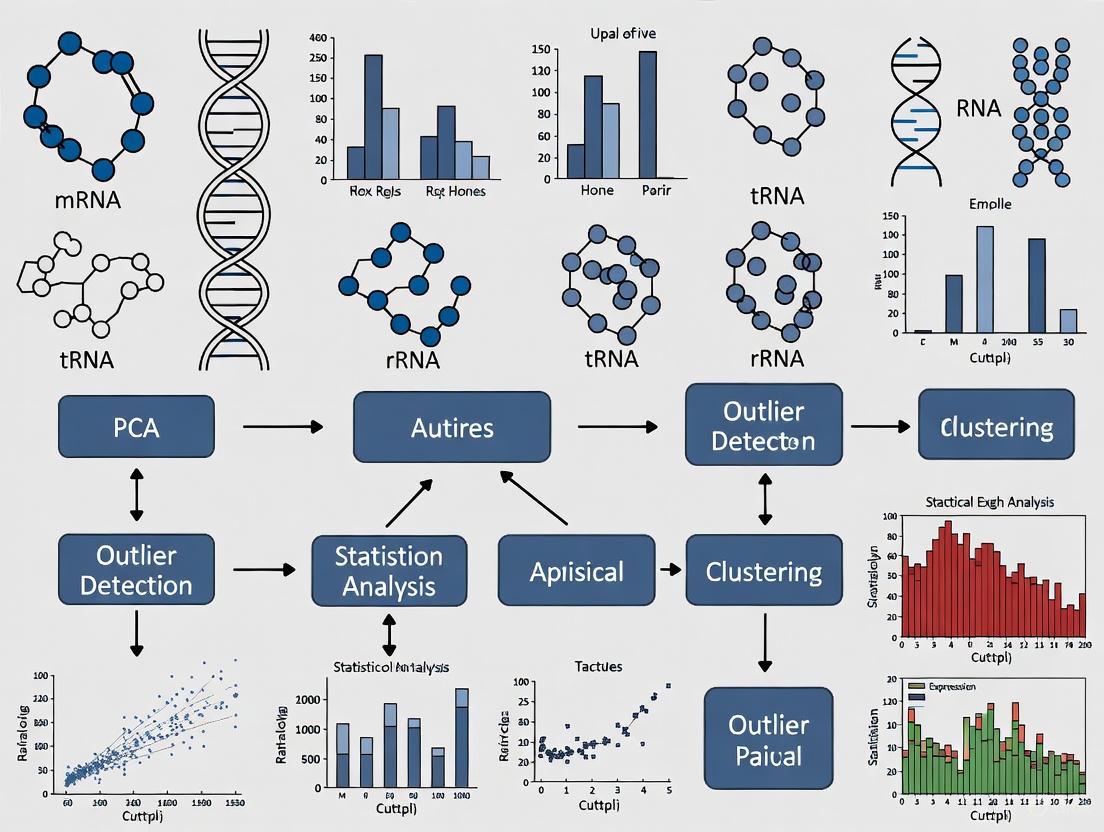
Visualization Workflow for Outlier Analysis
Effective identification and management of RNA-seq outliers requires a multifaceted approach combining robust statistical methods with biological reasoning. While methods like rPCA, OutSingle, and iLOO provide objective detection frameworks, researcher judgment remains essential for interpreting results within specific experimental contexts. Future directions in outlier management will likely involve improved integration of detection methods into standard analysis pipelines, development of more sophisticated classification algorithms distinguishing technical from biological outliers, and community standards for reporting outlier decisions in publications. By implementing these systematic approaches to outlier detection, researchers can significantly enhance the reliability and biological relevance of their RNA-seq findings.
In RNA sequencing (RNA-seq) data analysis, outliers—samples or observations that deviate markedly from others—present a complex challenge. They are traditionally viewed as technical artifacts to be removed to ensure data integrity. However, emerging evidence reveals that many outliers represent genuine biological variation with significant diagnostic value [11]. This technical support document examines both perspectives, providing frameworks for identifying, interpreting, and addressing outliers in research and diagnostic settings.
The fundamental challenge lies in distinguishing technical artifacts from biological signals. Technical outliers arise from multiple sources, including variations in RNA extraction, library preparation, sequencing depth, and instrumentation [1] [2]. Conversely, biological outliers may stem from genuine rare genetic variations, spontaneous transcriptional activation, or unique cellular responses [11]. Understanding this dual nature is crucial for making informed analytical decisions.
Detection Methodologies: Statistical Frameworks and Tools
Computational Tools for Outlier Detection
Several specialized algorithms have been developed to identify outliers in RNA-seq data. The table below summarizes key methods and their applications.
Table: RNA-Seq Outlier Detection Methods and Applications
| Method/Tool | Underlying Approach | Primary Application | Reference |
|---|---|---|---|
| OUTRIDER | Autoencoder with Negative Binomial distribution | Detecting aberrant gene expression in rare disease diagnostics | [12] |
| FRASER/FRASER2 | Splicing outlier detection | Identifying transcriptome-wide splicing defects, including minor spliceopathies | [13] [14] |
| OutSingle | Singular Value Decomposition (SVD) with Optimal Hard Threshold | Confounder-controlled outlier detection in gene expression data | [4] |
| Robust PCA (PcaGrid) | Robust principal component analysis | Accurate outlier sample detection in high-dimensional data with small sample sizes | [1] |
| iLOO (Iterative Leave-One-Out) | Probabilistic approach with leave-one-out design | Feature-level outlier detection in negative binomial distributed data | [15] |
Practical Workflow for Outlier Analysis
A robust outlier analysis strategy involves multiple steps, from quality control to biological interpretation. The following diagram illustrates a recommended workflow for handling outliers in RNA-seq data analysis:
Troubleshooting Guide: Frequently Asked Questions
Q1: How can I determine if an outlier sample results from technical error or genuine biological variation?
A1: Begin by examining quality control metrics. Technical outliers often exhibit:
- Low mapping percentages (<70% for human genome) [2]
- Abnormal GC content or gene length biases
- Irregular distribution of read coverage across transcripts
- Strand-specific biases in strand-preserving protocols
Biological outliers typically show:
- Normal QC metrics alongside specific aberrant expression patterns
- Co-regulation of functionally related genes [11]
- Reproducibility in independent experimental replicates
- Correlation with specific genetic variants or clinical phenotypes [13]
Q2: What is the minimum sample size required for reliable outlier detection?
A2: While some methods work with small sample sizes (n=2-6), detection power increases with larger cohorts. For rare biological event detection, studies with hundreds of samples dramatically improve identification of meaningful outliers [13]. Down-sampling analysis shows that even with only 8 individuals, approximately half of genes with extreme expression can be detected, with numbers increasing with sample size [11].
Q3: How do I handle outliers in single-cell RNA-seq data where dropout events are common?
A3: In scRNA-seq, embrace dropout patterns as potential biological signals rather than exclusively as noise:
- Use binarized expression (0/1 for undetected/detected) for co-occurrence clustering [16]
- Implement algorithms like M3Drop or scBFA that specifically model dropout characteristics
- Recognize that genes in the same pathway often exhibit similar dropout patterns across cell types
Q4: Can outlier removal improve differential expression analysis?
A4: Yes, when properly identified technical outliers are removed. One study demonstrated that removing outliers detected by robust PCA (PcaGrid) significantly improved the performance of differential gene detection and downstream functional analysis [1]. However, caution must be exercised—removing true biological outliers can lead to underestimation of natural biological variance and spurious conclusions.
Q5: How effective are NMD inhibitors in revealing splicing outliers?
A5: Cycloheximide (CHX) treatment significantly improves detection of transcripts subject to nonsense-mediated decay. Studies show CHX treatment increases expression of NMD-sensitive transcripts, enabling identification of splicing defects that would otherwise be masked [14]. Always include internal controls like SRSF2 transcripts to verify NMD inhibition efficacy.
Experimental Protocols
Protocol 1: Transcriptome-Wide Splicing Outlier Analysis
This protocol identifies individuals with rare spliceosome defects through intron retention patterns [13] [14]:
Sample Preparation:
- Collect whole blood in EDTA tubes
- Isolate peripheral blood mononuclear cells (PBMCs) using density gradient centrifugation
- Culture cells briefly with cycloheximide (CHX, 100μg/mL for 4-6 hours) to inhibit NMD
- Extract RNA using standard column-based methods
Library Preparation and Sequencing:
- Assess RNA integrity (RIN > 8 recommended)
- Perform ribosomal RNA depletion (do not use poly-A selection)
- Prepare strand-specific libraries with dUTP method
- Sequence on Illumina platform (minimum 30 million paired-end reads recommended)
Computational Analysis:
- Align reads to reference genome using STAR or HISAT2
- Run FRASER or FRASER2 to detect splicing outliers
- Focus on intron retention events in minor intron-containing genes (MIGs)
- Examine transcriptome-wide patterns rather than single-gene events
- Validate findings with Sanger sequencing of candidate variants
Protocol 2: Robust Outlier Sample Detection
This protocol identifies technical and biological outlier samples in a cohort study [1]:
Data Preprocessing:
- Perform standard RNA-seq quality control (FastQC, Trimmomatic)
- Align reads to reference genome/transcriptome
- Generate raw count matrix using featureCounts or HTSeq
Outlier Detection:
- Normalize counts using DESeq2 or edgeR median ratio method
- Apply robust PCA (PcaGrid function in rrcov R package)
- Calculate outlier distances for each sample
- Flag samples with distance > critical value (based on chi-square distribution)
- Compare with classical PCA to identify samples masked by non-robust methods
Validation:
- Correlate outlier status with clinical metadata and technical batch information
- Perform differential expression with and without outliers
- Use quantitative RT-PCR to validate key findings
The following diagram illustrates the experimental workflow for a comprehensive outlier analysis that balances both technical and biological considerations:
Research Reagent Solutions
Table: Essential Reagents and Resources for RNA-Seq Outlier Research
| Reagent/Resource | Function/Purpose | Application Example | Reference |
|---|---|---|---|
| Cycloheximide (CHX) | Nonsense-mediated decay (NMD) inhibition | Revealing aberrant transcripts degraded by NMD | [14] |
| RNase Inhibitors | Prevention of RNA degradation during isolation | Maintaining RNA integrity for accurate quantification | [2] |
| rRNA Depletion Kits | Removal of ribosomal RNA | Enhancing sequencing depth for mRNA | [13] [14] |
| Strand-Specific Library Prep Kits | Preservation of transcript orientation | Accurate identification of antisense transcripts | [2] |
| FRASER/FRASER2 Software | Splicing outlier detection | Identifying minor spliceopathies | [13] [17] |
| OUTRIDER Package | Aberrant expression detection | Diagnosing rare genetic disorders | [12] [14] |
| Robust PCA Algorithms | Outlier sample detection | Identifying technical artifacts in small sample studies | [1] |
Outliers in RNA-seq data present both challenges and opportunities. While technical artifacts must be identified and addressed to ensure data quality, biological outliers often contain valuable insights into rare genetic conditions, spontaneous transcriptional events, and novel regulatory mechanisms. By implementing robust detection methodologies, following standardized protocols, and maintaining a balanced perspective on the dual nature of outliers, researchers can maximize both the reliability and discovery potential of their RNA-seq analyses.
The field continues to evolve with new computational methods and experimental approaches that enhance our ability to distinguish biological signals from technical noise. Integrating these advances into standardized workflows will further unlock the diagnostic and research potential of transcriptomic outliers.
Troubleshooting Guides and FAQs for RNA-Seq Outlier Identification
Frequently Asked Questions (FAQs)
Q1: Why is outlier identification critical in RNA-Seq data analysis? Outliers in RNA-Seq data can significantly distort analytical results and lead to erroneous conclusions in downstream analyses, such as differential expression testing [18]. These extreme values may arise from technical artifacts, but recent research also identifies them as potential biological realities that should be investigated rather than automatically discarded [11]. Proper identification ensures the accuracy of transcript measurements and correct biological interpretations.
Q2: What is the fundamental difference between the IQR/Tukey's Fences and Z-score methods? The Interquartile Range (IQR) and Tukey's Fences method is a non-parametric approach based on data quartiles, making it robust to non-normal distributions and extreme values [19] [20]. In contrast, the Z-score method is parametric and assumes your data approximately follows a normal distribution, as it measures how many standard deviations a point is from the mean [21]. For RNA-Seq data, which often exhibits overdispersion and skewed distributions, Tukey's Fences is generally more reliable.
Q3: How do I choose the threshold (k-value) for Tukey's Fences?
The choice of k depends on how conservative you want to be:
- k = 1.5: Identifies "regular" outliers. This is a common default but can flag a high percentage of data points as outliers, especially in large samples [19] [22].
- k = 3.0: Identifies "far" or extreme outliers. This is more conservative and recommended for stringent outlier detection in RNA-Seq data to avoid removing biologically relevant but rare expression events [19] [11].
- k = 5.0: Used for very conservative identification of extreme over-expression in transcriptomic studies [11].
Q4: A sample in my RNA-Seq dataset was flagged as an outlier. Should I always remove it? Not necessarily. First, investigate the potential cause:
- Technical Error: Check for issues like low sequencing quality, adapter contamination, or high ribosomal RNA content using QC tools like RNA-QC-Chain or FastQC [18] [23]. If a technical error is confirmed, exclusion is justified.
- Biological Reality: Outlier expression may represent sporadic, genuine biological events [11]. If the outlier is reproducible and biological validation is possible, it might be worth further investigation instead of removal.
Comparison of Key Outlier Detection Methods
The following table summarizes the core components of the two primary outlier detection methods discussed.
| Feature | IQR & Tukey's Fences | Z-Score Method |
|---|---|---|
| Core Formula | IQR = Q3 - Q1Upper Fence = Q3 + k * IQRLower Fence = Q1 - k * IQR [24] [25] |
z = (x - μ) / σ [21] |
| Typical Threshold | k = 1.5 for regular outliersk = 3.0 for extreme outliers [19] |
z > 3 or z < -3 [21] |
| Distribution Assumption | Non-parametric; no assumption of normality [19] [20] | Parametric; assumes normal distribution [21] |
| Robustness to Extreme Values | High (uses quartiles, which are resistant to extremes) [26] | Low (mean and standard deviation are influenced by extremes) [19] |
| Primary Use Case in RNA-Seq | General-purpose outlier detection, especially for skewed data or data with potential outliers [11] | Can be used when data is known to be normally distributed, but less common for raw counts [21] |
Step-by-Step Experimental Protocols
Protocol 1: Identifying Outliers using Tukey's Fences in RNA-Seq Data
This protocol is ideal for gene expression values across samples.
- Prepare Data: Start with a normalized gene expression matrix (e.g., TPM, CPM). Work on a per-gene basis, analyzing the expression distribution of one gene across all samples [11].
- Calculate Quartiles:
- Compute Interquartile Range (IQR):
IQR = Q3 - Q1[24] [25]. - Establish Tukey's Fences:
- Lower Fence =
Q1 - k * IQR - Upper Fence =
Q3 + k * IQR - For a conservative approach in RNA-Seq, start with
k = 3[11].
- Lower Fence =
- Identify Outliers: Any sample where the gene's expression value falls below the Lower Fence or above the Upper Fence is considered an outlier for that gene [19] [20].
Protocol 2: Identifying Outliers using the Z-Score Method
Use this method with caution, primarily if the expression data is known to be normally distributed (e.g., after log-transformation).
- Prepare and Transform Data: Use a normalized and log-transformed expression matrix to better approximate a normal distribution.
- Calculate Mean and Standard Deviation:
- For a given gene, compute the mean (μ) expression across all samples.
- Compute the standard deviation (σ) of the expression values [21].
- Compute Z-Scores: For each sample's expression value (x) for the gene, calculate the Z-score:
z = (x - μ) / σ[21]. - Identify Outliers: Flag any sample with an absolute Z-score greater than your threshold (e.g.,
|z| > 3) as an outlier. A Z-score of 3 corresponds to a value more than 3 standard deviations from the mean, which is highly unlikely in a normal distribution [21].
Workflow and Decision Diagrams
The following diagram illustrates the logical relationship between the statistical concepts and the decision pathway for handling outliers in an RNA-Seq experiment.
The Scientist's Toolkit: Essential Research Reagents and Computational Solutions
| Item Name | Function / Purpose | Application Context |
|---|---|---|
| RNA-QC-Chain [18] | A comprehensive, all-in-one quality control pipeline for RNA-Seq data. It performs sequencing quality assessment, trims low-quality reads, filters ribosomal RNA, and identifies contamination. | Pre-processing of raw FASTQ files to ensure data quality before statistical analysis and outlier detection. |
| RSeQC [18] | Provides RNA-seq-specific quality control metrics based on alignment files, such as gene body coverage, read distribution, and strand specificity. | Post-alignment QC to identify biases that might lead to or explain outlier samples. |
| Normalized Expression Matrix (TPM/CPM) [11] | The starting material for outlier detection. Transcripts Per Million (TPM) or Counts Per Million (CPM) normalize for sequencing depth, allowing for sample comparison. | The fundamental data structure on which IQR or Z-score calculations are performed across samples for each gene. |
| Statistical Software (R/Python) | Provides the computational environment to calculate IQR, Tukey's Fences, Z-scores, and generate visualizations like boxplots and Q-Q plots. | The primary platform for implementing the statistical protocols outlined in this guide. |
| Tukey's Fences (k=3.0) [11] | A specific, conservative threshold for defining an "extreme outlier" in gene expression data, corresponding to a very low p-value under a normal assumption. | The recommended parameter for stringent outlier identification in RNA-Seq studies to avoid removing true biological signals. |
Frequently Asked Questions (FAQs)
FAQ 1: What is the evidence that outlier gene expression is a biological phenomenon and not just technical noise?
Recent research demonstrates that outlier gene expression patterns are a biological reality, reproducible across tissues and species. A 2025 study analyzed multiple large datasets, including outbred and inbred mice, human GTEx data, and different Drosophila species, finding comparable general patterns of outlier gene expression in all. Crucially, these outliers were fully reproducible in independent sequencing experiments, confirming they are not technical artifacts. The study also used a three-generation family analysis in mice to show that most extreme over-expression is not inherited but appears sporadically, suggesting it may be linked to "edge of chaos" effects in gene regulatory networks [11].
FAQ 2: My dataset has limited samples. Which outlier detection method is recommended for small sample sizes?
For datasets with a small number of samples, OutPyR is specifically designed for this scenario. It uses Bayesian inference to identify abnormal RNA-Seq gene expression counts, incorporating data-augmentation techniques to efficiently infer parameters of the underlying negative binomial process while assessing inference uncertainty [27]. This approach is particularly valuable when large sample sizes are not available.
FAQ 3: How can I control for confounders that might mask true biological outliers in my RNA-Seq data?
The OutSingle algorithm provides an effective solution for confounder control. It uses Singular Value Decomposition (SVD) with the recently discovered optimal hard threshold (OHT) method for noise detection. This approach offers a deterministic, computationally efficient way to control for confounders without the complexity of autoencoder-based methods [4]. For sample-level outlier detection, robust Principal Component Analysis (rPCA) methods, particularly PcaGrid, have demonstrated 100% sensitivity and specificity in detecting outlier samples in RNA-Seq data, even with small sample sizes [1].
FAQ 4: What statistical cutoff should I use to define an extreme outlier expression value?
While common practice often uses multiples of the interquartile range (IQR), research specifically focused on extreme outliers recommends a conservative threshold of Q3 + 5 × IQR for defining "over outliers" (OO) and Q1 - 5 × IQR for "under outliers" (UO). This corresponds to approximately 7.4 standard deviations above the mean in a normal distribution (P-value ≈ 1.4 × 10⁻¹³), providing a very stringent cutoff that satisfies multiple testing corrections [11].
Troubleshooting Guides
Issue: Difficulty Distinguishing Technical Artifacts from Biological Outliers
Problem: Suspected outliers in your data could be either technical errors or genuine biological signals.
Solution:
- Apply Reproducibility Testing: If resources allow, perform independent sequencing experiments on the same samples to verify if outlier patterns persist [11].
- Implement Robust PCA: Use rPCA methods (PcaGrid or PcaHubert) to objectively identify outlier samples rather than relying on subjective visual inspection of PCA biplots [1].
- Examine Co-regulation Patterns: Check if outlier genes form co-regulatory modules or known pathways, which suggests biological significance rather than random technical errors [11].
Issue: Outlier Detection Masked by Confounding Factors
Problem: True biological outliers are being hidden by technical batch effects or other confounding variables.
Solution:
- Apply OutSingle Algorithm: Implement this method which uses SVD with optimal hard thresholding to control for confounders while preserving true outlier signals [4].
- Utilize FRASER for Splicing Outliers: For aberrant splicing event detection, use FRASER which automatically controls for latent confounders while identifying statistically significant outlier splicing events [28].
- Leverage GTEx Data: Use large-scale reference datasets like GTEx to establish baseline covariation patterns specific to your tissue of interest [28].
Table 1: Prevalence of Extreme Outlier Genes Across Species and Tissues
| Species | Tissue | Sample Size | Genes with Extreme Outliers (≥1 per dataset) | Reference |
|---|---|---|---|---|
| Mouse (Outbred) | Multiple organs | 48 individuals | 3-10% of all genes (~350-1350 genes) | [11] |
| Human (GTEx) | Multiple tissues | 543 donors | Comparable patterns observed | [11] |
| Drosophila | Head & trunk | 27 individuals | Comparable patterns observed | [11] |
Table 2: Performance Comparison of Outlier Detection Methods
| Method | Approach | Strengths | Limitations | Use Case |
|---|---|---|---|---|
| OutSingle | Log-normal z-scores with SVD/OHT confounder control | Almost instantaneous computation; effective confounder control | Less performant on data with underexpressed outliers | Large datasets requiring fast processing [4] |
| rPCA (PcaGrid) | Robust principal component analysis | 100% sensitivity/specificity in tests; objective detection | Requires sufficient samples for PCA | Sample-level outlier detection [1] |
| OutPyR | Bayesian modeling of negative binomial distribution | Incorporates uncertainty assessment; works with small samples | Computationally demanding | Small datasets with limited samples [27] |
| FRASER | Beta-binomial distribution with autoencoder | Controls confounders; detects splicing outliers & intron retention | Complex implementation | Aberrant splicing detection [28] |
Experimental Protocols
Protocol 1: Identification of Extreme Expression Outliers Using IQR Method
Purpose: To conservatively identify extreme outlier expression values in RNA-Seq data.
Materials:
- Normalized expression matrix (TPM, CPM, or normalized counts)
- Statistical software (R, Python, or equivalent)
Procedure:
- Data Preparation: Use normalized transcript fragment count data without log-transformation. Avoid pre-filtering of potential outlier samples [11].
- Calculate Quartiles: For each gene across samples, compute the 1st quartile (Q1) and 3rd quartile (Q3), then determine the interquartile range (IQR = Q3 - Q1).
- Set Conservative Thresholds:
- For "over outliers" (OO): Q3 + 5 × IQR
- For "under outliers" (UO): Q1 - 5 × IQR
- Identify Outlier Genes: Flag any gene expression values exceeding these thresholds in any sample.
- Validation: Examine outlier genes for co-regulatory patterns or pathway enrichment to confirm biological significance.
Notes: This threshold corresponds to approximately 7.4 standard deviations above the mean in a normal distribution (P ≈ 1.4 × 10⁻¹³) [11].
Protocol 2: Confounder-Control in Outlier Detection Using OutSingle
Purpose: To detect outliers in RNA-Seq gene expression data while controlling for confounding effects.
Materials:
- RNA-Seq count matrix (genes × samples)
- OutSingle software (https://github.com/esalkovic/outsingle)
Procedure:
- Input Data Preparation: Format your data as a J × N count matrix where J is genes and N is samples.
- Log-Normal Transformation: Convert counts using log-normal transformation to calculate gene-specific z-scores.
- Apply Optimal Hard Threshold: Use Singular Value Decomposition (SVD) with the Optimal Hard Threshold (OHT) method to denoise the z-score matrix and control for confounders.
- Outlier Identification: Detect outliers from the confounder-corrected z-scores.
- Optional Outlier Injection: Use OutSingle's inverse procedure to inject artificial outliers masked by confounding effects for method validation.
Notes: This method is particularly effective for identifying outliers masked by confounders and is significantly faster than autoencoder-based approaches [4].
Method Selection Workflow
Research Reagent Solutions
Table 3: Essential Materials and Tools for RNA-Seq Outlier Research
| Reagent/Tool | Function | Example/Reference | Considerations |
|---|---|---|---|
| Reference RNA Materials | Benchmarking and quality control | Quartet Project reference materials [29] | Enables assessment of subtle differential expression |
| ERCC Spike-In Controls | Technical controls for quantification | External RNA Control Consortium [30] | Assess accuracy of absolute measurements |
| OutSingle Software | Outlier detection with confounder control | https://github.com/esalkovic/outsingle [4] | Fast, deterministic method |
| rPCA Methods (PcaGrid) | Sample-level outlier detection | rrcov R package [1] | Objective detection vs. visual PCA inspection |
| FRASER Algorithm | Aberrant splicing event detection | Beta-binomial model [28] | Detects intron retention and alternative splicing |
| GTEX Data Reference | Baseline covariation patterns | GTEx Project dataset [28] | Tissue-specific reference for confounder control |
Frequently Asked Questions
What defines an RNA-seq sample as an "outlier"? An outlier sample shows global gene expression or splicing patterns that are significantly different from other samples in the dataset, even when standard quality control metrics appear normal. These samples can dramatically influence analysis results—for example, a single outlier might generate over 100 differentially expressed genes that disappear when the sample is removed [6].
Which visualization methods best reveal outlier samples? Multidimensional scaling (MDS) plots and principal component analysis (PCA) plots are most commonly used. In these visualizations, outlier samples appear separated from the main cluster of other samples. Sample distances plots with dendrograms also help identify outliers by showing which samples have dissimilar expression patterns [31] [32].
My data has an outlier sample but its sequencing quality is good. Should I remove it? Yes, generally. If an sample is a clear visual outlier on MDS/PCA plots and is driving differential expression results that disappear upon its removal, it should likely be excluded from analysis. This remains true even if standard sequencing QC metrics are acceptable, as the outlier status may reflect underlying biological or technical issues not captured by standard QC [6].
What tools can formally identify outlier samples beyond visual inspection? Several specialized tools exist:
- FRASER/FRASER2: Detect splicing outliers, particularly useful for identifying rare diseases affecting spliceosome function [13] [33]
- OUTRIDER: Identifies expression outliers using a negative binomial distribution model [4] [14]
- OutSingle: Uses singular value decomposition for rapid outlier detection in gene expression data [4]
Can outlier samples actually be biologically meaningful? Yes. While often removed as technical artifacts, outliers can sometimes reveal true biological phenomena. Recent research shows that samples with excess intron retention outliers in minor intron-containing genes can indicate rare genetic disorders affecting the minor spliceosome, known as "minor spliceopathies" [13] [33].
How do I handle outliers in a diagnostic setting? In clinical RNA-seq analysis, outliers should be carefully investigated rather than automatically removed. Transcriptome-wide outlier patterns can increase diagnostic yield for rare diseases. Removing them might discard valuable diagnostic information, particularly when patterns suggest spliceosome dysfunction [13] [14].
Troubleshooting Guides
Problem: Single Sample Driving Differential Expression Results
Symptoms:
- Significant DEGs (100+) with all samples included
- Zero DEGs when one particular sample is removed
- Sample appears as visual outlier on MDS plot but has passing QC metrics
Investigation Steps:
- Confirm the outlier: Generate MDS and PCA plots to visually confirm the sample separates from others [6] [32]
- Check quality metrics: Verify mapping rates, read counts, and other standard metrics are comparable to other samples [31]
- Investigate biological causes: Use splicing outlier tools (FRASER) or expression outlier tools (OUTRIDER) to determine if the outlier pattern affects specific genes or pathways [13] [4]
- Examine experimental factors: Check if outlier corresponds to any known batch effects or processing differences
Resolution Paths:
- If technical artifact: Remove sample and proceed with analysis [6]
- If biological reality: Maintain sample but use robust statistical methods, or split analysis to investigate the outlier separately [13]
- If unclear provenance: Consider sample removal for conservative analysis, noting this decision in methods
Problem: Consistent Outlier Patterns Across Multiple Samples
Symptoms:
- Multiple samples cluster separately from main group
- Pattern correlates with known experimental factors (e.g., processing batch)
- Splicing or expression outliers affect specific functional groups of genes
Investigation Steps:
- Color PCA/MDS by experimental factors: Batch, processing date, sequencing lane, etc. [32]
- Test for batch effects: Use statistical methods to quantify variance explained by technical factors
- Analyze outlier gene patterns: Determine if outliers affect specific pathways (e.g., minor spliceosome genes) using FRASER or similar tools [13]
- Check for global splicing patterns: Examine whether outliers show excess intron retention in minor intron-containing genes, which might indicate spliceosome defects [33]
Resolution Paths:
- If batch effect: Include batch as covariate in analysis or use batch correction methods
- If biological subgroup: Analyze as separate group or include as factor in design matrix
- If spliceopathy pattern: Investigate further as potential rare disease diagnosis [13]
Experimental Protocols
Protocol 1: Comprehensive Outlier Detection in RNA-seq Data
Purpose: Systematically identify technical and biological outliers in RNA-seq datasets
Materials:
- Raw or normalized count matrix
- Sample metadata with experimental factors
- R or Python statistical environment
Procedure:
- Quality Control Assessment
- Calculate standard QC metrics: mapping rates, library sizes, gene detection counts [31]
- Check for samples with extreme values in any metric
Visual Outlier Detection
Statistical Outlier Detection
Differential Expression Sensitivity Analysis
- Perform DEG analysis with all samples
- Iteratively remove suspected outliers and re-run DEG analysis
- Note samples whose removal substantially changes results [6]
Biological Interpretation
- Identify genes driving outlier status
- Check if outlier genes belong to specific pathways (e.g., minor spliceosome)
- Correlate with available clinical or phenotypic data
Expected Results: Identification of samples that are technical outliers requiring removal, or biological outliers warranting further investigation.
Protocol 2: Splicing Outlier Analysis for Rare Disease Diagnosis
Purpose: Identify individuals with rare spliceosome disorders using transcriptome-wide splicing outlier patterns
Materials:
- RNA-seq data from whole blood or PBMCs
- FRASER/FRASER2 software
- Reference annotation of minor intron-containing genes
Procedure:
- Data Preparation
- Process RNA-seq data through standard alignment pipeline
- Generate splice junction counts for all samples
Splicing Outlier Detection
- Run FRASER on cohort to detect significant splicing outliers [13]
- Calculate outlier counts per sample for different splicing types (intron retention, exon skipping, etc.)
Minor Intron Analysis
- Extract minor intron-containing genes from reference [33]
- Calculate proportion of intron retention outliers in MIGs versus major introns
- Identify samples with significant enrichment of MIG intron retention (p < 0.05)
Variant Correlation
- For samples with MIG intron retention enrichment, examine minor spliceosome genes (RNU4ATAC, RNU6ATAC) for rare variants [33]
- Validate suspected variants through Sanger sequencing
Clinical Interpretation
- Correlate molecular findings with clinical presentation
- Compare to known minor spliceopathy phenotypes (e.g., microcephalic osteodysplastic primordial dwarfism) [13]
Expected Results: Identification of individuals with potential minor spliceopathies characterized by excess intron retention in minor intron-containing genes.
Comparative Methodologies
Table 1: RNA-seq Outlier Detection Methods
| Method | Primary Application | Statistical Approach | Strengths | Limitations |
|---|---|---|---|---|
| Visual Inspection (MDS/PCA) | Initial outlier screening | Dimensionality reduction | Fast, intuitive, requires no specialized tools | Subjective, may miss subtle outliers |
| FRASER/FRASER2 | Splicing outlier detection | Count-based modeling of splicing patterns | Specifically designed for splice defects, good for rare diseases | Computationally intensive, requires large sample sizes |
| OUTRIDER | Expression outlier detection | Negative binomial distribution with autoencoder | Specifically designed for outlier detection, handles confounders | Complex implementation, long run times |
| OutSingle | Expression outlier detection | Log-normal with SVD decomposition | Very fast execution, good performance | Newer method, less extensively validated |
| Z-score approaches | Simple outlier screening | Normal distribution assumption | Very simple to implement | Poor control of confounders, high false positive rate |
Table 2: Outlier Patterns in Rare Disease Contexts
| Outlier Pattern | Potential Biological Meaning | Associated Tools | Clinical Relevance |
|---|---|---|---|
| Excess intron retention in minor introns | Minor spliceosome dysfunction | FRASER, FRASER2 | RNU4atac-opathy disorders (MOPD1, Roifman syndrome) |
| Global splicing outliers | Major spliceosome defects | FRASER, OUTRIDER | Various Mendelian spliceosomopathies |
| Expression outliers in specific pathways | Haploinsufficiency or regulatory defects | OUTRIDER, OutSingle | Tissue-specific genetic disorders |
| Monoallelic expression outliers | Regulatory variants | OUTRIDER, custom approaches | Dominant disorders with cis-regulatory effects |
Research Reagent Solutions
Table 3: Essential Materials for RNA-seq Outlier Studies
| Reagent/Resource | Function | Application Notes |
|---|---|---|
| FRASER/FRASER2 software | Splicing outlier detection | Essential for identifying spliceopathies; requires RNA-seq data from multiple samples |
| OUTRIDER package | Expression outlier detection | Uses autoencoder to control for confounders; good for rare disease cohorts |
| OutSingle tool | Rapid expression outlier detection | Fast alternative to OUTRIDER; uses SVD for confounder control |
| PBMC isolation kit | Source of clinical RNA | Minimally invasive tissue source; expresses ~80% of intellectual disability panel genes |
| Cycloheximide | NMD inhibition | Allows detection of nonsense-mediated decay substrates; use during cell culture |
| Reference annotations | Minor intron identification | Essential for identifying minor intron-containing genes (~0.5% of all introns) |
| Salmon or similar | Transcript quantification | Provides count data for downstream outlier analysis |
Workflow Diagrams
Frequently Asked Questions
Q1: My RNA-seq data shows samples with extreme expression levels for hundreds of genes. Are these technical artifacts I should discard? Historically, such samples were often excluded as technical noise. However, recent research confirms that extreme outlier expression is a biological reality observed across tissues and species, including mice, humans, and Drosophila [11] [34]. These outliers are not purely technical artifacts and can provide valuable biological insights. Before discarding, you should:
- Verify Reproducibility: Check if the outlier pattern is reproducible in independent sequencing runs [11].
- Associate with Biology: Investigate if outlier genes are part of co-regulatory modules or known pathways, such as those involving prolactin and growth hormone [11].
- Check Heritability: Note that most extreme over-expression appears sporadically and is not inherited, which can help distinguish it from effects caused by genetic polymorphisms [11] [34].
Q2: My diagnostic pipeline for a rare disease keeps overlooking causal variants. What is a common type of pathogenic variant I might be missing? Your pipeline may be overlooking splice-disruptive variants. It is estimated that 15–30% of all disease-causing mutations may affect splicing [35] [36]. Standard clinical workflows often focus on variants in protein-coding regions and canonical splice sites, but many pathogenic variants lie in non-coding regions and can disrupt splicing regulation [35]. These include:
- Deep-intronic variants that create cryptic splice sites.
- Synonymous variants within exons that disrupt splicing enhancers or silencers.
- Variants affecting branch points or other regulatory elements [35] [36].
Q3: How can I distinguish a true, biologically relevant splicing outlier from background technical noise? Using dedicated statistical methods for splicing outlier detection is crucial. Tools like FRASER and FRASER2 are designed to identify aberrant splicing events, such as intron retention, from RNA-seq data [13]. A true biological signal often manifests as a coordinated pattern across multiple genes. For instance, an excess of intron retention outliers specifically in minor intron-containing genes (MIGs) can signal a defect in the minor spliceosome, potentially caused by variants in genes like RNU4ATAC [13]. Looking for these transcriptome-wide patterns provides a more robust signature than focusing on single-gene events.
Q4: What is monoallelic expression (MAE), and how can I detect it in my single-cell RNA-seq data? Monoallelic expression (MAE) occurs when a gene is expressed from only one of the two parental alleles [37] [38]. It can be constitutive, as seen in genomically imprinted genes, or random (rMAE), where the choice of allele is stochastic and can vary from cell to cell [37]. To detect it in scRNA-seq data, you need:
- Genotype Information: Whole-genome sequencing data or dense genotyping from the same individual to identify heterozygous single-nucleotide variants (SNVs) [37] [38].
- Allele-Resolved scRNA-seq Data: scRNA-seq data where reads covering these SNVs can be assigned to the maternal or paternal allele.
- Statistical Testing: A statistical framework (e.g., a chi-square test) to identify SNVs with significant allelic expression bias in a population of cells. An allele with a UMI fraction below 5% is often defined as exhibiting MAE [38].
Troubleshooting Guides
Guide 1: Investigating Extreme Expression Outliers
Problem: One or more samples in a dataset show extreme over- or under-expression for a large number of genes.
Investigation Workflow:
- Confirm it's not technical: Check standard QC metrics (sequencing depth, RNA quality, adapter contamination) to rule out obvious technical failures.
- Quantify the outliers: Use a conservative statistical cutoff to define outliers. A robust method is Tukey’s fences, defining extreme over-expression outliers (OO) as values above Q3 + 5 × IQR, which corresponds to a very low P-value in a normal distribution [11].
- Analyze the pattern:
Diagram 1: Workflow for investigating extreme expression outliers.
Guide 2: Diagnosing Splicing Defects in Rare Diseases
Problem: A patient with a suspected rare genetic disease has undergone genomic sequencing, but no definitive causative variant has been found.
Investigation Workflow:
- Re-analyze with splicing-aware tools: Re-process genomic data using computational tools designed to predict splice-altering variants, even those in deep intronic or synonymous regions [35] [36].
- Incorporate RNA-seq: If patient tissue (e.g., whole blood) is available, perform RNA-seq.
- Run splicing outlier detection: Use algorithms like FRASER/FRASER2 on the RNA-seq data to identify aberrant splicing events genome-wide [13].
- Look for pathway signatures: Don't just look at single genes. Search for patterns, such as an enrichment of intron retention in minor intron-containing genes, which points to a specific spliceopathy [13].
- Experimental validation: Use RT-PCR or other molecular assays to confirm the predicted splicing defect [36].
Guide 3: Detecting Monoallelic Expression in Single-Cell Data
Problem: Characterizing allele-specific expression patterns in a heterogeneous cell population.
Investigation Workflow:
- Data prerequisites: Obtain scRNA-seq data and matched genotyping (WGS or SNP array) for the same individual to identify informative heterozygous SNVs [37] [38].
- Data processing: Map scRNA-seq reads and assign them to cells and alleles using the genotype information. Filter low-quality SNVs and potential somatic mutations [38].
- Cell type identification: Classify cells into types using standard scRNA-seq clustering methods, as MAE can be cell-type specific [37].
- Statistical testing for MAE:
Diagram 2: Workflow for detecting monoallelic expression in single-cell data.
Quantitative Data Reference
The table below summarizes key quantitative findings from recent research on expression and splicing outliers.
| Outlier Category | Quantitative Finding | Context / Method | Source |
|---|---|---|---|
| Extreme Expression | ~3–10% of genes show extreme outlier expression in at least one individual. | Analysis of mouse transcriptome data (48 individuals) using a threshold of Q3 + 5 × IQR. | [11] |
| Extreme Expression | About 72% of genes in a dataset conform to a normal expression distribution; the remainder are potential outliers. | Shapiro-Wilk normality tests on RNA-seq data from multiple species. | [11] |
| Splicing Defects | 15–30% of disease-causing mutations are estimated to affect pre-mRNA splicing. | Review of splicing defects in rare diseases. | [35] [36] |
| Minor Splicing Defects | Identified 5 individuals with excess intron retention in minor intron-containing genes (MIGs) from a cohort of 385. | Splicing outlier analysis with FRASER/FRASER2 on rare disease cohort (GREGoR/UDN). | [13] |
The Scientist's Toolkit: Research Reagent Solutions
| Reagent / Tool | Function / Explanation | |
|---|---|---|
| FRASER / FRASER2 | Statistical methods to detect aberrant splicing patterns (like intron retention) from RNA-seq data in an unbiased, transcriptome-wide manner. | [13] |
| ERCC Spike-In Mix | A set of synthetic RNA controls used to standardize RNA quantification, determine the sensitivity, dynamic range, and technical variation of an RNA-seq experiment. | [39] |
| Unique Molecular Identifiers (UMIs) | Short random barcodes that label individual mRNA molecules before PCR amplification, allowing for accurate digital counting and correction of PCR bias and errors. | [39] |
| scRNA-seq with Genotyping | A combined approach where single-cell RNA-sequencing is performed alongside whole-genome sequencing of the same individual. This is essential for identifying heterozygous SNVs used to trace monoallelic expression. | [37] [38] |
| Poly-A Selection & rRNA Depletion | Two common methods for library preparation in RNA-seq. Poly-A selection enriches for mRNA in eukaryotes, while rRNA depletion is needed for studying non-polyadenylated RNAs (e.g., lncRNAs) or bacterial transcripts. | [39] |
Practical Implementation: Tools and Techniques for Effective Outlier Detection
What are FRASER and FRASER 2.0, and why are they important for RNA-seq analysis in rare disease research?
FRASER (Find RAre Splicing Events in RNA-seq) is a computational algorithm specifically designed to detect aberrant splicing events from RNA sequencing data. It was developed to address the limitation that approximately 15-30% of variants causing inherited diseases affect splicing, many of which are missed by standard prediction tools that rely on genome sequence alone [28]. The method provides a count-based statistical test for aberrant splicing detection while automatically controlling for latent confounders, which are widespread in RNA-seq data and can substantially affect detection sensitivity [28]. Unlike earlier methods, FRASER captures not only alternative splicing but also intron retention events, which typically doubles the number of detected aberrant events [28].
FRASER 2.0 represents a significant evolution of the original algorithm, introducing a more robust intron-excision metric called the intron Jaccard index that combines alternative donor, alternative acceptor, and intron retention signals into a single value [40]. This improvement came from the observation that FRASER's three original splice metrics were partially redundant and sensitive to sequencing depth [40] [41]. Through optimization of model parameters and filter cutoffs using candidate rare-splice-disrupting variants as independent evidence, FRASER 2.0 calls typically 10 times fewer splicing outliers while increasing the proportion of candidate rare-splice-disrupting variants by 10-fold [40]. This substantial reduction in outlier calls with minimal loss of sensitivity makes FRASER 2.0 particularly valuable for rare disease diagnostics, where reducing false positives is crucial for efficient diagnosis.
Key Methodological Components and Workflow
How do FRASER and FRASER 2.0 technically detect splicing outliers?
Both FRASER and FRASER 2.0 employ a sophisticated computational workflow that transforms raw RNA-seq data into statistically robust outlier calls. The core process involves multiple stages of data processing, normalization, and statistical testing.
Core Splicing Metrics
The original FRASER algorithm computes three primary metrics from RNA-seq data [28]:
- ψ5 metric: Quantifies alternative acceptor usage, defined as the fraction of split reads from an intron of interest over all split reads sharing the same donor.
- ψ3 metric: Quantifies alternative donor usage, analogously defined for the acceptor.
- θ metric: Represents splicing efficiency, defined as the fraction of split reads among split and unsplit reads overlapping a given donor or acceptor site.
FRASER 2.0 introduces a unified metric called the intron Jaccard index (J) that combines these signals [40]. For a given sample i and intron j, it is calculated as:
[ J{ij} = \frac{|D{ij} \cap A{ij}|}{|D{ij} \cup A{ij}|} = \frac{s{ij}}{\sum{d \in Lj} s{id} + \sum{a \in Rj} s{ia} + \sum{t \in {dj,aj}} u{it} - s_{ij}} ]
Where (s{ij}) denotes the count of split reads mapping to intron j, (dj) is the donor site, (aj) is the acceptor site, (Lj) is the set of introns using (dj), (Rj) is the set of introns using (aj), and (u{it}) denotes the count of non-split reads spanning the exon-intron boundary at a splice site t [40].
Denoising Autoencoder for Controlling Confounders
A key innovation in FRASER is the use of a denoising autoencoder to control for technical and biological confounders [28] [40]. Strong covariations in splicing metrics have been observed across RNA-seq datasets, arising from factors such as sex, population structure, batch effects, or variable RNA integrity [28]. The autoencoder models these covariations by fitting a low-dimensional latent space for each tissue separately using principal component analysis (PCA) on logit-transformed splicing metrics [28]. The optimal dimension for this latent space is determined by maximizing the area under the precision-recall curve when calling artificially injected aberrant values [28].
Statistical Testing and Outlier Calling
FRASER uses a beta-binomial distribution to model read counts and identify statistically significant outlier data points [28] [42]. After controlling for confounders via the autoencoder, the method calculates p-values representing the probability that an observed splicing metric deviates significantly from its expected value. These p-values are then corrected for multiple testing using false discovery rate (FDR) control, with default FDR < 0.1 and |Δψ| ≥ 0.3 for significance calling [40].
The following workflow diagram illustrates the key steps in FRASER's analysis process:
Comparative Analysis: FRASER vs. FRASER 2.0
What are the key differences between FRASER and FRASER 2.0, and how do they impact performance?
The evolution from FRASER to FRASER 2.0 brought significant improvements in precision and robustness. The table below summarizes the key methodological and performance differences between the two versions:
| Feature | FRASER (Original) | FRASER 2.0 |
|---|---|---|
| Core Metrics | Three partially redundant metrics: ψ5, ψ3, θ [28] | Single unified metric: Intron Jaccard Index [40] |
| Sensitivity to Sequencing Depth | Significant sensitivity observed [40] | Substantially reduced effect of sequencing depth [40] |
| Outlier Call Rate | Higher number of calls per sample [40] | 10x fewer splicing outliers on average [40] |
| Variant Enrichment | Baseline performance | 10x increase in proportion of candidate rare-splice-disrupting variants [40] |
| Intron Retention Detection | Captured through θ metric [28] | Integrated into Jaccard Index alongside other event types [40] |
| Multiple Testing Burden | Higher due to transcriptome-wide approach [40] | Reduced burden; option to test specific gene subsets [40] |
The performance improvements in FRASER 2.0 were validated on large datasets including 16,213 GTEx samples and 303 rare-disease samples, confirming both the reduction in outlier calls and maintenance of high sensitivity [40]. In practical diagnostic applications, FRASER 2.0 recovered 22 out of 26 previously identified pathogenic splicing cases with default cutoffs, and 24 when multiple-testing correction was limited to OMIM genes containing rare variants [40].
Experimental Protocols and Implementation
Standard Analysis Workflow
What are the key steps for implementing FRASER/FRASER 2.0 in a research pipeline?
The typical workflow for implementing FRASER or FRASER 2.0 involves both computational and analytical steps:
Data Preparation: Process raw RNA-seq data through alignment to generate BAM files. The DROP pipeline (v.1.1.3) is commonly used for count quantification and FraserDataSet creation [40].
Read Counting: Extract split reads and non-split reads using the FRASER package. Split reads are those whose ends align to two separated genomic locations of the same chromosome strand, providing evidence of splicing events [28]. Non-split reads spanning exon-intron boundaries are used for intron retention detection [28].
Quality Control and Filtering: Apply standard filters such as RNA integrity number (RIN) > 5.7, removal of tissues with <100 samples (for large studies), and intron-level filtering (95% of samples with n ≥ 1 read and at least one sample with an intron count ≥20) [40].
Model Fitting: Execute the FRASER algorithm with default parameters (FDR < 0.1, |Δψ| ≥ 0.3, minimal intron coverage ≥ 5 reads) or customized settings based on research goals [40].
Result Interpretation: Analyze outlier calls in the context of known rare variants, gene annotations, and potential clinical relevance.
Troubleshooting Common Experimental Issues
What challenges might researchers encounter when using FRASER, and how can they address them?
| Issue | Potential Causes | Recommended Solutions | ||
|---|---|---|---|---|
| Excessive outlier calls | Inadequate control for confounders; overly lenient thresholds [28] | Use FRASER 2.0; apply stricter filters (e.g., | Δψ | ≥ 0.3); limit testing to OMIM genes with rare variants [40] |
| Low concordance between replicates | Technical batch effects; poor RNA quality [28] | Check RNA integrity (RIN > 5.7); ensure consistent processing; include batch in autoencoder [40] | ||
| Missed validated splicing events | Overly stringent filtering; low sequencing depth [40] | Adjust count thresholds; increase sequencing depth; use FRASER 2.0 for better sensitivity [40] | ||
| Computational performance issues | Large sample sizes; whole transcriptome analysis [40] | Use gene-specific testing mode; increase computational resources; leverage BiocParallel for parallelization [40] [42] | ||
| Inconsistent results across metrics | Partial redundancy between ψ5, ψ3, and θ [40] | Implement FRASER 2.0 with unified Jaccard Index; prioritize events significant across multiple metrics [40] |
What computational tools and resources are essential for implementing FRASER in a research environment?
The table below outlines key resources in the FRASER ecosystem:
| Resource | Function | Implementation Details |
|---|---|---|
| FRASER R Package | Core analysis functionality [42] | Available through Bioconductor; supports aberrant(), calculatePvalues(), results() for key operations [42] |
| DROP Pipeline | Automated RNA-seq quantification and outlier detection [40] | Integrates FRASER with other outlier detection methods; processes BAM to results [40] |
| GTEx Dataset | Reference dataset for expected splicing patterns [28] | 7,842 RNA-seq samples from 48 tissues of 543 donors; provides baseline splicing distribution [28] |
| BiocParallel | Parallel computing framework [42] | Accelerates computation for large datasets; integrated in FRASER package [42] |
| GENCODE Annotation | Reference transcriptome [28] | Release 28 used in original FRASER publication; essential for annotation [28] |
FAQs on FRASER Applications and Limitations
What are the most common questions researchers have about implementing and interpreting FRASER results?
Q1: What types of splicing events can FRASER detect that other tools might miss? FRASER is particularly effective at detecting intron retention events, which are often missed by other splicing detection algorithms [28]. The original FRASER implementation typically doubled the number of detected aberrant events by capturing these retention events [28]. Additionally, FRASER can identify aberrant splicing from novel splice sites detected de novo from the RNA-seq data, not limited to previously annotated sites [28].
Q2: How does FRASER handle different tissue types in large cohort studies? FRASER is designed to model tissue-specific splicing patterns by fitting separate models for each tissue type [28]. In the GTEx analysis, FRASER created tissue-specific splice site maps containing on average 137,058 donor sites and 136,743 acceptor sites per tissue, with distinct covariation structures observed for each tissue [28]. This tissue-specific modeling is crucial as splicing regulation varies significantly across tissues.
Q3: What evidence validates FRASER's performance in diagnostic settings? Multiple studies have validated FRASER's diagnostic utility. In one analysis of rare disease samples, FRASER 2.0 recovered 22 out of 26 previously identified pathogenic splicing cases with default cutoffs [40]. Another study applying FRASER to 385 individuals from rare disease cohorts successfully identified five individuals with excess intron retention outliers in minor intron-containing genes, all of whom harbored rare, bi-allelic variants in minor spliceosome snRNAs [13].
Q4: How does FRASER address the multiple testing problem in transcriptome-wide analyses? FRASER employs beta-binomial testing with false discovery rate (FDR) correction, which reduces the number of calls by two orders of magnitude compared to commonly applied z-score cutoffs [28]. FRASER 2.0 further addresses this by offering an option to select specific genes for testing in each sample instead of a transcriptome-wide approach, which is particularly useful when prior information such as candidate variants is available [40].
Q5: What are the key considerations for sample size when using FRASER? While FRASER can work with smaller sample sizes, its denoising autoencoder benefits from larger cohorts. The fitted encoding dimension for the latent space grows approximately linearly with sample size, resulting in larger encoding dimensions in tissues with more samples [28]. For tissues with limited samples, researchers should consider leveraging cross-tissue resources or adjusting model parameters accordingly.
The following diagram illustrates the relationship between key splicing concepts and FRASER's detection approach:
FRASER and its enhanced version FRASER 2.0 represent specialized algorithms that significantly advance the detection of splicing outliers in RNA-seq data. Through their denoising autoencoder approach, beta-binomial statistical testing, and optimized metrics—particularly the unified intron Jaccard index in FRASER 2.0—these tools address critical challenges in rare disease diagnostics and splicing research. The evolution from FRASER to FRASER 2.0 demonstrates how methodological refinements can dramatically reduce false positive rates while maintaining sensitivity, making these algorithms invaluable for researchers and clinicians seeking to identify pathogenic splicing events in rare disease patients. As RNA-seq continues to play an expanding role in diagnostic settings, FRASER's ability to systematically detect aberrant splicing events positions it as an essential component in the modern genomic analysis toolkit.
Frequently Asked Questions
What is the primary function of the OUTRIDER tool? OUTRIDER (Outlier in RNA-Seq Finder) is a statistical algorithm designed to identify aberrantly expressed genes in RNA sequencing data. It uses an autoencoder to model read-count expectations based on gene covariation and identifies outliers as read counts that significantly deviate from a negative binomial distribution. It is particularly useful for rare disease diagnostic platforms [43].
When should I use a comparative analysis framework like CARE instead of OUTRIDER? The CARE (Comparative Analysis of RNA Expression) framework is particularly beneficial when analyzing ultra-rare cancers or diseases where no actionable mutations are found through DNA profiling alone. It identifies targetable overexpression genes and pathways by comparing a patient's tumor RNA-Seq profile to large compendiums of tumor data (e.g., over 11,000 samples). OUTRIDER is generally used for identifying aberrant expression within a dataset, while CARE is for placing a single sample in a broad disease context to nominate treatments [44].
My analysis has identified a list of outlier genes. What is the critical next step before concluding they are biologically relevant? Validation is a crucial next step. The golden standard is to validate findings with wet lab experiments. If that is not possible, you should use multiple data types and sources. For example, you can validate RNA-seq outliers with protein-level data (e.g., Western blot) or use publicly available datasets to see if the same conclusions are supported. Over-interpreting results without considering biological relevance is a common pitfall [45] [9].
What is a major statistical pitfall when performing differential expression analysis on single-cell RNA-seq data? A common mistake is grouping all cells from each condition together and performing differential gene expression tests at the individual cell level. The cells from each sample are not independent, and using a large number of cells can lead to artificially small p-values. The recommended best practice is to use a pseudo-bulk approach instead [45].
How does the choice of comparator cohort impact outlier detection in gene expression? The utility of RNA-Seq for identifying therapeutic targets is highly dependent on the comparator cohorts. Using large, uniformly processed datasets from multiple institutions and studies allows for the identification of molecularly similar tumors that may not be expected based on tumor histology alone. The impact of cohort selection on outlier detection is significant, and personalized comparator cohorts improve the identification of relevant overexpression outliers [44].
Troubleshooting Guides
Issue 1: Non-uniform p-value distribution in outlier detection
- Problem: When analyzing RNA-seq data to infer differential expression or outliers, the p-value distribution for null-hypothesis data is not uniform, leading to inaccurate estimates of False Discovery Rates (FDRs). This can cause a loss of power to detect true differential expression [46].
- Solution:
- Tool Selection: Use tools that accurately control for FDR. Studies have shown that the QLSpline implementation of QuasiSeq performs well in achieving a low and accurately estimated FDR when there are at least four biological replicates per condition. edgeR and DESeq2 are also among the next best-performing packages [46].
- Algorithmic Adjustment: For two-class datasets with a sufficient number of biological replicates (approximately 6 or more), an extension called Polyfit can be used with edgeR or DESeq. Polyfit adapts the Storey-Tibshirani procedure to address the problem of a non-uniform null p-value distribution [46].
Issue 2: Low diagnostic yield in RNA-driven analysis of rare diseases
- Problem: When using blood RNA-seq for rare disease diagnosis in cases where no candidate variants were found from prior exome/genome sequencing (ES/GS), the diagnostic uplift from a purely RNA-driven approach can be modest (e.g., 2.7%) [47].
- Solution:
- Strategy Change: Adopt an RNA-complementary approach instead of an RNA-driven one. Use RNA-seq to refine findings from DNA-sequencing, particularly for interpreting Variants of Uncertain Significance (VUS). This strategy has been shown to provide a much higher diagnostic uplift (60% in cases with candidate splicing VUS) [47].
- Pipeline Application: Employ a standardized pipeline like DROP for the detection of aberrant expression (AE) and aberrant splicing (AS) outliers. In the "RNA-complementary" approach, the OUTRIDER module of the DROP pipeline is often used specifically for cases where no aberrant splicing is detected, as abnormal transcripts may be degraded and thus mask splicing outliers [47].
Issue 3: Over-interpretation of data visualization
- Problem: Incorrectly interpreting the distance between points on a UMAP plot as a measure of biological similarity or difference [45].
- Solution:
- Remember that UMAP is a non-linear dimension reduction technique. The algorithm prioritizes the preservation of local structure over global distances. Therefore, the distance between clusters should not be over-interpreted. UMAP is excellent for visualization but should not be used for quantitative conclusions about relationships between distant cell clusters [45].
Experimental Protocols & Data
Table 1: Key RNA-seq Outlier Detection Tools and Their Applications
| Tool / Framework Name | Primary Function | Statistical Foundation | Key Application Context | Reference |
|---|---|---|---|---|
| OUTRIDER | Detects aberrantly expressed genes within a dataset | Autoencoder for covariation control, Negative Binomial distribution for outlier calling | Rare disease diagnostics; identifying aberrant expression in a cohort | [43] |
| CARE Framework | Identifies targetable overexpression by comparing a sample to large tumor compendiums | Z-score based outlier detection against personalized comparator cohorts | Precision oncology for rare pediatric and adult cancers; treatment nomination | [44] |
| DROP Pipeline | Detects Aberrant Expression (AE) and Aberrant Splicing (AS) | Multiple; incorporates OUTRIDER for AE analysis | Rare disease diagnostics, particularly following exome/genome sequencing | [47] |
Table 2: Performance Comparison of Differential Expression Packages
This table summarizes findings from a comparative analysis of several R packages for differential expression analysis, based on their ability to accurately estimate the False Discovery Rate (FDR) [46].
| Software Package | Model / Foundation | Recommended Minimum Replicates | Performance Note |
|---|---|---|---|
| QLSpline (QuasiSeq) | Quasi-likelihood with information sharing across genes | 4 | Achieves a low FDR that is accurately estimated, but has a slow run time. |
| edgeR | Negative Binomial model | Not specified | Next best performing package after QLSpline. |
| DESeq2 | Negative Binomial model with shrinkage estimation | Not specified | Next best performing package after QLSpline. |
| Polyfit (with DESeq) | Negative Binomial model with adapted FDR procedure | ~6 | Improves DESeq performance with sufficient replicates, making it comparable to edgeR/DESeq2. |
OUTRIDER Algorithm Workflow
CARE Framework Workflow
The Scientist's Toolkit: Research Reagent Solutions
| Item | Function in Experiment |
|---|---|
| PAXgene Blood RNA Tube | Stabilizes RNA in whole blood samples immediately upon drawing, ensuring an accurate representation of the transcriptome for rare disease studies [47]. |
| NEBNext Globin & rRNA Depletion Kit | Removes abundant globin and ribosomal RNA from blood-derived RNA samples, greatly improving the sequencing coverage of informative mRNAs [47]. |
| STAR Aligner | A robust and accurate tool for mapping RNA-seq reads to a reference genome, a critical step for downstream expression and splicing analysis [47]. |
| DROP Pipeline | An integrated computational pipeline for the detection of aberrant expression and aberrant splicing outliers in rare disease diagnostics [47]. |
| SpliceAI | An in-silico tool that predicts the impact of DNA sequence variants on mRNA splicing, which can be compared with empirical RNA-seq data for VUS interpretation [47]. |
The CARE Framework (Comprehensive, Adaptable, Research-Enabling) provides a structured approach for integrating advanced bioinformatics research, specifically RNA-seq outlier analysis, into clinical pediatric oncology practice. This framework bridges the critical gap between computational research findings and their practical application in clinical settings, ensuring that insights from transcriptomic data directly inform and improve patient care strategies. The framework's implementation is particularly vital in pediatric oncology, where treatment decisions must balance aggressive intervention with the long-term developmental and health outcomes of young patients.
In the context of RNA-seq outlier identification, the CARE Framework establishes standardized protocols for clinical laboratories to process, analyze, and interpret complex transcriptomic data. This enables healthcare teams to identify biologically significant expression patterns that may inform diagnosis, prognosis, or treatment selection. By providing clear guidelines for technical troubleshooting and quality control, the framework ensures the reliability and reproducibility of RNA-seq data, which is essential for making informed clinical decisions based on transcriptional outliers.
Technical Support Center: Troubleshooting RNA-seq Outlier Analysis
Frequently Asked Questions (FAQs)
Q1: What constitutes an "extreme outlier" in RNA-seq data, and how is this determined statistically? A1: In RNA-seq analysis, extreme outliers are data points representing gene expression levels that fall significantly outside the expected distribution across samples. Statistically, these are identified using Tukey's fences method, where outliers are defined as expression values falling below Q1 - k × IQR or above Q3 + k × IQR, where Q1 and Q3 represent the 1st and 3rd quartiles, and IQR is the interquartile range (Q3 - Q1) [11]. For rigorous analysis in pediatric oncology research, a conservative threshold of k = 5 is recommended, corresponding to approximately 7.4 standard deviations above the mean in a normal distribution (P-value ≈ 1.4 × 10⁻¹³) [11].
Q2: Are outlier expression patterns biologically relevant or merely technical artifacts? A2: Current evidence indicates that outlier expression patterns often represent biological reality rather than technical artifacts. Research across multiple datasets (including outbred and inbred mice, human GTEx data, and Drosophila species) demonstrates that these patterns occur universally across tissues and species and are reproducible in independent sequencing experiments [11]. In pediatric oncology contexts, these biologically relevant outliers may reveal unique tumor characteristics or patient-specific therapeutic targets.
Q3: How does sample size affect outlier gene detection in pediatric cancer studies? A3: Sample size significantly impacts outlier detection sensitivity. Resampling experiments demonstrate that as sample size decreases, the number of detectable outlier genes declines proportionally [11]. However, even with modest sample sizes (e.g., 8 individuals), approximately 50% of true outlier genes remain detectable, making analysis feasible for rare pediatric cancers where large cohorts are unavailable [11].
Q4: What percentage of genes typically show outlier expression patterns? A4: In comprehensive transcriptome datasets, approximately 3-10% of all genes (approximately 350-1350 genes) exhibit extreme outlier expression above overall expression in at least one individual when using a conservative threshold of k = 3 [11]. These percentages vary across tissues and patient populations, emphasizing the need for tissue-specific reference ranges in pediatric oncology applications.
Q5: How should clinical laboratories handle outlier samples in quality control processes? A5: Traditional quality control often removes outlier samples, but the CARE Framework recommends a stratified approach. Laboratories should first perform technical replication to distinguish true biological outliers from artifacts. Biologically validated outliers should be retained for further investigation as they may reveal clinically significant molecular subtypes or rare pathogenic mechanisms relevant to pediatric cancer progression or treatment response.
Troubleshooting Guides
Problem: High False Positive Rate in Outlier Detection
- Potential Cause: Overly lenient statistical thresholds (e.g., k < 3) or insufficient normalization for technical covariates.
- Solution: Implement conservative thresholds (k = 5), apply appropriate normalization methods (e.g., conditional quantile normalization), and incorporate technical covariates (batch, RNA quality) in the outlier detection model.
- Validation Protocol: Perform technical replication on putative outliers; true biological outliers should be reproducible across replicate sequencing.
Problem: Inconsistent Outlier Patterns Across Similar Patient Samples
- Potential Cause: Unexplained technical variability or heterogeneous patient populations.
- Solution: Implement standardized RNA extraction and library preparation protocols, increase sample size where possible, and stratify patients by clinical and molecular subtypes before outlier analysis.
- Clinical Consideration: In pediatric oncology, molecular subtypes may reflect distinct developmental origins of tumors, which should inform analytical stratification.
Problem: Integration of Outlier Findings with Clinical Decision-Making
- Potential Cause: Lack of framework for interpreting statistical outliers in clinical contexts.
- Solution: Develop institutional guidelines for clinical interpretation of transcriptional outliers, including thresholds for clinical actionability, correlation with functional validation data, and integration with other molecular and clinical findings.
Quantitative Data on RNA-seq Outlier Patterns
Statistical Thresholds for Outlier Detection
Table 1: Comparison of Statistical Thresholds for Outlier Detection in RNA-seq Data
| k-value | Standard Deviation Equivalence | Approximate P-value | Percentage of Genes Identified as Outliers | Recommended Use Case |
|---|---|---|---|---|
| 1.5 | 2.7 σ | 0.069 | Not reported | Exploratory analysis only |
| 3.0 | 4.7 σ | 2.6 × 10⁻⁶ | 3-10% | Standard research applications |
| 5.0 | 7.4 σ | 1.4 × 10⁻¹³ | <3% | Clinical applications in pediatric oncology |
Source: Adapted from outlier detection analysis in mouse datasets [11]
Outlier Patterns Across Biological Systems
Table 2: Prevalence of Outlier Gene Expression Across Species and Tissues
| Dataset | Sample Size | Tissues Analyzed | Genes with Outlier Expression | Tissue-Specific vs. Cross-Tissue Outliers |
|---|---|---|---|---|
| Outbred mice (DOM) | 48 individuals | 5 organs | ~350-1350 genes per tissue (k=3) | Majority tissue-specific |
| Human GTEx | 51 individuals | 3+ overlapping organs | Comparable to mouse patterns | Both tissue-specific and cross-tissue observed |
| Drosophila | 27-88 individuals | Head/trunk or whole flies | Consistent outlier percentages | Developmental stage and tissue specificity |
| Mouse inbred strain (C57BL/6) | 24 individuals | Brain | Reduced variance but persistent outliers | Evidence for non-genetic origins |
Source: Compiled from population transcriptome studies [11]
Experimental Protocols for Outlier Identification
Protocol 1: RNA-seq Data Preprocessing for Outlier Analysis
Methodology:
- Data Normalization: Convert raw counts to TPM (transcripts per million) or CPM (counts per million) to enable cross-sample comparison. Avoid log-transformation initially to preserve absolute expression values critical for outlier detection.
- Quality Assessment: Perform principal component analysis (PCA) to identify potential outlier samples driven by technical artifacts rather than biological variation.
- Batch Effect Correction: Implement ComBat or similar algorithms to adjust for technical batch effects while preserving biological outliers.
- Data Filtering: Remove genes with low expression (e.g., <10 counts in >90% of samples) to reduce multiple testing burden.
Technical Notes: For pediatric cancer samples with potential stromal contamination, consider implementing tumor purity estimation and adjustment to ensure outliers reflect malignant rather than microenvironmental cells.
Protocol 2: Statistical Identification of Expression Outliers
Methodology:
- Distribution Assessment: For each gene, assess conformity to normal distribution using Shapiro-Wilk normality test (p < 0.05 indicates non-normal distribution with potential outliers).
- Outlier Detection: Apply Tukey's fences method with k = 5 for stringent outlier identification:
- Calculate Q1 (25th percentile) and Q3 (75th percentile)
- Compute IQR = Q3 - Q1
- Identify outliers: Values < Q1 - 5×IQR or > Q3 + 5×IQR
- Multiple Testing Correction: Apply Benjamini-Hochberg FDR correction across all gene tests to control false discovery rate.
- Visualization: Generate Q-Q plots to visualize distribution deviations and identify extreme values.
Clinical Application: In pediatric oncology, establish gene-specific outlier thresholds based on normal tissue reference ranges when available to distinguish cancer-specific from normal variation.
Protocol 3: Biological Validation of Outlier Genes
Methodology:
- Technical Replication: Repeat RNA-seq analysis for samples with putative outliers to confirm technical reproducibility.
- Orthogonal Validation: Perform qRT-PCR or nanostring validation for top outlier genes using original RNA samples.
- Co-expression Analysis: Identify modules of co-regulated genes showing coordinated outlier patterns using weighted gene co-expression network analysis (WGCNA).
- Functional Annotation: Pathway enrichment analysis of outlier gene sets to identify biological processes potentially driving outlier expression.
Pediatric Oncology Consideration: Prioritize validation of outliers in genes with known roles in cancer pathways or developmental processes relevant to pediatric tumors.
Visualization of Analytical Workflows
RNA-seq Outlier Analysis Workflow
Transcriptomic Network Chaos Effect
Research Reagent Solutions for Pediatric Oncology Transcriptomics
Table 3: Essential Research Reagents for RNA-seq Outlier Studies in Pediatric Oncology
| Reagent/Category | Function | Pediatric Oncology Considerations |
|---|---|---|
| RNA Stabilization Reagents (e.g., RNAlater, PAXgene) | Preserve RNA integrity from tumor specimens | Critical for small biopsy samples common in pediatric tumors; enables multicenter studies |
| Library Preparation Kits (e.g., Illumina Stranded mRNA Prep) | Convert RNA to sequencing-ready libraries | Optimize for low-input samples from limited pediatric tumor material |
| RNA Spike-in Controls (e.g., ERCC RNA Spike-in Mix) | Monitor technical variability and sensitivity | Essential for quantifying detection limits in heterogeneous pediatric tumors |
| Hybridization Capture Reagents (e.g., Illumina Exome Panel) | Target sequencing to relevant genomic regions | Cost-effective focus on cancer genes when whole transcriptome sequencing is impractical |
| qRT-PCR Validation Kits (e.g., TaqMan RNA-to-Ct) | Orthogonal validation of outlier genes | Prioritize genes with potential clinical utility in pediatric oncology |
| Single-cell RNA-seq Kits (e.g., 10x Genomics) | Resolve cellular heterogeneity in tumors | Particularly valuable for developmental tumors with mixed cell populations |
Implementation in Pediatric Oncology Clinical Practice
The successful implementation of the CARE Framework requires careful consideration of several pediatric-specific factors. First, establishing clinical practice guidelines (CPGs) for the interpretation and application of RNA-seq outlier findings is essential for standardizing care across institutions [48]. These CPGs should include clear pathways for integrating transcriptomic outliers with conventional diagnostic and prognostic markers specific to pediatric cancers.
Second, the framework emphasizes empowering patient and family voices in the research and clinical application process [48]. This includes using patient-reported outcomes (PROs) and ensuring that communication about complex molecular findings is accessible to patients and caregivers. For adolescent patients specifically, providing developmentally appropriate explanations and involving them in decisions about how molecular information is used in their care is particularly important.
Finally, the framework addresses the need for personalized approaches in pediatric supportive care that are "consistent, evidence-based, and guided by clinical practice guidelines" [48]. This personalization is critical when applying RNA-seq outlier findings to clinical decision-making, as the functional significance of transcriptional outliers may vary substantially between patients, even with histologically similar tumors.
FAQs and Troubleshooting Guides
Section 1: Peripheral Blood Mononuclear Cell (PBMC) Isolation
Q1: What is the most critical step to ensure high PBMC purity and yield during density gradient centrifugation?
The most critical step is the proper setup and execution of the density gradient centrifugation. Key factors include using the correct density gradient medium (e.g., Ficoll-Paque or Lymphoprep with a density of 1.077 g/ml), ensuring the brake is OFF during centrifugation to prevent disturbing the formed layers, and carefully harvesting the mononuclear cell layer at the plasma-DGM interface without collecting the adjacent granulocyte or plasma layers [49] [50].
Troubleshooting Common PBMC Isolation Issues:
| Problem | Potential Cause | Solution |
|---|---|---|
| Low Cell Yield | Overloaded gradient; improper blood dilution; incomplete harvesting of interface. | Dilute blood 1:1 with PBS before layering. Use recommended blood-to-DGM volumes (e.g., 5 mL diluted blood on 3 mL Lymphoprep in a 14 mL tube) [50]. Ensure pipette tip is precisely at the opaque interface during harvest. |
| High Granulocyte Contamination | Brake applied during centrifugation; blood was not fresh or was stored incorrectly. | Always centrifuge with the brake OFF [49]. Process whole blood as soon as possible and use room temperature reagents. |
| Poor Cell Viability | Cells were subjected to mechanical stress during pipetting or washing; sterile technique was compromised. | Perform all steps gently. Resuspend pellets by gentle pipetting, do not vortex. Use a wash buffer like PBS supplemented with 0.5% BSA or 2% FBS [49]. |
| No Visible PBMC Layer | Layers were mixed during sample loading; centrifugation speed or time was incorrect. | Layer the diluted blood slowly and gently onto the DGM. Centrifuge at 800-1000 x g for 20-30 minutes as recommended [49] [50]. |
Q2: What is the recommended method for cryopreserving PBMCs for long-term storage?
For long-term biobanking, cryopreserve PBMCs using a controlled-rate freezing process and a specialized freezing medium. The recommended protocol is [49]:
- Prepare freezing medium consisting of 90% Fetal Bovine Serum (FBS) and 10% Dimethyl Sulphoxide (DMSO). Keep components on ice.
- Centrifuge the purified PBMCs to form a pellet.
- Resuspend the cell pellet in the freezing medium at a concentration of 5-10 x 10^6 cells/mL.
- Aliquot the cell suspension into cryovials (e.g., 1 mL per cryovial).
- Transfer the cryovials to a -80°C freezer overnight.
- The following day, move the vials to vapor phase liquid nitrogen for long-term storage.
Section 2: Inhibition of Nonsense-Mediated mRNA Decay (NMD)
Q3: Why would a researcher want to inhibit NMD, and what are the established methods to do so?
NMD is a conserved RNA decay pathway that degrades mRNAs containing premature termination codons (PTCs). While it serves as a quality control mechanism, it can also modulate the expression of normal transcripts involved in stress responses and adaptation [51]. Inhibiting NMD is a strategic approach to:
- Investigate NMD's role in cancer progression and its potential as a therapeutic target [52].
- Study the stability and function of natural NMD substrate transcripts.
- Understand how NMD evasion by certain PTCs influences genetic disease severity [53].
Established methods for NMD inhibition include both genetic knockout and pharmacological inhibition:
Table: Established Methods for NMD Inhibition in Research
| Method | Description | Key Considerations |
|---|---|---|
| Genetic Knockout | Using CRISPR-Cas9 to disrupt core NMD factors, such as SMG7, in cell lines [52]. | Provides a stable, permanent inhibition model. Requires validation of knockout efficiency and control for potential compensatory mechanisms (e.g., UPF1 autoregulation). |
| Pharmacological Inhibition | Using a small-molecule inhibitor of the NMD factor SMG1 (e.g., SMG1i at 0.3 µM) [52]. | Offers a rapid, transient inhibition. Allows for temporal control over the process. Potential for off-target effects must be considered. |
Q4: Our NMD inhibition experiment yielded unexpected results. How can we troubleshoot this?
Unexpected results in NMD inhibition experiments often stem from incomplete inhibition or unaccounted-for cellular feedback loops.
Troubleshooting NMD Inhibition Experiments:
| Problem | Investigation & Verification Steps |
|---|---|
| Ineffective Inhibition | Validate NMD Suppression: Confirm that known NMD substrate transcripts (e.g., SRSF11, HSPA1B) are stabilized using RT-qPCR. A successful inhibition should lead to a significant increase in their abundance [52].Check Reagent Viability: Ensure the SMG1 inhibitor is stored correctly and used at the validated concentration (e.g., 0.3 µM). For genetic models, confirm knockout via western blot or sequencing. |
| Confounding Feedback Loops | Check for Translation Feedback: NMD inhibition can trigger a feedback loop that boosts global translation initiation, potentially masking or altering phenotypes. Monitor translation rates or the expression of feedback-related genes like EIF4A2 [52].Monitor NMD Factor Expression: Many NMD factors (e.g., UPF1, SMG7) are autoregulated. Their mRNA levels may increase upon NMD inhibition, attempting to restore pathway activity. Measure their transcript levels as an internal control for inhibition efficacy [52]. |
| Variable Phenotypes | Control for Genetic Background: Use a "rescue" cell line where the knocked-out NMD factor (e.g., SMG7) is re-introduced. This confirms that observed phenotypes are specifically due to the loss of the NMD factor and not off-target effects [52].Context-Specificity: Be aware that the outcome of NMD inhibition (e.g., pro- vs anti-tumorigenic) can be highly dependent on the cellular model and context [52]. |
Section 3: Quality Control and Metrics for RNA-seq
Q5: What are the essential quality metrics for RNA-seq data, especially in the context of identifying outliers?
High-quality RNA-seq data is paramount for reliable outlier identification. Key metrics, as provided by tools like RNA-SeQC, can be categorized as follows [54]:
Table: Essential RNA-seq Quality Control Metrics
| Metric Category | Specific Metrics | Interpretation & Ideal Outcome |
|---|---|---|
| Read Counts & Alignment | Total Reads; Mapping Rate; Duplication Rate; rRNA Reads | High mapping rate (>80%) and low rRNA content indicate good library quality. High duplication can signal low sequencing depth or PCR bias. |
| Genomic Region Annotation | Transcript-Annotated Reads; Expression Profile Efficiency; Strand Specificity | High proportion of reads in exonic regions. Strand-specific protocols should show high strand specificity (e.g., 99%/1%) [54]. |
| Coverage Uniformity | 5'/3' Bias; Mean Coefficient of Variation; Gap Length | Low 5'/3' bias (near 1) and low CV indicate even transcript coverage. Few gaps in coverage are desirable. |
| Contamination | Genomic DNA (gDNA) Contamination | Tools like CleanUpRNAseq can detect and correct for gDNA contamination, which is vital for accurate gene expression quantification [55]. |
Q6: Our RNA-seq dataset has potential outlier samples. What is a robust method for detecting them?
Outliers in RNA-seq gene expression data can arise from technical artifacts or genuine biological aberrations. A robust method for their detection must control for confounding effects (e.g., batch, library preparation) that can mask true outliers.
The OutSingle (Outlier detection using Singular Value Decomposition) method is a recently developed, rapid, and effective approach [4].
- Log-Normal Z-scores: It first calculates gene-specific z-scores from log-transformed count data.
- Confounder Control via SVD: The core innovation uses Singular Value Decomposition (SVD) and an Optimal Hard Threshold (OHT) to denoise the z-score matrix, effectively removing major confounding variation and revealing the true outliers [4].
This method outperforms previous state-of-the-art models like OUTRIDER on some benchmark datasets with real biological outliers and is significantly faster [4].
The Scientist's Toolkit: Research Reagent Solutions
Table: Essential Materials for Featured Experiments
| Item | Function / Application | Example / Specification |
|---|---|---|
| Density Gradient Medium | Isolating PBMCs via centrifugation based on cell density. | Lymphoprep or Ficoll-Paque (density: 1.077 g/mL) [49] [50]. |
| SMG1 Inhibitor | Pharmacological inhibition of the NMD pathway. | hSMG1-inhibitor 11e (e.g., PC-35788 ProbeChem), used at 0.3 µM [52]. |
| Cryopreservation Medium | Long-term storage of PBMCs or other cell types. | 90% FBS + 10% DMSO [49]. |
| CleanUpRNAseq (R Package) | Detecting and correcting for genomic DNA contamination in RNA-seq data [55]. | Bioconductor package. |
| RNA-SeQC | Providing a comprehensive set of quality control metrics for RNA-seq data [54]. | Java program, can be run via command line or GenePattern. |
Workflow and Pathway Diagrams
PBMC Isolation and scRNA-seq Workflow
NMD Mechanism and Inhibition Points
OutSingle Outlier Detection Logic
Frequently Asked Questions (FAQs)
FAQ 1: What is the recommended comprehensive workflow for bulk RNA-seq data analysis from raw reads to count matrix?
A highly recommended practice is to use automated, community-curated workflows such as the nf-core RNA-seq pipeline [56]. This Nextflow-based workflow integrates multiple steps and tools, and specifically supports a "STAR-salmon" option [56]. This option provides a robust hybrid approach:
- Spliced Alignment with STAR: First, it uses the STAR aligner to perform splice-aware alignment of reads to the genome. This generates BAM files that are crucial for obtaining comprehensive quality control (QC) metrics [56].
- Alignment-Based Quantification with Salmon: Subsequently, the pipeline internally converts the genomic alignments to a transcriptome-compatible format and uses Salmon in its alignment-based mode to perform quantification. This leverages Salmon's statistical model to handle uncertainty in read assignment and generate accurate transcript and gene-level counts [56].
FAQ 2: I am getting file formatting errors when using STAR with my trimmed FASTQ files. What should I check?
This is a common integration issue [57]. Follow this troubleshooting checklist:
- Verify File Paths: Double-check that the paths to your trimmed FASTQ files in the STAR command are correct.
- Inspect File Integrity: Use command-line tools like
headto inspect the trimmed FASTQ files and ensure that the formatting, especially the header lines, has not been unintentionally corrupted during the trimming process [57]. - Confirm Paired-End Consistency: If using paired-end sequencing, ensure that both read files (R1 and R2) are correctly specified in the STAR command and that the reads are properly synchronized [57].
- Check Trimming Parameters: Review the parameters used with your trimming tool (e.g., Trimmomatic) to ensure they are appropriate for your data and do not produce malformed output [57].
FAQ 3: How should I handle extreme outlier expression values in my dataset before differential expression analysis?
Traditionally, extreme outlier expression values are treated as technical errors and removed. However, emerging research suggests they may have biological significance. Your approach should be guided by your research goals [11]:
- For Conventional Differential Expression Analysis: The standard practice is to identify and remove samples with extreme expression values, for example, through Principal Component Analysis (PCA) or specific denoising pipelines, to prevent them from skewing variance estimates and statistical models [11].
- For Investigating Biological Variability: If your research focus is the biology of outliers themselves, a conservative statistical cutoff can be used to identify them for separate study. One method is to use the Interquartile Range (IQR). You can define an "over outlier" (OO) for a gene as an expression value exceeding
Q3 + 5 * IQR, where Q3 is the third quartile. This stringent threshold (approximately equivalent to 7.4 standard deviations in a normal distribution) helps isolate extreme values for biological investigation [11].
Troubleshooting Guides
Common RNA-seq Pipeline Integration Issues
The table below summarizes specific problems, their likely causes, and solutions.
| Problem | Likely Cause | Solution |
|---|---|---|
| STAR alignment errors with trimmed FASTQ files [57] | Corrupted file headers or incorrect paths from trimming tool output. | Rerun trimming, inspect files with head, and ensure absolute paths are used. |
| Inconsistent results across species | Using default software parameters without species-specific optimization [58]. | Consult literature for tool parameters validated on your species of interest (e.g., plant pathogenic fungi) [58]. |
| High number of genes with extreme outlier expression | This can be a true biological effect of sporadic over-activation in specific samples rather than just technical noise [11]. | Apply a conservative statistical filter (e.g., IQR with k=5) to identify true biological outliers without discarding valuable data prematurely [11]. |
Tool Performance and Selection Guide
Based on a large-scale evaluation of 288 analysis pipelines across different species, here are performance considerations for key steps [58].
| Analysis Step | Tool Options | Performance Note |
|---|---|---|
| Filtering & Trimming | fastp, Trim_Galore | fastp was observed to significantly enhance processed data quality and is advantageous due to its rapid analysis and simple operation [58]. |
| Alignment | STAR, HISAT2 | STAR is a widely used, fast aligner specifically designed for RNA-seq data that can handle large genomes and identify splice junctions [59]. |
| Quantification | Salmon, kallisto | These tools use quasi-mapping or pseudo-alignment, which is much faster than traditional alignment and simultaneously handles read assignment uncertainty [56] [59]. |
| Differential Expression | DESeq2, edgeR, limma | These are common tools built on a negative binomial model (DESeq2, edgeR) or a linear-modeling framework (limma) for identifying differentially expressed genes [56] [11]. |
Experimental Protocols
Protocol: Implementing the nf-core RNA-seq Workflow
This protocol outlines how to execute the automated nf-core RNA-seq pipeline [56].
Prerequisites:
- A set of paired-end RNA-seq FASTQ files.
- A genome FASTA file for your target species.
- A GTF/GFF annotation file for the same species.
- A current version of Nextflow installed on an HPC cluster or cloud environment.
- A configured sample sheet in CSV format.
Sample Sheet Preparation: Create a comma-separated sample sheet with the following exact column headers [56]:
sample: The unique sample ID.fastq_1: The absolute or relative path to the R1 FASTQ file.fastq_2: The absolute or relative path to the R2 FASTQ file.strandedness: The library strandedness (auto,forward,reverse, orunstranded).
Execution Command: A basic command to launch the pipeline on an HPC cluster is:
--input: Path to your sample sheet.--genome: Identifier for a pre-built genome index or path to your own.-profile: Configuration profile for your execution environment (e.g.,singularityfor containers,cannonfor a specific cluster).
Protocol: Identification of Extreme Outlier Expression
This protocol describes a conservative method for identifying genes with extreme outlier expression values in a population sample, based on the research of [11].
- Input Data: Use normalized transcript count data, such as TPM (Transcripts Per Million) or CPM (Counts Per Million). Do not log-transform the data for this analysis.
- Calculate Quartiles: For each gene across all samples, calculate the first quartile (Q1, 25th percentile) and the third quartile (Q3, 75th percentile).
- Compute the Interquartile Range (IQR): For each gene, calculate IQR = Q3 - Q1.
- Set Outlier Thresholds:
- Over Outlier (OO) Threshold:
Q3 + (5 * IQR) - Under Outlier (UO) Threshold:
Q1 - (5 * IQR) - Using a factor of 5 (k=5) is a very conservative cutoff to define extreme outliers.
- Over Outlier (OO) Threshold:
- Identify Outlier Genes: A gene is classified as an "outlier gene" if its expression in any single sample exceeds the OO threshold or falls below the UO threshold.
Workflow and Pathway Diagrams
RNA-seq Analysis Workflow
This diagram illustrates the integrated steps from raw data to biological interpretation, highlighting key tools and potential integration points.
Outlier Identification Logic
This diagram outlines the logical steps and decision points for identifying extreme outlier expression in a dataset.
The Scientist's Toolkit
Research Reagent Solutions
This table lists essential materials and computational tools required for setting up an RNA-seq analysis workflow.
| Item | Function/Description |
|---|---|
| Paired-End RNA-seq Library | Provides more robust expression estimates compared to single-end layouts, effectively offering better accuracy for the same cost per base [56]. |
| Reference Genome (FASTA) | The nucleotide sequence of the target species' genome, required for aligning the sequencing reads to determine their genomic origin [56]. |
| Genome Annotation (GTF/GFF) | A file containing genomic coordinates of known genes, transcripts, and other features, used to assign aligned reads to specific genomic features for quantification [56]. |
| STAR Aligner | A widely used, fast aligner specifically designed for RNA-seq data that can handle large genomes and is adept at aligning split reads across splice junctions [56] [59]. |
| Salmon | A quantification tool that employs quasi-mapping to rapidly and accurately estimate transcript abundance, effectively handling the uncertainty in read assignment to genes or isoforms [56] [59]. |
| DESeq2 / edgeR / limma | Statistical software packages in R/Bioconductor for identifying differentially expressed genes from count matrices, using negative binomial or linear models [56] [11]. |
Technical Troubleshooting Guides
FAQ: Identifying and Validating Splicing Outliers in RNA-Seq Data
Q1: Our RNA-seq analysis identified potential splicing outliers, but we are getting many false positives. How can we improve the specificity of our detection?
- A: High false positive rates often stem from inadequate confounder control. Technical artifacts and strong biological batch effects can mimic true outlier signals.
- Solution: Implement a robust confounder control method. The OutSingle algorithm uses Singular Value Decomposition (SVD) with an Optimal Hard Threshold (OHT) to denoise data after initial z-score calculation, effectively removing confounding variation and revealing true biological outliers. This method has been shown to outperform other models like OUTRIDER in accuracy and speed [4].
- Actionable Protocol:
- Calculate gene-specific z-scores from log-transformed normalized count data.
- Apply the OutSingle package to this z-score matrix to perform SVD and apply OHT-based denoising.
- Re-calculate z-scores from the denoised matrix to obtain final, confounder-adjusted outlier calls [4].
Q2: How can we reliably detect outlier samples (as opposed to outlier genes) in a high-dimensional RNA-seq dataset with few replicates?
- A: Classical PCA, often used for sample clustering, is highly sensitive to outliers, which can distort the components and mask their own presence.
- Solution: Use Robust Principal Component Analysis (rPCA), specifically the PcaGrid algorithm. It is designed to be insensitive to outliers when constructing principal components, allowing for accurate and objective flagging of anomalous samples. It has demonstrated 100% sensitivity and specificity in benchmark tests on RNA-seq data [1].
- Actionable Protocol:
- Use the
PcaGridfunction from therrcovR package on your normalized gene expression matrix. - Visually inspect the resulting score plot; outlier samples will be clearly separated.
- The function provides robust distance measures that can be used to automatically flag outliers for further investigation [1].
- Use the
Q3: We suspect a trans-acting splicing factor mutation. How do we move from a single gene outlier to a transcriptome-wide signature?
- A: Isolated single-gene outliers are typically cis-acting. System-wide patterns, however, point to trans-acting factors. For minor spliceopathies, the key is to focus on coordinated intron retention across Minor Intron-containing Genes (MIGs).
- Solution: Employ specialized splicing outlier detection methods like FRASER or FRASER2 to perform a transcriptome-wide scan. Then, specifically test for an enrichment of splicing outliers (particularly intron retention) within the defined set of ~800 MIGs [33].
- Actionable Protocol:
- Process RNA-seq data through the FRASER/FRASER2 pipeline to generate outlier statistics for all introns/exons.
- Extract a list of MIGs from genomic annotations.
- Statistically compare the proportion of outlier MIGs in your sample against a cohort of controls. A significant excess indicates a minor spliceopathy [33].
Q4: Once we have candidate variants from DNA sequencing, how do we prioritize which ones to functionally validate for splice disruption?
- A: In silico predictors are essential for prioritization, but their performance varies.
- Solution: Use the best-performing deep learning-based splice prediction tools, which learn directly from sequence and gene model annotations. Current benchmarks indicate SpliceAI and Pangolin show superior overall performance in distinguishing disruptive and neutral variants [60].
- Actionable Protocol:
- Annotate your candidate variants with multiple splice prediction tools, with a focus on SpliceAI and Pangolin.
- Be aware that all tools perform less well on exonic variants compared to intronic ones. For critical exonic candidates, functional validation becomes even more important.
- Consult databases of experimentally validated variants, such as SpliceVarDB, to see if your variant or ones in close proximity have known spliceogenic effects [61] [60].
Experimental Protocols for Key Methodologies
Protocol 1: Transcriptome-Wide Splicing Outlier Analysis for Minor Spliceopathy Diagnosis
This protocol is based on the methodology established by Arriaga et al. (2025) [33].
- Objective: To identify individuals with rare diseases caused by trans-acting defects in the minor spliceosome by detecting a transcriptome-wide pattern of intron retention in Minor Intron-Containing Genes (MIGs).
- Materials:
- RNA-seq data (whole blood or relevant tissue) from the patient and a cohort of control samples.
- Computational resources for RNA-seq alignment and splicing analysis.
- FRASER or FRASER2 software package.
- Annotation file for MIGs (e.g., from GENCODE).
- Step-by-Step Procedure:
- Data Processing: Align RNA-seq reads to the reference genome using a splice-aware aligner (e.g., STAR).
- Splicing Quantification: Run the FRASER/FRASER2 pipeline on all samples to calculate percent spliced-in (PSI) metrics for all introns and exons, and to compute outlier statistics.
- Outlier Calling: For each sample, identify splicing events that are significant statistical outliers compared to the control cohort.
- MIG Filtering: Filter the list of outlier events to retain only those occurring within the pre-defined set of MIGs.
- Enrichment Analysis: Statistically test (e.g., using Fisher's exact test) whether the number of outlier MIGs in the patient sample is significantly higher than the average in the control cohort. A significant enrichment is diagnostic of a minor spliceopathy.
- Validation: Pursue DNA sequencing of minor spliceosome components (e.g., RNU4ATAC, RNU6ATAC) in the diagnosed individual to identify the causal genetic variant [33].
Protocol 2: Functional Validation of Splice-Disruptive Variants using Minigene/Midigene Assays
This protocol is synthesized from multiple sources detailing gold-standard functional validation [62] [61] [60].
- Objective: To experimentally confirm that a genetic variant alters the splicing of a specific gene.
- Materials:
- Wild-type genomic DNA fragment encompassing the exon of interest and its flanking introns.
- Minigene or midigene vector (e.g., with a CMV promoter).
- Site-directed mutagenesis kit.
- HEK293T cells or other suitable cell line.
- Transfection reagent.
- Reagents for RT-PCR and gel electrophoresis.
- Step-by-Step Procedure:
- Cloning: Clone the wild-type genomic DNA fragment into the minigene vector.
- Mutagenesis: Introduce the candidate variant into the wild-type construct using site-directed mutagenesis to create the mutant construct.
- Transfection: Independently transfect the wild-type and mutant minigene constructs into HEK293T cells.
- RNA Harvesting: Isolate total RNA 24-48 hours post-transfection.
- RT-PCR: Perform reverse transcription followed by PCR using primers that bind to the vector's constitutive exons flanking the cloned fragment.
- Analysis: Separate the PCR products by gel electrophoresis. Compare the splicing products of the wild-type and mutant constructs.
- A conclusive splice-altering variant will produce a different pattern of bands (e.g., a larger band indicating intron retention, a smaller band indicating exon skipping, or an additional band indicating a cryptic splice site) in the mutant compared to the wild-type.
- Quantify the percentage of aberrant splicing by measuring band intensity [62] [61].
Data Presentation and Workflow Visualization
Table 1: Performance Benchmark of Splice Prediction Tools on Experimentally Validated Variants
Table based on benchmarking studies using Massively Parallel Splicing Assays (MPSAs) and clinical variants [62] [60].
| Tool | Algorithm Type | Best For | Key Strength | Overall Performance |
|---|---|---|---|---|
| SpliceAI | Deep Learning | General purpose, intronic variants | High sensitivity, uses extensive sequence context | Top Tier |
| Pangolin | Deep Learning | General purpose | Competitive with SpliceAI, trained on gene models | Top Tier |
| ConSpliceML | Meta-classifier | Integrating multiple evidence types | Combines SpliceAI, SQUIRLS, and population constraint | High |
| SpliceRover | Deep Learning | ABCA4 NCSS variants | High performance on specific gene sets in benchmarks | Variable by dataset |
| MMSplice | Deep Learning/Hybrid | - | Combines multiple training data modules | Moderate |
| Alamut Visual | Consensus (MaxEntScan, etc.) | MYBPC3 NCSS variants | Interpretable, motif-based scores | Moderate |
| CADD | Machine Learning | - | Integrative score including splicing features | Moderate |
A collection of essential databases and tools for researchers in this field.
| Resource Name | Type | Function | Relevance to Case Study |
|---|---|---|---|
| SpliceVarDB | Database | Repository of >50,000 experimentally validated splicing variants [61] | Confirm if a variant is known to be splice-altering. |
| FRASER / FRASER2 | Software | Detect splicing outliers from RNA-seq data [33] | Core tool for identifying transcriptome-wide intron retention patterns. |
| OutSingle | Software | Detect and inject outliers in RNA-seq data with confounder control [4] | Improve specificity of gene expression outlier detection. |
| rrcov R Package | Software | Provides robust PCA methods (PcaGrid) [1] | Accurately detect outlier samples in a cohort. |
| IRFinder | Software | Precisely detect and quantify intron retention events [63] | Complementary tool for deep diving into IR signals. |
Visualized Workflows and Pathways
The following diagrams, generated using Graphviz, illustrate the core analytical pathway for diagnosing minor spliceopathies and the underlying biology.
Diagram 1: Minor Spliceopathy Molecular Pathway
Diagram 2: RNA-seq Diagnostic Workflow for Minor Spliceopathies
Technical Support Center: FAQs & Troubleshooting Guides
This technical support center provides specialized guidance for researchers and drug development professionals working on RNA sequencing (RNA-seq) for ultra-rare cancers. The content focuses on identifying therapeutic targets through transcriptome-wide outlier analysis, a method that has shown significant promise in diagnosing rare genetic conditions by detecting abnormal splicing patterns and extreme gene expression outliers. These approaches are particularly valuable for ultra-rare cancers where small patient populations and limited economic incentives have traditionally hampered therapeutic development [64]. The methodologies described here support the broader thesis that advanced RNA-seq analysis can reveal crucial biological insights often overlooked by conventional diagnostic approaches.
Frequently Asked Questions (FAQs)
Q1: What are the key indicators of a successful RNA-seq library for outlier detection? A successful RNA-seq library for outlier analysis must meet specific quality metrics:
- Library Size: Within recommended size range for your sequencing platform (Ion PGM or Proton systems)
- On-Target Reads: High percentage of on-target reads in sequencing data
- Adapter Contamination: Minimal adapter sequence contamination (e.g., absence of GGCCAAGGCG sequences at read beginnings)
- Strandedness Preservation: Maintained through directional ligation of unique 5' and 3' adapters to RNA prior to reverse transcription [65]
Q2: How does RNA-seq help identify therapeutic targets in ultra-rare cancers? RNA-seq enables identification of therapeutic targets through multiple mechanisms:
- Detection of aberrant splicing patterns caused by spliceosome dysfunction
- Identification of extreme expression outliers in individual patients
- Revelation of co-regulatory modules and pathways with sporadic over-activation
- Discovery of rare genetic variants with major effects on gene expression These approaches are particularly valuable for ultra-rare cancers, where conventional drug development faces significant economic barriers [64] [11].
Q3: What are the minimum input requirements for reliable RNA-seq in rare cancer studies? Input requirements vary by RNA type, with quality being paramount:
Table: RNA Input Requirements for Sequencing
| RNA Type | Recommended Amount | Quality Metric |
|---|---|---|
| PolyA-selected RNA | 1-500 ng | RIN >7 |
| rRNA-depleted RNA | 10-500 ng | RIN >7 |
| Total RNA | 1-100 ng | RIN >7 |
| FFPE/Degraded RNA | Case-specific | May omit fragmentation |
RIN (RNA Integrity Number) should ideally exceed 7, though poorer quality RNA can still generate libraries with modified protocols [65].
Q4: What constitutes an "extreme outlier" in gene expression analysis? Extreme outliers are statistically significant deviation from normal expression patterns:
- Over Outliers (OO): Expression values above Q3 + 5 × IQR (corresponds to ~7.4 standard deviations in normal distribution)
- Under Outliers (UO): Expression values below Q1 - 5 × IQR
- Statistical Significance: P-value approximately 1.4 × 10⁻¹³ under normal distribution assumptions These outliers are biological realities occurring universally across tissues and species, not technical artifacts [11].
Q5: Why focus on minor intron-containing genes in rare disease research? Minor intron-containing genes (MIGs) provide crucial diagnostic insights because:
- They represent only 0.5% of all introns but are removed by specialized minor spliceosome
- Pathogenic variants in minor spliceosome components (e.g., RNU4ATAC, RNU6ATAC) cause specific retention of minor introns
- This pattern represents a recognizable transcriptome-wide signature for specific genetic disorders
- MIG outlier analysis has successfully diagnosed individuals with RNU4atac-opathy and related conditions [13].
Troubleshooting Guides
Problem: RNA Degradation During Extraction Potential Causes and Solutions:
- RNase Contamination: Use RNase-free centrifuge tubes, tips, and solutions; wear masks and clean gloves; operate in separate clean area
- Improper Sample Storage: Use fresh samples or samples frozen in liquid nitrogen at -85°C to -65°C; store in separate packages to avoid repeated freeze-thaw cycles
- Electrophoresis Issues: Pre-treat tanks with 3% hydrogen peroxide or RNase removers; prepare buffers with RNase-free water; use fresh Loading Buffer [66]
Problem: Low RNA Yield or Purity Potential Causes and Solutions:
- Incomplete Homogenization: Optimize homogenization conditions; ensure sufficient lysis time (>5 minutes at room temperature)
- Too Much Sample: Reduce starting sample volume to prevent incomplete homogenization and ineffective RNA release
- Contaminants: Increase 75% ethanol rinses for polysaccharide removal; reduce shaking during centrifugation to minimize supernatant aspiration
- Incomplete Solubilization: Control drying time after ethanol wash; extend dissolution time or heat at 55-60°C for 2-3 minutes [66]
Problem: Genomic DNA Contamination Potential Causes and Solutions:
- High Sample Input: Reduce starting sample volume; increase volume of single-phase lysis reagent
- Insufficient DNA Removal: Add appropriate amount of HAc during lysis; use reverse transcription reagents with genome removal modules
- Amplification Issues: Design trans-intron primers to avoid genomic DNA amplification [66]
Problem: Identifying True Biological Outliers vs. Technical Artifacts Potential Causes and Solutions:
- Insufficient Statistical Stringency: Use conservative thresholds (k=5 in Tukey's fences method, corresponding to Q3 + 5 × IQR)
- Sample Size Considerations: Ensure adequate sample size (even 8 individuals can detect ~50% of outlier genes)
- Reproducibility Validation: Confirm outliers in independent sequencing experiments to verify biological reality [11]
Experimental Protocols & Methodologies
Protocol 1: Transcriptome-Wide Splicing Outlier Analysis
Purpose: Identify individuals with minor spliceopathies through systematic detection of splicing outliers.
Methodology:
- RNA Sequencing: Perform whole blood RNA-seq using standardized protocols
- Splicing Outlier Detection: Apply FRASER and FRASER2 algorithms to detect aberrant splicing events
- Intron Retention Focus: Specifically examine excess intron retention outliers in minor intron-containing genes (MIGs)
- Pattern Recognition: Identify individuals with transcriptome-wide signatures of spliceosome dysfunction
- Genetic Validation: Correlate splicing outliers with rare variants in spliceosome components [13]
Key Parameters:
- Cohort Size: 385 individuals (210 affected, 175 familial controls)
- Analysis Focus: Intron retention in minor introns (0.5% of all introns)
- Diagnostic Yield: Identified 5 individuals with bi-allelic variants in minor spliceosome snRNAs
Protocol 2: Extreme Outlier Gene Expression Analysis
Purpose: Detect sporadic extreme expression patterns that may reveal regulatory network disruptions.
Methodology:
- Data Normalization: Use TPM (transcripts per million) or CPM (counts per million) without log-transformation
- Outlier Identification: Apply Tukey's fences method with k=5 threshold (Q3 + 5 × IQR)
- Co-regulation Analysis: Identify outlier genes occurring as part of regulatory modules
- Inheritance Testing: Determine heritability through family studies (e.g., three-generation analysis)
- Biological Validation: Correlate with protein data and pathway analysis [11]
Key Parameters:
- Statistical Threshold: k=5 (corresponds to ~7.4 standard deviations, P ≈ 1.4 × 10⁻¹³)
- Data Types: Mouse, human, and Drosophila transcriptomes across multiple tissues
- Primary Finding: Most extreme over-expression is spontaneous, not inherited
Signaling Pathways & Experimental Workflows
RNA-seq Outider Analysis Workflow
Minor Spliceosome Disruption Pathway
Research Reagent Solutions
Table: Essential Research Materials for RNA-seq Outlier Studies
| Reagent/Kit | Function | Application Notes |
|---|---|---|
| Ion Total RNA-Seq Kit v2 | Whole transcriptome library prep | Compatible with barcoding; uses bead-based size selection |
| Dynabeads mRNA DIRECT Micro Kit | PolyA-selected RNA isolation | Ideal for low-input samples |
| RiboMinus Eukaryote System v2 | rRNA-depleted RNA preparation | Reduces ribosomal RNA contamination |
| FRASER/FRASER2 Algorithms | Splicing outlier detection | Identifies aberrant splicing events transcriptome-wide |
| Agilent RNA Kit with Bioanalyzer | RNA quality assessment | Determines RNA Integrity Number (RIN) critical for success |
| TRI Reagent/TRizol | Total RNA isolation | Effective for diverse sample types including challenging tissues |
Application to Ultra-Rare Cancers
The ULTRA program (Ultra-Rare Cancer Treatment Advancement) represents a pioneering approach to addressing the challenges of drug development for ultra-rare cancers. This public-private partnership focuses on:
- Economic Barrier Reduction: Conducting end-to-end therapeutic development for select ultra-rare cancers with well-established biologic vulnerabilities but limited economic incentive
- Collaborative Framework: Engaging academic institutions, government agencies, life science companies, and patient advocates
- Initial Indications: Clear cell sarcoma and desmoplastic small round cell tumors as pilot projects
- Reproducible Model Development: Creating frameworks for identifying preclinical candidates, designing clinical trials with small populations, gaining regulatory approval, and ensuring sustainable drug supply [64]
The transcriptomic approaches described in this technical support center directly support these efforts by providing methodologies to identify actionable therapeutic targets in these challenging disease contexts.
Overcoming Challenges: Technical Variations and Analytical Pitfalls
Troubleshooting Guides
FAQ: Addressing Common RNA-seq Pipeline Errors
1. My alignment tool (e.g., STAR) reports errors after read trimming. What should I check? This is often a file formatting or path issue. Verify the following:
- File Format: Ensure your trimmed FASTQ files are correctly formatted. For paired-end reads, specify both files correctly in the alignment command [57].
- File Integrity: Use command-line tools (e.g.,
head) to inspect the trimmed FASTQ files and confirm headers and sequences are intact and no unintended modifications occurred during trimming [57]. - Genome Index: Double-check the path to the genome index files used by the aligner is correct [57].
- Best Practice: Consider using established, curated pipelines such as those from nf-core to ensure seamless tool integration [57].
2. What is the single most important factor for a robust differential expression analysis? Biological replicates are absolutely critical. The number of biological replicates has a greater impact on the power to detect differentially expressed genes than sequencing depth. Biological replicates allow for accurate estimation of the biological variation within a sample group, which is essential for statistical testing [67] [68]. Increasing replicates is generally more beneficial than sequencing each sample to a greater depth [67].
3. How can I identify and manage outliers in my RNA-seq dataset? Outliers can significantly impact results. Methods are available to detect them prior to formal differential expression testing.
- Visualization: Use Principal Component Analysis (PCA) to visualize global variation in your dataset. Samples that cluster far from others in their group may be outliers [7].
- Statistical Detection: Specialized algorithms exist, such as the iterative Leave-One-Out (iLOO) approach, which uses a probabilistic measure to identify outlier observations within a homogeneous group [5].
4. My samples are clustering by preparation date rather than experimental group. What happened? This indicates a strong batch effect. Batch effects occur when technical factors (e.g., different library preparation days, researchers, or reagent kits) introduce systematic variation that can obscure biological signals [7] [67].
- Prevention: The best strategy is to design your experiment to avoid confounding batches with experimental groups. Process samples from all conditions simultaneously or randomize them across batches [67].
- During Analysis: If batches are unavoidable and not confounded with the experimental variable, record all batch metadata so that statistical models can be used to regress out this technical variation during analysis [7] [67].
Troubleshooting Bioinformatics Pipelines
The table below outlines common pipeline challenges and their solutions.
| Problem | Potential Cause | Solution |
|---|---|---|
| Low-quality reads [69] | Sequencing artifacts, adapter contamination. | Use quality control tools (e.g., FastQC) and trimming software (e.g., Trimmomatic) to remove low-quality sequences and adapters [69] [57]. |
| Tool compatibility errors [69] | Version conflicts, incorrect dependencies. | Use version control systems (e.g., Git) and document all tool versions. Utilize containerization (e.g., Docker, Singularity) for reproducible environments [69]. |
| High computational resource usage [69] | Large dataset size, inefficient algorithm parameters. | Optimize tool parameters, leverage workflow management systems (e.g., Nextflow, Snakemake) for efficient resource handling, or migrate analyses to cloud computing platforms [69]. |
| Error propagation [69] | A mistake in an early step (e.g., quality control) affects all downstream results. | Implement rigorous quality checks at each stage of the pipeline. Validate key results with known datasets or alternative methods where possible [69]. |
Experimental Protocols and Methodologies
Detailed Methodology: RNA-seq from Mouse Alveolar Macrophages
The following protocol is adapted from a study investigating alveolar macrophages in a murine lung transplant model [7].
1. Tissue Harvest and Single-Cell Preparation
- Perfusion: Flush the right ventricle with 10 ml of ice-cold Hanks' balanced salt solution.
- Digestion: Infiltrate lungs with a tissue digestion mixture containing collagenase D and DNase I.
- Dissociation: Perform mechanical dissociation using a GentleMACS dissociator alongside enzymatic digestion at 37°C for 30 minutes.
- Enrichment: Enrich for target cells using CD45 microbeads and a magnetic-activated cell sorting (MACS) system [7].
2. Fluorescence Activated Cell Sorting (FACS)
- Staining: Stain the single-cell suspension with fluorochrome-conjugated antibodies.
- Gating Strategy: Identify and sort alveolar macrophages using a defined gating strategy.
- Collection: Sort cells directly into cold buffer and pellet them immediately for RNA isolation [7].
3. RNA Isolation and Library Preparation
- RNA Extraction: Isolve RNA using a PicoPure RNA isolation kit. Assess RNA quality using an instrument like the Agilent TapeStation, accepting only samples with an RNA Integrity Number (RIN) > 7.0.
- mRNA Enrichment: Isolate mRNA from total RNA using poly(A) selection magnetic beads.
- cDNA Library Prep: Prepare sequencing libraries using an Ultra DNA Library Prep Kit. The described study sequenced libraries on an Illumina NextSeq 500 platform, achieving approximately 8 million aligned reads per sample [7].
4. Bioinformatics Data Processing
- Demultiplexing: Generate FASTQ files from the base call files (
bcl2fastq). - Alignment: Align reads to the appropriate reference genome (e.g., mm10 for mouse) using a splice-aware aligner like TopHat2.
- Gene Mapping: Assign aligned reads to genomic features (genes) using software such as HTSeq to generate a count table [7].
The Scientist's Toolkit: Research Reagent Solutions
| Item | Function |
|---|---|
| Collagenase D [7] | An enzyme blend for tissue dissociation, crucial for obtaining single-cell suspensions from complex tissues like lung. |
| CD45 Microbeads [7] | Magnetic beads conjugated to an antibody against the pan-leukocyte marker CD45, used to enrich for immune cells from a heterogeneous tissue digest. |
| PicoPure RNA Isolation Kit [7] | Designed for the purification of high-quality RNA from very small cell numbers, such as those obtained from sorted cell populations. |
| Poly(A) Selection Magnetic Beads [7] | Used to enrich for messenger RNA (mRNA) by binding the poly-A tail, thereby depleting ribosomal RNA and improving sequencing efficiency. |
| NEBNext Ultra DNA Library Prep Kit [7] | A common suite of reagents for preparing sequencing-ready cDNA libraries, including steps for end-repair, adapter ligation, and PCR enrichment. |
Data Presentation and Workflows
The following diagram illustrates the major sources of variation in an RNA-seq experiment and the corresponding quality control measures.
RNA-seq Bioinformatics Workflow
A generalized workflow for RNA-seq data analysis, from raw data to biological interpretation, is shown below.
The table below quantifies key recommendations for a robust RNA-seq experimental design.
| Design Factor | Recommendation | Rationale |
|---|---|---|
| Biological Replicates [67] | ≥ 3 per condition (more is better) | Provides power to estimate biological variance and detect differential expression more effectively than increased sequencing depth [67]. |
| Sequencing Depth [67] | ≥ 30 million reads for standard gene-level DE. ≥ 60 million for novel isoform detection. | Ensures sufficient coverage for reliable quantification, especially for lowly expressed transcripts. |
| Read Type [67] | Paired-end (≥ 50 bp) | Provides more information for accurate alignment across splice junctions, beneficial for isoform-level analysis. |
| RNA Quality [7] [67] | RIN > 7.0 | High-quality input RNA is critical for accurate representation of the transcriptome and library preparation success. |
Batch Effect Identification and Correction Strategies
Frequently Asked Questions
Q1: What is a batch effect and why is it a critical issue in RNA-seq analysis? A batch effect is a systematic, non-biological variation introduced into gene expression data due to technical differences in the experimental process, such as different sequencing runs, reagent lots, personnel, or sample preparation protocols [70] [71]. These effects are critical because they can obscure true biological signals, leading to misleading outcomes in differential expression analysis, false biomarker discovery, and irreproducible research findings [72] [71]. If uncorrected, batch effects can cause samples to cluster by technical artifacts rather than biological condition, compromising the reliability and interpretability of your data [73] [70].
Q2: How can I detect the presence of batch effects in my RNA-seq dataset? You can detect batch effects through both visual and quantitative methods:
- Visual Inspection: Use dimensionality reduction techniques like Principal Component Analysis (PCA) or UMAP plots. If your samples cluster predominantly by technical batch (e.g., sequencing run) rather than by biological group in these plots, it indicates a strong batch effect [74] [70].
- Quantitative Metrics: Employ metrics such as the k-nearest neighbor Batch Effect Test (kBET) or Local Inverse Simpson's Index (LISI) to statistically assess the degree of batch mixing in your data [74] [71]. These metrics evaluate the local neighborhood of cells or samples to determine if batch identity is a major driver of variation.
Q3: What are the main computational strategies for batch effect correction? There are two primary computational approaches:
- Correction Methods: These directly transform the data to remove batch-related variation. Examples include ComBat-seq (for count data) and
removeBatchEffectfrom the limma package (for normalized data) [75] [70]. - Statistical Modeling: Instead of pre-correcting the data, batch information is included as a covariate in the downstream statistical model for differential expression analysis, such as in DESeq2 or edgeR [70] [71]. This is often considered a more statistically sound approach as it avoids altering the raw data directly.
Q4: What is overcorrection and how can I identify it? Overcorrection occurs when a batch effect correction method is too aggressive and removes genuine biological variation along with the technical noise [76]. Key signs of overcorrection include:
- A significant loss of expected cluster-specific markers (e.g., canonical cell type markers are no longer detected) [74].
- A high degree of overlap between markers for distinct cell types or clusters [74].
- The absence of differential expression hits in pathways that are expected to be active based on the experimental conditions [74].
- The emergence of widespread, non-specific genes (like ribosomal genes) as top markers [74].
Q5: My experimental design is unbalanced (biological groups are not equally represented across batches). What should I do? Unbalanced designs are particularly problematic for some batch correction methods like ComBat, which can overfit the data and create artificial signals [73]. In this scenario, the recommended best practice is to avoid pre-correcting the data and instead account for the batch effect directly in your statistical model during differential expression testing. For instance, you can include "batch" as a blocking factor in your design matrix when using tools like limma, thus estimating the biological effect of interest while controlling for the batch confounder [73].
Troubleshooting Guides
Problem 1: Suspected Hidden Batch Effects
Symptoms: Your PCA plot shows strange subgroupings within a biological condition, or you have a known complex experimental timeline with multiple technicians. Solution:
- Perform Surrogate Variable Analysis (SVA): Use methods like SVA to estimate and account for hidden sources of variation when batch labels are unknown or incomplete [71].
- Validate with Known Biology: After applying any correction, check if well-established biological signals (e.g., known cell-type markers or pathway activations) are still present and strong [76].
Problem 2: Choosing a Correction Method for Single-Cell RNA-seq Data
Symptoms: After integration of multiple scRNA-seq datasets, distinct cell types are not aligning, or batches remain separate. Solution:
- Method Selection: Refer to benchmark studies. A 2025 study in Genome Research found that for scRNA-seq, Harmony was the only method consistently performing well without introducing measurable artifacts, while methods like MNN, SCVI, and LIGER often altered the data considerably [77].
- Use Multiple Metrics: Evaluate the success of integration using a combination of visual inspection (UMAP plots) and quantitative metrics like Adjusted Rand Index (ARI) for biological preservation and LISI for batch mixing [74] [71].
Problem 3: Correction Method Removes Expected Biological Signal
Symptoms: Key differentially expressed genes or expected cell population markers disappear after batch correction. Solution:
- Check for Confounding: Investigate if your biological variable of interest is perfectly correlated with a batch. If so, correction will inevitably remove some biological signal. This underscores the importance of balanced experimental design [73] [71].
- Adjust Correction Strength: Many methods allow you to tune the strength of correction (e.g., the
sigmaparameter in some algorithms). Reduce the correction strength and re-evaluate [78]. - Switch Methods: If using an aggressive method like adversarial learning (which is prone to removing biological signal), consider switching to a method like Harmony or a model that uses cycle-consistency, which have been shown to better preserve biology [78].
Comparative Data Tables
Table 1: Comparison of Common Batch Effect Correction Methods
| Method Name | Scope of Use | Key Principle | Strengths | Limitations |
|---|---|---|---|---|
| ComBat-seq [75] [70] | Bulk RNA-seq (count data) | Empirical Bayes framework with a reference batch. | Effective for known batches; preserves count data structure. | Requires known batch info; can be problematic for unbalanced designs [73]. |
| removeBatchEffect (limma) [70] [71] | Bulk RNA-seq (normalized data) | Linear model adjustment. | Fast, integrates well with limma DE workflow. | Assumes additive effects; not for direct use in DE analysis (use in design matrix instead) [70]. |
| Harmony [74] [77] [71] | scRNA-seq | Iterative clustering and integration based on PCA. | Good performance in benchmarks; low artifact introduction [77]. | Primarily corrects embeddings, not count matrix. |
| Mutual Nearest Neighbors (MNN) [74] [79] | scRNA-seq | Aligns cells across batches by finding mutual nearest neighbors. | Does not require all cell types to be in all batches. | Can introduce artifacts; computationally intensive [74] [77]. |
| SVA [71] | Bulk RNA-seq | Estimates and removes hidden surrogate variables. | Useful when batch factors are unknown. | High risk of overcorrection and removing biological signal if not carefully modeled [71]. |
Table 2: Quantitative Metrics for Evaluating Batch Correction Success
| Metric Name | What It Measures | Interpretation |
|---|---|---|
| k-nearest neighbor Batch Effect Test (kBET) [74] [71] | Tests if local cell/sample neighborhoods have a batch distribution similar to the global dataset. | A higher acceptance rate indicates better batch mixing. |
| Local Inverse Simpson's Index (LISI) [78] [71] | Measures the diversity of batches in the local neighborhood of each cell. | A higher LISI score indicates better batch mixing. |
| Adjusted Rand Index (ARI) [74] [71] | Quantifies the similarity between two clusterings (e.g., before/after correction). | Used to measure biological preservation; values closer to 1 indicate better preservation of true cell type/group clusters. |
| Average Silhouette Width (ASW) [71] | Measures how similar a cell is to its own cluster compared to other clusters. | Used for both batch mixing (batch ASW) and cell-type separation (cell-type ASW). |
Experimental Workflows and Visualization
The following diagram illustrates the standard workflow for identifying and correcting batch effects in an RNA-seq analysis pipeline.
| Item Name | Type | Function in Batch Effect Management |
|---|---|---|
| Balanced Experimental Design | Protocol | The most effective strategy; involves randomizing samples across batches so biological groups are equally represented, minimizing confounding [73] [72]. |
| R/Bioconductor | Software Environment | The primary platform for implementing statistical batch correction methods like ComBat-seq, limma, and SVA [70]. |
| Harmony | Software Package | A widely recommended R package for integrating single-cell datasets, shown to perform well with low artifact creation [74] [77]. |
| Seurat | Software Toolkit | A comprehensive R package for single-cell analysis that includes data integration functions, widely used in the community [74] [79]. |
| Pluto Bio | Web Platform | A commercial platform that offers batch effect correction and multi-omics data harmonization without requiring coding expertise [76]. |
| Quality Control (QC) Samples | Reagent/Standard | Using pooled QC samples or technical replicates across batches is a best practice for monitoring technical variation and aiding in later correction [72]. |
Frequently Asked Questions (FAQs)
1. What are the key metrics for assessing RNA quality? The three fundamental metrics for RNA quality are quantity, purity, and integrity [80] [81]. Quantity ensures you have sufficient RNA for your assay. Purity confirms the sample is free of contaminants like proteins or salts. Integrity confirms the RNA is not degraded.
2. How is RNA purity measured and what are the ideal values? RNA purity is typically assessed using UV absorbance ratios from spectrophotometry [80] [82].
- A260/A280 ratio: Measures protein contamination. The ideal ratio for pure RNA is approximately 2.0, with a range of 1.8–2.1 generally considered acceptable [80] [81].
- A260/A230 ratio: Measures contamination from salts or organic compounds. A ratio of >1.8 is generally considered pure [80] [82].
The table below summarizes these key purity metrics:
| Absorbance Ratio | Measures | Ideal Value | Acceptable Range |
|---|---|---|---|
| A260/A280 | Protein contamination | ~2.0 [80] | 1.8 – 2.1 [80] |
| A260/A230 | Salt/organic contamination | >1.8 [80] | >1.7 [82] |
3. What is the RNA Integrity Number (RIN) and how is it interpreted? The RNA Integrity Number (RIN) is a standardized score from 1 to 10 that quantifies RNA integrity, assigned by instruments like the Agilent Bioanalyzer [80]. A RIN of 10 represents perfectly intact RNA, while a RIN of 1 represents completely degraded RNA [80]. For sensitive downstream applications like RNA-seq, a high RIN (e.g., >8) is often recommended.
4. My RNA has a low A260/A280 ratio. What should I do? A low A260/A280 ratio (<1.8) typically indicates protein contamination [80]. To resolve this:
- Re-purity the sample: Perform an additional purification step, such as a phenol-chloroform extraction or using a clean-up kit, to remove residual proteins.
- Ensure proper technique: Avoid disturbing the interphase during RNA extraction.
5. I see a low A260/A230 ratio in my sample. What does this mean? A low A260/A230 ratio suggests contamination with salts, guanidine thiocyanate, or phenol [80] [82]. To address this:
- Re-precipitate the RNA: Ethanol precipitation can help remove these contaminants.
- Check wash buffers: Ensure that wash buffers during extraction were used and prepared correctly.
6. How does RNA quality impact outlier detection in RNA-seq analysis? High-quality RNA is a prerequisite for reliable outlier detection. Poor RNA quality (degradation or contamination) can:
- Introduce technical variation that masks true biological outliers.
- Cause a sample to be identified as an outlier for technical reasons rather than biological ones, leading to false positives.
- Skew gene expression counts, impacting the statistical models used by outlier detection algorithms like OUTRIDER [12] or OutSingle [4]. Therefore, rigorous RNA QC is essential before sequencing to ensure outliers reflect biology, not preparation artifacts.
Troubleshooting Guides
Problem: Inconsistent or Failed Downstream Reactions (e.g., cDNA synthesis)
Potential Cause 1: Contaminants inhibiting enzymatic reactions. Solution:
- Check purity ratios (A260/A280 and A260/A230) via spectrophotometry [80].
- If ratios are outside the acceptable range, re-purify the RNA. Be mindful that the impact of contaminants is more severe when working with low RNA concentrations [80].
- For critical applications, consider using fluorometry-based quantification, which is less affected by some contaminants [80].
Potential Cause 2: Degraded RNA. Solution:
- Assess integrity using gel electrophoresis or a bioanalyzer [81].
- On a gel, intact eukaryotic RNA should show sharp 28S and 18S ribosomal bands with a 2:1 intensity ratio [81]. Smearing indicates degradation.
- Check the RIN score. If degraded, repeat the RNA extraction, paying strict attention to RNase-free techniques and ensuring tissue is immediately stabilized post-collection.
Problem: Discrepancy Between Quantification Methods
Potential Cause: DNA contamination or reagent interference. Solution:
- Treat your RNA sample with DNase to remove genomic DNA, which can cause overestimation of RNA concentration in both spectrophotometry and non-specific dye-based assays [82].
- Understand the limitations of your method:
- The table below compares these two common quantification methods:
| Feature | Spectrophotometry | Fluorometry |
|---|---|---|
| Principle | UV light absorption at 260nm [80] | RNA-binding fluorescent dyes [80] |
| Sample Volume | Small (1-2 µL) [82] | Small (1-100 µL) [82] |
| Specificity | Low (measures all nucleic acids) [80] [82] | High (can be RNA-specific with right dye) [80] [82] |
| Sensitivity | 2 ng/µl [82] | Can detect as little as 1 pg/µl [82] |
| Purity Info | Yes (via A260/A280 & A260/A230) [80] | No [82] |
| Best For | Quick, initial quality check | Accurate quantification for low-concentration or precious samples [80] |
Experimental Protocols
Protocol 1: RNA Quality Assessment Using Spectrophotometry
This protocol provides a method for determining RNA concentration and purity [80] [81].
Research Reagent Solutions & Materials:
- Spectrophotometer (e.g., NanoDrop): Instrument for measuring UV absorbance.
- RNase-free water or TE buffer: Solution for diluting and blanking the instrument. TE buffer is preferred for stable pH [81].
- RNase-free pipette tips: To prevent sample degradation.
Methodology:
- Power on the spectrophotometer and initialize the software. Select the "RNA" measurement application.
- Clean the measurement pedestal with a lint-free tissue and RNase-free water.
- Load 1-2 µL of the blanking solution (the same buffer your RNA is in) and perform a blank measurement.
- Wipe the pedestal clean. Load 1-2 µL of your purified RNA sample.
- Record the measurements:
- Concentration: Read from the A260 value.
- Purity: Record the A260/A280 and A260/A230 ratios.
- Clean the pedestal thoroughly before the next sample.
Protocol 2: RNA Integrity Assessment Using Agarose Gel Electrophoresis
This protocol offers a cost-effective method to visually check for RNA degradation and DNA contamination [81].
Research Reagent Solutions & Materials:
- Agarose: Gel matrix for electrophoresis.
- Electrophoresis chamber and power supply: Equipment to run the gel.
- RNA stain (e.g., SYBR Safe, ethidium bromide): Fluorescent dye for nucleic acid visualization.
- Loading dye: For mixing with the sample for gel loading.
- RNase-free conditions: All equipment and solutions must be RNase-free to prevent sample degradation.
Methodology:
- Prepare a standard (non-denaturing) 1% agarose gel in 1x TAE buffer, incorporating the nucleic acid stain [81].
- Mix a small aliquot of your RNA sample (e.g., 100-500 ng) with an appropriate amount of DNA loading dye.
- Load the mixture into the gel well. Include an RNA ladder if available.
- Run the gel at 5-8 V/cm until the dye front has migrated sufficiently.
- Visualize the gel under UV light.
- Interpretation:
- Intact Eukaryotic RNA: Two sharp, clear bands (28S and 18S rRNA) with the upper band (28S) approximately twice the intensity of the lower band (18S) [81].
- Degraded RNA: A smear of low-molecular-weight RNA, faint or missing ribosomal bands, and/or equal intensity of the 28S and 18S bands.
- DNA Contamination: A high-molecular-weight band at the top of the gel well.
The Scientist's Toolkit: Essential Materials for RNA QC
| Item | Function |
|---|---|
| Spectrophotometer (NanoDrop) | Rapidly assesses RNA concentration and purity (A260/A280 & A260/A230 ratios) [82] [81]. |
| Fluorometer (Qubit) | Provides highly specific and sensitive RNA quantification, ideal for low-abundance samples or when DNA contamination is a concern [82]. |
| Bioanalyzer (Agilent 2100) | Provides an automated, quantitative assessment of RNA integrity (RIN score) using microfluidics technology [80] [81]. |
| Agarose Gel Electrophoresis System | A low-cost method for visual assessment of RNA integrity and detection of gross genomic DNA contamination [81]. |
| DNase I, RNase-free | Enzyme used to digest contaminating genomic DNA from RNA preparations [82]. |
| RNase-free Water/TE Buffer | Solvent for diluting and storing RNA; TE buffer helps maintain a stable pH for accurate spectrophotometry [81]. |
| RNase Decontamination Spray | Used to create an RNase-free work environment, critical for preventing sample degradation. |
Workflow and Relationship Diagrams
RNA Quality Control and Analysis Workflow
The following diagram illustrates the logical pathway for comprehensive RNA quality assessment and its connection to downstream data analysis, including outlier detection.
Relationship Between QC Failure and RNA-seq Outliers
This diagram conceptualizes how different types of RNA quality control failures can manifest as specific types of outliers in a subsequent RNA-seq Principal Component Analysis (PCA) plot.
Frequently Asked Questions (FAQs)
Q1: What are the key differences between Quartet and MAQC reference samples, and when should I use each?
The Quartet and MAQC reference materials serve complementary but distinct purposes in RNA-seq benchmarking. The MAQC reference materials (MAQC A and B), derived from ten cancer cell lines and human brain tissues, exhibit large biological differences (∼16,500 mean differentially expressed genes) [83]. They are ideal for validating an RNA-seq workflow's ability to detect strong expression signals and for initial pipeline setup.
In contrast, the Quartet reference materials are derived from immortalized B-lymphoblastoid cell lines from a Chinese family quartet (parents and monozygotic twin daughters) [83]. They feature subtle, clinically relevant biological differences (∼2,164 mean DEGs) [83], making them essential for assessing your pipeline's proficiency in detecting subtle differential expression, as required for clinical diagnostic purposes, disease subtyping, or staging [29].
Q2: My RNA-seq data shows an unexpected number of outlier samples. How can I determine if this is a technical artifact or a biological signal?
Systematically investigate using this approach: First, calculate the Signal-to-Noise Ratio (SNR) using Principal Component Analysis on your Quartet data [29]. Low SNR values indicate poor ability to distinguish biological signals from technical noise. Second, utilize ERCC spike-in controls to assess quantification accuracy independent of your sample biology [29]. Third, employ transcriptome-wide outlier detection tools (e.g., FRASER, FRASER2) to identify specific aberrant splicing patterns [13]. If outliers show random patterns across the transcriptome, suspect technical issues; if they cluster in specific functional groups (e.g., minor intron-containing genes), it may indicate true biological signal [13].
Q3: What are the most critical experimental factors causing inter-laboratory variation in RNA-seq results?
A multi-center benchmarking study identified several critical factors [29]:
- mRNA enrichment method (e.g., poly-A selection vs. ribo-depletion)
- Library strandedness
- Sequencing platform and depth
- Batch effects from processing samples across different flow cells or lanes
Bioinformatics choices, particularly in low-expression gene filtering, gene annotation sources, and differential analysis tools, also significantly impact results and contribute to inter-laboratory variation [29].
Q4: How can I implement Quartet reference materials in my lab's quality control workflow?
Implement a systematic QC workflow: First, incorporate Quartet samples in every batch of your RNA-seq experiments. The recommended design includes triplicates of each of the four Quartet samples plus MAQC A and B for broad dynamic range assessment [29]. Second, generate ratio-based reference datasets between specific Quartet samples (e.g., D5 vs. D6) to establish "ground truth" [83]. Third, calculate PCA-based SNR metrics for each batch to monitor technical performance over time [83]. Finally, leverage the Quartet Data Portal for accessing reference datasets, requesting materials, and using online quality assessment tools [84].
Troubleshooting Guides
Poor Detection of Subtle Differential Expression
| Symptom | Potential Causes | Diagnostic Steps | Solutions |
|---|---|---|---|
| Low SNR with Quartet samples [29] | Inadequate sequencing depth; High technical variation; Suboptimal library preparation | Calculate PCA-based SNR; Check correlation with Quartet reference datasets [83] | Increase sequencing depth; Optimize mRNA enrichment protocol; Implement batch effect correction |
| Inconsistent DEG identification [29] | Bioinformatics pipeline variations; Inappropriate normalization; Incorrect low-expression filtering | Compare multiple analysis pipelines; Validate with ERCC spike-ins [29] | Use recommended pipelines from Quartet study; Apply ratio-based normalization; Adopt consensus filtering thresholds |
| High inter-replicate variability [29] | RNA degradation; Library preparation inconsistencies; Sequencing artifacts | Check RNA integrity numbers; Review QC metrics from multiple replicates | Standardize RNA handling procedures; Use unique molecular identifiers; Technical replication |
Excessive Outlier Samples in Dataset
| Symptom | Potential Causes | Diagnostic Steps | Solutions |
|---|---|---|---|
| Global outlier patterns across transcriptome [11] | Sample degradation; Library construction failures; Sequencing errors | Check 3'/5' bias; Verify insert size distribution; Confirm base quality scores | Repeat library preparation; Use fresh RNA aliquots; Implement robust RNA preservation |
| Specific outlier patterns in splicing [13] | True biological signal (e.g., spliceosome mutations); Enrichment-based artifacts | Run FRASER/FRASER2 analysis; Check for enrichment in minor intron-containing genes [13] | Validate with orthogonal methods; Examine for known spliceopathy patterns; Use matched DNA sequencing |
| Batch-specific outliers [29] | Reagent lot variations; Personnel differences; Instrument drift | Perform PCA coloring by batch; Check correlation with reference samples | Randomize sample processing; Include inter-batch controls; Standardize protocols across personnel |
Low Correlation with Reference Datasets
| Symptom | Potential Causes | Diagnostic Steps | Solutions |
|---|---|---|---|
| Low correlation with Quartet reference datasets [83] | Platform-specific biases; Annotation differences; Computational workflow errors | Compare with both Quartet and MAQC datasets; Validate with TaqMan data [29] | Use recommended gene annotations; Adopt standardized bioinformatics pipelines; Cross-validate with orthogonal quantification |
| Poor ERCC spike-in correlation [29] | Improper spike-in dilution; Sequencing saturation; Mapping errors | Check linearity of ERCC quantification; Review spike-in mixing procedures | Precisely follow spike-in protocols; Optimize read mapping parameters; Use unique alignment only |
| Inaccurate ratio-based measurements [83] | Normalization errors; Cross-contamination; Sample mislabeling | Verify expected expression ratios between Quartet samples [83] | Apply ratio-based normalization methods; Implement strict sample tracking; Use unique barcodes |
Experimental Protocols
Protocol: RNA-seq Benchmarking Using Quartet and MAQC Reference Materials
Purpose: Systematically evaluate RNA-seq workflow performance for detecting subtle differential expression using Quartet and MAQC reference materials.
Materials:
- Quartet RNA reference materials (D5, D6, F7, M8)
- MAQC RNA reference materials (A and B)
- ERCC spike-in control mixes
- Standard RNA-seq library preparation reagents
- Sequencing platform (Illumina, MGI, or equivalent)
Procedure:
- Sample Preparation:
- Thaw RNA reference materials on ice
- Include ERCC spike-in controls according to manufacturer's instructions
- Prepare triplicate libraries for each of the 6 RNA samples (4 Quartet + 2 MAQC)
- Use consistent library preparation method (poly-A or RiboZero) across all samples
Sequencing:
- Sequence all libraries across balanced lanes/flowcells
- Aim for minimum 30 million reads per library
- Include balanced representation of all samples in each sequencing batch
Quality Control:
- Calculate PCA-based SNR using Quartet samples [83]
- Correlate gene expression with Quartet reference datasets
- Verify ERCC spike-in linearity (R² > 0.95)
- Assess inter-replicate variability
Data Analysis:
- Process data through standardized bioinformatics pipeline
- Compare DEG detection between Quartet and MAQC samples
- Evaluate accuracy using ratio-based "ground truths"
Troubleshooting: If SNR values are below 12 for Quartet samples, investigate technical variation sources and consider protocol optimization [29].
Protocol: Transcriptome-Wide Outlier Analysis for Rare Disease Diagnosis
Purpose: Identify individuals with rare genetic disorders through systematic detection of transcriptome-wide splicing outliers.
Materials:
- Patient RNA samples (whole blood, PBMCs, or tissue)
- FRASER/FRASER2 software packages
- High-performance computing resources
- Optional: Cycloheximide for NMD inhibition [14]
Procedure:
- Sample Processing:
- Extract high-quality RNA (RIN > 8)
- For blood samples, use PBMCs for optimal gene coverage [14]
- Optional: Treat with cycloheximide (100µg/mL for 4 hours) to inhibit NMD
RNA-seq Library Preparation:
- Use stranded mRNA-seq protocol
- Sequence to minimum 50 million reads per sample
- Include controls for quality monitoring
Outlier Detection:
- Process data through FRASER/FRASER2 pipeline [13]
- Focus on intron retention events in minor intron-containing genes (MIGs)
- Identify samples with excess outliers in specific functional categories
Validation:
- Confirm findings with orthogonal methods (RT-PCR, Sanger sequencing)
- Correlate with DNA sequencing variants
- Check for known spliceopathy patterns (e.g., RNU4ATAC-related disorders)
Expected Results: Successful identification should reveal specific outlier patterns, such as excess intron retention in MIGs for minor spliceopathies [13].
Signaling Pathways and Workflows
Research Reagent Solutions
Table: Essential Reference Materials and Their Applications in RNA-seq Benchmarking
| Reagent/Resource | Source/Provider | Key Applications | Performance Metrics |
|---|---|---|---|
| Quartet RNA Reference Materials | Quartet Project [83] [84] | Detecting subtle differential expression; Cross-laboratory reproducibility; Multi-batch integration | SNR > 12 acceptable [29]; 2,164 mean DEGs between family members [83] |
| MAQC RNA Reference Materials | MAQC/SEQC Consortium [29] | Assessing large differential expression; Pipeline validation; Platform comparisons | ∼16,500 mean DEGs between A and B [83] |
| ERCC Spike-in Controls | Thermo Fisher Scientific [29] | Quantification accuracy assessment; Technical variation monitoring; Normalization validation | Expected R² > 0.95 with nominal concentrations [29] |
| FRASER/FRASER2 Software | Bioconductor [13] [85] | Splicing outlier detection; Rare disease diagnostics; Quality monitoring | Identifies excess intron retention in MIGs for spliceopathies [13] |
| Quartet Data Portal | chinese-quartet.org [84] | Reference dataset access; Online quality assessment; Material requests | 40+ TB multi-level data; 3917 data files [84] |
Frequently Asked Questions (FAQs)
Q1: Why are RNA-seq data particularly prone to issues with false positives and the influence of low-expression genes? RNA-seq data are high-dimensional, meaning they contain measurements for thousands of genes from typically only a few biological replicates. This combination creates a challenging statistical scenario. Furthermore, the technology itself involves a random sampling process where low-expression genes may be indistinguishable from technical noise [86] [87]. The presence of these noisy, low-abundance genes can inflate variance estimates, thereby decreasing the statistical power to detect true differences and increasing the risk of false positives [87].
Q2: What is a robust statistical method, and how does it differ from traditional techniques? A robust statistical method provides valid results across a broad variety of non-ideal conditions, such as the presence of outliers or violations of standard assumptions [88] [89]. Traditional methods, like the ordinary least squares (OLS) estimation or t-tests based on classical mean and standard deviation, are highly sensitive to outliers. A single outlying data point can drastically bias their results [86] [88]. Robust methods, in contrast, resist this influence. A key concept is the "breakdown point," which is the maximum percentage of observations that can be replaced with outliers before the statistic becomes meaningless. The median, for example, has a high breakdown point of 50%, whereas the mean has a very low breakdown point [88].
Q3: How can I objectively identify an outlier sample in my RNA-seq dataset instead of relying on visual inspection? Visual inspection of PCA plots, the current standard, can be subjective. A robust alternative is to use Robust Principal Component Analysis (rPCA) methods, such as PcaGrid or PcaHubert [1]. These methods are specifically designed to fit the majority of the data first and then flag data points that deviate from it. In benchmark tests, the PcaGrid method has demonstrated 100% sensitivity and specificity in detecting outlier samples, providing an objective and statistically justified approach to a common problem in RNA-seq quality control [1].
Q4: Does filtering low-expression genes actually improve the detection of differentially expressed genes (DEGs)? Yes, when done appropriately. Research using benchmark datasets shows that filtering low-expression genes can increase both the sensitivity (True Positive Rate) and precision (Positive Predictive Value) of DEG detection [87]. Removing these genes reduces background noise, which allows statistical models to more accurately estimate biological variance. One study found that filtering out the bottom 15% of genes by average read count led to the discovery of 480 more DEGs compared to no filtering [87]. The key is to choose an optimal filtering threshold, as over-filtering will remove true biological signals.
Q5: My RNA-seq library yield is low. Could this be introducing bias into my data? Yes, low library yield is a common preparation issue that can lead to biased data and reduced power. Low yields often result from poor input quality, contaminants inhibiting enzymes, or inaccurate quantification [3]. These problems can cause uneven coverage and increase the impact of technical noise, which disproportionately affects low-expression genes and complicates the statistical identification of true DEGs. Ensuring a high-quality, high-yield library preparation is a critical wet-lab step that supports robust downstream statistical analysis.
Troubleshooting Guides
Guide 1: Troubleshooting High False Discovery Rates (FDR) in DEG Analysis
A high FDR means that many of the genes you identify as differentially expressed are likely false positives. This guide will help you diagnose and address the root causes.
Diagnosis Flowchart: The following diagram outlines a logical pathway to diagnose the source of a high FDR in your analysis.
Recommended Solutions:
- To Address Outlier Samples: Use the
PcaGridfunction from therrcovR package to objectively identify outlier samples. Re-run your DEG analysis with these samples removed or down-weighted [1]. - To Address Low-Expression Genes: Instead of using an arbitrary threshold, determine the optimal filtering cutoff for your specific RNA-seq pipeline. Calculate the average read count for each gene, then filter out genes below a certain percentile. A good starting point is to test thresholds between the 10th and 20th percentiles, as the optimal value often lies in this range and maximizes the number of detected DEGs [87].
- To Address Non-Robust Statistics: Employ a robust t-test method that uses β-divergence estimators for mean and variance, which are less sensitive to outliers [86]. Alternatively, consider nonparametric methods.
Guide 2: Optimizing Low-Expression Gene Filtering
Filtering low-expression genes is a balancing act. Removing too many genes sacrifices true signals, while removing too few leaves excessive noise. This guide helps you find the optimum.
Step-by-Step Protocol:
- Calculate Filter Statistic: For each gene, compute the average read count across all samples in your experiment. The average count is recommended as a specific and effective filtering statistic [87].
- Convert to Percentiles: Rank all genes based on their average read count and convert these ranks into percentiles.
- Iterative DEG Detection: Set a series of potential filtering thresholds (e.g., from the 5th to the 40th percentile in 5% increments). For each threshold, remove all genes with an average count below that percentile and perform your standard DEG analysis.
- Identify the Optimal Threshold: Plot the total number of DEGs detected against the filtering percentile. The threshold that corresponds to the peak number of DEGs is your optimal filter for that pipeline [87].
- Apply the Optimal Filter: Use this data-driven threshold to filter your gene set before performing your final, definitive DEG analysis.
Important Consideration: The optimal filtering threshold can be significantly affected by your choice of transcriptome annotation, quantification method, and DEG detection tool [87]. Therefore, this optimization process should be performed for each unique RNA-seq analysis pipeline.
Guide 3: Implementing a Robust Statistical Pipeline for DEG Identification
This guide provides a practical workflow for integrating robust methods into your RNA-seq analysis to mitigate the effects of outliers.
Robust RNA-seq Analysis Workflow:
Detailed Methodologies:
Experiment 1: Outlier Sample Detection with rPCA
- Objective: To objectively identify and remove technical outlier samples that inflate variance.
- Protocol: After generating your gene count matrix, use the
PcaGrid()function from therrcovR package. This function will return an objective measure of "outlierness" for each sample. Flag samples identified as outliers for removal or further investigation before proceeding with differential expression analysis [1].
Experiment 2: Data Cleaning with RNAdeNoise
- Objective: To subtract technical noise from the count data, especially beneficial for low to moderately expressed genes.
- Protocol: Use the
RNAdeNoiseR function (available on GitHub). The method models the observed count distribution as a mixture of a negative binomial signal and an exponential noise component. It fits an exponential curve to the low-count genes and subtracts the estimated noise contribution from all counts, thereby "cleaning" the data and improving DEG detection power [90].
Experiment 3: DEG Identification with Robust t-test
- Objective: To identify DEGs in a way that is resistant to outliers within the remaining data.
- Protocol: Implement a robust t-test that uses β-divergence estimators for the mean and variance, as described in [86]. This iterative method applies a weight function to down-weight the influence of outlying values during the calculation of test statistics, leading to higher sensitivity and specificity compared to edgeR, SAMSeq, and voom-limma in the presence of outliers [86].
The tables below summarize quantitative data from key studies on robust methods and filtering.
Table 1: Performance of Robust t-test vs. Other Methods in the Presence of 20% Outliers [86]
| Performance Measure | Robust t-test | edgeR | SAMSeq | voom+limma |
|---|---|---|---|---|
| Sensitivity (TPR) | 61.2% | Not Reported | Not Reported | Not Reported |
| Specificity (TNR) | 35.2% | Not Reported | Not Reported | Not Reported |
| Area Under Curve (AUC) | 74.5% | Not Reported | Not Reported | Not Reported |
| Misclassification Error Rate (MER) | 21.6% | 77.4% | 89.0% | 69.8% |
| False Discovery Rate (FDR) | 6.9% | Not Reported | Not Reported | Not Reported |
Table 2: Effect of Low-Expression Gene Filtering on DEG Detection [87]
| Filtering Threshold (Percentile) | Change in Number of Detected DEGs | Effect on True Positive Rate (TPR) | Effect on Precision (PPV) |
|---|---|---|---|
| No Filter | Baseline (0) | Baseline | Baseline |
| 15% | +480 DEGs | Increases | Increases |
| >30% | Number of DEGs decreases | Plateaus/Decreases | Continues to Increase |
The Scientist's Toolkit: Research Reagent Solutions
Table 3: Key Software Tools for Robust RNA-seq Analysis
| Tool / Package Name | Function | Brief Explanation |
|---|---|---|
| rrcov R Package | Outlier Sample Detection | Provides the PcaGrid and PcaHubert functions for robust PCA, enabling objective identification of outlier samples in high-dimensional data [1]. |
| RNAdeNoise | Data Cleaning | An R function that models and subtracts technical noise from RNA-seq count data, improving DEG detection for low to moderately expressed genes [90]. |
| Robust t-test (β-divergence) | Differential Expression Analysis | A statistical method that uses robust estimators for mean and variance to reduce the influence of outliers, implemented as described in [86]. |
| DESeq2 / edgeR | Standard DEG Analysis | While sensitive to outliers, these are standard tools. Their performance can be greatly improved by preceding them with the robust pre-processing steps outlined in this guide [86] [87]. |
FAQs on Tissue Selection and Data Interpretation
How do I choose the right clinically accessible tissue (CAT) for my RNA-seq study? No single CAT perfectly represents all disease-relevant tissues. When selecting a CAT, consider the biological context of your disease. A recent large-scale benchmark study found that 40.2% of genes expressed in non-accessible disease tissues were inadequately represented by at least one CAT at a standard sequencing depth of 50 million reads [91]. If your gene or condition of interest is not well-represented in common CATs like blood or fibroblasts, you may need to prioritize other tissues or employ deeper sequencing strategies.
Why does my RNA-seq dataset have so few outliers? A low outlier count can be technical or biological. Technically, standard sequencing depths (∼50–150 million reads) may fail to detect low-abundance transcripts where outliers occur [91]. Biologically, the baseline frequency of aberrant underexpression is extremely low, around 0.01% of all gene-sample pairs in one large benchmark [92]. Ensure your analysis method, like OUTRIDER or OutSingle, properly controls for confounders to avoid missing true biological outliers masked by technical variation [4].
What is the minimum required sequencing depth for my tissue of interest? The required depth depends on your target tissue and the expression level of your genes of interest. While standard depths (50-150 million reads) are common, ultra-deep sequencing (up to 1 billion reads) substantially improves the detection of lowly expressed genes and rare splicing events [91]. The following table summarizes gene detection saturation points in fibroblast samples at different sequencing depths, illustrating the gains from deeper sequencing [91]:
| Sequencing Depth | Cumulative Genes Detected | Key Utility |
|---|---|---|
| 50 million reads | ~14,000 genes | Standard practice; may miss low-abundance transcripts. |
| 200 million reads | ~17,000 genes | Detects most medium- and low-abundance genes. |
| 1 billion reads | ~18,000 genes (near saturation) | Enables detection of very rare transcripts and splicing events. |
How can I account for tissue-specific isoform expression when predicting aberrant expression? The transcript isoforms of a gene are often expressed at different proportions across tissues. Therefore, a genetic variant can have tissue-specific effects. Tools like the AbExp model integrate tissue-specific isoform proportions and expression variability with variant annotations to improve the tissue-specific prediction of aberrant underexpression [92]. Using a uniform, non-tissue-specific model will miss these important nuances.
Troubleshooting Guides
Problem: Inconsistent outlier calls between tissues. This is a common issue when a gene is expressed at low levels in one CAT but is robustly detected in another.
- Step 1: Check the baseline expression level of the gene in each tissue. Use resources like GTEx to determine if the gene is typically well-expressed in the CATs you are using.
- Step 2: Verify the sequencing depth. A gene with medium-to-low baseline expression may only reveal outliers at higher sequencing depths. Consult resources like MRSD-deep, which provides gene- and junction-level guidelines for required coverage [91].
- Step 3: Ensure your outlier detection model (e.g., OUTRIDER, OutSingle) has been fitted correctly for each tissue-specific dataset, as confounders can vary by tissue [4] [92].
Problem: A known pathogenic variant is not flagged as an expression outlier. This can happen if the variant's effect is subtle or tissue-specific.
- Step 1: Confirm the sequencing depth is sufficient to detect a change in expression, especially for lowly expressed genes. Pathogenic splicing abnormalities have been shown to be undetectable at 50 million reads but emerge clearly at 200 million to 1 billion reads [91].
- Step 2: Investigate the variant's mechanism. Does it cause a partial or complete loss of function? Does it affect splicing? Tools like LOFTEE can identify high-confidence loss-of-function variants, which are strongly enriched among underexpression outliers [92].
- Step 3: Use an integrative prediction model. Combine multiple lines of evidence using a tool like AbExp, which leverages variant annotations (e.g., from CADD, LOFTEE), tissue-specific isoform data, and expression variability to predict aberrant expression, often revealing signals missed by individual metrics [92].
Problem: A single sample appears to be a severe outlier, driving many differential expression results. This is a classic sample-level outlier problem, which can be identified with careful QC.
- Step 1: Perform multidimensional scaling (MDS) or principal component analysis (PCA). A true sample-level outlier will often separate dramatically from the rest of the cohort on one or more dimensions [6].
- Step 2: Check the number of outliers per sample. Samples with an extremely high number of outlier genes (e.g., >20) may indicate an unreliable model fit due to technical issues like low sequencing depth or biological reasons like underrepresented ancestry [92].
- Step 3: Consider an iterative approach. Use a method like the iLOO (Iterative Leave-One-Out) algorithm, which measures the deviation of an observation from the distribution of the remaining data to strengthen outlier identification [5].
- Recommendation: If a sample is a clear outlier with no technical justification and its inclusion drastically alters results, removal is often justified to preserve the integrity of the analysis [6].
Experimental Protocols and Workflows
Protocol: Implementing the OutSingle Algorithm for Outlier Detection OutSingle is a rapid method for detecting outliers in RNA-seq count data using a log-normal model and singular value decomposition (SVD) for confounder control [4].
- Input Data: Start with a raw count matrix (J genes x N samples).
- Log-Transform: Log-transform the count data. The assumption is that transformed counts follow a log-normal distribution.
- Calculate Z-scores: For each gene, calculate gene-specific z-scores from the log-transformed data.
- Confounder Control via SVD:
- Apply Singular Value Decomposition (SVD) to the z-score matrix.
- Use the Optimal Hard Threshold (OHT) method to determine how many singular values to keep, effectively denoising the matrix and removing confounding effects.
- Outlier Identification: Identify outlier counts based on the denoised z-score matrix. The method also allows for the injection of artificial outliers for benchmarking.
The workflow for this protocol is summarized in the diagram below:
Protocol: Building a Benchmark for Aberrant Expression Prediction This protocol outlines the steps used to create a large-scale benchmark for predicting aberrant underexpression from rare variants [92].
- Data Compilation: Gather RNA-seq and whole-genome sequencing data from a cohort like GTEx, spanning multiple tissues and individuals.
- Outlier Calling: Use an aberrant expression caller (e.g., OUTRIDER) on each tissue separately to identify genes that are significantly underexpressed (FDR < 0.05) in each sample.
- Data Filtering:
- Restrict analysis to protein-coding genes with sufficient average coverage (e.g., >450 read-pairs).
- Remove samples with an excessive number of outliers (e.g., >20), as this may indicate a poor model fit.
- Variant Annotation: For each gene-sample pair, compile rare variants (e.g., MAF < 0.1%) within the gene body and regulatory regions. Annotate them using tools like Ensembl VEP, LOFTEE, and CADD.
- Model Training: Train a machine learning model (e.g., AbExp) to integrate these variant annotations with tissue-specific features (like isoform proportion) to predict the OUTRIDER z-score or outlier status.
The workflow for this protocol is summarized in the diagram below:
The following table lists essential computational tools and resources for handling tissue-specific considerations in RNA-seq outlier detection.
| Tool / Resource | Function | Relevance to Tissue-Specificity |
|---|---|---|
| OUTRIDER [92] | Statistical method for detecting aberrantly expressed genes in RNA-seq data. | Used to define ground-truth outliers in benchmark datasets across multiple tissues. |
| OutSingle [4] | Fast outlier detection method using SVD for confounder control. | Its simple model can be easily applied and interpreted on a per-tissue basis. |
| AbExp [92] | Machine learning model predicting aberrant underexpression from rare variants. | Explicitly integrates tissue-specific isoform proportions and expression variability. |
| MRSD-deep [91] | A resource estimating the Minimum Required Sequencing Depth for genes/junctions. | Informs tissue-specific sequencing depth requirements to ensure adequate coverage. |
| LOFTEE & CADD [92] | Variant effect predictors (loss-of-function and deleteriousness). | Provides features on variant impact that are integrated into tissue-aware models like AbExp. |
| GTEx Dataset [92] | Public resource of human transcriptome data across multiple tissues. | Serves as a primary source for defining baseline tissue-specific gene expression. |
Frequently Asked Questions
What is the most critical factor for improving both sensitivity and specificity in RNA-Seq detection? Sample size (N) is one of the most critical factors. Analyses show that in underpowered experiments with small sample sizes (e.g., N=4 or less), results can be highly misleading due to high false positive rates and a failure to discover genuinely differentially expressed genes (DEGs). To minimize false positives and maximize true discoveries, a sample size of 6-7 is required to consistently decrease the false positive rate below 50% and raise detection sensitivity above 50% for 2-fold expression differences. Performance continues to improve with N=8-12, which is significantly better at recapitulating results from full-scale experiments [93].
Can raising the fold-change cutoff compensate for a small sample size? No, using a more stringent fold-change cutoff is not an effective substitute for increasing sample size. While this strategy is sometimes used to reduce the false discovery rate in underpowered experiments, it consistently results in inflated effect sizes and causes a substantial drop in detection sensitivity. Adequate sample size remains fundamental to reliable results [93].
Why is there significant variability in my differential expression results between runs? High variability, especially in false discovery rates (FDR) and sensitivity, is a known issue with low sample sizes. One study found that in the lung, the FDR ranged from 10% to 100% depending on which N=3 mice were selected for each genotype. This variability across trials drops markedly once the sample size reaches N=6. Consistency between trials improves with higher sample sizes because the overlap between sampled subsets increases [93].
What are the primary sources of technical variation affecting RNA-Seq accuracy and reproducibility? Large-scale, multi-center studies have identified that numerous factors in both the experimental and bioinformatics processes contribute to variation. Key experimental factors include the library preparation protocol (e.g., mRNA enrichment method and strandedness) and sequencing platform. On the bioinformatics side, variations can arise from every step of the pipeline, including the choice of gene annotation, genome alignment tool, expression quantification tool, normalization method, and differential expression analysis tool [29].
How can I computationally identify and remove hidden confounders in my RNA-Seq data?
Factor analysis can be employed to remove unwanted variation. Tools like svaseq (which provides Surrogate Variable Analysis (SVA) adapted for RNA-seq data) can be used to detect latent variables. After including covariates associated with the sample type, the inferred hidden confounders are computationally removed from the gene expression signal. This approach has been shown to substantially improve the empirical False Discovery Rate (eFDR) [94].
Troubleshooting Guides
Issue: High False Discovery Rate (FDR) in Differential Expression Analysis
Problem: Your analysis identifies a large number of Differentially Expressed Genes (DEGs), but you suspect many are false positives.
Investigation and Solutions:
Assess Sample Size:
- Action: Check the number of biological replicates (N) per group.
- Guidance: If N is less than 6, the experiment is likely underpowered. Empirical data from large-scale murine studies shows that false discovery rates are often above 50% for N=3 and only begin to consistently drop below 50% at N=6-7 [93]. The solution is to increase biological replication.
Apply Expression Level and Fold-Change Filters:
- Action: Filter out lowly expressed genes and require a minimum fold-change threshold.
- Guidance: After using factor analysis (e.g., SVA) to remove hidden confounders, apply additional filters. Requiring a minimum absolute fold-change (e.g., |log2(FC)| > 1, meaning a 2-fold change) and an Average Expression above a specific threshold can dramatically improve the agreement of DEGs across different analysis pipelines and sites. One benchmarking study showed that applying these filters after SVA correction reduced the number of DEG calls by about 50%, thereby improving specificity [94].
Review Experimental Factors:
- Action: Check your experimental protocol.
- Guidance: Consult large-scale benchmarking studies for best practice recommendations. Factors such as mRNA enrichment and strandedness during library preparation are primary sources of inter-laboratory variation. Ensuring your wet-lab methods follow recommended practices can reduce technical noise at the source [29].
Issue: Low Sensitivity in Detecting Subtle Differential Expression
Problem: Your experiment fails to detect known or expected differentially expressed genes, particularly those with small expression changes.
Investigation and Solutions:
Confirm Power for Subtle Changes:
- Action: Evaluate whether your study is designed to detect subtle expression differences.
- Guidance: Detecting subtle differential expression (e.g., between different disease subtypes) is more challenging than detecting large changes. Multi-center studies using reference materials with small biological differences (e.g., Quartet samples) show greater inter-laboratory variation and lower sensitivity compared to using samples with large differences (e.g., MAQC A/B samples). If your research question involves subtle changes, you must use a larger sample size and stringent quality controls tailored for this scenario [29].
Use Spike-In Controls:
- Action: Incorporate External RNA Control Consortium (ERCC) spike-in RNAs into your experiment.
- Guidance: Spike-in controls are synthetic RNAs at known concentrations added to your samples. They can be used to monitor technical performance and improve the accuracy of gene expression quantification, which is particularly helpful for identifying issues that lead to low sensitivity [95].
Benchmark with Specialized Reference Materials:
- Action: If possible, include reference materials designed to evaluate subtle differential expression.
- Guidance: Materials like the Quartet reference samples, which have small intrinsic biological differences, can be used to benchmark your entire RNA-seq workflow's sensitivity. A low Signal-to-Noise Ratio (SNR) calculated from Principal Component Analysis (PCA) on these samples indicates an inability to distinguish subtle biological signals from technical noise, prompting a review of your wet-lab and computational processes [29].
The following tables summarize key quantitative findings from recent large-scale RNA-seq studies to guide experimental design and parameter optimization.
Table 1: Impact of Sample Size on Detection Metrics (Murine RNA-Seq Study) [93]
| Sample Size (N) | Median False Discovery Rate (FDR) | Median Sensitivity | Key Observation |
|---|---|---|---|
| N = 3 | 28% - 38% (varies by tissue) | Very Low | High variability in FDR (10-100% depending on sample selection). |
| N = 5 | -- | -- | Fails to recapitulate the full gene signature found with N=30. |
| N = 6-7 | Consistently < 50% | > 50% | Minimum recommended threshold for a 2-fold change cutoff. |
| N = 8-12 | Significantly Lower | Significantly Higher (e.g., ~50% sensitivity at N=8 for some tissues) | Provides significantly better recapitulation of full-scale experiment results. |
Table 2: Effect of Bioinformatics Filtering on Differential Expression Calls [94]
| Analysis Step | Typical Number of DE Calls (Example from one pipeline) | Reduction from Raw | ||
|---|---|---|---|---|
| Raw Differential Expression Calls | ~8,078 | -- | ||
| After Factor Analysis (SVA) | ~8,078 | 0% | ||
| After SVA + Fold-Change Filter ( | log2(FC)>1 | ) | ~4,498 | 44% |
| After SVA + FC + Average Expression Filter | ~3,058 | 62% |
Experimental Protocols
Protocol: Down-Sampling to Determine Optimal Sample Size
This methodology is used to empirically determine the required sample size for a given experimental system by leveraging a large existing dataset [93].
- Generate a Gold Standard: Perform RNA-seq on a large cohort (e.g., N=30 per genotype) to establish a "gold standard" set of Differentially Expressed Genes (DEGs).
- Sub-Sample Data: For a given sample size N (ranging from 3 to 29), randomly select N samples from each condition without replacement.
- Perform DEG Analysis: Conduct differential expression analysis on this sub-sampled dataset using your chosen statistical thresholds (P-value, fold-change).
- Compare to Gold Standard:
- Sensitivity Calculation: Define as the percentage of gold standard DEGs detected in the sub-sampled signature.
- FDR Calculation: Define as the percentage of DEGs in the sub-sampled signature that are not present in the gold standard.
- Repeat and Analyze: Perform multiple Monte Carlo trials (e.g., 40) for each value of N. Plot the median FDR and sensitivity against the sample size to identify the point of diminishing returns.
Protocol: Multi-Center Benchmarking for Pipeline Assessment
This protocol outlines how large consortia assess the performance of various RNA-seq workflows, providing a model for evaluating your own pipeline [29].
- Select Reference Materials: Use well-characterized RNA reference samples. These can include:
- Samples with large biological differences (e.g., MAQC A and B).
- Samples with subtle biological differences (e.g., Quartet family cell lines).
- Synthetic spike-ins (e.g., ERCC controls).
- Defined mixture samples (e.g., T1: 3:1 mixture of two samples).
- Distribute Samples: Provide these materials to multiple laboratories, each employing their own in-house experimental protocols and/or bioinformatics pipelines.
- Generate and Process Data: Sequence the samples and analyze the data using the diverse set of workflows.
- Establish Ground Truth: Leverage the known properties of the reference materials, such as TaqMan qPCR datasets, known mixing ratios, and ERCC nominal concentrations, to create a benchmark.
- Compute Performance Metrics: For each laboratory and pipeline, calculate metrics such as:
- Accuracy of Expression: Correlation with TaqMan or ERCC concentration data.
- Signal-to-Noise Ratio (SNR): Based on Principal Component Analysis (PCA) to assess the ability to distinguish biological signals from technical noise.
- Accuracy of DEGs: The number of true positive and false positive DEGs identified against the built-in truth.
Workflow and Relationship Diagrams
RNA-Seq Parameter Optimization Logic
High FDR Troubleshooting Pathway
The Scientist's Toolkit: Research Reagent Solutions
Table 3: Essential Materials and Tools for Robust RNA-Seq Analysis
| Item | Function/Benefit |
|---|---|
| ERCC Spike-In Controls | Synthetic RNAs from the External RNA Control Consortium are spiked into samples at known concentrations to monitor technical performance, improve quantification accuracy, and aid in normalization [29] [95]. |
| Unique Molecular Identifiers (UMIs) | Short random nucleotide sequences added to each molecule during library preparation to label it uniquely. This allows for the bioinformatic correction of PCR amplification bias and more accurate digital counting of transcript molecules [95]. |
| Reference RNA Samples (e.g., MAQC, Quartet) | Well-characterized, stable reference materials (e.g., MAQC A/B, Quartet cell lines) used for inter-laboratory benchmarking, workflow validation, and quality control. They are critical for assessing performance on both large and subtle differential expression [29]. |
| Stranded Library Prep Kits | Library preparation protocols that preserve the strand orientation of the original RNA transcript. This improves genome annotation and is essential for the accurate analysis of anti-sense and overlapping transcripts [95]. |
| Factor Analysis Tools (e.g., SVA/svaseq) | Computational tools used to identify and remove sources of unwanted variation (hidden confounders) that are not related to the biological question, thereby substantially improving the empirical False Discovery Rate [94]. |
Ensuring Reliability: Benchmarking and Clinical Validation Approaches
Frequently Asked Questions (FAQs)
Q1: What is the Quartet Project, and why is it critical for RNA-seq benchmarking?
The Quartet Project is an international multi-omics initiative under the MAQC Society (MAQC-V) designed to enhance the reliability and reproducibility of large-scale omics data. It develops suites of multi-omics reference materials and reference datasets to provide a standardized "ground truth" for benchmarking. For RNA-seq analysis, this allows laboratories to evaluate their technical performance, identify batch effects, and optimize pipelines for accurate differential expression analysis and outlier detection, thereby ensuring findings are robust and comparable across different sites and platforms [96].
Q2: Are extreme outlier expressions in RNA-seq data technical noise or biological reality?
Emerging evidence indicates that extreme outlier expressions are often a biological reality, not just technical artifacts. One study found that outlier expression patterns, where a few individuals show extreme expression levels for specific genes, occur universally across tissues and species (mice, humans, Drosophila). These outliers frequently form co-regulatory modules and are largely spontaneous and not inherited. This suggests they may reflect "edge of chaos" effects within complex gene regulatory networks. Therefore, routinely discarding these outliers may remove biologically meaningful signal [11].
Q3: How can I determine if an outlier sample in my dataset is a technical outlier or has biological significance?
Distinguishing between technical and biological outliers requires a multi-faceted approach:
- Reproducibility: Check if the outlier pattern is reproducible in an independent sequencing experiment of the same sample, which would support a biological cause [11].
- Co-expression: Analyze if the outlier genes are part of co-expressed modules or known biological pathways [11].
- Sample Context: Use projects like the Quartet Project as a positive control. The built-in biological differences between the Quartet family members provide expected "real" signals, helping you gauge whether your outlier's magnitude is consistent with true biological variation or is more likely technical noise [97] [96].
- Statistical Methods: Employ robust outlier detection algorithms like the iterative leave-one-out (iLOO) approach, which is designed to handle the heavy-tailed distributions common in RNA-seq data [5].
Q4: What is the impact of batch effects and analysis tools on cross-laboratory reproducibility?
Batch effects and bioinformatic tool selection are major determinants of reproducibility. A multi-center single-cell RNA-seq study found that while pre-processing and normalization contributed to variability, batch-effect correction was the most critical factor for correctly classifying cells. Furthermore, the optimal bioinformatic method often depends on specific dataset characteristics, such as sample heterogeneity and the sequencing platform used. This underscores the need for careful pipeline selection and the use of reference materials to correct for these non-biological variations [98].
Troubleshooting Guide
This guide addresses common challenges in RNA-seq analysis identified through multi-laboratory benchmarking.
Problem: Inconsistent Identification of Differentially Expressed Genes (DEGs) Across Labs
Issue: Different laboratories analyzing the same biological material report different sets of DEGs.
| Potential Cause | Diagnostic Steps | Solution |
|---|---|---|
| Lack of Standardized Normalization | Check if different normalization methods (e.g., CPM, TMM, DESeq) were used. Compare positive control genes with known expression differences. | Use ratio-based profiling with Quartet RNA reference materials. This provides a common scale to correct systematic biases across datasets and labs [96]. |
| Variable Bioinformatics Pipelines | Audit the analysis tools and parameters used (e.g., aligners, differential expression tools). | Benchmark your pipeline against the Quartet reference datasets. Adopt a standardized, validated workflow, such as those recommended by the MAQC consortium [58] [96]. |
| Low Sequencing Depth or Quality | Evaluate per-sample sequencing depth, mapping rates, and gene detection counts. | Follow quality control (QC) guidelines. Use tools like fastp for effective read trimming and quality improvement, which can enhance subsequent alignment rates [58]. |
Problem: High Technical Variation Obscuring Subtle Biological Signals
Issue: Technical noise from library preparation, sequencing platforms, or lab protocols masks the true biological signal.
| Potential Cause | Diagnostic Steps | Solution |
|---|---|---|
| Platform-Specific Biases | Use Principal Component Analysis (PCA); technical replicates from the same lab should cluster tightly, while samples should separate by lab/platform. | Employ batch-effect correction algorithms (e.g., Seurat, Harmony, limma, ComBat) demonstrated to be effective in multi-platform studies [98]. |
| Insufficient Signal-to-Noise Ratio | Calculate the Signal-to-Noise Ratio (SNR) if using a study design with replicates. A low SNR indicates poor discriminability. | Utilize the Quartet Project's design. The four reference samples provide built-in biological truths, allowing you to quantitatively calculate SNR and benchmark your ability to detect true differences [97] [99]. |
Problem: Handling and Interpreting Extreme Outlier Expression Values
Issue: Deciding whether to remove samples or genes with extreme expression values.
| Potential Cause | Diagnostic Steps | Solution |
|---|---|---|
| Assumption of Technical Error | Use IQR-based methods (e.g., Tukey's fences) to identify extreme outliers conservatively. Check if outliers are reproducible. | Do not automatically discard outliers. Investigate their potential biological basis by testing for co-expression with other genes or pathway enrichment [11]. |
| Sporadic Biological Activation | Analyze if outlier genes are part of known pathways (e.g., prolactin and growth hormone pathways have been linked to outlier expression). | Consider that sporadic, non-inherited outlier expression may be a genuine biological phenomenon. Use a conservative statistical threshold (e.g., Q3 + 5 × IQR) to define extreme outliers for further biological investigation [11]. |
Table 1: Performance of Machine Learning Classifiers in Cancer Type Prediction from RNA-seq Data
This table summarizes a benchmark study evaluating classifiers on the PANCAN RNA-seq dataset. Models were validated with a 70/30 train-test split and 5-fold cross-validation [100].
| Classifier | 5-Fold Cross-Validation Accuracy (%) |
|---|---|
| Support Vector Machine (SVM) | 99.87 |
| Artificial Neural Networks (ANN) | Data Not Specified |
| Random Forest | Data Not Specified |
| Decision Tree | Data Not Specified |
| K-Nearest Neighbors (KNN) | Data Not Specified |
| AdaBoost | Data Not Specified |
| Quadratic Discriminant Analysis (QDA) | Data Not Specified |
| Naïve Bayes | Data Not Specified |
Table 2: Characterization of Outlier Gene Expression Across Species and Tissues
This table synthesizes findings from a study analyzing extreme outlier expression patterns in multiple RNA-seq datasets [11].
| Metric | Observation | Implication |
|---|---|---|
| Prevalence | ~3-10% of genes show extreme outlier expression in at least one individual (using k=3 IQR threshold). | Outlier expression is a common feature of transcriptomic networks. |
| Inheritance | Most extreme over-expression events are not inherited in a three-generation mouse family analysis. | Suggests a sporadic, non-genetic origin for many outliers. |
| Co-regulation | Outlier genes often occur as part of co-regulatory modules, some corresponding to known pathways. | Indicates a potential coordinated biological program behind some outliers. |
| Tissue Specificity | Some individuals show extreme numbers of outlier genes in only one out of several organs. | Outlier expression can be highly tissue-specific. |
Experimental Protocols
Protocol 1: A Minimally Invasive RNA-seq Workflow for Clinical Diagnostic Support
This protocol is designed to enhance variant interpretation in rare genetic disorders using accessible tissues [101].
- Sample Collection & Culture: Collect Peripheral Blood Mononuclear Cells (PBMCs) from patients.
- NMD Inhibition: Treat a portion of the cultured PBMCs with Cycloheximide (CHX). This inhibits nonsense-mediated decay (NMD), allowing for the detection of aberrant transcripts that would otherwise be degraded. Use an untreated control.
- RNA Extraction & Library Prep: Extract total RNA from both treated and untreated cells. Proceed with standard RNA-seq library preparation and sequencing.
- Bioinformatic Analysis:
- Aberrant Splicing Detection: Use tools like FRASER to identify aberrant splicing events.
- Outlier Expression Analysis: Use tools like OUTRIDER to detect abnormal expression levels.
- Variant Confirmation: Confirm suspected splicing defects and visualize the effect of CHX treatment in recovering NMD-sensitive transcripts.
NMD-Inhibited RNA-seq Workflow for Clinical Diagnostics
Protocol 2: Conservative Identification of Biological Outlier Expression
This protocol provides a method for identifying and analyzing extreme expression outliers without automatically discarding them as noise [11].
- Data Preparation: Use normalized count data (e.g., TPM, CPM). Avoid log-transformation for the initial outlier identification step to preserve the original distribution properties.
- Outlier Definition: For each gene, calculate the Interquartile Range (IQR). Define extreme outlier thresholds conservatively using Tukey's fences method with a high k-value (e.g., k=5). This corresponds to:
- Over Outlier (OO): Expression value > Q3 + 5 × IQR
- Under Outlier (UO): Expression value < Q1 - 5 × IQR
- Biological Validation:
- Reproducibility: If possible, check for the same outlier in an independently sequenced sample from the same individual.
- Co-expression Analysis: Perform gene co-expression network analysis to determine if the outlier gene is part of a larger, coordinately expressed module.
- Pathway Enrichment: Test if the set of genes showing outlier expression in a given sample is enriched for specific biological pathways.
Workflow for Identifying Biological Outliers
The Scientist's Toolkit: Research Reagent Solutions
Table 3: Key Reference Materials for RNA-seq Quality Control and Benchmarking
| Item | Function | Application in Quartet Project |
|---|---|---|
| Quartet DNA Reference Materials | Genomic DNA from four related cell lines (father, mother, twin daughters) providing a genetically-defined ground truth. | Serves as the foundational reference material for multi-omics profiling, enabling the assessment of technical performance from DNA through RNA to protein [99] [96]. |
| Quartet RNA Reference Materials | Processed RNA extracts from the four cell lines. | Allows labs to benchmark their entire RNA-seq workflow, from library prep to data analysis, and enables ratio-based profiling to correct for inter-laboratory biases [96]. |
| Quartet Protein Reference Materials | Protein extracts from the four cell lines. | Used to benchmark LC-MS/MS-based proteomics platforms, assessing reproducibility in protein identification and quantification across labs [97]. |
| Methylation Reference Datasets | Genome-wide quantitative methylation profiles for the Quartet DNA materials, generated using multiple protocols (WGBS, EMseq, TAPS). | Provides a "ground truth" for benchmarking epigenome sequencing technologies and analytical pipelines, assessing strand bias and cross-lab reproducibility [99]. |
In the context of RNA-seq outlier sample identification, evaluating the performance of a detection method is paramount for ensuring reliable and reproducible research outcomes. The core metrics used for this evaluation are Sensitivity (the ability to correctly identify true outlier samples), Specificity (the ability to correctly identify non-outlier, or inlier, samples), and Reproducibility (the consistency of results across repeated experiments or analyses) [1].
These metrics provide an objective framework to move beyond subjective "visual inspection" of plots, which has been a standard yet statistically unjustified practice in the field [1]. Accurate outlier detection is critical because technical outliers can inflate variance and reduce statistical power, while the inappropriate removal of true biological outliers can lead to an underestimation of natural biological variation and spurious conclusions [1].
Performance Metrics in Practice: A Comparative Table
The following table summarizes the reported performance of various outlier detection methods as documented in the literature. These results are typically derived from validation studies using simulated datasets where true outliers are known, or from real datasets with confirmed aberrant samples.
| Detection Method | Reported Sensitivity | Reported Specificity | Key Findings and Context |
|---|---|---|---|
| OutSingle [4] | Not Explicitly Quantified | Not Explicitly Quantified | Outperformed the state-of-the-art (OUTRIDER) on benchmark datasets with real biological outliers; noted for computational speed. |
| rPCA (PcaGrid) [1] | 100% (on tested simulated data) | 100% (on tested simulated data) | Achieved perfect performance on simulated datasets with varying degrees of outlier divergence ("outlierness"). |
| Iterative Method with Bagging [102] | Higher Accuracy | N/A (Simulations showed reduced bias) | The proposed iterative method yielded smaller bias and higher accuracy in outlier detection compared to conventional leave-one-out procedures in meta-analyses. |
| OUTRIDER [12] | High (per Precision-Recall analysis) | High (per Precision-Recall analysis) | Precision-recall analyses using simulated outliers demonstrated the importance of controlling for covariation and using significance-based thresholds. |
Experimental Protocols for Validating Metrics
Protocol: Validating Detection Methods Using Simulated Outliers
To quantitatively assess the sensitivity and specificity of an outlier detection method, a robust approach is to use simulated data where the ground truth is known [4] [1].
1. Dataset Generation:
- Baseline Data: Use tools like
Polyesterto simulate a baseline RNA-seq dataset that mirrors real biological conditions. A typical setup involves simulating 500 differentially expressed genes between two conditions, with a set number of biological replicates (e.g., n=3, 6, or 12 per group) [1]. - Outlier Injection: Artificially corrupt the baseline data to create outlier samples. Two main types are:
- High-"outlierness" (outlierH): Simulate samples with a completely different set of differentially expressed genes, representing samples from a wrong diagnosis or different population [1].
- Low-"outlierness" (outlierL): Simulate samples with a 50% overlap in DEGs with the baseline but with different fold changes, representing biological variants within the correct population [4] [1].
2. Method Application and Evaluation:
- Run the outlier detection algorithm (e.g., OutSingle, rPCA) on the combined dataset (baseline + simulated outliers) [4] [1].
- Compare the algorithm's predictions against the known ground truth.
- Calculate Metrics:
- Sensitivity: (True Positives) / (True Positives + False Negatives)
- Specificity: (True Negatives) / (True Negatives + False Positives)
Protocol: Assessing Reproducibility via Sensitivity Analysis
Reproducibility can be assessed by evaluating the stability of the analysis conclusions after the outlier removal process [102].
1. Iterative Outlier Detection:
- Apply an iterative detection method to a dataset to identify a set of potential outlier samples. This approach reduces the confounding impact that multiple outliers can have on each other's deviation scores when using a simple leave-one-out procedure [102].
2. Bagging and Re-analysis:
- Use bagging (bootstrap aggregating) to create multiple resampled datasets.
- On each resampled dataset, re-run the differential expression analysis pipeline both before and after the removal of the identified outliers.
- Evaluate Consistency: Compare the lists of differentially expressed genes (DEGs) and the estimates of effect size and heterogeneity across all bootstrap iterations. A robust and reproducible method will show consistent results and a significant reduction in bias and heterogeneity after outlier removal [102].
Visualizing the Outlier Detection and Validation Workflow
The following diagram illustrates the integrated workflow for method validation and application, connecting the experimental protocols with the key performance metrics.
The Scientist's Toolkit: Research Reagent Solutions
The table below lists key reagents and tools mentioned in the literature that are essential for conducting RNA-seq experiments and subsequent outlier detection analysis.
| Item | Function/Description | Relevance to Outlier Detection |
|---|---|---|
| ERCC Spike-In Mix [39] | A set of synthetic RNA controls of known concentration used to standardize RNA quantification. | Helps control for technical variation between runs, allowing researchers to distinguish technical artifacts from true biological outliers. [39] |
| UMIs (Unique Molecular Identifiers) [39] | Short random sequences used to tag individual mRNA molecules before PCR amplification. | Corrects for PCR bias and errors, leading to more accurate read counts and reducing a potential source of technical outliers. [39] |
| Ribo-Depletion Kits (e.g., RiboGone) [103] | Kits designed to remove ribosomal RNA (rRNA) from total RNA samples. | Critical for samples with low RNA quality (e.g., FFPE) or for analyzing non-polyadenylated RNA. Proper rRNA removal prevents skewed expression profiles that can be mistaken for outliers. [39] [103] |
| RNA Integrity Number (RIN) [103] | A quantitative measure of RNA quality based on electrophoretic traces. | A low RIN value is a primary indicator of a potentially problematic sample. It is a crucial first check before deep sequencing and analysis. [103] |
| Robust PCA (rPCA) Tools (e.g., PcaGrid, PcaHubert) [1] | Statistical algorithms implemented in R for objective outlier sample detection. | Provides a data-driven, non-subjective method for flagging outlier samples in high-dimensional data like RNA-seq, forming the basis for several modern detection protocols. [1] |
Frequently Asked Questions (FAQs)
Q1: My RNA-seq dataset has only 3 biological replicates per condition. Is outlier detection even feasible with such a small n? A: Yes, it is not only feasible but particularly critical. With small sample sizes, a single outlier can drastically skew results. Methods like rPCA (PcaGrid) have been specifically tested and shown to be accurate for high-dimensional data with small sample sizes, achieving high sensitivity and specificity even with n=3 [1].
Q2: What is the practical difference between a technical and a biological outlier, and how should I handle them? A: A technical outlier is caused by errors in sample preparation, sequencing, or other experimental procedures. A biological outlier arises from true, but extreme, biological variation within a cohort. Technical outliers should be removed, while biological outliers require careful investigation as they may represent important biological phenomena. The nature of an outlier must be determined by reviewing lab protocols and sample metadata, as statistical methods typically only flag the deviation, not its cause [1].
Q3: I've identified an outlier sample. Should I simply remove it and proceed with my differential expression analysis? A: While removal is common, a best practice is to perform a sensitivity analysis. Conduct your primary analysis both with and without the flagged outlier(s). If the key conclusions (e.g., the top differentially expressed genes) remain stable, it increases confidence in your findings. If conclusions change dramatically, it warrants a deeper investigation into the sample and the analysis method [102].
Q4: Why would I use a more complex method like OutSingle or rPCA instead of just looking at a PCA plot? A: Visual inspection of PCA plots is subjective and can be misleading, as the first principal components can themselves be pulled towards the outliers, masking the true data structure [1]. Automated, statistically-grounded methods like OutSingle [4] and rPCA [1] provide an objective, quantitative measure of "outlierness," which improves reproducibility and reduces unconscious bias.
Q5: How can I be sure my outlier detection method isn't incorrectly flagging valid samples? A: This is precisely why specificity is a key metric. You can gain confidence by:
- Using simulated data with known truths to validate the method's specificity, as done with PcaGrid [1].
- Checking if the flagged sample has other technical issues (e.g., low sequencing depth, low mapping rates, or poor RNA quality) that corroborate the statistical finding.
- Employing methods that control for confounders, which reduces false positives caused by technical batch effects [4] [12].
The identification of outliers in RNA-seq data is a critical step in pinpointing the molecular causes of rare diseases. While genome sequencing can detect DNA-level variants, RNA-seq reveals their functional consequences by capturing aberrant gene expression and splicing events. In a typical rare-disease diagnostic pipeline, a significant proportion of cases (approximately 60%) remain unsolved after exome or genome sequencing alone [47]. Computational frameworks like FRASER, OUTRIDER, and CARE are designed to bridge this diagnostic gap by systematically detecting these aberrant events from transcriptomic data, offering a powerful complementary approach to DNA-based diagnostics.
Frequently Asked Questions (FAQs)
Q1: What are the primary analytical targets of FRASER, OUTRIDER, and similar pipelines? These tools detect different types of aberrant molecular events in transcriptome data:
- FRASER is specialized for detecting aberrant splicing events, including alternative acceptor usage, alternative donor usage, and intron retention [28].
- OUTRIDER (Outlier in RNA-Seq Finder) is designed to identify genes with aberrant expression levels (over- or under-expression) that significantly deviate from the expected read counts in a sample cohort [104].
- The DROP Pipeline is a comprehensive framework that can integrate multiple outlier detection methods. Studies often use its modules for both aberrant expression (AE), frequently employing OUTRIDER, and aberrant splicing (AS), which can utilize FRASER, to provide a holistic RNA-seq analysis [47].
Q2: Why is it crucial for these methods to control for latent confounders? RNA-seq data contains widespread technical and biological covariations, such as those arising from sequencing center, batch effects, RNA integrity, population structure, or sex [28]. If not controlled for, these factors can drastically reduce the sensitivity and specificity of outlier detection. Both FRASER and OUTRIDER address this by using autoencoder-based algorithms to model and correct for these confounders, thereby isolating true biological outliers from technical noise [28] [104].
Q3: What evidence supports the real-world diagnostic utility of these tools? Clinical validation studies demonstrate their impact:
- In a cohort of 121 unsolved rare disease cases, RNA-seq analysis provided a diagnostic uplift of 2.7% (3/111) in cases with no prior candidate variants and helped reclassify 60% (6/10) of variants of uncertain significance (VUS) related to splicing [47].
- FRASER has been successfully applied to reprioritize pathogenic splicing events, such as an aberrant exon truncation in TAZ causing a mitochondrial disorder and a pathogenic intron retention in MCOLN1 causing mucolipidosis [28].
Q4: How does FRASER 2.0 improve upon the original FRASER algorithm? FRASER 2.0 introduces key optimizations that enhance its practicality for diagnostics [105]:
- Intron Jaccard Index: A novel, unified metric that combines signals from alternative donor, alternative acceptor, and intron retention into a single, robust value.
- Reduced False Positives: It typically calls 10 times fewer splicing outliers while increasing the proportion of candidate rare-splice-disrupting variants by 10-fold.
- Reduced Sequencing Depth Bias: The number of reported outliers is less dependent on sequencing depth.
- Targeted Analysis: An option to test only a pre-defined set of genes (e.g., OMIM genes) to lessen the multiple-testing correction burden.
Troubleshooting Guides
Issue 1: High Number of Outlier Calls
Problem: The analysis returns an overwhelming number of outlier splicing or expression events, making it difficult to prioritize candidates for diagnostic follow-up.
Solutions:
- For FRASER: Use FRASER 2.0. Its default parameters are optimized to drastically reduce the number of calls. If using the original FRASER, ensure you are using its beta-binomial test and multiple testing correction, which reduces calls by orders of magnitude compared to simple Z-score cutoffs [28] [105].
- Apply Gene Filters: Leverage FRASER 2.0's option to restrict testing to a specific gene set, such as genes from the OMIM database or those containing rare variants identified in the patient. This limits the multiple-testing burden and focuses the analysis on the most biologically relevant candidates [105].
- Check for Confounders: Verify that the autoencoder has effectively controlled for latent variables. Inspecting the data for remaining strong batch effects or covariates is recommended.
- Adjust Outlier Stringency: For expression-based outliers, the definition of "extreme" can be adjusted. Using a very conservative cutoff like Q3 + 5 × IQR (Interquartile Range) can help isolate the most significant events [11].
Issue 2: Validating Splicing Predictions from DNA Variants
Problem: A DNA variant is predicted to affect splicing (e.g., by SpliceAI), but you need functional validation from RNA-seq.
Solutions:
- Integrated Analysis Workflow: Follow the protocol used in clinical studies [47]:
- Identify the variant of uncertain significance (VUS) with a SpliceAI score ≥ 0.2.
- Run the RNA-seq data through FRASER and manually inspect the locus in a genome browser (e.g., IGV).
- Confirm aberrant splicing if FRASER reports |Δψ| ≥ 0.2 with a nominal p-value < 0.05 or if visual inspection shows ≥ 15 reads supporting the mis-spliced transcript.
- Check for concordance between the predicted mRNA effect (e.g., exon skipping) and the observed RNA-seq result.
The diagram below illustrates this validation workflow.
Issue 3: Handling Suspected Expression Outliers
Problem: You need to identify genes with statistically significant aberrant expression in one or a few samples within a cohort.
Solutions:
- Use OUTRIDER: Employ the OUTRIDER algorithm, which is specifically designed for this task. It models read-count expectations using an autoencoder to account for gene covariation and then identifies outliers based on a negative binomial distribution [104].
- Cohort Size Consideration: Be aware that the number of detectable outlier genes is dependent on cohort size. As the sample size decreases, the power to detect outliers also decreases, though studies show that even with 8 individuals, about half of the outlier genes can still be detected [11].
- Inspect Affected Pathways: Outlier expression often occurs in co-regulatory modules or known biological pathways. Analyzing the functional context of outlier genes can help prioritize those with biological plausibility for the disease phenotype [11].
The table below summarizes the core characteristics of FRASER and OUTRIDER, two specialized tools that are often integrated within larger analytical frameworks like CARE/DROP.
| Feature | FRASER / FRASER 2.0 | OUTRIDER |
|---|---|---|
| Primary Target | Aberrant Splicing [28] | Aberrant Expression [104] |
| Key Metrics | ψ5, ψ3, θ, Intron Jaccard Index (v2.0) [28] [105] | RNA-seq read counts [104] |
| Core Algorithm | Denoising Autoencoder (PCA-based) [28] | Denoising Autoencoder [104] |
| Statistical Test | Beta-binomial [28] | Negative Binomial [104] |
| Controls Confounders | Yes [28] | Yes [104] |
| Key Output | Significantly aberrant splice sites | Aberrantly expressed genes (FDR-adjusted p-values) |
| Main Advantage | Detects both alternative splicing and intron retention; optimized for rare disease [28] | Provides significance-based thresholds for expression outliers [104] |
The Scientist's Toolkit: Essential Research Reagents & Materials
The following table lists key reagents and materials used in a standard blood RNA-seq workflow for rare disease diagnostics, as derived from the cited experimental protocols [47].
| Item | Function in the Experiment |
|---|---|
| PAXgene Blood RNA Tube | Stabilizes the RNA transcriptome in whole blood samples at the point of collection to preserve RNA integrity. |
| PAXgene Blood RNA Kit (Qiagen) | For the extraction of high-quality total RNA from stabilized whole blood. |
| NEBNext Globin & rRNA Depletion Kit | Removes abundant globin mRNA and ribosomal RNA from human blood total RNA to enrich for informative transcripts. |
| NEBNext Ultra Directional RNA Library Prep Kit | Prepares sequencing-ready libraries from the enriched RNA. |
| Illumina Novaseq 6000 | Platform for high-throughput sequencing (typically ~100M paired-end 150bp reads per sample). |
| STAR Aligner | Performs accurate alignment of RNA-seq reads to the human reference genome (GRCh37/hg19). |
| DROP Pipeline | An integrated computational framework to run aberrant expression (OUTRIDER) and aberrant splicing (FRASER) analyses in a coordinated manner [47]. |
Experimental Protocol: Validating Splicing VUS with RNA-seq
This detailed protocol is adapted from a 2025 translational medicine study [47].
Objective: To functionally validate whether a Variant of Uncertain Significance (VUS) predicted to affect splicing actually leads to aberrant splicing in the patient's transcriptome.
Step-by-Step Methodology:
- Patient Cohort & Sample Collection:
- Recruit patients with suspected Mendelian disorders who remain undiagnosed after ES/GS. Collect whole blood in PAXgene Blood RNA Tubes and store as per manufacturer's instructions.
RNA Extraction & Library Preparation:
- Extract total RNA using the PAXgene Blood RNA Kit.
- Use the NEBNext Globin and rRNA Depletion Kit to deplete unwanted RNAs.
- Prepare the sequencing library with the NEBNext Ultra Directional RNA Library Prep Kit.
- Sequence on an Illumina Novaseq 6000 to a depth of approximately 100 million paired-end (150 bp) reads per sample.
Computational Analysis & Outlier Detection:
- Alignment: Align raw sequencing reads to the appropriate reference genome (e.g., GRCh37) using STAR in two-pass mode.
- Quality Control: Perform QC with tools like RSeQC.
- Aberrant Splicing Detection: Process the aligned BAM files with the DROP pipeline, which utilizes FRASER to identify statistically significant outlier splicing events.
- Visual Inspection: Simultaneously, load the BAM files into the Integrative Genomics Viewer (IGV) to visually inspect the splicing patterns around the genomic coordinates of the VUS.
Validation & Interpretation Criteria: Aberrant splicing is considered confirmed if AT LEAST ONE of the following criteria is met:
- FRASER Output: The splicing event involving the VUS-located junction shows a |Δψ| ≥ 0.2 and a nominal p-value < 0.05.
- IGV Inspection: Manual review in IGV shows at least 15 RNA-seq reads unambiguously supporting the mis-spliced mRNA transcript (e.g., exon skipping, intron retention).
- Finally, assess if the observed aberrant splicing consequence (e.g., exon skipping) matches the effect predicted by tools like SpliceAI. This concordance strongly supports the variant's pathogenicity.
Frequently Asked Questions (FAQs)
1. What are ERCC RNA Spike-In Controls and why are they critical for RNA-seq experiments?
ERCC RNA Spike-In Controls are a set of 92 synthetic, unlabeled, polyadenylated transcripts developed by the External RNA Controls Consortium (ERCC) and the National Institute of Standards and Technology (NIST) [106] [107]. They are spiked into RNA samples after isolation but before library preparation. They are essential for:
- Establishing Ground Truth: They provide known, absolute concentrations of RNA transcripts, enabling objective assessment of an experiment's technical performance independent of the biological sample [106] [108] [109].
- Technical Performance Metrics: They allow researchers to measure key assay parameters, including sensitivity (limit of detection), dynamic range, and accuracy of differential expression measurements [106] [110].
- Correcting Normalization Bias: They are crucial for identifying and correcting for global shifts in gene expression that are missed by standard normalization methods (e.g., Reads Per Million), which can otherwise lead to erroneous biological conclusions [109].
2. I've generated my RNA-seq data. How do I specifically analyze the ERCC spike-in data to assess performance?
Analysis can be performed using specialized software packages. The erccdashboard R package is a powerful tool for a comprehensive performance evaluation [111].
- Function: It analyzes ERCC spike-in control ratio mixtures to assess technical performance in differential gene expression experiments [111].
- Workflow: The package requires an expression table, sample names, and specific experimental parameters like the ERCC dilution factor and the mass of total RNA used. It then runs a dashboard that generates diagnostic figures and reports on performance [111].
- Alternative: The Torrent Suite Software offers an ERCC_Analysis Plugin for users of specific Ion AmpliSeq panels [106] [107].
3. Why might my ERCC spike-in reads show up as an over-represented sequence in only one sample, and what should I do?
This is not typical and suggests a technical issue during library preparation for that specific sample, such as an inconsistent spike-in volume or problems with the sample's RNA quality or quantity [112]. For your analysis:
- For Alignment: If you are aligning to a common model organism (e.g., human, mouse, A. thaliana), the ERCC sequences are unlikely to align to the endogenous genome at a significant rate. You can proceed with alignment to the standard reference [112].
- For Quantification: It is good practice to separate the counts for ERCC transcripts from your endogenous genes during quantification. You can then use the ERCC counts for performance assessment and the remaining counts for differential expression analysis. The ERCC transcripts themselves should be removed from the final list of endogenous differentially expressed genes [112].
4. What is the fundamental difference between the two main ERCC kit configurations?
The choice of kit depends on the specific performance metric you wish to evaluate. The key differences are summarized below [106] [107]:
| Kit Configuration | ERCC RNA Spike-In Mix (Cat. No. 4456740) | ERCC ExFold Spike-In Mixes (Cat. No. 4456739) |
|---|---|---|
| Components | Contains only Spike-In Mix 1 | Contains both Spike-In Mix 1 and Mix 2 |
| Primary Application | Assess platform dynamic range and limit of detection | Assess accuracy of differential gene expression measurements |
| Experimental Use | Added to a single sample condition | Mix 1 is added to one condition (e.g., Control), Mix 2 to another (e.g., Treatment) |
5. Can ERCC spike-ins be used for global normalization of RNA-seq data, and what are the caveats?
While ERCC spike-ins can be used for normalization, this approach requires caution. Some studies and community experiences suggest that the behavior of ERCC spike-ins may not perfectly mirror that of endogenous genes, and fluctuations in their read counts can sometimes lead to poor normalization [113]. They are most reliable for normalization in experiments where a global shift in gene expression is suspected, as standard methods like TMM or RPM can introduce artifacts in these scenarios [109]. It is advisable to compare the results of spike-in normalization with other methods and to consult the literature for your specific experimental context.
Experimental Protocol: Using ERCC Spike-Ins for Performance Assessment
This protocol outlines the key steps for incorporating ERCC RNA Spike-In Controls into an RNA-seq experiment to establish ground truth and assess technical performance.
1. Experimental Design and Spike-In Addition
- Kit Selection: Choose the appropriate kit based on your needs (see FAQ #4). For differential expression, use the ERCC ExFold Spike-In Mixes (Cat. No. 4456739) [106].
- Spike-In Procedure: After total RNA isolation and quality control, add a defined volume of the diluted ERCC spike-in mix to a known mass of your total RNA sample. A typical starting condition is adding 1 µL of a 1:100 diluted spike-in mix to 0.5 µg of total RNA [111]. Consistency in volumes and masses across all samples is critical.
2. Library Preparation and Sequencing
- Proceed with your standard RNA-seq library preparation protocol from the point of spike-in addition onward. This includes steps like reverse transcription, adapter ligation, and PCR amplification [110].
- The spike-in transcripts will be processed alongside your endogenous RNA, undergoing all the same technical steps.
3. Data Analysis and Performance Dashboard
- Alignment and Quantification: Map your sequencing reads to a combined reference genome that includes both your organism's genome and the ERCC transcript sequences. Generate a count table that includes reads for both endogenous genes and ERCC transcripts.
- Run the
erccdashboard:
4. Interpretation of Results
The erccdashboard output provides several diagnostic plots and metrics:
- Linearity and Dynamic Range: A plot of log(reads) vs. log(input concentration) should show a linear relationship over a wide range, confirming the experiment's dynamic range [108].
- Differential Expression Accuracy: The dashboard will assess how accurately your platform detects the known, pre-defined fold-changes between Mix 1 and Mix 2 [106] [111].
- Sensitivity (Limit of Detection): It helps identify the lowest concentration at which an ERCC transcript can be reliably detected [106].
Experimental Workflow for ERCC Spike-in Utilization
The following diagram illustrates the complete workflow for using ERCC spike-ins in an RNA-seq experiment, from experimental design to data analysis.
Research Reagent Solutions
The following table lists key materials and tools essential for experiments utilizing ERCC spike-in controls.
| Item | Function / Description |
|---|---|
| ERCC RNA Spike-In Mix (4456740) | A pre-formulated blend of 92 transcripts for assessing dynamic range and limit of detection [106]. |
| ERCC ExFold Spike-In Mixes (4456739) | Contains two mixes (1 & 2) with defined ratios for validating differential gene expression measurements [106] [107]. |
| erccdashboard R Package | A bioinformatics tool for comprehensive analysis of ERCC spike-in control data and generating performance reports [111]. |
| ERCC_Analysis Plugin (Torrent Suite) | Software for analyzing ERCC spike-in data specifically on the Ion Torrent sequencing platform [106] [107]. |
| Nuclease-free Water | Provided in the ERCC kits for dilution and sample preparation, ensuring no RNase contamination [106]. |
Frequently Asked Questions (FAQs)
Q1: What are the primary methods for identifying outlier samples in RNA-seq data? Outlier detection methods range from visual approaches to computational algorithms. Multidimensional scaling (MDS) plots and Principal Component Analysis (PCA) are fundamental visualization tools that reveal samples clustering away from the main group [6] [31]. For a more quantitative approach, the OutSingle algorithm uses a log-normal model of count data combined with Singular Value Decomposition (SVD) for confounder control to detect outliers masked by technical noise [4]. Another established tool is OUTRIDER, which employs a negative binomial distribution and an autoencoder to model expected expression and flag significant deviations [4].
Q2: How can a single outlier sample impact differential gene expression (DGE) results? A single outlier can drastically skew DGE results. In one documented case, the presence of a single outlier sample led to the identification of over 100 differentially expressed genes. When this outlier was removed, the analysis resulted in zero DEGs, demonstrating that the outlier was solely responsible for driving the apparent differential expression [6]. Outliers can create false positive or false negative results, severely compromising the biological validity of the study.
Q3: Why might a sample be an outlier even if its basic sequencing QC metrics are good? Standard sequencing quality control (QC) metrics like read count and mapping rates assess technical aspects of the data [31]. A sample can pass these checks but still be a biological outlier due to reasons such as:
- Hidden technical factors: Undetected batch effects or library preparation issues.
- Biological contamination: The sample is contaminated with an unexpected cell type or organism [6].
- Unaccounted biological variance: An unknown genetic or environmental factor affecting the sample.
- Sample misidentification or swapping.
Q4: Can we remove an outlier sample identified in a PCA plot and re-run the analysis? Yes, it is a standard practice to remove clear outlier samples and regenerate the analysis to see if data clustering improves [31]. The decision should be justified by the strength of the evidence that the sample is an outlier and by a significant change in the results upon its removal, as seen in the DGE example above [6].
Q5: How does RNA-seq data correlate with protein-level data from IHC? RNA-seq can be a robust complementary tool to immunohistochemistry (IHC). Studies show strong correlations (coefficients ranging from 0.53 to 0.89) between mRNA levels and protein expression for key cancer biomarkers like ESR1 (ER), PGR (PR), and ERBB2 (HER2) [114]. RNA-seq thresholds can be established to accurately reflect clinical IHC classifications, though the correlation can be influenced by factors like tumor purity and the tumor microenvironment [114].
Troubleshooting Guides
Problem 1: Suspected Outlier in DGE Analysis
Symptoms:
- MDS or PCA plots show one sample far from the others in the experimental group [6].
- DGE results change dramatically (e.g., hundreds of DEGs become zero) when a single sample is excluded [6].
Investigation and Resolution Protocol:
Confirm with Visualizations:
Review All QC Metrics:
Apply Computational Detection:
- Run an outlier detection algorithm like OutSingle or OUTRIDER to get a statistical assessment [4].
- These tools can help identify outliers that are masked by confounding factors.
Investigate Biological Cause:
- Examine the genes driving the separation of the outlier sample in the MDS/PCA plot [6]. This can provide clues about the cause (e.g., contamination).
Make an Informed Decision:
- If the sample is a clear outlier with no justifiable biological reason, remove it and re-run the analysis.
- Document the reason for removal and the impact on the results transparently.
Problem 2: Inconsistent Correlation between RNA-seq and IHC Results
Symptoms:
- A biomarker shows a positive result in RNA-seq but is negative by IHC, or vice versa.
Investigation and Resolution Protocol:
Verify Technical Procedures:
Consider Biological Context:
- Tumor Purity: RNA-seq measures expression from all cells in the sample. A low tumor cell percentage can dilute the tumor-derived signal in RNA-seq data [114].
- Tumor Microenvironment (TME): Expression from immune or stromal cells in the TME can influence the RNA-seq signal. For example, PD-L1 (CD274) expression on immune cells is a key factor [114].
- Post-Transcriptional Regulation: mRNA levels may not always directly correlate with protein abundance due to regulatory mechanisms.
Leverage Complementary Strengths:
- Use RNA-seq for standardized, quantitative, and multiplexed assessment of many biomarkers at once.
- Use IHC to provide spatial context and confirm protein expression at the cellular level, confirming that the signal originates from tumor cells.
Table 1: Correlation Between RNA-seq and IHC for Key Biomarkers
| Biomarker | Common Name | Spearman Correlation (Range) | Key Considerations |
|---|---|---|---|
| ESR1 | Estrogen Receptor (ER) | 0.53 - 0.89 | Strong correlation; RNA-seq cut-offs can predict IHC status [114] |
| PGR | Progesterone Receptor (PR) | 0.53 - 0.89 | Strong correlation; RNA-seq cut-offs can predict IHC status [114] |
| ERBB2 | HER2 | 0.53 - 0.89 | Strong correlation; important for therapy selection [114] |
| CD274 | PD-L1 | ~0.63 | Moderate correlation; expression in TME is significant [114] |
| AR | Androgen Receptor | 0.53 - 0.89 | Strong correlation [114] |
| MKI67 | Ki-67 | 0.53 - 0.89 | Strong correlation; proliferation marker [114] |
Table 2: Outlier Detection Tools for RNA-seq Data
| Tool | Statistical Model | Key Feature | Confounder Control | Reference |
|---|---|---|---|---|
| OutSingle | Log-normal | Fast; uses SVD and optimal hard threshold (OHT) | Yes (via SVD/OHT) | [4] |
| OUTRIDER | Negative Binomial | Uses autoencoder for non-biased parameter inference | Yes (via autoencoder) | [4] |
| Z-score approach | Log-normal | Simple and fast | No | [4] |
Experimental Workflow: Outlier Identification
The following diagram illustrates the logical workflow for identifying and handling outlier samples in an RNA-seq experiment.
Logic for Identifying RNA-seq Outliers
The Scientist's Toolkit
Table 3: Essential Research Reagent Solutions for RNA-seq Analysis
| Item | Function | Example / Note |
|---|---|---|
| Salmon | Fast and accurate quantification of transcript abundance from RNA-seq data. | Used in tutorials for mapping reads to a reference transcriptome [32]. |
| DESeq2 | R package for differential expression analysis. Models count data using a negative binomial distribution. | Used for statistical testing of differences between groups [32]. |
| edgeR | R package for differential expression analysis. Used for creating MDS plots and DGE analysis. | Another robust method for RNA-seq analysis [6]. |
| Kallisto | Pseudo-aligner for rapid transcriptome quantification. | An alternative to Salmon for read quantification [114]. |
| tximport | R tool to import transcript-level abundance and counts for gene-level analysis. | Prepares output from quantifiers like Salmon for DESeq2 [32]. |
| OutSingle | Tool for statistical detection of outliers in RNA-seq count data. | Specifically designed for outlier detection with confounder control [4]. |
| OUTRIDER | Tool for detecting aberrant gene expression in RNA-seq data. | Uses an autoencoder to model expected expression [4]. |
Frequently Asked Questions
FAQ 1: What are the primary sources of cross-platform inconsistency in sequencing data? Inconsistencies often stem from fundamental differences in probe selection strategies and experimental protocols. For example, cDNA microarrays use long cDNA clones as probes, while platforms like Affymetrix use short, chemically synthesized oligonucleotides. Each method has distinct limitations; cDNA probes can be misidentified, while oligonucleotide probes are susceptible to issues if the reference sequence used for their design is inaccurate [115]. Sequence-based matching of probes, rather than relying on gene identifiers, has been shown to significantly improve cross-platform consistency [115].
FAQ 2: How can we improve the consistency of results when comparing data from different sequencing platforms? A key strategy is to use sequence-based matching of probes. Research demonstrates that restricting analysis to probes where the Affymetrix oligonucleotide sequence is contained within the Agilent cDNA clone sequence significantly improves correlation between platforms compared to simple gene identifier-based matching [115]. Ensuring that measurements are both Unigene-matched and sequence-matched yields the most consistent results for gene expression ratios and difference calls [115].
FAQ 3: Should extreme outlier expression values in RNA-seq data always be treated as technical errors? Not necessarily. While often removed as noise, evidence suggests extreme outliers can be a biological reality. Reproducible outlier expression occurs across species and tissues, forming co-regulatory modules. These outliers are frequently spontaneous and not inherited, potentially reflecting complex dynamics within gene regulatory networks [11]. Investigating these patterns can provide biological insights, as methods like FRASER use splicing outlier detection to diagnose rare diseases [116].
FAQ 4: What quality control metrics are crucial for cross-platform sequencing studies? Rigorous quality control is essential. For RNA-seq, this includes evaluating RNA quality and sequencing depth. During alignment and analysis, using outlier detection tools like FRASER and FRASER2 helps identify aberrant splicing events [116]. For cross-platform comparisons, assessing the correlation of normalized counts or expression ratios between techniques for a set of core genes is a critical metric of consistency [115].
Experimental Protocols for Cross-Platform Evaluation
Protocol 1: Sequence-Based Matching for Microarray Data
This protocol is adapted from methods used to compare Agilent cDNA and Affymetrix oligonucleotide microarrays [115].
- Sequence Acquisition: Retrieve all mRNA sequences from a reference database (e.g., NCBI Unigene).
- Probe Mapping: Identify the location of all Affymetrix probe sequences within their corresponding mRNAs using available map files.
- Clone Identification: For Agilent arrays, use the provided probe information to find the GenBank sequence identifier most similar to the clone used on the array.
- Matching: Classify a measurement as "sequence-matched" only if the Affymetrix probe sequence is entirely contained within the sequence of the corresponding Agilent clone. Measurements based on the same Unigene but without sequence overlap are considered "non-sequence-matched."
Protocol 2: Identifying Splicing Outliers in RNA-seq Data
This protocol is based on approaches used to diagnose rare diseases through transcriptome-wide patterns [116].
- Sample Preparation & Sequencing: Isolate high-quality RNA (e.g., from whole blood). Prepare libraries and sequence using a standardized RNA-seq protocol.
- Quality Control & Alignment: Perform initial QC to remove samples with insufficient RNA quality. Align the RNA-seq reads to the reference genome.
- Splicing Outlier Detection: Run specialized outlier detection methods such as FRASER or FRASER2 on the aligned data to identify aberrant splicing events across the transcriptome.
- Pattern Analysis: Examine the results for transcriptome-wide patterns. For example, an excess of intron retention outliers in minor intron-containing genes (MIGs) can indicate disruptions in the minor spliceosome, prompting investigation of genes like RNU4ATAC.
Protocol 3: Conservative Identification of Extreme Expression Outliers
This protocol uses a robust method to define extreme expression outliers for biological investigation [11].
- Data Normalization: Normalize transcript count data (e.g., to TPM or CPM) without log-transformation.
- Calculate Distribution Metrics: For each gene, calculate the 1st quartile (Q1), 3rd quartile (Q3), and interquartile range (IQR).
- Set Outlier Threshold: Use Tukey's fences method with a high k-value (e.g., k=5) to conservatively define outliers. This corresponds to values above
Q3 + 5 * IQR(over-outliers, OO) or belowQ1 - 5 * IQR(under-outliers, UO). - Gene-Level Analysis: Classify a gene as an "outlier gene" if it shows at least one OO or UO among the sampled individuals.
Data Presentation: Platform Comparison and Outlier Statistics
Table 1: Comparison of Microarray Platform Characteristics and Consistency
| Feature | cDNA Microarray (e.g., Agilent) | Oligonucleotide Microarray (e.g., Affymetrix) | Impact on Cross-Platform Consistency |
|---|---|---|---|
| Probe Type | Long cDNA clones (hundreds of bases) [115] | Short, synthesized oligonucleotides (e.g., 25mers) [115] | Fundamental difference requiring sequence alignment for comparison. |
| Probe Reliability | Up to ~30% of probes may be misidentified [115] | Reliability depends on accuracy of reference sequence used for design [115] | Both platforms introduce uncertainty in gene identity. |
| Matching Method | Gene identifier-based (e.g., Unigene ID) | Gene identifier-based (e.g., Unigene ID) | Lower consistency in expression ratios and difference calls [115]. |
| Matching Method | Sequence-based (Affymetrix probe within cDNA clone) | Sequence-based (Affymetrix probe within cDNA clone) | Significantly improved consistency in expression ratios and difference calls [115]. |
Table 2: Statistics on Extreme Expression Outliers in RNA-seq Data [11]
| Metric | Value / Description | Context |
|---|---|---|
| Defining Threshold | Q3 + 5 * IQR | A very conservative cutoff for "over-outliers" (OO). |
| Equivalent in Normal Distribution | ~7.4 standard deviations above mean (P ≈ 1.4 × 10⁻¹³) | Demonstrates the extremity of the defined outliers. |
| Percentage of Outlier Genes (at k=3) | ~3-10% of all genes (approx. 350-1350 genes) | The number of genes showing at least one extreme outlier in an individual. |
| Inheritance Pattern | Most extreme over-expression is not inherited | Suggests a sporadic, non-genetic origin for many outliers. |
Workflow and Relationship Visualizations
Workflow for Cross-Platform Sequencing Analysis
Causes and Solutions for Inconsistency
The Scientist's Toolkit: Research Reagent Solutions
Table 3: Key Reagents and Tools for Cross-Platform Sequencing Studies
| Item Name | Function / Description | Example Use Case |
|---|---|---|
| FRASER / FRASER2 | Statistical methods for identifying aberrant splicing events from RNA-seq data. | Diagnosing rare diseases by detecting transcriptome-wide splicing outliers in patient cohorts [116]. |
| Sequence-Matched Probes | A computational approach where probes from different platforms are aligned by nucleotide sequence rather than gene identifier. | Improving correlation and consistency of gene expression ratios between cDNA and oligonucleotide microarray platforms [115]. |
| High-Stringency Wash Buffers | Used in microarray hybridization to remove non-specifically bound cDNA, reducing background noise. | Essential for obtaining clean signal data in both Agilent and Affymetrix microarray protocols [115]. |
| TRIzol LS / RNeasy Kits | Reagents for the isolation and purification of high-quality, intact total RNA from cell lines or tissues. | Preparing RNA for microarray hybridization (e.g., from breast cancer cell lines) to ensure reliable results [115]. |
| Minor Intron-Containing Genes (MIGs) | A specific set of genes (~0.5% of human introns) spliced by the minor spliceosome. | Serving as a biomarker for identifying "minor spliceopathies" when a pattern of intron retention outliers is detected [116]. |
Troubleshooting Guide: RNA Extraction and Quality Control
Q1: My extracted RNA appears degraded. What could be the cause and how can I fix it?
RNA degradation is a common issue that can compromise downstream RNA-seq applications, including outlier analysis.
- Causes:
- Solutions:
- Ensure a sterile, RNase-free environment by using RNase-decontamination products on work surfaces and using filtered pipette tips [66].
- Wear gloves at all times and use RNase-free tubes [66].
- Store RNA samples at -70°C and aliquot them to avoid repeated freeze-thaw cycles [66].
- For tissues, ensure they are flash-frozen in liquid nitrogen immediately after collection and stored at -85°C to -65°C [66].
Q2: I am observing genomic DNA contamination in my RNA samples. How can I address this?
gDNA contamination can lead to false positives in splicing or expression outlier calls.
- Causes:
- Solutions:
- Reduce the starting sample volume or increase the volume of the lysis reagent [66].
- During the lysis step, add an appropriate amount of acetic acid (HAc) to help separate DNA into the organic phase [66].
- Use a DNase I digestion step during the RNA cleanup process [117].
- Use reverse transcription reagents that include a genomic DNA removal module [66].
Q3: My RNA yield is low. What are the potential reasons?
- Causes:
- Solutions:
- Optimize homogenization conditions for your specific tissue or cell type [66].
- Ensure the TRIzol (or equivalent) volume is proportional to the sample amount to avoid neutral pH, which can cause DNA to dissolve in the aqueous phase with RNA [66].
- When discarding the supernatant after precipitation, aspirate slowly instead of decanting to avoid losing the pellet [66].
- Control the ethanol drying time. If the pellet is over-dried, heat the suspension at 55–60°C for 2–3 minutes to aid dissolution [66].
Experimental Protocols for RNA-seq Outlier Identification
The following protocols are adapted from recent studies that successfully utilized RNA-seq for diagnostic reclassification.
Protocol: Splicing Outlier Analysis for Rare Disease Diagnosis
This protocol is based on a study that identified pathogenic non-coding variants via splicing disruptions in a cohort of 30 cases from the Utah Penelope Program and the Undiagnosed Diseases Network [118].
Step 1: Sample Preparation and Sequencing
- Source: Whole blood and/or skin fibroblasts.
- Library Preparation: Two primary methods were used:
- Sequencing: Sequence on an Illumina platform to a depth of 30–75 million paired-end (150 bp) reads per sample [118].
Step 2: Data Alignment and Processing
- Alignment: Align FASTQ reads to the human reference genome (e.g., GRCh37/hg19) using the STAR aligner (v2.7.8a) [118].
- Gene-level Quantification: Assign reads to genomic features (exons) using the
featureCountsfunction from the Rsubread package with a strand-specific, paired-end setting. Use an annotation like GENCODE v41lift37 [118].
Step 3: Splicing and Expression Outlier Detection
- Normalization: Normalize the gene count matrix using size factors estimated with DESeq2 [118].
- Outlier Detection: Employ specialized statistical tools to identify aberrant splicing and expression:
- OUTRIDER (v1.14.0): For identifying gene expression outliers. This tool uses an autoencoder to model gene expression while correcting for hidden confounders [118].
- FRASER/FRASER2: For detecting aberrant splicing events, such as intron retention, exon skipping, and cryptic splice-site activation [13]. These tools were critical for identifying transcriptome-wide patterns, such as excess intron retention in minor intron-containing genes (MIGs), which led to the diagnosis of rare "minor spliceopathies" [13].
Step 4: Visualization and Validation
- Manual Inspection: Use the Integrative Genomics Viewer (IGV) to visually confirm splicing outliers (e.g., using Sashimi plots) [118].
- Variant Reclassification: Integrate RNA-seq functional evidence with existing DNA sequencing data to reclassify Variants of Uncertain Significance (VUS) following ACMG guidelines. In the referenced study, this led to the reclassification of 8 variants as pathogenic [118].
Protocol: Comparative RNA Expression Analysis (CARE) for Oncology
This protocol outlines the CARE framework used to identify targetable overexpression outliers in a pediatric myoepithelial carcinoma case, leading to a successful treatment response [44].
Step 1: Tumor RNA-seq and Comparator Cohort Assembly
- Tumor Profiling: Isolate RNA from a patient's tumor sample (e.g., a metastatic lung nodule) and perform standard RNA-seq [44].
- Cohort Selection: Compare the patient's tumor RNA-seq profile to a large, uniformly processed compendium of public tumor RNA-seq data (e.g., over 11,000 profiles). Construct multiple "personalized" comparator cohorts for robustness:
- Pan-cancer: Compare against all available tumor types.
- Pan-disease: Compare against (1) tumors with the same diagnosis, (2) molecularly similar tumors (nearest neighbors based on Spearman correlation), and (3) a hybrid cohort [44].
Step 2: Identification of Expression Outliers and Pathways
- Outlier Calling: Identify genes in the patient's tumor that are significant expression outliers (e.g., exceeding a defined Z-score threshold) relative to each comparator cohort [44].
- Pathway Analysis: Perform gene set enrichment analysis on the outlier genes to identify overrepresented oncogenic pathways (e.g., FGFR signaling, PDGF signaling, cell cycle regulation) [44].
Step 3: Target Nomination and Clinical Correlation
- Targetable Genes: Prioritize outlier genes that are known to be clinically actionable or are part of a druggable pathway (e.g., receptor tyrosine kinases, cyclins) [44].
- Immunohistochemical Validation: Correlate RNA findings with protein-level expression. For example, the CARE analysis identified CCND2 (cyclin D2) overexpression, which was validated by a positive CDK4 immunohistochemical stain on the patient's tumor tissue [44].
- Treatment: Nominate and administer targeted therapies based on the identified pathway. In the case study, ribociclib (a CDK4/6 inhibitor) was administered and produced a durable clinical response with stable disease [44].
Data Presentation: Quantitative Outcomes of RNA-seq in Diagnostics
| Metric | Value | Details |
|---|---|---|
| Cohort Size | 30 participants | 11 males, 19 females [118]. |
| Diagnostic Resolution | 10 definitive + 1 likely (27%) | Aligned with increased diagnostic yield of 10–35% from prior studies [118]. |
| Resolving Tissue Source | Blood: 55% (6/11)Fibroblasts: 27% (3/11)Both: 18% (2/11) | Highlights importance of tissue type selection [118]. |
| Molecular Mechanisms Identified | Exon skipping: 46% (6/13)Intron retention: 15% (2/13)Cryptic splice-site: 8% (1/13)Positional enrichment: 15% (2/13)Multiple effects: 15% (2/13) | Shows the range of functional impacts detected [118]. |
| Variant Reclassification | 8 variants | 5 VUS and 3 likely pathogenic variants reclassified as pathogenic [118]. |
| Time to Resolution | Median 9 weeks | From RNA-seq analysis to diagnostic resolution [118]. |
Table 2: Key Research Reagent Solutions for RNA-seq Outlier Studies
| Reagent / Tool | Function in Workflow | Application Note |
|---|---|---|
| Poly-A Selection | Enriches for poly-adenylated mRNA from total RNA. | Standard for mRNA sequencing in eukaryotic samples; used in the UDN cohort [118] [39]. |
| Ribo-Zero Depletion | Removes ribosomal RNA (rRNA) without biasing against non-polyA transcripts. | Essential for studying non-coding RNA or bacterial transcripts; used in the Utah Penelope Program [118] [39]. |
| ERCC Spike-in Mix | A set of synthetic RNA controls of known concentration. | Added to samples to control for technical variation, determine sensitivity, and standardize quantification across experiments [39]. |
| Unique Molecular Identifiers (UMIs) | Short random barcodes ligated to each cDNA molecule before amplification. | Corrects for PCR amplification bias and errors, providing a more accurate count of original RNA molecules, especially crucial for low-input samples [39]. |
| DNase I | Enzyme that degrades double- and single-stranded DNA. | Critical for removing genomic DNA contamination during RNA cleanup, which prevents false-positive splicing or variant calls [117]. |
Visualizing the Workflows
The following diagrams illustrate the core analytical pipelines for the two primary protocols described in this guide.
RNA-seq Splicing Outlier Analysis
Comparative RNA Expression (CARE)
Conclusion
RNA-seq outlier analysis represents a paradigm shift in transcriptomic interpretation, transforming potential technical nuisances into valuable biological insights. The integration of robust statistical methods, specialized detection tools, and comprehensive validation frameworks enables researchers to reliably distinguish meaningful biological outliers from technical artifacts. Current applications in rare disease diagnosis and oncology demonstrate substantial clinical impact, including increased diagnostic yields and identification of novel therapeutic targets. Future directions should focus on standardizing analytical pipelines across laboratories, expanding reference datasets for rare conditions, and developing integrated multi-omics approaches that combine outlier detection with other genomic data types. As evidence accumulates, RNA-seq outlier analysis is poised to become an essential component of precision medicine, particularly for conditions with complex genetic architecture and limited treatment options. The field requires continued development of best practices, larger-scale validation studies, and enhanced computational methods to fully realize the potential of transcriptomic outliers in biomedical research and clinical applications.
References
- 1. Robust principal component analysis for accurate outlier ... [https://bmcbioinformatics.biomedcentral.com/articles/10.1186/s12859-020-03608-0]
- 2. A survey of best practices for RNA-seq data analysis [https://genomebiology.biomedcentral.com/articles/10.1186/s13059-016-0881-8]
- 3. How to Troubleshoot Sequencing Preparation Errors (NGS ... [https://www.cd-genomics.com/resource-sequencing-preparation-troubleshooting.html]
- 4. a novel method of detecting and injecting outliers in RNA ... [https://pmc.ncbi.nlm.nih.gov/articles/PMC10089674/]
- 5. An Iterative Leave-One-Out Approach to Outlier Detection ... [https://www.rna-seqblog.com/an-iterative-leave-one-out-approach-to-outlier-detection-in-rna-seq-data/]
- 6. RNA Seq Sample Outlier [https://www.biostars.org/p/9589403/]
- 7. A Beginner's Guide to Analysis of RNA Sequencing Data [https://pmc.ncbi.nlm.nih.gov/articles/PMC6096346/]
- 8. 10 Best Practices for Effective RNA-Seq Data Analysis - Focal [https://www.getfocal.co/post/10-best-practices-for-effective-rna-seq-data-analysis]
- 9. What are common pitfalls in RNA-seq data analysis? [https://synapse.patsnap.com/article/what-are-common-pitfalls-in-rna-seq-data-analysis]
- 10. Enhancing biological signals and detection rates in single-cell ... [https://pubmed.ncbi.nlm.nih.gov/34850101/]
- 11. Patterns of extreme outlier gene expression suggest an edge ... [https://genomebiology.biomedcentral.com/articles/10.1186/s13059-025-03709-0]
- 12. OUTRIDER: A Statistical Method for Detecting Aberrantly ... [https://www.sciencedirect.com/science/article/pii/S0002929718304014]
- 13. Transcriptome-wide outlier approach identifies individuals ... [https://www.sciencedirect.com/science/article/pii/S0002929725003507]
- 14. Cracking rare disorders: a new minimally invasive RNA- ... [https://www.nature.com/articles/s41525-025-00502-7]
- 15. An Iterative Leave-One-Out Approach to Outlier Detection in ... [https://journals.plos.org/plosone/article?id=10.1371/journal.pone.0125224]
- 16. Embracing the dropouts in single-cell RNA-seq analysis [https://www.nature.com/articles/s41467-020-14976-9]
- 17. Transcriptome-wide outlier approach identifies individuals ... [https://pubmed.ncbi.nlm.nih.gov/39802771/]
- 18. RNA-QC-chain: comprehensive and fast quality control for ... [https://bmcgenomics.biomedcentral.com/articles/10.1186/s12864-018-4503-6]
- 19. Outlier calculator - using Tukey's fences, Z-score [https://www.statskingdom.com/outlier-calculator.html]
- 20. Tukey Fences for Outliers [https://community.ibm.com/community/user/ai-datascience/blogs/moloy-de1/2021/03/23/points-to-ponder]
- 21. Z-Score: Definition, Formula, Calculation & Interpretation [https://www.simplypsychology.org/z-score.html]
- 22. Probability of observing outliers using Tukey's fences [https://aakinshin.net/posts/tukey-outlier-probability/]
- 23. An RNA-Seq QC Overview [https://www.rna-seqblog.com/an-rna-seq-qc-overview/]
- 24. Quartile Calculator | Interquartile Range Calculator [https://www.calculatorsoup.com/calculators/statistics/quartile-calculator.php]
- 25. How to Find Interquartile Range (IQR) | Calculator & ... [https://www.scribbr.com/statistics/interquartile-range/]
- 26. Interquartile range [https://en.wikipedia.org/wiki/Interquartile_range]
- 27. OutPyR: Bayesian inference for RNA-Seq outlier detection [https://www.sciencedirect.com/science/article/pii/S1877750320305433]
- 28. Detection of aberrant splicing events in RNA-seq data ... [https://www.nature.com/articles/s41467-020-20573-7]
- 29. A real-world multi-center RNA-seq benchmarking study ... [https://www.nature.com/articles/s41467-024-50420-y]
- 30. A comprehensive assessment of RNA-seq accuracy ... [https://pmc.ncbi.nlm.nih.gov/articles/PMC4321899/]
- 31. RNA-Seq Results Frequently Asked Questions [https://web.genewiz.com/faqs/rna-seq-results]
- 32. Introduction to Bulk RNAseq data analysis [https://bioinformatics-core-shared-training.github.io/cruk-summer-school-2021/RNAseq/Markdowns/07_Data_Exploration.html]
- 33. Transcriptome-wide outlier approach identifies individuals ... [https://pubmed.ncbi.nlm.nih.gov/40975062/]
- 34. Patterns of extreme outlier gene expression suggest an edge ... [https://pubmed.ncbi.nlm.nih.gov/40926263/]
- 35. Splicing defects in rare diseases: transcriptomics and ... [https://pmc.ncbi.nlm.nih.gov/articles/PMC10516351/]
- 36. Genome-wide functional annotation and interpretation of ... [https://www.nature.com/articles/s10038-025-01424-z]
- 37. A Comprehensive Characterization of Monoallelic ... [https://www.frontiersin.org/journals/cell-and-developmental-biology/articles/10.3389/fcell.2021.702897/full]
- 38. A Comprehensive Characterization of Monoallelic Expression ... [https://pmc.ncbi.nlm.nih.gov/articles/PMC8548578/]
- 39. RNA-Seq Frequently Asked Questions [https://web.genewiz.com/faqs/rna-seq]
- 40. Improved detection of aberrant splicing with FRASER 2.0 ... [https://pmc.ncbi.nlm.nih.gov/articles/PMC10716352/]
- 41. Improved detection of aberrant splicing using the Intron ... [https://www.medrxiv.org/content/10.1101/2023.03.31.23287997v1]
- 42. FRASER: Find RAre Splicing Events in RNA-Seq Data - bioc [https://bioc.r-universe.dev/FRASER]
- 43. OUTRIDER: A Statistical Method for Detecting Aberrantly Expressed Genes in RNA Sequencing Data - PubMed [https://pubmed.ncbi.nlm.nih.gov/30503520/]
- 44. Comparative analysis of RNA expression identifies ... [https://www.nature.com/articles/s41698-025-00918-5]
- 45. Common mistakes when analyzing single-cell RNAseq data [https://divingintogeneticsandgenomics.com/post/common-mistakes-when-analyzing-single-cell-rnaseq-data/]
- 46. Error estimates for the analysis of differential expression from ... [https://pmc.ncbi.nlm.nih.gov/articles/PMC4179614/]
- 47. Blood RNA-seq in rare disease diagnostics: a comparative ... [https://translational-medicine.biomedcentral.com/articles/10.1186/s12967-025-06609-w]
- 48. Supportive Care in Pediatric Oncology: Opportunities and ... [https://pmc.ncbi.nlm.nih.gov/articles/PMC10705083/]
- 49. How to isolate PBMCs from whole blood using density ... [https://www.reprocell.com/blog/cls/protocol-isolating-pbmcs-whole-blood]
- 50. Protocol for Isolating Mononuclear Cells from Whole Blood [https://www.stemcell.com/isolating-mononuclear-cells-from-whole-blood-by-density-gradient-centrifugation.html]
- 51. Nonsense mediated RNA decay regulation by cellular stress [https://pmc.ncbi.nlm.nih.gov/articles/PMC2841721/]
- 52. Inhibition of nonsense-mediated mRNA decay reduces ... [https://pmc.ncbi.nlm.nih.gov/articles/PMC10480688/]
- 53. The impact of nonsense-mediated mRNA decay on genetic ... [https://pmc.ncbi.nlm.nih.gov/articles/PMC6858879/]
- 54. RNA-SeQC: RNA-seq metrics for quality control and ... [https://pmc.ncbi.nlm.nih.gov/articles/PMC3356847/]
- 55. An R/Bioconductor Package for Detecting and Correcting ... [https://pubmed.ncbi.nlm.nih.gov/39189209/]
- 56. [Tutorial] Bulk RNA-seq DE analysis [https://informatics.fas.harvard.edu/resources/tutorials/differential-expression-analysis/]
- 57. Issues with Bioinformatics Pipeline Integration for RNA-Seq ... [https://community.france-bioinformatique.fr/t/issues-with-bioinformatics-pipeline-integration-for-rna-seq-analysis/5406]
- 58. A comprehensive workflow for optimizing RNA-seq data analysis [https://bmcgenomics.biomedcentral.com/articles/10.1186/s12864-024-10414-y]
- 59. RNA-Seq Data Analysis - PMC - NIH [https://pmc.ncbi.nlm.nih.gov/articles/PMC12125953/]
- 60. Benchmarking splice variant prediction algorithms using ... [https://genomebiology.biomedcentral.com/articles/10.1186/s13059-023-03144-z]
- 61. SpliceVarDB: A comprehensive database of experimentally ... [https://www.sciencedirect.com/science/article/pii/S000292972400288X]
- 62. Benchmarking deep learning splice prediction tools using ... [https://pmc.ncbi.nlm.nih.gov/articles/PMC8360004/]
- 63. Intron Retention as a Mode for RNA-Seq Data Analysis - PMC [https://pmc.ncbi.nlm.nih.gov/articles/PMC7358572/]
- 64. Ultra-Rare Cancer Treatment Advancement Program ... [https://fnih.org/our-programs/ultra-rare-cancer-treatment-advancement/]
- 65. Ion Total RNA-Seq Kit v2, 48 reactions - FAQs [https://www.thermofisher.com/store/v3/products/faqs/4479789]
- 66. Troubleshooting Guide: RNA Extraction for Sequencing [https://rna.cd-genomics.com/resource/troubleshooting-rna-extraction.html]
- 67. Experimental design considerations | Introduction to RNA-seq ... [https://hbctraining.github.io/Intro-to-rnaseq-fasrc-salmon-flipped/lessons/02_experimental_planning_considerations.html]
- 68. RNA-seq Data: Challenges in and Recommendations for ... [https://pmc.ncbi.nlm.nih.gov/articles/PMC4230301/]
- 69. Bioinformatics Pipeline Troubleshooting [https://www.meegle.com/en_us/topics/bioinformatics-pipeline/bioinformatics-pipeline-troubleshooting]
- 70. How to Analyze RNAseq Data for Absolute Beginners 21 [https://ngs101.com/how-to-analyze-rnaseq-data-for-absolute-beginners-21-a-comprehensive-guide-to-batch-effects-covariates-adjustment/]
- 71. Why You Must Correct Batch Effects in Transcriptomics Data? [https://www.metwarebio.com/transcriptomics-batch-effect-correction/]
- 72. Assessing and mitigating batch effects in large-scale omics ... [https://pmc.ncbi.nlm.nih.gov/articles/PMC11447944/]
- 73. Are we tricking ourselves with batch effect correction? [https://www.biostars.org/p/145083/]
- 74. Batch Effect in Single-cell RNA-seq: Frequently Asked ... [https://www.elucidata.io/blog/batch-effect-in-single-cell-rna-seq-frequently-asked-questions-and-answers]
- 75. Highly effective batch effect correction method for RNA-seq ... [https://www.sciencedirect.com/science/article/pii/S200103702400432X]
- 76. Stop Wasting Time: Correcting Batch Effects in Multi-Omics ... [https://pluto.bio/resources/Learning%20Series/batch-effects-multi-omics-data-analysis]
- 77. Batch correction methods used in single-cell RNA ... [https://pubmed.ncbi.nlm.nih.gov/40623818/]
- 78. Integrating single-cell RNA-seq datasets with substantial ... [https://bmcgenomics.biomedcentral.com/articles/10.1186/s12864-025-12126-3]
- 79. Batch Effect Correction [https://www.10xgenomics.com/analysis-guides/introduction-batch-effect-correction]
- 80. RNA quantification and quality control methods [https://www.qiagen.com/us/knowledge-and-support/knowledge-hub/bench-guide/rna/rna-analysis/quantification-and-analysis-of-rna]
- 81. RNA quality control: 3 Key Considerations [https://bitesizebio.com/13527/how-to-quality-control-check-your-rna-samples/]
- 82. Methods of RNA Quality Assessment [https://www.promega.com/resources/pubhub/methods-of-rna-quality-assessment/]
- 83. Quartet RNA reference materials improve the quality of ... [https://pmc.ncbi.nlm.nih.gov/articles/PMC11251996/]
- 84. The Quartet Data Portal: integration of community-wide ... [https://genomebiology.biomedcentral.com/articles/10.1186/s13059-023-03091-9]
- 85. Transcriptome-wide outlier approach identifies individuals ... [https://www.medrxiv.org/content/10.1101/2025.01.02.24318941v1]
- 86. Biomarker Identification from RNA-Seq Data using a ... [https://pmc.ncbi.nlm.nih.gov/articles/PMC6016759/]
- 87. Effect of low-expression gene filtering on detection ... [https://pmc.ncbi.nlm.nih.gov/articles/PMC4983442/]
- 88. What are Robust Statistics? [https://statisticsbyjim.com/basics/robust-statistics/]
- 89. Robust statistical methods and the credibility movement of ... [https://pmc.ncbi.nlm.nih.gov/articles/PMC12490515/]
- 90. Modeling and cleaning RNA-seq data significantly improve ... [https://bmcbioinformatics.biomedcentral.com/articles/10.1186/s12859-022-05023-z]
- 91. The utility of ultra-deep RNA sequencing in Mendelian ... [https://www.sciencedirect.com/science/article/pii/S0002929725003696]
- 92. Aberrant gene expression prediction across human tissues [https://www.nature.com/articles/s41467-025-58210-w]
- 93. Optimized murine sample sizes for RNA sequencing ... [https://www.nature.com/articles/s41467-025-65022-5]
- 94. Sensitivity, specificity, and reproducibility of RNA-Seq ... [https://pmc.ncbi.nlm.nih.gov/articles/PMC5168849/]
- 95. RNA Sequencing | RNA-Seq | GENEWIZ from Azenta [https://www.genewiz.com/public/services/next-generation-sequencing/rna-seq]
- 96. Multiomics Quartet Project (MAQC-V) [https://www.nature.com/collections/ibadeahigd]
- 97. Quartet protein reference materials and datasets for multi ... [https://genomebiology.biomedcentral.com/articles/10.1186/s13059-023-03048-y]
- 98. A multi-center study benchmarking single-cell RNA ... [https://pmc.ncbi.nlm.nih.gov/articles/PMC11245320/]
- 99. Quantitative methylation reference datasets of Quartet DNA ... [https://www.researchsquare.com/article/rs-6420165/latest]
- 100. Machine learning approach to identify significant genes ... [https://www.sciencedirect.com/science/article/pii/S2699940425000803]
- 101. Cracking rare disorders: a new minimally invasive RNA ... [https://pmc.ncbi.nlm.nih.gov/articles/PMC12120114/]
- 102. Sensitivity analysis with iterative outlier detection for ... - PubMed [https://pubmed.ncbi.nlm.nih.gov/38318993/]
- 103. RNA-seq FAQs [https://www.takarabio.com/learning-centers/next-generation-sequencing/rna-seq/rna-seq-faqs?srsltid=AfmBOoovUMXneP-ohuDnsCs7KjtJTBIHqnTtR13UovbRX1WmbFjoU4Xz]
- 104. OUTRIDER – a statistical method for detecting aberrantly ... [https://www.rna-seqblog.com/outrider-a-statistical-method-for-detecting-aberrantly-expressed-genes-in-rna-sequencing-data/]
- 105. Improved detection of aberrant splicing with FRASER 2.0 ... [https://www.sciencedirect.com/science/article/pii/S0002929723003671]
- 106. ERCC RNA Spike-In Mix 1 Kit | Buy Online | Invitrogen™ [https://www.thermofisher.com/order/catalog/product/4456740]
- 107. ERCC RNA Spike-In Mix - FAQs [https://www.thermofisher.com/store/v3/products/faqs/4456740]
- 108. Synthetic spike-in standards for RNA-seq experiments - PMC [https://pmc.ncbi.nlm.nih.gov/articles/PMC3166838/]
- 109. The Overlooked Fact: Fundamental Need for Spike-In ... [https://pmc.ncbi.nlm.nih.gov/articles/PMC4760223/]
- 110. How to Improve Your RNA-Seq Data with UMIs and ERCC ... [https://blog.genewiz.com/how-to-improve-your-rna-seq-data-with-umis-and-ercc-rna]
- 111. Assessing Differential Gene Expression Experiments with ... [https://www.bioconductor.org/packages//release/bioc/vignettes/erccdashboard/inst/doc/erccdashboard.html]
- 112. ERCC spike in [https://www.biostars.org/p/217961/]
- 113. How does edgeR do ERCC spike-in normalization? [https://www.biostars.org/p/212836/]
- 114. RNA sequencing and immunohistochemistry jointly ... [https://www.nature.com/articles/s41598-025-12780-3]
- 115. Sequence-matched probes produce increased cross- ... [https://pmc.ncbi.nlm.nih.gov/articles/PMC419626/]
- 116. Transcriptome-wide outlier approach identifies individuals ... [https://www.sciencedirect.com/science/article/abs/pii/S0002929725003507]
- 117. Troubleshooting Guide for RNA Cleanup [https://www.neb.com/en-us/tools-and-resources/troubleshooting-guides/troubleshooting-guide-for-rna-cleanup?srsltid=AfmBOoqjD3QnYaKTBMXHvIjcPkGASu9jpLwSxNhskdt-CvR1HKxM_eG6]
- 118. RNA sequencing provides functional insights and ... [https://pmc.ncbi.nlm.nih.gov/articles/PMC12619422/]