Strand-Specific RNA-Seq: A Technical Guide to Principles, Methods, and Advanced Applications for Precision Transcriptomics
Strand-specific RNA-seq is an advanced next-generation sequencing protocol that preserves the directional origin of RNA transcripts, a critical feature lost in conventional methods.
Strand-Specific RNA-Seq: A Technical Guide to Principles, Methods, and Advanced Applications for Precision Transcriptomics
Abstract
Strand-specific RNA-seq is an advanced next-generation sequencing protocol that preserves the directional origin of RNA transcripts, a critical feature lost in conventional methods. This article provides a comprehensive resource for researchers and drug development professionals, covering the foundational principles of why strandedness matters for accurate gene expression quantification and discovery of regulatory antisense RNAs. It details current methodological approaches, including comparisons of major library preparation kits and protocols for low-input samples. The guide also addresses practical troubleshooting, optimization strategies, and validation metrics that demonstrate the superior accuracy of stranded protocols. Finally, it explores cutting-edge applications in variant calling and single-cell analysis, positioning strand-specific RNA-seq as an indispensable tool for precise transcriptomics in biomedical research.
Decoding Strandedness: Why RNA Direction is Fundamental to Accurate Transcriptomics
Within the broader thesis of strand-specific RNA-seq research, the choice between stranded and non-stranded library preparation is not merely technical but fundamental to biological interpretation. This guide elucidates the core conceptual and practical differences, framing them as a critical decision point for accurate transcriptional landscape analysis in research and drug development.
Core Conceptual Difference
The fundamental distinction lies in whether the sequencing protocol retains the original orientation (strandedness) of the RNA molecule.
- Non-Stranded (Unstranded) RNA-seq: During cDNA library preparation, information about the original strand of the RNA transcript (sense vs. antisense) is lost. A read can originate from either the sense (coding) strand or the antisense (template) strand of DNA, creating ambiguity.
- Stranded (Strand-Specific) RNA-seq: The protocol incorporates molecular markers (e.g., dUTP, adaptor orientation) that preserve the strand information. Each sequenced read can be unequivocally assigned to its transcriptional origin.
This difference has profound implications for data analysis and biological insight, as summarized in the table below.
Comparative Analysis: Implications for Data Interpretation
| Feature | Non-Stranded RNA-seq | Stranded RNA-seq |
|---|---|---|
| Core Protocol | Lacks strand preservation markers. | Incorporates strand preservation (e.g., dUTP second strand marking). |
| Read Assignment | Ambiguous. Reads map to either genomic strand. | Unambiguous. Reads map to the genomic strand of origin. |
| Gene Quantification | Inflated or inaccurate for genes with overlapping antisense transcription. | Accurate, even in complex genomic regions. |
| Antisense RNA Detection | Cannot reliably distinguish antisense from sense signal. | Essential for detecting and quantifying antisense lncRNAs, NATs. |
| Overlapping Genes | Cannot resolve expression of genes on opposite strands in overlapping loci. | Clearly resolves expression from both strands. |
| Applications | Suitable for basic differential gene expression in well-annotated, non-complex genomes. | Required for: de novo transcriptome assembly, lncRNA/NAT studies, viral RNA detection, precise annotation in complex genomes. |
| Data Analysis | Simpler alignment, but interpretation is limited. | Requires strand-aware aligners (e.g., STAR, HISAT2) and appropriate settings. |
| Cost & Complexity | Historically slightly cheaper and simpler. | Modern kits have minimized the complexity and cost difference. |
Quantitative Impact on Data
| Data Metric | Non-Stranded Protocol Effect | Stranded Protocol Effect | Supporting Evidence |
|---|---|---|---|
| Misassignment Rate | Up to 15-30% of reads in complex mammalian genomes can be misassigned. | Near 0% misassignment when protocols are optimized. | Studies on mouse and human transcriptomes show significant misalignment in overlapping regions for non-stranded data. |
| Antisense Detection | Essentially non-detectable as a distinct signal. | Enables precise quantification; antisense transcripts can comprise 20-30% of annotated transcripts in some cell types. | ENCODE and other consortia mandate stranded protocols for comprehensive annotation. |
| Differential Expression False Positives | Increased rate in regions of bidirectional or overlapping transcription. | Significantly reduced false positives and more accurate fold-change estimates. | Benchmarking studies demonstrate improved specificity in simulated and real datasets with stranded data. |
Detailed Experimental Protocol for a Standard Stranded Workflow
Protocol: Stranded RNA-seq Library Prep with dUTP Second Strand Marking (Illumina TruSeq Stranded)
Principle: During second-strand cDNA synthesis, dTTP is replaced with dUTP. The dUTP-marked second strand is subsequently degraded prior to PCR amplification, ensuring only the first strand (representing the original RNA orientation) is amplified and sequenced.
- RNA Fragmentation & Priming: Purified total RNA (typically 100ng-1μg) is fragmented using divalent cations at elevated temperature (e.g., 94°C for specific duration). Fragmentation randomizes along transcript length. Primers are annealed to the RNA.
- First Strand cDNA Synthesis: Reverse transcriptase and dNTPs synthesize the first strand cDNA. This strand is complementary to the original RNA template.
- Second Strand cDNA Synthesis (dUTP Incorporation): RNA is removed. DNA Polymerase I, RNase H, and a dNTP mix containing dUTP instead of dTTP synthesizes the second strand. This strand is marked with uracil.
- End Repair, A-tailing, and Adapter Ligation: The double-stranded cDNA is end-repaired, a single 'A' nucleotide is added to the 3' ends, and directional adapters are ligated. These adapters have different sequences at their two ends, preserving strand information.
- dUTP Strand Degradation: The reaction is treated with Uracil-Specific Excision Reagent (USER), which enzymatically degrades the dUTP-containing second strand. Only the first-strand cDNA with adapters remains.
- Library Amplification: PCR amplifies the remaining strand using primers complementary to the adapter sequences. The final library molecules represent only the original RNA strand.
- Sequencing: The library is sequenced, typically from the "Read1" end, which corresponds to the 3' end of the original RNA molecule.
Visualization of Workflows and Logical Relationships
Diagram Title: Stranded vs. Non-Stranded RNA-seq Workflow Comparison
Diagram Title: Impact of Strandedness Choice on Data Output
The Scientist's Toolkit: Key Research Reagent Solutions
| Item | Function in Stranded RNA-seq |
|---|---|
| dUTP Nucleotide Mix | The critical reagent for second-strand marking. Replaces dTTP to create a degradable strand, enabling strand preservation. |
| Uracil-Specific Excision Reagent (USER Enzyme) | Enzyme mix (Uracil DNA Glycosylase and DNA Glycosylase-Lyase Endonuclease VIII) that specifically degrades the dUTP-containing second cDNA strand. |
| Directional Adapter Oligos | Asymmetric adapters with distinct sequences for 5' and 3' ends. During ligation, they attach in a fixed orientation, preserving strand information in the final library molecule. |
| Strandedness-Preserving Reverse Transcriptase | High-fidelity RTase for robust first-strand synthesis, which becomes the final template for sequencing. |
| Ribo-depletion/RiboZero Reagents | For ribosomal RNA removal in total RNA-seq. Stranded versions are designed to work compatibly with dUTP protocols without interfering with strand marking. |
| Strand-Aware Alignment Software (e.g., STAR, HISAT2) | Critical for analysis. Must be run with the --outSAMstrandField or equivalent parameter set correctly (e.g., intronMotif or XS attribute) to utilize the stranded information during read mapping. |
| Strand-Specific Quantification Tools (e.g., featureCounts, HTSeq) | Must be configured with the correct library type parameter (e.g., -s reverse or -s yes) to assign reads to the correct genomic feature strand. |
Within the broader thesis of strand-specific RNA-seq (ssRNA-seq) research, the ability to accurately determine the transcriptional orientation of RNA molecules is not merely a technical refinement but a foundational necessity. Standard, non-strand-specific RNA-seq protocols discard this directional information, creating a fundamental ambiguity in data interpretation. This loss leads to the misannotation of antisense transcription, erroneous quantification of overlapping genes, and the inability to resolve complex genomic loci. For researchers, scientists, and drug development professionals, these errors can derail the identification of bona fide therapeutic targets and biomarkers. This whitepaper details the technical origins of this ambiguity, its quantitative impact, and provides validated experimental protocols to recover strandedness.
Quantitative Impact of Strand Ambiguity
The following tables summarize key quantitative data on the prevalence and consequences of lost strand information.
Table 1: Prevalence of Overlapping Gene Architectures in Model Genomes
| Genome | % of Genes in Antisense Overlaps | % of Loci with Sense-Intronic Antisense Transcription | Citation |
|---|---|---|---|
| Human (GRCh38) | 20-30% | ~15% | ENCODE 2020 |
| Mouse (GRCm39) | 15-25% | ~12% | FANTOM5 |
| Drosophila (BDGP6) | 5-10% | <5% | ModENCODE |
Table 2: Misannotation Rates in Non-Strand-Specific vs. Strand-Specific Protocols
| Analysis Task | Non-Strand-Specific Error Rate | Strand-Specific Error Rate | Common Consequence |
|---|---|---|---|
| Quantifying Overlapping Gene Pairs | Up to 40% | <5% | False differential expression calls |
| Novel lncRNA Discovery | High False Positive Rate (>50%) | High Precision (>90%) | Erroneous functional assignment |
| Viral Integration Site Mapping | Ambiguous | Unambiguous | Incorrect pathogenicity model |
Core Experimental Protocols for Strand-Specific RNA-seq
Protocol 1: dUTP Second-Strand Marking (Illumina-Compatible) This is the most widely adopted method for preserving strand information during library preparation.
Reagents: Fragmented RNA, Random Hexamers, SuperScript II Reverse Transcriptase, dNTPs (including dUTP in place of dTTP), RNase H, E. coli DNA Polymerase I, T4 DNA Polymerase, T4 PNK, Uracil-Specific Excision Enzyme (USER).
Procedure:
- First-Strand Synthesis: Synthesize cDNA using reverse transcriptase and random primers with standard dATP, dCTP, dGTP, and dUTP.
- Second-Strand Synthesis: Use RNase H to nick the RNA template, followed by E. coli DNA Polymerase I and T4 DNA Polymerase to synthesize the second strand. This second strand incorporates dUTP.
- Library Construction: Proceed with end-repair, A-tailing, and adapter ligation.
- dUTP Strand Digestion: Prior to PCR amplification, treat the library with USER enzyme, which cleaves at uracil residues, thereby degrading the second (dUTP-containing) strand. Only the original first strand is amplified.
- PCR Amplification: Amplify the single-stranded library to generate sequencing-ready fragments. The final sequence is complementary to the original RNA strand.
Protocol 2: Ligase-Based Strand Orientation (Illumina SENSE, SMARTer) This method uses directional adapters ligated directly to the RNA molecule.
Reagents: Full-length RNA, T4 RNA Ligase 2, Truncated, Splint Oligos, RNA-specific Adapters (with blocked ends), Reverse Transcriptase.
Procedure:
- RNA Adapter Ligation: Ligate a defined, blocked adapter sequence only to the 3' end of the RNA molecule using T4 RNA Ligase 2 and a splint oligonucleotide.
- Reverse Transcription: Prime cDNA synthesis from the ligated adapter using a complementary primer.
- cDNA Adapter Addition: Add a second adapter to the 3' end of the cDNA via template-switching or ligation.
- Amplification: PCR amplify the cDNA. The adapter sequences preserve the original 5'-to-3' orientation of the RNA.
Visualization of Workflows and Logical Relationships
Diagram 1: Consequences of Lost vs. Preserved Strand Information
Diagram 2: dUTP Strand-Specific RNA-seq Experimental Workflow
The Scientist's Toolkit: Key Research Reagent Solutions
| Reagent / Kit | Manufacturer Example | Primary Function in ssRNA-seq |
|---|---|---|
| dUTP Nucleotide Mix | Thermo Fisher, NEB | Incorporated during second-strand synthesis to enzymatically mark and enable later degradation of the non-original strand. |
| Uracil-Specific Excision Reagent (USER) | New England Biolabs | Enzyme mixture (Uracil DNA Glycosylase + DNA Glycosylase-Lyase Endonuclease VIII) that cleaves DNA at dUTP sites, enabling strand-specific selection. |
| Illumina Stranded mRNA Prep | Illumina | Commercial kit implementing the dUTP method for poly-A-selected RNA. |
| SMARTer Stranded RNA-Seq Kit | Takara Bio | Commercial kit utilizing a ligation-based method that preserves strand information from total RNA. |
| NEBNext Ultra II Directional RNA | New England Biolabs | Commercial kit based on the dUTP second-strand marking method. |
| RNase H | Multiple | Nicks RNA in RNA:DNA hybrids to initiate second-strand synthesis. |
| T4 RNA Ligase 2, Truncated | New England Biolabs | Crucial for ligation-based methods; catalyzes template-directed ligation of adapters to RNA 3' ends with high specificity. |
| Ribo-Zero / rRNA Depletion Kits | Illumina, Thermo Fisher | Strand-specific rRNA removal probes are essential for maintaining strand integrity during ribosomal RNA depletion from total RNA samples. |
Strand-specific RNA sequencing (ssRNA-seq) is an indispensable methodological advancement that allows researchers to unambiguously determine the transcript strand of origin. This capability is foundational for the discovery and functional characterization of non-canonical genomic features, namely antisense RNAs, long non-coding RNAs (lncRNAs), and overlapping genes. This whitepaper details the core biological insights these elements provide and the experimental paradigms enabled by ssRNA-seq within the broader thesis that precise transcriptional mapping is critical for understanding genomic complexity and regulatory networks in health and disease.
Core Biological Features and Quantitative Insights
Antisense RNAs (asRNAs)
Antisense RNAs are transcribed from the opposite strand of a protein-coding or other non-coding gene locus, often overlapping with the sense transcript. They are key regulators of gene expression at the transcriptional and post-transcriptional levels.
Table 1: Prevalence and Characteristics of Antisense Transcription
| Feature | Quantitative Finding | Model System/Study | Implication |
|---|---|---|---|
| Genome-wide prevalence | 20-50% of protein-coding loci have antisense transcripts | Human, mouse, Arabidopsis | Widespread regulatory potential |
| Average length | ~1-2 kb, generally shorter than sense mRNA | Mammalian cells | Distinct biogenesis and stability |
| Expression level | Typically 1-10% of corresponding sense mRNA level | Various cell lines | Fine-tuning regulatory role |
| Correlation with sense | Both positive (stabilizing) and negative (silencing) correlations observed | Cancer models, development | Context-dependent function |
Long Non-Coding RNAs (lncRNAs)
lncRNAs are transcripts >200 nucleotides with low protein-coding potential. They function via diverse mechanisms, including chromatin remodeling, transcriptional interference, and as molecular scaffolds or decoys.
Table 2: Key Quantitative Data on lncRNAs
| Feature | Quantitative Finding | Model System/Study | Implication |
|---|---|---|---|
| Number of loci | ~20,000-60,000 predicted human loci | GENCODE, FANTOM | Vast, unannotated transcriptome |
| Tissue specificity | Significantly higher than protein-coding genes (τ = 0.39 vs 0.18) | Human tissue atlas | Cell-type specific regulators |
| Subcellular localization | ~30% nuclear, ~15% cytoplasmic, ~55% both | RNA fractionation studies | Informs mechanistic hypotheses |
| Conservation | Lower sequence conservation, higher promoter conservation | Cross-species comparison | Function often in cis-regulation |
| Disease association | >30% of GWAS SNPs map to lncRNA loci | NHGRI-EBI GWAS Catalog | Therapeutic target potential |
Overlapping Genes
Overlapping genes are genomic loci where transcriptional units occupy the same genomic coordinates on opposite strands or in different reading frames. They are hotspots for regulatory interaction and evolutionary innovation.
Table 3: Metrics of Gene Overlap in Complex Genomes
| Feature | Quantitative Finding | Genome | Functional Consequence |
|---|---|---|---|
| Overlap frequency | Up to 30% of genes involved in some form of overlap | Vertebrates, plants, viruses | High regulatory density |
| Overlap type prevalence | 5' UTR overlaps most common (~40%), followed by 3' UTR (~30%) | Human genome | Potential for translational interference |
| Conservation | Overlaps are often lineage-specific | Comparative genomics | Rapid evolution of regulation |
| Mutation constraint | Higher constraint in overlap regions | Population genomics | Functional importance |
Experimental Protocols for Discovery and Validation
Strand-Specific RNA-seq Library Construction (dUTP Second Strand Marking)
This is the gold-standard protocol for generating strand-oriented sequencing libraries.
Detailed Protocol:
- RNA Extraction & Ribodepletion: Isolate total RNA using TRIzol or column-based methods. Treat with DNase I. Perform ribosomal RNA depletion using hybridization-based probes (e.g., Ribo-Zero) to enrich for non-coding RNAs.
- Fragmentation & First Strand Synthesis: Fragment 100ng-1µg of RNA (e.g., 94°C for 8 minutes in divalent cations). Reverse transcribe using random hexamers and dNTPs with SuperScript II/III reverse transcriptase.
- Second Strand Synthesis with dUTP: Synthesize the second strand using E. coli DNA Polymerase I, RNase H, and a dNTP mix where dTTP is replaced by dUTP. This marks the second strand.
- End-Repair, A-tailing, and Adapter Ligation: Perform standard end-repair and 3' A-tailing. Ligate double-stranded sequencing adapters.
- dUTP Strand Digestion: Treat the library with Uracil-Specific Excision Reagent (USER) or Uracil-DNA Glycosylase (UDG) followed by heat/alkali, which selectively degrades the dUTP-containing second strand. This results in a library where only the first-strand cDNA (complementary to the original RNA) is amplified.
- PCR Amplification & Sequencing: Amplify the single-stranded library with indexed primers for 10-15 cycles. Purify and quantify. Sequence on an Illumina platform (≥50 million paired-end 150bp reads recommended).
Functional Validation of asRNA/lncRNA: CRISPR Interference (CRISPRi) Knockdown
Detailed Protocol:
- Design sgRNAs: Design 3-5 sgRNAs targeting the promoter or transcriptional start site (TSS) of the target non-coding RNA. Use a non-targeting sgRNA as control.
- Lentiviral Delivery: Clone sgRNAs into a lentiviral vector expressing dCas9-KRAB (transcriptional repressor). Package lentivirus in HEK293T cells.
- Transduction & Selection: Transduce target cells at low MOI (<1) and select with puromycin (or relevant antibiotic) for 72+ hours.
- Phenotypic Assay: Harvest cells 7-10 days post-selection.
- qRT-PCR Validation: Quantify knockdown efficiency (>70% target reduction) using strand-specific primers.
- Assay Readout: Measure impact on overlapping sense gene expression (qRT-PCR), cellular phenotype (proliferation, differentiation), or pathway activity (reporter assay, Western blot).
- Rescue Experiment: Express an RNAi-resistant version of the target lncRNA/asRNA from an orthogonal promoter to confirm phenotype specificity.
Signaling Pathways and Regulatory Networks
lncRNAs and asRNAs often function within key signaling pathways relevant to cancer and development.
Diagram Title: Non-coding RNA regulation of a signaling pathway (e.g., TGF-β/SMAD).
The Scientist's Toolkit: Research Reagent Solutions
Table 4: Essential Reagents for ssRNA-seq and Functional Studies
| Reagent Category | Specific Item/Kit | Function in Research |
|---|---|---|
| Stranded Library Prep | Illumina Stranded Total RNA Prep with Ribo-Zero Plus | Integrated ribodepletion and strand marking via dUTP for high-throughput workflows. |
| Ribodepletion | NEBNext rRNA Depletion Kit (Human/Mouse/Rat) | Efficient removal of cytoplasmic and mitochondrial rRNA to enhance ncRNA detection. |
| Strand-Specific RT | SuperScript IV Reverse Transcriptase | High-temperature, high-fidelity reverse transcription critical for complex RNA. |
| CRISPR Functional Screens | dCas9-KRAB Lentiviral Particle (Pooled sgRNA) | For genome-wide CRISPRi screens targeting lncRNA promoters. |
| RNA Capture/Enrichment | myBaits Expert Viral RNA Panel | Hybrid capture for overlapping viral/host transcripts. |
| Single-Cell ssRNA-seq | 10x Genomics Chromium Single Cell 3' Gene Expression | Captures strand-of-origin information at single-cell resolution. |
| In Situ Visualization | RNAscope HiPlex Assay | Multiplexed, single-molecule FISH for validating expression and localization of as/lncRNAs. |
| RNA-Protein Interaction | Pierce Magnetic RNA-Protein Pull-Down Kit | Validate lncRNA interactions with chromatin modifiers or transcription factors. |
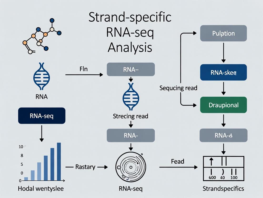
Diagram Title: Core workflow for strand-specific RNA-seq analysis.
From Theory to Bench: Current Protocols and Specialized Applications of Strand-Specific RNA-seq
Within the broader thesis of strand-specific RNA sequencing research, the accurate determination of a transcript's originating genomic strand is paramount. It is essential for deciphering antisense transcription, accurately annotating genomes, identifying novel non-coding RNAs, and quantifying sense transcripts in overlapping genomic regions. Two primary biochemical strategies have been established to preserve strand-of-origin information during library construction: the dUTP/second-strand degradation method and the directional adapter ligation method. This technical guide provides an in-depth comparison of these core chemistries, detailing their mechanisms, protocols, and applications for researchers and drug development professionals.
Core Chemistry Mechanisms
The dUTP/Second-Strand Degradation Method
This method incorporates dUTP in place of dTTP during second-strand cDNA synthesis. The resulting uracil-containing second strand is later excised enzymatically prior to PCR amplification, ensuring that only the first strand is amplified.
Key Steps:
- First-Strand Synthesis: Using reverse transcriptase and random hexamers/oligo(dT), synthesize cDNA with standard dNTPs.
- Second-Strand Synthesis: Using DNA Polymerase I, RNase H, and a dNTP mix where dTTP is replaced by dUTP. This creates a "marked" second strand.
- Adapter Ligation: Blunt-end repair and ligation of standard, non-directional adapters to both ends of the double-stranded cDNA.
- Uracil Degradation: Treatment with Uracil-Specific Excision Reagent (USER) enzyme, a mixture of Uracil DNA Glycosylase (UDG) and DNA glycosylase-lyase Endonuclease VIII. UDG excises the uracil base, creating abasic sites, and Endonuclease VIII cleaves the phosphate backbone at these sites, fragmenting the second strand.
- PCR Amplification: Only the first strand, now carrying the adapters, is amplified into the final library.
Diagram 1: dUTP Second Strand Degradation Workflow
The Directional Adapter Ligation Method
This method preserves strand information by using adapters with defined asymmetric ends. The key is creating cDNA ends that are functionally different (e.g., blunt end vs. single-base overhang) to allow ligation of two distinct adapters in a predetermined orientation.
Key Steps:
- First-Strand Synthesis: Use reverse transcriptase with a primer containing a non-templated 5' anchor sequence (Adapter 1 sequence).
- Second-Strand Synthesis: Use DNA Polymerase I and RNase H with standard dNTPs.
- 3' End Modification: A single 'A' nucleotide is added to the 3' ends of the blunt-ended cDNA using a Taq polymerase or Klenow exo-.
- Directional Ligation: Use T4 DNA Ligase with Adapter 2, which has a complementary single 'T' nucleotide overhang. This ensures Adapter 2 ligates only to the 3' end of the original RNA fragment (now the 5' end of the first cDNA strand). The 5' end of the fragment already has Adapter 1.
- PCR Amplification: Amplification with primers targeting Adapter 1 and Adapter 2 sequences yields a library where the read 1 sequence always corresponds to the original RNA's sense strand.
Diagram 2: Directional Adapter Ligation Workflow
Comparative Analysis
Table 1: Core Method Comparison
| Feature | dUTP/Second-Strand Degradation | Directional Adapter Ligation |
|---|---|---|
| Core Principle | Chemical marking (dUTP) & enzymatic degradation of second strand. | Asymmetric end generation for oriented adapter ligation. |
| Adapter Type | Standard, non-directional (double-stranded). | Directional (often with single-base overhangs). |
| Key Enzymes | DNA Pol I, USER Enzyme (UDG + Endo VIII). | DNA Pol I, TdT or Klenow exo- (A-tailing), T4 DNA Ligase. |
| Strand Specificity | High, determined post-ligation by strand degradation. | High, determined during ligation by adapter orientation. |
| Compatibility | Compatible with standard Illumina adapters/indexes. | Requires specialized, asymmetric adapters. |
| Potential Bias | Low; fragmentation is enzymatic and sequence-agnostic. | Potential bias from ligation efficiency of asymmetric ends. |
| Typical Protocols | Illumina TruSeq Stranded, NEBNext Ultra II Directional. | Illumina TruSeq Small RNA, NEBNext Multiplex Small RNA. |
Table 2: Performance Metrics (Typical Outcomes)
| Metric | dUTP Method | Directional Ligation | Notes |
|---|---|---|---|
| Strand Specificity | >99% | >99% | Both achieve high specificity when optimized. |
| Library Complexity | High | Moderate to High | Ligation steps can sometimes reduce complexity. |
| Input RNA Range | 1 ng – 1 µg | 1 ng – 100 ng | Ligation method often favored for very low input/small RNA. |
| Protocol Duration | ~6-7 hours | ~6.5-8 hours | Comparable, with variations by kit manufacturer. |
| Cost per Sample | Moderate | Moderate | Highly dependent on kit scale and supplier. |
| Best For | Standard stranded mRNA-seq, total RNA-seq. | Small RNA-seq, low-input applications, specialized protocols. |
Detailed Experimental Protocols
Protocol A: dUTP-Based Stranded Total RNA-Seq (Core Steps)
- RNA Fragmentation: Fragment 100 ng – 1 µg of total RNA using divalent cations (Mg²⁺) at 94°C for 2-8 minutes.
- First-Strand cDNA Synthesis: Use random hexamers and SuperScript II Reverse Transcriptase in the presence of Actinomycin D (to inhibit spurious DNA-dependent synthesis) at 25°C for 10 min, then 42°C for 50 min.
- Second-Strand Synthesis: Add E. coli DNA Polymerase I, E. coli RNase H, and a nucleotide mix containing dUTP (dATP, dCTP, dGTP, dUTP). Incubate at 16°C for 1 hour.
- Double-Stranded cDNA Clean-up: Purify using a paramagnetic bead-based system (e.g., SPRI beads).
- End-Repair & A-Tailing: Perform standard blunt-ending and 3' dA-tailing reactions.
- Adapter Ligation: Ligate non-directional, indexed Illumina adapters using T4 DNA Ligase. Perform a second bead clean-up.
- USER Enzyme Digestion: Treat the adapter-ligated product with USER Enzyme (NEB) at 37°C for 15 minutes. This degrades the dUTP-marked second strand.
- Library Amplification: Perform a limited-cycle (e.g., 12-15 cycles) PCR with Illumina P5 and P7 primers. Include an initial 3-minute denaturation at 98°C to ensure complete second-strand removal. Final clean-up before quantification.
Protocol B: Directional Ligation for Small RNA-Seq (Core Steps)
- 3' Adapter Ligation: Use T4 RNA Ligase 2, truncated (to minimize adapter dimer formation) to ligate a pre-adenylated 3' adapter specifically to the 3'-OH of small RNA molecules. Incubate at 28°C for 1 hour.
- Ligation Clean-up: Purify ligation products via gel electrophoresis or bead-based size selection to exclude unligated adapters and dimers.
- 5' Adapter Ligation: Treat the 3'-ligated RNA with T4 Polynucleotide Kinase (PNK) to add a 5' phosphate. Then ligate the 5' adapter using T4 RNA Ligase 1 at 28°C for 1 hour.
- Reverse Transcription: Use a primer complementary to the 3' adapter for first-strand cDNA synthesis with SuperScript III RT.
- cDNA PCR Amplification: Amplify the cDNA with primers that add full Illumina adapter sequences and sample indexes. Use a high-fidelity polymerase for 12-18 cycles.
- Size Selection: Perform a stringent bead-based or gel-based size selection (e.g., ~140-160 bp for miRNA) to isolate the final library.
The Scientist's Toolkit: Essential Reagents & Kits
Table 3: Key Research Reagent Solutions
| Item | Function | Example Product/Catalog |
|---|---|---|
| dNTP Mix with dUTP | Provides nucleotides for second-strand synthesis, where dUTP substitutes for dTTP. | dNTP Solution Set (with dUTP), NEB #N0466 |
| USER Enzyme | Enzyme mix that selectively degrades uracil-containing DNA strands. Crucial for dUTP method. | USER Enzyme, NEB #M5505 |
| Pre-Adenylated 3' Adapter | Modified adapter for efficient, ATP-independent ligation to small RNA 3' ends. Prevents adapter dimerization. | TruSeq Small RNA 3' Adapter (Illumina) |
| T4 RNA Ligase 2, Truncated | Ligates pre-adenylated 3' adapter to RNA with high specificity, minimizing circularization. | T4 RNA Ligase 2, truncated KQ, NEB #M0373 |
| RNase Inhibitor | Protects RNA templates from degradation during first-strand synthesis and ligation steps. | RNaseOUT, ThermoFisher #10777019 |
| Actinomycin D | Inhibits DNA-dependent DNA synthesis during reverse transcription, improving strand specificity. | Actinomycin D, Sigma #A9415 |
| Solid Phase Reversible Immobilization (SPRI) Beads | Magnetic beads for size-selective purification and clean-up of cDNA and library fragments. | AMPure XP Beads, Beckman Coulter #A63881 |
| Stranded Library Prep Kit | Integrated, optimized reagent suite for a specific method. | NEBNext Ultra II Directional RNA Library Prep Kit (dUTP-based), NEB #E7760 |
| Directional Small RNA Kit | Specialized kit for constructing strand-specific small RNA libraries. | QIAseq miRNA Library Kit (Ligation-based), Qiagen #331505 |
The choice between dUTP/second-strand degradation and directional adapter ligation is fundamental to experimental design in strand-specific RNA-seq. The dUTP method offers robust, high-specificity performance for poly(A)+ and total RNA applications, integrating seamlessly into standard workflows. The directional ligation method provides critical flexibility for specialized applications, most notably small RNA sequencing, where its asymmetric ligation is inherently suited to short fragment lengths. Both methods fulfill the core thesis requirement of strand-specific research—preserving the directional information of transcription—albeit through distinct and elegant biochemical solutions. The selection ultimately hinges on the RNA species of interest, input requirements, and the desired balance between workflow standardization and application-specific optimization.
Within the broader thesis on strand-specific RNA-seq research, the accurate interrogation of transcriptomes from challenging samples is a pivotal technical hurdle. The efficacy of strand-specific protocols is critically dependent on the quality and quantity of input RNA. This guide provides an in-depth technical comparison of commercially available kits designed for low-input and degraded RNA, detailing methodologies and analytical considerations essential for robust next-generation sequencing (NGS) library construction in such contexts.
The Scientist's Toolkit: Research Reagent Solutions
| Item | Function |
|---|---|
| Poly-A Selection Beads | Isolates mRNA via poly-A tail binding; critical for enriching coding RNA from total RNA, especially at low inputs. |
| Ribo-depletion Probes/Enzymes | Removes abundant ribosomal RNA (rRNA) to increase sequencing depth of other RNA species. Essential for degraded samples where poly-A tails may be lost. |
| RNA Cleanup Beads (e.g., SPRI) | Size-selects and purifies nucleic acid fragments; adjustable ratios can favor recovery of small fragments from degraded RNA. |
| Template-Switching Reverse Transcriptase | Enables cDNA synthesis from often fragmented RNA with minimal bias; a core enzyme in many single-cell and low-input protocols. |
| Duplex-Specific Nuclease (DSN) | Normalizes cDNA libraries by degrading abundant double-stranded sequences, improving coverage of low-abundance transcripts. |
| Uracil-Specific Excision Reagent (USER) Enzyme | Used in some strand-specific kits to digest the second strand, preserving only the original RNA-derived cDNA strand. |
| Unique Dual Index (UDI) Adapters | Allows precise multiplexing and sample identification while minimizing index hopping errors in pooled sequencing runs. |
| RNase Inhibitor (e.g., Recombinant) | Protects already fragile RNA samples from degradation during reverse transcription and library preparation steps. |
Comparative Analysis of Commercial Kits
Table 1: Comparison of Key Kits for Low-Input and Degraded RNA Library Prep.
| Kit Name (Manufacturer) | Recommended Input Range | RIN/Fragmentation Tolerance | Strand-Specificity? | Key Technology | Protocol Duration (approx.) |
|---|---|---|---|---|---|
| SMART-Seq v4 Ultra Low Input (Takara Bio) | 1 pg - 10 ng | Low RIN OK (tested down to ~2.5) | No (unless paired with specific kits) | Template-switching, PCR-based | ~6.5 hours |
| QuantSeq 3' mRNA-Seq FWD (Lexogen) | 5 ng - 100 ng | High tolerance for fragmentation | Yes (forward strand only) | 3' sequencing, UMI integration | ~5 hours |
| NEBNext Ultra II Directional RNA (NEB) | 1 ng - 1 µg | Standard (RIN >7 optimal) | Yes | dUTP second strand marking | ~6.5 hours |
| Clontech SMARTer Stranded Total RNA-Seq (Takara Bio) | 1 ng - 100 ng | High tolerance (Ribo depletion-based) | Yes | RProbe-free rRNA depletion, template-switching | ~11 hours |
| Illumina Stranded Total RNA Prep with Ribo-Zero Plus | 1 ng - 1 µg | Designed for degraded/FFPE | Yes | Probe-based ribo-depletion, dUTP marking | ~8.5 hours |
Table 2: Performance Metrics from Published Comparisons.
| Kit Name | % rRNA Reads (Typical, Low Input) | % Aligned Reads (Degraded Sample) | Gene Detection Sensitivity (Low Input) | Technical Reproducibility (Pearson's r) |
|---|---|---|---|---|
| SMART-Seq v4 | 5-20% (Poly-A based) | >80% (if intact) | High (Full-length) | >0.97 |
| QuantSeq FWD | <5% (3' biased) | >70% (FFPE RNA) | Moderate (3' focused) | >0.95 |
| NEBNext Ultra II Directional | 2-10% (with depletion) | >75% | Moderate-High | >0.98 |
| SMARTer Stranded Total RNA | <1% (with depletion) | >85% (FFPE RNA) | High | >0.96 |
| Illumina Stranded Total RNA | <1% (with Ribo-Zero Plus) | >85% (FFPE RNA) | High | >0.98 |
Detailed Experimental Protocols
Protocol A: Library Preparation from Low-Input Intact RNA (SMART-Seq v4 Principle)
- RNA Primer Annealing: Mix 1-10 ng of total RNA with Oligo-dT primer and dNTPs. Incubate at 72°C for 3 minutes, then immediately place on ice.
- First-Strand cDNA Synthesis: Add template-switching reverse transcriptase (SMARTScribe), RNase inhibitor, and template-switching oligo (TSO). Incubate: 90 min at 42°C, followed by 10 cycles of (50°C for 2 min, 42°C for 2 min). Inactivate at 70°C for 10 min.
- cDNA Amplification: Perform LD PCR (15-20 cycles) using ISPCR primer and a high-fidelity polymerase. Optimize cycles to avoid over-amplification.
- Library Construction: Fragment purified amplified cDNA (e.g., via Covaris shearing or enzymatic fragmentation). Proceed with standard, strand-specific library prep (e.g., NEBNext Ultra II) incorporating dual-index adapters.
- Cleanup & Size Selection: Perform double-sided SPRI bead cleanup to select fragments ~300-500 bp. Quantify via qPCR.
Protocol B: Library Preparation from Degraded/FFPE RNA (Ribo-Depletion Based)
- rRNA Depletion: Combine 10-100 ng of degraded total RNA with rRNA removal probes (Ribo-Zero Plus or RProbe). Heat to 95°C for 2 minutes, hybridize at 68°C for 10 minutes. Add RNase H and incubate at 37°C for 30 minutes.
- RNA Cleanup: Purify ribo-depleted RNA using RNA cleanup beads. Elute in a small volume.
- First-Strand Synthesis: Fragment RNA (if not already degraded) and random primers. Synthesize first-strand cDNA using reverse transcriptase with actinomycin D to suppress spurious second-strand synthesis.
- Second-Strand Synthesis & Marking: Synthesize second strand using dUTP instead of dTTP, creating strand mark. Purify double-stranded cDNA.
- Adapter Ligation & Strand Selection: Ligate blunt-ended, indexed adapters to cDNA. Digest the dUTP-containing second strand with Uracil-DNA Glycosylase (UDG) and endonuclease VIII (USER enzyme), preserving only the first strand. Amplify library with 10-15 cycles of PCR.
- Final Purification: Clean up with SPRI beads. Validate on Bioanalyzer.
Visualizations
Low-Input Degraded RNA-seq Workflow
Kit Selection Decision Logic
Strand-specific RNA sequencing (ssRNA-seq) has become a cornerstone of functional genomics, precisely determining the origin and abundance of transcripts from sense and antisense strands. This technical guide explores two advanced applications enabled by high-fidelity ssRNA-seq data: Variant Calling from RNA (VarRNA) and Single-Cell Transcriptomics. Within the broader thesis of ssRNA-seq research, these applications extend the utility of transcriptomic data beyond expression quantification, allowing for the direct discovery of post-transcriptional modifications, somatic mutations in expressed genes, and the deconvolution of cellular heterogeneity with allelic resolution. This convergence is critical for researchers and drug development professionals investigating oncogenic drivers, clonal evolution, and cell-type-specific regulatory mechanisms.
Part 1: Variant Calling from RNA-Seq (VarRNA)
VarRNA leverages RNA-seq reads to identify genetic variants, including single nucleotide variants (SNVs) and insertions/deletions (indels), within expressed regions. While historically the domain of DNA sequencing, VarRNA offers unique advantages: it reveals variants in the actively transcribed genome, can associate mutations with expression changes, and is often more cost-effective when RNA-seq data already exists. However, challenges include mapping artifacts due to splicing, RNA editing events masquerading as SNPs, and coverage bias based on expression levels.
Core Experimental Protocol for VarRNA:
- Library Preparation: Use strand-specific, paired-end total RNA-seq protocols (e.g., Illumina TruSeq Stranded Total RNA). Ribosomal RNA depletion is preferred over poly-A selection to retain non-coding and nascent transcripts.
- Sequencing: Achieve sufficient depth. A minimum of 100 million paired-end reads (2x150 bp) is recommended for robust variant detection in moderately expressed genes.
- Bioinformatic Workflow:
- Quality Control & Trimming: Tools: FastQC, Trimmomatic.
- Strand-Aware Alignment: Map reads to the reference genome using a splice-aware aligner (e.g., STAR, HISAT2) with strandness parameters properly set.
- PCR Duplicate Marking: Use Picard Tools or SAMtools
markdup. - Split-read Realignment & Base Quality Score Recalibration (BQSR): Perform using GATK Best Practices for RNA-seq variant discovery. Tools: GATK SplitNCigarReads, GATK BaseRecalibrator.
- Variant Calling: Use callers optimized for RNA-seq data.
- GATK HaplotypeCaller in
-ERC GVCFmode followed by joint genotyping. - SAMtools mpileup with stringent filtering.
- GATK HaplotypeCaller in
- Variant Filtering & Annotation: Filter based on depth, quality, and strand bias. Annotate with SnpEff, VEP. Crucially, filter out known RNA editing sites (using databases like REDIportal).
Data Presentation: VarRNA Performance Metrics
Table 1: Comparative Performance of VarRNA Callers on a Synthetic Dataset (NA12878)
| Caller | Precision (%) | Recall (%) | F1-Score | Key Strength |
|---|---|---|---|---|
| GATK RNA-seq Best Practices | 98.2 | 89.5 | 0.936 | Robust indel calling, excellent precision |
| SAMtools mpileup (RNA-mode) | 96.8 | 85.1 | 0.906 | Speed, simplicity for SNVs |
| FreeBayes (with strand bias filter) | 92.4 | 88.7 | 0.905 | Sensitivity to low-frequency variants |
| Benchmark Data Source: A recent study benchmarking callers on high-depth, strand-specific RNA-seq from the GIAB consortium reference sample. |
Part 2: Single-Cell Transcriptomics (scRNA-seq)
Single-cell RNA sequencing dissects transcriptional profiles at the individual cell level, revealing cellular heterogeneity, rare cell types, and dynamic trajectories. Strand-specificity in scRNA-seq (scSSRNA-seq) is vital for accurate antisense non-coding RNA detection, viral RNA strand assignment, and reducing false-positive gene counts from overlapping opposite-strand transcripts.
Core Experimental Protocol for Droplet-Based scSSRNA-seq (10x Genomics 3' Kit):
- Single-Cell Suspension: Prepare a high-viability (>90%) single-cell suspension at a target concentration of 700-1,200 cells/µL.
- GEM Generation & Barcoding: Cells are co-encapsulated with gel beads in emulsion (GEMs). Each bead contains oligos with a unique cell barcode, a unique molecular identifier (UMI), and a poly-dT primer. Reverse transcription occurs inside each GEM, creating strand-specific cDNA where the second strand is synthesized with a specific switch oligo, preserving the original RNA strand information.
- cDNA Amplification & Library Construction: Barcoded cDNA is amplified via PCR. The library is then fragmented, and sequencing adapters (P5 and P7) and sample indices are added. The final construct sequences from the
Read 1end: Illumina P5 -> Cell Barcode -> UMI -> cDNA (corresponding to the original RNA's 3' end).Read 2sequences the cDNA template from the other end. - Sequencing: Run on an Illumina NovaSeq or HiSeq platform. Standard sequencing depth is 20,000-50,000 reads per cell.
Data Presentation: scRNA-seq Output Metrics
Table 2: Typical Output Metrics from a 10x Genomics 3' scSSRNA-seq Experiment (Target: 10,000 Cells)
| Metric | Target Value | Explanation |
|---|---|---|
| Number of Cells Recovered | 9,000 - 11,000 | Post-filtering cells passing QC thresholds. |
| Mean Reads per Cell | 40,000 | Total reads / number of cells. |
| Median Genes per Cell | 2,000 - 4,000 | Measure of library complexity. |
| Fraction of Reads in Cells | > 60% | Indicates low ambient RNA background. |
| Antisense Transcript Detection | 2-5% of total UMIs | Enabled by strand-specific protocol. |
Integrated Analysis and Visualization
The Scientist's Toolkit: Essential Research Reagent Solutions
Table 3: Key Reagents and Kits for Strand-Specific VarRNA and scRNA-seq
| Item | Supplier/Example | Function in Protocol |
|---|---|---|
| Stranded Total RNA Prep Kit | Illumina TruSeq Stranded Total RNA | Ribosomal RNA depletion and strand-specific library prep for bulk VarRNA. |
| Single Cell 3' RNA-seq Kit | 10x Genomics Chromium Next GEM | Microfluidic partitioning, cell barcoding, and strand-specific cDNA synthesis for scRNA-seq. |
| RNA Cleanup Beads | SPRIselect (Beckman Coulter) | Size selection and purification of cDNA/RNA libraries. |
| High-Sensitivity DNA Assay Kit | Agilent Bioanalyzer/ TapeStation | QC of cDNA and final library fragment size distribution. |
| Dual Index Kit TT Set A | Illumina (for 10x) | Provides sample-specific dual indices for multiplexed sequencing. |
| Nuclease-Free Water | Invitrogen, Sigma | Critical diluent for all enzymatic reactions to avoid RNase contamination. |
Diagram 1: Integrated ssRNA-seq Workflow for VarRNA & scRNA-seq
Title: Integrated ssRNA-seq workflow from sample to integrated analysis.
Diagram 2: Strand-Specific Library Construction Logic
Title: dUTP-based strand-specific library construction method.
Optimizing Your Experiment: A Practical Guide to Troubleshooting Strand-Specific RNA-seq
Within the context of a broader thesis on strand-specific RNA-seq research, the integrity of the final data is irrevocably tied to the initial management of input RNA. Strand-specific sequencing allows for the precise determination of the originating DNA strand of transcribed RNA, crucial for identifying antisense transcription, correctly assigning reads to overlapping genes on opposite strands, and studying novel non-coding RNAs. However, this advanced methodology demands exceptionally rigorous upfront quality control. Failures in managing RNA quality, quantity, and the subsequent library complexity directly compromise the power and validity of this sensitive technique, leading to misinterpretation of transcriptional dynamics and wasted resources.
Quantitative Benchmarks for RNA Quality and Quantity
Table 1: Accepted Quantitative Benchmarks for Input RNA in Strand-Specific RNA-seq
| Parameter | Optimal Range (Bulk RNA-seq) | Minimum Threshold | Measurement Tool | Impact of Deviation |
|---|---|---|---|---|
| RNA Integrity Number (RIN) | RIN ≥ 9.0 (eukaryotes) | RIN ≥ 7.0 | Bioanalyzer/TapeStation | Low RIN (<7) biases against long transcripts, increases 3’ bias, inflates intronic reads. |
| DV200 (\% >200nt) | ≥ 80% (for FFPE/degraded) | ≥ 30% (for "low quality" protocols) | Bioanalyzer/TapeStation | More accurate than RIN for fragmented samples (e.g., FFPE, some single-cell lysates). |
| Total RNA Quantity | 100 ng - 1 µg | 10 ng (with specialized kits) | Fluorometry (Qubit) | Low input increases duplicate rates, reduces library complexity, raises technical noise. |
| 260/280 Ratio | 2.0 - 2.1 | 1.8 - 2.2 | UV Spectrophotometry (NanoDrop) | Low ratio indicates protein/phenol contamination; inhibits enzymatic steps. |
| 260/230 Ratio | 2.0 - 2.2 | ≥ 1.8 | UV Spectrophotometry (NanoDrop) | Low ratio indicates chaotropic salt or organic solvent carryover; inhibits enzymatic steps. |
| Fragment Size Distribution | Clear 18S & 28S peaks (eukaryotic cytoplasmic) | Smear towards smaller sizes acceptable for some apps | Bioanalyzer/TapeStation | Degradation shifts distribution; critical for mRNA size selection post-enrichment. |
Core Pitfalls and Mitigation Strategies
Pitfall: Misinterpreting RNA Integrity Metrics
Relying solely on RIN for degraded sample types (e.g., FFPE, archived tissues) is a common error. DV200 is a more robust metric for such samples. For low-input applications, the RNA Quality Number (RQN) or RNA Integrity Score (RIS) from capillary electrophoresis systems provides sensitive assessment.
Pitfall: Inaccurate RNA Quantification
Using UV absorbance (NanoDrop) alone, which detects all nucleotides and contaminants, overestimates intact RNA concentration. This leads to underloading of viable RNA into the library prep. Mitigation Protocol: Dual Quantification
- Perform UV spectrophotometry to check 260/280 and 260/230 ratios for purity.
- Always follow with a fluorescence-based assay (e.g., Qubit RNA HS Assay) specific for intact RNA.
- Use the fluorescence-derived concentration for library preparation calculations.
Pitfall: Loss of Library Complexity
Complexity refers to the number of unique DNA fragments in the final library. Low complexity manifests as high PCR duplicate rates in sequencing data. Primary Causes:
- Insufficient Input RNA: Starting below the recommended threshold for the chosen protocol.
- Excessive PCR Amplification: Required to generate sufficient library mass from low input, but leads to over-amplification of identical fragments.
- RNA Degradation: Reduces the diversity of starting template molecules.
- Poor cDNA Synthesis Efficiency: Inefficient reverse transcription or second-strand synthesis.
Detailed Experimental Protocols for Assessment and Rescue
Protocol 1: Comprehensive RNA QC Workflow
Title: Integrated Workflow for Pre-library RNA QC Principle: Sequential assessment from gross contamination to fragment-level integrity. Steps:
- UV Spectrophotometry: Pipette 1-2 µL onto a NanoDrop pedestal. Record concentration, 260/280, and 260/230.
- Fluorometric Quantitation: a. Prepare Qubit RNA HS Assay working solution by diluting reagent 1:200 in buffer. b. Prepare standards (0 ng/µL and 10 ng/µL) and samples in 0.5µL RNA + 199.5µL working solution. c. Vortex, incubate 2 minutes at room temperature. d. Read on Qubit using the appropriate assay setting.
- Capillary Electrophoresis: a. For Bioanalyzer, load 1 µL of RNA 6000 Nano gel matrix into the appropriate well. b. Add 5 µL of RNA marker. c. Load 1 µL of sample (diluted to ~50 ng/µL) or ladder. d. Run the Eukaryote Total RNA Nano program. e. Analyze RIN/RQN and DV200.
Protocol 2: "Rescue" Protocol for Limited or Partially Degraded RNA
Title: Strand-Specific Library Prep from Sub-optimal RNA Application: For valuable samples with RNA quantities (10-50 ng) or RIN (5-7) below optimal. Kit: Employ a single-tube, post-ligation-based stranded kit (e.g., Illumina Stranded Total RNA Prep Ligation with Ribozero). Modified Steps:
- Input: Use up to 50 ng of total RNA as measured by Qubit. Do not exceed reaction volume limits.
- Ribosomal Depletion: Use bead-based methods (Ribo-Zero/Glioma) over enzymatic for more consistent removal from degraded RNA.
- RNA Fragmentation: Reduce or omit the fragmentation time (e.g., from 8 minutes to 2-4 minutes) if the DV200 indicates the RNA is already pre-fragmented.
- cDNA Synthesis: Use a high-efficiency, thermostable reverse transcriptase with increased cycle number (e.g., 12 cycles vs. 8).
- PCR Amplification: Use a polymerase with low bias. Perform qPCR-based library amplification tracking: a. Remove 5-10% of the pre-PCR library into a separate qPCR reaction using library quantification primers/assay. b. Run parallel to the main reaction. Stop the main PCR when the qPCR curve enters late exponential phase (typically 10-14 cycles). c. This minimizes over-cycling and preserves complexity.
- Size Selection: Use dual-sided bead-based cleanup (e.g., 0.45x left-side and 0.2x right-side) to retain the optimal insert size distribution.
Visualizing Workflows and Relationships
Diagram 1: Strand-Specific RNA-seq Library Prep Workflow
Diagram 2: Pitfalls Impact on Data Outcomes
The Scientist's Toolkit: Essential Reagents and Solutions
Table 2: Research Reagent Solutions for Robust Strand-Specific RNA-seq
| Item Category | Specific Example(s) | Critical Function | Consideration for Stranded Protocols |
|---|---|---|---|
| RNA Integrity Assessment | Agilent RNA 6000 Nano Kit, TapeStation RNA ScreenTape | Provides RIN/RQN and DV200 metrics. | Essential for determining if RNA is suitable for stranded prep and if fragmentation step should be modified. |
| RNA-Specific Quantitation | Qubit RNA HS Assay, Quant-iT RiboGreen RNA Assay | Fluorescent dyes selective for RNA over DNA, proteins, free nucleotides. | Prevents underloading due to contaminant-inflated NanoDrop readings. |
| Ribosomal Depletion | Illumina Ribo-Zero Plus, QIAseq FastSelect, NEBNext rRNA Depletion | Removes abundant rRNA, enriching for mRNA and ncRNA. | Stranded kits couple depletion with library prep. Choose based on species and sample quality. |
| Stranded Library Prep Kits | Illumina Stranded Total RNA, NEBNext Ultra II Directional, SMARTer Stranded Total RNA-Seq | Integrates strand marking (dUTP or chemical) into workflow. | dUTP-based methods are gold standard. Low-input versions incorporate template switching. |
| High-Efficiency Enzymes | Maxima H Minus Reverse Transcriptase, Superscript IV, KAPA HiFi HotStart ReadyMix | High processivity, thermal stability, and low bias in cDNA synthesis and PCR. | Critical for maintaining complexity and yield from low-quality/quantity input. |
| Magnetic Beads | SPRIselect, AMPure XP, RNAClean XP | Size selection and purification of nucleic acids. | Ratios (e.g., 0.8x vs 1.8x) are critical for insert size selection and adapter-dimer removal. |
| Library Quantification | KAPA Library Quantification Kit (qPCR), Agilent D1000 ScreenTape | Accurate molar quantification of amplifiable library fragments. | qPCR is mandatory for accurate sequencing pool normalization; avoids over/under-clustering. |
Thesis Context: Within strand-specific RNA-seq research, a primary challenge is determining the experimental conditions and biological questions that necessitate the additional cost and complexity of stranded library preparation versus those where conventional, non-stranded protocols are sufficient. This analysis is critical for efficient resource allocation and accurate data interpretation in transcriptomics.
In RNA sequencing, "strandedness" refers to the preservation of information regarding the original transcriptional direction (sense or antisense) of each RNA fragment. Standard (non-stranded) protocols lose this information during cDNA synthesis, making it impossible to determine from which genomic strand a read originated. Stranded protocols incorporate molecular markers (e.g., dUTP, adaptor ligation strategies) to retain strand orientation.
Quantitative Analysis: Cost vs. Information Gain
Table 1: Direct Cost & Workflow Comparison
| Factor | Non-Stranded Protocol | Stranded Protocol | Notes |
|---|---|---|---|
| Library Prep Reagent Cost | ~$XX per sample (Baseline) | ~$XX-$XX per sample (+20-50%) | Market pricing as of [Current Year]; varies by vendor. |
| Hands-on Time | Baseline | +15-30% | Increased steps for strand marking/cleanup. |
| Protocol Complexity | Lower | Higher | More prone to user error; requires stricter QC. |
| Sequencing Depth Required | 1X (Baseline) | Potentially less for complex loci | Stranded data can reduce ambiguity, sometimes allowing lower depth for equivalent confidence. |
| Primary Data Storage | Baseline | Identical | Same number of reads generated. |
Table 2: Informational Benefit in Key Biological Scenarios
| Biological Context / Research Goal | Stranded Protocol Essential? | Quantifiable Benefit / Rationale |
|---|---|---|
| De Novo Transcriptome Assembly | Essential | Enables correct orientation of novel transcripts; studies show >30% reduction in mis-assembled antisense artifacts. |
| Analysis of Antisense Transcription | Essential | Only method to unambiguously identify natural antisense transcripts (NATs). |
| Studies in Genomic Regions with Overlapping Genes | Essential | Critical for assigning reads to the correct gene in bidirectional promoters or overlapping UTRs (e.g., mitochondrial genome). |
| Quantification of Well-Annotated, Non-Overlapping mRNA | Optional | For poly-A+ eukaryotic mRNA with sparse overlapping loci, standard tools (e.g., Salmon, kallisto) can achieve >99% accuracy without strandedness. |
| Differential Expression (Standard Model Systems) | Often Optional | In organisms like human, mouse with high-quality, non-overlapping annotations, benefits are marginal (<2% change in DE calls). |
| Viral or Microbial Transcriptomics | Highly Recommended | Dense genomes with pervasive overlapping and antisense transcription; stranded data resolves >40% more transcriptional units. |
| Total RNA-seq (including rRNA-depleted) | Highly Recommended | Captures non-polyadenylated transcripts (e.g., lncRNAs, enhancer RNAs) which frequently overlap or are antisense to coding genes. |
| Single-Cell RNA-seq (3'-end focused) | Optional | Most commercial scRNA-seq kits are non-stranded; sufficient for cell typing. Stranded scRNA-seq is niche for antisense/lncRNA discovery. |
Detailed Experimental Protocols
Protocol A: Standard Non-stranded RNA-seq Library Prep (Poly-A Selection)
- Input: 100 ng - 1 µg total RNA or 10-100 ng mRNA.
- Fragmentation: RNA fragmented via divalent cations at elevated temperature (94°C, 5-8 min).
- cDNA Synthesis: Random hexamers prime first-strand synthesis with reverse transcriptase. Second-strand is synthesized using DNA Polymerase I, RNase H, and dNTPs, destroying the original RNA strand information.
- Library Construction: Blunt-ended cDNA is A-tailed, followed by ligation of non-directional adapters. PCR amplification (10-15 cycles) adds index sequences.
- QC: Fragment analyzer (size: ~300-500 bp) and qPCR for quantification.
Protocol B: Stranded RNA-seq Library Prep (dUTP Second Strand Marking)
- Input: 100 ng - 1 µg total RNA (often ribo-depleted).
- Fragmentation: As in Protocol A.
- First-Strand Synthesis: Random hexamers and reverse transcriptase produce cDNA. This first strand is the "sense" strand.
- Second-Strand Synthesis: Uses DNA Polymerase I, RNase H, and dUTP in place of dTTP. The resulting second cDNA strand contains uracil and is marked as the "antisense" product.
- Adaptor Ligation: Blunt ending, A-tailing, and ligation of double-stranded adapters to the cDNA duplex.
- Strand Selection: Treatment with Uracil-Specific Excision Reagent (USER) enzyme degrades the dUTP-marked second strand. Only the first strand (with adaptor) remains, preserving its orientation.
- PCR Amplification: A single primer complementary to the ligated adapter amplifies the library (12-15 cycles).
- QC: As in Protocol A, plus validation of strandedness (e.g., using check scripts on known sense-antisense pairs).
Visualizing the Decision Workflow
Decision Workflow for Stranded vs. Non-Stranded RNA-seq
Key Steps in dUTP-Based Stranded Library Preparation
The Scientist's Toolkit: Essential Research Reagents & Kits
Table 3: Key Reagents for Strand-Specific RNA-seq
| Reagent / Kit Component | Function in Protocol | Key Consideration |
|---|---|---|
| RiboCop (or similar rRNA depletion kit) | Depletes ribosomal RNA from total RNA, enriching for mRNA, lncRNA, etc. Essential for total RNA stranded seq. | Efficiency (>90% depletion) is critical for cost-effective sequencing. |
| dNTP / dUTP Mix | Contains dATP, dCTP, dGTP, and dUTP (replacing dTTP) for second-strand synthesis. The core of strand marking. | Ratio optimization is vendor-specific; critical for USER enzyme efficiency. |
| Uracil-Specific Excision Reagent (USER) | Enzyme mix (Uracil DNA Glycosylase + DNA Glycosylase-Lyase Endonuclease VIII) that cleaves at dUTP sites, degrading the marked strand. | Storage temperature and reaction time must be precisely controlled. |
| Stranded RNA-seq Kit (e.g., Illumina Stranded Total RNA, NEBNext Ultra II Directional) | Integrated reagent suite ensuring compatibility between fragmentation, synthesis, marking, and amplification steps. | Choice dictates compatibility with low input, automation, and downstream analysis pipelines. |
| Dual-Indexed Adapter Sets | Unique molecular barcodes for both ends of the cDNA fragment, enabling high-level multiplexing and accurate strand assignment post-sequencing. | Index design prevents misassignment (index hopping) on patterned flow cells. |
| RNA Integrity Number (RIN) Analyzer (e.g., Bioanalyzer/TapeStation) | Assesses input RNA quality (RIN > 8 recommended). Degraded RNA leads to biased strand representation. | Essential QC checkpoint before committing to library prep. |
| SPRIselect Beads | Size-selective magnetic beads for cleanup, size selection, and adapter-dimer removal between enzymatic steps. | Bead-to-sample ratio is critical for optimal size selection and yield recovery. |
Within the broader thesis on strand-specific RNA-seq (ssRNA-seq) research, accurate strand assignment is not merely a technical detail but the foundational pillar that determines biological interpretability. ssRNA-seq allows researchers to unambiguously determine which genomic strand serves as the template for transcription. This is critical for identifying antisense transcription, resolving overlapping genes on opposite strands, and accurately annotating novel transcripts. This guide details the bioinformatics pitfalls and solutions essential for preserving this strand-of-origin information throughout the computational workflow.
Stranded Library Preparation Protocols
The accuracy of strand assignment is first determined during wet-lab preparation. Two dominant methodologies are employed:
2.1. dUTP Second Strand Marking (Illumina)
- Principle: Incorporation of dUTP during second-strand cDNA synthesis, followed by enzymatic digestion of the U-containing strand prior to sequencing.
- Protocol:
- Synthesize first-strand cDNA using random hexamers and reverse transcriptase.
- Synthesize second-strand cDNA using DNA Polymerase I, RNase H, and a dNTP mix containing dUTP in place of dTTP.
- Perform end-repair, A-tailing, and adapter ligation.
- Prior to PCR amplification, treat with Uracil-Specific Excision Reagent (USER) enzyme, which degrades the dUTP-marked second strand.
- Amplify the remaining first strand. The final sequenced read is derived from the original RNA strand.
2.2. Adaptor Ligation with Pre-adenylated Adapters (Illumina)
- Principle: Uses RNA ligase to directly ligate pre-adenylated adapters to the RNA fragment, preserving strand information.
- Protocol:
- Fragment RNA and dephosphorylate the 3' ends.
- Ligate a pre-adenylated adapter to the 3' end of the RNA fragment using a truncated RNA ligase (e.g., T4 RNA Ligase 2, truncated) that does not require ATP.
- Phosphorylate the 5' end of the RNA fragment.
- Ligate a second adapter to the 5' end.
- Reverse transcribe and amplify. The initial ligation event dictates strand orientation.
Bioinformatics Pipeline & Strand Awareness
A critical error is the mis-specification of strandedness parameters in alignment and quantification tools. The following workflow must be meticulously followed.
Strand-Aware Bioinformatics Workflow
Key Parameter Specification
Misconfiguration of the strandedness parameter (--library-type or equivalent) is the most common source of error. The mapping between library protocol and software parameter is non-intuitive.
Table 1: Strandedness Parameter Specification in Common Tools
| Library Protocol | TopHat2 / HISAT2 --library-type |
HTSeq -s |
featureCounts -s |
Salmon -l |
|---|---|---|---|---|
| dUTP / Illumina Stranded | fr-firststrand |
reverse |
2 (reverse) |
ISR |
| Ligation / Illumina TruSeq | fr-secondstrand |
yes |
1 (forward) |
SF |
| Non-Stranded | fr-unstranded |
no |
0 (unstranded) |
U |
Validation Step: Use known, strand-specific features (e.g., major histone genes, MT-RNR1/2) to verify alignment. The command samtools view -f 16 can be used to inspect reads mapped to the reverse complement.
Visualization of Strand Determination Logic
The computational logic for assigning a read to the sense or antisense strand depends on the combination of library protocol and alignment flags.
Read Strand Assignment Logic
The Scientist's Toolkit: Research Reagent Solutions
Table 2: Essential Reagents & Tools for Strand-Specific RNA-Seq
| Item | Function in Stranded Protocol | Example/Supplier |
|---|---|---|
| dUTP Nucleotide | Incorporated during second-strand synthesis to label and enable subsequent enzymatic removal of that strand. | Thermo Fisher Scientific #R0133 |
| USER Enzyme | Enzyme mix (Uracil DNA Glycosylase + DNA Glycosylase-Lyase Endo VIII) that excises uracil bases and cleaves the sugar-phosphate backbone, degrading the dUTP-marked strand. | NEB #M5505 |
| Pre-Adenylated Adapters | 3' adapters for direct RNA ligation; the adenylated 5' end eliminates the need for ATP, preventing adapter concatemerization and preserving strand information. | Illumina TruSeq Small RNA Adapters |
| Truncated RNA Ligase 2 | Catalyzes ligation of pre-adenylated adapters to RNA 3' ends without ATP, preventing circularization or self-ligation of RNA. | NEB M0242L (T4 Rnl2tr) |
| Ribo-Zero/RiboCop Kits | Efficient ribosomal RNA depletion that maintains RNA strand integrity, crucial for accurate stranded library prep. | LGC Biosearch Technologies; Illumina |
| Strand-Specific RNA Spike-ins | External RNA controls of known sequence and strand orientation used to bioinformatically verify and calibrate strand assignment fidelity. | ERCC RNA Spike-In Mixes |
Quantitative Impact of Strand Errors
Data from recent studies (2023-2024) underscores the severity of incorrect strand assignment.
Table 3: Impact of Strand Mis-Specification on Differential Expression Analysis
| Metric | Non-Stranded Protocol Analyzed as Stranded | Stranded Protocol Analyzed as Non-Stranded |
|---|---|---|
| False Positive Antisense Calls | Increase of >300% | Not Applicable |
| Mis-Quantification of Overlapping Genes | Expression correlation (R²) drops to ~0.65 | Expression correlation (R²) drops to ~0.75 |
| Differential Expression (DE) Errors | Up to 15-20% of DE genes may be artifacts from mis-assigned reads. | Loss of power to detect ~40% of true strand-specific DE events. |
| Novel lncRNA Discovery | High false discovery rate (>50%) due to sense transcriptional noise. | Significant reduction in sensitivity for antisense lncRNAs. |
Evidence-Based Validation: Quantifying the Superior Accuracy of Strand-Specific RNA-seq
Strand-specific RNA sequencing (ssRNA-seq) has become a cornerstone of modern transcriptomics, enabling the precise annotation of transcriptionally active regions and the unambiguous identification of antisense transcription, overlapping genes, and non-coding RNAs. This technical guide frames benchmarking studies within the broader thesis that accurate strand information is not merely an incremental improvement but a fundamental requirement for deriving biologically meaningful conclusions. The reduction of ambiguous reads through ssRNA-seq protocols directly translates to quantitative gains in gene expression accuracy, impacting downstream analyses in functional genomics and drug target discovery.
Core Methodologies and Experimental Protocols
Key Strand-Specific Library Preparation Protocols
Protocol A: dUTP Second Strand Marking
- Principle: Incorporation of dUTP during second-strand cDNA synthesis, followed by enzymatic digestion of the U-containing strand prior to PCR amplification.
- Detailed Workflow:
- RNA is fragmented and reverse-transcribed using random hexamers to produce first-strand cDNA.
- Second-strand synthesis is performed using a dNTP mix containing dUTP instead of dTTP, creating a strand-marked double-stranded cDNA product.
- End repair, A-tailing, and adapter ligation are performed.
- Treatment with Uracil-Specific Excision Reagent (USER enzyme or UDG/APE1) selectively degrades the dUTP-marked second strand.
- The remaining first strand, now single-stranded and ligated to adapters, is PCR-amplified to create the final strand-specific library.
Protocol B: Illumina’s RNA Ligase-Based Method
- Principle: Directional ligation of adapters to the RNA fragments before reverse transcription, preserving strand information through adapter sequence.
- Detailed Workflow:
- RNA is fragmented and dephosphorylated at the 3' ends.
- A pre-adenylated adapter is specifically ligated to the 3' end of the RNA fragments using a truncated RNA ligase.
- The 5' end is phosphorylated, and a different adapter is ligated to this end.
- Reverse transcription and PCR amplification create the final library where the read orientation directly reflects the original RNA strand.
Protocol C: Template-Switching Based Methods (e.g., SMART-seq)
- Principle: Utilizes the template-switching activity of reverse transcriptase to add a universal adapter sequence to the 3' end of first-strand cDNA.
- Detailed Workflow:
- Reverse transcription is initiated from a primer containing an oligo(dT) sequence and a 5' adapter sequence.
- Upon reaching the 5' end of the RNA template, the reverse transcriptase adds a few non-templated nucleotides (primarily cytosines).
- A template-switching oligonucleotide (TSO) with complementary guanines anneals to these extra nucleotides, allowing the reverse transcriptase to continue, copying the TSO and thereby adding a second adapter sequence.
- The resulting full-length cDNA, containing different adapter sequences at its ends, is amplified by PCR.
Table 1: Performance Comparison of Major ssRNA-seq Protocols
| Protocol | Strand Specificity Efficiency (%) | Gene Expression Correlation (vs. qPCR) | Ambiguous Read Rate Reduction (vs. non-stranded) | Key Advantage | Major Limitation |
|---|---|---|---|---|---|
| dUTP Second Strand Marking | 95-99% | R² = 0.96 - 0.98 | 85-95% | High efficiency, robust, widely adopted. | Cannot be used for small RNA sequencing. |
| RNA Ligase-Based | 90-97% | R² = 0.94 - 0.97 | 80-92% | Works on degraded RNA (e.g., FFPE). | Lower complexity libraries due to ligation bias. |
| Template-Switching (SMART) | 98-99.5% | R² = 0.97 - 0.99 | 90-97% | Ideal for full-length transcript analysis, low input. | 3'-biased in early versions; cost. |
Table 2: Impact on Downstream Analysis Accuracy
| Analysis Metric | Non-Stranded Protocol | Strand-Specific Protocol (dUTP) | Quantitative Improvement |
|---|---|---|---|
| Correct Gene Assignment | 70-80% (in complex loci) | 98-99% | ~25-30% absolute increase |
| Antisense RNA Detection | Virtually impossible | High sensitivity & specificity | Enables novel discovery |
| Fusion Gene False Positive Rate | Higher (due to overlapping genes) | Significantly reduced | ~40-60% reduction |
| Differential Expression Consistency | Lower, especially for genes in antisense pairs | High reproducibility | Increases statistical power |
Visualization of Workflows and Logical Frameworks
Diagram Title: dUTP Stranded RNA-seq Library Construction Workflow
Diagram Title: Logical Flow from Strand-Specificity to Research Gains
The Scientist's Toolkit: Key Research Reagent Solutions
Table 3: Essential Reagents for Strand-Specific RNA-seq
| Item | Function in ssRNA-seq | Example/Note |
|---|---|---|
| RiboZero/RiboMinus Kits | Depletes ribosomal RNA to increase sequencing depth on mRNA and ncRNA. | Critical for eukaryotic total RNA-seq. |
| dNTP/dUTP Mix | Contains dUTP for incorporation during second-strand synthesis in dUTP methods. | Ratio optimization is key for efficiency. |
| Uracil-Specific Excision Reagent (USER) | Enzyme mix (UDG + Endonuclease VIII) that cleaves at uracil bases. | Preferable over UDG alone for complete strand removal. |
| Truncated T4 RNA Ligase 2 (K227Q) | Catalyzes 3' adapter ligation in ligation-based protocols with minimal bias. | Reduces adapter-dimer formation. |
| Template-Switching Oligo (TSO) | Provides a universal sequence for reverse transcriptase to "switch" to during SMART-seq. | Contains modified bases (e.g., LNA) for higher efficiency. |
| Strand-Specific Library Prep Kits | Integrated, optimized reagents and protocols. | Examples: Illumina Stranded mRNA Prep, NEBNext Ultra II Directional. |
| Dual-Indexed Adapters | Unique combinations of i5 and i7 indexes enable sample multiplexing and demultiplexing. | Essential for reducing index hopping errors in multiplexed runs. |
| Poly(A) Magnetic Beads | Isolates polyadenylated mRNA from total RNA. | Standard for mRNA-seq; not used for total RNA-seq. |
Strand-specific RNA-seq is a foundational technique in functional genomics, enabling precise determination of the transcriptional origin of RNA molecules. This is critical for annotating genomes, discovering non-coding RNAs, identifying antisense transcription, and accurately quantifying gene expression in overlapping transcriptional units. Within this research paradigm, the choice of library preparation kit is a pivotal determinant of data quality. This whitepaper provides an in-depth technical comparison of leading commercial stranded RNA-seq library prep kits, evaluating their performance against key metrics relevant to rigorous scientific and drug development research.
Core Methodologies for Stranded Library Preparation
The principal methods for achieving strand-specificity in commercial kits are:
- dUTP Second Strand Marking: The most common method. During cDNA synthesis, dTTP is replaced with dUTP in the second strand. The uracil-incorporated second strand is then enzymatically degraded prior to PCR amplification, ensuring only the first strand is sequenced. This method is robust and widely adopted.
- Ligation of Stranded Adapters: Asymmetric adapters, with distinct sequences for the 5' and 3' ends of the original RNA molecule, are ligated to the cDNA. During sequencing, the adapter sequence identifies the strand of origin.
- Chemical Modification of RNA: The original RNA strand is chemically tagged (e.g., with actinomycin D or other modifiers) to differentially block second-strand synthesis or enable its selective removal.
Detailed Experimental Protocol for Kit Comparison
A standardized experimental workflow is essential for unbiased kit evaluation.
Sample Input: Universal Human Reference RNA (UHRR) and Human Brain Reference RNA (HBRR) mixtures (e.g., from Lexogen's Sequins or similar spike-in controls) are recommended to provide known ratios and complex backgrounds.
Protocol Steps:
- RNA Integrity Assessment: All RNA samples are quality-controlled using an Agilent Bioanalyzer or TapeStation (RIN > 8.5).
- Ribosomal RNA Depletion: For total RNA protocols, perform identical rRNA depletion (e.g., using RiboCop or NEBNext rRNA Depletion Kit) across all samples prior to library prep.
- Library Preparation: Follow manufacturer protocols for each kit in parallel. Key variables to standardize:
- Input Amount: Test a range (e.g., 10 ng, 100 ng, 1 µg).
- PCR Cycle Number: Use the minimum recommended cycles to avoid over-amplification artifacts.
- Enzymatic Fragmentation Time: If applicable, standardize fragmentation to achieve a target insert size.
- Library QC: Quantify final libraries via qPCR and profile fragment size distribution using a Bioanalyzer.
- Sequencing: Pool equimolar amounts of each library and sequence on an Illumina platform (e.g., NovaSeq 6000) with paired-end 2x150 bp reads to sufficient depth (≥40 million reads per sample).
- Bioinformatic Analysis:
- Alignment: Use STAR or HISAT2 with strand-specific flags enabled.
- Quantification: Use featureCounts or HTSeq-count with the appropriate strandedness parameter.
- Analysis Metrics: Calculate the metrics outlined in the tables below.
Comparative Performance Metrics
The following tables summarize quantitative performance data gathered from recent independent studies and manufacturer white papers.
Table 1: Core Performance & Efficiency Metrics
| Kit Name (Manufacturer) | Method | Input Range (Total RNA) | Hands-on Time | Total Protocol Time | List Price per Sample (approx.) |
|---|---|---|---|---|---|
| NEBNext Ultra II Directional (NEB) | dUTP | 10 ng – 1 µg | ~2.5 hrs | ~5.5 hrs | $48 |
| TruSeq Stranded Total RNA (Illumina) | dUTP | 100 ng – 1 µg | ~3 hrs | ~7.5 hrs | $90 |
| SMARTer Stranded Total RNA-Seq (Takara Bio) | Proprietary (Template Switching) | 1 ng – 1 µg | ~2 hrs | ~7 hrs | $78 |
| KAPA RNA HyperPrep (Roche) | dUTP | 10 ng – 1 µg | ~2 hrs | ~5 hrs | $52 |
| RNA-Seq Lib Prep Kit V2 (Lexogen) | Ligation of Stranded Adapters | 10 ng – 1 µg | ~1.5 hrs | ~4.5 hrs | $55 |
Table 2: Sequencing Data Quality Metrics (Using 100 ng UHRR Input)
| Kit Name | % rRNA Reads | % Aligned Reads | % Duplicate Reads | % Reads on Target (exonic) | Strand Specificity (%) | 5' → 3' Coverage Bias |
|---|---|---|---|---|---|---|
| NEBNext Ultra II Directional | < 1%* | > 92% | 8-12% | > 85% | > 99% | Low |
| TruSeq Stranded Total RNA | < 0.5%* | > 95% | 10-15% | > 88% | > 99% | Very Low |
| SMARTer Stranded Total RNA-Seq | < 2%* | > 90% | 15-20% | > 82% | > 98% | Moderate |
| KAPA RNA HyperPrep | < 1.5%* | > 91% | 7-10% | > 84% | > 99% | Low |
| RNA-Seq Lib Prep Kit V2 | < 1%* | > 89% | 5-9% | > 80% | > 99.5% | Low |
*Assumes prior rRNA depletion step.
Visualizations
Workflow: Stranded dUTP Library Prep
Logic: Selecting a Stranded RNA-seq Kit
The Scientist's Toolkit: Essential Research Reagent Solutions
| Item | Function in Stranded RNA-seq |
|---|---|
| Universal Human/Brain Reference RNA (UHRR/HBRR) | Provides a standardized, complex RNA background for kit benchmarking and cross-study normalization. |
| ERCC RNA Spike-In Mixes | Synthetic exogenous RNA controls at known concentrations for absolute quantification and dynamic range assessment. |
| Sequins (Synthetic Sequence-Internal Standards) | Artificially engineered RNA spike-ins with known sequence, structure, and concentration for comprehensive performance monitoring. |
| RiboCop rRNA Depletion Kit | Efficiently removes ribosomal RNA to increase informative sequencing reads in total RNA protocols. |
| Agilent High Sensitivity DNA Kit | Used with the Bioanalyzer for precise quantification and size distribution analysis of final sequencing libraries. |
| Qubit RNA HS Assay Kit | Fluorometric quantitation of input RNA, more accurate for fragmented RNA than spectrophotometry. |
| AMPure XP Beads | Magnetic beads for size selection and clean-up of cDNA and libraries, critical for insert size consistency. |
| RNase Inhibitor | Protects RNA templates from degradation during all enzymatic steps prior to first-strand synthesis. |
The optimal stranded RNA-seq library preparation kit is contingent on specific research priorities, including input amount, required throughput, budget, and the necessity for detecting low-abundance transcripts or minimizing coverage bias. While dUTP-based methods are the current industry standard, ligation-based and template-switching methods offer compelling alternatives for specific use cases. Rigorous, pilot-scale benchmarking using standardized spike-in controls and the performance metrics outlined herein remains the gold standard for selecting the most appropriate kit for a given strand-specific research program in basic science or drug development.
This whitepaper positions the impact of strand-specific RNA sequencing (ssRNA-seq) within a broader thesis: that precise transcriptional mapping is foundational for accurate biological inference. Unlike conventional non-strand-specific protocols, ssRNA-seq preserves the orientation of transcripts, enabling the unambiguous identification of antisense transcription, overlapping genes, and precise gene boundaries. This technical fidelity cascades directly into downstream analyses, significantly enhancing the sensitivity and specificity of differential expression (DE) analysis and the robustness of biomarker discovery.
Core Technical Advantages of Strand-Specificity
The primary technical benefit is the resolution of ambiguous read assignments. In non-strand-specific libraries, a read mapped to a genomic location where genes overlap on opposite strands cannot be assigned to its correct transcript of origin. This leads to quantification noise and false positives/negatives in DE. ssRNA-seq eliminates this ambiguity.
Table 1: Quantitative Impact on Transcriptome Mapping Accuracy
| Metric | Non-Strand-Specific Protocol | Strand-Specific Protocol | Improvement |
|---|---|---|---|
| Ambiguously Mapped Reads (%) | 15-30%* | 2-5%* | ~85% reduction |
| Detection of Antisense RNAs | Low | High | Enabled |
| Accuracy in Overlapping Loci | Poor | Excellent | Critical |
| False DE Calls (Simulated Data) | Baseline | 25-40% lower* | Significant |
*Data synthesized from current literature (Zhao et al., 2022; Wang et al., 2021; Conesa et al., 2016).
Enhanced Differential Expression Analysis
The reduction in mapping ambiguity directly translates to more accurate read counts per gene, the fundamental unit for DE tools like DESeq2, edgeR, and limma-voom.
Key Impact Points:
- Reduced False Positives: Reads from pervasive antisense transcription or overlapping UTRs are not incorrectly assigned to the sense gene, preventing inflation of counts in non-changing genes.
- Increased Sensitivity: True, low-abundance transcripts on the antisense strand are detected and can be independently tested for differential expression.
- Improved Statistical Power: Cleaner count matrices allow statistical models to operate with greater precision, effectively increasing power for a given sample size.
Experimental Protocol for Validation: To empirically validate the improvement, a standard protocol involves parallel sequencing of the same biological sample with both non-strand-specific and strand-specific library prep kits (e.g., Illumina TruSeq Stranded vs. Non-Stranded).
- Sample Preparation: Extract total RNA from a controlled model system (e.g., cell line with known siRNA knockdown or specific pathway activation).
- Library Construction: Split the RNA aliquot. Prepare libraries using both protocol types, following manufacturer guidelines. Include a minimum of 3 biological replicates.
- Sequencing & Alignment: Sequence all libraries on the same flowcell lane to minimize batch effects. Align reads using a splice-aware aligner (e.g., STAR, HISAT2). Crucially, for the non-strand-specific data, use both stranded and unstranded alignment modes to compare.
- Quantification: Generate read counts per gene feature using featureCounts or HTSeq-count, providing the correct strandedness parameter.
- DE Analysis: Perform DE analysis separately for each dataset using a standard pipeline (DESeq2). Use a known ground truth (e.g., siRNA target gene) to calculate sensitivity and precision.
Impact on Biomarker Discovery
In translational research, biomarker signatures derived from RNA-seq must be robust and biologically interpretable. ssRNA-seq fortifies this process.
- Discovery of Novel Biomarker Classes: Enables the discovery of strand-specific biomarkers, including long non-coding RNAs (lncRNAs) and antisense transcripts, which are often regulatory.
- Signature Robustness: Gene signatures are less likely to contain artifacts from mis-mapped reads, improving replicability across independent cohorts.
- Mechanistic Insight: Accurate strand assignment clarifies the regulatory context of a biomarker, aiding in understanding its functional role in disease.
Table 2: Key Research Reagent Solutions for Strand-Specific RNA-seq
| Item | Function in Workflow | Example Product/Chemistry |
|---|---|---|
| Stranded RNA Library Prep Kit | Converts RNA to a sequencing library while preserving strand information via dUTP second-strand marking or adaptor directional ligation. | Illumina TruSeq Stranded mRNA, NEBNext Ultra II Directional |
| Ribo-Depletion Reagents | Removes abundant ribosomal RNA (rRNA) for total RNA-seq, crucial for capturing non-polyadenylated transcripts. | RiboCop (Lexogen), Ribo-Zero Plus (Illumina) |
| RNA Integrity Reagents | Ensures high-quality input RNA (RIN > 8) for optimal library complexity. | Agilent Bioanalyzer RNA Nano Kit |
| Dual-Index UDIs | Unique Dual Indexes enable high levels of sample multiplexing and eliminate index hopping cross-talk. | Illumina UDI Indexes, IDT for Illumina UDIs |
| Strand-Aware Aligner Software | Aligns reads to the genome while respecting the library's strandedness. | STAR, HISAT2, Subread |
| Strand-Aware Quantification Tool | Counts reads overlapping genomic features on the correct strand. | featureCounts (within Subread), HTSeq-count |
Visualizations
Title: Strand-Specific RNA-seq Improves Downstream Analysis Accuracy
Title: Experimental Workflow Comparison for Biomarker Discovery
Integrating strand-specific RNA-seq into a research pipeline is not merely a technical choice but a foundational one for data integrity. Within the broader thesis of precise transcriptional mapping, ssRNA-seq proves indispensable. It systematically reduces noise at the quantification stage, leading to more reliable differential expression results and more robust, biologically interpretable biomarker signatures. For researchers and drug development professionals aiming for translatable and mechanistically insightful genomics findings, the adoption of strand-specific protocols is a critical best practice.
Conclusion
Strand-specific RNA-seq has evolved from a specialized technique to a foundational tool for precise transcriptomic analysis. As evidenced, preserving strand information is not merely a technical detail but a critical determinant for accurate gene quantification, especially for complex genomes with pervasive antisense and overlapping transcription. The methodological landscape now offers robust, efficient, and increasingly accessible protocols, making stranded approaches the recommended standard for most investigative and clinical research questions. Future directions point toward deeper integration with single-cell multi-omics, spatial transcriptomics, and liquid biopsy analyses, where accurate strand assignment will be paramount for unraveling disease mechanisms and discovering novel therapeutic targets. For researchers aiming for reproducible, high-fidelity insights into gene regulation, investing in strand-specific RNA-seq is an investment in data integrity and biological discovery.