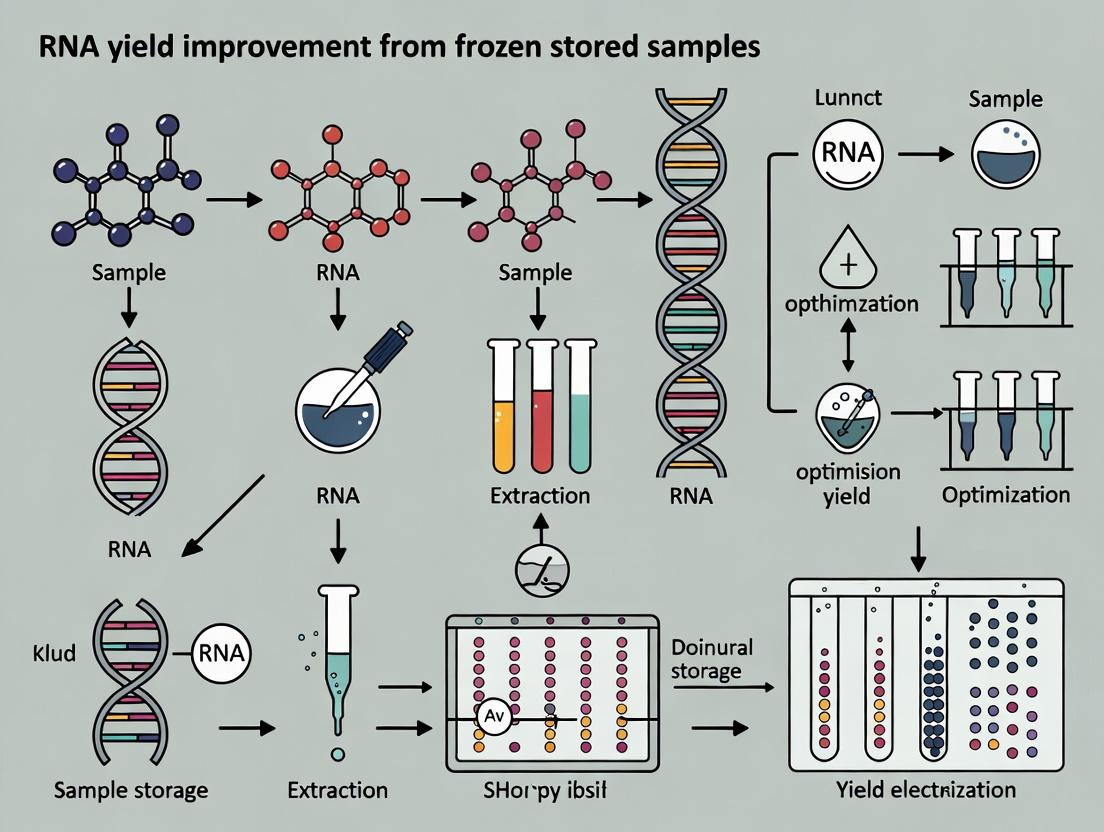
Unlocking Biobank Potential: A Practical Guide to Maximizing RNA Yield and Integrity from Frozen Archived Samples
Obtaining high-quality RNA from frozen archival tissues and blood samples remains a significant bottleneck in biomedical research and drug development.
Unlocking Biobank Potential: A Practical Guide to Maximizing RNA Yield and Integrity from Frozen Archived Samples
Abstract
Obtaining high-quality RNA from frozen archival tissues and blood samples remains a significant bottleneck in biomedical research and drug development. This article provides a comprehensive, evidence-based guide for researchers aiming to unlock the value of biobanked samples. We first explore the fundamental science behind RNA degradation during freeze-thaw cycles. We then detail optimized methodological workflows—including specific thawing protocols, preservation buffer addition, and mechanical handling—that have been proven to significantly improve RNA Integrity Numbers (RIN) and yield. A dedicated troubleshooting section addresses common pitfalls like low yield, degradation, and contamination. Finally, we present comparative validation data from recent studies, demonstrating the successful application of these optimized protocols on long-term archived human samples for downstream sequencing and analysis. This guide synthesizes the latest research to empower scientists to reliably extract robust transcriptional data from precious frozen specimens.
The Science of Degradation: Understanding Key Factors That Compromise RNA in Frozen Samples
Welcome to the Technical Support Center. This resource is designed to support researchers within the broader thesis context of improving RNA yield from frozen stored samples. Below are troubleshooting guides and FAQs addressing common experimental issues related to sample thawing and RNase activity.
Frequently Asked Questions & Troubleshooting
Q1: My RNA yields from frozen tissue samples are consistently low and degraded. Could my thawing method be the problem? A: Yes, the thawing method is a critical, often overlooked variable. Thawing at room temperature allows the sample exterior to reach permissive temperatures for RNase activity while the interior remains frozen. This creates a gradient where RNases become active and begin degrading RNA before the sample is fully homogenized in a lysis buffer containing RNase inhibitors. Rapid thawing on wet ice is strongly recommended to minimize this window of opportunity for RNase activity.
Q2: I need to process multiple samples quickly. Is thawing on ice really necessary, or can I use a warmer water bath to speed things up? A: Thawing in a warm water bath is strongly discouraged. While faster, it dramatically increases RNase activity. The goal is to transition the sample from a frozen state (RNases inactive) to complete lysis in a denaturing buffer as quickly as possible, but without applying external heat. Ice-thawing is the best compromise. For high-throughput, pre-chill your lysis buffer and use a cold metal block placed on ice to ensure consistent, rapid cooling for all samples during thawing.
Q3: After thawing my cell pellets on ice and adding TRIzol, my RNA Integrity Number (RIN) is still poor. What else should I check? A: Focus on the post-thaw handling time. The moment the pellet is just thawed, you must immediately add and vortex it with the lysis reagent. Even on ice, a fully thawed pellet is vulnerable. Ensure your lysis buffer (e.g., TRIzol, QIAzol) is highly denaturing and is itself RNase-free. Also, verify that your frozen samples were flash-frozen in optimal conditions (e.g., in a preservative like RNAlater or immediately in liquid nitrogen) before storage, as poor initial freezing causes ice crystal damage and release of RNases.
Q4: Is there quantitative data comparing RNA integrity from different thawing methods? A: Yes. Studies consistently show significant differences in RNA quality metrics based on thawing protocol. See the summarized data below.
Table 1: Impact of Thawing Method on RNA Quality from Frozen Tissue Samples
| Thawing Method | Average RNA Yield (µg/mg tissue) | Average RIN (RNA Integrity Number) | % of Samples with RIN > 7 | Key Observation |
|---|---|---|---|---|
| Ice/Wet Ice (0-4°C) | 1.8 ± 0.3 | 8.2 ± 0.7 | 95% | Optimal balance; minimizes RNase activation. |
| Room Temperature (25°C) | 1.1 ± 0.4 | 5.5 ± 1.2 | 20% | Significant degradation; high variability. |
| Warm Water Bath (37°C) | 0.9 ± 0.5 | 4.0 ± 1.5 | 5% | Severe and rapid degradation; not recommended. |
| Direct Lysis (Frozen Grinding) | 2.0 ± 0.2 | 8.8 ± 0.3 | 100% | Gold standard but not always practical. |
Experimental Protocol: Standardized Thawing Test for RNase Activity Impact
Objective: To empirically determine the impact of ice-thawing vs. room temperature thawing on RNA yield and integrity from your specific sample type.
Materials: See "Research Reagent Solutions" below.
Methodology:
- Sample Preparation: Divide a homogeneous tissue sample or cell pellet into multiple, identical aliquots (e.g., 4 x 20mg). Flash-freeze all aliquots immediately in liquid nitrogen. Store at -80°C for at least 24 hours.
- Thawing Conditions:
- Group A (Ice): Remove one aliquot from -80°C and immediately place it on a pre-chilled (wet ice) weigh boat or tube rack. Allow it to thaw completely.
- Group B (Room Temp): Remove a second aliquot and place it directly on a lab benchtop (22-25°C). Allow it to thaw completely.
- Time Tracking: Record the time to complete thaw for each condition.
- Immediate Lysis: The instant the sample is thawed (no visible ice), add the appropriate volume of pre-chilled denaturing lysis buffer (e.g., TRIzol). For tissue, immediately begin homogenization.
- RNA Isolation: Complete the RNA extraction protocol according to the manufacturer's instructions for all samples in parallel.
- Quality Assessment: Quantify RNA yield (ng/µL) via spectrophotometry. Assess integrity using a Bioanalyzer or TapeStation to generate an RIN or equivalent.
- Analysis: Compare yield and integrity metrics between Group A and Group B. Perform statistical analysis (e.g., t-test) on replicate experiments (n≥3).
Visualization: RNase Activity Dynamics During Thawing
The Scientist's Toolkit: Research Reagent Solutions
| Item | Function in Thawing/RNA Isolation | Key Consideration |
|---|---|---|
| Denaturing Lysis Buffer (e.g., TRIzol, QIAzol) | Immediately inactivates RNases upon contact with thawed tissue/cells. Disrupts cells and dissolves RNA. | Must be pre-chilled and added the instant the sample is thawed. |
| RNase Inhibitors (e.g., recombinant RNasin) | Added to non-denaturing or mild lysis buffers to non-covalently inhibit RNase activity. | A supplementary safeguard, not a substitute for rapid processing and denaturing buffers. |
| RNAlater Stabilization Solution | Penetrates tissue to stabilize and protect RNA at room temperature post-collection, before freezing. | Simplifies logistics but does not eliminate need for proper thawing before RNA isolation. |
| Pre-Chilled Metal Block or Weight Boat | Provides a high-thermal conductivity surface on ice for rapid, uniform thawing of multiple samples. | Pre-chill on ice before use. Avoid foam racks which insulate. |
| Liquid Nitrogen (or Dry Ice Slurry) | For initial flash-freezing of samples. Rapid freezing minimizes ice crystal formation and cellular damage. | Essential for preserving RNA integrity before the thawing step is even considered. |
| Bioanalyzer/TapeStation & RNA Screentapes | Microfluidic capillary electrophoresis systems for objective assessment of RNA integrity (RIN/DVN). | Critical for quantifying the result of your thawing protocol on RNA quality. |
Technical Support Center & FAQ
Troubleshooting Guide
Issue: Low RNA Yield from Frozen Tissue Problem: RNA concentration is below the expected range for the tissue mass processed. Solution Steps:
- Verify Homogenization: Ensure the tissue is fully homogenized. No visible clumps should remain. Incomplete homogenization is the leading cause of low yield.
- Check Lysis Buffer Volume: Confirm the lysis buffer-to-tissue ratio is correct. For dense tissues, a ratio of 100 µl per 10 mg may be insufficient. Increase volume proportionally.
- Audit Sample Thawing: Avoid letting the sample thaw during aliquoting. Use pre-chilled tools and work on dry ice. RNA degradation begins rapidly upon thawing.
- Validate RNase Inactivation: Ensure the lysis buffer contains a sufficient concentration of a strong chaotropic salt (e.g., guanidinium thiocyanate) and is fresh.
- Re-evaluate Aliquot Mass: The optimal input mass may be lower than used. Overloading can inhibit homogenization and binding. See Table 1 for recommended starting masses.
Issue: Poor RNA Integrity (Low RIN/RQN) Problem: RNA is degraded, as shown by Bioanalyzer/TapeStation profiles. Solution Steps:
- Minimize Ischemic Time: Review sample collection SOPs. For frozen tissues, the time between excision and freezing must be minimized (<30 minutes ideal).
- Optimize Freezing: Snap-freeze in liquid nitrogen or an isopentane bath cooled by LN2. Slow freezing causes ice crystal formation and cellular disruption.
- Prevent Freeze-Thaw: Aliquot tissue before freezing. Never thaw the primary specimen. Use a cryostat to shave off material from a frozen block while it remains frozen.
- Use Nuclease-Free Tools: All consumables (tubes, pestles) must be certified nuclease-free. Clean work surfaces with RNase decontaminants.
Frequently Asked Questions (FAQs)
Q1: What is the single most important factor for maximizing RNA recovery from frozen tissue? A: Complete and rapid homogenization of the tissue in a denaturing lysis buffer. The buffer must fully penetrate the sample to inactivate RNases immediately. Sample mass must be balanced against lysis buffer volume to achieve this.
Q2: How do I determine the optimal starting tissue mass for a new tissue type? A: Perform a mass titration experiment. Process identical aliquots of the same frozen sample at different masses (e.g., 5 mg, 10 mg, 20 mg, 30 mg) using a standardized protocol. Plot mass input vs. total RNA yield and RNA integrity number (RIN). The optimal mass is at the plateau of the yield curve before RIN declines. See Table 1 and Protocol 1.
Q3: My tissue is very fibrous (e.g., heart, skin). How can I improve homogenization? A: For fibrous tissues, mechanical disruption is key. Use:
- TissueLyser II with stainless steel beads for high-throughput processing.
- Pre-cooled ceramic or steel mortar and pestle for grinding under liquid nitrogen before adding lysis buffer.
- Increased homogenization time in a bead mill (e.g., 3 x 2 minute cycles with cooling).
Q4: Can I re-freeze leftover lysate for RNA extraction later? A: No. Lysate should be processed immediately for RNA purification. While the lysate is inhibitory to RNases, long-term storage even at -80°C can lead to degradation. Proceed to the binding column or phase separation step without delay.
Q5: How does aliquot size affect RNA yield variability? A: Smaller aliquots reduce repeated freeze-thaw cycles of the master sample, preserving integrity. However, very small aliquots (<5mg) can lead to greater relative loss during handling and increase sampling error if the tissue is heterogeneous. An optimal range balances these factors.
Data Presentation
Table 1: Recommended Starting Tissue Mass and Lysis Buffer Volumes for RNA Extraction Data synthesized from current manufacturer protocols (Qiagen, Thermo Fisher, Zymo Research) and recent literature on frozen tissue optimization.
| Tissue Type | Optimal Starting Mass Range (mg) | Recommended Lysis Buffer Volume (µl) | Homogenization Method Note |
|---|---|---|---|
| Liver / Spleen | 15 - 30 mg | 300 - 600 µl | Homogenizes easily. Avoid overloading. |
| Brain (gray matter) | 20 - 40 mg | 400 - 800 µl | Soft tissue. Use gentle mechanical disruption. |
| Heart / Muscle | 10 - 25 mg | 500 - 1000 µl | Fibrous. Requires vigorous bead-beating or grinding. |
| Tumor (solid) | 15 - 35 mg | 450 - 900 µl | Highly variable. Necrotic areas yield less. |
| Lung | 25 - 50 mg | 750 - 1500 µl | Often low yield per mg. Higher mass may be needed. |
| Skin / Fibrous | 5 - 15 mg | 500 - 1000 µl | Very tough. Pre-grind under LN2 is essential. |
| Adipose | 50 - 100 mg | 1000 - 2000 µl | Low RNA cellularity. High mass required. |
Experimental Protocols
Protocol 1: Tissue Mass Titration for Yield Optimization
Objective: To determine the ideal tissue input mass for maximum RNA yield and integrity from a specific frozen tissue type.
Materials: See "The Scientist's Toolkit" below. Procedure:
- Sample Preparation: On dry ice, use a pre-cooled scalpel or cryostat to aliquot a frozen tissue block into masses of 5, 10, 20, and 30 mg (n=3 per mass). Keep all pieces on dry ice.
- Homogenization: Place each aliquot into a pre-chilled 2ml tube containing 1.4mm ceramic beads. Immediately add an appropriate volume of lysis buffer (see Table 1 guide, e.g., 300µl per 10mg).
- Disruption: Homogenize in a bead mill (e.g., Bertin Precellys) at 6,500 rpm for 30 seconds. Cool samples on ice for 30 seconds. Repeat for a total of 3 cycles.
- RNA Extraction: Centrifuge lysate (12,000 x g, 2 min, 4°C). Transfer supernatant to a fresh tube. Complete extraction using your standard silica-membrane column protocol (e.g., RNeasy).
- Quantification & QC: Elute RNA in 30µl RNase-free water. Quantify using a fluorometric assay (Qubit). Assess integrity with a microfluidics platform (Bioanalyzer).
- Analysis: Calculate total yield (µg) = concentration x elution volume. Plot mean yield (±SD) vs. input mass. Plot mean RIN vs. input mass. The optimal mass is at the yield plateau with highest RIN.
Protocol 2: Cryostat Sectioning for Sub-Aliquoting Frozen Blocks
Objective: To obtain small, reproducible tissue masses from a large frozen specimen without thawing.
Materials: Cryostat, disposable blades, specimen chucks, OCT compound (optional), brush, pre-cooled collection tubes. Procedure:
- Embedding: Mount the frozen tissue block on a cryostat chuck using a minimum amount of OCT, or directly freeze a small piece of tissue onto the chuck.
- Equilibration: Allow the chuck and tissue to equilibrate to the cryostat chamber temperature (-20°C) for at least 15 minutes.
- Sectioning: Set section thickness to 20-50 µm. Trim the block face until full tissue surface is exposed. Begin collecting sections.
- Weighing: Using a cold brush, carefully transfer a roll of sections to a pre-weighed, pre-chilled microtube on dry ice. Cap and briefly remove from dry ice to weigh. The mass difference is the tissue mass.
- Processing: Immediately return the tube to dry ice. Proceed directly to homogenization by adding lysis buffer to the tube containing the frozen sections.
Mandatory Visualization
Title: Workflow for Generating Optimized Frozen Tissue Aliquots
The Scientist's Toolkit: Research Reagent Solutions
| Item | Function & Rationale |
|---|---|
| Denaturing Lysis Buffer (e.g., Qiazol, TRIzol, or RLT Plus) | Contains guanidinium salts to immediately denature proteins and inactivate RNases, preserving RNA during homogenization. |
| Silica-Membrane Spin Columns (e.g., RNeasy, PureLink) | Selective binding of RNA in high-salt conditions, allowing efficient washing and elution of pure RNA. |
| RNase-Free DNase I (e.g., RNase-Free DNase Set) | Digests genomic DNA bound to the column membrane, preventing DNA contamination in the eluate. |
| β-Mercaptoethanol or 1-Thioglycerol | Reducing agent added to lysis buffer (1% v/v) to disrupt disulfide bonds and aid in protein denaturation, crucial for fibrous tissues. |
| RNase-Free Beads (Ceramic, 1.4mm) | Used in bead-mill homogenizers for rapid, high-throughput mechanical disruption of frozen tissue in lysis buffer. |
| Cryostat | Instrument to thinly slice frozen tissue blocks, enabling precise, non-thawing sampling for mass titration experiments. |
| Fluorometric RNA Assay Kit (e.g., Qubit RNA HS) | Provides accurate RNA quantification without interference from contaminants like DNA or free nucleotides. |
| Microfluidics-based Analyzer (e.g., Bioanalyzer, TapeStation) | Assesses RNA integrity (RIN/RQN) and detects degradation, essential for QC of extraction optimization. |
Technical Support Center
Troubleshooting Guides & FAQs
Q1: Why did my RNA RIN drop significantly after just 2-3 freeze-thaw cycles, even when stored at -80°C? A: RNA is highly susceptible to degradation by RNases, which can be transiently activated or become more accessible during the thawing phase. Each freeze-thaw cycle causes physical stress (ice crystal formation, pH shifts) that can compromise RNA secondary structure and lead to strand breaks. Even at -80°C, repeated warming to 0-4°C during thawing allows for brief enzymatic activity. Quantitative data from recent studies is summarized in Table 1.
Q2: What is the most critical step during the thawing process to minimize RIN loss? A: The single most critical step is rapid, consistent thawing on ice (0-4°C). Avoid thawing at room temperature or in your hands. Thawing on ice keeps the sample in a temperature range that minimizes RNase activity. Once thawed, keep the sample on ice and proceed immediately to cDNA synthesis or further analysis. Do not re-freeze the original aliquot.
Q3: How should I aliquot my RNA to prevent the need for freeze-thaw cycles? A: Aliquot the purified RNA into small, single-use volumes immediately after isolation. The volume should be the exact amount needed for one downstream application (e.g., one qRT-PCR reaction). Use nuclease-free tubes. Store aliquots at -80°C. This is the gold-standard practice for preserving RIN in long-term studies.
Q4: Can I use RNase inhibitors during storage to protect against freeze-thaw degradation? A: While RNase inhibitors are essential during the extraction and working phases, they are generally not recommended for long-term storage in frozen samples. Their protective activity can diminish over time, and they may not prevent the physical degradation caused by ice crystals. The best practice remains proper aliquoting and avoiding thaw cycles.
Q5: My samples have undergone multiple freeze-thaw cycles. Are they still usable for qPCR? A: It depends on the application and the extent of degradation. For qPCR targeting short amplicons (<150 bp), moderately degraded RNA (RIN > 5) may still yield usable data. However, for RNA-Seq, microarray, or full-length transcript analysis, a high RIN (>7 or >8) is typically required. Always check the RIN and DV200 (percentage of fragments > 200 nucleotides) metrics. See Table 2 for application-specific RIN thresholds.
Table 1: Impact of Freeze-Thaw Cycles on RNA Integrity Number (RIN)
| Freeze-Thaw Cycles | Mean RIN (Human Liver Tissue) | Mean RIN (Cultured Cells) | Storage Temperature | Key Observation |
|---|---|---|---|---|
| 0 (Fresh Aliquots) | 8.7 ± 0.3 | 9.5 ± 0.2 | -80°C | Baseline integrity. |
| 1-2 Cycles | 8.1 ± 0.4 | 8.9 ± 0.3 | -80°C | Slight but significant decrease (p<0.05). |
| 3-5 Cycles | 6.8 ± 0.7 | 7.5 ± 0.6 | -80°C | Moderate degradation; not suitable for sensitive assays. |
| >5 Cycles | 5.2 ± 1.1 | 5.9 ± 0.9 | -80°C | Severe degradation; only usable for very short amplicon PCR. |
| 0 Cycles (Improper) | 4.5 ± 1.5 | 5.0 ± 1.8 | -20°C | Demonstrates temperature stability is crucial. |
Data synthesized from recent literature (2022-2024).
Table 2: Recommended Minimum RIN Values for Common Downstream Applications
| Application | Recommended Minimum RIN | Alternative Metric (if RIN is low) | Notes for Freeze-Thawed Samples |
|---|---|---|---|
| qRT-PCR (amplicon <100 bp) | 5.0 | Cq value shift < 2 vs. high-RIN control | Most tolerant; assess via control gene stability. |
| Microarray Analysis | 7.0 | N/A | High RIN critical for accurate genome-wide expression. |
| Bulk RNA-Seq | 7.0 | DV200 > 70% | Ribosomal RNA profile may be distorted after multiple cycles. |
| Single-Cell RNA-Seq | 8.0 | DV200 > 80% | Extremely sensitive to degradation; avoid any thaw cycles. |
| Northern Blot | 8.5 | N/A | Requires largely intact full-length transcripts. |
Experimental Protocols
Protocol 1: Systematic Quantification of Freeze-Thaw Impact on RIN
Objective: To measure the direct correlation between number of freeze-thaw cycles and RNA integrity in stored samples.
Materials: See "The Scientist's Toolkit" below. Method:
- Sample Preparation: Isolate total RNA from a homogeneous source (e.g., cell pellet pool) using a silica-column method. Elute in nuclease-free water.
- Baseline Analysis: Determine initial RIN using a Bioanalyzer or TapeStation (Agilent). This is the "Cycle 0" timepoint.
- Aliquot Strategy: Divide the RNA solution into 10 equal single-use aliquots.
- Freeze-Thaw Cycling:
- Keep Aliquot 1 as the uncycled control (store at -80°C until final analysis).
- Subject Aliquots 2-10 to programmed cycles. One cycle consists of: a. Thawing on wet ice for 5 minutes. b. Immediate vortex mixing for 5 seconds. c. Quick centrifugation to collect liquid. d. Returning to -80°C for a minimum of 1 hour.
- Sampling: After 1, 2, 3, 5, and 7 cycles, remove one aliquot (in chronological order) for final analysis. Do not re-freeze.
- Final Analysis: Thaw all sampled aliquots on ice. Analyze RNA concentration and integrity (RIN) using the same instrument and settings as in Step 2.
- Data Analysis: Plot RIN (y-axis) vs. Number of Freeze-Thaw Cycles (x-axis). Perform linear regression analysis to determine the rate of degradation.
Protocol 2: Optimized Long-Term Storage and Thawing for Maximum RIN Preservation
Objective: To establish a best-practice workflow for frozen RNA samples intended for high-sensitivity applications.
Method:
- Isolation & Initial Assessment: Isolve RNA. Check concentration and RIN. Only proceed if RIN > 8.0.
- Immediate Aliquoting: In a nuclease-free environment, dilute RNA to a standard working concentration (e.g., 50 ng/µL) in nuclease-free water or a weak buffer (e.g., 0.1 mM EDTA, pH 8.0). Avoid Tris buffers as they can depress freezing point.
- Aliquoting: Dispense into low-binding, nuclease-free PCR tubes in volumes needed for a single downstream reaction (e.g., 10 µL for one cDNA synthesis).
- Flash-Freezing (Optional but Recommended): Place aliquots in a float and submerge in a dry ice/ethanol bath for 2 minutes before transferring to a -80°C freezer. This prevents slow freezing and large ice crystal formation.
- Storage: Store boxes of aliquots at -80°C in a dedicated, rarely opened freezer. Use freezer racks to minimize time the door is open.
- Thawing for Use: a. Pre-chill a cooler with wet ice. b. Remove only the number of aliquots needed from -80°C. c. Immediately place them on pre-chilled wet ice. d. Allow 5-10 minutes for complete thawing. e. Gently flick and quick-spin the tube. f. Keep on ice at all times until added to the reaction mix.
- Record Keeping: Maintain a detailed log for each sample stock, tracking the freeze-thaw history of the master stock and the usage of individual aliquots.
Visualizations
Title: Mechanism of RNA Degradation via Freeze-Thaw Cycles
Title: Optimal RNA Handling Workflow for Frozen Storage
The Scientist's Toolkit: Research Reagent Solutions
| Item | Function & Rationale |
|---|---|
| Silica-Membrane RNA Isolation Kits (e.g., Qiagen RNeasy, Zymo Research) | Provides high-purity RNA free of contaminants that can exacerbate degradation during freezing. Consistent yield is critical for aliquotting. |
| Nuclease-Free Water (not DEPC-treated) | The ideal resuspension buffer for long-term storage. Lacks ions that can catalyze hydrolysis and avoids Tris buffers that inhibit freezing. |
| Low-Binding, Nuclease-Free Microtubes/PCR Tubes | Minimizes RNA adsorption to tube walls, which is a significant and often overlooked source of loss during freeze-thaw and pipetting. |
| β-Mercaptoethanol (β-Me) or Alternative Reducing Agents | Essential additive to lysis buffer to denature RNases during the initial homogenization step, setting the stage for high-integrity RNA. |
| RNA Integrity Number (RIN) Analysis Kits (Agilent Bioanalyzer RNA kits, TapeStation) | Gold-standard for quantitatively assessing RNA degradation. Must be used to establish baseline and monitor freeze-thaw impact. |
| Portable Cooler Rack & Wet Ice | Critical for maintaining the 0-4°C thawing environment. Pre-cooling is essential to prevent the start of thawing at warmer temperatures. |
| Digital Freezer Temperature Loggers | Ensures the storage freezer maintains a consistent -80°C. Temperature fluctuations can induce partial thawing and degrade RNA even without opening the door. |
Frequently Asked Questions & Troubleshooting Guides
Q1: Our RNA yields from frozen mouse liver are consistently lower than from human frozen PBMCs, despite using identical extraction protocols. What could explain this interspecies variation?
A: Interspecies differences in tissue composition, RNase activity, and metabolism are key factors. Murine tissues, particularly liver and spleen, often have higher baseline RNase activity than many human tissues. Furthermore, the lipid content and cell wall/tissue matrix toughness can differ, affecting lysis efficiency.
- Troubleshooting Steps:
- Pre-homogenize: For dense murine tissues, perform a mechanical cryogenic grinding step (using a mortar and pestle cooled by liquid nitrogen) before adding lysis buffer to ensure complete tissue disruption.
- Increase Lysis: Increase the volume or potency of the lysis buffer. Consider buffers with higher concentrations of guanidinium isothiocyanate and/or β-mercaptoethanol for murine tissues.
- Reduce Time-to-Lysis: Keep tissue frozen until immediately immersed in lysis buffer and process samples individually to minimize thaw-time.
Q2: When extracting RNA from frozen whole blood vs. isolated PBMCs, we see significant yield and quality differences. What is the best approach?
A: Whole blood contains high levels of globin mRNAs and RNases from erythrocytes and platelets, which can dominate sequencing libraries and degrade RNA. Isolating PBMCs or using leukocyte depletion filters prior to freezing/freezing improves yield and specificity for immune cell transcriptomics.
- Comparative Data:
| Sample Type | Avg. RNA Yield (per 1mL blood) | RIN (RNA Integrity Number) | Key Contaminants/Challenges |
|---|---|---|---|
| Frozen Whole Blood (PAXgene/TRIzol LS) | 2 - 5 µg | 5 - 7 | Hemoglobin, Platelet-derived RNA, High RNase |
| Frozen PBMCs (from Ficoll) | 1 - 3 µg | 7 - 9 | Lower yield, Ficoll procedure introduces variability |
| Buffy Coat | 3 - 6 µg | 6 - 8 | Mixed leukocyte population, Erythrocyte contamination |
Q3: Our RNA Integrity Number (RIN) is poor (>7) from long-term frozen samples. How can we improve this for retrospective studies?
A: RIN degradation is often due to pre-freezing handling and storage conditions, not just time.
- Protocol for Maximizing RNA Integrity from Frozen Tissues:
- Collection: Excise tissue rapidly. For human samples, reduce ischemia time (e.g., <30 minutes). For murine, perfuse if possible.
- Stabilization: For tissues >5mg, slice into <5mm slices and immerse in 10x volume of RNAlater or similar stabilizing reagent. Incubate at 4°C overnight before freezing. For blood, use PAXgene or Tempus tubes immediately.
- Freezing: Flash-freeze stabilized samples in liquid nitrogen or an isopentane/dry ice slurry. Do not use -80°C for initial freezing.
- Storage: Store at -150°C (liquid nitrogen vapor phase) if possible, or a dedicated, rarely opened -80°C freezer. Avoid frost-free cycles.
- Extraction: Use a silica-membrane column-based method with on-column DNase digestion. For very degraded samples, kits designed for FFPE (Formalin-Fixed Paraffin-Embedded) samples can recover more short fragments.
Q4: What are the critical control points when comparing RNA-seq data from different species and sample types?
A: The major variables are ribosomal RNA (rRNA) depletion efficiency, library prep bias, and normalization. Mouse RNA has a different rRNA sequence, requiring specific probes for depletion. Globin mRNA from blood must also be depleted.
- Experimental Workflow Diagram:
Title: Workflow for Cross-Species RNA Analysis
Q5: Can you recommend a standardized protocol for benchmarking RNA extraction methods across these sample types?
A: Yes, use this controlled benchmarking protocol.
- Benchmarking Protocol:
- Sample Sets: Create aliquots from a single, well-mixed source: e.g., frozen human liver homogenate, frozen mouse liver homogenate, and fresh whole blood stabilized in PAXgene.
- Parallel Extraction: Extract RNA from 5 replicates of each sample type using:
- Method A: Traditional TRIzol/chloroform.
- Method B: Silica-membrane column kit (general).
- Method C: Silica-membrane kit (designed for tough/rich tissues).
- Quantification: Use fluorometry (Qubit) for accurate yield and UV spec for purity (260/280, 260/230).
- Quality Assessment: Run on Bioanalyzer or TapeStation for RIN/DIN.
- Functional QC: Perform a downstream qRT-PCR for a housekeeping gene (e.g., GAPDH, β-actin) and a long mRNA target (>2kb) to assess integrity.
The Scientist's Toolkit: Research Reagent Solutions
| Item | Function | Key Consideration |
|---|---|---|
| RNAlater Stabilization Solution | Penetrates tissue to stabilize and protect RNA immediately post-collection. | For tissues >5mm, injection or slicing is needed for full penetration. |
| PAXgene Blood RNA Tubes | Chemically stabilizes intracellular RNA in whole blood immediately upon draw. | Critical for gene expression "snapshot"; required for whole blood biobanking. |
| TRIzol LS Reagent | Monophasic solution of phenol/guanidine for lysis and initial stabilization of liquid samples. | Effective for diverse samples; requires careful phase separation. |
| Silica-Membrane Spin Columns | Selective binding of RNA in high-salt buffers, with wash steps to remove contaminants. | Choose kits with on-column DNase I treatment. Species-specific versions exist. |
| β-Mercaptoethanol (BME) | Reducing agent added to lysis buffers to inhibit RNases by breaking disulfide bonds. | Use in a fume hood. Can be substituted with newer, less toxic alternatives. |
| RNase-Free DNase I | Enzyme that degrades genomic DNA contamination without harming RNA. | Essential for applications like qRT-PCR and RNA-seq. |
| Magnetic Bead-Based Purifiers | Solid-phase reversible immobilization (SPRI) beads for clean-up and size selection. | Ideal for high-throughput automation and FFPE/degraded RNA. |
| RiboErase (rRNA Depletion Kits) | Remove abundant ribosomal RNA to increase sequencing coverage of mRNA. | Must select species-specific (human/mouse/rat/bacterial). |
- Signaling Pathway: Cellular RNA Degradation Mechanisms
Title: RNase-Mediated RNA Degradation Pathway
Proven Protocols: Step-by-Step Workflows to Enhance RNA Yield from Tissues and Blood
Troubleshooting Guides & FAQs
Q1: My RNA yield is low after thawing a large tissue sample (e.g., >50mg) on ice. What is the likely cause and solution? A: The likely cause is localized RNase reactivation and RNA degradation in the outer layers of the sample that thaw first, while the core remains frozen. For large samples, surface-area-to-volume ratio is low, making slow, passive thawing on ice insufficient.
- Solution: Implement cryoextraction (pulverization). While the sample is fully frozen in liquid nitrogen, use a chilled mortar and pestle or a dedicated cryomill to pulverize it into a fine powder. You can then immediately transfer aliquots of the powder to lysis buffer. This ensures uniform and rapid exposure to denaturing conditions.
Q2: I thawed small cell pellets (e.g., 1x10^6 cells) at -20°C overnight as per an old protocol, but my RNA Integrity Number (RIN) was poor. Why? A: Thawing at -20°C is a slow process that allows ice crystals to recrystallize and grow, mechanically shearing cellular organelles and membranes, leading to RNase release and RNA degradation before lysis buffer can inactivate them.
- Solution: For small, dense samples like cell pellets, rapid thawing directly in lysis buffer is superior. Remove the sample from -80°C storage and immediately add an appropriate volume of strong denaturing lysis buffer (e.g., containing guanidinium thiocyanate). Vortex swiftly to thaw and homogenize simultaneously.
Q3: During cryoextraction, my powder clumps and melts. How do I prevent this? A: This occurs due to heat transfer from the environment and tools, causing partial thaw.
- Solution: Pre-chill all tools (mortar, pestle, spatula, weighing boat) in liquid nitrogen. Work quickly in small batches. Keep the sample submerged in liquid nitrogen until the moment of grinding. Perform the grinding in short, forceful bursts, re-chilling tools and sample frequently.
Q4: Is there a definitive best practice for thawing for RNA extraction? A: The optimal method is sample-size dependent. The core principle is to minimize the time the sample spends in a transitional, partially thawed state where RNases are active but not yet inhibited.
| Sample Size / Type | Recommended Thawing Strategy | Rationale | Expected Impact on RNA Yield/Quality (Relative) |
|---|---|---|---|
| Small Cell Pellets (<5x10^6 cells) | Direct Lysis: Immediate addition of lysis buffer to frozen pellet. | Maximizes speed of RNase inactivation; avoids a discrete thawing phase. | Highest yield & integrity. |
| Small Tissue Biopsies (1-10 mg) | Rapid Thaw on Ice (<2 mins) followed by immediate lysis/homogenization. | Small mass allows quick, uniform thawing; ice keeps temperature low. | High yield & integrity. |
| Large Tissue Pieces (>50 mg) | Cryoextraction: Pulverization under liquid N₂, then transfer powder to buffer. | Creates homogeneous, large-surface-area material for instant, uniform lysis. | Superior integrity vs. simple thawing; maximizes yield from large samples. |
| Archival Samples (long-term @ -80°C) | Cryoextraction recommended. | Repeated freeze-thaw cycles from storage access compound damage; cryoextraction allows single-use aliquots. | Best practice to recover quality from precious archives. |
Detailed Experimental Protocol: Cryoextraction for RNA Yield Optimization
Title: Protocol for Tissue Pulverization for Optimal RNA Recovery.
Principle: Mechanical disruption of frozen tissue to achieve a homogeneous powder, enabling instantaneous and complete lysis.
Materials (The Scientist's Toolkit):
| Research Reagent / Material | Function |
|---|---|
| Liquid Nitrogen (N₂) | Cryogenic coolant to maintain tissue in a brittle, fully frozen state. |
| Pre-chilled Mortar and Pestle (or Cryomill) | Tools for mechanical pulverization; pre-chilling prevents localized thawing. |
| RNase-free Spatula & Weigh Boats | For handling powdered tissue without introducing RNases or moisture. |
| Denaturing Lysis Buffer (e.g., QIAzol or TRI Reagent) | Immediately inactivates RNases upon contact with tissue powder. |
| Safety Gear: Cryogenic Gloves, Face Shield, Lab Coat | Protection against liquid nitrogen splash and flying debris. |
Methodology:
- Preparation: Fill a dewar flask with liquid nitrogen. Submerge clean mortar, pestle, spatula, and weigh boat to pre-chill for at least 10 minutes.
- Sample Transfer: Using tongs, transfer the frozen tissue block from -80°C storage directly into the liquid nitrogen-filled mortar. Ensure it remains submerged.
- Pulverization: Firmly grip the chilled pestle and crush the tissue with decisive, downward pressure. Use a grinding motion if necessary. Re-apply liquid nitrogen to the mortar as it evaporates to keep the sample and tools fully frozen.
- Powder Handling: Using the chilled spatula, quickly scoop the fine powder into a pre-chilled weigh boat. Without delay, transfer a measured aliquot (e.g., 10-50 mg) to a tube containing the appropriate volume of pre-dispensed lysis buffer.
- Immediate Lysis: Vortex or pipette mix the powder and buffer vigorously immediately upon addition. The powder will dissolve/lyse rapidly. Proceed with standard RNA extraction steps.
Strategic Thawing Decision Pathway
RNA Degradation Pathways During Suboptimal Thawing
Technical Support Center: Troubleshooting & FAQs
Q1: My RNA yield after using the Reverse-Add protocol is lower than expected. What are the primary causes? A: Low yield is often due to RNase activation or inefficient cell lysis during the critical thawing phase. Ensure the preservation buffer (e.g., RNA stabilization reagent) is added directly to the frozen sample pellet as it begins to thaw, not after it is fully liquid. Verify buffer-to-sample volume ratio (typically 5:1 to 10:1). Incomplete homogenization of the sample in the buffer is another common culprit.
Q2: I observe significant RNA degradation (low RIN/RQI scores) in my unprotected thawed samples despite using Reverse-Add. How can I improve integrity? A: This indicates RNase activity preceded buffer contact. Key steps:
- Speed is critical: Pre-align all tools. The thawing buffer must be added within the first 5-10 seconds of removing the sample from -80°C.
- Buffer Temperature: Use ice-cold preservation buffer to slow thawing and enzymatic activity.
- Inhibitants: Ensure your preservation buffer contains potent RNase inhibitors (e.g., Guanidinium salts >0.5M, or specific proteinase inhibitors).
Q3: What is the optimal centrifugation speed and time for pelleting cells after the Reverse-Add thawing step? A: Follow the protocol standardized for your sample type. General guidelines are in the table below.
Q4: Can I use the Reverse-Add method with whole blood or tissue samples? A: Yes, but protocols differ.
- Whole Blood: Add a commercial RNA stabilization buffer (e.g., PAXgene) immediately to thawing blood, then proceed with erythrocyte lysis and leukocyte pellet isolation.
- Tissue: For small tissue fragments (<30mg), homogenize directly in the cold preservation buffer using a rotor-stator homogenizer. For larger pieces, snap-freeze in liquid N2 after dissection, then pulverize while frozen, adding buffer to the powder.
Detailed Experimental Protocol for Reverse-Add Method
Title: Protocol for RNA Isolation from Unprotected Frozen Cell Pellets Using the Reverse-Add Thawing Technique.
Materials:
- Unprotected frozen cell pellet (stored at -80°C)
- Ice-cold RNA Preservation Buffer (e.g., TRIzol, QIAzol, or homemade buffer with Guanidine Thiocyanate)
- Ice-cold PBS
- Microcentrifuge tubes, pre-cooled
- Pipettes and ice-cold tips
- Vortex mixer
- Centrifuge pre-cooled to 4°C
Procedure:
- Preparation: Pre-cool a microcentrifuge to 4°C. Aliquot the required volume of ice-cold RNA Preservation Buffer into a tube on ice.
- Critical Thawing Step: Remove the sample tube containing the unprotected frozen cell pellet from -80°C storage. Immediately (within 5-10 seconds) add the ice-cold Preservation Buffer directly onto the frozen pellet.
- Immediate Lysis: As the pellet thaws in the buffer, immediately vortex or pipette mix vigorously for 15-20 seconds to ensure complete and rapid lysis.
- Incubation: Incubate the lysate at room temperature for 5 minutes to ensure complete dissociation of nucleoprotein complexes.
- Processing: Proceed directly with RNA extraction according to the chosen method's standard protocol (e.g., phase separation for TRIzol, or column-based purification).
Table 1: Comparison of RNA Yield and Integrity from Unprotected Frozen Pellets Using Different Thawing Methods
| Thawing Method | Average RNA Yield (μg per 10^6 cells) | Average RIN | % of Intact 18S & 28S rRNA |
|---|---|---|---|
| Reverse-Add (with Buffer) | 8.5 ± 1.2 | 8.7 ± 0.4 | 95% |
| Traditional (PBS first, then Buffer) | 5.1 ± 2.1 | 6.1 ± 1.5 | 65% |
| Direct Lysis in Warm Buffer | 7.0 ± 1.5 | 7.5 ± 0.8 | 85% |
| Slow-Thaw on Ice (no buffer) | 3.2 ± 1.8 | 4.0 ± 2.0 | 40% |
Table 2: Recommended Preservation Buffer Components for Reverse-Add Protocol
| Component | Example | Typical Concentration in Buffer | Primary Function |
|---|---|---|---|
| Chaotropic Salt | Guanidine Thiocyanate | 4M | Denatures proteins/RNases, inactivates nucleases. |
| Detergent | N-Laurylsarcosine | 0.5-1% | Solubilizes membranes, aids in protein denaturation. |
| Reducing Agent | β-mercaptoethanol | 0.1-1% | Breaks disulfide bonds in proteins. |
| Acidic Buffer | Sodium Acetate, pH 4-5 | 25-50mM | Maintains acidic pH to inhibit RNase and aid phase separation. |
| RNase Inhibitor | Recombinant RNase Inhibitor | 0.2-0.4 U/μL | Provides additional, specific enzymatic inhibition. |
The Scientist's Toolkit: Research Reagent Solutions
| Item | Function in Reverse-Add Protocol |
|---|---|
| RNA Stabilization Buffer (e.g., TRIzol/ QIAzol) | A monophasic solution of phenol and guanidine thiocyanate. Immediately inactivates RNases upon contact with the thawing sample, preserving RNA integrity. |
| RNase-free Tubes & Tips | Prevents introduction of exogenous RNases during the critical sample handling steps. |
| Cooled Microcentrifuge (4°C) | Maintains sample temperature during post-thaw processing steps to minimize RNase activity. |
| Portable Ice Bucket | Allows for rapid transport and handling of ice-cold preservation buffer at the freezer. |
| Cryogenic Sample Vials | For initial sample freezing; ensures rapid freezing to -80°C to minimize pre-storage degradation. |
| Homogenizer (for tissues) | For immediate mechanical disruption of tissue samples in preservation buffer post-thaw. |
Diagrams
Diagram 1: Reverse-Add vs Traditional Thawing Workflow
Diagram 2: Mechanism of RNA Preservation in Buffer
Troubleshooting Guides & FAQs
Q1: My RNA yield from frozen tissue samples is consistently low. What are the primary causes related to lysis? A: Low yield from frozen samples typically stems from incomplete cellular disruption or RNase activity. Key lysis-related causes are:
- Insufficient Homogenization: Dense or fibrous frozen tissue (e.g., heart, tumor) requires more rigorous mechanical disruption.
- Suboptimal Lysis Buffer Chemistry: The buffer may not effectively solubilize all membrane types (e.g., nuclear envelopes) or inhibit RNases post-homogenization.
- Sample Thawing: Allowing the sample to thaw before adding lysis buffer leads to rapid RNase activation and degradation.
- Buffer-to-Sample Ratio: Too high a ratio dilutes the lysate, affecting binding in subsequent column-based steps; too low leads to viscous, poorly homogenized lysates.
Q2: How do I choose between mechanical homogenization methods (e.g., rotor-stator vs. bead mill) for my frozen sample type? A: The choice depends on tissue toughness and throughput needs.
| Homogenization Method | Best For Sample Types | Throughput | Key Consideration for Yield |
|---|---|---|---|
| Rotor-Stator (Probe) | Soft tissues (liver, spleen), soft tumors. | Low to medium | Can cause localized heat; keep tube on ice. Use disposable probes to avoid cross-contamination. |
| Bead Mill (Bead Beater) | Tough, fibrous tissues (muscle, skin), plant, bacterial cells, micro-dissected samples. | Medium to high (with multi-tube adapters) | Bead size and material are critical. Ceramic/silica beads provide more abrasive force for tough tissues. |
| Rotor-Based (Blade) | Homogenizing many samples simultaneously in a tube with lysis buffer. | High | Ensure sufficient buffer volume for effective homogenization. |
| Manual (Dounce, Pestle) | Very small or precious samples. | Very Low | Requires skill for consistency; keep mortar/pestle chilled with liquid N₂. |
Q3: My post-lysis lysate is extremely viscous and clogs purification columns. How do I resolve this? A: Viscosity is often due to genomic DNA (gDNA) release. Solutions include:
- Increase Mechanical Shearing: Homogenize longer or use a smaller-gap homogenizer probe.
- Optimize Buffer: Use a lysis buffer containing a high concentration of a chaotropic salt (e.g., guanidine isothiocyanate) and a detergent (e.g., β-mercaptoethanol or N-Lauroylsarcosine) to denature proteins and nucleases.
- DNase Treatment: Perform an on-column or in-solution DNase I digest step during purification.
- Sample Dilution: Dilute the lysate with more lysis buffer before loading onto a column, though this may reduce yield concentration.
Q4: How critical is temperature control during lysis for RNA yield from frozen samples? A: Critical. The protocol must prevent thawing until the sample is fully immersed in a denaturing lysis buffer. Best practice is to pulverize the frozen tissue under liquid nitrogen and then immediately transfer the powder to chilled lysis buffer. Keep samples on ice or at 4°C during homogenization.
Experimental Protocols for Maximum Yield
Protocol 1: Optimized Lysis for Tough Fibrous Tissue (e.g., Muscle, Heart) Objective: Achieve complete disruption while maintaining RNA integrity.
- Pre-chill a bead mill homogenizer chamber or a rotor-stator probe at 4°C.
- Weigh frozen tissue (≤30 mg) in a pre-chilled tube. Do not let it thaw.
- Immediately add 600 µL of a denaturing lysis buffer (e.g., QIAzol or TRIzol) or a high-salt guanidine-HCl buffer with β-mercaptoethanol.
- For bead milling: Add ceramic beads (2-3 mm diameter). Homogenize at 6 m/s for 45 seconds. Place on ice for 2 minutes. Repeat 1-2 cycles.
- For rotor-stator: Homogenize at full speed for 20 seconds. Place on ice for 30 seconds. Repeat 2-3 cycles.
- Incubate the lysate at room temperature for 5 minutes to ensure complete dissociation of nucleoprotein complexes.
- Centrifuge at 12,000 x g for 5 minutes at 4°C to remove insoluble debris.
- Transfer the clear supernatant to a new tube for RNA purification.
Protocol 2: Comparative Yield Analysis of Lysis Buffers Objective: Quantify the impact of lysis buffer chemistry on RNA yield and integrity (RIN) from frozen liver.
- Divide pulverized frozen liver powder from a single source into 5 x 20 mg aliquots.
- Homogenize each aliquot identically (bead mill, 2x 30s cycles) in 600 µL of one of five buffers:
- Buffer A: TRIzol
- Buffer B: QIAzol
- Buffer C: Guanidine-HCl + β-mercaptoethanol + detergent
- Buffer D: Guanidine-Thiocyanate + Sarkosyl
- Buffer E: Standard Phenol-free column kit buffer
- Purify RNA from each lysate using the manufacturer's recommended method.
- Quantify yield (ng/µL) via spectrophotometry (A260).
- Assess integrity via Bioanalyzer (RIN) and purity via A260/A280 ratio.
Results Table:
| Lysis Buffer | Avg. Yield (ng RNA/mg tissue) | Avg. A260/A280 | Avg. RIN |
|---|---|---|---|
| TRIzol | 875 ± 45 | 2.05 | 8.5 |
| QIAzol | 890 ± 52 | 2.07 | 8.6 |
| Gua-HCl + Detergent | 810 ± 65 | 2.01 | 8.2 |
| Gua-Thiocyanate + Sarkosyl | 920 ± 40 | 2.03 | 8.7 |
| Standard Kit Buffer | 650 ± 70 | 1.95 | 7.8 |
Visualizations
The Scientist's Toolkit: Research Reagent Solutions
| Reagent/Material | Function in Optimized Lysis |
|---|---|
| Denaturing Lysis Buffer (e.g., TRIzol/QIAzol) | Contains phenol and guanidine thiocyanate. Simultaneously lyses cells, denatures proteins (including RNases), and stabilizes RNA. |
| Guanidine Hydrochloride/Isothiocyanate | Chaotropic salt. Disrupts hydrogen bonding, denatures proteins, inactivates RNases, and promotes nucleic acid binding to silica membranes. |
| β-Mercaptoethanol or DTT | Reducing agent. Breaks disulfide bonds in proteins, aiding denaturation and inactivating RNases. |
| N-Lauroylsarcosine (Sarkosyl) | Ionic detergent. Effective at solubilizing membranes, especially nuclear envelopes, and inhibits RNases. |
| Ceramic/Silica Beads (1-5 mm) | Provide abrasive mechanical force in bead mills for disrupting tough, fibrous, or frozen tissues. |
| RNase-Free Mortar & Pestle | For manual pulverization of frozen tissue under liquid nitrogen before buffer addition. |
| Liquid Nitrogen | Keeps tissue completely frozen during weighing and pulverization, preventing thaw-associated degradation. |
| Phase Lock Gel Tubes | For phenol-chloroform separation. Creates a tight gel barrier, improving recovery and preventing interface carryover. |
Troubleshooting Guides & FAQs
Q1: My RNA yield from frozen tissue is consistently low and degraded. What are the most critical steps? A: Ensure rapid, flash-freezing in liquid nitrogen immediately after collection. Thawing must be prevented during storage or transport. For processing, keep the tissue frozen and use a pre-chilled mortar and pestle or cryomill to pulverize it under liquid nitrogen. Immediately add the powder to lysis buffer containing a strong denaturant (e.g., guanidinium thiocyanate). Inadequate homogenization is the most common cause of low yield.
Q2: How can I prevent RNA degradation when isolating from whole EDTA blood? A: Process EDTA blood samples within 2-4 hours of draw if kept at 4°C. For storage, consider freezing stabilized whole blood using specialized RNA-stabilizing tubes. If using standard EDTA tubes, separate the buffy coat within 30 minutes and proceed with lysis or freeze it at -80°C. Add β-mercaptoethanol to the lysis buffer to inhibit RNases released from granulocytes. Avoid repeated freeze-thaw cycles of whole blood.
Q3: My buffy coat RNA purity (A260/A280) is suboptimal, showing protein contamination. How do I improve it? A: This often results from incomplete lysis of red blood cells or granulocytes. For buffy coats, perform an additional red blood cell lysis step (using ammonium chloride solution) before the main RNA lysis. Ensure the lysis buffer is thoroughly mixed and vortexed with the cell pellet. During purification, include an additional wash step with 80% ethanol, and ensure the wash buffer is completely removed before elution.
Q4: Are there specific considerations for long-term frozen storage of these sample types? A: Yes. Store pulverized frozen tissue powder or buffy coat pellets at -80°C or in liquid nitrogen vapor phase. Avoid -20°C freezers. Use RNase-free, airtight tubes. For long-term archival (>5 years), liquid nitrogen storage is superior. Document freeze-thaw cycles meticulously, as each cycle significantly degrades RNA.
Table 1: Comparative RNA Yield and Quality Metrics from Frozen Samples
| Sample Type | Optimal Starting Amount | Expected Total RNA Yield | Minimum RIN (RNA Integrity Number) | Key Storage Condition |
|---|---|---|---|---|
| Frozen Tissue | 10-30 mg | 2-8 µg/mg tissue | 7.0 | Flash-frozen, <-80°C, no thaw |
| EDTA Whole Blood | 2.5-10 mL | 0.5-2 µg/mL blood | 6.5 (from buffy coat) | Processed <4h, or stabilized/frozen |
| Buffy Coat | Derived from 5-10 mL blood | 2-10 µg per preparation | 8.0 | Isolated promptly, <-80°C |
Experimental Protocols
Protocol 1: RNA Isolation from Flash-Frozen Tissue (Optimized for Yield)
- Homogenization: Preheat a mortar and pestle with liquid nitrogen. Add frozen tissue and pulverize to a fine powder under constant N₂ coverage.
- Lysis: Transfer powder to a tube containing 1 mL of Qiazol (or TRIzol) lysis reagent. Vortex vigorously for 60 seconds.
- Phase Separation: Add 200 µL of chloroform, shake vigorously for 15 sec, incubate 3 min at RT. Centrifuge at 12,000 x g for 15 min at 4°C.
- RNA Precipitation: Transfer the upper aqueous phase to a new tube. Add 0.5 mL of 100% isopropanol and 1 µL of glycogen (20 mg/mL). Incubate at -20°C for 1 hour.
- Wash: Centrifuge at 12,000 x g for 30 min at 4°C. Discard supernatant. Wash pellet with 1 mL of 80% ethanol. Centrifuge at 7,500 x g for 5 min at 4°C.
- Elution: Air-dry pellet for 5-10 min. Dissolve in 30-50 µL of RNase-free water.
Protocol 2: RNA Isolation from Buffy Coat
- Buffy Coat Separation: Centrifuge EDTA blood at 400 x g for 10 min at 4°C. Carefully aspirate the buffy coat (mononuclear cell layer).
- RBC Lysis (Optional): Resuspend buffy coat in 5x volume of RBC lysis buffer (e.g., ammonium chloride). Incubate on ice for 10 min. Centrifuge at 300 x g for 10 min.
- Cell Lysis: Lyse cell pellet in 350-700 µL of RLT buffer (with β-mercaptoethanol) by vortexing.
- Purification: Follow the protocol of a silica-membrane column kit (e.g., RNeasy). Include an on-column DNase I digestion step for 15-30 min.
- Elution: Elute RNA in 30-50 µL of RNase-free water.
Visualization
Title: Optimal Workflow for Frozen Tissue RNA Isolation
Title: Critical Path for Blood/Buffy Coat RNA Preservation
The Scientist's Toolkit: Research Reagent Solutions
Table 2: Essential Materials for RNA Isolation from Frozen Biospecimens
| Item | Function | Key Consideration |
|---|---|---|
| RNase-free Tubes & Tips | Prevent exogenous RNase contamination. | Use certified, disposable supplies. |
| Denaturing Lysis Reagent (e.g., Qiazol, TRIzol) | Immediately inactivate RNases, dissolve sample. | Contains guanidinium salts and phenol. |
| Liquid Nitrogen | For flash-freezing and cryogenic grinding. | Essential for preserving tissue integrity. |
| β-Mercaptoethanol (or DTT) | A reducing agent that denatures RNases. | Critical for nucleated blood cells. |
| Silica-membrane Spin Columns | Selective binding and purification of RNA. | Allows for DNase I on-column treatment. |
| DNase I, RNase-free | Removes genomic DNA contamination. | Essential for downstream applications like qPCR. |
| RNA Stabilization Tubes (e.g., PAXgene) | Chemically stabilizes RNA in whole blood at collection. | Eliminates need for immediate processing. |
| Glycogen (or Linear Acrylamide) | Carrier to precipitate small amounts of RNA. | Improves recovery from low-yield samples. |
Troubleshooting Common Pitfalls: Solutions for Low Yield, Degradation, and Contamination
Troubleshooting Guides & FAQs
FAQ 1: What are the critical RNA integrity metrics I should check after extraction from frozen samples?
Answer: After extracting RNA from frozen samples, three primary metrics should be evaluated:
- RNA Integrity Number (RIN): A score from 1 (degraded) to 10 (intact). For most downstream applications (e.g., qRT-PCR, RNA-Seq), a RIN ≥ 7 is recommended. Frozen samples often yield RINs between 5 and 8 due to ice crystal damage and RNase activity during freeze-thaw cycles.
- Absorbance Ratios (A260/A280 & A260/A230): Assess purity.
- A260/A280: Ideal range is 1.8-2.0. Lower values suggest protein contamination.
- A260/A230: Ideal range is 2.0-2.2. Lower values indicate contamination by chaotropic salts, phenol, or carbohydrates.
- Gel Electrophoresis: Visualizes the 18S and 28S ribosomal RNA bands. Sharp, distinct bands with a 28S:18S intensity ratio of approximately 2:1 indicate high integrity. Smearing, faint bands, or a ratio reversal suggest degradation.
Table: Acceptable vs. Problematic RNA Quality Metrics from Frozen Samples
| Metric | Target (Ideal) | Acceptable for Most Applications | Indication of Problem |
|---|---|---|---|
| RIN | ≥ 8.0 | ≥ 7.0 | < 6.0 (Severe degradation risk) |
| A260/A280 | 1.9 - 2.1 | 1.8 - 2.2 | < 1.8 (Protein/organic contam.) |
| A260/A230 | 2.0 - 2.3 | 1.8 - 2.4 | < 1.8 (Salt/carbohydrate contam.) |
| 28S:18S Ratio | 2.0:1 | 1.5:1 - 2.5:1 | < 1.0:1 (Degradation) |
FAQ 2: My RNA from frozen tissue has a good A260/A280 ratio but a low A260/A230 ratio. What does this mean?
Answer: This specific pattern is common in RNA from frozen samples and indicates contamination with guanidine salts (from lysis buffers) or carbohydrates/glycogen (common in tissues like liver, spleen). These contaminants absorb strongly at 230nm, depressing the A260/A230 ratio. They can inhibit enzymatic downstream reactions like reverse transcription and PCR.
Solution: Perform an additional ethanol precipitation or use a commercial clean-up kit (e.g., silica-membrane columns) specifically designed to remove these salts and small organic compounds. Ensure the wash buffers contain sufficient ethanol.
FAQ 3: My gel shows faint or smeared rRNA bands, but the bioanalyzer gives a RIN > 7. Which should I trust?
Answer: Trust the RIN from the bioanalyzer or Fragment Analyzer system. Traditional agarose gel electrophoresis is less sensitive and can be misleading due to:
- Overloading or underloading the gel.
- Ethidium bromide staining inefficiency.
- Inability to resolve small degradation products. Capillary electrophoresis systems (which generate the RIN) provide a more objective, quantitative, and high-resolution assessment of RNA integrity across the full size range. The smearing on the gel may represent minor degradation not heavily weighted in the RIN algorithm, but the RIN is the current gold standard for most peer-reviewed journals.
FAQ 4: How can I quickly check for gross RNA degradation without a bioanalyzer?
Answer: Perform a rapid microfluidic gel electrophoresis using a dedicated system (e.g., TapeStation, Fragment Analyzer) or a high-resolution agarose gel (e.g., 1% E-Gel). Protocol: Quick-Check Agarose Gel Electrophoresis
- Prepare Gel: Use a pre-cast 1% agarose E-Gel or prepare a 1% agarose gel in 1X TAE buffer with a safe DNA/RNA stain.
- Prepare Sample: Mix 1 µL of extracted RNA with 5 µL of RNA loading dye (containing denaturing agents like formamide/formaldehyde if checking integrity).
- Run: Load the mixture alongside an RNA ladder. Run at 5-6 V/cm for 20-30 minutes.
- Visualize: Image on a gel doc system. Look for sharp 18S and 28S bands in eukaryotic RNA. Prokaryotic RNA will show a 16S and 23S band pattern with 5S band.
FAQ 5: What are the most critical steps during frozen tissue processing to preserve RNA quality?
Answer: The phase between tissue thawing and homogenization/lysis is the most critical. Follow this workflow:
Diagram Title: Critical Workflow for RNA Preservation from Frozen Tissue
Key Protocol: Rapid Homogenization in Denaturing Buffer
- Pre-cool a bead mill homogenizer or rotor-stator probe to 4°C.
- Pre-fill a tube with an appropriate volume of a denaturing lysis buffer (e.g., containing guanidine isothiocyanate or phenol) and keep it on ice.
- Immediately upon thawing, place the tissue piece into the lysis buffer and homogenize for 30-60 seconds.
- Crucial: Ensure the sample is fully submerged and homogenized before the tissue thaws completely. This rapidly inactivates RNases released upon cell disruption.
The Scientist's Toolkit: Research Reagent Solutions
Table: Essential Reagents for RNA Quality Assessment from Frozen Samples
| Reagent / Kit | Primary Function | Key Consideration for Frozen Samples |
|---|---|---|
| Denaturing Lysis Buffer (Guanidine-based) | Immediately inactivate RNases upon tissue disruption. | Ensure sufficient volume for complete tissue immersion. |
| RNA Clean-up Kit (Silica-membrane) | Remove contaminants (salts, organics) that affect A260/A230 ratios. | Essential after precipitation or when ratios are suboptimal. |
| RNase Inhibitors | Protect RNA during subsequent handling steps (e.g., elution, dilution). | Add to elution buffers or reaction mixes post-purification. |
| Fluorometric RNA Assay Dye (e.g., Qubit RNA BR) | Accurate RNA quantification without interference from contaminants. | More reliable than A260 absorbance for degraded/contaminated samples. |
| RNA Integrity Assay Kit (e.g., Bioanalyzer RNA Kit) | Provide precise RIN and electrophoregram. | The gold standard for integrity assessment prior to costly experiments. |
| Nuclease-Free Water & Plasticware | Provide an RNase-free environment for sample handling. | Always use certified nuclease-free tubes and tips. |
Diagram Title: Relationship Between Degradation Causes and Quality Metrics
Troubleshooting Guide
Q1: My RNA yield from frozen tissue is consistently low. Where should I start troubleshooting? A: Begin by systematically assessing the three most critical failure points: 1) Incomplete tissue lysis, 2) RNA degradation during processing, and 3) Inefficient binding/elution from the purification column. For frozen samples, the lysis step is paramount. Ensure the tissue is kept frozen during initial disruption (using a mortar/pestle chilled with liquid N₂ or a pre-cooled bead mill) and immediately submerged in a denaturing lysis buffer.
Q2: How can I confirm if lysis is incomplete? A: Visually inspect the lysate post-homogenization. A completely lysed sample should be homogenous and viscous due to released genomic DNA. Persistent tissue granules indicate incomplete lysis. Centrifuge a small aliquot (e.g., 30 seconds at 12,000 x g). A large, visible pellet suggests poor lysis. For quantitative data, measure the total protein concentration of the supernatant; low protein yield correlates with incomplete cell disruption.
Q3: I see good lysis but poor RNA binding to the column. What are the causes? A: Common causes include:
- Ethanol Concentration: The binding solution must contain the correct final ethanol/isopropanol concentration (typically 25-35% for ethanol). Verify volumes.
- Column Overloading: Exceeding the column's binding capacity (usually 100 µg for midi columns) leads to RNA flow-through. Dilute the lysate and use multiple columns.
- pH Imbalance: Lysate pH is crucial for silica membrane binding. Ensure no carryover of acidic phenol or other contaminants if using TRIzol-based methods.
Q4: What are the most common mistakes during the elution step that reduce yield? A:
- Insufficient Elution Volume: Using less than the recommended volume (e.g., 30 µL for a micro column) fails to fully saturate the membrane.
- Elution Buffer Placement: Applying elution buffer to the center of the membrane, not uniformly across it.
- Inadequate Incubation: Immediate centrifugation does not allow the buffer to fully hydrate the membrane. A 1-5 minute incubation at room temperature can increase yield by 10-30%.
- Using DEPC-Water Instead of Buffer: Nuclease-free water has a low ionic strength and poor buffering capacity (often pH <7.0), leading to suboptimal elution efficiency. Always use provided elution buffer or TE buffer (pH 8.0).
Q5: How can I prevent RNA degradation in frozen-thawed samples? A: Maintain a cold, RNase-inhibited chain of custody. Thaw samples on ice in the presence of a denaturing lysis buffer. Add a potent RNase inhibitor (e.g., 1 U/µL recombinant RNase inhibitor) immediately upon homogenization if using mild lysis buffers. Avoid repeated freeze-thaw cycles of both samples and reagents.
FAQs
Q: Does the temperature of the elution buffer matter? A: Yes. Pre-warming the elution buffer to 55-70°C can significantly improve the elution efficiency of high molecular weight RNA and the overall yield by 15-50%, as shown in Table 1. Ensure the buffer is RNase-free.
Q: Should I perform a DNase digest on-column or in-solution after elution? A: For maximum RNA yield from precious frozen samples, on-column digestion is preferred. It minimizes sample handling and loss. In-solution digestion requires repurification, leading to an inevitable 10-20% loss.
Q: What is the single most impactful protocol modification for frozen samples? A: Implementing a mechanical homogenization step (e.g., bead beating) while the sample is still suspended in lysis buffer. This is superior to manual grinding for fibrous or tough frozen tissues and ensures complete lysis before endogenous RNases can act.
Table 1: Impact of Elution Conditions on RNA Yield from Frozen Liver Tissue
| Condition | Average Yield (µg/mg tissue) | RIN Value | 28S/18S Ratio |
|---|---|---|---|
| Elution with H₂O, no incubation | 1.2 ± 0.3 | 7.1 ± 0.5 | 1.4 ± 0.2 |
| Elution with Buffer (pH 8.0), no incubation | 1.8 ± 0.4 | 7.3 ± 0.4 | 1.5 ± 0.1 |
| Elution with Buffer, 5 min incubation at RT | 2.4 ± 0.3 | 7.5 ± 0.3 | 1.7 ± 0.1 |
| Elution with Buffer pre-heated to 65°C, 2 min incubation | 3.1 ± 0.5 | 7.0 ± 0.6 | 1.6 ± 0.2 |
Table 2: RNA Yield Loss from Common Errors
| Error Point | Estimated Yield Loss (%) | Primary Effect |
|---|---|---|
| Incomplete Homogenization | 40-70 | Incomplete cell lysis |
| Ethanol Concentration off by 10% | 20-40 | Inefficient binding/flow-through |
| Column Overloading (2x capacity) | 30-50 | RNA flow-through |
| Skipping On-Column DNase I step | N/A (gDNA contamination) | Impacts downstream qPCR |
| Single Elution with minimal volume | 10-30 | Residual RNA on membrane |
Experimental Protocol: Optimized RNA Extraction from Frozen Tissue
Title: Protocol for High-Yield RNA Isolation from Frozen Stored Tissue Samples.
Reagents: TRIzol or equivalent phenol-guanidine isothiocyanate lysis reagent, Chloroform, 100% Ethanol, 70% Ethanol (in nuclease-free water), Silica-membrane spin column kit, DNase I (RNase-free), Elution Buffer (10 mM Tris-HCl, pH 8.0).
Procedure:
- Pre-cool a bead mill homogenizer or mortar/pestle with liquid nitrogen.
- Weigh 20-30 mg of frozen tissue. Do not thaw.
- Immediately add tissue to a tube containing 1 mL of TRIzol and pre-cooled sterile beads. Homogenize at 4°C for 2x 45 seconds.
- Incubate the homogenate for 5 minutes at room temperature.
- Add 200 µL chloroform. Shake vigorously for 15 seconds. Incubate 3 minutes.
- Centrifuge at 12,000 x g for 15 minutes at 4°C.
- Transfer the upper aqueous phase to a new tube. Add 1.5x volume of 100% ethanol. Mix thoroughly.
- Apply the mixture to a silica column. Centrifuge at 10,000 x g for 30 seconds. Discard flow-through.
- Wash with 700 µL Wash Buffer 1. Centrifuge. Discard flow-through.
- Prepare DNase I Digestion Mix: 40 µL DNase I buffer + 10 µL DNase I (1 U/µL) per column. Apply directly to membrane. Incubate at RT for 15 minutes.
- Wash with 700 µL Wash Buffer 1, then 2x with 500 µL Wash Buffer 2 (ethanol-based). Centrifuge after each wash. Dry column with an extra spin.
- Elute: Apply 50 µL of pre-heated (65°C) Elution Buffer to the center of the membrane. Incubate for 2 minutes at RT. Centrifuge at maximum speed for 1 minute. Re-apply the eluate to the column for a second spin to increase concentration.
Visualizations
Title: Optimized RNA Extraction Workflow from Frozen Tissue
Title: Root Cause Analysis for Low RNA Yield
The Scientist's Toolkit: Research Reagent Solutions
| Item | Function in Frozen Sample RNA Extraction |
|---|---|
| Denaturing Lysis Buffer (e.g., TRIzol, QIAzol) | Contains phenol and guanidine thiocyanate to immediately denature proteins and RNases upon contact with tissue, preserving RNA integrity. |
| Cryogenic Beads (Ceramic or Steel) | Used in bead mill homogenizers to mechanically disrupt tough, frozen tissue while it is submerged in lysis buffer. |
| RNase Inhibitor (Recombinant) | Added to non-denaturing or mild lysis buffers to provide an extra layer of protection against residual RNase activity. |
| Silica-Membrane Spin Columns | Selective binding of RNA in high-salt, ethanol-adjusted solutions. The solid-phase allows efficient washing and DNase treatment. |
| RNase-Free DNase I | Digests genomic DNA bound to the silica membrane, eliminating the need for a post-elution cleanup step and minimizing loss. |
| Warm Elution Buffer (Tris-HCl, pH 8.0) | Pre-heated to 55-70°C, this low-ionic strength, slightly alkaline buffer maximizes RNA desorption from the silica membrane. |
| RNA Stabilization Tubes (e.g., RNAlater-ICE) | For pre-storage, these allow gradual permeation at sub-freezing temperatures to stabilize RNA in intact tissue. |
Troubleshooting Guides & FAQs
Q1: My RNA yield from frozen tissue is consistently low. What are the primary failure points during sample collection and storage? A: Low yield most often stems from inadequate RNase inactivation prior to freezing. RNases remain active during slow freezing. Immediate stabilization is critical.
- Protocol: For tissue samples, do not simply drop them into liquid nitrogen. Immerse the sample in at least 10 volumes of a commercial RNase-inactivating stabilization reagent (e.g., RNAlater) and incubate at 4°C for 12-24 hours before transferring to -80°C. For cells, use a lysis buffer containing strong chaotropic agents (e.g., guanidine thiocyanate) immediately upon harvesting.
Q2: I see degraded RNA (low RIN/RQI) even when I start with high-yield samples. What could be happening during the extraction process itself? A: Degradation during extraction typically indicates introduced RNase contamination from the environment, reagents, or equipment.
- Protocol:
- Decontaminate workspace: Clean surfaces with an RNase-deactivating solution (e.g., 0.1% Diethyl pyrocarbonate (DEPC)-treated water or commercial RNaseZap solutions). Use dedicated, aerosol-barrier pipette tips.
- In-process inactivation: Ensure your lysis buffer is fresh and contains potent chaotropic salts. Perform initial homogenization/lysis in this buffer at room temperature, not on ice, to fully denature RNases.
- Toolkit: Always include β-mercaptoethanol (1% v/v) in lysis buffers if using silica-membrane columns, as it inactivates RNases by reducing disulfide bonds.
Q3: Are there differences in strategy when working with frozen biofluids (e.g., plasma) versus frozen cells? A: Yes. Biofluids contain abundant circulating RNases and require immediate, robust inhibition.
- Protocol for Plasma/Serum: Collect blood into tubes containing a stabilizing additive. For existing frozen samples, thaw on ice and immediately add a denaturing detergent (e.g., 1% SDS) or a specific RNase inhibitor cocktail before proceeding with an acid-phenol:chloroform or column-based extraction designed for low-input biofluids.
Q4: Can I re-stabilize an already frozen sample that was not initially treated with an RNase inhibitor? A: No. RNase activity occurs during the freeze-thaw cycle. Once thawed, any RNA not already degraded will be exposed to active RNases. The only recourse is to proceed quickly with a strongly denaturing extraction protocol, but yields will reflect pre-extraction degradation.
The Scientist's Toolkit: Key Research Reagent Solutions
| Item | Function & Rationale |
|---|---|
| RNAlater Stabilization Solution | Penetrates tissues to inactivate RNases prior to freezing, preserving RNA integrity for long-term archival storage. |
| Guanidine Thiocyanate (GTC) | Powerful chaotropic agent in lysis buffers (e.g., in QIAzol or TRIzol). Denatures RNases instantly upon cell/tissue disruption. |
| β-Mercaptoethanol (BME) | Reducing agent added to lysis buffers. Disrupts disulfide bonds in RNase proteins, aiding their denaturation. |
| RNase Inhibitor Proteins | Recombinant proteins (e.g., RNasin) that non-covalently bind and inhibit specific RNases (like RNase A). Used in cDNA synthesis and other enzymatic reactions. |
| Acid-Phenol:Chloroform | During extraction, RNA partitions into the aqueous phase while proteins (including RNases) are retained in the organic phase or interphase, providing a critical purification step. |
| Silica-Membrane Columns | Bind RNA in the presence of high-concentration chaotropic salts (GTC), which both promote binding and continue to inhibit RNases during wash steps. |
Table 1: Comparison of RNA Integrity from Murine Liver Tissue Frozen Under Different Conditions (n=5 per group).
| Storage Condition | Average Yield (μg/mg tissue) | Average RIN | % of Samples with RIN > 7 |
|---|---|---|---|
| Snap-frozen in LN₂, no stabilizer | 1.2 ± 0.3 | 5.8 ± 1.2 | 20% |
| Immersed in RNAlater, then -80°C | 1.8 ± 0.2 | 8.5 ± 0.5 | 100% |
Table 2: Efficacy of Common Decontamination Methods on RNase Inactivation.
| Surface Treatment | Protocol (Contact Time) | Result (RNase Activity Post-Treatment) |
|---|---|---|
| 70% Ethanol | Wipe (1 min) | Reduced by ~50% |
| 0.1% DEPC-treated water | Soak/Spray (10 min) | Reduced by ~90% |
| Commercial RNaseZap | Spray/Wipe (2 min) | Reduced to undetectable levels |
Experimental Protocols
Protocol 1: Optimal Tissue Collection & Stabilization for RNA-seq.
- Materials: RNAlater, sterile surgical tools, cryovials, -80°C freezer.
- Method:
- Dissect tissue sample rapidly (< 30 sec post-excision).
- Submerge tissue in ≥10 volumes of RNAlater in a tube.
- Incubate at 4°C for 12-24 hours to allow complete penetration.
- Remove tissue from solution, blot briefly, place in a dry cryovial, and store at -80°C indefinitely.
Protocol 2: RNA Extraction with Concurrent RNase Inactivation.
- Materials: TRIzol reagent, BME, chloroform, isopropanol, 75% ethanol (in DEPC-water), silica spin columns.
- Method:
- Homogenize stabilized frozen tissue in 1 ml TRIzol per 50 mg tissue. Add 10 μl BME per 1 ml TRIzol. Homogenize at room temp.
- Incubate homogenate at room temp for 5 min.
- Add 0.2 ml chloroform, shake vigorously, incubate 3 min.
- Centrifuge at 12,000g for 15 min at 4°C.
- Transfer aqueous phase to a new tube. Mix with an equal volume of 70% ethanol.
- Load mixture onto a silica column. Centrifuge and proceed with manufacturer's wash and elution steps.
Workflow & Pathway Visualizations
Troubleshooting Guide & FAQs
FAQ 1: My RNA yield is acceptable, but my qRT-PCR results show erratic Cq values and poor amplification in no-template controls (NTCs). What is the likely cause and solution? Answer: This is a classic sign of genomic DNA (gDNA) contamination. DNA can serve as a template during the RT-qPCR, leading to false-positive signals and inaccurate quantification of RNA. Effective DNase I treatment is required.
- Solution: Perform an on-column or in-solution DNase I digestion step. For on-column treatment (recommended), add the DNase I enzyme mix directly onto the silica membrane after the wash steps and incubate for 15-20 minutes at room temperature. Follow with additional wash buffers to inactivate and remove the enzyme.
FAQ 2: After DNase treatment, my RNA yield dropped significantly. Did the DNase degrade my RNA? Answer: Pure DNase I, in the absence of divalent cations like Mg²⁺ or Ca²⁺, has minimal RNase activity. Yield loss is more commonly due to:
- Incomplete inactivation/removal of DNase: Residual active DNase can degrade RNA during subsequent storage or use. Ensure you perform the recommended wash steps with buffers containing EDTA (a chelator that inactivates DNase by removing Mg²⁺) after the incubation.
- RNA loss during extra handling: The additional steps (incubation, washes) increase mechanical loss. Use a carrier RNA in the DNase incubation mix if working with low-input samples (<100 ng) to improve recovery.
FAQ 3: My RNA appears intact (good RIN) but downstream enzymatic reactions (reverse transcription, cDNA synthesis) are inefficient. What inhibitors could be present? Answer: Common inhibitors co-purified from frozen tissue samples include:
- Hemoglobin/Heme (from blood).
- Ionic detergents (e.g., SDS if used in lysis).
- Salts (guanidinium, sodium, potassium).
- Ethanol from incomplete wash buffer evaporation.
- Phenols or proteoglycans from certain tissues. These can inhibit polymerase and reverse transcriptase enzymes.
- Solution: Implement an additional purification step. Perform an ethanol or isopropanol reprecipitation of the RNA after the initial column purification. This effectively removes salts, small organic molecules, and residual solvents. For particularly challenging samples, use a lithium chloride precipitation, which selectively precipitates RNA while leaving many inhibitors in solution.
FAQ 4: What is the most critical factor for successful DNase treatment? Answer: The presence of the correct cofactor. DNase I absolutely requires Mg²⁺ for activity. Always ensure your DNase I reaction buffer contains 1-10 mM MgCl₂ or MgSO₄. Incubation without Mg²⁺ will result in failed digestion.
Key Experimental Protocols
Protocol 1: On-Column DNase I Digestion (Post-RNA Isolation)
- Perform standard RNA isolation on your frozen sample using a silica-membrane column kit.
- After the final wash step (with Wash Buffer 2, usually containing ethanol), remove all residual wash buffer by centrifugation.
- Prepare the DNase I mix: Combine 10 µL of 10X DNase I Reaction Buffer (with Mg²⁺), 5 µL of recombinant DNase I (1 U/µL), and 85 µL of nuclease-free water per sample.
- Apply the entire 100 µL mix directly onto the center of the dry silica membrane.
- Incubate at room temperature (20-25°C) for 15 minutes.
- Add 500 µL of Wash Buffer 1 (typically containing guanidine salts) to the column and centrifuge. Discard flow-through.
- Add 600 µL of Wash Buffer 2 (ethanol-based) and centrifuge. Discard flow-through. Repeat this wash step a second time.
- Centrifuge the empty column for 2-3 minutes to dry the membrane completely.
- Elute RNA with 30-50 µL of nuclease-free water or TE buffer (pH 8.0).
Protocol 2: Ethanol Reprecipitation for Inhibitor Removal
- Take up to 100 µL of your purified RNA in nuclease-free water.
- Add 1/10 volume of 3 M Sodium Acetate (pH 5.2) and mix.
- Add 2.5 volumes of ice-cold 100% Ethanol. Mix thoroughly by vortexing.
- Incubate at -20°C for a minimum of 30 minutes (or overnight for maximum recovery).
- Centrifuge at >12,000 x g for 30 minutes at 4°C. A small, glassy pellet should be visible.
- Carefully decant the supernatant without disturbing the pellet.
- Wash the pellet with 500 µL of ice-cold 75% Ethanol (made with nuclease-free water). Centrifuge at 12,000 x g for 10 minutes at 4°C.
- Carefully remove all ethanol and air-dry the pellet for 5-10 minutes. Do not over-dry.
- Resuspend the RNA pellet in an appropriate volume of nuclease-free water or TE buffer.
Table 1: Impact of DNase Treatment and Reprecipitation on RNA Quality and Downstream Application Performance
| Sample Condition (from frozen mouse liver) | RNA Yield (µg/mg tissue) | A260/A280 Ratio | A260/A230 Ratio | qRT-PCR Cq for Gapdh (RNA) | qRT-PCR Cq in NTC | cDNA Synthesis Yield (ng/µg input RNA) |
|---|---|---|---|---|---|---|
| Standard Column Purification Only | 1.45 ± 0.12 | 2.08 ± 0.03 | 1.95 ± 0.10 | 18.5 ± 0.3 | 34.2 ± 1.5 | 480 ± 25 |
| + On-Column DNase I Treatment | 1.32 ± 0.10 | 2.10 ± 0.02 | 2.08 ± 0.05 | 18.7 ± 0.2 | Undetected (40) | 465 ± 30 |
| Standard Purification + EtOH Reprecipitation | 1.20 ± 0.15 | 2.11 ± 0.01 | 2.20 ± 0.02 | 18.4 ± 0.3 | 35.1 ± 2.1 | 520 ± 20 |
| DNase Treatment + EtOH Reprecipitation | 1.08 ± 0.08 | 2.12 ± 0.01 | 2.25 ± 0.01 | 18.6 ± 0.2 | Undetected (40) | 545 ± 15 |
Visualizations
Title: RNA Purification Workflow with DNase & Re-precipitation
Title: How Contaminants Inhibit Enzymatic Reactions
The Scientist's Toolkit: Research Reagent Solutions
Table 2: Essential Materials for DNA/Inhibitor-Free RNA Isolation
| Item | Function & Rationale |
|---|---|
| Silica-Membrane Spin Columns | Selective binding of RNA in high-salt conditions, allowing efficient removal of proteins and contaminants via washing. |
| Recombinant DNase I (RNase-free) | Enzymatically degrades double- and single-stranded DNA. Recombinant form minimizes risk of RNase contamination. |
| 10X DNase I Reaction Buffer (with MgCl₂) | Provides optimal pH and ionic strength. The Mg²⁺ is an essential cofactor for DNase I activity. |
| Wash Buffers (with Ethanol & EDTA) | Ethanol maintains RNA binding during washes. EDTA in the final wash(es) chelates Mg²⁺, inactivating DNase I. |
| Carrier RNA (e.g., Poly-A, tRNA) | Added during lysis or DNase step for low-input samples to improve RNA recovery by competing for non-specific binding sites. |
| 3M Sodium Acetate (pH 5.2) | Used in ethanol precipitation to provide counter-ions for RNA and adjust pH for optimal precipitation efficiency. |
| Nuclease-Free Water (pH ~7.0) | For elution and reagent preparation. Free of nucleases and ions that could degrade RNA or inhibit enzymes. Avoid DEPC-treated water for downstream enzymatic steps. |
| Anion-Exchange Columns | Alternative to silica for specific applications; can separate RNA from certain inhibitors like humic acids. |
Data-Driven Validation: Comparative Analysis of Optimized Protocols and Downstream Success
Technical Support Center
FAQs & Troubleshooting Guides
Q1: My RNA yields from frozen tissue are consistently lower than expected with the standard protocol. What are the most common causes? A: Low yield is typically due to inefficient lysis or RNase activity during thawing. Frozen tissue can form ice crystals that disrupt cells but also create physical barriers to lysis buffer penetration. Ensure samples are not allowed to thaw slowly at room temperature before homogenization. Immediate immersion in lysis buffer is critical. Verify that your lysis buffer volume is sufficient for the tissue mass and that mechanical homogenization (e.g., bead beating) is thorough.
Q2: I see a significant discrepancy between RNA yield and quality (RIN). My yield is high, but my RIN is low (<7). What went wrong? A: High yield with low RIN suggests co-purification of degraded RNA fragments or failure to inhibit RNases during the initial sample processing phase. This is common if the homogenization step is prolonged, generating heat, or if the sample was partially thawed before lysis. Ensure all equipment and solutions are RNase-free, and that the lysis buffer contains potent, fresh RNase inhibitors. On-column DNase digestion steps, if too lengthy, can also impact RIN.
Q3: The optimized protocol uses a different homogenization method. How do I troubleshoot if my homogenizer is not available? A: The core principle is rapid, complete disruption while keeping the sample cold. If the specified mechanical homogenizer (e.g., a specific bead mill) is unavailable, you must calibrate an alternative. For a rotor-stator homogenizer, use short, 5-10 second pulses with the tube on ice between pulses to prevent overheating. The endpoint is a visually uniform lysate. You may need to adjust the lysis buffer volume slightly. Always perform a parallel comparison with a control sample using the standard method to validate your adapted protocol.
Q4: During the optimized protocol's phase separation step, the interphase is thick and cloudy. Should I proceed? A: No. A thick, cloudy interphase indicates incomplete homogenization or excessive cellular debris carryover, which will contaminate the RNA and affect yield and purity. To troubleshoot, re-homogenize the sample with more vigorous mechanical disruption or a slightly increased volume of lysis buffer. Centrifuging the lysate at high speed (12,000 x g, 5 min, 4°C) before transferring the supernatant to the phase separation step can help.
Q5: My final RNA eluate has a low concentration (< 20 ng/µL). Can I concentrate it without affecting the RIN? A: Yes, but avoid vacuum centrifugation which can promote degradation. Use a speed vacuum concentrator at low heat (≤30°C) or ethanol precipitation. For ethanol precipitation, add 1/10 volume 3M sodium acetate (pH 5.2) and 2.5 volumes ice-cold 100% ethanol, incubate at -20°C for 30+ minutes, and centrifuge at >12,000 x g for 30 min at 4°C. Wash with 80% ethanol and resuspend in a smaller volume of RNase-free water or TE buffer. Re-assess concentration and RIN afterwards.
Experimental Protocols
Protocol 1: Standard TRIzol/Chloroform Method for Frozen Tissue
- Pre-chill: Pre-cool centrifuge to 4°C. Keep samples on dry ice.
- Homogenization: Weigh 20-30 mg of frozen tissue. Immediately place it in 1 mL of TRIzol reagent. Homogenize using a rotor-stator homogenizer (10-15 seconds at room temperature).
- Phase Separation: Incubate homogenate 5 min at RT. Add 0.2 mL chloroform, shake vigorously for 15 sec, incubate 2-3 min. Centrifuge at 12,000 x g for 15 min at 4°C.
- RNA Precipitation: Transfer the colorless upper aqueous phase to a new tube. Add 0.5 mL isopropyl alcohol, mix, incubate 10 min at RT. Centrifuge at 12,000 x g for 10 min at 4°C.
- Wash: Remove supernatant. Wash pellet with 1 mL 75% ethanol. Vortex, centrifuge at 7,500 x g for 5 min at 4°C.
- Resuspension: Air-dry pellet for 5-10 min. Dissolve in 30-50 µL RNase-free water.
Protocol 2: Optimized Bead-Beating Lysis & Silica-Membrane Column Method
- Pre-cool: Pre-cool a microcentrifuge tube rack and lysis buffer on ice.
- Bead-Beating Homogenization: Place 20-30 mg frozen tissue directly into a tube containing 600 µL RLT Plus buffer (with β-mercaptoethanol) and 2.8mm ceramic beads. Immediately homogenize in a bead mill for 2 cycles of 45 seconds at 6 m/s, with 60-second rests on ice between cycles.
- Clarification: Centrifuge lysate at 12,000 x g for 2 min at 4°C. Transfer supernatant to a new tube.
- Ethanol Adjustment: Add 1 volume of 70% ethanol to the lysate and mix thoroughly by pipetting.
- Column Purification: Apply mixture to a silica-membrane column. Centrifuge at ≥8000 x g for 30 sec. Discard flow-through.
- Washes: Perform two wash steps with buffer RW1 and RPE (as per kit instructions) with appropriate centrifugation.
- DNase Digestion: Apply on-column DNase I digestion for 15 min at RT.
- Final Wash & Elution: Complete final RPE wash. Elute RNA in 30-50 µL RNase-free water by centrifugation.
Data Presentation
Table 1: Comparison of RNA Yield from Frozen Mouse Liver Tissue (n=6)
| Method | Tissue Mass (mg) | Average Yield (µg) | Yield per mg Tissue (µg/mg) |
|---|---|---|---|
| Standard TRIzol | 25 | 12.4 ± 2.1 | 0.50 ± 0.08 |
| Optimized Bead/Column | 25 | 21.8 ± 1.7 | 0.87 ± 0.07 |
Table 2: Comparison of RNA Integrity Number (RIN) from Frozen Mouse Liver Tissue (n=6)
| Method | Average RIN | % of Samples with RIN ≥ 8 | 28S/18S rRNA Ratio |
|---|---|---|---|
| Standard TRIzol | 7.1 ± 0.9 | 50% | 1.5 ± 0.3 |
| Optimized Bead/Column | 8.6 ± 0.4 | 100% | 1.9 ± 0.2 |
The Scientist's Toolkit: Key Research Reagent Solutions
| Item | Function in Optimized Protocol |
|---|---|
| RLT Plus Lysis Buffer | A guanidine-thiocyanate-based buffer that rapidly denatures proteins and RNases, stabilizing RNA immediately upon tissue disruption. |
| β-Mercaptoethanol (BME) | Added to RLT buffer, it acts as a reducing agent to break disulfide bonds in proteins, aiding in complete denaturation and lysis. |
| Ceramic Beads (2.8mm) | Provide vigorous, uniform mechanical shearing for complete tissue disruption while the sample is bathed in lysis buffer, minimizing RNase activity. |
| Silica-Membrane Spin Columns | Selectively bind RNA in high-salt conditions, allowing efficient removal of contaminants, proteins, and salts through sequential washes. |
| RNase-Free DNase I | Digests genomic DNA directly on the silica membrane, removing DNA contamination without requiring separate organic extraction steps. |
| RNase-Free Water (no EDTA) | Used for final elution, as EDTA in TE buffer can interfere with downstream applications like quantitative reverse transcription PCR. |
Diagrams
Optimized RNA Extraction Workflow from Frozen Tissue
Key Factors Affecting RNA Yield and Integrity
Troubleshooting Guides & FAQs
Q1: My recovered RNA from frozen samples has a good A260/A280 ratio (>1.8) but shows poor amplification and high Ct values in qRT-PCR. What could be wrong? A: Good purity ratios do not guarantee functional integrity. The issue is likely RNA degradation or the presence of inhibitors from the frozen tissue storage or extraction process. Perform an RNA Integrity Number (RIN) analysis using a Bioanalyzer or TapeStation. A RIN >7 is generally required for reliable qRT-PCR. Ensure complete removal of extraction reagents like guanidinium salts or phenol by performing an additional ethanol wash and letting the pellet dry sufficiently. Dilute the RNA template in the RT reaction to dilute potential inhibitors.
Q2: I observe significant discordance between technical replicates in my qRT-PCR assay when using RNA from long-term frozen archives. How can I improve reproducibility? A: This often stems from inconsistent reverse transcription. Use a master mix for all cDNA synthesis reactions. Include a genomic DNA elimination step. Optimize and consistently use the same input RNA mass (e.g., 500 ng) rather than volume. Always run triplicate qPCR reactions. Verify that your RNA is homogenously dissolved; avoid repeated freeze-thaw cycles of the RNA stock by aliquoting.
Q3: My negative controls (no-template control, no-RT control) show amplification in late cycles (Ct >35). Is this a concern for my gene expression data? A: Yes. Amplification in no-RT controls indicates genomic DNA contamination. You must treat your RNA sample with DNase I, preferably using an on-column digestion protocol during RNA purification. Always include a no-RT control for each sample/primer set to monitor this. Amplification in the no-template control indicates primer-dimer formation or reagent contamination; re-design primers or use a hot-start polymerase.
Q4: How do I choose a valid reference gene for normalization when studying frozen samples of varying quality? A: Classic "housekeeping" genes (e.g., GAPDH, ACTB) are often unstable in degraded or compromised samples. You must validate reference genes for your specific sample set. Select 3-5 candidate genes and use algorithms like geNorm or NormFinder to assess their stability across all your sample conditions (different storage times, tissues). The table below summarizes a typical validation result.
Table 1: Reference Gene Stability Analysis for Frozen Myocardial Tissue Samples (n=24)
| Candidate Gene | geNorm M-value | NormFinder Stability Value | Recommended? |
|---|---|---|---|
| 18S rRNA | 0.82 | 0.45 | No (high variance) |
| GAPDH | 0.65 | 0.38 | No |
| HPRT1 | 0.32 | 0.15 | Yes |
| PPIA | 0.28 | 0.12 | Yes |
| YWHAZ | 0.30 | 0.14 | Yes |
Q5: What is an acceptable delta-delta Ct (ΔΔCt) standard deviation or standard error for concluding concordance in expression? A: There is no universal cutoff, but the precision of your assay determines biological relevance. The standard error (SE) of the mean ΔΔCt should ideally be <0.5 on a log2 scale. An SE of 0.5 translates to a ~1.4-fold change confidence interval. For conclusive concordance, the 95% confidence interval (mean ΔΔCt ± 1.96*SE) should not span zero. Use technical triplicates and biological replicates (n>=5) to reduce variance.
Detailed Experimental Protocols
Protocol 1: RNA Integrity and Purity Assessment for Frozen Samples
- RNA Quantification: Use a fluorometric assay (e.g., Qubit RNA HS Assay) for accurate concentration, as it is less affected by contaminants than A260 absorbance.
- Quality Control: Analyze 100-500 ng RNA on an Agilent Bioanalyzer 2100 using the RNA Nano Kit.
- Data Interpretation: Record the RIN (1-10). Proceed with qRT-PCR only if RIN >7. Visually inspect the electrophoretogram for a smooth decline from 18S to 5S rRNA peaks and minimal low-molecular-weight fragmentation.
Protocol 2: DNase Treatment and Reverse Transcription for Degradation-Sensitive Samples
- DNase I Treatment: To 1 µg of RNA in 8 µL nuclease-free water, add 1 µL of 10x DNase I Buffer and 1 µL of RNase-free DNase I (1 U/µL). Incubate at 25°C for 15 minutes.
- DNase Inactivation: Add 1 µL of 50 mM EDTA and incubate at 65°C for 10 minutes.
- Reverse Transcription: Use a high-fidelity reverse transcriptase (e.g., SuperScript IV). For each reaction: combine DNase-treated RNA (11 µL), 1 µL of 50 µM Oligo(dT)20, and 1 µL of 10 mM dNTPs. Heat to 65°C for 5 min, then place on ice. Add 4 µL of 5x SSIV Buffer, 1 µL of 100 mM DTT, 1 µL of RNaseOUT, and 1 µL of SuperScript IV. Incubate: 23°C for 10 min, 55°C for 10 min, 80°C for 10 min. Store cDNA at -20°C.
Protocol 3: qPCR Setup and Analysis for Concordance Validation
- Reaction Mix: Use a SYBR Green master mix. Per 20 µL reaction: 10 µL 2x SYBR Green Master Mix, 0.8 µL each of 10 µM forward and reverse primers, 2 µL cDNA (diluted 1:10 from RT reaction), 6.4 µL nuclease-free water.
- Cycling Conditions: 95°C for 3 min; 40 cycles of 95°C for 10 sec, 60°C for 30 sec; followed by a melt curve analysis from 65°C to 95°C, increment 0.5°C.
- Data Analysis: Calculate average Ct for technical triplicates. Use the ΔΔCt method with validated stable reference genes. Perform statistical analysis (e.g., t-test) on ΔCt values.
Visualizations
Workflow for RNA QC and qRT-PCR Validation
Impact of RNA Integrity on cDNA and PCR Results
The Scientist's Toolkit: Key Research Reagent Solutions
Table 2: Essential Materials for RNA Integrity Validation and qRT-PCR
| Item | Function | Example Product |
|---|---|---|
| Fluorometric RNA Assay Kit | Accurately quantifies RNA concentration without interference from common contaminants (e.g., salts, protein). | Qubit RNA HS Assay Kit |
| Microfluidic Capillary Electrophoresis System | Assesses RNA integrity (RIN) and detects degradation. Critical for pre-screening frozen sample RNA. | Agilent Bioanalyzer 2100 / RNA Nano Kit |
| RNase-Free DNase I | Digests genomic DNA contamination prior to RT, crucial for accurate gene expression from complex frozen tissues. | Turbo DNase (Thermo Fisher) |
| High-Efficiency Reverse Transcriptase | Synthesizes cDNA from potentially degraded or inhibitor-containing RNA with high yield and fidelity. | SuperScript IV Reverse Transcriptase |
| SYBR Green qPCR Master Mix | Sensitive detection of PCR product amplification. Enables melt curve analysis for amplicon specificity. | Power SYBR Green Master Mix |
| Validated Reference Gene Assays | Pre-designed, optimized primers/probes for candidate reference genes to expedite normalization validation. | TaqMan Gene Expression Assays (e.g., HPRT1, PPIA) |
Technical Support & Troubleshooting Center
Frequently Asked Questions (FAQs)
Q1: My RNA yield from a 15-year-old frozen tissue block is extremely low. What are the most likely causes and solutions?
A: Low yield is commonly due to hydrolysis and fragmentation. Ensure all steps are performed cold. Pre-treat samples with proteinase K in a high-salt buffer before homogenization to maximize recovery of protein-bound RNA. Increase the starting tissue mass by 50-100% compared to fresh samples. Use a silica-membrane column specifically validated for fragmented RNA.
Q2: The RNA Integrity Number (RIN) from my archived samples is consistently below 5.0. Can I still proceed with RNA-Seq?
A: Yes, but library preparation must be adapted. Use ribosomal RNA depletion instead of poly-A selection, as fragmentation preferentially degrades mRNA poly-A tails. Employ library generation kits designed for low-input and degraded RNA (e.g., with single-stranded adapters and minimal purification steps). Expect lower mapping rates and bias toward 3' ends.
Q3: I suspect genomic DNA contamination in my RNA prep from old samples. How do I verify and address this?
A: Perform a no-reverse transcriptase (-RT) control in your downstream qPCR assay. A significant signal indicates gDNA contamination. Solutions: 1) Use an on-column DNase I digestion step during the RNA extraction protocol (preferred). 2) Perform a rigorous post-extraction DNase I treatment with a subsequent clean-up. Note that excessive DNase can degrade RNA.
Q4: What is the best method for homogenizing tough, old frozen tissue without generating excessive heat?
A: Avoid rotor-stator homogenizers. Use a bead mill homogenizer (e.g., TissueLyser) with stainless steel or ceramic beads, keeping samples submerged in liquid nitrogen or lysis buffer during the process. Alternatively, use a manual polypropylene pestle for microcentrifuge tubes with pre-chilled lysis buffer.
Q5: How should I adjust my QC methods for potentially degraded RNA?
A: Move beyond RIN. Use the Fragment Analyzer or Bioanalyzer to view the smear profile. Calculate the DV200 value (percentage of fragments > 200 nucleotides), which is a more reliable predictor of sequencing success for degraded samples. Use fluorometric assays (Qubit RNA HS) for accurate concentration over spectrophotometry (A260), which is skewed by fragments.
Experimental Protocol: Optimized RNA Extraction from Decade-Old FFPE or Frozen Tissues
Principle: Combine aggressive proteinase K digestion to reverse crosslinks/denature proteins with a high-capacity silica-membrane binding in the presence of a high-concentration chaotropic salt.
Materials:
- Deparaffinization Solution (Xylene) for FFPE only
- Proteinase K (≥ 800 mAU/mL activity)
- High-Salt Lysis Buffer (e.g., containing 4M guanidine thiocyanate, 0.1M Tris-HCl pH 7.5, 0.5% N-Lauroylsarcosine)
- β-Mercaptoethanol
- Molecular-grade Ethanol (96-100%)
- Silica-membrane spin columns (rated for short fragments)
- DNase I (RNase-free)
- Wash Buffers (containing ethanol)
- RNase-free Elution Buffer or Water
Procedure:
- Preparation: For frozen tissue, cut 10-20 mg sections on dry ice. For FFPE, cut 10 µm sections and departaffinize with xylene/ethanol series.
- Digestion: Incubate tissue in 200-300 µL of High-Salt Lysis Buffer with 1% β-ME and 2 µL Proteinase K at 56°C for 30-60 minutes (frozen) or 3 hours (FFPE). Vortex intermittently.
- Binding: Add 1 volume of 70% ethanol to the lysate, mix, and apply to a silica column. Centrifuge at ≥ 10,000 x g for 30 sec.
- DNase Treatment: Add 80 µL of in-column DNase I incubation mix directly to the membrane. Incubate at RT for 15 min.
- Washing: Perform two washes with the provided wash buffers. Centrifuge thoroughly to dry the membrane.
- Elution: Elute RNA in 20-30 µL of pre-warmed (65°C) elution buffer by centrifugation. A second elution with a fresh aliquot can increase yield.
Table 1: Impact of Storage Duration on RNA Quality Metrics
| Storage Duration (Years) | Sample Type | Average Yield (ng/mg tissue) | Average RIN | Average DV200 (%) | Successful NGS Library Prep? |
|---|---|---|---|---|---|
| 0-2 (Fresh/Frozen) | Liver (Frozen) | 850 ± 120 | 8.5 ± 0.4 | 95 ± 3 | Yes (Poly-A) |
| 10 | Liver (Frozen) | 310 ± 85 | 5.2 ± 1.1 | 65 ± 12 | Yes (rRNA depletion) |
| 15 | Breast Tumor (FFPE) | 55 ± 30 | 2.1 ± 0.5 | 42 ± 8 | Yes (rRNA depletion) |
| 20 | Brain (Frozen) | 180 ± 60 | 4.8 ± 0.9 | 58 ± 10 | Limited (rRNA depletion) |
Table 2: Comparison of Library Prep Methods for Degraded RNA
| Method | Principle | Minimum Input (ng) | Recommended DV200 | Pros | Cons |
|---|---|---|---|---|---|
| Standard Poly-A Selection | Oligo-dT binding | 100 | >70% | Low ribosomal RNA | Fails on fragmented RNA |
| Ribosomal RNA Depletion | Probe-based removal of rRNA | 10 | >30% | Captures non-polyA RNA; works on degraded samples | Higher cost; species-specific probes |
| Single-Stranded / Low-Input Kits | Ligation of ssAdaptors | 1-10 | >20% | High efficiency for low/deg. samples | Complex protocol; higher duplication rates |
Visualizations
Diagram 1: Workflow for RNA Recovery from Archived Samples
Diagram 2: RNA Degradation Pathways in Biobanks
The Scientist's Toolkit: Key Research Reagent Solutions
| Item | Function & Rationale |
|---|---|
| High-Salt Lysis Buffer with Guanidine Thiocyanate | Immediate denaturation of RNases and proteins; promotes binding of small RNA fragments to silica. |
| Proteinase K (High Purity) | Digests histones and proteins that trap nucleic acids, crucial for reversing formalin crosslinks in FFPE and unpacking frozen tissue matrices. |
| Silica-Membrane Columns for miRNA/fragment RNA | Optimized pore size and chemistry to retain RNA fragments as short as 20-50 nt, maximizing yield from degraded samples. |
| RNase-Free DNase I (Recombinant) | Removes genomic DNA contamination on-column, preventing false positives in sensitive downstream assays like qPCR or sequencing. |
| Ribonuclease Inhibitors | Added to lysis buffer or during tissue disruption to inhibit any residual RNase activity before complete denaturation. |
| rRNA Depletion Probes (Human/Mouse/Rat) | Essential for sequencing degraded RNA where poly-A tails are lost; enriches for mRNA and other coding/non-coding RNAs. |
| Single-Stranded DNA/RNA Library Prep Kits | Designed to efficiently convert severely degraded or low-input RNA into sequencer-compatible libraries, bypassing the need for a second-strand synthesis step. |
Technical Support Center: Troubleshooting & FAQs
Q1: I am getting consistently low RNA yield from my archived frozen tissue. What are the primary causes? A: Low yield from archived samples is often due to RNA degradation during long-term storage or suboptimal homogenization. Ensure samples were flash-frozen and stored at -80°C without freeze-thaw cycles. For fibrous or tough tissues, consider using a bead-based homogenizer instead of manual grinding. Verify that the lysis buffer volume is sufficient to completely cover the tissue.
Q2: My RNA has a poor A260/A280 ratio (<1.8) after extraction from frozen samples. How can I improve purity? A: A low A260/A280 ratio indicates protein contamination. This is common in archived samples where proteins may become cross-linked. Solution: Add an additional proteinase K digestion step (incubate at 56°C for 10 minutes) after initial lysis and before adding binding buffer. Perform an extra wash step with the provided wash buffer.
Q3: How does the RIN (RNA Integrity Number) score from my frozen samples impact downstream applications? A: Archived samples often have lower RIN scores. See the table below for compatibility with applications.
| RIN Score Range | Recommended Downstream Application |
|---|---|
| 8.0 - 10.0 | Ideal for RNA-Seq, microarrays, and cDNA library construction. |
| 6.0 - 7.9 | Suitable for RT-qPCR (targets <500 bp), Northern blot. |
| 4.0 - 5.9 | Only reliable for short-amplicon RT-qPCR (<200 bp). |
| < 4.0 | Not recommended for most applications; consider targeted assays. |
Q4: The column from my kit frequently clogs during centrifugation with frozen tissue lysate. How do I prevent this? A: Clogging is caused by incomplete homogenization or excessive tissue input. Follow this protocol adjustment:
- After homogenization, centrifuge the crude lysate at 12,000 x g for 5 minutes at 4°C.
- Carefully pipette the supernatant into a new tube, avoiding the pelleted debris.
- Only load this cleared supernatant onto the binding column. Do not exceed 30 mg of starting tissue per extraction.
Q5: I need to extract RNA from a large batch of archived samples. Which kit characteristic is most critical for consistency? A: For high-throughput consistency, choose a kit with:
- Scalability: A 96-well plate-based format.
- Automation Compatibility: Validated for use with liquid handlers.
- Robust Lysis: A single, universal lysis buffer that works across your sample types.
Experimental Protocol: Comparative Evaluation of RNA Yield and Integrity
Objective: To compare the performance of three commercial RNA extraction kits for archived frozen liver tissue stored for 5 years at -80°C.
Materials: See "The Scientist's Toolkit" below. Method:
- Tissue Sectioning: For each sample and kit, obtain three 10 µm-thick cryosections (≈20 mg total) from the same frozen block using a cryostat. Keep on dry ice.
- Homogenization: Place sections directly in the kit's specified lysis buffer. Homogenize using a rotor-stator homogenizer (3 x 15 sec pulses) on ice.
- Extraction: Precisely follow each manufacturer's protocol (Kits A, B, C). Include the optional on-column DNase I digestion step for all.
- Elution: Elute RNA in 30 µL of nuclease-free water.
- QC Analysis:
- Yield: Quantify using a fluorometric assay (e.g., Qubit RNA HS Assay).
- Purity: Measure A260/A280 and A260/A230 ratios on a spectrophotometer.
- Integrity: Analyze RIN on a Bioanalyzer or TapeStation.
- Downstream Validation: Perform a one-step RT-qPCR for a housekeeping gene (e.g., Gapdh) and a long amplicon (1 kb) to assess functionality.
Quantitative Data Summary:
| Kit | Average Yield (µg per 20 mg tissue) | Average A260/A280 | Average RIN | RT-qPCR CT (Gapdh) |
|---|---|---|---|---|
| Commercial Kit A | 4.2 ± 0.5 | 2.08 ± 0.03 | 7.5 ± 0.4 | 22.3 ± 0.3 |
| Commercial Kit B | 5.8 ± 0.7 | 1.95 ± 0.05 | 8.2 ± 0.3 | 21.8 ± 0.2 |
| Commercial Kit C | 3.6 ± 0.6 | 2.10 ± 0.02 | 6.1 ± 0.8 | 23.1 ± 0.5 |
Visualization: Comparative Extraction Workflow
Workflow for Extracting RNA from Frozen Tissue
Visualization: Factors Impacting RNA Integrity
Key Factors for RNA Quality from Frozen Samples
The Scientist's Toolkit: Essential Research Reagent Solutions
| Item | Function in Experiment |
|---|---|
| Rotor-Stator Homogenizer | Provides mechanical shearing to disrupt tough, frozen tissue and release RNA into lysis buffer. |
| Silica-Membrane Spin Columns | Selective binding of RNA molecules under high-salt conditions, separating them from contaminants. |
| DNase I (RNase-Free) | Enzymatically degrades genomic DNA co-purified with RNA, preventing false positives in PCR. |
| β-Mercaptoethanol or DTT | Reducing agent added to lysis buffer to disrupt disulfide bonds in proteins and inactivate RNases. |
| RNA-specific Fluorescent Dye (e.g., Qubit RNA HS) | Accurate quantitation of RNA yield without interference from DNA or common contaminants. |
| Bioanalyzer RNA Nano Chips | Microfluidic electrophoresis for assessing RNA Integrity Number (RIN) and sample degradation. |
| RNase Inhibitor | Added to reactions post-extraction (e.g., RT step) to protect RNA from trace RNase activity. |
| Nuclease-Free Water | Used for RNA elution and reagent preparation to prevent enzymatic degradation of samples. |
Conclusion
Maximizing RNA yield from frozen stored samples is not a singular trick but a holistic strategy grounded in understanding degradation mechanics and implementing meticulous, sample-tailored protocols. Key takeaways include the paramount importance of controlled thawing in the presence of stabilizers, the careful optimization of tissue aliquot size, and the minimization of freeze-thaw cycles. The validated methodologies presented, such as the reverse addition of lysis buffer, provide a practical pathway to rescue high-integrity RNA from archival biobanks previously considered suboptimal. This capability profoundly impacts biomedical and clinical research by unlocking vast retrospective cohorts for transcriptomic analysis, enabling novel biomarker discovery, and enhancing the translational potential of long-term studies. Future directions should focus on standardizing these pre-analytical workflows across biobanks and further automating cryoextraction techniques to improve reproducibility and throughput in large-scale studies.