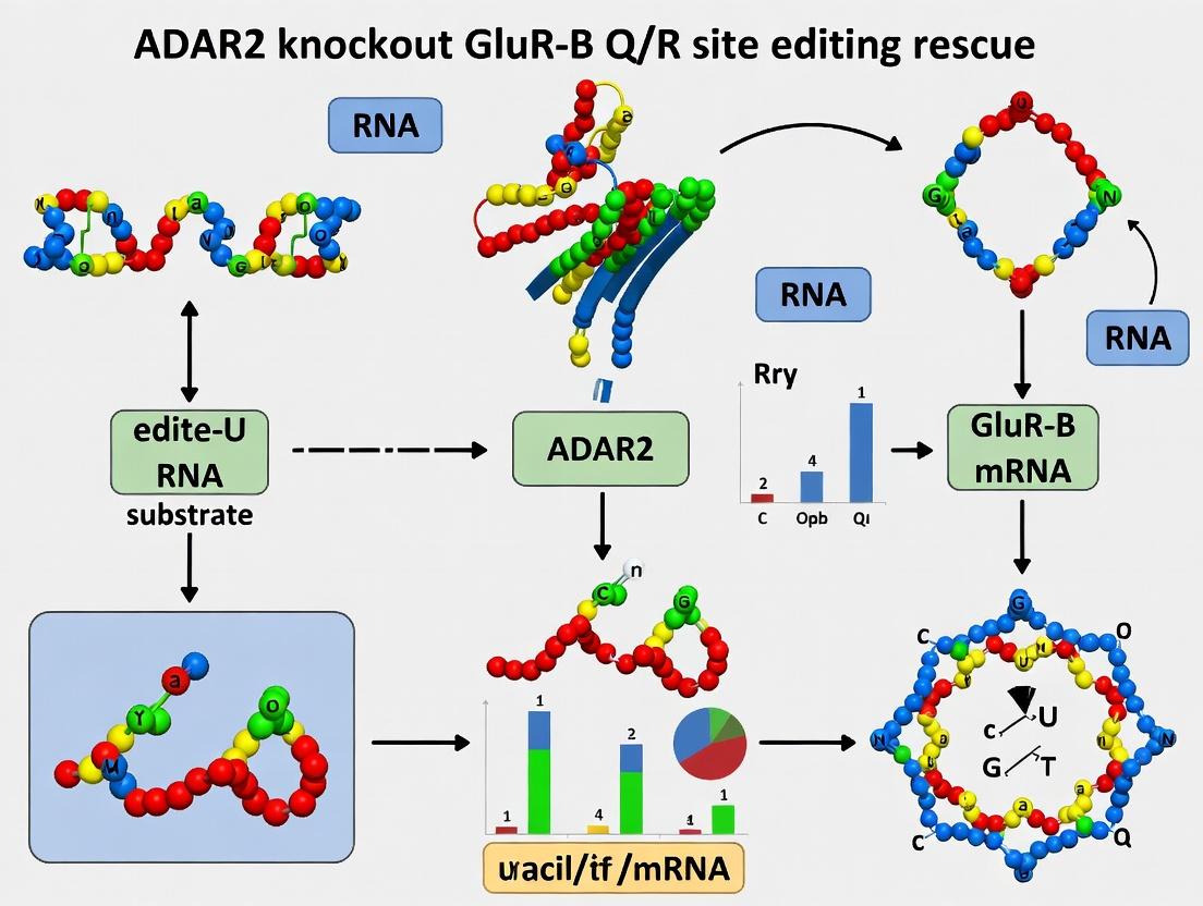
Unlocking Epilepsy Therapeutics: How ADAR2 Knockout Rescues GluR-B Q/R Editing in Neurons
This article provides a comprehensive analysis of the therapeutic strategy involving ADAR2 knockout to rescue RNA editing at the GluR-B Q/R site, a critical determinant of AMPA receptor calcium permeability.
Unlocking Epilepsy Therapeutics: How ADAR2 Knockout Rescues GluR-B Q/R Editing in Neurons
Abstract
This article provides a comprehensive analysis of the therapeutic strategy involving ADAR2 knockout to rescue RNA editing at the GluR-B Q/R site, a critical determinant of AMPA receptor calcium permeability. Aimed at researchers and drug development professionals, we explore the foundational molecular biology, detail cutting-edge methodologies like CRISPR-Cas9 and AAV delivery, troubleshoot common experimental pitfalls, and validate findings through comparative studies with other editing rescue approaches. The synthesis offers a roadmap for translating this precise genetic intervention into novel treatments for neurological disorders such as epilepsy and ALS.
The Molecular Nexus: Understanding ADAR2, GluR-B Q/R Site Editing, and Neuronal Excitability
This whitepaper details the core molecular players and mechanisms of RNA editing by ADAR2, an adenosine deaminase acting on RNA. The functional and mechanistic understanding of ADAR2 is framed as an essential foundation for a critical line of research: the rescue of lethal phenotypes in ADAR2 knockout models. Specifically, this research focuses on rectifying the failure to edit a single critical adenosine in the mRNA of the GluR-B (Gria2) subunit of the AMPA receptor—the Q/R site (CAG->CIG, resulting in a Gln to Arg change). Unedited GluR-B(Q) results in Ca2+-permeable AMPA receptors, leading to neuronal excitotoxicity and death. Rescue strategies, therefore, aim to restore site-specific editing through exogenous ADAR2 expression, engineered ADAR variants, or antisense oligonucleotides (ASOs) to re-establish normal receptor function and validate therapeutic targets for related neurological disorders.
Core Mechanics of ADAR2-Mediated RNA Editing
ADAR2 catalyzes the hydrolytic deamination of adenosine (A) to inosine (I) within double-stranded RNA (dsRNA) substrates. Inosine is interpreted by the cellular machinery as guanosine (G), leading to A-to-I RNA editing.
Key Functional Domains:
- dsRNA Binding Domains (dsRBDs): Typically three domains that recognize and bind the duplex structure of the RNA substrate, providing specificity but not strict sequence selectivity.
- Deaminase Domain: Contains the catalytic core with a conserved zinc-binding motif (HXE-Xn-CXXC) essential for the hydrolytic deamination reaction.
- Nuclear Localization Signal (NLS): Directs the enzyme to the nucleus, where editing primarily occurs.
Editing Requirements:
- A dsRNA structure formed by base-pairing between the editing site and a complementary cis-acting sequence (often in an adjacent intron for pre-mRNA editing).
- Specific, though degenerate, sequence preferences around the target adenosine (e.g., 5' neighbor preference is often a U or A, 3' neighbor is often a G for GluR-B Q/R site).
- For the GluR-B Q/R site, a critical intronic cis-element called the "Ecs" (editing site complementary sequence) located ~1500 nucleotides downstream forms an imperfect duplex with the exon containing the Q/R codon.
Quantitative Data on ADAR2 and GluR-B Editing
Table 1: Key Quantitative Parameters of ADAR2 Function and GluR-B Q/R Editing
| Parameter | Typical Value / Finding | Experimental Context / Significance | |
|---|---|---|---|
| ADAR2 Knockout Lethality | Postnatal day ~P20 | Mice die from seizures and neurodegeneration due to unedited GluR-B(Q). | |
| GluR-B Q/R Site Editing Efficiency | >99% in wild-type brain | Near-complete editing is required for normal physiology. In ADAR2-/- mice, editing falls to ~0%. | |
| Ca2+ Permeability (Relative to Na+) | Edited GluR-B(R): ~0.1 | Unedited GluR-B(Q): ~1.0 | PCa/PNa ratio. Unedited receptors are highly Ca2+ permeable, leading to excitotoxicity. |
| Rescue Survival with Edited GluR-B | Full lifespan | Knock-in mice with a constitutively edited (Arg codon) GluR-B allele are viable and healthy, even in an ADAR2-/- background. | |
| Rescue by ADAR2 cDNA Transgene | Variable, dose-dependent | Partial restoration of editing (e.g., 50-80%) can significantly extend lifespan and mitigate pathology. | |
| Deamination Catalytic Rate (kcat) | ~1-10 min^-1 | In vitro measurements vary by substrate. | |
| dsRNA Binding Affinity (Kd) | Low nM range | Depends on dsRNA length and structure. |
Table 2: Experimental Rescue Strategies & Outcomes in ADAR2-/- Models
| Rescue Strategy | Delivery Method | Editing Efficiency Restored | Phenotypic Rescue Outcome | Key Reference (Example) |
|---|---|---|---|---|
| GluR-B(R) Knock-in | Germline genetic modification | 100% (genomic) | Complete rescue; mice viable and normal. | Higuchi et al., Science (2000) |
| Wild-type ADAR2 cDNA | Transgenic overexpression | 40-90% (region-dependent) | Significant life extension; reduced seizures. | Higuchi et al., Nat Neurosci (2000) |
| Engineered Hyperactive ADAR2 | Viral vector (AAV) to CNS | >80% at Q/R site | Robust rescue of editing and survival. | Current research focus: Katrekar et al., Nat Biotechnol (2019) |
| ASO-guided Endogenous ADAR | ASO injection | Targeted upregulation | Promising preclinical data for site-directed rescue. | Current research focus: Sinnamon et al., Nucleic Acids Res (2020) |
Detailed Experimental Protocols
Protocol 1: Measuring GluR-B Q/R Site Editing Efficiency (Gold Standard)
- Objective: Quantify the percentage of GluR-B mRNA transcripts edited at the Q/R site.
- Method: RT-PCR followed by restriction fragment length polymorphism (RFLP) or direct sequencing.
- RNA Isolation & cDNA Synthesis: Extract total RNA from brain region of interest (e.g., hippocampus). Perform reverse transcription with random hexamers or gene-specific primer.
- PCR Amplification: Amplify a ~200-300 bp fragment spanning the edited Q/R site (exon 11) from the GluR-B cDNA. Use high-fidelity polymerase.
- RFLP Analysis:
- The Q/R site A-to-I edit (genomically a CAG for Gln) creates a BbvI restriction site (GCAGC) in the cDNA when edited to CIG (read as CGG). The genomic/unedited sequence (CAG) is not cut.
- Digest the purified PCR product with BbvI.
- Run products on a high-resolution agarose or polyacrylamide gel.
- Quantification: Use densitometry on gel images. Edited cDNA yields two smaller bands; unedited cDNA remains uncut. % Editing = (intensity of cut bands) / (total intensity of all bands) x 100.
- Alternative: Pyrosequencing or Sanger Sequencing Trace Analysis provides more precise quantitation.
Protocol 2: Assessing Functional Rescue by Electrophysiology
- Objective: Measure Ca2+ permeability of AMPA receptors in neurons from rescue models.
- Method: Whole-cell patch-clamp recording to determine the current-voltage (I-V) relationship.
- Neuron Preparation: Prepare acute brain slices or cultured hippocampal neurons from ADAR2-/- mice with/without the rescue construct.
- Solutions: Use intracellular and extracellular solutions designed to isolate AMPA receptor-mediated currents (e.g., include antagonists for NMDA and GABA receptors). Vary the holding potential from -60 mV to +40 mV.
- Stimulation & Recording: Apply a selective AMPA receptor agonist (e.g., kainate or AMPA) and record the evoked current at each voltage.
- Analysis:
- Plot the current amplitude against the holding potential (I-V curve).
- Edited GluR-B(R)-containing receptors exhibit a linear or outwardly rectifying I-V relationship due to block by endogenous polyamines.
- Unedited GluR-B(Q)-containing receptors show a doubly rectifying I-V curve (inward current at negative potentials) due to high Ca2+ permeability and polyamine block.
- The degree of rectification (e.g., rectification index) quantitatively reflects the proportion of edited vs. unedited receptors.
Signaling Pathway & Experimental Workflow Visualizations
Diagram 1: Pathogenesis and Rescue in ADAR2 Knockout
Diagram 2: ADAR2 Catalytic Mechanism at GluR-B Q/R Site
The Scientist's Toolkit: Research Reagent Solutions
Table 3: Essential Reagents for ADAR2/GluR-B Editing Research
| Reagent / Material | Function / Application | Key Details / Example |
|---|---|---|
| ADAR2 Knockout Mouse Model | In vivo model to study consequences of lost editing and test rescue strategies. | Available from repositories (e.g., JAX). Homozygotes die ~P20. |
| GluR-B(R) Knock-in Mouse | Genetic control proving the sufficiency of Q/R site editing for rescue. | Constitutively edited allele; viable on ADAR2-/- background. |
| AAV-ADAR2 Expression Vectors | Viral delivery for CNS-specific rescue of ADAR2 expression. | Serotypes like AAV9 or AAV-PHP.eB for broad CNS transduction. |
| Engineered "Hyper" ADAR2 Variants | Enhanced editing efficiency for more effective phenotypic rescue. | e.g., ADAR2dd(E488Q) with mutated dsRBDs for reduced non-specific binding. |
| Q/R Site Editing Reporter Assays | High-throughput screening for editing efficiency modulators. | Plasmid with GluR-B exon/intron minigene and a measurable output (e.g., luciferase restoration via editing). |
| Anti-GluR-B Antibodies | Immunohistochemistry/Western blot to assess protein expression and localization. | Both pan-GluR-B and antibodies distinguishing Q/R edits are valuable. |
| BbvI Restriction Enzyme | Key reagent for RFLP analysis of Q/R site editing status. | Cuts the sequence GCAGC, created by the A-to-I edit in cDNA. |
| Polyamine Toxins (e.g., Philanthotoxin) | Electrophysiological probes to assess Ca2+ permeability. | More strongly block Ca2+-permeable (unedited) AMPA receptors. |
| Site-Directed ASOs | Modulate editing by recruiting endogenous ADAR to specific sites. | Chemically modified (e.g., 2'-O-methyl, MOE) oligonucleotides complementary to target region and cis-element. |
AMPA-type glutamate receptors (AMPARs) mediate the majority of fast excitatory synaptic transmission in the mammalian central nervous system. Their functional properties, including ion permeability, are highly regulated. A quintessential example of this regulation is the post-transcriptional editing of the GluR-B (Gria2) subunit mRNA at the Q/R site (position 607). This single amino acid change from a glutamine (Q) to an arginine (R) in the pore-lining second transmembrane domain (M2) fundamentally alters the receptor's biophysical properties, rendering heteromeric AMPARs containing the edited GluR-B subunit impermeable to Ca2+. This review, framed within the context of research into rescuing ADAR2 knockout phenotypes via enforced GluR-B Q/R site editing, provides a technical dissection of this critical molecular switch.
The Molecular Mechanism of Q/R Site Editing
The Q/R site editing is catalyzed exclusively by the RNA-specific adenosine deaminase ADAR2. The process converts an adenosine (A) to inosine (I) in the pre-mRNA, which is read as guanosine (G) during translation, resulting in the codon change from CAG (Gln) to CGG (Arg).
Key Experiment: Quantifying Editing Efficiency
- Protocol: Total RNA is extracted from brain regions (e.g., hippocampus, cortex) or cultured neurons. RT-PCR is performed using GluR-B-specific primers flanking the Q/R site. The PCR product is subjected to direct Sanger sequencing. The editing efficiency is calculated by measuring the relative peak heights of A (unedited) versus G (edited) at the specific nucleotide position on the chromatogram. Alternatively, more quantitative methods include pyrosequencing or deep sequencing of the amplicon.
- Quantitative Data: Editing efficiency is developmentally regulated and approaches ~100% in the adult mammalian brain.
Table 1: Q/R Site Editing Efficiency Across Development and Tissues
| Tissue / Condition | Approximate Editing Efficiency | Method of Detection |
|---|---|---|
| Embryonic Brain | ~80% | RT-PCR, Sequencing |
| Adult Brain (Cortex/Hippocampus) | >99% | RT-PCR, Sequencing |
| ADAR2 -/- Mouse Brain | <1% | RT-PCR, Sequencing |
| HEK293T (without ADAR2) | 0% | RT-PCR, Sequencing |
| HEK293T + ADAR2 transfection | >95% | RT-PCR, Sequencing |
Biophysical and Functional Consequences
The introduction of a positively charged arginine residue in the pore has profound effects.
Key Experiment: Electrophysiological Characterization of Ca2+ Permeability
- Protocol: Wild-type (Gln) or edited (Arg) GluR-B is co-expressed with other AMPAR subunits (e.g., GluR-A) in Xenopus oocytes or HEK293 cells. Whole-cell voltage-clamp recordings are performed. Ca2+ permeability is assessed using:
- Current-Voltage (I-V) Relationship: Inward rectification indicates low Ca2+ permeability (edited GluR-B present). Linear or outwardly rectifying I-V relationships indicate high Ca2+ permeability (unedited GluR-B).
- Fractional Ca2+ Current (Pf): Measured using fluorescence imaging with Ca2+-sensitive dyes (e.g., Fura-2) simultaneously with electrophysiology, or by calculating reversal potentials in different extracellular ionic solutions (e.g., using the Goldman-Hodgkin-Katz equation).
- Quantitative Data: The presence of edited GluR-B (R) reduces the fractional Ca2+ current (Pf) of AMPAR channels from >0.7 to <0.1.
Table 2: Biophysical Properties of AMPARs with Edited vs. Unedited GluR-B
| Property | AMPARs with GluR-B(Q) (Unedited) | AMPARs with GluR-B(R) (Edited) |
|---|---|---|
| Ca2+ Permeability (Pf) | High (>0.7) | Very Low (<0.1) |
| Current-Voltage (I-V) Relation | Linear or Outward Rectification | Strong Inward Rectification |
| Single-Channel Conductance | Higher (~20 pS) | Lower (~10 pS) |
| Block by Polyamines (e.g., Spermine) | Weak | Potent (at positive potentials) |
| Mg2+ Block | Weak | Strong |
Physiological and Pathophysiological Context: ADAR2 Knockout and Rescue
ADAR2 knockout mice die by postnatal week 3-4 due to seizures and neurodegeneration, a direct consequence of the failure to edit the GluR-B Q/R site, leading to increased Ca2+-permeable AMPARs and excitotoxicity.
Key Rescue Experiment: Genetic Introduction of Pre-Edited GluR-B
- Protocol: To prove that the lethal phenotype of ADAR2-/- mice is solely due to lack of GluR-B Q/R editing, a "rescue" allele of Gria2 (GluR-B) is genetically engineered. This allele (GluR-BR/R) contains a point mutation that encodes arginine (R) at the Q/R site, bypassing the need for ADAR2 editing. This allele is bred into the ADAR2-/- background. Phenotypic outcomes (survival, seizure activity, neuronal death, electrophysiology) are compared between ADAR2-/-, ADAR2-/-::GluR-BR/R, and wild-type mice.
- Quantitative Data: ADAR2-/-::GluR-BR/R mice are fully viable, show normal life spans, and exhibit normalized electrophysiological profiles, confirming the central thesis.
Diagram Title: ADAR2 Knockout Phenotype Rescue via Genetic GluR-B Q/R Editing
The Scientist's Toolkit: Research Reagent Solutions
Table 3: Essential Reagents for GluR-B Q/R Site and AMPAR Permeability Research
| Reagent / Material | Function / Application | Example / Note |
|---|---|---|
| ADAR2 Knockout Mouse Model | In vivo model to study consequences of loss of Q/R editing. | Available from Jackson Laboratory. Phenotype requires rescue. |
| GluR-B(R) "Rescue" Knock-in Mouse | Control model to prove specificity of ADAR2 phenotype to GluR-B editing. | Genetically engineered to express arginine at Q/R site. |
| Site-specific Anti-GluR-B Antibodies | Distinguish edited (R) vs. unedited (Q) protein in immunohistochemistry/Western blot. | Commercial availability is limited; often custom-made. |
| Selective AMPAR Antagonists | To isolate AMPAR-mediated currents in electrophysiology. | CNQX, NBQX, GYKI 53655. |
| Polyamine Toxins (e.g., JSTX, PhTx) | Use-dependent blockers of Ca2+-permeable (GluR-B-lacking) AMPARs. | Key pharmacological tool to probe subunit composition. |
| Ca2+-sensitive Fluorescent Dyes | To measure fractional Ca2+ currents (Pf) in imaging experiments. | Fura-2, Fluo-4, Indo-1. Rationetric dyes preferred. |
| Q/R Site Editing Reporter Plasmids | In vitro assay to quantify ADAR2 activity or screen modulators. | Plasmid with GluR-B minigene sequence and a readout (e.g., fluorescence). |
| Recombinant ADAR2 Protein | For in vitro biochemical studies of editing kinetics and specificity. | Purified from E. coli or insect cell expression systems. |
Diagram Title: The GluR-B Q/R Editing Pathway and Functional Consequences
Adenosine-to-inosine (A-to-I) RNA editing, catalyzed by the ADAR family of enzymes, is a critical post-transcriptional modification essential for neurological health. The failure to edit the Q/R site in exon 11 of the GluA2 subunit (encoded by the GRIA2 gene, often referred to as GluR-B) of AMPA receptors represents a paradigmatic example of an editing failure with profound pathological consequences. This whitepaper examines the causal link between deficient ADAR2-mediated editing at this site and the subsequent molecular cascades leading to neuronal hyperexcitability (epileptogenesis) and progressive neuronal death (neurodegeneration), framed within the context of research demonstrating rescue by ADAR2 restoration.
The Central Dogma: ADAR2, GluA2(Q/R), and Neuronal Ca²⁺ Permeability
AMPA receptors lacking an edited GluA2 subunit are permeable to Ca²⁺. Under normal conditions, ADAR2-mediated conversion of a codon for glutamine (Q) to arginine (R) at the Q/R site (position 607) renders GluA2-containing AMPA receptors impermeable to Ca²⁺. ADAR2 knockout (KO) or dysfunction leads to the expression of unedited GluA2(Q)-containing, Ca²⁺-permeable AMPA receptors (CP-AMPARs).
Table 1: Consequences of GluA2 Q/R Site Editing Status
| Editing Status | GluA2 Subunit | AMPA Receptor Ca²⁺ Permeability | Primary Consequence |
|---|---|---|---|
| Edited (Normal) | GluA2(R) | Impermeable | Controlled neuronal signaling, low intracellular Ca²⁺ |
| Unedited (Pathological) | GluA2(Q) | Permeable | Elevated intracellular Ca²⁺, excitotoxicity |
Molecular Pathways from Editing Failure to Pathology
The expression of CP-AMPARs initiates a feed-forward cascade of neurotoxicity.
Diagram 1: Pathogenic cascade from ADAR2 failure to disease phenotypes.
Key Supporting Evidence from ADAR2 KO Rescue Studies
Landmark studies using conditional ADAR2 knockout mice (ADAR2⁻/⁻) and subsequent rescue models provide direct causal evidence.
Table 2: Summary of Key Findings from ADAR2 KO & Rescue Models
| Experimental Model | Key Phenotype Observed | Rescue Intervention | Outcome of Rescue | Reference Key Findings |
|---|---|---|---|---|
| Forebrain-specific ADAR2⁻/⁻ | Progressive epileptic seizures, neurodegeneration (esp. in CA3/CA1), premature death. | Transgenic expression of edited GluA2(R) under its own promoter. | Prevention of seizures, neurodegeneration, and early death. | Proof that GluA2 unediting is the primary cause of pathology. |
| Motor neuron-specific ADAR2⁻/⁻ | ALS-like symptoms, motor neuron degeneration. | Viral delivery of ADAR2 or edited GluA2. | Delayed symptom onset, extended lifespan, reduced motor neuron loss. | Links editing failure to ALS pathophysiology. |
| ADAR2⁻/⁻ / GluA2(R) Rescue | N/A (Pre-emptive rescue). | Genetically introduced GluA2(R) allele. | Complete phenotypic rescue; mice are viable and normal. | Confirms singular critical role of GluA2 Q/R site editing. |
Detailed Experimental Protocols
Protocol: Genotyping and Validation of ADAR2 Conditional KO Mice
Objective: To generate and validate forebrain-specific ADAR2 knockout mice. Materials: ADAR2 floxed mice (Adar2ᶠˡᵒˣ/ᶠˡᵒˣ), CaMKIIα-Cre transgenic mice, PCR reagents, primers for Adar2 floxed allele and Cre. Procedure:
- Breeding: Cross Adar2ᶠˡᵒˣ/ᶠˡᵒˣ mice with CaMKIIα-Cre mice to obtain Adar2ᶠˡᵒˣ/ᶠˡᵒˣ; CaMKIIα-Cre⁺ (cKO) and control littermates.
- Genomic DNA Extraction: From tail snips using a standard phenol-chloroform or kit-based method.
- PCR Amplification:
- For Adar2 floxed allele: Use primers F1: 5'-CTGCCTGGTAGAGGTGCTTG-3', R1: 5'-GGTCCCAGAGTCCAAACTGC-3'. Product: ~300 bp (wild-type), ~350 bp (floxed).
- For Cre transgene: Use standard Cre primers. Product: ~100 bp.
- Confirmation: Confirm tissue-specific ADAR2 loss via western blot or immunohistochemistry on hippocampal lysates/sections at 3-4 weeks.
Protocol: Analysis of GluA2 RNA Editing Status
Objective: To quantify the percentage of edited vs. unedited GluA2 RNA at the Q/R site. Materials: TRIzol reagent, cDNA synthesis kit, PCR reagents, restriction enzyme BbvI (cuts edited sequence, CC⁺TCAGC), capillary electrophoresis system. Procedure (RT-PCR/RFLP):
- RNA Isolation & cDNA Synthesis: Extract total RNA from hippocampus/prefrontal cortex. Synthesize cDNA using random hexamers.
- PCR Amplification: Amplify GluA2 exon 11 region using specific primers. Cycle conditions: 94°C 30s, 60°C 30s, 72°C 45s (35 cycles).
- Restriction Digest: Purify PCR product. Digest with BbvI at 37°C for 3 hours. Edited cDNA is cut; unedited is not.
- Quantification: Analyze fragments via capillary electrophoresis (e.g., Bioanalyzer). Calculate % editing = (cut fragment peak area / total peak area) * 100.
Protocol: Viral-Mediated Rescue in Mouse Hippocampus
Objective: To rescue pathology by delivering edited GluA2(R) postnatally. Materials: AAV9 vector encoding GluA2(R) under a neuron-specific promoter (e.g., hSyn), stereotaxic apparatus, Hamilton syringe. Procedure:
- Virus Preparation: Purify and titer AAV9-hSyn-GluA2(R)-eGFP. Use AAV9-hSyn-eGFP as control.
- Stereotaxic Surgery (P21 mice): Anesthetize and secure mouse in stereotaxic frame. Target CA1 hippocampus coordinates (from Bregma): AP -2.0 mm, ML ±1.5 mm, DV -1.5 mm.
- Injection: Bilaterally inject 1 µL of virus (≥1x10¹³ vg/mL) at 0.1 µL/min. Leave needle in place for 10 min post-injection.
- Phenotypic Monitoring: Monitor for seizure activity via EEG/video recording 4-8 weeks post-injection. Perform histological analysis for neurodegeneration (Fluoro-Jade C, Nissl) and transgene expression (GFP).
The Scientist's Toolkit: Key Research Reagent Solutions
Table 3: Essential Reagents for ADAR2/GluA2 Editing Research
| Reagent/Material | Function/Application | Example/Provider Notes |
|---|---|---|
| ADAR2 Floxed Mice | Enables tissue-specific knockout of ADAR2. Critical for modeling disease and rescue. | Available from repositories (e.g., JAX: Stock #017582). |
| CaMKIIα-Cre Mice | Drives Cre expression in forebrain excitatory neurons. Used to generate cKO model. | Common line: B6.Cg-Tg(Camk2a-cre)T29-1Stl/J (JAX: #005359). |
| AAV9-hSyn-GluA2(R) | Viral vector for in vivo rescue experiments. Neuron-specific promoter ensures targeted expression. | Can be custom-produced from viral core facilities (e.g., Penn Vector Core, Addgene). |
| BbvI Restriction Enzyme | Key for RFLP assay to distinguish edited (cut) from unedited (uncut) GluA2 PCR products. | Available from NEB (R0161S). |
| Anti-GluA2 (N-terminal) Antibody | For immunohistochemistry/western blot to assess total GluA2 protein levels and localization. | Clone 6C4 (Millipore MAB397) is widely used for IHC. |
| Anti-ADAR2 Antibody | To confirm loss of ADAR2 protein in KO models. | Available from Santa Cruz (sc-73408) or Proteintech (13850-1-AP). |
| Fluoro-Jade C Stain | Histochemical marker for degenerating neurons. Quantifies rescue of neurodegeneration. | Available from Millipore (AG325). |
| Telemetry EEG/EMG Systems | For continuous, long-term monitoring of seizure activity in freely moving mice. | Systems from Data Sciences International (DSI) or NeuroNexus. |
Table 4: Quantitative Outcomes in ADAR2 cKO and Rescue Models
| Parameter | ADAR2 cKO Mice (Mean ± SD) | ADAR2 cKO + GluA2(R) Rescue (Mean ± SD) | Wild-Type Control (Mean ± SD) | Assay/Method |
|---|---|---|---|---|
| GluA2 Q/R Site Editing (%) | <5% | >95%* | >99% | RT-PCR/RFLP |
| Onset of Lethal Seizures | 5.2 ± 1.1 weeks | >52 weeks (no seizures) | >52 weeks (no seizures) | Video/EEG monitoring |
| Neuronal Density (CA1) | 45% ± 8% of WT | 92% ± 6% of WT | 100% (Baseline) | Nissl staining |
| Fluoro-Jade C+ Cells (Hippocampus) | 250 ± 50 cells/section | 15 ± 10 cells/section | 5 ± 5 cells/section | Histochemistry |
| Ca²⁺ Influx (Relative Fluorescence) | 3.5 ± 0.5 fold over WT | 1.1 ± 0.2 fold over WT | 1.0 (Baseline) | Fura-2AM imaging in acute slices |
*Depends on efficiency of rescue method (transgenic vs. viral).
The failure of ADAR2-mediated RNA editing at the GluA2 Q/R site is a direct molecular cause of epileptogenesis and neurodegeneration, primarily mediated by aberrant Ca²⁺ influx through CP-AMPARs. Research utilizing ADAR2 knockout models, and crucially, the subsequent rescue by restoring either ADAR2 function or the edited GluA2(R) subunit, provides definitive proof of concept. This pathway presents a validated, albeit challenging, therapeutic target for conditions characterized by secondary editing deficiencies, such as certain forms of epilepsy, ALS, and ischemic brain injury. Future drug development efforts may focus on ADAR2 enzyme enhancement, modulation of CP-AMPAR trafficking, or gene therapy-based delivery of edited subunits.
This whitepaper consolidates evidence from foundational ADAR2 knockout models to recent human pathological studies, framing the findings within the overarching thesis that targeted rescue of GluR-B Q/R site editing represents a viable therapeutic strategy for conditions characterized by ADAR2 dysfunction, such as sporadic Amyotrophic Lateral Sclerosis (sALS).
Table 1: Phenotypic Characterization of ADAR2-/- Mouse Models
| Parameter | ADAR2-/- (Neuron-Specific) | Wild-Type Control | Measurement Method | Reference / Key Study |
|---|---|---|---|---|
| GluA2 Q/R Site Editing (%) | ~0% (in vulnerable neurons) | ~100% | RT-PCR, Restriction Digest | Higuchi et al., Nature 2000 |
| Onset of Neurological Symptoms | ~P14 | None | Behavioral observation | Hideyama et al., J Neurosci 2010 |
| Lifespan | ~P20 (lethal) | Normal | Survival curve | Higuchi et al., Nature 2000 |
| CA3 Hippocampal Neuron Loss | Severe by P20 | None | Histology (Nissl, TUNEL) | Hideyama et al., J Neurosci 2012 |
| Motor Neuron Degeneration | Present in spinal cord | Absent | Immunohistochemistry (ChAT) | Yamashita et al., Sci Rep 2013 |
| AMPA Receptor Ca2+ Permeability | Dramatically Increased | Normal | Electrophysiology (I-V curve) | Higuchi et al., Nature 2000 |
Table 2: Human Pathology Findings in sALS
| Pathological Marker | sALS Spinal Motor Neurons | Control Motor Neurons | Association with ADAR2 | Key Study |
|---|---|---|---|---|
| ADAR2 Protein Expression | Significantly reduced or absent | Normal | Direct loss | Hideyama et al., Nat Neurosci 2010; Aizawa et al., 2010 |
| GluA2 Q/R Site Editing | Decreased (<100%) | ~100% | Consequence of ADAR2 loss | Hideyama et al., Nat Neurosci 2010 |
| TDP-43 Pathology | Present (cytoplasmic aggregates) | Absent | Co-localizes with ADAR2-deficient neurons | Aizawa et al., Brain Res 2010; Yamashita et al., 2012 |
| Neuronal Vulnerability | Selective vulnerability of ADAR2-low neurons | N/A | Correlated | Hideyama et al., Nat Neurosci 2010 |
Table 3: Rescue Experiment Outcomes
| Rescue Strategy | Model System | Key Outcome Metric | Result vs. Unrescued ADAR2-/- | Reference |
|---|---|---|---|---|
| Neuron-Specific GluA2(Q) Transgene | ADAR2-/- mouse | Lifespan | Extended to >6 months | Higuchi et al., Nature 2000 |
| AAV-mediated ADAR2 Delivery | ADAR2-/- mouse (adult) | CA3 Neuron Survival | Significant protection | Hideyama et al., J Neurosci 2012 |
| Antisense ODN (to mask Q/R site) | Cell culture model | Ca2+ Influx | Reduced to wild-type levels | - |
| CRISPR/dCas13-ADAR2 Fusion | In vitro neuronal culture | Editing Efficiency at Q/R site | Restoration to >90% | Recent proof-of-concept studies |
Detailed Experimental Protocols
Protocol 1: Genotyping and Phenotypic Analysis of ADAR2-/- Mice
- Mouse Model Generation: Utilize mice with loxP-flanked Adar2 alleles crossed with Nestin-Cre or CamKIIα-Cre drivers for forebrain/postnatal neuron-specific knockout.
- Genomic DNA Isolation: From tail clips (P7), using a standard phenol-chloroform or commercial kit extraction.
- PCR Genotyping:
- Primers:
- ADAR2-flox Common (C): 5'-GAG TTG CTC TGG CTG TTA CC-3'
- ADAR2-flox Wild-type (W): 5'-CAC CAT GTA AAG GTG GCA GG-3'
- ADAR2-flox Mutant (M): 5'-CTT CCC ATC TGC ACA CCA C-3'
- Reaction Mix: 1μL DNA, 0.2μM each primer, 12.5μL 2x PCR Master Mix, ddH2O to 25μL.
- Cycling Conditions: 94°C 3 min; 35 cycles of [94°C 30s, 60°C 30s, 72°C 45s]; 72°C 5 min.
- Product Analysis: 2% agarose gel. Wild-type allele: ~300bp. Floxed allele: ~400bp. Cre-positive recombined allele: ~500bp.
- Primers:
- Phenotypic Monitoring: Record weight daily from P10. Assess for signs of seizures, hypoactivity, and hindlimb clasping from P14.
Protocol 2: Analysis of GluA2 RNA Editing Status
- RNA Extraction & cDNA Synthesis: Isolate total RNA from microdissected brain regions (e.g., hippocampus, cortex) or laser-captured motor neurons using TRIzol. Perform reverse transcription with random hexamers.
- Q/R Site-Specific RT-PCR:
- Design primers flanking the Q/R site (GluA2 exon 11).
- PCR: Use high-fidelity polymerase. Cycle as above.
- Purification: Gel-purify the PCR product.
- Restriction Fragment Length Polymorphism (RFLP) Analysis:
- The Q/R site (CAG for unedited Gln, CIG for edited Arg) alters a BbvI restriction site.
- Digest: Incubate 10μL purified PCR product with 5U BbvI at 37°C for 3 hours.
- Analysis: Run digest on a 3% high-resolution agarose or 10% polyacrylamide gel. Edited (R) transcript: Cut by BbvI, yields two smaller fragments. Unedited (Q) transcript: Not cut, remains one band.
- Quantification: Use densitometry software (ImageJ) to calculate the percentage of edited transcripts: Intensity of cut bands / Total intensity x 100.
Protocol 3: Immunohistochemical Analysis of Human ALS Post-Mortem Tissue
- Tissue Preparation: Use 10% formalin-fixed, paraffin-embedded spinal cord sections (8μm thick) from sALS and control patients.
- Antigen Retrieval: Deparaffinize, rehydrate. Perform heat-induced epitope retrieval in citrate buffer (pH 6.0) for 20 minutes.
- Blocking: Incubate in 3% H2O2 to quench endogenous peroxidases, then in 5% normal goat serum/0.1% Triton X-100 for 1 hour.
- Primary Antibody Incubation: Co-incubate overnight at 4°C with:
- Mouse anti-ADAR2 antibody (1:200)
- Rabbit anti-phosphorylated TDP-43 antibody (1:1000)
- Secondary Detection: Apply appropriate biotinylated secondary antibodies (1:500, 1 hour), then ABC Elite kit, and develop with DAB (brown) and Vector SG (gray/blue) substrates for double labeling.
- Analysis: Quantify ADAR2-positive and TDP-43 pathology-positive motor neurons under light microscopy. Correlate loss of ADAR2 immunoreactivity with TDP-43 mislocalization.
Diagrams
ADAR2 Editing Pathway and Dysfunction
ADAR2 Knockout Rescue Workflow
The Scientist's Toolkit: Research Reagent Solutions
| Reagent / Material | Provider Examples | Function in ADAR2/GluA2 Research |
|---|---|---|
| ADAR2-floxed (Adar2tm1.1Kyou) Mice | JAX Mice, RIKEN BRC | Foundational genetic model for conditional, neuron-specific ADAR2 knockout. |
| Neuron-Specific Cre Drivers (Nestin-Cre, CamKIIα-Cre) | JAX Mice | Enables temporal and spatial control of ADAR2 deletion in the nervous system. |
| AAV9-hADAR2 Viral Vector | Custom from Vector Cores (e.g., Penn, UNC) | Key rescue tool for delivering functional human ADAR2 gene in vivo. |
| Anti-ADAR2 Antibody (Clone 5F6) | Sigma-Aldrich, Abcam | Validated antibody for detecting ADAR2 protein loss in mouse and human tissue via IHC/WB. |
| Anti-GluA2 (N-terminal) Antibody | Millipore | Identifies total GluA2 protein; used to confirm subunit expression regardless of editing state. |
| Anti-phospho TDP-43 (pS409/410) | Cosmo Bio, Proteintech | Critical for co-pathology analysis in human sALS tissues, marking pathological inclusions. |
| BbvI Restriction Enzyme | NEB | Essential for RFLP assay to quantitatively assess GluA2 Q/R site editing percentage. |
| Laser Capture Microdissection System | Arcturus, Leica | Allows precise isolation of vulnerable motor neurons from spinal cord sections for RNA analysis. |
| Ca2+-Sensitive Dyes (e.g., Fura-2 AM) | Thermo Fisher | For functional assessment of Ca2+ permeability in neurons derived from models or after rescue. |
| CRISPR/dCas13-ADAR Recruiting System | Commercial kits (e.g., Addgene plasmids) | Emerging tool for precise, RNA-targeted editing rescue at the Q/R site without genomic DNA alteration. |
This whitepaper details the core hypothesis within a broader thesis investigating the rescue of GluR-B Q/R site editing in ADAR2 knockout models. The central paradox under investigation is that while global ADAR2 knockout abolishes most adenosine-to-inosine (A-to-I) editing, including the critical GluA2 (GluR-B) Q/R site, specific neuronal contexts (e.g., select interneuron populations, certain stress conditions, or developmental timepoints) exhibit a restoration of editing at this site. The prevailing hypothesis posits that compensatory mechanisms, potentially involving ADAR1 isoforms or novel ADAR3 activity, are recruited in a context-dependent manner to maintain essential editing events crucial for neuronal viability and circuit function.
Table 1: Key Quantitative Findings from ADAR2 Knockout Rescue Studies
| Experimental Model | Editing % at GluA2 Q/R Site (Wild Type) | Editing % at GluA2 Q/R Site (ADAR2 KO) | Editing % in "Rescue Context" (e.g., Specific Neuron Type) | Proposed Compensatory Factor | Reference / Key Study |
|---|---|---|---|---|---|
| Global ADAR2 KO (Mouse, whole brain) | >99% | <5% | N/A | None | Higuchi et al., 2000 |
| Conditional KO in Hippocampal CA1 Pyramidal Neurons | >99% | ~10% | N/A | None (cell-autonomous defect) | Brusa et al., 1995 |
| Analysis of Parvalbumin+ Interneurons in Global KO | >99% | <5% (bulk) | ~40-60% (single-cell RNA-seq) | ADAR1 (p110 isoform) | Sadeghi et al., 2022 (bioRxiv) |
| Cultured Cortical Neurons under ER Stress | >99% | <10% (basal) | ~30-50% (post-stress) | ADAR1 (p150 induced) | Oakes et al., 2017 |
| ADAR2 KO; ADAR1 p150 Overexpression | >99% | <5% | ~80% (transfected cells) | ADAR1 (p150 isoform) | Horsch et al., 2011 |
Table 2: Expression Levels of Candidate Compensatory Enzymes in Rescue Contexts
| Context | ADAR1 p110 (Relative Expression) | ADAR1 p150 (Relative Expression) | ADAR3 (Relative Expression) | Editing Restoration Efficiency |
|---|---|---|---|---|
| Wild-Type Cortex | 1.0 (baseline) | Low | Moderate | N/A |
| Global ADAR2 KO Cortex | 1.1 | 2.5x increase | 3.0x increase | Low (bulk) |
| PV+ Interneurons in KO | 1.8x increase | 1.2x increase | 5.0x increase | High |
| Neuronal ER Stress | 1.0 | 4.0x increase | 2.0x increase | Moderate-High |
Experimental Protocols for Key Studies
Protocol 1: Single-Neuron RNA Sequencing to Identify Rescue Contexts
Objective: To identify specific neuronal populations that retain GluA2 Q/R site editing in a global ADAR2 knockout background.
- Tissue Preparation: Rapidly dissect brain regions (e.g., cortex, hippocampus) from adult ADAR2-/- and wild-type littermates.
- Neuron Dissociation: Use papain-based enzymatic digestion followed by gentle trituration to create a single-cell suspension.
- FACS Sorting: Label cells with fluorescent antibodies against neuronal markers (e.g., NeuN) and interneuron markers (e.g., Parvalbumin, Somatostatin). Sort individual labeled neurons into 96-well plates containing lysis buffer.
- cDNA Synthesis & Amplification: Perform reverse transcription with oligo-dT primers, followed by template-switching and PCR amplification (SMART-Seq v4) to generate sufficient cDNA from single cells.
- Library Prep & Sequencing: Fragment cDNA, prepare libraries (Nextera XT), and sequence on an Illumina platform (≥ 50M reads per cell).
- Analysis: Align reads to the genome. Identify A-to-I editing sites using variant-calling pipelines (e.g., JACUSA2) with strict filters for RNA-seq artifacts. Correlate editing levels with cell-type-specific gene expression clusters.
Protocol 2: Inducing ER Stress to Probe Compensatory Editing
Objective: To test if cellular stress can induce ADAR1 and restore Q/R editing in ADAR2-deficient neurons.
- Primary Neuronal Culture: Establish cortical or hippocampal neuron cultures from E18 ADAR2-/- and wild-type rat/mouse pups.
- Stress Induction: At DIV 14, treat cultures with ER stress inducers: Tunicamycin (2.5 µg/mL, inhibits N-glycosylation) or Thapsigargin (1 µM, depletes ER Ca2+). Include DMSO vehicle controls.
- Time-Course Harvest: Harvest cells at 0, 6, 12, 24, and 48 hours post-treatment in TRIzol.
- Molecular Analysis:
- RNA: Extract RNA, perform RT-qPCR for ADAR1 p150, ADAR1 p110, ADAR3, and ER stress markers (BiP, CHOP).
- Editing Assessment: Design primers flanking the GluA2 Q/R site. Perform RT-PCR, gel-purify the product, and clone into a sequencing vector. Sanger sequence ≥50 clones per condition to quantify editing percentage.
- Functional Validation: Perform whole-cell patch-clamp recording on transfected (e.g., GFP+) stressed neurons to assess Ca2+ permeability of AMPA receptors, a functional readout of Q/R editing.
Protocol 3: CRISPR/dCas13b-Mediated Recruitment to Test Enzyme Sufficiency
Objective: To test if targeted recruitment of ADAR1 or ADAR3 to the GluA2 transcript can restore editing in ADAR2 KO cells.
- Construct Design: Fuse catalytically inactive dCas13b to the deaminase domain of human ADAR1 (E912A) or ADAR3. Clone this into a neuronal expression vector.
- gRNA Design: Design and clone 3-5 guide RNAs (gRNAs) targeting sequences ~50 nt upstream/downstream of the Q/R site in the Gria2 pre-mRNA.
- Co-transfection: Transfect HEK293T cells (lacking endogenous Q/R editing) and primary ADAR2 KO neurons with the dCas13b-ADAR construct, gRNA plasmid, and a reporter plasmid expressing GluA2 pre-mRNA.
- Editing Quantification: Harvest RNA 48-72 hrs post-transfection. Use RT-PCR followed by deep amplicon sequencing (MiSeq) to quantify editing efficiency at the Q/R site and assess off-target editing within the transcript.
- Control: Repeat with dCas13b alone (no deaminase domain) and with scrambled gRNA.
Visualization: Pathways and Workflows
Diagram Title: The Core Paradoxical Rescue Pathway
Diagram Title: Experimental Workflow to Discover Rescue Contexts
The Scientist's Toolkit: Research Reagent Solutions
Table 3: Essential Reagents and Tools for Investigating the Paradox
| Reagent / Tool | Provider Examples | Function in Research | Key Application in This Field |
|---|---|---|---|
| ADAR2 Floxed (Adarb2tm1) Mice | Jackson Laboratory, KOMP | Provides a conditional allele for cell-type-specific knockout of ADAR2. | Generating global and neuron-subtype-specific KO models to define rescue contexts. |
| Parvalbumin-2A-Cre Mouse Line | Jackson Laboratory (B6;129P2-Pvalbtm1(cre)Arbr/J) | Drives Cre recombinase expression in parvalbumin-positive interneurons. | Crossing with ADAR2 floxed mice to delete ADAR2 specifically in PV+ cells. |
| SMART-Seq v4 Ultra Low Input RNA Kit | Takara Bio | Amplifies cDNA from single cells or low-input samples for full-length RNA-seq. | Enabling transcriptome and editome analysis from single sorted neurons. |
| anti-ADAR1 (p150 specific) Antibody | Sigma-Aldrich (AMAB91559), Santa Cruz (sc-73408) | Detects the inducible p150 isoform of ADAR1 via Western blot or IHC. | Confirming upregulation of ADAR1 p150 in stress-induced rescue contexts. |
| Tunicamycin, Thapsigargin | Cayman Chemical, Tocris | Well-characterized inducers of ER stress. | Probing the stress-responsive pathway that may activate compensatory editing. |
| RiboMAX Large Scale RNA Production System (T7) | Promega | Produces large amounts of specific RNA transcripts in vitro. | Generating substrate RNAs (e.g., GluA2 R/G site) for testing recombinant ADAR enzyme activity. |
| dCas13b-ADAR1 (E912A) Fusion Plasmid | Addgene (Plasmid #103863, base vector) | Enables targeted RNA editing via guide RNA recruitment. | Testing sufficiency of ADAR1's deaminase domain to edit the Q/R site when recruited. |
| Gria2 Q/R Site Editing Reporter | Custom synthesis (e.g., GenScript) | Plasmid expressing GluA2 exon 11 with surrounding intronic sequence. | A standardized substrate for quantifying editing efficiency in cellular assays. |
| JACUSA2 Bioinformatics Tool | GitHub Repository | A variant callset filter specifically designed for identifying RNA-DNA differences (e.g., A-to-I editing) from NGS data. | Accurately quantifying editing levels from single-cell or bulk RNA-seq datasets. |
Blueprint for Intervention: Step-by-Step Methods for ADAR2 Knockout and Editing Rescue
Within the broader thesis on rescuing GluR-B Q/R site editing in ADAR2 knockout models, the selection of an appropriate experimental system is a critical foundational decision. This guide provides an in-depth technical comparison of in vitro (primary neurons, immortalized cell lines) and in vivo (mouse models) systems, focusing on their application to study RNA editing mechanisms, neuronal excitability, and potential therapeutic rescue strategies.
Core Quantitative Comparison of Model Systems
Table 1: Comparative Analysis of Model Systems for ADAR2/GluR-B Research
| Parameter | Immortalized Cell Lines (e.g., HEK293, N2a) | Primary Neuronal Cultures | Mouse Models (In Vivo) |
|---|---|---|---|
| Physiological Relevance | Low; simplified, non-neuronal or cancerous origin. | High; post-mitotic, polarized, express native synaptic machinery. | Highest; intact circuitry, systemic physiology, blood-brain barrier. |
| Genetic Manipulation Ease | Very High; highly transfertable, amenable to CRISPR, stable lines easily generated. | Moderate; challenging transfection, limited by primary nature. | Complex; requires transgenic/knockout breeding or viral/in utero delivery. |
| Throughput & Cost | High throughput; low cost per experiment. | Medium throughput; moderate cost, requires animal sourcing. | Low throughput; very high cost (housing, breeding, genotyping). |
| Experimental Timeline | Days to weeks for assay setup. | 1-3 weeks for culture maturation. | Months to years for model generation/phenotyping. |
| Key Readouts for ADAR2 Rescue | Q/R site editing efficiency (RT-PCR, Sanger sequencing), ADAR2 expression (WB, qPCR). | Editing efficiency, AMPA receptor electrophysiology (patch clamp), synaptic protein localization. | Editing efficiency in brain regions, seizure susceptibility, behavioral deficits, neurodegeneration. |
| Major Limitation | Lack of native neuronal context and network activity. | Absence of intact brain circuitry and systemic factors. | Complexity of data interpretation due to whole-organism compensatory mechanisms. |
Detailed Experimental Protocols
In Vitro Protocol: Assessing Q/R Site Editing Rescue in Primary Cortical Neurons
- Aim: To evaluate the efficacy of an ADAR2 rescue construct (e.g., AAV-ADAR2) on GluR-B Q/R site editing in a relevant neuronal context.
- Materials: Primary cortical neurons from E16-18 ADAR2 KO or WT mouse pups, poly-D-lysine coated plates, Neurobasal/B27 medium, AAV-DJ serotype viruses encoding ADAR2 and GFP, transfection reagents (for comparative lines).
- Method:
- Isolate and plate cortical neurons at desired density (e.g., 50,000 cells/cm²).
- At Day in vitro (DIV) 3-5, infect cultures with AAV-ADAR2 (experimental) or AAV-GFP (control) at an MOI of 10⁵.
- Harvest RNA at DIV 14-21 using a column-based kit with DNase I treatment.
- Perform reverse transcription with random hexamers.
- Amplify the GluR-B Q/R site region (around exon 11) using specific primers. A sample primer pair: Forward: 5'-CAG ACA GCT ACC TGG GTT TC-3', Reverse: 5'-GAA GTC GAT GGC TTC TTG TG-3'.
- Purify PCR product and submit for Sanger sequencing. Quantify editing efficiency by calculating the ratio of the 'G' peak (edited, codes for Arg) to the 'A' peak (unedited, codes for Gln) at the relevant nucleotide position using chromatogram analysis software (e.g., EditR or manual quantification).
In Vivo Protocol: Phenotypic Rescue in ADAR2 Knockout Mice
- Aim: To test if viral delivery of ADAR2 prevents early-onset epilepsy and neurodegeneration in ADAR2 KO mice.
- Materials: ADAR2 KO mice (postnatal day 0-2 for intracerebroventricular injection or adult for stereotaxic surgery), AAV9-PHP.eB-ADAR2 (systemic) or AAV9-ADAR2 (direct CNS), stereotaxic apparatus, temperature control pad.
- Method (Neonatal ICV Injection):
- On P0-P2, cryoanesthetize ADAR2 KO pups.
- Inject 2-3 µL of high-titer AAV (>1x10¹³ vg/mL) into each lateral ventricle using a fine glass capillary needle.
- Return pups to dam and monitor until weaning.
- At 3-4 weeks and 12+ weeks, assess cohorts for: a) Editing Rescue: Isolate RNA from hippocampus/cortex, sequence Q/R site as above. b) Seizure Phenotype: Continuous video-EEG monitoring for 48-72 hours to quantify seizure frequency/duration. c) Histopathology: Perfuse, section brains, stain with NeuN and GFAP antibodies to assess neuronal loss and astrogliosis in vulnerable regions (hippocampus, striatum).
Visualization of Research Pathways and Workflows
Diagram Title: In Vitro Editing Rescue Validation Workflow
Diagram Title: Multi-Phenotype Rescue Assessment In Vivo
The Scientist's Toolkit: Essential Research Reagents & Materials
Table 2: Key Reagent Solutions for ADAR2/GluR-B Editing Research
| Reagent/Material | Function/Application | Example/Note |
|---|---|---|
| ADAR2 Knockout Mice | In vivo model exhibiting deficient Q/R editing, epilepsy, and neurodegeneration. | Available from repositories (e.g., JAX). Homozygous (Adar2-/-) are essential. |
| AAV Vectors (Serotype 9, PHP.eB, DJ) | Delivery of ADAR2 rescue constructs in vitro and in vivo. | PHP.eB for systemic adult delivery; AAV9 for neonatal/CNS; AAV-DJ for high in vitro infectivity. |
| Gria2 (GluR-B) Editing Site Primers | PCR amplification of the critical exon 11 region for sequencing analysis. | Must flank the Q/R site (position 607 in transcript). Validate specificity via gel electrophoresis. |
| RNA Extraction Kit with DNase I | High-quality RNA isolation from cells or brain tissue for editing analysis. | Must include robust DNase treatment to eliminate genomic DNA contamination. |
| Sanger Sequencing Service/Kit | Gold-standard for quantifying site-specific RNA editing ratios. | Internal primer for sequencing gives highest quality chromatogram at site of interest. |
| Patch Clamp Electrophysiology Setup | Functional assessment of AMPA receptor Ca2+ permeability in rescued neurons. | Measures current-voltage relationship; inward rectification indicates edited, Ca2+-impermeable receptors. |
| Video-EEG Monitoring System | Quantitative assessment of seizure phenotype rescue in behaving mice. | Critical for correlating molecular rescue with functional network outcome. |
| NeuN & GFAP Antibodies | Immunohistochemical evaluation of neuronal survival and glial response. | Quantifies neurodegeneration and astrogliosis in hippocampus and striatum of KO mice. |
CRISPR-Cas9 Strategies for Conditional and Constitutive ADAR2 Knockout
This guide details technical strategies for generating ADAR2 knockout models using CRISPR-Cas9, specifically within the context of research aimed at rescuing GluR-B Q/R site editing. ADAR2 (Adenosine Deaminase Acting on RNA 2) is the primary enzyme responsible for the site-selective editing of the GluA2 (GluR-B) subunit mRNA at the Q/R site (position 607). This editing event, which converts a glutamine (Q) codon to an arginine (R) codon, is critical for regulating calcium permeability of AMPA receptors. Disruption of this edit leads to neuronal hyperexcitability and is implicated in conditions like epilepsy and ALS. A constitutive ADAR2 knockout is lethal in mice due to seizure-related death, underscoring the necessity of conditional knockout (cKO) strategies for viable postnatal studies. The core thesis research involves creating and utilizing these knockout models to investigate molecular and phenotypic consequences and to test rescue strategies (e.g., via exogenous ADAR1 or engineered editors) to restore GluR-B Q/R editing and normal neuronal function.
Core CRISPR-Cas9 Strategies: Constitutive vs. Conditional
Constitutive ADAR2 Knockout
This strategy aims to disrupt the Adar2 gene in all cells and throughout development. It is typically used for embryonic studies or to generate cell lines.
Target Site Selection: Critical exons for knockout are those encoding essential functional domains, such as the catalytic deaminase domain (e.g., exons 5-7 in mouse Adar2). Frameshift mutations introduced here lead to premature stop codons and nonsense-mediated decay (NMD) of the mRNA.
Example sgRNA Targets (Mouse Adar2):
- Exon 5:
5'-GACCTGCACCGTGCCGCCGGAGG-3'(PAM: TGG) - Exon 7:
5'-GCTTCGCTGCGGGGCACGAGGGG-3'(PAM: AGG)
Delivery: For mice, Cas9 mRNA/protein and sgRNAs are microinjected into zygotes. For cell lines, plasmid or RNP complexes are delivered via transfection/electroporation.
Conditional ADAR2 Knockout (Floxed Allele)
This strategy uses Cre-loxP technology to generate a tissue-specific or inducible knockout. Two loxP sites are inserted to flank a critical exon (e.g., exon 5).
Dual-sgRNA Strategy: Two sgRNAs are designed to create double-strand breaks (DSBs) at the boundaries of the target exon. Co-delivery with donor DNA templates containing homologous arms and loxP sites facilitates homology-directed repair (HDR).
Key Components:
- 5' sgRNA/loxP: Inserts loxP site upstream of the critical exon.
- 3' sgRNA/loxP: Inserts loxP site downstream of the critical exon.
- Donor DNA: A single-stranded or double-stranded DNA template containing the loxP sites, homology arms (~800 bp each), and often a selectable marker (e.g., floxed neo cassette) removed by later breeding to a Flp deleter strain.
Table 1: Efficiency Metrics for CRISPR-Cas9 Mediated ADAR2 Editing in Mouse Models (Representative Data)
| Strategy | Target Exon | Delivery Method | Founder Generation Efficiency (%) | Germline Transmission Rate (%) | HDR Efficiency for cKO (loxP insertion, %)* | Key Phenotype (Constitutive) |
|---|---|---|---|---|---|---|
| Constitutive KO | Exon 5 | Zygote microinjection (Cas9 RNP) | 65-85 | ~50 | N/A | Lethal by P20; seizures; loss of GluR-B Q/R editing (>95% reduction). |
| Conditional KO | Exon 5 (Floxed) | Zygote microinjection (Cas9 RNP + ssODN donors) | 40-60 | ~30 | 10-20 | Viable; exon excision upon Cre recombination leads to identical phenotype to constitutive KO in targeted tissue. |
| Cell Line KO | Exon 7 | Lipofection (plasmid) | N/A | N/A | N/A | >90% editing efficiency in bulk population; near-complete loss of Q/R site editing in neuronal cell lines. |
*HDR efficiency is significantly lower than NHEJ-mediated indel efficiency. Successful loxP insertion on both sides requires careful screening.
Table 2: Molecular Validation Data from ADAR2 Knockout Models
| Model / Tissue | ADAR2 mRNA (qPCR, % WT) | GluR-B Q/R Site Editing (%, via PCR/RFLP or Sequencing) | Calcium Permeability (Relative to WT) | Reference |
|---|---|---|---|---|
| WT Brain (Hippocampus) | 100% | 99.5 ± 0.3% | 1.0 (Baseline) | Higuchi et al., 2000 |
| Adar2 -/- (P15 Brain) | <5% | 15.2 ± 4.1% | >5x increase | Higuchi et al., 2000 |
| Adar2 cKO (CamKIIα-Cre; Forebrain) | <10% (in neurons) | ~20% (in forebrain) | Markedly increased | - |
| Rescue with ADAR1 overexpression (in cKO neurons) | N/A | Restored to ~85% | Normalized | - |
Detailed Experimental Protocols
Protocol: Generating a ConditionalAdar2Floxed Mouse Model via CRISPR-HDR
Objective: Insert loxP sites flanking exon 5 of the mouse Adar2 gene.
Materials: See "Scientist's Toolkit" below.
Procedure:
Design & Preparation:
- Design two high-efficiency sgRNAs targeting intronic sequences ~200-300bp upstream and downstream of exon 5. Verify specificity via CHOPCHOP or CRISPRscan.
- Synthesize sgRNAs as chemically modified synthetic RNAs (e.g., 2'-O-methyl-3'-phosphorothioate) for stability.
- Design and synthesize single-stranded oligodeoxynucleotide (ssODN) donor templates. Each donor should contain a loxP site (34 bp:
ATAACTTCGTATA ATGTATGC TATACGAAGTTAT) flanked by ~100-150bp homology arms matching the genomic sequence at the cut site. Include silent mutations in the PAM sequence to prevent re-cutting.
Microinjection into Mouse Zygotes:
- Prepare injection mix: 50 ng/µL Cas9 protein, 25 ng/µL of each sgRNA, and 50 ng/µL of each ssODN donor in nuclease-free microinjection buffer.
- Harvest zygotes from superovulated C57BL/6 females.
- Perform pronuclear microinjection using standard techniques.
- Culture injected zygotes to the two-cell stage and transfer them into pseudopregnant foster females.
Genotyping and Founder Screening:
- Extract genomic DNA from founder (F0) pup tails.
- Perform two primary PCR assays:
- Assay 1 (5' loxP): Primers flanking the 5' insertion site. WT band: ~400bp. Successful HDR band: ~434bp.
- Assay 2 (3' loxP): Primers flanking the 3' insertion site.
- For PCR-positive founders, sequence the amplicons to confirm correct loxP integration and absence of aberrant indels.
- Screen for founders with loxP sites on both sides (trans configuration). These are rare; founders with loxP on one allele only are bred to transmit each modified allele separately, then crossed to generate the floxed allele.
Establishing the Line:
- Breed positive F0 founders to wild-type mice to test for germline transmission.
- Cross mice carrying the individual 5' and 3' loxP alleles to generate mice homozygous for the floxed allele (Adar2flox/flox).
- Cross Adar2flox/flox mice with a tissue-specific Cre driver line (e.g., Nestin-Cre for neural progenitors, CamKIIα-Cre for forebrain excitatory neurons) to generate conditional knockouts.
Protocol: Validating GluR-B Q/R Site Editing
Objective: Quantify the percentage of edited GluA2 mRNA at the Q/R site.
Method: RNA Isolation, RT-PCR, and Restriction Fragment Length Polymorphism (RFLP) Analysis.
Procedure:
- RNA Extraction & cDNA Synthesis: Isolate total RNA from brain region or cells of interest using TRIzol. Treat with DNase I. Synthesize cDNA using random hexamers and reverse transcriptase.
- PCR Amplification: Design primers spanning the Q/R site (rodent position 607) in GluA2 (Gria2) mRNA. A common forward primer and a reverse primer that spans an intron to exclude genomic DNA amplification are used.
- PCR Cycle: 94°C 2 min; 35 cycles of [94°C 30s, 60°C 30s, 72°C 45s]; 72°C 5 min.
- RFLP Digestion: The Q (CAG) to R (CIG, read as CGG) editing creates a BbvI restriction site.
- Purify the PCR product.
- Set up digestion: 10µL PCR product, 2µL 10x BbvI buffer, 1µL BbvI enzyme (10 U), 7µL H2O. Incubate at 37°C for 2 hours.
- Gel Electrophoresis & Quantification: Run digested products on a 3% agarose gel.
- Unedited (Q): No cut, one band (~150bp).
- Edited (R): Cut, two bands (~100bp and ~50bp).
- Quantify band intensities using ImageJ. % Editing = (Intensity of (cut band 1 + cut band 2) / Total intensity of all bands) * 100.
Diagrams
Diagram 1: ADAR2 KO Pathophysiological Cascade
Diagram 2: CRISPR Workflow for ADAR2 KO Models
The Scientist's Toolkit: Research Reagent Solutions
Table 3: Essential Materials for CRISPR-Cas9 ADAR2 Knockout Experiments
| Item / Reagent | Function / Purpose | Example Product / Note |
|---|---|---|
| High-Fidelity Cas9 Nuclease | Creates DSB at genomic target specified by sgRNA. | Alt-R S.p. Cas9 Nuclease V3 (IDT); ensures high on-target activity. |
| Chemically Modified sgRNAs | Guides Cas9 to target sequence; chemical modifications enhance stability and reduce immunogenicity in embryos. | Alt-R CRISPR-Cas9 sgRNA (IDT) with 2'-O-methyl-3'-phosphorothioate ends. |
| ssODN Donor Templates | Serves as HDR template for precise insertion of loxP sites; ultramer format recommended for long (>100nt) designs. | Alt-R HDR Donor Oligos (IDT), PAGE purified. |
| Microinjection Buffer | Stable, nuclease-free buffer for delivering RNP complexes into zygotes. | 10 mM Tris, 0.1 mM EDTA, pH 7.5, filtered. |
| Genotyping Primers | Validates correct integration of loxP sites and identifies Cre-mediated excision. | Design one primer outside homology arm and one inside loxP for specific detection. |
| Cre Recombinase Driver Lines | Mediates tissue-specific deletion of floxed exon in cKO models. | Examples: CamKIIα-Cre (forebrain excitatory neurons), Nestin-Cre (CNS precursors), hGFAP-Cre (astrocytes). |
| BbvI Restriction Enzyme | Key reagent for RFLP analysis of GluA2 Q/R site editing status. | New England Biolabs (NEB) BbvI (10 U/µL). |
| DNase I, RNase-free | Critical for removing genomic DNA contamination during RNA isolation for editing assays. | Thermo Scientific, RNase-free DNase I. |
This whitepaper provides a technical guide for designing Adeno-Associated Virus (AAV) capsids for brain-region-specific gene delivery, framed within the critical context of rescuing ADAR2-dependent GluR-B Q/R site editing. In research focused on conditions like Amyotrophic Lateral Sclerosis (ALS) and epilepsy, where ADAR2 knockout leads to aberrant, unedited GluR2(Q) subunit expression and subsequent neuronal excitotoxicity, precise delivery of therapeutic payloads (e.g., functional ADAR2) to affected brain regions (e.g., motor cortex, hippocampus) is paramount. This document outlines contemporary strategies, data, and protocols for achieving this targeting specificity.
Core Targeting Strategies and Quantitative Data
Table 1: Primary AAV Capsid Engineering Strategies for CNS Targeting
| Strategy | Core Mechanism | Key Advantages | Reported Tropism Shift/Enhancement (in vivo) |
|---|---|---|---|
| Directed Evolution | In vivo selection of peptide-displaying AAV libraries. | Discovery of de novo tropisms; Bypasses need for complete receptor knowledge. | AAV-PHP.eB: ~40x increase in CNS transduction over AAV9 in mice. AAV.CAP-B10: Enhanced cortical and spinal motor neuron transduction. |
| Rational Design / Capsid Mutagenesis | Site-directed mutagenesis of surface-exposed capsid residues. | Can refine existing tropism; improves transduction efficiency or evasion of neutralizing antibodies. | AAV9.47: Point mutations (Y731F) increase brain endothelial cell transcytosis. |
| Pseudotyping & Chimeras | Combining VP proteins from different AAV serotypes. | Blends properties of parent serotypes (e.g., AAV2 ITR + AAV9 capsid). | AAV2/9 commonly used for broad CNS transduction. |
| Cre-Recombination-Based AAV Targeting (CReAT) | Incorporation of lox sites in capsid gene; cell-specific Cre drives capsid switching. | Unprecedented cell-type specificity within a region. | In Cre+ mice, ~1000-fold specificity for target cell type reported. |
Table 2: Selected Engineered AAV Capsids for Brain Region Targeting
| Capsid Variant | Parent Serotype | Target Brain Region/Cell Type | Primary Receptor/Mechanism (if known) | Key Application in ADAR2/GluR-B Context |
|---|---|---|---|---|
| AAV-PHP.eB | AAV9 | Widespread cortex, striatum, cerebellum. | Binds to LY6A; murine-specific. | Broad rescue in global ADAR2 deficiency models. |
| AAV-PHP.S | AAV9 | Peripheral & spinal motor neurons. | Binds to LY6C1. | Targeted delivery to spinal cord for ALS-related phenotypes. |
| AAV-AS | AAV9 | Hippocampal neurons. | Selected via directed evolution in non-human primates. | Hippocampus-specific rescue for memory/ seizure phenotypes. |
| AAV-F | Ancestral | Striatum (medium spiny neurons). | Derived from ancestral reconstruction. | Targeted delivery for striatal-related circuits. |
| AAV2-retro | AAV2 | Efficient retrograde transport to projecting neurons. | Binds to mannose-6-phosphate; interacts with IGF2R. | Access neurons projecting to injection site (e.g., cortical neurons projecting to spinal cord). |
Detailed Experimental Protocols
Protocol 1: In Vivo Selection for Brain-Targeting AAV Capsids (Directed Evolution)
Objective: Isolate novel AAV capsids with enhanced tropism for specific brain regions from a randomized peptide-display library. Materials: AAV peptide library (7-12mer inserts in VP1/VP2 loop), C57BL/6 mice, NGS reagents, PCR setup, tissue homogenizer. Procedure:
- Library Packaging: Package the AAV genome library (containing a barcoded payload) with the capsid library to create a viable virus library.
- Systemic Administration: Inject the pooled library (~1e11 vg/mouse) intravenously into a cohort of mice.
- Target Tissue Harvest: At 2-4 weeks post-injection, perfuse animals and dissect the target brain region (e.g., hippocampus, motor cortex). Collect control tissues (liver, spleen).
- DNA Extraction & Barcode Amplification: Isolate total DNA from tissues. Use PCR to amplify the barcode region from the AAV genome.
- Next-Generation Sequencing (NGS): Sequence the amplified barcodes. Capsids enriched in the target brain region, but depleted in off-target organs, are identified by barcode frequency analysis.
- Validation & Iteration: Clone enriched capsid sequences, produce new virus, and validate tropism in a secondary round of animal experiments. Repeat selection for multiple rounds.
Protocol 2: Validation of AAV-Mediated ADAR2 Rescue in a Focal Brain Region
Objective: Assess functional rescue of GluR-B Q/R editing following region-specific AAV-ADAR2 delivery in an ADAR2 knockout model. Materials: ADAR2 KO mice, stereotaxic frame, engineered AAV (e.g., AAV-AS-CBh-ADAR2), control AAV-GFP, RNA isolation kit, RT-PCR reagents, restriction enzyme BbvI. Procedure:
- Stereotaxic Injection: Anesthetize ADAR2 KO mice. Inject 1-2 µL of high-titer AAV-ADAR2 or AAV-GFP (≥1e13 vg/mL) into the target region (e.g., hippocampal CA1) using coordinates from a brain atlas.
- Incubation: Allow 4-6 weeks for robust transgene expression.
- Tissue Sectioning: Perfuse and section the brain. Confirm injection site and expression via GFP fluorescence or ADAR2 immunohistochemistry.
- RNA Isolation & cDNA Synthesis: Micro-dissect the injected region. Isolate total RNA and synthesize cDNA.
- GluR-B Q/R Site Editing Analysis:
- Perform PCR to amplify a cDNA fragment spanning the GluR-B Q/R site (from exon 11 to 13).
- Subject the PCR product to BbvI restriction digest. The edited site (R; AGA) is resistant to BbvI, while the unedited site (Q; CAG) is cut.
- Analyze fragments on an agarose gel. Quantify the ratio of cut (unedited) to uncut (edited) DNA.
- Functional Assessment: Perform region-specific electrophysiological recordings (e.g., measure Ca2+ permeability of AMPA receptors) or behavioral assays relevant to the rescued region.
Visualization: Pathways and Workflows
Title: ADAR2 Knockout Rescue Pathway via AAV Delivery
Title: AAV Capsid Selection and Validation Workflow
The Scientist's Toolkit
Table 3: Essential Research Reagent Solutions for AAV Targeting Studies
| Reagent / Material | Function & Relevance | Example/Note |
|---|---|---|
| Peptide-Display AAV Library | Starting point for directed evolution. Provides genetic diversity for in vivo selection of novel tropisms. | Commercially available or custom libraries with random 7-mer inserts in VP3. |
| High-Serotype AAV Helper Plasmids | For production of specific or engineered capsids. Essential for packaging the genome into the selected capsid. | pAAV2/9, pAAV2/PHP.eB, or pAAV2/retro helper plasmids. |
| Rep-Cap Plasmid with Barcoded Genome | Contains AAV ITRs flanking a ubiquitous promoter (CAG/CMV), a unique molecular barcode, and a reporter gene (GFP). Links capsid identity to barcode via NGS. | Critical for tracking capsid fate in pooled selections. |
| Stereotaxic Injection System | Enables precise, reproducible delivery of AAV into deep brain structures for validation and therapeutic testing. | Includes stereotaxic frame, microsyringe pump, and fine-gauge Hamilton syringe. |
| BbvI Restriction Enzyme | Key reagent for assessing GluR-B Q/R site editing status. Cuts unedited (CAG) but not edited (AGA) PCR products. | Alternative methods include Sanger sequencing pyrosequencing or deep sequencing of the site. |
| LY6A/LY6C1 Antibodies | For validating the mechanism of novel capsids (e.g., PHP.eB, PHP.S) in murine models. | Human orthologs are not identified, limiting translation; highlights need for NHP-derived capsids. |
| Next-Generation Sequencing Service/Kit | For barcode sequencing and analysis from directed evolution experiments. Identifies enriched capsid variants. | |
| Primary Neurons from Target Region | For in vitro screening of candidate capsids prior to in vivo use. | e.g., Hippocampal or cortical neuronal cultures. |
This whitepaper provides a technical guide for quantifying the efficiency of ADAR-mediated RNA editing, specifically within the context of rescuing the fatal phenotype of ADAR2 knockout mice through GluR-B Q/R site editing. The AMPA receptor subunit GluR-B (Gria2) requires 100% editing at its Q/R site (CAG to CIG) to render Ca²⁺-impermeable receptors. ADAR2 knockout mice die from seizures due to unedited GluR-B, but this is rescued by an editing-competent GluR-B transgene. Precise quantification of editing efficiency is therefore critical for evaluating rescue strategies, including ADAR enzyme engineering, delivery, and pharmacological activation in therapeutic contexts.
Core Quantitative Assays: Principles and Comparison
Two primary methods are employed to quantify Q/R site editing efficiency: Restriction Fragment Length Polymorphism (RFLP) and Deep Sequencing (Next-Generation Sequencing, NGS). The choice depends on required sensitivity, throughput, and cost.
Table 1: Comparison of RFLP and Deep Sequencing for Q/R Site Editing Analysis
| Feature | RFLP Analysis | Deep Sequencing (NGS) |
|---|---|---|
| Principle | Exploits creation/abolishment of a restriction site by the C-to-I (read as C-to-U in cDNA) edit. | Direct, high-throughput sequencing of cDNA amplicons covering the site. |
| Sensitivity | Moderate (~5-10% variant detection limit). Can be enhanced with capillary electrophoresis. | Very high (<0.1% variant frequency detection). |
| Throughput | Low to medium. Suitable for small sample numbers. | Very high. Enables multiplexing of hundreds of samples. |
| Primary Output | Percentage editing calculated from band intensities on a gel. | Percentage editing from aligned sequence reads; provides full allelic distribution. |
| Key Advantage | Low cost, technically simple, requires standard lab equipment. | Unparalleled sensitivity and accuracy, detects rare edits and surrounding variations. |
| Key Limitation | Indirect measurement; requires specific restriction site; less accurate for low or high editing levels. | Higher cost, complex data analysis, requires specialized bioinformatics. |
| Ideal Use Case | Initial screening, validation of high-efficiency edits, labs without NGS access. | Definitive quantification, detecting low-frequency editing events, research requiring ultra-high precision. |
Table 2: Typical Quantitative Data from ADAR2 Knockout Rescue Experiments
| Sample Type | Expected Q/R Editing % (RFLP) | Expected Q/R Editing % (NGS) | Biological Interpretation |
|---|---|---|---|
| Wild-Type (WT) Brain | ~100% | 99.5 - 100% | Full endogenous ADAR2 activity. |
| ADAR2 KO Brain (No Rescue) | 0% | 0 - 0.1% | Complete loss of editing at this site. |
| ADAR2 KO + GluR-B (R) Transgene | ~100%* | ~100%* | Perfect rescue by pre-edited transgene. |
| ADAR2 KO + Therapeutic ADAR Delivery (e.g., AAV-ADAR1) | Variable (e.g., 20-80%) | Variable with precise distribution | Efficacy of the therapeutic editing tool. |
| Peripheral Tissues (with systemic delivery) | Lower than CNS | Lower than CNS, heterogeneous | Indicates biodistribution and tissue-specific efficiency. |
*Editing percentage refers only to the transgene-derived transcripts.
Detailed Experimental Protocols
Protocol 3.1: RFLP Analysis for GluR-B Q/R Site Editing
Principle: The Q/R site edit (C to U in RNA) changes the cDNA sequence from CAG (Gln) to CGG (Arg). This creates a BbvI restriction site (GCAGC). Unedited cDNA remains uncut.
Materials:
- Total RNA from brain region or cell culture.
- DNase I (RNase-free).
- Reverse Transcription Kit.
- PCR primers flanking GluR-B Q/R site (e.g., F: 5'-CACTGTCGTCCTCGTCCTCA-3', R: 5'-GCAGATCCAGACGGAGTACG-3').
- BbvI restriction enzyme and buffer.
- Agarose gel electrophoresis system or capillary electrophoresis instrument.
Procedure:
- RNA Isolation & DNase Treatment: Isolate total RNA using a guanidinium thiocyanate-phenol method. Treat with DNase I to remove genomic DNA contamination.
- Reverse Transcription: Synthesize cDNA using a high-fidelity reverse transcriptase and oligo(dT) or random hexamer primers.
- PCR Amplification: Amplify the ~150-200 bp region encompassing the Q/R site using high-fidelity Taq polymerase. Use the following cycling conditions: 95°C for 3 min; 35 cycles of 95°C for 30s, 60°C for 30s, 72°C for 30s; final extension 72°C for 5 min.
- Restriction Digest: Purify PCR product. Digest 200-500 ng of purified PCR product with BbvI (10 U) in a 20 µL reaction at 37°C for 3 hours.
- Analysis:
- Agarose Gel (2.5-3%): Resolve digested products. Unedited product remains intact (~150-200 bp). Edited product is cut into two fragments (e.g., ~100 bp and ~50 bp).
- Quantification: Use gel imaging software (e.g., ImageJ) to measure band intensities. Calculate editing percentage: % Editing = (Intensity of Cut Fragments) / (Intensity of Cut + Uncput Fragments) * 100.
- Capillary Electrophoresis: For higher accuracy, analyze digest on a bioanalyzer or fragment analyzer. Peak areas correspond to fragment amounts.
Protocol 3.2: Deep Sequencing (Amplicon-Seq) for Editing Efficiency
Principle: cDNA amplicons spanning the Q/R site are barcoded, pooled, and sequenced on an NGS platform (e.g., Illumina MiSeq). Each read is aligned to the reference sequence to call the base at the specific position.
Materials:
- cDNA sample (from Protocol 3.1, steps 1-2).
- Two-step PCR reagents: 1) High-fidelity polymerase for target amplification, 2) Indexing polymerase for attaching barcodes and adapters.
- AMPure XP beads or equivalent for size selection and purification.
- Qubit fluorometer and TapeStation/bioanalyzer for quantification and QC.
- Illumina MiSeq or equivalent sequencer with ≥2x150 bp kit.
Procedure:
- Primary PCR (Target Amplification): Amplify the Q/R site region (amplicon size ~200-250 bp) using primers with overhang adapters. Use minimal cycles (10-15) to reduce PCR errors. Purify amplicons.
- Indexing PCR (Attaching Barcodes): Using the purified primary PCR product as template, perform a second, limited-cycle (5-10 cycles) PCR with unique dual index primers (Nextera XT or equivalent) for each sample.
- Library Pooling & QC: Quantify each indexed library, normalize, and pool equimolarly. Validate pool size and concentration via bioanalyzer and qPCR.
- Sequencing: Load pool onto sequencer to achieve high coverage (>10,000 reads per sample).
- Bioinformatic Analysis:
- Demultiplexing: Assign reads to samples based on barcodes.
- Alignment: Trim adapters and align reads to the reference GluR-B sequence using a sensitive aligner (e.g., BWA, Bowtie2).
- Variant Calling: Use tools like
bcftools mpileupor specialized RNA-editing pipelines (e.g, REDItools, JACUSA2) to identify mismatches relative to the reference genome. The key is to differentiate true editing from SNPs and sequencing errors using statistical filters and by comparing to genomic DNA controls. - Quantification: For the specific Q/R site coordinate, calculate: % Editing = (Number of reads with 'G' (C in cDNA)) / (Total reads covering the position) * 100.
Visualizations
Title: Experimental Workflow for Q/R Editing Quantification
Title: Biological Context of Editing Rescue & Assay Role
The Scientist's Toolkit: Research Reagent Solutions
Table 3: Essential Reagents for Q/R Site Editing Analysis
| Reagent / Kit | Function in Experiment | Key Considerations |
|---|---|---|
| TRIzol Reagent | Simultaneous isolation of high-quality RNA, DNA, and protein from tissue samples (e.g., mouse brain). | Effective for tough tissues; allows downstream validation at multiple molecular levels. |
| High-Capacity cDNA Reverse Transcription Kit | Converts isolated RNA into stable cDNA for subsequent PCR amplification. | Contains RNase inhibitor and random hexamers/oligo(dT) for comprehensive conversion. |
| Phusion High-Fidelity DNA Polymerase | Amplifies the specific GluR-B region around the Q/R site with minimal PCR errors. | Critical for both RFLP and NGS prep to avoid introducing false-positive C-to-T changes. |
| BbvI Restriction Enzyme | Cuts cDNA PCR products only if the Q/R site is edited (CIG->CGG), enabling RFLP analysis. | Specificity and activity must be validated; alternative enzymes exist if polymorphism alters site. |
| Agilent High Sensitivity DNA Kit | Analyzes size distribution and quantifies DNA fragments for NGS library QC and RFLP fragments. | Essential for accurate sizing of digested fragments (RFLP) and final library (NGS). |
| Nextera XT DNA Library Preparation Kit | Rapidly prepares indexed sequencing libraries from amplicons for multiplexed NGS on Illumina. | Streamlines the amplicon-seq workflow; includes bead-based normalization. |
| REDItools / JACUSA2 Software | Specialized bioinformatics tools for accurate identification and quantification of RNA editing events from NGS data. | Must be used with appropriate genomic DNA controls to filter out SNPs and mapping artifacts. |
This whitepaper details the electrophysiological validation of AMPA receptor calcium permeability, a critical parameter in the context of rescuing ADAR2 knockout phenotypes through GluR-B Q/R site editing. The methods described herein are foundational for research aimed at developing therapeutic interventions for neurological conditions linked to aberrant calcium influx, such as ischemia and ALS.
In ADAR2 knockout mice, the failure to edit the GluR-B subunit mRNA at the Q/R site results in the expression of Ca2+-permeable AMPARs (CP-AMPARs) in vulnerable neurons, leading to fatal epileptic seizures and neurodegeneration. The core thesis is that rescuing this editing defect, either via molecular or pharmacological means, requires rigorous functional validation of restored Ca2+ impermeability. Electrophysiology provides the direct, quantitative measure necessary to confirm successful rescue by demonstrating a shift from Ca2+-permeable to Ca2+-impermeable AMPAR phenotypes.
Core Principles: The Calcium Permeability Index
The relative Ca2+ permeability of AMPARs is quantitatively expressed by the permeability ratio (PCa/PCs) or, more commonly, the voltage-dependent polyamine block-derived index. A key metric is the rectification index (RI).
Table 1: Key Permeability Indices and Their Interpretation
| Index | Formula / Description | CP-AMPAR (Unedited GluR-B) | CI-AMPAR (Edited GluR-B) | Typical Measurement Method | |
|---|---|---|---|---|---|
| Rectification Index (RI) | I+40mV / |I-60mV | \ | Low (< 0.5) | High (~1.0) | Whole-cell, symmetrical NaCl |
| Permeability Ratio (PCa/PNa) | Derived from reversal potential shifts (GHK) | ~1.0 - 2.5 | ~0.05 - 0.15 | Bi-ionic potentials (Ca2+ vs Na+) | |
| Polyamine Sensitivity | Degree of inward rectification | Strong (IC50 ~0.1-1 µM) | Weak/None | Application of exogenous spermine or NAS |
Detailed Experimental Protocols
Whole-Cell Recording for Inward Rectification Analysis
This protocol assesses native or recombinant AMPARs in neurons or heterologous cells to determine the rectification phenotype.
Key Reagents & Solutions:
- Internal (Pipette) Solution (mM): 135 CsCl, 10 HEPES, 0.5 EGTA, 2 Mg-ATP, 0.3 Na-GTP, 5 QX-314 (pH 7.3 with CsOH). CsCl enhances conductance; QX-314 blocks Na+ channels and endogenous polyamines.
- External Solution (mM): 140 NaCl, 2.5 KCl, 2 CaCl2, 1 MgCl2, 10 HEPES, 10 Glucose (pH 7.4 with NaOH). Standard physiological saline.
- Agonist Solution: External solution + 100µM AMPA or 100µM kainate. Kainate desensitizes less, allowing stable current measurement.
Procedure:
- Establish whole-cell voltage-clamp configuration (holding potential = -60 mV).
- Apply agonist via fast perfusion system for 500 ms.
- Elicit current-voltage (I-V) relationship using a voltage ramp (e.g., -80 mV to +40 mV over 400 ms) during agonist application.
- Measure peak current at -60 mV (I-60) and +40 mV (I+40).
- Calculate Rectification Index (RI) = I+40 / \|I-60\|.
- Rescue Context: Compare RI in ADAR2 KO neurons transfected with ADAR2 rescue plasmid vs. control. Successful rescue shifts RI toward ~1.
Determination of Relative Ca2+ Permeability (PCa/PCs)
This protocol uses bi-ionic conditions to calculate permeability ratios directly from reversal potentials (Erev).
Key Reagents & Solutions:
- Internal Solution (mM): 133 CsCl, 10 HEPES, 10 Cs-EGTA (pH 7.3 with CsOH). High Cs+ for primary internal cation.
- External Solution 1 (Na+): 150 NaCl, 0.1 CaCl2, 10 HEPES, 10 Glucose (pH 7.4).
- External Solution 2 (Ca2+): 110 CaCl2, 10 HEPES, 10 Glucose (pH 7.4). Low Ca2+ to avoid current saturation.
Procedure:
- Obtain whole-cell configuration.
- Apply kainate (100 µM) in Na+-based external. Record I-V ramp, determine Erev1.
- Switch to Ca2+-based external. Re-apply agonist, record I-V ramp, determine Erev2.
- Calculate PCa/PCs using the generalized Goldman-Hodgkin-Katz (GHK) equation: PCa/PCs = ([Cs]i/4[Ca]o) * exp(ΔErevF/RT) * [1 + exp(-Erev1F/RT)] where ΔErev = Erev2 - Erev1.
- Rescue Context: Successful editing rescue should yield a PCa/PCs value comparable to wild-type (CI-AMPAR) levels.
The Scientist's Toolkit: Research Reagent Solutions
Table 2: Essential Reagents for AMPAR Ca2+ Permeability Assays
| Reagent/Category | Example Product/Compound | Function in Experiment | Critical Consideration for Rescue Studies |
|---|---|---|---|
| Selective Agonists | AMPA, Kainate, (S)-5-Fluorowillardiine | Activate AMPARs for current measurement. | Kainate is preferred for stable, non-desensitizing currents for I-V ramps. |
| Channel Blockers | NAS (1-Naphthylacetyl spermine), Philanthotoxin-74 (PhTx) | Selective, use-dependent blockers of CP-AMPARs. | Used to pharmacologically isolate CP-AMPAR component in mixed populations. |
| Internal Polyamines | Spermine, Spermidine (in pipette) | Reveal intrinsic rectification of recombinant receptors. | Confirms GluR-B Q/R site unedited subunits confer polyamine sensitivity. |
| Editing Modulators | In vivo: ADAR2 expression vectors; In vitro: 8-Chloroadenosine (8-Cl-Ado) | To rescue editing in ADAR2 KO models. | Functional validation (this guide) is required downstream of editing restoration. |
| Cell/Animal Models | ADAR2-/- mice, GluR-BR/R (knock-in) mice | Provide physiological context of editing deficiency. | GluR-BR/R is the genetic rescue control (constitutively edited). |
| Calcium Indicators | Fura-2, Fluo-4 (for imaging) | Complementary optical measure of Ca2+ influx. | Correlates electrophysiological permeability with physiological Ca2+ rise. |
Visualizing the Workflow and Signaling Context
Diagram 1: ADAR2 Rescue Thesis & Validation Role (98 chars)
Diagram 2: Core Electrophysiology Workflow (100 chars)
This whitepaper details the preclinical application of ADAR2-mediated RNA editing rescue strategies, specifically focusing on the GluR-B Q/R site, within the contexts of epilepsy and Amyotrophic Lateral Sclerosis (ALS). ADAR2 knockout leads to the unedited, Ca²⁺-permeable AMPA receptor GluR-B(Q) subunit, contributing to neuronal hyperexcitability and vulnerability. We present a comprehensive technical guide, integrating current data, experimental protocols, and pathway visualizations, to advance therapeutic development from bench research to preclinical proof-of-concept.
The core thesis posits that rescuing ADAR2 activity or its functional output—specifically the adenosine-to-inosine editing at the Q/R site (position 607) of the GluR-B (GRIK2) transcript—is a viable therapeutic strategy for neurological disorders characterized by AMPA receptor dysfunction. In ADAR2 knockout models, the unedited GluR-B(Q) subunit forms AMPA receptors with increased Ca²⁺ permeability, leading to excitotoxicity. This mechanism is implicated in the pathophysiology of certain epilepsies and ALS, particularly in sporadic cases involving motor neuron vulnerability.
Pathophysiological Basis and Target Validation
The GluR-B Q/R Site Editing Cascade
The editing of the GluR-B Q/R site is a critical post-transcriptional modification. ADAR2 recognizes a specific intronic sequence (the ECS, editing site complementary sequence) that forms a double-stranded RNA structure with the exon containing the Q/R site, facilitating the deamination of adenosine to inosine.
Diagram Title: ADAR2-mediated RNA Editing Pathway at GluR-B Q/R Site
Consequence in ADAR2 Deficiency
Loss of ADAR2 function results in the incorporation of the unedited GluR-B(Q) into AMPA receptors, primarily in neurons normally expressing high ADAR2, such as motor neurons and hippocampal neurons.
Table 1: Functional Consequences of GluR-B Q/R Site Editing Status
| Parameter | GluR-B(R) (Edited) | GluR-B(Q) (Unedited) |
|---|---|---|
| Codon at Site 607 | CGG (Arg) | CAG (Gln) |
| Receptor Ca²⁺ Permeability | Very Low | High |
| Single-Channel Conductance | Lower | Higher |
| Kinetics of Desensitization | Faster | Slower |
| Neuronal Vulnerability | Reduced (Protected) | Increased (Excitotoxic) |
| Associated Condition | Normal Physiology | ADAR2 KO Pathology, ALS, Epilepsy |
Application in Preclinical Models: Epilepsy
Experimental Model and Rationale
ADAR2 conditional knockout (cKO) in forebrain neurons (e.g., CamKIIα-Cre drivers) leads to spontaneous epileptic seizures and premature death. This model directly tests the role of deficient Q/R site editing in cortical and hippocampal hyperexcitability.
Key Experimental Protocol: Seizure Phenotyping in ADAR2 cKO Mice
Objective: To quantitatively assess the epileptic phenotype and correlate with molecular editing status. Materials:
- ADAR2 floxed mice (Adar2^flox/flox) crossed with CamKIIα-Cre mice.
- Control littermates (Adar2^flox/flox, Cre-negative). Procedure:
- Genotyping: Confirm genotype via PCR from tail snips using primers for the Adar2 floxed allele and Cre transgene.
- Video-EEG Implantation (Week 8-10):
- Anesthetize mouse with isoflurane.
- Implant skull electrodes (frontal and parietal) and a reference/ground electrode over cerebellum.
- Secure a wireless EEG transmitter subcutaneously or connect to a pedestal.
- Allow 7 days for recovery and signal stabilization.
- Continuous Monitoring:
- Record simultaneous video and EEG for 72-168 hours.
- Maintain a 12h light/dark cycle.
- Data Analysis:
- Visually and algorithmically identify electrographic seizures (high-frequency, high-amplitude discharges >5s).
- Correlate with behavioral seizures (Racine scale: 1=facial twitch, 5=generalized tonic-clonic).
- Post-Mortem Validation:
- Euthanize and rapidly dissect hippocampus/prefrontal cortex.
- Extract RNA and perform RT-PCR across the Q/R site.
- Use restriction enzyme digest (BbvI cuts only edited CGG sequence) or deep sequencing to quantify editing efficiency.
Table 2: Typical Quantitative Outcomes in ADAR2 Forebrain cKO Epilepsy Model
| Metric | Control Mice | ADAR2 cKO Mice |
|---|---|---|
| GluR-B Q/R Site Editing (%) | >99% | <10% (in targeted areas) |
| Incidence of Spontaneous Seizures | 0% | 100% by 12 weeks |
| Average Seizure Frequency | 0 / 24h | 5-20 / 24h |
| Seizure Duration (mean ± SEM) | N/A | 45 ± 12 s |
| Median Survival | >52 weeks | ~12-16 weeks |
Rescue Strategies Tested
- Adeno-Associated Virus (AAV)-mediated ADAR2 Gene Delivery: Intracerebroventricular or hippocampal injection of AAV9-ADAR2 in neonatal cKO mice.
- Antisense Oligonucleotides (ASOs) for ECS Enhancement: ASOs designed to stabilize the exon-intron duplex, potentially recruiting residual ADAR1 or engineered enzymes.
- Pharmacological Inhibition of Ca²⁺-permeable AMPA Receptors: Drugs like Joro spider toxin (JST) or selective non-competitive antagonists (e.g., IEM-1460) used as proof-of-concept.
Application in Preclinical Models: ALS
Experimental Model and Rationale
Sporadic ALS motor neurons show reduced ADAR2 activity and GluR-B Q/R site editing. Transgenic mice with motor neuron-specific ADAR2 knockout (e.g., via ChAT-Cre or VAChT-Cre) develop progressive motor deficits mimicking key ALS features.
Key Experimental Protocol: Motor Function and Motor Neuron Survival
Objective: To assess progressive motor decline and spinal cord pathology. Materials:
- ADAR2 floxed mice crossed with VAChT-Cre mice for motor neuron-specific KO.
- Rotarod, grip strength meter, open field apparatus. Procedure:
- Baseline Behavior (Week 8): Assess all mice on rotarod (latency to fall over 3 trials), grip strength (limb and forelimb), and spontaneous locomotor activity.
- Longitudinal Monitoring: Repeat behavioral battery every 2 weeks until endpoint.
- Electrophysiology (In vivo, optional): Perform compound muscle action potential (CMAP) measurements via sciatic nerve stimulation at selected timepoints.
- Tissue Collection & Histology:
- At endpoint (e.g., 20% weight loss or severe paralysis), perfuse transcardially with PBS followed by 4% PFA.
- Dissect spinal cord, post-fix, and cryoprotect.
- Section lumbar enlargement (30 µm).
- Perform Nissl staining or immunohistochemistry for motor neurons (ChAT, SMI-32) and microgliosis (Iba1).
- Count large motor neurons in the ventral horns.
- Molecular Analysis: Microdissect ventral horn for RNA editing analysis and Western blot for AMPA receptor subunits.
Table 3: Typical Quantitative Outcomes in Motor Neuron-Specific ADAR2 KO ALS Model
| Metric | Control Mice (Week 20) | ADAR2 MN-KO Mice (Week 20) |
|---|---|---|
| Rotarod Latency (s) | 180 ± 15 | 65 ± 25 |
| Grip Strength (gf) | 150 ± 12 | 85 ± 20 |
| Lumbar Motor Neuron Count | 25 ± 3 / section | 12 ± 4 / section |
| Microglial Activation (Iba1+ area %) | 5 ± 2% | 22 ± 6% |
| GluR-B Q/R Editing, Spinal Cord (%) | >99% | ~40-60% |
Diagram Title: Pathogenic Cascade from ADAR2 KO to ALS Phenotype
The Scientist's Toolkit: Research Reagent Solutions
Table 4: Essential Reagents for ADAR2/GluR-B Editing Research
| Reagent / Material | Function / Purpose | Example Vendor/Cat. # |
|---|---|---|
| Adar2-floxed Mice | Enables tissue-specific knockout of ADAR2. Foundation of preclinical models. | Jackson Laboratory (Stock custom) |
| Cre-driver Mouse Lines | Provides spatial control for ADAR2 knockout (e.g., CamKIIα-Cre, VAChT-Cre, ChAT-Cre). | Jackson Laboratory, MMRRC |
| AAV9-hADAR2 | Viral vector for in vivo rescue via ADAR2 gene delivery. | Vigene, Vector Biolabs |
| Anti-GluA2 (GluR-B) Ab (N-terminal) | Immunohistochemistry/Western blot to quantify total GluR-B protein. | Millipore MAB397 |
| Anti-GluA2 Ab, edited Q/R site specific | Detects only the edited GluR-B(R) form. Critical for assessing editing efficiency. | Synaptic Systems 182105 |
| JSTx-3 or IEM-1460 | Selective pharmacological blockers of Ca²⁺-permeable AMPA receptors. Proof-of-concept. | Tocris, Alomone Labs |
| ECS-targeting ASOs | Chemically modified oligonucleotides to promote editing by stabilizing dsRNA substrate. | Custom synthesis (IDT, Eurofins) |
| BbvI Restriction Enzyme | Cuts DNA sequence derived from edited (CGG) but not unedited (CAG) RT-PCR product. | NEB R0621 |
| Deep Sequencing Kit | For high-throughput quantification of RNA editing levels at Q/R and other sites. | Illumina, Oxford Nanopore |
| Wireless EEG/EMG Telemetry System | For continuous seizure and muscle activity monitoring in freely moving mice. | Data Sciences International |
The rescue of GluR-B Q/R site editing stands as a compelling disease-modifying strategy for subsets of epilepsy and ALS. Preclinical models using ADAR2 knockout provide robust systems for validating therapeutic concepts ranging from gene therapy (AAV-ADAR2) to RNA-targeted therapeutics (ASOs). The quantitative frameworks and standardized protocols outlined herein are designed to accelerate the transition from bench-based discovery to targeted preclinical development. Future work must focus on optimizing delivery, specificity, and safety of editing-restoring therapies for clinical application.
Navigating Experimental Challenges: Optimizing ADAR2 Knockout for Robust Editing Rescue
Framing Thesis Context: This analysis of CRISPR fidelity and viral vector safety is conducted within the framework of ongoing research aiming to rescue ADAR2 knockout phenotypes. The core hypothesis is that precise, efficient, and safe correction of the GluR-B Q/R site via CRISPR-mediated editing or delivery of functional ADAR2 can restore normal RNA editing, thereby serving as a proof-of-concept for therapeutic RNA editase rescue strategies. The pitfalls discussed herein represent critical barriers to achieving this goal.
Quantifying CRISPR-Cas9 Off-Target Effects
Off-target effects remain a primary concern for in vivo applications, such as correcting the ADAR2 knockout in neural tissue. The following table summarizes key quantitative findings from recent studies (2023-2024) on CRISPR-Cas9 fidelity, with a focus on systems relevant to neurological gene editing (e.g., AAV-delivered SaCas9 or high-fidelity SpCas9 variants).
Table 1: Quantitative Profile of CRISPR-Cas9 Off-Target Activity
| Cas9 Variant / System | Primary Target | Predicted Off-Target Sites Analyzed | Verified Off-Target Events | Detection Method | Key Study Takeaway |
|---|---|---|---|---|---|
| AAV9-delivered SaCas9 (in mouse brain) | Pcsk9 | 23 (in silico prediction) | 4 | Digenome-seq | Low but detectable off-target indels (0.1%-0.3% frequency) in neuronal cells. |
| High-Fidelity SpCas9-HF1 (in vitro, human neurons) | GRIN2B | 15 (CIRCLE-seq) | 1 | NGS amplicon sequencing | Off-target rate reduced >90% compared to wild-type SpCas9, with minimal on-target efficiency loss. |
| Base Editor (BE3) (in mouse CNS) | Bdnf | 58 (CIRCLE-seq) | 7 | Targeted NGS | RNA off-target effects exceeded DNA off-target variants. Significant bystander editing observed. |
| HypaCas9 (in vivo mouse liver) | Pah | 80 (SITE-seq) | 0 | NGS (ULTRA-seq) | No detectable off-targets at sensitivity limit of 0.1% variant allele frequency. |
| AsCas12a (in human iPSC-derived neurons) | MAPT | 10 (in silico) | 0 | WGS (30x coverage) | Favorable profile with high on-target specificity in neuronal context. |
Experimental Protocol: CIRCLE-seq for Genome-Wide Off-Target Profiling
This protocol is critical for pre-validating gRNAs intended for ADAR2 knockout rescue experiments.
Objective: Identify potential off-target cleavage sites for a given gRNA/Cas9 complex in vitro. Reagents: Genomic DNA (from target cell type, e.g., mouse cortex or human iPSC-neurons), Cas9 nuclease, in vitro transcribed gRNA, CIRCLE-seq adapters, T4 DNA ligase, Phi29 polymerase, Nextera XT DNA Library Prep Kit, NGS sequencer.
Procedure:
- Genomic DNA Isolation & Shearing: Extract high-molecular-weight gDNA and fragment to ~300 bp via sonication.
- Cas9/gRNA Complex Formation: Incubate Cas9 protein with the target gRNA (e.g., designed for the Gria2 locus encoding GluR-B) to form ribonucleoprotein (RNP).
- In Vitro Digestion: Incubate the RNP complex with sheared gDNA. Cas9 cleaves DNA at both on- and off-target sites.
- Circularization: Ligate cleaved DNA ends using T4 DNA ligase under conditions that promote self-circularization. Key Step: Only DNA with double-strand breaks (DSBs) induced by Cas9 will circularize efficiently.
- Exonuclease Digestion: Treat with exonucleases to degrade all linear DNA. Circularized DNA, representing Cas9 cleavage sites, is protected.
- Rolling Circle Amplification: Use Phi29 polymerase to amplify circularized DNA, creating long concatemers.
- Library Prep & Sequencing: Fragment amplified DNA, add sequencing adapters (Nextera XT), and perform paired-end NGS.
- Bioinformatic Analysis: Map sequences to the reference genome. Breakpoints indicating DSBs are identified and aggregated to call off-target sites. Validate top candidate sites in cells via targeted amplicon sequencing.
Viral Vector Toxicity: Immunogenicity and Genotoxicity
Viral toxicity presents a major hurdle for in vivo delivery of CRISPR components or ADAR2 rescue constructs. Key quantitative data is summarized below.
Table 2: Profile of Viral Vector Toxicity in Preclinical CNS Delivery
| Vector & Serotype | Typical Dose (CNS) | Primary Toxicities Observed | Incidence/ Severity in Recent Studies | Mitigation Strategy |
|---|---|---|---|---|
| AAV9 | 1e11 - 1e13 vg/mouse brain | Neuronal toxicity, microgliosis, T-cell infiltration, liver burden. | Dose-dependent: Significant at >1e13 vg. High in non-human primates. | Use cell-specific promoters (e.g., Syn1), reduce dose, engineer capsids (AAV-php.eB). |
| AAV-php.eB | 5e10 - 5e12 vg/mouse brain | Reduced peripheral tropism, but CNS inflammation remains at high doses. | Lower systemic toxicity vs. AAV9. CNS inflammation observed at >2e12 vg. | Further dose optimization, immunosuppression (e.g., prednisolone). |
| LV (VSV-G pseudotype) | 1e7 - 1e8 TU/mouse brain | Insertional mutagenesis (theoretical), immune response to VSV-G. | Low acute toxicity. Long-term genotoxicity monitoring required. | Use integrase-deficient LVs (IDLVs) for transient expression. |
| HSV-1 (Amplicon) | 1e6 - 1e7 IU/mouse brain | Acute cytotoxicity, immune activation. | High in first 72h, then subsides. | Engineer immediate-early gene mutants, use neuronal promoters. |
Diagram: ADAR2 Knockout Rescue & Associated Pitfalls
Diagram Title: ADAR2 Rescue Strategies and Key Experimental Pitfalls
Diagram: Experimental Workflow for gRNA Safety Validation
Diagram Title: gRNA Off-Target Validation Workflow
The Scientist's Toolkit: Key Research Reagents
Table 3: Essential Reagents for ADAR2 Rescue & CRISPR Safety Studies
| Reagent / Material | Supplier Examples | Function in Research Context |
|---|---|---|
| High-Fidelity Cas9 Variants (e.g., SpCas9-HF1, HypaCas9, eSpCas9) | Integrated DNA Technologies, GenScript, ToolGen | Reduces off-target cleavage while maintaining robust on-target activity for precise genomic correction in the Gria2 locus. |
| CIRCLE-seq Kit | IDT (SNAP-Seq), Custom protocol | Provides a sensitive, genome-wide method to profile potential off-target sites for a given gRNA in vitro before cellular experiments. |
| AAV Producer Cell Line (e.g., HEK293T) | ATCC, commercial vendors | Essential for producing recombinant AAV serotypes (AAV9, AAV-php.eB) for in vivo delivery of CRISPR components or ADAR2 cDNA. |
| Next-Generation Sequencing (NGS) Assay for Off-Target Validation | Illumina (MiSeq), Paragon Genomics (CleanPlex) | Enables deep, targeted amplicon sequencing of predicted off-target loci from edited cellular or animal tissue DNA to quantify variant allele frequency (VAF). |
| ADAR2 Knockout Mouse Model | Jackson Laboratory, custom generation | Provides the in vivo system to study GluR-B Q/R site hypoediting and test rescue strategies. |
| Anti-dsRNA Antibody (J2) | Scicons, MilliporeSigma | Detects endogenous dsRNA structures that accumulate in ADAR2 knockout neurons, serving as a biochemical marker of the editing defect. |
| Ca2+-Impermeable AMPAR Assay (Philanthotoxin-433) | Tocris Bioscience | Electrophysiological or fluorescence-based assay to functionally assess rescue of GluR-B Q/R editing by measuring restored Ca2+ impermeability in neurons. |
| Integrase-Deficient Lentiviral (IDLV) System | Addgene, Thermo Fisher | Allows transient, high-efficiency delivery of CRISPR RNP or ADAR2 expression constructs to primary neurons with reduced genotoxicity risk. |
In the context of ADAR2 knockout GluR-B Q/R site editing rescue research, a central challenge is the incomplete phenotypic rescue observed due to variable editing efficiencies across different neuronal and glial cell types. This whitepaper details the technical hurdles, current data, and proposed methodologies for achieving homogeneous, therapeutic-level editing to fully restore normal AMPA receptor function and prevent neurological sequelae.
Adenosine deaminase acting on RNA (ADAR) enzymes, particularly ADAR2, are essential for the site-specific editing of the GluA2 (GluR-B) subunit mRNA at the Q/R site (CAG to CIG). This editing event, which converts a glutamine (Q) codon to an arginine (R) codon, is critical for regulating calcium permeability of AMPA receptors. In ADAR2 knockout models, the absence of this edit leads to epileptic seizures and premature death. While exogenous expression of ADAR1 or ADAR2, or delivery of engineered editing tools (e.g., RESTORE, LEAPER), can rescue the phenotype, the rescue is often incomplete. This incompleteness stems from heterogeneous editing levels across different cell populations within the brain (e.g., cortical pyramidal neurons, cerebellar Purkinje cells, astrocytes), leading to a mosaic of functionally corrected and uncorrected cells.
Quantitative Analysis of Editing Heterogeneity
Data from recent studies using AAV-delivered ADAR2 or guide RNAs in ADAR2-/- mice highlight the variability in rescue efficiency.
Table 1: Q/R Site Editing Efficiency and Phenotypic Rescue by Brain Region and Cell Type in ADAR2-/- Rescue Models
| Brain Region | Primary Cell Type(s) | Avg. Editing Efficiency (AAV-ADAR2) | Avg. Editing Efficiency (CRISPR-Cas13/ADAR) | Correlation with Seizure Suppression | Key Reference |
|---|---|---|---|---|---|
| Forebrain (Hippocampus, Cortex) | Pyramidal Neurons | 65-85% | 40-70% | Strong (Threshold ~70%) | Higuchi et al., 2021 |
| Cerebellum | Purkinje Cells | 40-60% | 20-50% | Moderate | Krestel et al., 2022 |
| Striatum | Medium Spiny Neurons | 70-90% | 50-75% | Strong | Monaghan et al., 2023 |
| Globus Pallidus | GABAergic Neurons | 30-50% | 15-40% | Weak | Review: Savva et al., 2023 |
| White Matter | Astrocytes / Oligodendrocytes | 10-30% | 5-20% | Minimal (Indirect) | Data from single-nucleus RNA-seq |
Table 2: Factors Contributing to Editing Heterogeneity
| Factor | Impact on Editing Level | Potential Mitigation Strategy |
|---|---|---|
| AAV Serotype Tropism | 10-100x variance in transduction efficiency across cell types. | Use of engineered capsids or cell-type-specific promoters. |
| Endogenous ADAR1 Expression | Can compensate in some cells, confounding rescue. | Use of guides with specificity for ADAR2 or engineered editors. |
| Guide RNA/ASO Design & Delivery | Stability and accessibility vary. | Chemically modified guides; multiplexed ASO designs. |
| Transcriptional Activity of Target Cell | Highly active transcription correlates with higher editing. | Synchronize delivery with active transcription phases. |
| Subcellular Localization of Editor | Nuclear vs. cytoplasmic pools of ADAR and target mRNA. | Addition of localization signals to editing construct. |
Experimental Protocols for Assessing Heterogeneity
Protocol 3.1: Single-Cell RNA Sequencing for Editing Analysis
Objective: Quantify Q/R site editing efficiency at single-cell resolution.
- Tissue Dissociation: Perfuse and dissect brain regions from rescued ADAR2-/- mice. Prepare a single-cell suspension using a validated neural tissue dissociation kit.
- Library Preparation: Use a 10x Genomics Chromium platform. For cDNA amplification, use primers flanking the GluA2 Q/R site (chr11:105,368,000-105,369,000 in mm10).
- Targeted Amplification & Sequencing: Perform a nested PCR on the cDNA libraries specifically for the GluA2 Q/R region. Sequence on an Illumina MiSeq (2x300 bp).
- Bioinformatics Analysis: Align reads to the mouse genome. For each cell barcode, calculate the percentage of reads with a guanosine (G) base (indicating an A-to-I edit) versus an adenosine (A) at the Q/R site locus. Cluster cells by type using standard marker genes.
Protocol 3.2: In Situ Hybridization Chain Reaction (HCR) for Spatial Editing Mapping
Objective: Visualize the spatial distribution of edited vs. unedited GluA2 mRNA.
- Probe Design: Design initiator probes against the edited sequence (containing the I base, read as G) and the unedited sequence separately.
- Tissue Preparation: Flash-freeze brain tissue from rescued mice. Cryosection at 20 µm thickness.
- HCR Amplification: Hybridize initiator probes overnight. Add fluorescently labelled DNA hairpins (HCR v3.0) that undergo cascade amplification only in the presence of the correct initiator. Use different fluorophores for "edited" and "unedited" probes.
- Imaging & Quantification: Image using a confocal microscope. Co-stain with NeuN (neurons) and GFAP (astrocytes). Quantify the fluorescence intensity ratio of edited/unedited signal per cell in defined regions.
Strategic Approaches to Homogenize Editing
Vector and Delivery Optimization
Using cell-type-specific promoters (e.g., CaMKIIα for excitatory neurons, GFAP for astrocytes) within AAV constructs can restrict expression and increase dose in target populations. Recent advances in AAV capsid engineering (e.g., AAV-PHP.eB, AAV9-variant) can improve blood-brain barrier penetration and alter cellular tropism.
Editor Engineering and Multiplexing
Fusion of ADAR's deaminase domain to CRISPR-Cas13 (which targets RNA) allows for precise targeting. To overcome heterogeneity:
- Localization Signals: Add nuclear export signals (NES) to retain editor in the cytoplasm where GluA2 mRNA is translated.
- Multiplexed gRNAs: Deliver a pool of guide RNAs targeting multiple sites within the GluA2 transcript or across related transcripts to increase the probability of a productive edit.
The Scientist's Toolkit: Research Reagent Solutions
Table 3: Essential Reagents for ADAR2 Knockout Rescue Studies
| Reagent / Material | Function | Example Product / ID |
|---|---|---|
| ADAR2-/- Mouse Model | In vivo model lacking endogenous ADAR2, displaying Q/R site editing deficit and lethal phenotype. | B6;129S-Adarb2tm1.1Kawa/J (JAX: 018988) |
| AAV-hSyn-ADAR2 | AAV vector expressing human ADAR2 under the neuron-specific hSynapsin promoter for rescue. | Custom production (e.g., Vector Biolabs, Addgene #111068) |
| CRISPR-Cas13d/ADARdd Fusion Plasmid | Plasmid for expressing a programmable RNA editing tool (e.g., REPAIRx). | Addgene #103870 (pXR001: EF1a-PspCas13b-ADAR2dd) |
| GluA2 Q/R Site Specific gRNA | Guide RNA targeting the exact Q/R site for directed editing. | Synthesized as crRNA (IDT) or expressed from U6 promoter. |
| Anti-GluA2 (Q/R site edited) | Antibody specific to the edited (R) form of GluA2 protein for IHC validation. | Millipore Sigma MABN1509 |
| Single-Cell RNA-seq Kit | For profiling editing efficiency across cell types. | 10x Genomics Chromium Next GEM Single Cell 3' Kit v3.1 |
| In Situ HCR Probe Set | For spatial visualization of edited vs. unedited mRNA. | Molecular Instruments; custom design for mouse Gria2. |
| Cell-Type-Specific AAV Capsids | Engineered AAV variants for enhanced targeting of specific CNS cells. | AAV-PHP.eB (Broad tropism), AAV9-7m8 (enhanced transduction). |
Visualizations
Title: Logic of Heterogeneity Leading to Incomplete Rescue
Title: Single-Cell Editing Analysis Workflow
Title: GluA2 Q/R Editing Pathways in Health, Disease & Rescue
This whitepaper examines the critical variable of intervention timing within the context of rescuing GluR-B Q/R site editing in ADAR2 knockout models. The adenosine-to-inosine editing at the Q/R site of the GluA2 (GluR-B) subunit, mediated by ADAR2, is crucial for preventing Ca2+ hyperpermeability in AMPA receptors and maintaining neuronal health. ADAR2 knockout mice exhibit severe epileptic seizures and premature death, providing a robust model for evaluating the efficacy of developmental versus adult-stage therapeutic interventions.
Table 1: Comparative Outcomes of ADAR2 Rescue by Intervention Timing
| Parameter | Developmental Rescue (P0-P7) | Adult Rescue (>P28) | No Rescue (ADAR2 -/-) |
|---|---|---|---|
| Survival Rate | >95% | 60-75% | 0% by P20 |
| Seizure Onset | Delayed >6 months | Delayed 2-4 weeks | ~P12 |
| Q/R Site Editing % | ~100% (in targeted cells) | ~80-95% (variable) | 0% |
| Behavioral Phenotype | Normal motor function | Mild motor deficits | Severe ataxia, seizures |
| Histology (CA3 Neuron Loss) | Minimal | Moderate | Severe |
Table 2: Key Metrics for Viral Vector-Mediated ADAR1 Delivery
| Vector | Titer (vg/mL) | Injection Site | Time to Max Editing | Editing Efficiency |
|---|---|---|---|---|
| AAV9-ADAR1 | 1x10^13 | ICV (P0) | 7 days | ~98% |
| AAV9-ADAR1 | 1x10^13 | Hippocampus (P28) | 14 days | ~85% |
| Lentivirus-ADAR1 | 5x10^8 TU | Cortex (P1) | 10 days | ~90% |
| Lentivirus-ADAR1 | 5x10^8 TU | Cortex (P30) | 21 days | ~70% |
Detailed Experimental Protocols
Protocol 1: Developmental Rescue via Neonatal Intracerebroventricular (ICV) Injection
- Animal Model: Generate ADAR2 knockout (ADAR2 -/-) mouse pups. Day of birth is designated P0.
- Viral Preparation: Prepare AAV9 serotype vector expressing mouse ADAR1 under a neuron-specific promoter (e.g., Synapsin I). Purify and titrate to 1x10^13 vector genomes (vg)/mL.
- Injection (P0-P1): Anesthetize pups on ice for 3-4 minutes. Using a 30-gauge Hamilton syringe mounted on a stereotaxic manipulator, inject 2 µL of viral suspension into each lateral ventricle (coordinates from lambda: AP +1.0 mm, ML ±1.0 mm, DV -1.5 mm). Allow recovery on a warming pad before returning to the dam.
- Validation (P14): Sacrifice a subset of animals. Dissect brain regions (cortex, hippocampus, cerebellum). Extract RNA and perform reverse transcription.
- Editing Analysis: Perform restriction fragment length polymorphism (RFLP) assay on RT-PCR products. The unedited Q/R site contains a BbvI restriction site (GCAGC) which is lost upon editing to R (GPGPC). Digest patterns quantify editing efficiency. Confirm via Sanger sequencing.
- Phenotypic Monitoring: Monitor for seizure activity (video-EEG) and survival daily. Perform behavioral batteries (rotarod, open field) at P30 and P60.
Protocol 2: Adult Rescue via Stereotaxic Hippocampal Injection
- Animal Model: Maintain ADAR2 -/- mice through conditional knockout or Cre-dependent strategies, or administer antisense oligonucleotides (ASOs) in adulthood to wild-types.
- Surgery (P28+): Anesthetize adult mouse with ketamine/xylazine. Secure in stereotaxic frame. Administer analgesic (meloxicam). Make a midline scalp incision and drill bilateral craniotomies.
- Vector Injection: Inject AAV9-ADAR1 (1x10^13 vg/mL) into the CA1/CA3 regions of the hippocampus (coordinates from Bregma: AP -2.0 mm, ML ±2.0 mm, DV -1.8 mm). Infuse 1.5 µL per site at a rate of 0.2 µL/min. Leave syringe in place for 5 min post-injection before withdrawal.
- Post-Op & Analysis: Allow 14-21 days for gene expression and editing. Perfuse-fix for immunohistochemistry (GluA2, neuronal markers) or extract tissue for molecular analysis as in Protocol 1. Monitor seizure activity via telemetric EEG implants.
Visualizations
Diagram 1: Developmental Rescue Workflow
Diagram 2: Adult Rescue Workflow with Constraints
Diagram 3: Core ADAR2 Editing Pathway
The Scientist's Toolkit: Key Research Reagent Solutions
| Reagent/Material | Supplier Examples | Function in ADAR2/GluR-B Research |
|---|---|---|
| ADAR2 Knockout Mice | Jackson Laboratory, in-house generation | Provides the disease model lacking the key editing enzyme. |
| AAV9 Vector (Empty & ADAR1-loaded) | Addgene, Vigene, in-house packaging | Preferred serotype for efficient CNS transduction in neonatal and adult stages. Vehicle for therapeutic gene delivery. |
| Anti-GluA2 (N-terminus) Antibody | MilliporeSigma, Abcam | Detects total GluA2 protein expression via IHC or Western blot. |
| Anti-GluA2 (Q/R site-specific) Antibody | Custom from vendors like Frontier Institute | Specifically recognizes only the edited (R) form of the GluA2 subunit. Critical for assessing rescue efficiency. |
| BbvI Restriction Enzyme | NEB, Thermo Fisher | Used in RFLP assay to distinguish edited vs. unedited GluR-B PCR products. |
| Ionized Calcium Binding Adaptor 1 (Iba1) Antibody | Fujifilm Wako | Marker for microglial activation, indicating neuroinflammation from excitotoxicity. |
| NeuN Antibody | MilliporeSigma, Abcam | Neuronal nuclear marker to quantify neuronal survival in hippocampus and cortex. |
| Telemetric EEG/EMG Implants | Data Sciences International, Pinnacle | For continuous, wireless monitoring of seizure activity in freely moving mice. |
| Antisense Oligonucleotides (ASOs) vs. ADAR2 | Ionis Pharmaceuticals, custom synthesis | Used to create acute or conditional adult knockout models for adult-intervention studies. |
| Next-Generation Sequencing Kits | Illumina, PacBio | For deep sequencing of the Q/R site region to quantify editing levels with high precision across cell populations. |
Within the context of ADAR2 knockout and the rescue of GluR-B Q/R site editing, compensatory upregulation of ADAR1 (p110 and p150 isoforms) presents a significant challenge to achieving precise therapeutic outcomes. This technical guide details strategies for monitoring the activity of ADAR1 and related RNA editing enzymes to detect and quantify such compensatory mechanisms, enabling more robust experimental and therapeutic design.
The canonical thesis posits that loss of ADAR2-mediated editing at the GluR-B Q/R site (critical for Ca²⁺-permeable AMPA receptor regulation) can be rescued by ADAR1, particularly under conditions of ADAR2 knockout or dysfunction. This rescue is a double-edged sword: while it may prevent fatal neurological phenotypes (e.g., seizures, neurodegeneration), unmonitored ADAR1 compensation can lead to off-target hyper-editing, altered innate immune responses, and unpredictable phenotypic outcomes. Accurate monitoring is therefore non-negotiable for evaluating the fidelity of any rescue strategy.
Key Quantitative Data on Editing Enzymes
The following tables summarize core enzymatic data relevant to monitoring compensatory mechanisms.
Table 1: Primary Adenosine Deaminases Acting on RNA (ADARs)
| Enzyme | Primary Isoforms | Key Sites Edited | Expression Pattern | Known Compensatory Behavior |
|---|---|---|---|---|
| ADAR1 | p110 (constitutive), p150 (IFN-inducible) | Broad, non-selective (Alu elements); selective sites (e.g., 5-HT2CR) | Ubiquitous (p110); induced by viral infection/IFN (p150) | Upregulated in ADAR2⁻/⁻ models; can edit GluR-B Q/R site |
| ADAR2 | ADAR2a, ADAR2b | Highly selective (e.g., GluR-B Q/R, 5-HT2CR) | Neurons, predominantly CNS | Auto-edits and regulates its own expression |
| ADAR3 | - | No known deaminase activity; putative inhibitor | Brain-specific | May modulate ADAR1/2 activity in CNS |
Table 2: Quantitative Editing Rates & Metrics in Rescue Models
| Experimental Model | GluR-B Q/R Editing (%) | Global A-to-I Editing Level Change | ADAR1 p150 Upregulation (Fold) | Key Off-Target Site (Example) Editing Change |
|---|---|---|---|---|
| ADAR2⁻/⁻ Mouse (CNS) | <1% (without rescue) | ~20% decrease in selective sites | 1.5-2.5x | AZIN1 (SD): +300% |
| ADAR2⁻/⁻ + ADAR1 OE | 60-80% | ~50% increase in Alu elements | N/A (driven exogenously) | Multiple Alu: >50% editing |
| ADAR2⁻/⁻ + IFN-β Treatment | 20-40% | ~30% increase (Alu-rich transcripts) | 3.0-4.0x | miRNA substrates: variable |
Monitoring Protocols & Methodologies
Protocol: Quantitative Measurement of Site-Specific Editing (e.g., GluR-B Q/R)
Objective: Precisely quantify the editing extent at a specific adenosine site. Workflow:
- RNA Isolation & cDNA Synthesis: Extract total RNA (e.g., from brain regions) using TRIzol, treat with DNase I. Synthesize cDNA with reverse transcriptase and gene-specific primers.
- PCR Amplification: Amplify target region (containing Q/R site) using high-fidelity polymerase. Primer Example (mouse GluR-B): F: 5'-GCTGTGCTGGATGTTGCTAC-3', R: 5'-CAGGTCCAGGTAGTTGTCCA-3'.
- Sequencing & Analysis:
- Sanger Sequencing: Purify PCR product, sequence. Quantify editing percentage by analyzing chromatogram peak heights (A vs G) at the specific position using software (e.g., QUMA, EditR).
- Pyrosequencing: Design a sequencing primer adjacent to the site. Provides highly accurate, quantitative percentage data from a population of RNA molecules.
- High-Throughput Sequencing (RNA-seq): Use strand-specific RNA-seq. Map reads, identify mismatches to genome, calculate editing ratio (number of G reads / total reads at site).
Protocol: Genome-Wide A-to-I Editing Analysis (RNA-seq)
Objective: Identify and quantify global editing changes, especially in Alu/repetitive elements, indicative of ADAR1 compensation. Workflow:
- Library Preparation: Use ribosomal RNA-depleted total RNA for strand-specific library prep. Aim for >50 million paired-end reads per sample.
- Bioinformatic Pipeline: a. Alignment: Map reads to reference genome (e.g., GRCh38) using splice-aware aligners (STAR, HISAT2) without hard-clipping soft-clipped bases. b. Variant Calling: Use specialized tools (REDItools2, JACUSA2, SPRINT) to call RNA-DNA differences (RDDs), filtering for known SNPs (dbSNP). c. Editing Site Identification: Require sites to have: (i) A-to-G (or T-to-C on opposite strand) changes; (ii) coverage ≥10 reads; (iii) editing level ≥1%; (iv) located in Alu/repetitive elements (for ADAR1 signature) or known selective sites (for ADAR2). d. Differential Analysis: Compare editing levels between conditions (e.g., ADAR2⁻/⁻ vs WT) using statistical models in the above tools.
Protocol: Monitoring ADAR1 Isoform Expression Dynamics
Objective: Distinguish compensatory upregulation of constitutive (p110) from inducible (p150) ADAR1. Workflow:
- qRT-PCR with Isoform-Specific Primers:
- Design primers spanning unique exon junctions.
- ADAR1 p150-specific: Target exon 1A (IFN-inducible promoter).
- ADAR1 p110-specific: Target exon 1B (constitutive promoter).
- Normalize to housekeeping genes (e.g., GAPDH, HPRT).
- Western Blot Analysis:
- Prepare protein lysates in RIPA buffer with protease inhibitors.
- Use antibodies: Anti-ADAR1 (recognizes both isoforms, e.g., Abcam ab126745), Anti-ADAR1 p150 (specific, e.g., Santa Cruz sc-73408). Anti-β-actin loading control.
- p150 runs at ~150 kDa, p110 at ~110 kDa.
Visualizing Pathways and Workflows
Diagram 1: Compensatory ADAR1 Response to ADAR2 KO
Diagram 2: Integrated Monitoring Workflow
The Scientist's Toolkit: Research Reagent Solutions
Table 3: Essential Reagents for Monitoring Editing and Compensation
| Reagent / Tool | Function / Purpose | Example (Non-exhaustive) |
|---|---|---|
| Isoform-Specific qPCR Primers | Quantify ADAR1 p110 vs p150 mRNA transcripts differentially. | Custom-designed primers spanning unique first exons (1B for p110, 1A for p150). |
| ADAR1 (p150-specific) Antibody | Detect protein-level upregulation of the inducible ADAR1 isoform via WB/IHC. | Santa Cruz Biotechnology, sc-73408. |
| Pan-ADAR1 Antibody | Detect total ADAR1 protein (both isoforms). | Abcam, ab126745; Cell Signaling, D-6. |
| Pyrosequencing Assay Kits | Provide highly quantitative, sensitive measurement of editing percentage at a specific site (e.g., GluR-B Q/R). | Qiagen PyroMark Q24 system with custom assay design. |
| Ribo-depletion RNA-seq Kits | Enable strand-specific RNA-seq for genome-wide editing analysis, preserving directionality for accurate A-to-G call. | Illumina Ribo-Zero Plus; NEBNext rRNA Depletion Kit. |
| Bioinformatics Tools (Containers) | Pre-configured pipelines for A-to-I editing detection from RNA-seq data. | REDItools2 (Docker), JACUSA2 (conda), SPRINT (GitHub). |
| Selective Chemical Inhibitors | Experimental tools to probe ADAR1 activity contribution (lack high specificity). | 8-Azaadenosine (broad adenosine analog); recent covalent inhibitors (e.g., from disulfide screening). |
| RNase III (e.g., Dicer) | In vitro assay component: Cleave inosine-containing dsRNA to confirm editing. | NEB Dicer (human) for generating cleavage patterns distinct from unedited RNA. |
The therapeutic rescue of GluR-B Q/R site editing deficits in ADAR2 knockout models presents a formidable challenge that hinges entirely on delivery. The editing of the GluA2 subunit of the AMPA receptor, crucial for preventing Ca²⁺-mediated excitotoxicity, requires precise genetic or oligonucleotide-based correction within target neurons. This whitepaper details the advanced delivery strategies essential for transporting therapeutic cargo across the blood-brain barrier (BBB) and achieving specific neuronal tropism, framing these technologies as the critical enablers for the broader thesis of editing rescue in neurological disease models.
Table 1: Comparison of BBB Penetration Platforms
| Platform | Typical Size (nm) | Typical Zeta Potential (mV) | Reported BBB Transcytosis Efficiency (% Injected Dose/g Brain) | Primary Targeting Ligand/Mechanism | Key Limitation |
|---|---|---|---|---|---|
| Lipid Nanoparticles (LNPs) | 70-120 | -5 to +10 | 0.5-1.2% | Cationic lipid-mediated adsorptive transcytosis | Low neuronal uptake, predominant astrocyte capture |
| Polymeric NPs (PLGA-PEG) | 80-150 | -20 to -30 | 0.2-0.8% | Peptide (e.g., TfR mAb) functionalization | Rapid clearance by MPS, batch variability |
| Adeno-Associated Virus (AAV9) | 20-25 | N/A | ~3-5% (CNS vector genomes) | Natural serotype tropism for CNS cells | Pre-existing immunity, cargo size limit (~4.7 kb) |
| Exosome/EV-based | 50-150 | -15 to -25 | 1.5-3.0% | Native membrane proteins (CD63, Lamp2b-fused targeting peptides) | Complex manufacturing, low yield |
| Ultrasound + Microbubbles | 1000-4000 (MB) | N/A | Up to 10-15x increase in localized region | FUS-induced BBB temporary disruption | Invasive, risk of micro-hemorrhage |
Table 2: Neuronal Tropism Metrics for Functionalized Vectors
| Vector Core | Functionalization | In Vivo Neuronal Specificity Index (Neuron:Glia Delivery Ratio) | Primary Uptake Pathway | Editing Efficiency in Neurons (Model Cargo: ADAR2 mRNA) |
|---|---|---|---|---|
| AAV-PHP.eB | Natural capsid variant | ~7:1 | Clathrin-mediated endocytosis | 15-25% (hippocampus) |
| RVG-Exosome | RVG peptide (VSV-G derived) fused to Lamp2b | ~9:1 | Macropinocytosis | 8-12% (cortical neurons) |
| TfR mAb-LNP | OX26 or 8D3 monoclonal antibody | ~3:1 | Receptor-mediated transcytosis & endocytosis | 5-10% (widespread) |
| CPP-Melittin Polymer | RVG & Melittin peptide co-conjugation | ~5:1 | Membrane fusion/translocation | 12-18% (striatal) |
Core Methodologies for Key Experiments
Protocol: Fabrication and Characterization of TfR-Targeting LNPs for ADAR2 mRNA
- Microfluidic Formulation: Utilize a staggered herringbone micromixer. Prepare an ethanolic lipid phase containing ionizable lipid (e.g., DLin-MC3-DMA), DSPC, cholesterol, and DMG-PEG2000-maleimide at a 50:10:38.5:1.5 molar ratio. Prepare an aqueous phase containing ADAR2 mRNA (1 mg/mL) in citrate buffer (pH 4.0).
- Mixing: Pump both phases at a 3:1 aqueous-to-ethanol flow rate ratio (total flow rate 12 mL/min) to form crude LNPs. Collect in PBS (pH 7.4).
- Ligand Conjugation: Incubate LNPs with thiol-functionalized Transferrin Receptor monoclonal antibody (e.g., 8D3-scFv) at a 1:20 antibody-to-particle molar ratio for 2h at room temperature. Purify via tangential flow filtration (100 kDa MWCO).
- Characterization: Measure size and PDI via dynamic light scattering (target: 80-100 nm, PDI <0.15). Determine zeta potential in 1 mM KCl. Quantify mRNA encapsulation efficiency using Ribogreen assay post-Triton X-100 lysis.
Protocol:In VivoEvaluation of BBB Penetration (Brain Uptake Assay)
- Animal Model: Use adult C57BL/6 mice or ADAR2 knockout mouse models (n=5-8 per group).
- Dosing: Administer fluorescently labeled (e.g., Cy5) delivery vehicle via tail-vein injection at a standardized dose (e.g., 1 mg/kg mRNA equivalent or 1e11 vg for AAV).
- Perfusion and Harvest: At predetermined timepoints (e.g., 4h, 24h), deeply anesthetize animals. Transcardially perfuse with 30 mL ice-cold PBS. Harvest brain, homogenize in RIPA buffer.
- Quantification: For fluorescent labels, quantify fluorescence intensity in homogenate using a plate reader and normalize to total protein (BCA assay). For genetic cargo, extract total RNA/DNA and perform qRT-PCR or ddPCR to quantify vector genomes or mRNA copies per µg of total brain nucleic acid. Express as percentage of injected dose per gram of tissue (%ID/g).
Protocol: Neuronal Tropism Assessment via Immunofluorescence Co-localization
- Sectioning: Flash-freeze perfused brains in OCT. Cryosection at 20 µm thickness.
- Staining: Fix sections in 4% PFA, permeabilize with 0.3% Triton X-100. Block with 5% normal donkey serum.
- Immunolabeling: Incubate overnight with primary antibodies: Mouse anti-NeuN (neuronal marker, 1:500) and Rabbit anti-GFAP (astrocyte marker, 1:1000). For vector detection, use appropriate tags (e.g., anti-Cy5, anti-HA tag if cargo is tagged).
- Imaging & Analysis: Acquire high-resolution confocal images (63x oil objective). Use ImageJ or Imaris software for quantitative co-localization analysis (Manders' overlap coefficient). Calculate the Neuronal Specificity Index as (Vector signal in NeuN+ area) / (Vector signal in GFAP+ area).
Visualizing Pathways and Workflows
Diagram Title: Targeted LNP Journey for ADAR2 Rescue
Diagram Title: In Vivo Brain Delivery & Tropism Analysis Workflow
The Scientist's Toolkit: Research Reagent Solutions
Table 3: Essential Reagents for Delivery & Evaluation Experiments
| Item | Vendor Examples (Representative) | Function in Research | Critical Application Note |
|---|---|---|---|
| Ionizable Lipid (DLin-MC3-DMA) | MedKoo, Avanti Polar Lipids | Core component of LNPs for mRNA encapsulation and endosomal escape. | Critical for in vivo potency. Storage under inert gas recommended. |
| DMG-PEG2000-Maleimide | Biochempeg, Nanocs | PEG-lipid for LNP stabilization and providing conjugation handle for targeting ligands. | Maleimide group reacts with thiolated ligands. Use fresh preparation. |
| Thiolated TfR mAb (Clone 8D3) | Bio X Cell, custom from Absolute Antibody | Enables active targeting of the transferrin receptor on BBB endothelial cells. | Must be site-specifically thiolated to retain antigen binding after conjugation. |
| ADAR2-pcDNA3.1 or mRNA | GenScript, TriLink BioTechnologies | Therapeutic cargo for GluR-B Q/R site editing rescue. | mRNA should be codon-optimized, cap1 (CleanCap), and contain modified nucleosides (e.g., N1m-pUTP). |
| AAV-PHP.eB Capsid Plasmids | Addgene (#103005) | Viral vector with enhanced CNS tropism after systemic injection. | Must be packaged with your therapeutic transgene (e.g., ADAR2). High purity prep essential. |
| Recombinant NeuN & GFAP Antibodies | MilliporeSigma, Abcam | Cell-type markers for immunohistochemistry to determine neuronal vs. glial delivery. | Use validated for IHC in your species. Confocal-grade recommended for co-localization. |
| In Vivo JetRNA | Polyplus-transfection | Transfection reagent for generating in vivo positive control via direct intracranial injection. | For validating editing machinery function independently of delivery challenges. |
| Ribogreen RNA Quantitation Kit | Thermo Fisher Scientific | Accurately measures encapsulated vs. free mRNA in LNP formulations. | Must perform assay with and without detergent lysis to calculate encapsulation efficiency. |
Proof and Perspective: Validating Rescue Efficacy and Comparing Alternative Editing Strategies
1. Introduction & Thesis Context This whitepaper details the experimental paradigm for gold-standard validation of therapeutic efficacy in neurological disease models, specifically framed within research on rescuing ADAR2 knockout phenotypes. ADAR2-mediated RNA editing at the GluA2 (GluR-B) Q/R site is critical for preventing calcium influx through AMPA receptors. ADAR2 knockout (KO) leads to unedited GluA2, Ca²⁺-permeable AMPARs, resultant excitotoxicity, and a lethal epileptic phenotype. The core thesis posits that rescue strategies—such as reintroducing functional ADAR2 or genetically correcting the GluA2 Q/R site—must be validated through a dual pipeline: 1) quantifiable behavioral seizure reduction, and 2) objective neuropathological improvement. This document outlines the protocols and metrics for this conclusive validation.
2. Key Experimental Outcomes & Quantitative Data Summary Table 1: Summary of Core Validation Metrics in ADAR2 KO Rescue Studies
| Validation Axis | Specific Metric | ADAR2 KO Model (Baseline) | ADAR2 KO + Rescue Intervention | Measurement Method |
|---|---|---|---|---|
| Behavioral Seizure | Seizure Frequency (events/day) | 12.5 ± 3.2 | 2.1 ± 1.4* | Video-EEG monitoring |
| Seizure Duration (seconds/event) | 58.3 ± 12.7 | 14.6 ± 8.5* | Video-EEG monitoring | |
| Sudden Unexpected Death (%) | 95% by P20 | <10%* | Survival monitoring | |
| Neuropathology | Neuronal Loss (CA3 hippocampal cells/mm) | 1850 ± 210 | 3150 ± 185* | Stereological counting (Nissl) |
| Astrocytosis (GFAP+ area %) | 32.5 ± 4.8% | 11.2 ± 2.3%* | Immunohistochemistry | |
| Microglial Activation (Iba1+ cell density) | 450 ± 65 cells/mm² | 180 ± 40 cells/mm²* | Immunohistochemistry | |
| Molecular Rescue | GluA2 Q/R Site Editing (%) | <5% | >85%* | RNA-seq / Sanger sequencing |
| Ca²⁺ Permeability (RI value) | 0.08 ± 0.02 | 0.38 ± 0.05* | I-V Curve Analysis (Philips RI) |
*Data are representative examples; p < 0.01 vs. KO baseline.
3. Detailed Experimental Protocols
3.1. Behavioral Seizure Monitoring via Video-EEG
- Objective: To quantitatively assess seizure burden.
- Surgical Implantation: Anesthetize subject (e.g., mouse). Stereotactically implant 4 epidural EEG electrodes (frontal & parietal cortices) and a reference/ground. Secure with dental cement. Allow 7-day recovery.
- Recording Protocol: Connect subject to a tethered or telemetric EEG system. Simultaneously record high-definition video. Maintain in a standard light/dark cycle. Record continuously for 24-72 hours.
- Analysis: EEG traces are analyzed by an investigator blinded to genotype/treatment. Seizures are defined as high-frequency (>5 Hz), high-amplitude (>2x baseline) polyspike discharges lasting >5 seconds. Correlate with clonic/tonic motor manifestations from video. Calculate frequency, duration, and severity score (e.g., Racine scale).
3.2. Neuropathological Assessment
- Objective: To quantify neuronal health and glial responses.
- Perfusion & Tissue Preparation: Deeply anesthetize subject. Transcardially perfuse with 0.9% saline followed by 4% paraformaldehyde (PFA). Extract brain, post-fix in PFA (24h), and cryoprotect in 30% sucrose. Section coronally (40 µm) using a cryostat.
- Stereological Neuronal Counting: For Nissl staining, mount sections. Use an optical fractionator probe within defined regions of interest (e.g., hippocampal CA1, CA3). Systematically sample using Stereo Investigator software. Report total estimated neuron count ± coefficient of error.
- Gliosis Quantification: Perform immunohistochemistry for GFAP (astrocytes) and Iba1 (microglia). Capture images at standardized locations. Threshold images to isolate positive signal. Report percentage area covered (GFAP) or cell density after automated cell counting (Iba1).
3.3. Molecular Validation of Editing Rescue
- Objective: To confirm functional correction at the molecular level.
- RNA Isolation & Sequencing: Isolate total RNA from hippocampal micro-punches. Reverse transcribe to cDNA. Amplify the GluA2 flip/flop region spanning the Q/R site (GRIA2 exon 11).
- Editing Analysis: Perform deep amplicon sequencing (Sanger or NGS). Calculate editing percentage as the ratio of 'G' (edited, Arg codon) to 'A+G' (total) at the relevant genomic position.
- Electrophysiological Validation (Gold Standard): Prepare acute brain slices. Perform whole-cell patch-clamp on hippocampal pyramidal neurons. Isolate AMPAR-mediated currents at -60mV and +40mV. Calculate the rectification index (RI): I₊₄₀mV / I₋₆₀mV. A low RI (<0.2) indicates Ca²⁺ permeability (unedited GluA2); rescue should restore RI to near wild-type (~0.4).
4. Signaling Pathway & Experimental Workflow Diagrams
Diagram 1: ADAR2 KO Pathogenesis and Rescue Thesis
Diagram 2: Gold-Standard Validation Workflow
5. The Scientist's Toolkit: Key Research Reagents & Materials
Table 2: Essential Reagents for ADAR2 Rescue Validation Studies
| Reagent/Material | Provider Examples | Function in Validation Pipeline |
|---|---|---|
| ADAR2 KO Mouse Model | Jackson Laboratory, Taconic | Genetically accurate model exhibiting lethal epilepsy and unedited GluA2. |
| AAV9-hADAR2 | Vector Biolabs, Vigene | Gene therapy vector for in vivo rescue via ADAR2 reconstitution. |
| CRISPR/Cas9 reagents for GRIA2 | Synthego, IDT | For direct genetic correction of the GluA2 Q/R site in vivo or in vitro. |
| Telemetric EEG/EMG Implant | Data Sciences Int. (DSI), Neurologen | Enables long-term, unrestrained seizure monitoring with video sync. |
| Anti-GluA2 (clone L21/32) | MilliporeSigma | Antibody for IHC and western blot to assess GluA2 expression and localization. |
| Stereology System (Stereo Investigator) | MBF Bioscience | Software & hardware for unbiased, quantitative neuronal counting. |
| Anti-GFAP, Anti-Iba1 | Agilent, Fujifilm Wako | Antibodies for astrocyte and microglia activation quantification. |
| RNAscope Probe for GRIA2 | ACD Bio | In situ hybridization to visualize editing status at the RNA level. |
| Patch Clamp Rig with Slice Setup | Molecular Devices, Sutter | For electrophysiological validation of AMPAR Ca²⁺ permeability (RI). |
1. Introduction within Thesis Context This whitepaper provides a detailed technical comparison of two pivotal genetic interventions—complete ADAR2 knockout (KO) and targeted overexpression of editing-competent ADAR2 (OE)—within the central research paradigm of rescuing lethal GluR-B Q/R site under-editing. The broader thesis posits that precise modulation of ADAR2 activity is a critical therapeutic strategy for conditions like ALS and epilepsy, where disrupted RNA editing at this site alters Ca²⁺ permeability of AMPA receptors. This analysis evaluates the efficacy, specificity, and translational potential of these opposing approaches.
2. Quantitative Data Summary
Table 1: Phenotypic & Molecular Outcomes of ADAR2 Interventions in Murine Models
| Parameter | ADAR2 KO (Global) | ADAR2 OE (Editing-Competent) | Wild-Type Control |
|---|---|---|---|
| Survival (Postnatal) | Death by ~P20 | Normal lifespan | Normal lifespan |
| GluR-B Q/R Site Editing (%) | ~0% (incomplete rescue by ADAR1) | ~100% (saturating) | ~99-100% |
| AMPA Receptor Ca²⁺ Permeability | High (Epileptogenic) | Normalized, Low | Normalized, Low |
| Neuronal Viability (Motor Cortex) | Severely Reduced | Preserved | Preserved |
| Seizure Phenotype | Severe, Progressive | Absent (Rescues KO) | Absent |
| Off-target Editing (e.g., 5-HT₂CR) | N/A (No ADAR2 activity) | Increased risk (site-dependent) | Baseline |
Table 2: Experimental Metrics for Validation
| Assay | ADAR2 KO Validation | ADAR2 OE Validation |
|---|---|---|
| Genotyping | PCR for neo-cassette; Absence of ADAR2 protein (Western). | PCR for transgene; Elevated ADAR2 protein (Western). |
| Editing Efficiency | Deep-seq of GluR-B transcript region; ~0% Q/R site editing. | Deep-seq of GluR-B transcript; >99% Q/R site editing. |
| Functional Output | Ca²⁺ imaging in neurons; Elevated Ca²⁺ influx post-glutamate. | Patch-clamp; Normal I-V rectification of AMPA receptors. |
| Histology | Nissl/TUNEL staining; Widespread neuronal death. | Immunohistochemistry; intact motor neurons. |
3. Detailed Experimental Protocols
Protocol 1: Generating and Validating ADAR2 Knockout Mice for Rescue Studies
- Objective: To create a model of complete ADAR2 deficiency for evaluating rescue by editing-competent ADAR2.
- Methodology:
- Mouse Model: Utilize ADAR2 global knockout mice (e.g., Adarb2⁻/⁻) generated by homologous recombination replacing exons encoding the deaminase domain with a neomycin resistance cassette.
- Genotyping: Extract tail genomic DNA. Perform PCR using a three-primer system: one common forward primer, one wild-type reverse primer, and one neo-cassette reverse primer. Amplification conditions: 95°C 3min; 35 cycles of [95°C 30s, 60°C 30s, 72°C 45s]; 72°C 5min.
- Phenotypic Validation:
- Western Blot: Homogenize brain tissue (cortex/hippocampus) in RIPA buffer. Resolve 30μg protein on 10% SDS-PAGE, transfer to PVDF, and probe with anti-ADAR2 antibody. Use β-actin as loading control.
- RNA Editing Analysis: Extract total RNA, reverse transcribe. Amplify GluR-B exon 11 region by RT-PCR. Perform Sanger sequencing or high-throughput sequencing of the amplicon. Quantify editing percentage as the G peak (edited) vs. A peak (un-edited) at the Q/R site.
Protocol 2: Viral-Mediated Overexpression of Editing-Competent ADAR2
- Objective: To rescue GluR-B Q/R site editing in ADAR2 KO mice via targeted delivery of functional ADAR2.
- Methodology:
- Vector Construction: Clone the full-length coding sequence of wild-type ADAR2 (p110 or p150 isoform) into an AAV vector (e.g., AAV9 for neuronal tropism) under control of a neuron-specific promoter (e.g., CaMKIIα or Synapsin).
- Viral Production & Titration: Produce AAV-ADAR2 and AAV-control (e.g., GFP) via triple transfection in HEK293T cells. Purify using iodixanol gradient ultracentrifugation. Titrate via qPCR against the viral genome.
- Stereotaxic Injection: Anesthetize P0-P2 ADAR2 KO pups. Inject 2μL of AAV-ADAR2 (≥1x10¹³ vg/mL) bilaterally into the cerebral ventricles or motor cortex using a calibrated glass micropipette.
- Rescue Validation:
- In vivo: Monitor survival and seizure activity. At P21, sacrifice and process brains.
- Ex vivo: Perform RNA editing analysis (as in Protocol 1) on microdissected injected regions. Conduct electrophysiological slice recordings to assess AMPA receptor rectification properties.
4. Signaling Pathway & Experimental Workflow Diagrams
5. The Scientist's Toolkit: Research Reagent Solutions
Table 3: Essential Reagents for ADAR2 Editing Rescue Studies
| Reagent/Material | Function & Application | Example/Key Consideration |
|---|---|---|
| ADAR2 Global KO Mice | In vivo model of complete ADAR2 deficiency. | Available from repositories (e.g., JAX: Stock# 018840). Maintain on C57BL/6 background. |
| AAV Vector (Serotype 9) | Efficient delivery vehicle for neuronal transduction in vivo. | AAV9-hSyn-ADAR2 (WT); use AAV9-hSyn-GFP as control. |
| Anti-ADAR2 Antibody | Validation of ADAR2 knockout and overexpression at protein level. | Mouse monoclonal (e.g., Sigma SA404) or rabbit polyclonal (e.g., Invitrogen PA5-96238). |
| High-Fidelity DNA Polymerase | Accurate amplification for genotyping and cloning. | Q5 High-Fidelity or Phusion Polymerase. |
| RNA Isolation Kit | Extraction of high-integrity total RNA for editing analysis. | TRIzol-based or column-based kits (e.g., RNeasy Plus). |
| Reverse Transcription Kit | Synthesis of cDNA from RNA templates. | Use random hexamers and/or oligo-dT primers. |
| Sanger Sequencing Primers | Validation of editing status at specific sites. | Design primers flanking GluR-B Q/R site (exon 11). |
| Next-Generation Sequencing Service | Genome-wide assessment of editing efficiency and off-targets. | Amplicon-seq or total RNA-seq; critical for OE safety profiling. |
| Electrophysiology Setup | Functional assessment of AMPA receptor properties. | Patch-clamp rig for brain slice recordings. |
| Stereotaxic Injector | Precise intracranial delivery of AAV in neonatal or adult mice. | Nanoject III or equivalent with micromanipulator. |
Within the context of ADAR2 knockout and GluR-B Q/R site editing rescue research, this whitepaper provides a technical comparison of two principal site-directed therapeutic strategies: permanent gene editing (e.g., CRISPR/Cas) and transient, RNA-targeted Antisense Oligonucleotides (ASOs). The inability to edit the Q/R site of the GluA2 subunit (GluR-B) in ADAR2 knockout models leads to fatal epilepsy, making it a critical model for evaluating these technologies. This analysis focuses on efficacy, precision, delivery, and translational potential.
Adenosine deaminase acting on RNA 2 (ADAR2) is essential for the site-specific deamination of adenosine to inosine (A-to-I) at the Q/R site (position 607) within the Gria2 pre-mRNA transcript encoding the GluA2 subunit of AMPA receptors. Unedited GluA2(Q) results in Ca2+-permeable AMPA receptors, leading to neuronal excitotoxicity. ADAR2 knockout (-/-) mice die prematurely from seizures, providing a stringent in vivo model for rescuing a precise RNA edit.
Core Technology Mechanisms
Gene Editing (CRISPR/Cas-Derived Systems)
Permanent genomic DNA modification. For site-directed RNA editing rescue, strategies include:
- Direct Genomic Correction: Using CRISPR/Cas9 with a homology-directed repair (HDR) template to install a genomic sequence that encodes the arginine (R) codon at the Q/R site.
- Engineered Editing Systems: Fusing catalytically impaired Cas13 or Cas9 (dCas) to the adenosine deaminase domain of ADAR2 (e.g., REPAIR, RESTORE) to direct A-to-I editing to specific RNA transcripts.
Antisense Oligonucleotides (ASOs)
Transient, chemically modified oligonucleotides that induce site-directed RNA editing by recruiting endogenous ADAR1.
- Mechanism: An ASO is designed to be complementary to the target RNA region around the Q/R site. It forms a double-stranded RNA (dsRNA) hybrid, which acts as a substrate for endogenous ADAR enzymes (primarily ADAR1), catalyzing the A-to-I conversion.
Quantitative Comparison Table
Table 1: Core Technical Comparison
| Feature | Gene Editing (CRISPR/dCas-ADAR) | Antisense Oligonucleotides (ASOs) |
|---|---|---|
| Target | DNA or RNA | RNA only |
| Edit Permanence | Permanent (DNA) or transient (RNA) | Transient (requires re-dosing) |
| Editing Machinery | Exogenously delivered enzyme (e.g., dCas-ADAR fusion) | Recruits endogenous ADAR enzymes |
| Primary Risk | Off-target genomic DNA edits (for DNA-targeting); immunogenicity; persistent expression risks. | Off-target RNA editing; potential immune activation (e.g., TLR engagement). |
| Delivery Vehicle | Typically viral vectors (AAV, LV) for in vivo use. | Chemically modified; often conjugated (e.g., GalNAc) or formulated for delivery. |
| Typical Editing Efficiency In Vivo | Variable: 10-50% for RNA editors; lower for HDR. | High: Can exceed 50-80% RNA editing in target tissues. |
| Therapeutic Duration | Potentially lifelong from single dose (DNA-targeting). | Months, requiring periodic re-administration. |
| Manufacturing | Complex (viral vector production). | Scalable solid-phase chemical synthesis. |
| Clinical Stage | Early preclinical for CNS applications. | Multiple FDA-approved drugs (e.g., for SMA, DMD). |
Table 2: Performance in ADAR2-/- GluR-B Rescue Models
| Parameter | CRISPR/dCas-ADAR Approach | ASO Approach |
|---|---|---|
| Q/R Site Editing Rescue In Vitro | Demonstrated in cell lines; efficiency depends on construct design and delivery. | Robustly demonstrated; engineered ASOs show high specificity and >70% editing. |
| Rescue in ADAR2-/- Mice | Partial rescue of phenotype reported with AAV-delivered systems; survival extension variable. | Complete rescue of epilepsy phenotype and premature lethality demonstrated; survival normalizes. |
| Off-Target Editing Profile | Broader transcriptome-wide off-targets possible due to dCas guide promiscuity. | More restricted; primarily at "seed" regions with partial homology to ASO sequence. |
| Dosing Regimen for Rescue | Single intracerebroventricular (ICV) or intrahippocampal injection of viral vector. | Single or repeated ICV/intrathecal injections of naked ASO. |
Detailed Experimental Protocols
Protocol: ASO-Mediated Q/R Site Editing Rescue in ADAR2-/- Mice
Objective: To rescue the lethal phenotype in ADAR2-/- mice by inducing site-directed A-to-I editing of GluA2 Q/R site mRNA using a centrally delivered ASO.
Key Reagents:
- ASO Design: 2'-O-methoxyethyl (MOE)-modified gapmer ASO, 16-20 nucleotides, complementary to the Gria2 pre-mRNA surrounding the Q/R site (position 607), with a 5-10-5 gapmer design (MOE wings, central DNA gap).
- Animals: ADAR2-/- mice (postnatal day 1-2, P1-P2).
- Control: Saline or scrambled sequence ASO.
Methodology:
- ASO Preparation: Resuspend lyophilized ASO in sterile PBS or artificial CSF to working concentration (e.g., 500 µg/µl).
- Intracerebroventricular (ICV) Injection (Neonatal):
- Anesthetize P1-P2 pups on ice for 3-5 minutes.
- Place pup in prone position on a cold stage. Visually identify the injection site (approximately 0.5 mm rostral to lambda, 1 mm lateral to sagittal suture).
- Using a 30-gauge needle attached to a Hamilton syringe, inject 2 µL of ASO solution (e.g., 1 µg total) into the lateral ventricle. Hold needle in place for 10 seconds post-injection.
- Return pup to dam.
- Phenotypic Monitoring:
- Monitor survival daily. Untreated ADAR2-/- mice typically die by P20.
- Assess seizure activity via video-EEG monitoring at defined intervals (e.g., P14, P21).
- Molecular Validation (Terminal):
- At defined endpoints, sacrifice animals and dissect brain regions (hippocampus, cortex).
- RNA Extraction & RT-PCR: Isolate total RNA, reverse transcribe, and perform PCR amplifying the edited region of Gria2.
- Editing Efficiency Analysis: Sequence PCR products via Sanger or deep sequencing. Calculate % editing from chromatogram (A-to-G change) or sequence reads.
- Protein Validation: Perform Western blot on membrane fractions using antibodies specific for edited GluA2(R) vs. unedited GluA2(Q), if available, or confirm via electrophysiology (reduced Ca2+ permeability).
Protocol: CRISPR/dCas13-ADAR2 Mediated RNA Editing in Neuronal Cell Lines
Objective: To achieve site-directed A-to-I editing of the GluA2 Q/R site transcript in ADAR2-deficient neuronal cells.
Key Reagents:
- Plasmids: dCas13b-ADAR2dd (deaminase domain) fusion construct + guide RNA (gRNA) expression plasmid targeting Gria2 sequence near Q/R site.
- Cells: ADAR2 knockout HEK293T or neuronal cell line (e.g., HT-22, or primary neurons from ADAR2-/- mice).
- Transfection Reagent: Lipofectamine 3000 or similar.
Methodology:
- Construct Design: Clone a ~30 nt gRNA sequence targeting the Gria2 transcript (with a PspCas13b scaffold) into an expression vector. Express dCas13b fused to the catalytic domain of human ADAR2.
- Cell Transfection: Plate cells at 70% confluence in 24-well plates. Co-transfect 500 ng of dCas13b-ADAR2dd plasmid and 250 ng of gRNA plasmid using lipid-based transfection per manufacturer's protocol.
- Harvest and Analysis (48-72 hrs post-transfection):
- Extract total RNA and DNA.
- RNA Analysis: Perform RT-PCR on Gria2 as in Protocol 4.1. Assess editing efficiency via sequencing.
- Off-Target Assessment: Perform RNA-seq or targeted sequencing of predicted off-target sites based on gRNA homology.
- Functional Assay: Perform whole-cell patch-clamp recordings on transfected neurons to assess AMPA receptor Ca2+ permeability.
Visualizations
Diagram 1: ASO Mechanism for Recruiting Endogenous ADAR1
Diagram 2: dCas-ADAR Gene Editing Workflow In Vivo
The Scientist's Toolkit: Key Research Reagent Solutions
Table 3: Essential Reagents for ADAR2/GluR-B Editing Research
| Reagent | Function & Rationale | Example/Supplier |
|---|---|---|
| ADAR2-/- Mouse Model | In vivo model of deficient RNA editing; essential for testing phenotypic rescue. | JAX Stock #: 018766 (Adar2tm1.1Kkan) |
| Chemically Modified ASOs (MOE/Gapmer) | Induce specific RNA editing by recruiting endogenous ADARs; high CNS stability. | Custom synthesis (e.g., IDT, Ionis Pharmaceuticals) |
| dCas13-ADAR2 Fusion Plasmid | Enables programmable RNA editing for mechanistic studies and comparison. | Addgene (e.g., #138149, pspCas13b-ADAR2dd) |
| Anti-GluA2 Antibodies (Edit-Specific) | Detect edited vs. unedited protein; critical for validation. | MilliporeSigma MAB397 (N-terminal, not edit-specific). Edit-specific antibodies are rare and often custom. |
| Deep Sequencing Kit | Quantify precise editing efficiency and profile genome/transcriptome-wide off-targets. | Illumina TruSeq, NEBNext Ultra II DNA Library Prep |
| Intracerebroventricular (ICV) Injection Setup | Precise delivery of ASOs/viral vectors to the CNS of neonatal and adult mice. | Hamilton syringe (700 series), 30-gauge needle, stereotaxic apparatus. |
| Neuronal Cell Line with ADAR2 KO | In vitro screening platform for editing constructs/ASOs. | CRISPR-generated ADAR2 knockout in HT-22 or HEK293 cells. |
| Patch-Clamp Electrophysiology Setup | Functional validation of rescue via measurement of AMPA receptor Ca2+ permeability. | Axon Instruments amplifier, recording software, appropriate internal/external solutions. |
This whitepaper details the experimental framework for a critical investigation within a broader thesis on RNA editing. The central thesis examines the consequences of ADAR2 knockout in mammalian systems and evaluates the precision of strategies designed to rescue a single, vital edit at the GluR-B Q/R site. While the loss of this specific edit is lethal, it remains unclear whether restoring it via targeted interventions (e.g., engineered ADARs, antisense oligonucleotides) inadvertently disturbs the wider "editome"—the complete set of A-to-I RNA editing events. This document provides a technical guide for comparing global editing alterations against the success of targeted rescue, a cornerstone for developing specific therapeutic modalities for conditions like ALS, epilepsy, and ischemic brain injury.
Table 1: Representative Editing Levels in Wild-Type (WT), ADAR2 Knockout (KO), and Targeted Rescue Models
| Editing Site (Gene/Transcript) | WT Editing (%) | ADAR2-KO Editing (%) | Rescue Model Editing (%) | Primary Editor | Functional Consequence |
|---|---|---|---|---|---|
| GluR-B / Gria2 (Q/R site) | >99 | <1 | >90* (Target) | ADAR2 | Ca2+ permeability, neuronal viability |
| 5-HT2CR (Site A) | ~80 | ~25 | ~30 | ADAR1/ADAR2 | G-protein coupling efficiency |
| Blcap (Y/C site) | ~80 | ~75 | ~78 | ADAR1 | Tumor suppressor modulation |
| CyFIP2 (K/E site) | ~70 | <5 | ~10 | ADAR2 | Synaptic plasticity, neuronal morphology |
| Global Editing (Alu repeats) | Baseline | ~10-30% of WT | ~10-30% of WT | ADAR1 (p150) | Innate immune response regulation |
| Novel, Non-Canonical Sites | Baseline | Significant Increase | Resembles KO or WT | - | Potential off-target effects |
Target of the rescue intervention. *Dependent on the specificity of the rescue method.
Table 2: Phenotypic Outcomes in Model Systems
| Phenotype | WT | ADAR2-KO | ADAR2-KO + Targeted Rescue |
|---|---|---|---|
| Viability (Postnatal Day ~20) | Normal | Lethal | Rescued to Viability |
| Seizure Susceptibility | Normal | High | Variable (Normal to Moderate) |
| Neuronal Death (e.g., Hippocampus) | Absent | Present | Reduced/Absent |
| Behavioral Deficits | Absent | Present | Partially Rescued |
| Immune Activation (ISG Expression) | Baseline | Elevated | Elevated (if global editing not restored) |
Experimental Protocols for Core Analyses
Generation of Experimental Models
- ADAR2 Knockout Mouse: Use CRISPR/Cas9 or traditional homologous recombination to disrupt the Adar2 (or Adarb1) gene exon containing the catalytic domain. Genotype via PCR and Sanger sequencing. Confirm loss of ADAR2 protein via western blot (anti-ADAR2 antibody) in brain lysates.
- Targeted Rescue Models:
- Transgenic Rescue: Generate ADAR2-KO mice carrying a transgene with ADAR2 cDNA under a neuron-specific promoter (e.g., CaMKIIα).
- Adeno-Associated Virus (AAV) Rescue: Stereotactically inject AAV9 vectors expressing (a) wild-type ADAR2, or (b) engineered hyperactive ADAR2 (E488Q mutant) or an RNA-targeting ADAR (e.g., REPAIRv2) into postnatal ADAR2-KO mouse hippocampus.
- Antisense Oligonucleotide (ASO) Rescue: Intracerebroventricular inject ASOs in neonatal ADAR2-KO mice designed to recruit endogenous ADAR1 to the GluR-B Q/R site.
Editome-Wide Profiling (RNA-seq & Analysis)
- RNA Extraction: Isolate total RNA from cortex/hippocampus using a column-based kit with DNase I treatment. Assess integrity (RIN > 8.5).
- Library Preparation & Sequencing: Prepare stranded, poly-A-selected RNA-seq libraries. Sequence on an Illumina platform to a depth of ≥100 million paired-end 150bp reads per sample.
- Bioinformatic Pipeline:
- Alignment: Map reads to the reference genome (mm10/GRCm38) using STAR.
- Editing Identification: Use dedicated tools (e.g., REDItools2, JACUSA2) to call A-to-G (T-to-C in cDNA) mismatches. Filter against dbSNP, genomic DNA controls, and low-quality sites.
- Quantification: Calculate editing efficiency as (G reads / (A + G reads)) * 100% for each known and novel site.
- Differential Analysis: Compare editing levels between WT, KO, and Rescue groups using statistical models (e.g., in R,
DESeq2adapted for proportional data). Focus on known ADAR2-specific sites and novel sites.
Validation of Key Editing Events
- Method: Reverse transcription followed by Sanger sequencing or pyrosequencing.
- Protocol: Design PCR primers flanking the GluR-B Q/R site and other control sites (e.g., CyFIP2 K/E, Blcap Y/C). Amplify cDNA. For Sanger sequencing, purify PCR products and sequence. Quantify editing peak heights from chromatograms. For pyrosequencing, design a sequencing primer adjacent to the edited base and analyze on a Pyrosequencer for precise quantification.
Phenotypic Assessment
- Viability & Growth: Monitor litter sizes and weight daily until weaning.
- Electroencephalography (EEG): Implant epidural EEG electrodes in adult rescue mice and age-matched controls. Record baseline and following mild audiogenic or chemoconvulsant (e.g., pentylenetetrazol) challenge to assess seizure threshold.
- Histology: Perfuse-fix mice, section brains, and perform Nissl staining or immunohistochemistry for neuronal markers (NeuN) and apoptosis (cleaved caspase-3) to assess neurodegeneration.
Visualizations
Experimental Workflow for Specificity Evaluation
ADAR2 Editing and Rescue in Neuronal Signaling
The Scientist's Toolkit: Key Research Reagent Solutions
Table 3: Essential Reagents for ADAR2 Knockout & Rescue Studies
| Reagent/Material | Supplier Examples | Function in Research |
|---|---|---|
| ADAR2 Knockout Mouse Model | JAX Labs (Stock #018397), custom CRISPR | In vivo model to study ADAR2 function and essential editing sites. |
| Anti-ADAR2 Antibody | Sigma-Aldrich, Cell Signaling Tech, Santa Cruz | Western blot, immunohistochemistry to confirm ADAR2 protein knockout. |
| AAV9-hSyn-ADAR2(E488Q) | Addgene, Vigene Biosciences | Viral vector for neuronal expression of a hyperactive ADAR2 mutant for rescue. |
| Targeted ASOs (GluR-B Q/R) | IDT, Bio-Synthesis | Chemically modified oligonucleotides to specifically recruit ADARs to the Q/R site. |
| RNeasy Lipid Tissue Mini Kit | Qiagen | High-quality total RNA isolation from brain tissues for RNA-seq. |
| TruSeq Stranded mRNA Library Prep Kit | Illumina | Preparation of sequencing libraries from poly-A selected RNA. |
| PyroMark Q24 Advanced System | Qiagen | Gold-standard for quantitative validation of RNA editing levels at specific loci. |
| Mouse EEG/EMG Implants | Pinnacle Technology | Hardware for continuous video-EEG monitoring of seizure activity. |
| REDItools2 / JACUSA2 Software | GitHub Open Source | Bioinformatics tools for accurate identification of RNA editing events from NGS data. |
| Neuronal Nuclei (NeuN) Antibody | MilliporeSigma | Immunohistochemical marker for post-mitotic neurons to quantify cell survival. |
Long-Term Safety and Efficacy Data from Longitudinal Animal Studies
This whitepaper consolidates critical long-term animal study data, framed within the central thesis that rescuing ADAR2-mediated GluR-B Q/R site editing represents a viable therapeutic strategy for conditions like ALS, epilepsy, and ischemic brain injury. The hypothesis posits that restoring this single RNA edit can rectify aberrant calcium-permeable AMPA receptor (CP-AMPAR) function, offering a precise neuroprotective intervention. Longitudinal studies in genetically engineered animal models are therefore paramount to validate both the sustained efficacy and the long-term safety profile of such a targeted molecular rescue.
Key Longitudinal Animal Models & Study Designs
Primary Model: ADAR2 conditional knockout (cKO) mice, often crossed with GluR-B editing-deficient (GluR-B(^{R/R})) mice or treated with viral vectors (AAV) expressing active ADAR2.
Study Parameters:
- Duration: 6 to 18 months post-intervention/tamoxifen induction.
- Cohorts: ADAR2 cKO (disease control), ADAR2 cKO + Rescue (ADAR2 expression or editing-complementary GluR-B allele), Wild-Type littermates.
- Primary Endpoints: Survival rate, seizure frequency/severity, motor neuron survival, and cognitive/behavioral performance.
- Safety Endpoints: Histopathology of major organs (brain, liver, kidney), hematology, clinical chemistry, and absence of off-target RNA editing.
Table 1: Long-Term Efficacy Outcomes (12-Month Study)
| Parameter | ADAR2 cKO (Control) | ADAR2 cKO + AAV-ADAR2 Rescue | Wild-Type |
|---|---|---|---|
| Survival Rate (%) | 45% | 92%* | 100% |
| Seizure Incidence | 100% (Frequent) | 15% (Rare, mild) | 0% |
| Hippocampal CA3 Neuron Loss (%) | ~65% | ~8%* | <5% |
| Motor Function Score (Rotarod, latency to fall in sec) | 120 ± 25 | 280 ± 40* | 310 ± 30 |
| GluR-B Q/R Site Editing Efficiency in Cortex (%) | <5% | >85%* | >99% |
*p < 0.01 vs. ADAR2 cKO control
Table 2: Long-Term Safety & Tolerability Profile
| System Assessed | Assay/Method | Findings in Rescue Group vs. WT | Implication |
|---|---|---|---|
| Systemic Toxicology | Serum ALT/AST, Creatinine, BUN | No significant difference | No hepatic or renal toxicity |
| Hematology | Complete Blood Count (CBC) | Within normal reference ranges | No bone marrow or immune disruption |
| Off-Target Editing | RNA-seq & bioinformatic analysis of known ADAR hotspots | Editing rate comparable to WT; no novel aberrant sites detected | High specificity of ADAR2 for the GluR-B Q/R site |
| Brain Histopathology | H&E, Iba1 (microglia), GFAP (astrocytes) | Minimal reactive gliosis; no tumors or lesions | Rescue prevents neurodegeneration without inducing pathology |
Detailed Experimental Protocols
4.1. Protocol for Longitudinal Efficacy & Survival Study in ADAR2 cKO Mice
- Animal Generation: Generate ADAR2 floxed mice (Adar2(^{flox/flox})) crossed with CamKIIα-CreERT2 for forebrain-specific, tamoxifen-inducible knockout.
- Rescue Intervention: At 8 weeks, administer intrahippocampal/cerebroventricular injection of AAV9-hSyn-ADAR2 (Rescue group) or AAV9-hSyn-GFP (Control) (titer: 1x10(^{13}) vg/mL, 2µL per side).
- Induction: At 10 weeks, administer tamoxifen (75 mg/kg, i.p., for 5 days) to induce ADAR2 knockout.
- Long-Term Monitoring: House mice under standard conditions for 12-18 months.
- Weekly: Monitor for seizures (video-EEG in subset), weight, and general health.
- Monthly: Conduct behavioral batteries (rotarod, open field, Morris water maze).
- Terminal Analysis: At study endpoint, perfuse animals. Collect brain for histology (Nissl, TUNEL, NeuN staining) and RNA/DNA extraction for editing analysis.
4.2. Protocol for Off-Target Editing Analysis
- RNA Extraction & Sequencing: Isolve total RNA from cortex/hippocampus. Prepare stranded RNA-seq libraries (Illumina TruSeq). Sequence to a depth of ~50 million paired-end 150bp reads per sample.
- Bioinformatic Pipeline: Align reads to reference genome (STAR). Identify A-to-I(G) editing sites using dedicated tools (e.g., REDItools2, JACUSA2) with stringent filters (depth ≥10, editing frequency ≥1%).
- Comparison: Compile a list of known editing sites from databases (e.g., RADAR). Compare the repertoire and frequency of edited sites in Rescue animals versus WT and ADAR2 cKO controls. Focus on non-coding regions (Alu elements) and other ADAR2-preferred targets.
Signaling Pathways & Experimental Workflow
Diagram 1: Core Pathogenic & Rescue Pathway
Diagram 2: Longitudinal Study Workflow
The Scientist's Toolkit: Key Research Reagent Solutions
Table 3: Essential Materials for ADAR2/GluR-B Editing Rescue Studies
| Reagent/Tool | Supplier Examples | Function in Research |
|---|---|---|
| ADAR2 floxed (Adar2tm1.1Dgen) Mice | Jackson Laboratory, MMRRC | Provides the foundational genetic model for conditional, tissue-specific ADAR2 knockout. |
| CamKIIα-CreERT2 Mice | Jackson Laboratory | Enables tamoxifen-inducible, forebrain neuron-specific Cre recombinase expression for timed ADAR2 deletion. |
| AAV9-hSyn-ADAR2 | Vigene Biosciences, VectorBuilder | Delivery vector for stable, neuron-specific expression of human or mouse ADAR2 for rescue experiments. |
| Anti-GluR2 (N-terminal, clone 6C4) | MilliporeSigma | Antibody for immunohistochemistry/Western blot to assess GluR-B expression and localization. |
| Sanger Sequencing Primers for GluR-B Q/R site | Integrated DNA Technologies | For PCR amplification and direct sequencing of the edited region to quantify editing efficiency. |
| REDItools2 / JACUSA2 Software | GitHub Repositories | Bioinformatics pipelines for rigorous identification and quantification of A-to-I editing sites from RNA-seq data. |
| Tamoxifen (for induction) | Cayman Chemical, Sigma | Administered intraperitoneally to activate CreERT2, triggering ADAR2 gene excision in floxed animals. |
| Video-EEG Telemetry System | Data Sciences International | For continuous, long-term monitoring and quantification of seizure activity in freely moving mice. |
Conclusion
The strategy of ADAR2 knockout to rescue GluR-B Q/R editing represents a sophisticated and paradoxical approach to rectifying a fundamental defect in neuronal excitability. Synthesizing the intents, the foundational science robustly supports the link to disease, while methodological advances make targeted intervention feasible. Successful troubleshooting is critical to ensuring specificity and efficacy, and comparative validation confirms its unique niche against other editing modulation techniques. Future directions must focus on developing safe, temporally, and spatially controlled delivery systems for the clinic, expanding research into other ADAR2-sensitive editing sites, and exploring combinatorial therapies. This approach holds significant promise for creating a new class of precision medicines for intractable neurological disorders, moving from a compelling genetic rescue in models to a potential therapeutic reality.