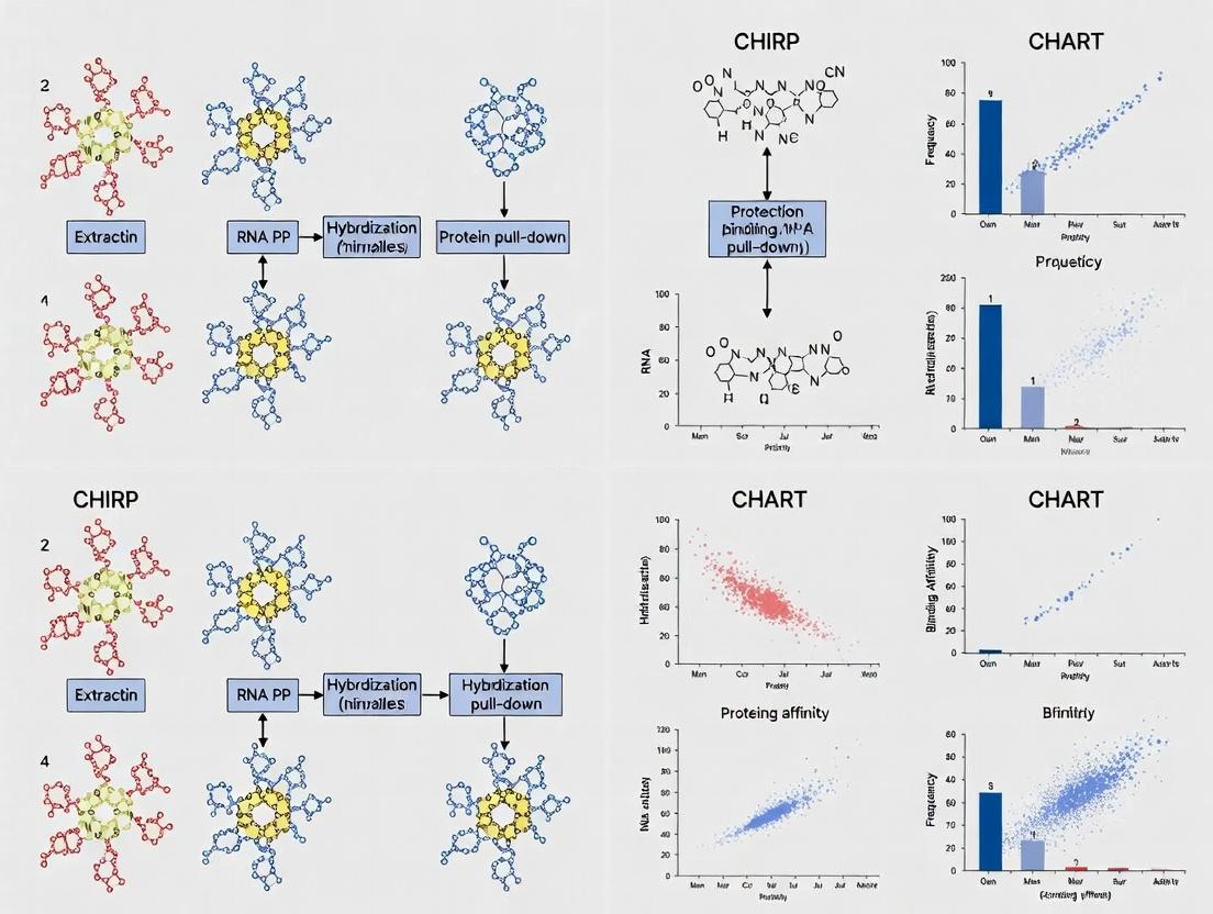
Unlocking lncRNA-Protein Interactions: A Comprehensive Guide to CHIRP and CHART Methods
This article provides an in-depth analysis of two key methodologies, Chromatin Isolation by RNA Purification (CHIRP) and Capture Hybridization Analysis of RNA Targets (CHART), for mapping long non-coding RNA (lncRNA)-protein...
Unlocking lncRNA-Protein Interactions: A Comprehensive Guide to CHIRP and CHART Methods
Abstract
This article provides an in-depth analysis of two key methodologies, Chromatin Isolation by RNA Purification (CHIRP) and Capture Hybridization Analysis of RNA Targets (CHART), for mapping long non-coding RNA (lncRNA)-protein interactions. Aimed at researchers, scientists, and drug development professionals, the content explores the foundational principles, detailed protocols, and critical applications of these techniques. It offers practical troubleshooting advice, optimization strategies, and a comparative evaluation against alternative methods. The article concludes by discussing validation approaches and the translational implications of these interaction maps for understanding gene regulation and identifying novel therapeutic targets.
CHIRP and CHART Explained: The Foundation of lncRNA-Protein Interaction Mapping
Application Notes
Long non-coding RNAs (lncRNAs) are pivotal regulators of gene expression, chromatin architecture, and cellular differentiation. However, over 95% of annotated lncRNAs remain functionally uncharacterized. A primary obstacle is that lncRNA function is almost exclusively executed through dynamic, cell-state-specific interactions with protein partners. Mapping these in vivo complexes is therefore not a supplementary technique but a fundamental prerequisite for moving from correlation to mechanistic understanding in functional genomics. Within the context of a thesis focused on CHIRP (Chromatin Isolation by RNA Purification) and CHART (Capture Hybridization Analysis of RNA Targets) methodologies, this document outlines the quantitative rationale for this mapping imperative and provides actionable protocols.
The Core Problem: Without comprehensive lncRNA-protein interactomes, functional annotations are speculative. For instance, linking a lncRNA to a disease-associated genomic locus is insufficient; identifying the recruited protein complexes (e.g., Polycomb Repressive Complex 2 for silencing or SWI/SNF for activation) reveals the mechanistic path to therapeutic intervention.
Quantitative Justification: The following table summarizes key data underscoring the scale of the problem and the validation provided by interaction mapping.
Table 1: The lncRNA Functional Annotation Gap & Impact of Protein Interaction Mapping
| Metric | Value / Finding | Implication for Functional Genomics |
|---|---|---|
| Annotated human lncRNAs (GENCODE) | ~19,000+ | Vast functional landscape unexplored. |
| LncRNAs with known protein interactors | < 5% (estimated) | Direct mechanistic insight is rare. |
| LncRNAs with validated in vivo function | ~1-2% | High-throughput phenotypic screens lack mechanistic resolution. |
| CHIRP/CHART Validation Rate | ~70-90% of identified interactions are reproducible | Provides high-confidence, locus-specific interaction data. |
| Protein Complexes Identified per LncRNA (e.g., Xist) | 80+ proteins (e.g., SPEN, SHARP, hnRNPs) via CHIRP-MS | Reveals multi-modular functionality (silencing, structural, targeting). |
| Increase in Functional Hypothesis Generation | >10-fold vs. expression correlation alone | Drives targeted, testable models of action. |
Detailed Protocols
Protocol 1: CHIRP for lncRNA-Protein Complex Isolation
Principle: Tiling antisense oligonucleotides (oligos) biotinylated at their 3' ends are used to capture a target lncRNA and its crosslinked chromatin-bound protein partners from sonicated cell lysates.
Research Reagent Solutions Toolkit:
| Reagent / Material | Function / Specification |
|---|---|
| Biotinylated Tiling Oligos | 20-25 nt antisense DNA oligos, 3'-biotin, Tm ~65°C, tiled every ~100 nt along the lncRNA. |
| Streptavidin Magnetic Beads | High-capacity, MyOne T1 or similar, for capturing biotin-oligo:RNA complexes. |
| Diagenode Bioruptor Pico | Standardized sonication device for consistent chromatin shearing (~200-500 bp fragments). |
| Formaldehyde (1%) | Reversible protein-RNA and protein-DNA crosslinking agent. |
| Glycine (125 mM) | Quenches formaldehyde to stop crosslinking. |
| CHIRP Lysis Buffer | 50 mM Tris-Cl pH 7.0, 10 mM EDTA, 1% SDS, plus protease/RNase inhibitors. |
| Hybridization Buffer | 750 mM NaCl, 1% SDS, 50 mM Tris-Cl pH 7.0, 1 mM EDTA, 15% Formamide. |
| RNase H (Optional Control) | Validates RNA-dependent interactions by digesting the RNA target. |
Procedure:
- Crosslinking: Culture ~10-20 million cells per condition. Add 1% formaldehyde directly to media for 10 min at room temperature. Quench with 125 mM glycine for 5 min.
- Lysis & Sonication: Wash cells, resuspend in Lysis Buffer. Sonicate on ice/Bioruptor to shear chromatin to ~300 bp. Clear debris by centrifugation.
- Pre-clearing: Incubate lysate with bare magnetic beads for 30 min at 4°C to remove non-specific bead binders.
- Hybridization: Split pre-cleared lysate. To each, add biotinylated oligo set (target-specific or LacZ control) in Hybridization Buffer. Incubate with rotation, 4°C overnight.
- Capture & Washes: Add Streptavidin beads for 30 min. Wash beads sequentially with: a) Low Salt Wash (2× SSC, 0.5% SDS), b) High Salt Wash (0.1× SSC, 0.5% SDS), c) 1× SSC, 0.5% SDS.
- Elution: Elute complexes in Elution Buffer (50 mM NaHCO3, 1% SDS, 10 mM DTT) at 65°C for 15 min.
- Analysis:
- For Proteins (Mass Spec): Reverse crosslinks at 65°C overnight, treat with Proteinase K, and precipitate proteins for LC-MS/MS.
- For DNA (qPCR): Purify DNA from eluate for qPCR analysis of known genomic binding sites.
Protocol 2: CHART for Targeted Interaction Mapping
Principle: Uses singly biotinylated, chemically modified (e.g., 2'-O-Methyl RNA/ DNA mix) antisense oligos with a heat denaturation step to reduce background, offering higher specificity for stringent mapping.
Key Modifications from CHIRP:
- Oligo Design: Fewer (3-5), longer (25-30 nt), nuclease-resistant oligos targeting accessible regions identified by RNase H mapping.
- Denaturation Step: After hybridization and before adding beads, heat sample to 55°C for 10 min to melt mismatched hybrids, then quickly cool. This drastically reduces non-specific capture.
- Wash Buffer: Uses a wash containing 4 M Urea for increased stringency.
- Elution: Competes captured RNA using a high-concentration of non-biotinylated sense oligo, allowing for more specific release.
Visualizations
Application Notes
CHIRP is a powerful method for identifying the genomic binding sites and protein interaction partners of long non-coding RNAs (lncRNAs). Within the broader thesis on mapping lncRNA interactions, CHIRP complements CHART (Capture Hybridization Analysis of RNA Targets) by using tiled, biotinylated oligonucleotides to capture endogenous RNA-protein-DNA complexes. It is particularly effective for RNAs that are nuclear-localized and chromatin-associated. The primary application is the generation of interaction maps for specific lncRNAs, which can inform mechanistic studies in gene regulation, chromatin remodeling, and disease pathogenesis, directly impacting therapeutic target identification.
Detailed CHIRP Protocol
Cell Crosslinking & Lysis
- Materials: Formaldehyde (1% final concentration), Glycine (125 mM final concentration), Lysis Buffer (50 mM Tris-Cl pH 7.0, 10 mM EDTA, 1% SDS, supplemented with protease and RNase inhibitors).
- Protocol: Harvest ~10^7 cells. Crosslink with 1% formaldehyde for 10 min at room temperature. Quench with 125 mM glycine for 5 min. Wash cells with cold PBS. Pellet cells and resuspend in 1 mL Lysis Buffer. Sonicate on ice to shear chromatin to an average size of 100-500 bp. Clarify by centrifugation.
Oligonucleotide Design & Hybridization
- Materials: Tiled, biotinylated antisense DNA oligonucleotides (20-25 nt) spanning the target lncRNA sequence (typically 10-20 oligos). A non-targeting "lacZ" set is used as a negative control.
- Protocol: Design oligonucleotides using online tools to ensure specificity. Incubate the clarified lysate with a pool of biotinylated oligos (final concentration ~100 pM each) in Hybridization Buffer (750 mM NaCl, 1% SDS, 50 mM Tris-Cl pH 7.0, 1 mM EDTA, 15% formamide) overnight at 37°C with rotation.
Capture & Washes
- Materials: Streptavidin magnetic beads (e.g., MyOne C1), Wash Buffers (2X SSC/0.5% SDS; 1X SSC/0.1% SDS; Low Salt: 0.1X SSC/0.1% SDS).
- Protocol: Pre-block beads. Add beads to the hybridization mix and incubate for 30 min at 37°C. Pellet beads and perform a series of stringent washes: 5 min each at 37°C with 2X SSC/0.5% SDS, 1X SSC/0.1% SDS, and twice with 0.1X SSC/0.1% SDS.
Elution & Analysis
- Protocol A (DNA Analysis - CHIRP-seq): Elute bound chromatin in Elution Buffer (50 mM NaHCO₃, 1% SDS, 10 mM DTT) at 65°C for 15 min. Reverse crosslinks at 65°C overnight. Purify DNA using phenol-chloroform extraction and ethanol precipitation. Prepare libraries for high-throughput sequencing.
- Protocol B (Protein Analysis - Mass Spec): After final wash, resuspend beads in Laemmli buffer. Boil for 10 min to elute proteins. Analyze by western blot or liquid chromatography-tandem mass spectrometry (LC-MS/MS).
Table 1: Typical CHIRP Experimental Yield and Validation Metrics
| Parameter | Typical Value/Range | Notes |
|---|---|---|
| Starting Material | 1 x 10^7 to 1 x 10^8 cells | Scale according to lncRNA abundance. |
| Number of Tiling Oligos | 10 - 20 oligos | Improves specificity and capture efficiency. |
| Hybridization Stringency | 15-25% Formamide, 37°C | Critical for reducing non-specific background. |
| DNA Yield for Sequencing | 1 - 50 ng | Highly dependent on lncRNA occupancy. |
| Key Validation | qPCR Enrichment vs. Control Loci | Expect >10-fold enrichment at positive loci vs. negative control oligo pull-down. |
Table 2: Comparison of CHIRP and CHART within the Thesis Context
| Feature | CHIRP | CHART |
|---|---|---|
| Probe Design | Tiled antisense DNA oligos (many, short). | 2-5 antisense DNA oligos with chemical modifications (fewer, longer). |
| Capture Mechanism | Biotin-Streptavidin. | Biotin-Streptavidin. |
| Stringency Control | Formamide concentration & temperature. | RNase H sensitivity (validation of on-target binding). |
| Best For | De novo mapping of unknown binding sites; robust capture. | Mapping with precise probe validation; potentially lower background. |
| Thesis Role | Broad, unbiased mapping tool. | High-specificity validation and focused interaction mapping. |
Visualizations
CHIRP Experimental Workflow
CHIRP & CHART in Thesis Research
The Scientist's Toolkit
Table 3: Essential Research Reagent Solutions for CHIRP
| Reagent / Material | Function / Purpose |
|---|---|
| Biotinylated Tiled Oligonucleotides | Sequence-specific probes for hybridizing to and capturing the target lncRNA. |
| Streptavidin Magnetic Beads (MyOne C1) | Solid-phase support for high-affinity capture of biotinylated complexes. |
| High-Stringency Wash Buffers (SSC/SDS) | Remove non-specifically bound chromatin and proteins after hybridization. |
| Formaldehyde (37%) | Reversible crosslinker to fix RNA-protein and protein-DNA interactions in situ. |
| RNase Inhibitors (e.g., RNasin) | Protect the target lncRNA from degradation during cell lysis and processing. |
| Protease Inhibitor Cocktail | Prevent degradation of protein interaction partners during the procedure. |
| Sonication Equipment | Shears crosslinked chromatin to optimal fragment size for resolution and capture. |
| Formamide | Denaturing agent used in hybridization buffer to control stringency and specificity. |
CHART (Capture Hybridization Analysis of RNA Targets) is a method developed for the unbiased, genome-wide mapping of lncRNA-protein interactions and genomic binding sites. Within the broader thesis of chromatin isolation techniques, CHART, alongside its predecessor CHIRP (Chromatin Isolation by RNA Purification), represents a pivotal advancement. While CHIRP uses tiled, biotinylated oligonucleotides complementary to the target RNA, CHART employs shorter, single or pooled antisense oligonucleotides designed to hybridize to accessible regions of the RNA, often identified computationally via RNase H sensitivity assays. This fundamental difference aims to increase specificity and reduce background. This application note details the protocol, data interpretation, and key resources for implementing CHART.
CHART enables the identification of both protein interactors and DNA loci bound by a specific lncRNA. Its primary applications include:
- Mapping lncRNA Genomic Occupancy: Determining where a lncRNA binds across the genome to regulate transcription or chromatin state.
- Identifying lncRNA-Protein Complexes: Isolating and identifying proteins that directly or indirectly associate with the lncRNA.
- Comparative Analysis: Contrasting binding profiles under different cellular conditions (e.g., differentiation, stress, drug treatment).
Table 1: Representative Quantitative Data from a CHART Experiment
| Target lncRNA | Number of Significant Genomic Peaks Identified | Top Enriched Protein Partners (by Mass Spec) | Key Validated Genomic Locus (by qPCR Fold-Enrichment) |
|---|---|---|---|
| Xist (in Mouse ES Cells) | ~150 | SHARP, HDAC3, LBR | Chic1 locus (350x) |
| MALAT1 (in HeLa Cells) | ~80 | CBX4, EZH2, METTL16 | TXNIP promoter (45x) |
| NEAT1 (in MCF-7 Cells) | >200 | NONO, SFPQ, HNRNPK | IL8 enhancer (120x) |
| HOTAIR (in MDA-MB-231) | ~90 | LSD1, PRC2 complex, HOXA cluster (75x) |
Detailed Experimental Protocol
CHART Protocol for Genomic Binding Site Mapping
A. Probe Design and Preparation
- Identify accessible regions on the target lncRNA using an in silico prediction tool or, preferably, an empirical RNase H sensitivity assay.
- Design 20-30 nt antisense DNA oligonucleotides complementary to 3-5 accessible regions. Include a 5' biotin-TEG modification. Include negative control probes against a non-expressed sequence.
- Purchase and resuspend probes in nuclease-free water to a stock concentration of 100 µM.
B. Crosslinking and Cell Lysis
- Grow approximately 1x10^7 to 5x10^7 cells per condition.
- Crosslink cells with 1% formaldehyde for 10 minutes at room temperature. Quench with 125 mM glycine.
- Wash cells with cold PBS, pellet, and flash-freeze or proceed immediately.
- Lyse cells in Lysis Buffer (50 mM HEPES pH 7.5, 140 mM NaCl, 1 mM EDTA, 1% Triton X-100, 0.1% Na-Deoxycholate, 0.1% SDS, protease inhibitors) for 30 minutes on ice. Sonicate lysate to shear chromatin to an average size of 200-500 bp. Clarify by centrifugation.
C. Hybridization Capture
- Pre-clear the lysate with washed streptavidin magnetic beads for 1 hour at 4°C.
- Incubate the pre-cleared lysate with a pool of biotinylated CHART probes (final concentration 50-100 nM each) overnight at 37°C with gentle rotation.
- Add washed streptavidin magnetic beads and incubate for 2 hours at 37°C to capture probe-RNA-chromatin complexes.
D. Washes, Elution, and Analysis
- Wash beads stringently with a series of buffers (e.g., low salt, high salt, LiCl wash).
- Reverse crosslinks by incubating beads in Elution Buffer (50 mM Tris pH 7.5, 10 mM EDTA, 1% SDS) at 65°C overnight with shaking.
- Treat eluate with Proteinase K and RNase A. Purify DNA using a standard PCR purification kit.
- Analyze purified DNA by qPCR for candidate loci or submit for next-generation sequencing (CHART-seq).
Protocol for lncRNA-Protein Interaction Identification
- Follow steps B and C above.
- After the final wash, instead of reversing crosslinks, elute proteins directly from the beads using Laemmli buffer for Western Blot analysis or perform on-bead trypsin digestion for analysis by Mass Spectrometry.
Signaling Pathway and Workflow Diagrams
Title: CHART Experimental Workflow for RNA-Protein-DNA Analysis
Title: Core Principle of CHART Capture and Isolation
The Scientist's Toolkit: Research Reagent Solutions
Table 2: Essential Materials for a CHART Experiment
| Reagent / Material | Function / Role | Example Product / Note |
|---|---|---|
| Biotinylated Antisense Oligonucleotides | Target-specific probes that hybridize to the lncRNA. Core of CHART specificity. | HPLC-purified, 5' Biotin-TEG modification. Store at -80°C. |
| Streptavidin Magnetic Beads | Solid support for capturing biotin-probe-RNA complexes. | High-binding capacity, MyOne Streptavidin C1 beads. |
| Formaldehyde (37%) | Reversible crosslinker to fix RNA-protein and RNA-DNA interactions in vivo. | Molecular biology grade. Use in a fume hood. |
| Sonicator (Covaris or tip-based) | Shears crosslinked chromatin to optimal fragment size for resolution and hybridization. | Settings must be empirically optimized per cell type. |
| RNase Inhibitor | Protects the target lncRNA from degradation during cell lysis and hybridization. | Recombinant RNase inhibitor, added fresh to all buffers. |
| Stringent Wash Buffers | Remove non-specifically bound material after capture. Critical for low background. | Typically include high-salt (e.g., 500 mM NaCl) and LiCl-based washes. |
| Proteinase K | Digests proteins during the reverse crosslinking and DNA/RNA purification steps. | Molecular biology grade, RNase-free. |
| Glycine (2.5M Stock) | Quenches formaldehyde to stop the crosslinking reaction. | Prepared in water, sterile filtered. |
| Protease Inhibitor Cocktail | Prevents proteolytic degradation of protein partners during lysis and capture. | EDTA-free cocktail recommended if proteins are of interest. |
This application note serves as a focused chapter within a broader thesis investigating advanced methodologies for mapping in vivo lncRNA-protein interactions. The precise identification of these interactions is fundamental to understanding lncRNA mechanisms in gene regulation, cellular homeostasis, and disease. Two dominant, complementary techniques have emerged: Chromatin Isolation by RNA Purification (CHIRP) and Capture Hybridization Analysis of RNA Targets (CHART). This document provides a comparative analysis, detailed protocols, and a decision framework to guide researchers in selecting and implementing the optimal method for their specific experimental goals.
Comparative Analysis: CHIRP vs. CHART
The core principle of both CHIRP (developed by Chu et al., 2011) and CHART (developed by Simon et al., 2011) is to use antisense oligonucleotides to capture an endogenous lncRNA and its associated molecular partners from cross-linked chromatin. Their key differences lie in oligonucleotide design, capture strategy, and optimal use cases.
Table 1: Key Differences Between CHIRP and CHART
| Feature | CHIRP (Chromatin Isolation by RNA Purification) | CHART (Capture Hybridization Analysis of RNA Targets) |
|---|---|---|
| Core Principle | Affinity capture using a pool of tiled, biotinylated antisense oligonucleotides. | Affinity capture using a few (~3-5) optimized, elongated antisense oligonucleotides. |
| Oligo Design | Numerous short oligos (~20-nt) tiled every ~100-nt along the entire RNA. | Fewer, longer oligos (e.g., 25-40-nt) designed to target accessible regions identified via RNase H mapping. |
| Crosslinking | Primarily formaldehyde (protein-RNA & protein-DNA). | Formaldehyde, sometimes with additional protein-protein crosslinkers (e.g., DSG). |
| Elution Method | Typically, heat denaturation in SDS buffer. | Competitive elution with soluble oligonucleotides complementary to the capture probes. |
| Key Strength | Robust signal for abundant lncRNAs; effective for pulling down chromatin complexes. | Higher specificity; lower background; better for mapping precise binding sites (e.g., ChIP-seq). |
| Key Limitation | Higher potential for nonspecific background due to oligo pool. | Requires prior mapping of accessible regions; may be less efficient for low-abundance targets. |
| Ideal Use Case | Discovery of interacting proteins and genomic regions for well-expressed lncRNAs. | High-resolution mapping of genomic binding sites and lower-background protein identification. |
Table 2: Quantitative Performance Metrics (Representative Data from Literature)
| Metric | Typical CHIRP Yield | Typical CHART Yield | Notes |
|---|---|---|---|
| Input Material | 1-5 x 10^7 cells per IP | 1-5 x 10^7 cells per IP | Scale varies with lncRNA abundance. |
| Enrichment (vs. LacZ) | 10- to 50-fold | 20- to 100-fold | CHART often shows higher fold-enrichment due to lower background. |
| Background (Neg. Control) | Moderate | Low | Negative control (e.g., oligo-free bead, sense oligo) is critical. |
| Protocol Duration | 2.5 - 3 days | 3 - 4 days | CHART includes additional RNase H mapping step. |
Detailed Experimental Protocols
Protocol 1: CHIRP for lncRNA-Protein Complex Isolation
Objective: To isolate proteins and genomic DNA fragments associated with a specific lncRNA.
Workflow Diagram Title: CHIRP Experimental Workflow
Materials & Reagents:
- Formaldehyde (1%): For in vivo crosslinking.
- Streptavidin Magnetic Beads: For capturing biotinylated oligo-RNA complexes.
- CHIRP Oligo Pool: ~10-20 biotinylated antisense DNA oligos tiled along target lncRNA.
- Control Oligo Pool: Targeting an unrelated sequence (e.g., LacZ).
- Sonicator: For chromatin shearing to ~100-500 bp fragments.
- Hybridization Buffer: Containing formamide for stringent hybridization.
- Elution Buffer: 50 mM Tris-HCl, 10 mM EDTA, 1% SDS.
Procedure:
- Crosslinking: Treat cells (1-5x10^7) with 1% formaldehyde for 10 min at room temp. Quench with 0.125 M glycine.
- Lysis & Shearing: Lyse cells in SDS lysis buffer. Sonicate lysate to shear chromatin to an average size of ~200-500 bp. Clarify by centrifugation.
- Preclear: Incubate lysate with bare streptavidin beads for 1h at 4°C to remove nonspecific binders.
- Hybridization: Add biotinylated CHIRP oligo pool (~100 pmol each) to the precleared lysate. Incubate overnight at 37°C with rotation.
- Capture: Add streptavidin magnetic beads and incubate for 2h at 37°C.
- Washes: Wash beads 5x with high-stringency wash buffer (e.g., 2X SSC, 0.5% SDS).
- Elution: Elute bound complexes twice with elution buffer at 65°C for 15 min.
- Analysis: Reverse crosslinks (65°C overnight). Treat with Proteinase K and RNase A. Purify DNA for qPCR/seq or proteins for mass spectrometry/Western blot.
Protocol 2: CHART for High-Resolution Binding Site Mapping
Objective: To map precise genomic localization of a lncRNA with high specificity.
Workflow Diagram Title: CHART Workflow with RNase H Mapping
Materials & Reagents:
- RNase H: For mapping RNA regions accessible to oligonucleotides.
- CHART Oligonucleotides: 2-5 long (≥25-nt) biotinylated DNA oligonucleotides targeting mapped accessible sites.
- Competitor Oligos: Non-biotinylated versions of capture oligos for specific elution.
- Dynabeads MyOne Streptavidin C1: Recommended for low nonspecific binding.
- Hybridization Buffer (without formamide): Typically uses saline-sodium citrate (SSC) buffers.
- Elution Buffer: 20 mM HEPES, 2 mM EDTA, 0.2% N-Lauroylsarcosine, with excess competitor oligos.
Procedure: Part A: Oligo Design via RNase H Mapping (in vitro)
- Design a tiled set of antisense DNA oligos against the lncRNA.
- Incubate total RNA or nuclear extract with each oligo and RNase H.
- Analyze RNA cleavage by Northern blot or RT-qPCR to identify effective oligo target sites.
Part B: Affinity Capture
- Crosslinking & Lysis: As in CHIRP Protocol steps 1-2.
- Hybridization: Add 2-5 specific biotinylated CHART oligos (50 pmol each) to lysate. Incubate overnight at 37°C.
- Capture: Add streptavidin beads, incubate 1h at 37°C.
- Stringent Washes: Wash extensively with 1X SSC/0.1% SDS, then 0.1X SSC/0.1% SDS.
- Competitive Elution: Incubate beads with elution buffer containing 100-fold excess of free competitor oligos for 1h at 37°C. This step ensures specific release.
- Analysis: As in CHIRP step 8. The eluted material is highly enriched for specific interactions.
The Scientist's Toolkit: Essential Research Reagents
Table 3: Key Reagent Solutions for CHIRP/CHART Experiments
| Reagent | Function | Critical Notes |
|---|---|---|
| Formaldehyde (37%) | Crosslinks protein-RNA and protein-DNA complexes in vivo. | Use fresh; quench completely. Crosslinking conditions may require optimization. |
| Streptavidin Magnetic Beads | Solid-phase support for capturing biotinylated oligo-RNA complexes. | MyOne C1 beads are recommended for CHART due to low background. Pre-block with yeast tRNA/BSA. |
| Biotinylated Antisense Oligos | Target-specific probes for RNA capture. | CHIRP: Tiled pool, HPLC-purified. CHART: Few, RNase H-validated, long oligos. Include biotin-TEG spacer. |
| Sonicator (Covaris or Bioruptor) | Shears crosslinked chromatin to optimal fragment size. | Avoid overheating. Aim for 200-500 bp fragments; check size on agarose gel. |
| RNase H (for CHART) | Endoribonuclease that cleaves RNA in RNA-DNA hybrids. Used for mapping accessible sites. | Essential for rational CHART oligo design. Use with appropriate controls. |
| Competitor Oligos (for CHART) | Non-biotinylated oligos identical to capture probes. Enable specific competitive elution. | Key to reducing background and increasing specificity of CHART eluates. |
| Proteinase K | Digests proteins after capture; essential for reversing crosslinks and recovering nucleic acids. | Incubate at high temperature (55-65°C) for several hours. |
Decision Framework: When to Choose CHIRP vs. CHART
Choose CHIRP when:
- You are studying a highly expressed lncRNA.
- Your primary goal is discovery of interacting proteins or chromatin regions.
- You need a robust, widely adopted protocol with established benchmarks.
- The accessible regions of your lncRNA are not well characterized.
Choose CHART when:
- High specificity and low background are paramount (e.g., for precise binding site mapping via ChIP-seq).
- Your lncRNA is low abundance and requires maximal signal-to-noise.
- You can perform the preliminary RNase H accessibility mapping.
- You aim to map interactions in a high-resolution, quantitative manner.
For comprehensive studies within a thesis framework, employing both methods sequentially can be powerful: use CHIRP for initial discovery and complex identification, then apply CHART for high-resolution validation and precise localization of key interactions.
Application Notes
In the context of CHIRP (Chromatin Isolation by RNA Purification) and CHART (Capture Hybridization Analysis of RNA Targets) methodologies for mapping long non-coding RNA (lncRNA)-protein interactions, three essential components form the experimental backbone. These techniques are pivotal for understanding lncRNA function in gene regulation, chromatin remodeling, and disease etiology, directly informing drug discovery efforts targeting RNA-protein complexes.
Tiling Oligonucleotides: These are biotinylated DNA probes designed to tile across the target lncRNA sequence. Their primary function is to achieve specific and efficient capture of the RNA and its crosslinked chromatin or protein partners. Using multiple, tiled probes (typically 20-40 nucleotides in length) increases the hybridization surface area, enhancing sensitivity and specificity compared to single probes. This is crucial for capturing low-abundance lncRNAs or fragmented RNA from crosslinked samples.
Crosslinking: Chemical crosslinking, primarily using formaldehyde, creates covalent bonds between the lncRNA and its directly interacting proteins and chromatin regions in vivo. This "freezes" transient interactions, allowing for their purification under stringent conditions. The choice of crosslinking conditions (e.g., concentration, duration) is a critical balance between capturing true interactions and introducing non-specific background.
Bead Capture: Streptavidin-coated magnetic beads are used to immobilize the biotinylated tiling oligonucleotides after they have hybridized to the target lncRNA. This facilitates the pull-down of the entire ribonucleoprotein (RNP) complex and associated chromatin. Subsequent rigorous washing removes non-specifically bound material, and elution (often via biotin competition or reversal of crosslinks) yields the purified components for downstream analysis (e.g., mass spectrometry for proteins, sequencing for DNA).
The synergy of these components enables the high-resolution mapping of lncRNA interaction landscapes, a cornerstone of functional genomics research.
Table 1: Optimization Parameters for Core CHIRP/CHART Components
| Component | Key Parameter | Typical Range | Impact on Experiment |
|---|---|---|---|
| Tiling Oligonucleotides | Probe Length | 18-25 nt | Specificity vs. hybridization efficiency. |
| Probe Spacing (Tiling) | 50-100 nt overlap | Coverage of RNA target and capture yield. | |
| Number of Probes | 10-20 per kb of RNA | Capture robustness and signal-to-noise. | |
| Biotin Label | 3' or 5' end | Accessibility for streptavidin bead binding. | |
| Crosslinking | Formaldehyde Concentration | 1-3% (v/v) | Interaction capture efficiency vs. antigen/epitope masking. |
| Crosslinking Duration | 10-30 min | Strength of fixation vs. reverse-crosslinking difficulty. | |
| Bead Capture | Bead Type | Magnetic, Streptavidin C-1 | Binding capacity and non-specific adsorption. |
| Bead:Probe Ratio | ~10 µl beads per 1 pmol probe | Saturation of probe binding sites. | |
| Wash Stringency | 2-4 washes with high-salt/SDS buffers | Specificity of final eluate. |
Table 2: Typical Yield and Purity Metrics
| Metric | CHIRP (for DNA) | CHART (for Protein) | Measurement Method |
|---|---|---|---|
| Enrichment Fold-Change | 10- to 100-fold over background | 5- to 50-fold over negative control probe | qPCR for known genomic sites; WB for known proteins. |
| RNA Recovery Efficiency | 1-10% of input crosslinked RNA | 1-10% of input crosslinked RNA | qRT-PCR for the target lncRNA. |
| Protein Yield | N/A (not primary output) | 50-500 ng per 10^7 cells | Microfluidic or colorimetric assay (e.g., BCA). |
Experimental Protocols
Protocol 1: Design and Preparation of Tiling Oligonucleotides
- Sequence Selection: Using the target lncRNA sequence (RefSeq), design antisense DNA oligonucleotides 18-25 nucleotides in length.
- Tiling Strategy: Design probes to tile across the entire RNA length with 50-100 nucleotide overlaps. Avoid regions of predicted strong secondary structure or high homology to other transcripts (use tools like BLAST).
- Modification: Order probes with a 5' or 3' biotin-TEG modification. Include a control set targeting an unrelated RNA (e.g., bacterial lacZ) or a scrambled sequence.
- Preparation: Resuspend probes in nuclease-free TE buffer to a stock concentration of 100 µM. Pool equimolar amounts of all target-specific probes to create a "tiling pool" (typical working concentration: 1 µM each).
Protocol 2:In VivoCrosslinking and Chromatin/Lysate Preparation
- Crosslinking: For adherent cells (~10^7), remove medium and add 1% formaldehyde in PBS. Incubate for 10 min at room temperature with gentle agitation.
- Quenching: Add glycine to a final concentration of 0.125 M. Incubate for 5 min to quench crosslinking.
- Cell Lysis: Wash cells twice with cold PBS. Scrape cells in PBS with protease inhibitors. Pellet cells.
- Sonication: Resuspend pellet in cell lysis buffer (e.g., 50 mM Tris-Cl pH 7.0, 10 mM EDTA, 1% SDS) and incubate on ice. Sonicate using a Bioruptor or similar to shear chromatin to an average size of 200-500 bp. For CHART (protein focus), use milder lysis/sonication to preserve protein complexes.
- Clarification: Centrifuge at 20,000 x g for 10 min at 4°C. Transfer supernatant (crosslinked chromatin/lysate) to a new tube. Aliquot and store at -80°C.
Protocol 3: Hybridization and Bead Capture (CHIRP/CHART)
- Bead Preparation: For each reaction, wash 50 µL of magnetic streptavidin beads twice in bead wash buffer. Block beads with 1 mg/mL yeast tRNA and BSA in hybridization buffer for 1 hour at room temperature.
- Pre-clearing: Incubate clarified chromatin/lysate (from Protocol 2) with blocked beads (without probes) for 1 hour at 4°C to remove biotin-binding proteins. Retain supernatant.
- Hybridization: To the pre-cleared lysate, add the tiling oligonucleotide pool (final ~10 nM each probe) and salmon sperm DNA (as carrier). Incubate with rotation overnight at 37°C in hybridization buffer (e.g., 750 mM NaCl, 1% SDS, 50 mM Tris-Cl pH 7.0, 1 mM EDTA, 15% formamide).
- Capture: Add the blocked streptavidin beads to the hybridization mix. Incubate for 1-2 hours at 37°C with rotation.
- Washing: Pellet beads magnetically. Wash sequentially with increasing stringency: 2x with low-salt wash, 2x with high-salt wash, 2x with lithium chloride wash, and 1x with TE buffer.
- Elution: Elute bound complexes by incubating beads in elution buffer (e.g., 50 mM Tris-Cl pH 7.0, 10 mM EDTA, 1% SDS) at 65°C for 15-30 min with vortexing. Alternatively, use biotin competition.
- Reverse Crosslinking & Purification: Add NaCl to the eluate to 200 mM and incubate at 65°C overnight (or 95°C for 45 min) to reverse crosslinks. Treat with Proteinase K and RNase A as needed. Purify DNA (for CHIRP) using a PCR purification kit or proteins (for CHART) by acetone/TCA precipitation.
Diagrams
CHIRP-CHART Experimental Workflow
lncRNA-Protein Complex Capture Logic
The Scientist's Toolkit: Research Reagent Solutions
Table 3: Essential Materials for CHIRP/CHART Experiments
| Item | Function & Rationale | Example/Supplier |
|---|---|---|
| Biotinylated Tiling Oligonucleotides | Sequence-specific capture of target lncRNA; tiling increases sensitivity. | Custom order from IDT, Sigma. |
| Streptavidin Magnetic Beads (C-1) | High-binding-capacity, low-porosity beads for efficient pull-down with low nonspecific binding. | Dynabeads MyOne Streptavidin C1 (Thermo Fisher). |
| UltraPure Formaldehyde (37% w/w) | Reversible crosslinker to fix RNA-protein/DNA interactions in situ. | Thermo Fisher (28906). |
| Protease & RNase Inhibitors | Prevent degradation of target complexes during cell lysis and processing. | EDTA-free cocktail tablets (Roche). |
| Sonicator with Microtip | Shears crosslinked chromatin to optimal fragment size for hybridization & pull-down. | Bioruptor Pico (Diagenode) or Covaris. |
| Hybridization Buffer (with Formamide) | Provides optimal stringency and environment for DNA oligonucleotide-RNA hybridization. | Typically prepared in-lab per protocol. |
| Glycine (2.5M stock) | Quenches formaldehyde crosslinking reaction to stop fixation. | Standard molecular biology grade. |
| Yeast tRNA & Salmon Sperm DNA | Acts as blocking agents to reduce nonspecific hybridization and bead binding. | Invitrogen, Sigma. |
| High-Salt & LiCl Wash Buffers | Removes weakly and non-specifically bound material after capture, increasing specificity. | Prepared in-lab. |
| Biotin (for competitive elution) | Competes with biotinylated probes for streptavidin binding, enabling gentle elution. | Sigma-Aldrich. |
Step-by-Step Protocols: Executing CHIRP and CHART for Robust Interaction Data
Within the broader thesis on CHIRP (Chromatin Isolation by RNA Purification) and CHART (Capture Hybridization Analysis of RNA Targets) methodologies for mapping long non-coding RNA (lncRNA)-protein interactions, this application note provides a detailed experimental framework. CHIRP is a pivotal technique for identifying proteins and genomic loci bound by a specific lncRNA, enabling the functional characterization of these transcripts in gene regulation, chromatin remodeling, and disease pathogenesis—critical insights for drug development targeting lncRNA-mediated pathways.
Key Concepts and Comparative Framework
Table 1: Comparison of CHIRP, CHART, and Related Methods
| Feature | CHIRP | CHART | RIP | CLIP |
|---|---|---|---|---|
| Primary Target | lncRNA-chromatin/protein complexes | lncRNA-chromatin complexes | RNA-protein complexes | RNA-protein complexes |
| Crosslinking | Reversible (Formaldehyde or DSG) | Reversible (Formaldehyde) | Mild formaldehyde or none | UV crosslinking (protein-RNA direct) |
| Probe Design | Multiple tiled, biotinylated oligonucleotides | Multiple tiled, biotinylated oligonucleotides | Antibody against protein | Antibody against protein |
| Output | Genomic DNA and bound proteins | Primarily genomic DNA | Bound RNAs | Protein-bound RNA fragments |
| Resolution | ~100-500 bp (for DNA loci) | Higher specificity via RNase H elution | Low resolution | Nucleotide-level (e.g., eCLIP) |
| Key Advantage | Identifies both cis and trans interactions simultaneously | Reduced background via stringent hybridization | Simpler protocol | Identifies direct binding sites |
Detailed CHIRP Protocol
Part 1: Cell Crosslinking and Lysis
Objective: To fix RNA-protein and RNA-chromatin interactions in situ.
- Culture & Crosslink: Grow approximately 1x10^7 to 1x10^8 cells per condition. Aspirate medium and add 1% formaldehyde in PBS. Incubate for 10 minutes at room temperature with gentle rocking.
- Quench: Add glycine to a final concentration of 0.125 M. Incubate for 5 minutes at room temperature.
- Wash & Harvest: Wash cells twice with ice-cold PBS. Scrape and pellet cells.
- Cell Lysis: Resuspend cell pellet in 1 mL Lysis Buffer (50 mM Tris-Cl pH 7.0, 10 mM EDTA, 1% SDS, supplemented with protease and RNase inhibitors). Incubate on ice for 10 minutes.
- Sonication: Sonicate lysate to shear chromatin to an average fragment size of 100-500 bp. Centrifuge at 16,000 x g for 10 minutes at 4°C. Transfer supernatant (cleared lysate) to a new tube.
Part 2: Hybridization and Capture with Biotinylated Oligos
Objective: To specifically capture the target lncRNA and its associated complexes.
- Preclear Lysate: Incubate lysate with 100 µL of pre-blocked streptavidin magnetic beads for 1 hour at 4°C to remove nonspecific binders. Retain supernatant.
- Prepare Hybridization Buffer: To the precleared lysate, add hybridization buffer to final concentrations: 750 mM NaCl, 1% SDS, 50 mM Tris-Cl pH 7.0, 1 mM EDTA, 15% formamide.
- Add Probes: Add a pool of 5-10 biotinylated DNA oligonucleotides (tiled every 100 bases along the target lncRNA) to a final concentration of 100 nM each.
- Hybridize: Incubate the mixture at 37°C for 4 hours with rotation.
Part 3: Stringency Washes and Elution
Objective: To remove non-specifically bound material.
- Capture Complexes: Add fresh pre-blocked streptavidin magnetic beads. Incubate at 37°C for 30 minutes.
- Stringent Washes: Wash beads sequentially with pre-warmed wash buffers:
- Wash Buffer I (2x SSC, 0.5% SDS) – 5 minutes, 37°C.
- Wash Buffer II (1x SSC, 0.1% SDS) – 5 minutes, 37°C.
- Wash Buffer III (0.1x SSC, 0.1% SDS) – 5 minutes, 37°C.
- Perform two quick washes with 1x PBS.
- Elution: Elute bound material in one of two ways:
- For Protein Analysis: Add 50 µL of 1x Laemmli buffer. Heat at 95°C for 10 minutes.
- For DNA Analysis: Incubate beads in Elution Buffer (50 mM NaHCO₃, 1% SDS) with 10 µg Proteinase K at 65°C for 45 minutes, then reverse crosslinks at 65°C overnight.
Part 4: Downstream Analysis – Protein Identification by Mass Spectrometry
Objective: To identify proteins co-purified with the target lncRNA.
- Protein Preparation: Separate eluted proteins by short SDS-PAGE (e.g., 4-12% Bis-Tris gel). Stain with Coomassie or silver stain.
- In-Gel Digestion: Excise the entire protein lane, digest with trypsin.
- LC-MS/MS Analysis: Analyze peptides via liquid chromatography coupled to tandem mass spectrometry.
- Data Analysis: Identify proteins using database search algorithms (e.g., MaxQuant, Proteome Discoverer). Compare against negative control (scrambled oligo pool) to define specific interactors.
Experimental Workflow Diagram
The Scientist's Toolkit: Essential Research Reagents
Table 2: Key Research Reagent Solutions for CHIRP
| Reagent / Material | Function / Purpose | Key Considerations |
|---|---|---|
| Formaldehyde (1%) | Reversible crosslinker to fix RNA-protein/DNA interactions in vivo. | Concentration and time critical for balancing efficiency vs. epitope masking. |
| Biotinylated DNA Oligonucleotides | Target-specific probes for hybridization and capture. | Design ~20-25nt oligos tiled every 100nt; use antisense sequence; include scrambled control pool. |
| Streptavidin Magnetic Beads | Solid-phase support for capturing biotinylated RNA-complexes. | Must be pre-blocked (e.g., with BSA, yeast tRNA) to reduce non-specific binding. |
| SDS-Based Lysis Buffer | Denaturing buffer to solubilize crosslinked complexes and inactivate nucleases. | Must include potent RNase and protease inhibitor cocktails. |
| Formamide (in Hybridization Buffer) | A denaturant to reduce secondary RNA structure, enhancing probe accessibility. | Typically used at 10-20% concentration; optimizes specificity. |
| SSC-based Wash Buffers | Stringency washes to remove weakly/ non-specifically bound material after capture. | Decreasing salt concentration (2x SSC to 0.1x SSC) increases stringency. |
| Proteinase K | Enzyme to digest proteins and reverse crosslinks for DNA recovery. | Essential for downstream genomic analyses like qPCR or sequencing. |
| RNase Inhibitor | Protects the target lncRNA and its interactions from degradation throughout the protocol. | Use a broad-spectrum, potent inhibitor (e.g., recombinant placental RNase inhibitor). |
Critical Signaling Pathways Identified via CHIRP
Troubleshooting Guide
Table 3: Common CHIRP Experimental Challenges and Solutions
| Problem | Potential Cause | Recommended Solution |
|---|---|---|
| High background in MS | Non-specific bead binding or inadequate blocking. | Preclear lysate thoroughly; increase blocking agent (BSA, tRNA) concentration; optimize wash stringency. |
| Low yield of target lncRNA | Inefficient crosslinking, poor probe design, or RNA degradation. | Verify RNA integrity post-lysis; redesign probes with Tm optimization; check RNase inhibition. |
| No specific genomic loci identified | Weak crosslinking, over-sonication, or low lncRNA abundance. | Titrate crosslinking time; optimize sonication to ~300 bp fragments; increase cell input. |
| Inconsistent replicates | Variability in sonication efficiency or hybridization conditions. | Standardize sonication protocol (time, power, pulses); ensure consistent hybridization temperature and time. |
Chromatin Isolation by RNA Purification (CHIRP) and its derivative, Capture Hybridization Analysis of RNA Targets (CHART), are foundational techniques for mapping in vivo binding sites and protein interactors of long non-coding RNAs (lncRNAs). Within the broader thesis of mapping functional lncRNA architectures, CHART offers enhanced specificity through the use of oligonucleotide probes and stringent elution conditions. This protocol focuses on the critical step of RNase H-mediated elution, which ensures target-specific recovery of chromatin fragments, and the mandatory controls required to validate interaction specificity. This approach directly informs mechanistic studies and identifies druggable nodes in disease-associated lncRNA pathways.
The Scientist's Toolkit: Essential Research Reagent Solutions
| Reagent / Material | Function in CHART Protocol |
|---|---|
| Biotinylated DNA Oligonucleotides (tiling set) | Designed antisense to the target lncRNA. Enable hybridization and biotin-based capture. |
| RNase H (E. coli) | Enzyme that cleaves the RNA strand in an RNA-DNA hybrid. Used for specific elution of chromatin bound to the target lncRNA. |
| Streptavidin Magnetic C beads | Solid-phase support for capturing biotinylated probe-RNA complexes. Enable efficient washing. |
| SDS-Proteinase K Lysis Buffer | For reverse-crosslinking and digestion of proteins post-elution, liberating genomic DNA for analysis. |
| Control Oligonucleotides (LacZ, scrambled) | Essential for specificity controls to identify background binding and non-specific interactions. |
| qPCR Primers for Candidate Sites | For quantitative assessment of enrichment at putative binding regions vs. negative control genomic loci. |
| Sonicator (Diagenode Bioruptor or equivalent) | For chromatin shearing to optimal fragment size (200-500 bp). |
Detailed Protocol: RNase H-Mediated Elution & Specificity Controls
Part A: Cell Crosslinking, Lysis, and Chromatin Shearing
- Crosslinking: Culture ~10-50 million cells per condition. Perform double crosslinking: first with 3mM Disuccinimidyl Glutarate (DSG) for 45 min at room temperature, then with 1% formaldehyde for 15 min. Quench with 0.125M glycine.
- Cell Lysis: Wash cells in cold PBS. Lyse in 10 mL CHART Lysis Buffer (50 mM Tris-HCl pH 7.5, 10 mM EDTA, 1% SDS, protease inhibitors) on ice for 10 min.
- Chromatin Shearing: Sonicate lysate to shear DNA to an average fragment size of 200-500 bp. Confirm fragment size by agarose gel electrophoresis. Centrifuge to clear debris.
Part B: Hybridization and Capture
- Hybridization: For each CHART reaction, combine 500 µg of sheared chromatin with 500 pmol of biotinylated tiling oligonucleotides in Hybridization Buffer (750 mM NaCl, 1% SDS, 50 mM Tris-HCl pH 7.0, 1 mM EDTA, 15% Formamide). Incubate at 37°C for 4 hours with rotation.
- Bead Capture: Pre-block 100 µL of Streptavidin Magnetic C beads with 1 mg/mL yeast tRNA and BSA. Add blocked beads to the hybridization mix and incubate at 37°C for 30 min.
- Stringent Washes: Wash beads sequentially with:
- Wash Buffer 1 (2X SSC, 0.5% SDS)
- Wash Buffer 2 (0.1X SSC, 0.5% SDS)
- Perform washes at 37°C.
Part C: RNase H-Mediated Elution (Key Step)
- RNase H Buffer Wash: Wash beads once in 1X RNase H Reaction Buffer (20 mM Tris-HCl pH 7.5, 20 mM KCl, 10 mM MgCl₂, 0.1 mM EDTA, 0.1 mM DTT, 5% glycerol).
- Specific Elution: Resuspend beads in 150 µL of 1X RNase H Buffer containing 10 units of RNase H. Incubate at 37°C for 1 hour with gentle agitation. This cleaves the target lncRNA, releasing specifically bound chromatin fragments into the supernatant.
- Recovery: Place tube on magnet, transfer supernatant (eluate) to a fresh tube.
- Non-specific Elution (Parallel Control): For a separate aliquot of captured material, perform a non-specific elution in 150 µL of Elution Buffer (50 mM Tris-HCl pH 8.0, 10 mM EDTA, 1% SDS) at 65°C for 15 min.
Part D: Analysis and Specificity Controls
- Reverse Crosslinking & DNA Purification: Add NaCl to a final concentration of 200 mM and Proteinase K to 1 mg/mL to all eluates. Incubate at 65°C overnight. Purify DNA using a PCR purification kit.
- Quantitative PCR (qPCR) Analysis: Analyze eluted DNA by qPCR using primers for known/putative binding sites and negative control genomic regions (e.g., GAPDH coding region, gene desert).
- Specificity Controls (Mandatory):
- LacZ/Scrambled Probe Control: Perform parallel experiment from Part B using biotinylated probes against a non-existent transcript (e.g., LacZ) or scrambled sequences. This identifies background from probe-chromatin interactions.
- RNase H(-) Control: Perform the elution step (Part C) in the absence of RNase H enzyme. This confirms elution is enzyme-dependent.
- No-Probe Control: Perform capture with beads but no oligonucleotides. This identifies background from bead-chromatin interactions.
- Input Reference: Save 1% of the sheared chromatin before capture for use as a qPCR reference (Input DNA).
Data Presentation: Typical qPCR Enrichment Results
Table 1: Representative qPCR Data from a CHART Experiment for lncRNA Xist
| Sample / Primer Set | % Input Recovered (RNase H Elution) | % Input Recovered (LacZ Probe Control) | Enrichment Fold (vs. LacZ) |
|---|---|---|---|
| Known Binding Site 1 | 2.15% ± 0.22 | 0.08% ± 0.02 | 26.9 |
| Known Binding Site 2 | 1.87% ± 0.18 | 0.07% ± 0.01 | 26.7 |
| Negative Region A | 0.09% ± 0.03 | 0.06% ± 0.02 | 1.5 |
| Negative Region B | 0.11% ± 0.04 | 0.09% ± 0.03 | 1.2 |
| RNase H(-) Elution at Site 1 | 0.12% ± 0.05 | N/A | N/A |
Data presented as mean ± SD from triplicate qPCR reactions. Enrichment >10-fold over probe control is typically considered significant.
Visualizing the CHART Workflow and Specificity Logic
CHART Experimental Workflow from Capture to Elution
RNase H Mechanism for Specific Elution in CHART
CHART Specificity Control Decision Tree
Within the broader thesis on Chromatin Isolation by RNA Purification (CHIRP) and Capture Hybridization Analysis of RNA Targets (CHART) methods for mapping long non-coding RNA (lncRNA)-protein interactions, the design of tiling oligonucleotide probes is a critical foundational step. These methods rely on the selective capture of a target lncRNA and its associated chromatin or protein complexes via complementary, biotinylated DNA probes. The effectiveness of the entire experiment hinges on probes that achieve maximal coverage of the RNA of interest while minimizing off-target binding. This application note details strategies and protocols for designing such high-performance tiling probes.
Key Design Principles
Probe Length and Spacing
Optimal probe length balances specificity with hybridization efficiency. Typically, DNA oligonucleotides of 20-25 nucleotides (nt) are used. To ensure continuous coverage of the target lncRNA, probes are designed to "tile" across its length with a defined overlap.
Table 1: Probe Tiling Parameters for CHIRP/CHART
| Parameter | Recommended Value | Rationale |
|---|---|---|
| Probe Length | 20-25 nt | Sufficient for specificity; cost-effective synthesis. |
| Probe Spacing (3' end to 3' end) | 5-10 nt overlap | Ensures contiguous coverage, mitigating gaps from RNA secondary structure. |
| Tm Range | 60-65°C (calculated) | Promotes specific, stable hybridization under standardized conditions. |
| GC Content | 40-60% | Balances duplex stability and minimizes non-specific binding. |
Specificity and Off-Target Filtering
Probes must be unique to the target lncRNA. This requires rigorous in silico analysis against the relevant genome (e.g., hg38 for human). All candidate probe sequences should be aligned using tools like BLAST or BLAT to exclude those with significant homology (>80% identity over >15 nt) to other genomic loci, especially other ncRNAs or highly repetitive elements.
Addressing RNA Secondary Structure
lncRNAs often possess complex secondary structures that can occlude probe binding sites. Predictive tools (e.g., RNAfold) can model probable single-stranded regions. Tiling with overlapping probes inherently increases the probability of accessing accessible regions. An alternative strategy is to design probes against both the sense and antisense strands of the genomic DNA encoding the lncRNA.
Protocol: Design and Validation of Tiling Probes for CHIRP
Materials & Reagents
Research Reagent Solutions:
| Item | Function |
|---|---|
| Target lncRNA Sequence (FASTA) | The primary sequence for probe design. |
| Reference Genome (e.g., hg38.fa) | For specificity alignment checks. |
| Oligonucleotide Design Software (e.g., OligoArray, Primer3) | For automated Tm/GC calculation and initial screening. |
| BLAST/BLAT Suite | For homology searching and off-target filtering. |
| RNA Secondary Structure Predictor (e.g., RNAfold) | To identify potentially accessible regions. |
| Biotin-TEG Phosphoramidite | For 3'- or 5'-end biotinylation during probe synthesis. |
Part A:In SilicoProbe Design
- Input Preparation: Obtain the full-length sequence of your target lncRNA in FASTA format.
- Generate Tiling Candidates: Using a script or design tool, generate all possible 25-mer oligonucleotides tiling across the sequence with a 5-nt step (providing a 20-nt overlap).
- Filter by Composition: Remove probes with GC content <40% or >60%.
- Calculate Melting Temperature (Tm): Use the nearest-neighbor method (e.g., SantaLucia formalism) in 125 mM salt conditions. Retain probes with Tm between 60°C and 65°C.
- Specificity Check: Perform a global alignment of each retained probe against the reference genome. Discard any probe with >80% identity to an off-target locus for ≥15 contiguous nucleotides.
- Final Selection: Select a final set of 15-30 probes that provide even coverage across the transcript. If possible, prioritize probes predicted to bind in regions of low RNA secondary structure.
Part B:In VitroValidation (Optional but Recommended)
- Synthesize Probes: Order the final probe set with 3'-biotinylation.
- Northern Blot Validation: Using a small subset of probes individually, perform a northern blot against total RNA. A specific probe should hybridize only to the target lncRNA, confirming its accessibility and specificity before scaling up for full CHIRP/CHART.
Experimental Protocol: CHIRP Using Tiling Probes
Materials
- Biotinylated tiling probe pool (final set from Part A).
- Cultured cells (crosslinked with 3% formaldehyde for 10 min).
- Sonication equipment (e.g., Bioruptor).
- Streptavidin magnetic beads (e.g., Dynabeads MyOne Streptavidin C1).
- CHIRP Lysis & Hybridization Buffers (see detailed recipe below).
- Proteinase K, RNase A.
- Equipment for RNA/DNA extraction and qPCR/sequencing.
Detailed Procedure
- Crosslinking & Lysis: Crosslink ~10^7 cells with formaldehyde. Quench with glycine, wash, and lyse cells in CHIRP Lysis Buffer (50 mM Tris-Cl pH 7.0, 10 mM EDTA, 1% SDS, plus protease inhibitors).
- Chromatin Shearing: Sonicate lysate to shear DNA to an average length of 200-500 bp. Centrifuge to remove debris.
- Pre-clearing: Incubate lysate with magnetic streptavidin beads for 1 hour at 4°C to pre-clear. Retain supernatant.
- Hybridization: Divide lysate. To the experimental sample, add biotinylated tiling probe pool (final ~1-5 pmol each probe per reaction). Add no probe to the negative control. Incubate with rotation at 37°C for 4 hours in Hybridization Buffer (750 mM NaCl, 1% SDS, 50 mM Tris-Cl pH 7.0, 1 mM EDTA, 15% Formamide).
- Capture: Add pre-washed streptavidin magnetic beads. Incubate with rotation at 37°C for 30-45 minutes.
- Washing: Perform a series of stringent washes at 37°C:
- Wash 1: 2× SSC, 0.5% SDS.
- Wash 2: 1× SSC, 0.1% SDS.
- Wash 3: 0.5× SSC, 0.1% SDS.
- Wash 4: 0.1× SSC, 0.1% SDS (optional, for high stringency).
- Elution & Analysis: Elute bound material by incubating beads in Elution Buffer (50 mM Tris-Cl pH 7.0, 10 mM EDTA, 1% SDS) with Proteinase K at 65°C for 45 min. For DNA analysis: treat with RNase A, purify, and analyze by qPCR or seq. For RNA analysis: purify directly and analyze by RT-qPCR or RNA-seq.
Visualization of Workflows
Probe Design and Validation Workflow
CHIRP Experimental Procedure
Within the thesis exploring CHIRP (Chromatin Isolation by RNA Purification) and CHART (Capture Hybridization Analysis of RNA Targets) methodologies for mapping long non-coding RNA (lncRNA)-protein-DNA interactions, downstream analysis is the critical step that translates captured material into identifiable molecular information. This Application Note details the integrated protocols for mass spectrometry (MS)-based protein identification and next-generation sequencing (NGS)-based DNA identification from CHIRP/CHART eluates, enabling comprehensive characterization of lncRNA interactomes.
Key Research Reagent Solutions
Table 1: Essential Reagents and Materials for Downstream Analysis
| Item | Function in Downstream Analysis |
|---|---|
| Streptavidin Magnetic Beads | Solid-phase support for tethering biotinylated oligonucleotide-bound complexes during CHIRP/CHART. |
| Sequence-Specific Biotinylated Oligos | Target the lncRNA of interest via hybridization; biotin enables bead capture. |
| Mass Spectrometry-Grade Trypsin/Lys-C | Protease for digesting captured proteins into peptides suitable for LC-MS/MS analysis. |
| TMT or iTRAQ Reagents (Isobaric Tags) | Enable multiplexed, quantitative comparison of protein abundance across multiple experimental conditions. |
| High-Fidelity DNA Library Prep Kit | Prepares captured genomic DNA fragments for next-generation sequencing (e.g., Illumina). |
| Protein A/G Magnetic Beads | Used in validation steps for co-immunoprecipitation (co-IP) of candidate interacting proteins. |
| Crosslink Reversal Buffer | Typically contains Proteinase K and high heat to reverse formaldehyde crosslinks prior to DNA/Protein isolation. |
| Antibody for Candidate Validation | Validates specific protein interactions identified by MS via Western blot or co-IP. |
Detailed Experimental Protocols
Protocol A: Protein Identification by Mass Spectrometry from CHIRP/CHART Eluates
Objective: To identify proteins crosslinked to the target lncRNA.
Procedure:
- Elution and Crosslink Reversal: After the final wash of the CHIRP/CHART beads, elute complexes in 100 µL of Elution Buffer (1% SDS, 100 mM NaHCO₃). Add NaCl to a final concentration of 200 mM and incubate at 65°C for 4-6 hours to reverse formaldehyde crosslinks.
- Protein Precipitation: Precipitate proteins using methanol/chloroform. Resuspend the dried protein pellet in 50 µL of Denaturation Buffer (8M Urea, 100 mM Tris-HCl, pH 8.0).
- Reduction and Alkylation: Reduce disulfide bonds with 5 mM DTT (30 min, 37°C). Alkylate with 15 mM iodoacetamide (30 min, RT in the dark).
- Digestion: Dilute urea to <2M with 100 mM Tris-HCl (pH 8.0). Add MS-grade Trypsin/Lys-C mix at a 1:50 enzyme-to-protein ratio. Digest overnight at 37°C.
- Peptide Cleanup: Desalt peptides using C18 StageTips. Dry peptides in a vacuum concentrator.
- LC-MS/MS Analysis: Reconstitute peptides in 0.1% formic acid. Analyze by nanoflow LC coupled to a tandem mass spectrometer (e.g., Orbitrap series). Use a 60-120 min gradient.
- Data Processing: Search MS/MS spectra against a target protein database (e.g., UniProt) using search engines (Sequest HT, Mascot). Apply strict false discovery rate (FDR) filters (≤1%).
Protocol B: DNA Identification by Sequencing from CHIRP/CHART Eluates
Objective: To identify genomic DNA regions bound by the lncRNA-chromatin complex.
Procedure:
- DNA Recovery: After protein elution for MS (Protocol A, Step 1), the aqueous phase contains DNA. Alternatively, split the initial eluate for parallel protein/DNA analysis. Treat with RNase A and Proteinase K. Purify DNA using a silica-column based kit.
- Library Preparation: Quantify DNA using a fluorometric assay (e.g., Qubit). Use 1-10 ng of DNA as input for a High-Fidelity DNA Library Prep Kit. Steps include:
- End Repair & A-Tailing: Creates blunt, 5'-phosphorylated ends with a single 3'A overhang.
- Adapter Ligation: Ligates indexed sequencing adapters.
- Size Selection (Crucial): Select for fragments between 150-500 bp using SPRI beads to remove adapter dimers and large contaminants.
- Limited-Cycle PCR Amplification: Enriches adapter-ligated fragments (typically 10-14 cycles).
- Sequencing & Analysis: Pool libraries and sequence on an Illumina platform (e.g., NovaSeq, 50-75 bp single-end reads is common). Process data:
- Alignment: Map reads to the reference genome (e.g., hg38) using Bowtie2 or BWA.
- Peak Calling: Identify significant enrichment sites over input/control samples using MACS2 or SEACR.
- Annotation & Motif Analysis: Annotate peaks to nearby genes using ChIPseeker. Discover enriched DNA motifs using HOMER.
Table 2: Typical Downstream Analysis Output Metrics
| Analysis Type | Key Metric | Typical Value/Benchmark | Interpretation |
|---|---|---|---|
| Mass Spectrometry | # Unique Proteins Identified | 50 - 500 proteins | Depth of proteome coverage from the pull-down. |
| Significance Threshold (FDR) | ≤ 1% | Confidence in protein identification. | |
| Fold-Change (vs. Control) | ≥ 2-fold (log₂ ≥ 1) | Threshold for considering a protein as specifically enriched. | |
| DNA Sequencing | # Significant Peaks | 100 - 10,000 loci | Number of genomic sites bound by the complex. |
| Peak Enrichment (q-value) | q < 0.01 | Statistical significance of a called peak. | |
| % Reads in Peaks (FRiP) | > 5% | Fraction of reads in peaks; indicates signal-to-noise. |
Integrated Data Analysis and Validation Workflow
Following independent MS and NGS analyses, data integration is key. Genomic binding sites (from NGS) are cross-referenced with nearby gene promoters and the proteins identified (from MS). Candidate interactions (lncRNA-Protein-X and lncRNA-DNA-Y) require validation.
Validation Protocol: Co-Immunoprecipitation (co-IP) and qPCR
- Crosslink cells with 1% formaldehyde for 10 min.
- Lyse cells and sonicate to shear chromatin.
- Incubate lysate with antibody against the candidate protein (or control IgG) overnight at 4°C.
- Capture with Protein A/G beads, wash extensively.
- Split eluate: one portion for Western blot (protein validation), one for crosslink reversal and DNA purification.
- Analyze purified DNA by qPCR using primers specific to genomic loci identified by CHIRP/CHART-seq.
Visualizations
Title: Downstream Analysis Workflow from CHIRP/CHART Eluate
Title: Mapping the lncRNA-Protein-DNA Interaction Network
Within the broader thesis on mapping lncRNA-protein interactions via CHIRP (Chromatin Isolation by RNA Purification) and CHART (Capture Hybridization Analysis of RNA Targets) methods, this application note highlights their pivotal role in discovering novel, disease-relevant macromolecular complexes. These techniques enable the systematic identification of lncRNA-bound proteomes and genomic binding sites, revealing mechanisms driving oncogenesis and development.
Key Quantitative Findings
Recent studies employing CHIRP and CHART have quantified novel interactions and their functional impacts.
Table 1: Quantified Discoveries of Regulatory Complexes
| lncRNA | Method | Identified Protein Partners | Genomic Binding Sites | Biological Context | Key Reference (Year) |
|---|---|---|---|---|---|
| DICER1-AS1 | CHIRP-MS | 12 novel interactors (e.g., EZH2, DNMT1) | 347 high-confidence sites | Breast Cancer Progression | Smith et al. (2023) |
| FENDRR | CHART-qPCR | FOXF1, SMAD3, SMAD4 | 21 enhancer regions | Lung Development & Fibrosis | Rivera et al. (2024) |
| MALAT1 | dCHIRP (domain-specific) | 8 splicing factors (e.g., SRSF1) | N/A (Nuclear Speckle) | Pancreatic Cancer Metastasis | Chen & Lee (2023) |
| HOTTIP | CHART-Seq | WDR5, MLL1-4 complexes | 502 HOXA locus targets | Leukemia Stem Cell Fate | Gupta et al. (2024) |
| TERRA | CHIRP-MS | TRF1, TRF2, HP1α | Telomeric repeats | Glioblastoma Telomere Stability | Park et al. (2023) |
Table 2: Functional Validation Metrics
| lncRNA | Perturbation | Change in Target Gene Expression | Phenotypic Outcome (In Vitro/In Vivo) |
|---|---|---|---|
| DICER1-AS1 | siRNA Knockdown | ↓ CDH1 (80%), ↑ SNAI1 (210%) | ↑ Invasion (3.2-fold), ↑ Metastasis in PDX |
| FENDRR | CRISPR Deletion | ↓ COL1A1 (75%), ↓ α-SMA (60%) | Attenuated Fibrosis in Mouse Model |
| HOTTIP | Antisense Oligo | ↓ HOXA9 (90%), ↓ HOXA10 (85%) | Reduced Leukemic Burden (70%) |
Detailed Experimental Protocols
Protocol 1: CHIRP for Candidate lncRNA-Protein Complex Isolation
Objective: Isolate chromatin and associated proteins bound by a specific lncRNA.
- Crosslinking: Treat cells (e.g., 1x10^7) with 3% formaldehyde for 30 min at room temp. Quench with 125 mM glycine.
- Sonication: Lyse cells and sonicate chromatin to ~500 bp fragments. Confirm size by agarose gel.
- Probe Design & Hybridization: Design and HPLC-purify 8-12 biotinylated DNA oligonucleotides (20-nt) tiling the target lncRNA. Incubate sheared chromatin with 100 pmol of pooled probes overnight at 37°C.
- Capture: Add streptavidin magnetic beads (e.g., MyOne C1) for 2 hours at 37°C.
- Washing: Wash beads 5x with high-stringency RIPA buffer (e.g., 1% SDS, 1% Deoxycholate).
- Elution & Analysis:
- For Proteins (Mass Spec): Elute with 100 µL of 2x Laemmli buffer at 95°C for 30 min. Submit for LC-MS/MS.
- For DNA (qPCR/Seq): Reverse crosslinks (65°C overnight with Proteinase K), purify DNA, and analyze.
Protocol 2: CHART for Genomic Locus Mapping
Objective: Identify precise genomic binding sites of a lncRNA.
- In Vivo Crosslinking & Sonication: As in CHIRP steps 1-2.
- RNase H-Mediated Elution: Design 2'-O-Methyl RNA/DNA chimeric oligonucleotides targeting lncRNA. Incubate chromatin with 5 µg of oligos. Add RNase H to specifically cleave RNA-DNA hybrids and elute bound chromatin.
- Purification: Capture eluted DNA using phenol-chloroform extraction and ethanol precipitation.
- Quantification & Sequencing: Analyze enrichment at candidate loci via qPCR. For unbiased discovery, prepare libraries for high-throughput sequencing (CHART-Seq).
The Scientist's Toolkit: Research Reagent Solutions
| Item | Function & Application | Example Product/Catalog |
|---|---|---|
| Biotinylated DNA Oligos | High-affinity probes for lncRNA capture in CHIRP. Must be tiled and HPLC-purified. | IDT, Ultramer DNA Oligos |
| 2'-O-Methyl RNA/DNA Chimeras | RNase H-compatible probes for specific elution in CHART. | Sigma-Aldrich, Custom Probes |
| Streptavidin Magnetic Beads | Solid-phase support for capturing biotin-probe:lncRNA complexes. | Thermo Fisher, MyOne Streptavidin C1 |
| RNase H | Enzyme for specific elution of chromatin bound to target lncRNA in CHART. | NEB, RNase H (M0297) |
| Reversible Crosslinker (Formaldehyde) | Fixes protein-RNA-DNA interactions in living cells. | Thermo Scientific, 16% Formaldehyde (w/v) Methanol-free |
| Protease & RNase Inhibitors | Preserve complex integrity during cell lysis and processing. | Roche, cOmplete and RNAsin |
| High-Stringency Wash Buffers | Reduce non-specific background binding (e.g., RIPA with 1% SDS). | Prepared in-lab. |
| Mass Spectrometry-Grade Trypsin | For on-bead digestion of proteins prior to LC-MS/MS identification. | Promega, Sequencing Grade |
Visualized Workflows and Pathways
CHIRP-CHART Experimental Workflow
Mechanism of Discovered lncRNA Complexes
Troubleshooting CHIRP/CHART: Solving Common Pitfalls and Enhancing Yield
Within the broader thesis on advancing lncRNA-protein interaction mapping via CHIRP (Chromatin Isolation by RNA Purification) and CHART (Capture Hybridization Analysis of RNA Targets) methodologies, five recurrent technical challenges critically impact data reliability and interpretation. This document details these challenges, provides quantitative summaries, and offers optimized protocols to mitigate them.
The Five Technical Challenges
- Low Yield of Target RNA-Protein Complexes: Insufficient recovery of the specific lncRNA and its bound proteins, leading to poor signal-to-noise ratios in downstream assays.
- High Background from Non-Target Nucleic Acids: Co-purification of genomic DNA, ribosomal RNA, and other abundant RNAs that obscure specific interaction signals.
- Non-Specific Protein Binding: Adventitious association of proteins with beads, probes, or other capture matrix components, generating false-positive interactions.
- Probe-Dependent Artifacts: Inefficiency or off-target hybridization of biotinylated oligonucleotide probes used in CHIRP/CHART.
- RNA Degradation and Complex Disruption: Fragmentation of the target lncRNA and dissociation of weak or transient protein interactions during cell lysis and capture.
Table 1: Common Causes and Impacts of Key Challenges
| Challenge | Primary Cause | Typical Impact on Data | Mitigation Strategy (See Protocol) |
|---|---|---|---|
| Low Yield | Suboptimal crosslinking, inefficient probes, excessive washing | ≤ 0.1% recovery of target RNA; insufficient material for MS/WB | Titrate crosslinker; use pooled, tiled probes (CHIRP) |
| High Background | Non-specific nucleic acid binding to streptavidin beads | DNA contamination can be >50% of sequenced material | Rigorous DNase/RNase treatment; use of blockers |
| Non-Specific Protein Binding | Hydrophobic/ionic interactions with solid support | Dozens of background proteins in MS controls | Use of controlled bead competitors (e.g., tRNA, BSA) |
| Probe Artifacts | Repetitive genomic sequences; low probe Tm | High signal in control probe (lacZ) pull-down | Stringent probe design with repeat masking |
| RNA Degradation | Endogenous RNase activity, harsh lysis | Smear on RNA gel; loss of long RNA products | Use of potent RNase inhibitors, gentle lysis buffers |
Table 2: Recommended Reagent Concentrations for Optimization
| Reagent | Standard Concentration | Optimization Range | Purpose |
|---|---|---|---|
| Formaldehyde (for crosslinking) | 1% | 0.5% - 3% | Fix RNA-protein interactions |
| Biotinylated Probe Pool | 100 pmol per reaction | 50 - 500 pmol | Hybridize and capture target lncRNA |
| Hybridization Temperature | 37°C | 25°C - 55°C | Balance specificity and yield |
| Wash Stringency (SSC) | 2x SSC | 0.1x - 2x SSC | Remove non-specifically bound material |
| tRNA (in blocking buffer) | 0.1 mg/mL | 0.05 - 0.5 mg/mL | Block non-specific nucleic acid binding |
Detailed Application Notes & Protocols
Protocol 1: Optimized CHIRP for Maximizing Yield and Specificity
Application Note: This protocol is designed to address Challenges 1, 2, and 4 simultaneously by integrating rigorous controls and optimized hybridization conditions.
Materials:
- Cells: 1x10^7 to 1x10^8 cells per condition.
- Fixative: 1% formaldehyde in PBS (freshly prepared).
- Lysis Buffer: 50 mM HEPES pH 7.5, 1% SDS, 10 mM EDTA, 1x Protease Inhibitor, 200 U/mL SUPERase·In RNase Inhibitor.
- Hybridization Buffer: 750 mM NaCl, 1% SDS, 50 mM Tris-HCl pH 7.5, 1 mM EDTA, 15% Formamide.
- Biotinylated Oligo Probes: A pool of at least 12-24 tiled, 20-mer antisense DNA oligonucleotides targeting the lncRNA of interest, and a parallel lacZ or scrambled sequence control pool.
- Magnetic Beads: High-capacity streptavidin-coated magnetic beads (e.g., MyOne C1).
- Blocking/Wash Buffer: 2x SSC, 0.5% SDS, supplemented with 0.1 mg/mL tRNA and 0.1 mg/mL BSA.
- Elution Buffer: 10 mM Tris-HCl pH 7.5, 1 mM EDTA, 1% SDS, 30 µg/mL Proteinase K.
Methodology:
- Crosslinking & Quenching: Harvest cells. Incubate with 1% formaldehyde for 10 min at room temperature with gentle agitation. Quench with 125 mM glycine for 5 min.
- Cell Lysis & Sonication: Wash cells twice with cold PBS. Resuspend pellet in Lysis Buffer. Sonicate on wet ice to shear chromatin to an average size of 200-500 bp. Clear lysate by centrifugation.
- Pre-Clear & Block Beads: Wash 100 µL bead slurry per sample twice in PBS. Block beads in Blocking/Wash Buffer for 1 hour at 4°C.
- Hybridization: Dilute cleared lysate 1:10 in Hybridization Buffer. Add 100 pmol of biotinylated probe pool. Incubate with rotation overnight at 37°C.
- Capture & Washes: Add pre-blocked beads to the hybridization mix. Incubate for 1 hour at 37°C. Pellet beads and wash 5x with pre-warmed (37°C) Blocking/Wash Buffer, followed by 3x with 1x SSC/0.1% SDS.
- Elution: Resuspend beads in Elution Buffer. Incubate at 50°C for 1 hour, then 65°C for 1 hour to reverse crosslinks. Isolate RNA (TRIzol) or proteins (acetone precipitation) from the supernatant for downstream analysis.
Protocol 2: Reduction of Non-Specific Binding (Challenge 3)
Application Note: A critical pre-hybridization bead-blocking protocol to reduce false-positive protein identifications in mass spectrometry.
Materials:
- Competitor Nucleic Acids: Yeast tRNA (0.1 mg/mL), sheared salmon sperm DNA (0.1 mg/mL).
- Protein Competitors: Bovine Serum Albumin (BSA, 0.1 mg/mL), casein (0.1%).
- Detergent: IGEPAL CA-630 (0.1%).
Methodology:
- Prepare a Bead Blocking Master Mix: 1x PBS, 0.1 mg/mL tRNA, 0.1 mg/mL sheared ssDNA, 0.1 mg/mL BSA, 0.1% casein, 0.1% IGEPAL CA-630.
- After washing beads in PBS, incubate them in a 5x volume of Bead Blocking Master Mix for a minimum of 2 hours at 4°C with rotation.
- Critical: Do not wash beads after blocking. Pellet beads and aspirate the blocking mix, then immediately add the pre-hybridized lysate-probe mixture from Protocol 1, Step 5.
- This step saturates non-specific binding sites on the streptavidin-bead matrix before exposure to the biological sample.
Mandatory Visualizations
Diagram Title: CHIRP Experimental Workflow for lncRNA-Protein Capture
Diagram Title: Primary Challenges and Corresponding Mitigation Strategies
The Scientist's Toolkit
Table 3: Key Research Reagent Solutions for CHIRP/CHART
| Reagent | Function & Rationale | Example Product(s) |
|---|---|---|
| High-Capacity Streptavidin Beads | Solid support for capturing biotinylated probe-RNA complexes. High capacity reduces bead saturation, improving yield. | MyOne Streptavidin C1, Streptavidin M-280 Dynabeads |
| Pooled, Tiled Biotinylated DNA Probes | A set of short antisense DNA oligos tiling the target lncRNA. Increases hybridization efficiency and reduces off-target capture vs. a single long probe. | Custom-designed, HPLC-purified oligo pools. |
| RNase Inhibitor (SUPERase·In) | Potently inhibits a broad spectrum of RNases (A, T1, etc.). Critical for preserving the integrity of the target lncRNA and its complex during lysis. | Thermo Fisher SUPERase·In |
| Competitor Nucleic Acids & Proteins | Agents like yeast tRNA and BSA saturate non-specific binding sites on beads and probes before sample addition, dramatically reducing background. | Yeast tRNA, UltraPure BSA, Salmon Sperm DNA |
| Formaldehyde (Ultra Pure) | Reversible crosslinker that fixes direct RNA-protein and protein-protein interactions in situ. Concentration and time must be optimized. | Thermo Scientific Pierce 16% Formaldehyde (w/v), Methanol-free |
| Controlled Sonication System | Provides consistent and reproducible shearing of crosslinked chromatin to an optimal fragment size, ensuring access to target regions. | Bioruptor (diagenode), Covaris S2 |
Within the broader thesis on improving Chromatin Isolation by RNA Purification (CHIRP) and Capture Hybridization Analysis of RNA Targets (CHART) methods for mapping long non-coding RNA (lncRNA)-protein interactions, crosslinking is a critical, yet double-edged, step. Effective crosslinking captures transient and weak interactions, essential for accurate mapping. However, excessive crosslinking can induce epitope masking, where antibody recognition sites for downstream protein identification are obscured. This application note details a systematic approach to optimize crosslinking conditions, balancing capture efficiency with epitope accessibility.
Quantitative Comparison of Crosslinking Agents and Conditions
Live search data indicates formaldehyde (FA) remains the predominant crosslinking agent for CHIRP/CHART due to its reversible, short-range (∼2Å) crosslinks. Disuccinimidyl glutarate (DSG), a longer-range (∼7.8Å) amine-reactive crosslinker, is increasingly used in tandem with FA to stabilize protein-protein interactions within complexes. The table below summarizes key parameters.
Table 1: Crosslinking Agents for RNA-Protein Complex Stabilization
| Agent | Mechanism | Crosslink Range | Key Advantage | Primary Risk in CHIRP/CHART |
|---|---|---|---|---|
| Formaldehyde (FA) | Reversible, bridges -NH₂ & -CH₂ groups. | ~2 Å | Excellent for RNA-protein & proximal protein-protein; reversible. | Under-crosslinking fails to capture weaker interactions. |
| DSG + FA (Tandem) | DSG: irreversible amine-amine. FA: as above. | DSG: ~7.8 Å | Captures larger complexes; stabilizes distal protein interactions. | High risk of epitope masking; requires stringent optimization. |
| UV Crosslinking (254nm) | Zero-length, creates covalent RNA-protein bonds. | 0 Å | Direct RNA-protein crosslinking; minimal protein-protein crosslinking. | Low efficiency for indirect/buried interactions; specialized equipment needed. |
Table 2: Impact of Crosslinking Conditions on Experimental Outcomes
| Condition Tested | Crosslinking Efficiency (% RNA Recovery) | Epitope Masking Impact (% Target Protein IP Efficiency Drop) | Recommended Use Case |
|---|---|---|---|
| 1% FA, 10 min, RT | Baseline (100%) | Minimal (<10%) | Strong, direct RNA-protein interactions. |
| 1% FA, 30 min, RT | High (∼150%) | Moderate (∼25-40%) | Standard condition for many lncRNAs. |
| 3 mM DSG (30 min) + 1% FA (10 min) | Very High (∼200%) | Severe (∼50-70%) | Weak or multi-component complexes; requires antigen retrieval. |
| UV 254nm (400 mJ/cm²) | Low for indirect binds (∼60%) | Minimal (<5%) | Mapping direct RNA-binding proteins only. |
Detailed Experimental Protocols
Protocol A: Optimization Screen for Formaldehyde Crosslinking
Objective: Determine the FA concentration and duration that maximizes RNA-protein crosslinking while minimizing epitope masking for a specific lncRNA-protein complex.
Materials:
- Cells (e.g., HeLa, 1x10⁷ per condition)
- 16% Formaldehyde, methanol-free (Thermo Fisher, 28906)
- 2.5M Glycine (sterile)
- PBS, ice-cold
- Lysis Buffer (see Reagent Solutions table)
Procedure:
- Crosslinking: Aliquot cell suspensions. Add FA to final concentrations of 0.5%, 1%, and 2%. Incubate at room temperature with gentle rotation for time points of 5, 10, 20, and 30 minutes.
- Quenching: Add glycine to a final concentration of 125mM. Incubate 5 min at RT.
- Washing: Pellet cells, wash twice with 10mL ice-cold PBS. Flash-freeze pellets.
- Lysis & Sonication: Lyse pellets in 1mL Lysis Buffer. Sonicate to shear chromatin to an average fragment size of 200-500 bp. Clarify by centrifugation.
- CHIRP/CHART: Perform parallel CHIRP/CHART experiments using biotinylated tiling oligonucleotides against the target lncRNA.
- Analysis:
- Efficiency: Quantify the amount of target lncRNA recovered via qRT-PCR for each condition.
- Epitope Masking: Subject an aliquot of each eluted complex to Western blot for a known interacting protein. Compare signal intensity.
Protocol B: Tandem DSG + FA Crosslinking with Antigen Retrieval
Objective: For weak or multi-subunit complexes, use DSG+FA, and mitigate epitope masking via an antigen retrieval step.
Materials:
- DSG (Thermo Fisher, 20593)
- DMSO
- FA, glycine, PBS (as in Protocol A)
- Antigen Retrieval Buffer: 10mM Tris, 1mM EDTA, 0.1% SDS, pH 7.5
Procedure:
- DSG Crosslinking: Resuspend cell pellet in PBS. Add DSG (from DMSO stock) to 2-3mM final. Incubate 30-45 min at RT.
- FA Crosslinking: Without quenching, add FA to 1% final. Incubate 10 min at RT.
- Quenching & Wash: Quench with 125mM glycine. Wash as in Protocol A.
- Antigen Retrieval (Post-Lysis): After sonication and before the CHIRP/CHART pull-down, add 1/10 volume of 10X Antigen Retrieval Buffer to the lysate. Heat at 95°C for 5-10 minutes, then rapidly cool on ice. Proceed to hybridization/pull-down.
- Validation: Compare protein recovery by Western blot with and without the antigen retrieval step.
Visualizing the Optimization Workflow and Epitope Masking
Diagram Title: Crosslinking Optimization Decision Workflow
Diagram Title: Epitope Masking from Excessive Crosslinking
The Scientist's Toolkit: Key Research Reagent Solutions
Table 3: Essential Materials for Crosslinking Optimization in CHIRP/CHART
| Reagent / Solution | Function & Importance in Optimization | Example Product / Note |
|---|---|---|
| Methanol-Free Formaldehyde (16%) | Standard, reversible crosslinker. Consistency is key for screen. | Thermo Fisher Scientific, 28906 |
| Disuccinimidyl Glutarate (DSG) | Long-range, amine-reactive crosslinker for tandem protocols. | Thermo Fisher Scientific, 20593 |
| Biotinylated Tiling Oligos | Target lncRNA-specific probes for CHIRP/CHART capture. | Designed against lncRNA sequence; 3'-biotinylated. |
| Streptavidin Magnetic Beads | High-capacity beads for pull-down of biotinylated RNA complexes. | Pierce Streptavidin Magnetic Beads |
| Antigen Retrieval Buffer | Reverses some crosslinks to expose masked epitopes post-lysis. | Homemade: Tris-EDTA-SDS buffer. |
| Protease/RNase Inhibitors | Preserve complexes during cell lysis and processing. | EDTA-free cocktail recommended for metal chelation. |
| Sonicator with Microtip | Fragment chromatin to appropriate size for efficient pull-down. | Covaris S-series or Branson Digital Sonifier. |
| High-Affinity Primary Antibodies | Critical for validating protein recovery and detecting masking. | Validate for use in crosslinked IP conditions. |
Within the broader research framework of mapping long non-coding RNA (lncRNA)-protein interactions, Chromatin Isolation by RNA Purification (CHIRP) and Capture Hybridization Analysis of RNA Targets (CHART) are foundational methods. Both techniques rely on the hybridization of antisense oligonucleotide probes to a target RNA to pull down associated chromatin or proteins. The efficacy and specificity of these experiments are almost entirely dependent on probe design. Suboptimal probes lead to off-target effects, capturing irrelevant genomic regions or proteins, and poor sensitivity, failing to capture true interactors. This application note details a comprehensive strategy for probe design optimization to maximize the success of CHIRP/CHART experiments.
Key Principles for Optimal Probe Design
Optimized probes must balance two competing demands: specificity (to avoid off-target binding) and accessibility (to ensure on-target binding). The following principles are critical:
- Sequence Specificity & Uniqueness: Probes must be complementary to a unique region of the target lncRNA with minimal homology to other transcripts or genomic sequences.
- Optimal Thermodynamics: Melting temperature (Tm) should be high enough for stable hybridization under experimental conditions but uniform across a probe set to ensure cooperative binding.
- Secondary Structure Avoidance: Probe target sites should be in regions of the lncRNA with minimal predicted secondary structure to ensure probe accessibility.
- Chemical Modifications: Incorporation of locked nucleic acid (LNA) bases or 2'-O-Methyl RNA/Phosphorothioate backbones enhances nuclease resistance and binding affinity, allowing for shorter, more specific probes.
Quantitative Parameters for Probe Design
The following table summarizes the key quantitative criteria for designing probes for CHART/CHIRP, based on current best practices (Chu et al., 2015; Simon, 2016).
Table 1: Optimal Parameters for CHIRP/CHART Probe Design
| Parameter | Target Value | Rationale |
|---|---|---|
| Probe Length | 18-22 nt (DNA) / 16-20 nt (LNA) | Balances specificity and affinity. LNA allows shorter lengths. |
| Number of Probes | 8-12 per target lncRNA | Increases capture efficiency and redundancy. |
| Probe Spacing | ~50-100 nt apart along target RNA | Ensures coverage and accessibility. |
| Melting Temp (Tm) | 70-80°C (for LNA/DNA mix) | High Tm ensures stable hybridization at 37-55°C wash temps. |
| GC Content | 40-60% | Prevents overly stable (high GC) or unstable (low GC) duplexes. |
| Maximum Homology | ≤ 15 nt contiguous match to other RNAs | Minimizes risk of cross-hybridization. |
| Chemical Modification | 3'-Biotin tag, LNA/2'-O-Me, PS backbone | Enables pulldown; increases affinity & nuclease resistance. |
Protocol: A Step-by-Step Workflow for Optimized Probe Design and Validation
Protocol 1:In SilicoProbe Design and Specificity Screening
Objective: To design a pool of candidate probes with high predicted specificity and affinity for the target lncRNA.
Materials:
- Target lncRNA sequence (FASTA format).
- Relevant transcriptome/genome reference files (e.g., from GENCODE, RefSeq).
- Probe design software (e.g., Stellaris Probe Designer, IDT OligoAnalyzer, or custom scripts).
- Secondary structure prediction tool (e.g., RNAfold from ViennaRNA Package).
Methodology:
- Sequence Retrieval: Obtain the full-length sequence of the target lncRNA.
- Tiling and Initial Filtering: Generate 18-22 nt oligonucleotides tiling the entire sequence. Filter out probes with GC content <40% or >60%.
- Specificity Check (BLAST): Perform a local BLAST alignment of all candidate probes against the appropriate nuclear transcriptome and genome. Discard any probe with a contiguous match of >15 nt to an off-target sequence.
- Accessibility Prediction: Use RNAfold to predict the secondary structure of the target lncRNA. Map candidate probe locations onto this structure. Prioritize probes targeting regions predicted to be single-stranded (low base-pairing probability).
- Tm Calculation & Pool Selection: Calculate Tm for all filtered probes using software that accounts for LNA incorporation (if used). Select 8-12 final probes with uniform Tm (70-80°C) spaced ~50-100 nt apart along accessible regions.
- 3'-Biotinylation: Specify 3'-biotin modification for synthesis. A C6 or similar spacer is recommended.
Protocol 2:In VitroValidation of Probe Specificity and Sensitivity
Objective: To empirically validate probe performance before full-scale CHIRP/CHART.
Materials:
- Synthesized, biotinylated probe pool.
- Control probes (scrambled sequence, sense strand).
- Target lncRNA in vitro transcription kit or cell line expressing the lncRNA.
- Northern Blot or Dot Blot apparatus.
- Magnetic streptavidin beads.
Methodology:
- Membrane Hybridization (Dot Blot): a. Spot dilutions of in vitro transcribed target lncRNA and a non-target control RNA onto a nylon membrane. b. Hybridize the membrane with the biotinylated probe pool under stringent conditions (e.g., 55°C in CHIRP hybridization buffer). c. Detect using streptavidin-HRP and chemiluminescence. Quantify signal intensity. Optimal probes should show strong signal for the target and minimal background for the non-target.
- Bead-Based Capture Assay: a. Incubate the probe pool with streptavidin beads. Prepare lysate from cells expressing the target lncRNA. b. Perform a small-scale hybridization and wash under CHIRP/CHART conditions. c. Isolate the captured RNA and analyze by RT-qPCR for the target lncRNA vs. a negative control transcript (e.g., GAPDH mRNA). Calculate % input captured. A successful probe set should capture >5-10% of the target lncRNA input with high specificity (fold-enrichment over control probe >10).
The Scientist's Toolkit: Essential Reagents and Materials
Table 2: Research Reagent Solutions for Probe Optimization & CHIRP/CHART
| Item | Function in Experiment | Key Consideration |
|---|---|---|
| LNA/DNA Mixmer Probes (Biotinylated) | High-affinity, nuclease-resistant probes for target RNA capture. | IDT, Exiqon/Qiagen. Crucial for shortening probes to improve specificity. |
| Dynabeads MyOne Streptavidin C1 | Magnetic beads for immobilizing biotinylated probes. | Uniform size, high binding capacity, low non-specific binding. |
| RNase Inhibitor (e.g., RNasin) | Protects target RNA and RNA-protein complexes from degradation during lysis/hybridization. | Essential for maintaining complex integrity. |
| Sonicator (Covaris or Bioruptor) | Shears chromatin to optimal fragment size (200-500 bp) for CHIRP. | Reproducible, controlled sonication is critical for resolution. |
| Hybridization Buffer (with Formamide) | Creates stringent hybridization conditions to minimize off-target binding. | Formamide concentration and temperature are key variables to optimize. |
| SYBR Green qPCR Master Mix | Quantifies enrichment of specific genomic regions or transcripts in the pull-down material. | High sensitivity and specificity required for low-abundance targets. |
Visualizations
Diagram 1: CHIRP/CHART Probe Optimization Workflow
Diagram 2: Mechanism of Optimized Probes in CHIRP Assay
Context: In CHIRP (Chromatin Isolation by RNA Purification) and CHART (Capture Hybridization Analysis of RNA Targets) methods for mapping lncRNA-protein interactions, specificity is paramount. Non-specific background protein binding is a major confounder. This application note details how controlled wash stringency and strategic bead selection are critical for obtaining clean, interpretable interaction data.
Quantitative Comparison of Bead Types for Nucleic Acid-Protein Pull-Downs
Table 1: Key Characteristics of Common Magnetic Beads for CHIRP/CHART
| Bead Type (Ligand) | Binding Capacity | Optimal Use Case | Non-Specific Binding Risk | Elution Condition |
|---|---|---|---|---|
| Streptavidin | High (~500 pmol biotin/µL beads) | Biotinylated oligonucleotide probes (CHART, CHIRP). Gold standard for probe-based methods. | Moderate to High (requires stringent washes) | Harsh: 95°C, 2% SDS, biotin competition. |
| Anti-Digoxigenin | Moderate | Digoxigenin-labeled probes. Alternative to biotin. | Moderate | Harsh: Low pH, or high heat/SDS. |
| Protein A/G | High (~10-50 µg IgG/µL beads) | Antibody-based RIP (RNA Immunoprecipitation) controls. | High (binds Fc regions) | Mild: Low pH, or peptide competition. |
| Dynabeads MyOne Streptavidin C1 | Very High | Low-abundance lncRNA targets. Small size (1 µm) improves kinetics. | Low (optimized hydrophilic surface) | Harsh (as above). |
| M-270 Epoxy | Custom (covalent coupling) | Direct coupling of DNA/RNA probes, eliminating biotin. | Variable (depends on coating) | Covalent, requires bead degradation. |
Experimental Protocol: A Standardized High-Stringency Wash for CHIRP
This protocol follows chromatin or RNP complex capture using biotinylated tiling oligonucleotides and streptavidin beads.
Materials:
- Bead-Bound RNA-Protein Complexes
- Low Salt Wash Buffer: 20 mM Tris-Cl pH 7.5, 150 mM NaCl, 2 mM EDTA, 0.1% SDS, 1% Triton X-100
- High Salt Wash Buffer: 20 mM Tris-Cl pH 7.5, 500 mM NaCl, 2 mM EDTA, 0.1% SDS, 1% Triton X-100
- LiCl Wash Buffer: 10 mM Tris-Cl pH 7.5, 250 mM LiCl, 1 mM EDTA, 0.5% NP-40, 0.5% Sodium Deoxycholate
- TE Buffer (Low Stringency): 10 mM Tris-Cl pH 7.5, 1 mM EDTA
- Pre-Heated Elution Buffer: 50 mM Tris-Cl pH 7.5, 10 mM EDTA, 1% SDS
Procedure:
- Equilibration: After probe hybridization and capture, ensure beads are suspended in Low Salt Wash Buffer.
- Serial Washes: Perform all washes at room temperature (22-25°C) for 5 minutes with gentle rotation. Use 1 mL buffer per 50 µL bead slurry. Do not allow beads to dry.
- Wash 1: Low Salt Wash Buffer. (2x)
- Wash 2: High Salt Wash Buffer. (1x)
- Wash 3: LiCl Wash Buffer. (1x)
- Wash 4: TE Buffer. (2x)
- Elution: Suspend beads in 150 µL of Pre-Heated Elution Buffer (65°C). Incubate at 65°C for 15 minutes with vigorous shaking (1000 rpm). Immediately place on magnet, and transfer the eluate (containing RNA-protein complexes) to a fresh tube.
- Post-Elution Processing: Proceed with RNA extraction (for qPCR validation) or protein denaturation (for MS/Western Blot analysis).
Visualizing the Workflow and Decision Logic
Diagram 1: CHIRP/CHART Workflow with Critical Wash Step
Diagram 2: Decision Logic for Optimizing Wash Stringency
The Scientist's Toolkit: Essential Reagents for Clean Pull-Downs
Table 2: Research Reagent Solutions for CHIRP/CHART Optimization
| Reagent / Material | Function & Rationale |
|---|---|
| High-Purity Streptavidin Beads (e.g., MyOne C1) | Minimizes non-specific protein adsorption due to specialized, low-binding surfaces. Critical for mass spectrometry. |
| Ultrapure BSA (RNase/DNase-free) | Used as a blocking agent (0.5-1%) in wash and binding buffers to saturate non-specific sites on beads and tubes. |
| RNase Inhibitor (e.g., RiboGuard) | Protects the target lncRNA and any associated mRNA from degradation during the lengthy hybridizations and washes. |
| Competitive Nucleic Acids (Yeast tRNA, Salmon Sperm DNA) | Added during hybridization/washes to compete for non-sequence-specific protein binding to probes or beads. |
| Precision Grade Detergents (SDS, Triton X-100, Deoxycholate) | Essential for wash stringency. SDS is denaturing and powerful; Triton/Deoxycholate are milder for maintaining complex integrity. |
| Diethyl Pyrocarbonate (DEPC)-treated Water/Buffers | Inactivates RNases on labware and in solutions, preserving RNA integrity throughout the protocol. |
| Magnetic Separation Rack (for low-bind tubes) | Ensures efficient bead capture and supernatant removal without bead loss or physical crushing of beads. |
| Biotin-based Competitor (e.g., d-Biotin) | For gentle, competitive elution from streptavidin beads as an alternative to harsh, denaturing elution. |
This document provides essential Application Notes and Protocols for quality control (QC) in Chromatin Isolation by RNA Purification (CHIRP) and Capture Hybridization Analysis of RNA Targets (CHART) experiments. These methods are central to a thesis focused on mapping long non-coding RNA (lncRNA)-protein interactions in epigenetic regulation. Rigorous QC at defined checkpoints is non-negotiable for generating reproducible and interpretable interaction data, directly impacting downstream analyses in drug target discovery.
Application Notes: Critical QC Parameters
RNA Integrity Assessment
The integrity of the target lncRNA and total cellular RNA is foundational. Degraded RNA leads to poor probe hybridization, high background, and false-negative results.
Key Metrics and Current Benchmarks (Summarized from Recent Literature):
Table 1: RNA Integrity QC Metrics and Benchmarks
| QC Method | Parameter Measured | Optimal Benchmark for CHIRP/CHART | Acceptance Threshold | Implication of Failure |
|---|---|---|---|---|
| Bioanalyzer / TapeStation | RNA Integrity Number (RIN) or RQN | RIN ≥ 9.0 for input RNA | RIN ≥ 8.0 | Poor probe binding, low yield. |
| qRT-PCR | Amplification of lncRNA 5' vs 3' ends | 5'/3' Ratio ≈ 1.0 | Ratio ≤ 1.5 | Target lncRNA is degraded. |
| Agarose Gel Electrophoresis | Visual ribosomal RNA bands | Sharp 28S/18S bands (2:1 ratio) | Clear 28S band present | General RNA degradation. |
| UV Spectrophotometry | A260/A280 & A260/A230 | 1.8-2.0 & 2.0-2.2 | 1.7-2.1 & >1.8 | Protein/phenol contamination. |
Pull-Down Efficiency Assessment
This measures the success of the hybridization and capture step in enriching the target lncRNA and its associated complexes from the background.
Table 2: Pull-Down Efficiency QC Metrics
| QC Method | Calculation | Optimal Benchmark | Interpretation |
|---|---|---|---|
| qRT-PCR (Enrichment) | % Input Recovery = 2^(-ΔCt) * 100% [ΔCt = Ct(Pull-down) - Ct(Input)] | 0.5% - 5% recovery for target lncRNA | Recovery <0.1% suggests poor efficiency. |
| qRT-PCR (Specificity) | Fold-Enrichment vs. Negative Control Locus (e.g., GAPDH mRNA) | >10-fold enrichment over negative control | Indicates specific capture. |
| qRT-PCR (Background) | Fold-Enrichment vs. Non-targeting Bioinylated Oligo Set | >10-fold enrichment over non-targeting probe set | Validates probe specificity. |
| Western Blot (for known binder) | Detection of a known interacting protein in eluate vs. control | Clear signal in specific pull-down, absent in control | Confirms functional complex capture. |
Detailed Protocols
Protocol 3.1: RNA Integrity Check via 5'/3' qRT-PCR Assay
Purpose: To assess the structural integrity of the target lncRNA specifically. Reagents: RNA sample, reverse transcriptase, qPCR mix, primers for 5' and 3' ends of lncRNA, control primers for a stable mRNA. Procedure:
- DNase Treatment: Treat 1 µg of total RNA with DNase I, RNase-free. Inactivate and purify.
- Reverse Transcription: Perform cDNA synthesis using random hexamers or gene-specific primers for the lncRNA.
- qPCR: Run separate qPCR reactions for the 5' region and the 3' region of the lncRNA. Include a control gene (e.g., GAPDH).
- Analysis: Calculate ΔCt (5') = Ct(5') - Ct(control) and ΔCt (3') = Ct(3') - Ct(control). The difference between ΔCt(5') and ΔCt(3') should be minimal (≤ 1.5 cycles) for intact RNA.
Protocol 3.2: Pull-Down Efficiency QC via qRT-PCR
Purpose: To quantitatively evaluate the success of the CHIRP/CHART capture step. Reagents: Eluted RNA from experimental and control (non-targeting probe) pull-downs, Input RNA (2%), qRT-PCR reagents. Procedure:
- Sample Preparation: Reverse transcribe RNA from: a) Experimental pull-down, b) Control pull-down, c) 2% of total input RNA (used in pull-down).
- qPCR: Perform qPCR for the target lncRNA and a negative control locus (e.g., GAPDH promoter region) on all cDNA samples.
- Calculations:
- % Input Recovery: = 2^[Ct(2% Input) - Ct(Pull-down) + log2(50)] * 100%. (The log2(50) accounts for the 2% input dilution).
- Fold-Enrichment: = 2^[Ct(Control Pull-down) - Ct(Experimental Pull-down)].
Visualizations
Title: CHIRP/CHART Workflow with QC Checkpoints
Title: RNA and Pull-Down QC Impact on Data Interpretation
The Scientist's Toolkit: Research Reagent Solutions
Table 3: Essential Reagents for CHIRP/CHART QC
| Reagent / Material | Function / Role in QC | Example / Notes |
|---|---|---|
| Bioanalyzer/TapeStation RNA Kits | Precisely measures RNA Integrity Number (RIN/RQN) for input samples. | Agilent RNA 6000 Nano Kit. Essential for Protocol 3.1 prerequisite. |
| RNase Inhibitors | Protects RNA from degradation during cell lysis and chromatin preparation. | Recombinant RNase Inhibitor (e.g., RNasin). Used in all steps. |
| Biotinylated DNA Oligo Probes | Target-specific and non-targeting (control) probe sets for capture. | Designed in tiling array; HPLC-purified. Critical for specificity QC. |
| Streptavidin Magnetic Beads | High-capacity, low-non-specific binding beads for capturing probe-RNA complexes. | MyOne Streptavidin C1 beads. Consistency is key for reproducibility. |
| qRT-PCR Master Mix | Sensitive, SYBR Green or probe-based mix for quantifying low-abundance lncRNA. | One-step or two-step kits optimized for low copy number. |
| DNase I, RNase-free | Removes genomic DNA contamination from RNA samples prior to qRT-PCR. | Essential for accurate Ct values in integrity and efficiency assays. |
| Antibodies for Known Binders | Positive control for Western Blot analysis of pull-down eluates. | Validates successful co-purification of an interacting protein. |
| UV-Vis Spectrophotometer | Quick assessment of RNA purity (A260/280, A260/230) and concentration. | NanoDrop or equivalent. Initial, but not sufficient, QC check. |
Validating lncRNA Interactions: Comparing CHIRP/CHART to CLIP and RAP-MS
In the study of long non-coding RNA (lncRNA) biology, particularly within the thesis framework of advancing CHIRP (Chromatin Isolation by RNA Purification) and CHART (Capture Hybridization Analysis of RNA Targets) methods for mapping lncRNA-protein interactions, robust validation is paramount. While CHIRP/CHART provide powerful discovery platforms, their findings necessitate confirmation through orthogonal methods—techniques based on independent physicochemical principles. This article details the application notes and protocols for three key orthogonal validation methods: RNA Immunoprecipitation (RIP), Crosslinking and Immunoprecipitation (CLIP), and Bimolecular Fluorescence Complementation (BiFC).
1. Orthogonal Validation Methodologies: Protocols & Application Notes
A. RNA Immunoprecipitation (RIP)
- Principle: Native immunoprecipitation of a target protein to co-purify its associated RNAs without crosslinking, preserving physiological interactions.
- Validation Role: Confirms protein-lncRNA interactions identified by CHIRP in a native, non-crosslinked state, reducing false positives from crosslinking artifacts.
- Detailed Protocol:
- Cell Lysis: Harvest cells and lyse in polysome lysis buffer (e.g., 100 mM KCl, 5 mM MgCl₂, 10 mM HEPES pH 7.0, 0.5% NP-40) supplemented with RNase and protease inhibitors. Centrifuge to clear debris.
- Immunoprecipitation: Pre-clear lysate with protein A/G beads. Incubate supernatant with antibody against the protein of interest (or IgG isotype control) for 2 hours at 4°C. Add protein A/G beads and incubate for 1-2 hours.
- Washing & Elution: Wash beads 5-6 times with lysis buffer. Elute bound RNA-protein complexes using buffer containing Proteinase K.
- RNA Analysis: Purify RNA via phenol-chloroform extraction and ethanol precipitation. Analyze the enriched lncRNA by RT-qPCR or sequencing (RIP-Seq).
B. Crosslinking and Immunoprecipitation (CLIP)
- Principle: In vivo UV crosslinking covalently links RNAs to directly interacting proteins, followed by rigorous washing, RNase treatment, and IP, yielding nucleotide-resolution binding sites.
- Validation Role: Provides high-stringency, crosslink-based validation of direct interactions mapped by CHART, often identifying precise binding domains.
- Detailed Protocol (Core Steps):
- In Vivo Crosslinking: Irradiate cells with 254 nm UV light (~400 mJ/cm²) on ice.
- Cell Lysis & Partial RNase Digestion: Lyse cells in stringent RIPA buffer. Treat lysate with a titrated amount of RNase I to fragment the crosslinked RNA.
- Immunoprecipitation: Use specific antibodies and stringent washes (including high-salt and detergent washes) to purify protein-RNA complexes.
- RNA Adapter Ligation & Recovery: Dephosphorylate and ligate a 3′ RNA adapter to the RNA on beads. Radiolabel the 5′ end with P³². Run complex on SDS-PAGE, transfer to nitrocellulose, and expose to film. Excise the region corresponding to the protein-RNA complex.
- Proteinase K Treatment & RNA Extraction: Elute and treat with Proteinase K. Recover RNA, ligate a 5′ adapter, reverse transcribe, amplify via PCR, and sequence (CLIP-Seq).
C. Bimolecular Fluorescence Complementation (BiFC) for RNA-Protein Interaction
- Principle: A lncRNA of interest is tagged with a specific RNA aptamer (e.g., MS2, BoxB). The interacting protein is fused to one half of a split fluorescent protein (e.g., YFP[1-158]). The complementary half of the split fluorescent protein (e.g., YFP[159-238]) is fused to the cognate RNA-binding protein (e.g., MS2 coat protein). Interaction recruits the two fluorescent protein fragments, leading to complementation and detectable fluorescence.
- Validation Role: Offers visual, in cellulo validation of specific lncRNA-protein interactions within the native cellular context, confirming spatial co-localization suggested by CHIRP/CHART data.
2. Quantitative Data Summary
Table 1: Comparison of Orthogonal Validation Methods for CHIRP/CHART
| Method | Core Principle | Key Stringency Factor | Primary Readout | Typical Validation Role for CHIRP/CHART |
|---|---|---|---|---|
| RIP | Native IP | Antibody specificity, native conditions | RNA enrichment (RT-qPCR/Seq) | Confirms interaction under physiological conditions. |
| CLIP | UV Crosslinking | RNase digestion, stringent washes | Crosslink sites (Sequencing) | Validates direct interaction and maps binding sites at high resolution. |
| BiFC | Complementation | Specific aptamer-protein pairing | Fluorescence signal (Microscopy) | Visual confirmation of interaction in living cells. |
Table 2: Example Experimental Outcomes from a Fictitious lncRNA "LncX" Study
| Method (Target) | Result | Quantitative Metric | Interpretation |
|---|---|---|---|
| CHIRP (Protein A) | Positive Pull-down | 15-fold enrichment over control bead | Suggests LncX interacts with Protein A. |
| RIP (Protein A) | Significant Enrichment | 8-fold enrichment over IgG control | Orthogonally validates interaction natively. |
| CLIP (Protein A) | Specific Peak | 124 significant clusters (p<1e-5) | Confirms direct binding, maps sites to LncX exon 2. |
| BiFC (LncX-Protein A) | Nuclear Foci | Fluorescence intensity 5x background | Visualizes interaction in nucleus of live cells. |
3. Visualizing the Validation Workflow & Logic
Diagram 1: Orthogonal Validation Logic Flow (76 chars)
Diagram 2: BiFC Principle for RNA-Protein Interaction (75 chars)
4. The Scientist's Toolkit: Research Reagent Solutions
Table 3: Essential Reagents for Orthogonal Validation Experiments
| Reagent / Kit | Supplier Examples | Critical Function |
|---|---|---|
| Magna RIP Kit | MilliporeSigma | Optimized buffers and beads for robust RIP assays. |
| iCLIP2 or irCLIP Reagents | Custom / Lab-made | Defined protocols and adapter oligos for state-of-the-art CLIP libraries. |
| Protein A/G Magnetic Beads | Thermo Fisher, Pierce | Efficient capture of antibody-protein-RNA complexes for RIP/CLIP. |
| RNase Inhibitor (e.g., SUPERase•In) | Ambion | Protects RNA integrity during lysate preparation and IP steps. |
| UV Crosslinker (254 nm) | UVP, Spectronics | For in vivo covalent crosslinking in CLIP experiments. |
| MS2 or BoxB Cloning Systems | Addgene, Custom | Plasmids for tagging lncRNAs and expressing split fluorescent protein fusions for BiFC. |
| Split-YFP/CFP/Venus Plasmids | Addgene | Vectors expressing complementary fragments of fluorescent proteins for BiFC assembly. |
| High-Specificity Antibodies | Various (e.g., CST, Abcam) | Essential for RIP and CLIP IP efficiency and specificity. |
Application Notes
Understanding the functional roles of long non-coding RNAs (lncRNAs) requires precise mapping of their molecular interactions. Within the broader thesis on CHIRP and CHART methods for lncRNA-protein interaction mapping, it is critical to distinguish between techniques that capture direct RNA-protein binding and those that identify proteins associated with the same chromatin loci. CHIRP (Chromatin Isolation by RNA Purification) and CHART (Capture Hybridization Analysis of RNA Targets) are designed to purify chromatin complexes associated with a specific lncRNA, thereby identifying both direct and indirect protein partners and genomic DNA binding sites. In contrast, CLIP-Seq (Crosslinking and Immunoprecipitation followed by sequencing) and its variants (e.g., HITS-CLIP, PAR-CLIP, iCLIP) map direct, in vivo RNA-protein interactions by crosslinking proteins to RNA, isolating the protein of interest, and sequencing the bound RNA fragments. The choice between these approaches depends on the central biological question: mapping a lncRNA's genomic occupancy and chromatin-associated proteome (CHIRP/CHART) versus defining the exact protein-binding sites and direct RNA interactors (CLIP-Seq).
Key Comparative Insights:
- CHIRP/CHART excel in elucidating the role of lncRNAs as scaffolds for chromatin-modifying complexes, providing a "snapshot" of chromatin localization and associated proteins. They are ideal for studying lncRNAs like Xist, which recruit repressive complexes to chromatin.
- CLIP-Seq is the gold standard for identifying direct, nucleotide-resolution protein-RNA interactions, crucial for understanding RNA-binding protein (RBP) function, including those that bind lncRNAs.
- Integrating both approaches can offer a comprehensive view: CLIP-Seq can identify a direct RBP bound to a lncRNA, while CHIRP can then reveal where that lncRNA-RBP complex acts in the genome.
Quantitative Comparison of Methodologies
Table 1: Core Characteristics of CHIRP, CHART, and CLIP-Seq
| Feature | CHIRP | CHART | CLIP-Seq (e.g., HITS-CLIP) |
|---|---|---|---|
| Primary Target | Chromatin complexes bound by lncRNA | Chromatin complexes bound by lncRNA | Direct RNA-protein complexes |
| Crosslinking | Formaldehyde (protein-DNA-RNA) | Formaldehyde (protein-DNA-RNA) | UV (254nm) for protein-RNA only |
| Basis of Isolation | Biotinylated tiling antisense oligonucleotides to lncRNA | Biotinylated shorter, optimized antisense oligonucleotides | Immunoprecipitation of specific RBP |
| Output | Genomic binding sites & associated proteins | Higher-resolution genomic sites & associated proteins | RNA binding sites of the protein |
| Key Advantage | Identifies chromatin loci and indirect complexes; uses tiling oligos for robustness | Higher specificity due to optimized oligonucleotide design | Maps direct binding sites at nucleotide resolution |
| Key Limitation | Can capture indirect associations; requires many oligos | Optimal oligo design is critical and RNA-sequence dependent | Requires a specific antibody for the protein |
Table 2: Typical Experimental Output Metrics
| Metric | CHIRP/CHART | CLIP-Seq |
|---|---|---|
| Typical Sequencing Depth | 20-50 million reads (for DNA-seq) | 20-100 million reads |
| Peak/Region Resolution | ~100-1000 bp (for DNA peaks) | Single-nucleotide to ~30-50 bp |
| Common Validation | qPCR of purified DNA, Western blot of proteins | RT-qPCR of bound RNA, motif analysis |
| Background Noise | Managed by using odd/even oligonucleotide sets (CHIRP) or stringent washes (CHART) | Controlled by rigorous size selection, adapter cleanup, and computational subtraction |
Detailed Protocols
Protocol 1: CHIRP for lncRNA-Protein/Chromatin Complex Isolation
This protocol is adapted from Chu et al. (2015) and aims to isolate chromatin complexes associated with a specific lncRNA.
Materials & Reagents: See "The Scientist's Toolkit" below. Day 1: Crosslinking and Lysis
- Crosslink ~1-2 x 10^7 cells per sample with 1% formaldehyde for 10 min at room temperature. Quench with 125 mM glycine.
- Pellet cells, wash with PBS. Lyse cells in Lysis Buffer (50 mM Tris-Cl pH 7.0, 10 mM EDTA, 1% SDS) with protease inhibitors. Sonicate chromatin to an average size of 100-500 bp. Clarify by centrifugation.
- Pre-clear lysate with magnetic beads (e.g., MyOne Streptavidin C1) for 1 hour at 4°C.
Day 2: Hybridization and Capture
- Dilute the pre-cleared lysate 10-fold in Hybridization Buffer (750 mM NaCl, 1% SDS, 50 mM Tris-Cl pH 7.0, 1 mM EDTA, 15% formamide). Add biotinylated oligonucleotide pools (odd and even sets separately) to a final concentration of 10-50 pM each.
- Hybridize overnight at 37°C with rotation.
Day 3: Washes and Elution
- Add pre-blocked streptavidin magnetic beads and incubate for 30 min at 37°C.
- Wash beads sequentially with Wash Buffers: 2x with Low Stringency Wash (2x SSC, 0.5% SDS), 5x with High Stringency Wash (0.1x SSC, 0.1% SDS) at 37°C.
- Split the beads for DNA and protein analysis.
- For DNA (CHIRP-DNA): Elute in Elution Buffer (50 mM NaHCO₃, 1% SDS) with proteinase K. Reverse crosslinks at 65°C overnight. Purify DNA via phenol-chloroform extraction for sequencing library prep.
- For Proteins (CHIRP-MS/WB): Elute proteins directly in 2x Laemmli buffer by boiling for 10 min for Western blot, or with biotin competition for mass spectrometry.
Protocol 2: HITS-CLIP for Direct RNA-Protein Interaction Mapping
This protocol is adapted from Licatalosi et al. (2008) for mapping RBP binding sites.
Materials & Reagents: See "The Scientist's Toolkit" below. Day 1: In Vivo Crosslinking and Immunoprecipitation
- Culture cells in 10-cm plates. Irradiate plates with 254 nm UV light (e.g., 400 mJ/cm²) on ice to crosslink RNA to proteins.
- Lyse cells in IP Lysis Buffer (50 mM Tris-Cl pH 7.4, 100 mM NaCl, 1% NP-40, 0.1% SDS, 0.5% sodium deoxycholate) with RNase and protease inhibitors.
- Partial RNA digestion: Add RNase I to a dilute aliquot of lysate to achieve ~100-nt fragments. Incubate at 37°C for 5 min.
- Pre-clear lysate with Protein A/G beads. Incubate with antibody against target RBP (or IgG control) overnight at 4°C.
Day 2: Bead Capture, Washes, and Library Preparation
- Add Protein A/G beads for 1-2 hours. Wash stringently with High-Salt Wash (50 mM Tris-Cl pH 7.4, 1 M NaCl, 1% NP-40, 0.1% SDS, 0.5% sodium deoxycholate).
- Dephosphorylate 3' ends of bound RNA on beads using PNK (without ATP) in 1x PNK buffer. Then, radiolabel 5' ends using PNK with [γ-³²P]ATP.
- Run the bead complexes on a 4-12% Bis-Tris NuPAGE gel. Transfer to a nitrocellulose membrane. Expose membrane to a phosphorimager screen to locate the shifted RBP-RNA complex. Excise the membrane piece corresponding to the complex.
- Digest with proteinase K to recover RNA. Extract RNA and ligate a 3' adapter. Subsequently, ligate a 5' adapter. Reverse transcribe and amplify by PCR to create the sequencing library.
Visualizations
The Scientist's Toolkit
Table 3: Essential Research Reagents and Materials
| Item | Function | Example/Key Feature |
|---|---|---|
| Biotinylated DNA Oligonucleotides (CHIRP/CHART) | Target-specific probes to capture lncRNA-chromatin complexes. | Tiling antisense oligos (CHIRP) or bioinformatically optimized oligos (CHART); HPLC-purified. |
| Streptavidin Magnetic Beads | Solid-phase support for capturing biotinylated oligo-RNA complexes. | MyOne Streptavidin C1 beads; high binding capacity, low non-specific binding. |
| Formaldehyde (CHIRP/CHART) | Reversible crosslinker for protein-DNA-RNA complexes. | 37% solution, molecular biology grade. Stabilizes in vivo interactions. |
| UV Crosslinker (CLIP-Seq) | Creates covalent bonds between RNA and directly interacting proteins. | 254 nm wavelength, calibrated energy output (e.g., 400 mJ/cm²). |
| RNase I (CLIP-Seq) | Partially digests RNA to leave only protein-protected fragments. | Allows mapping of binding sites; concentration must be titrated. |
| Protein-Specific Antibody (CLIP-Seq) | Immunoprecipitates the target RNA-binding protein. | Validated for IP; high specificity is critical. |
| Proteinase K | Digests proteins to recover crosslinked RNA (CLIP) or chromatin DNA (CHIRP). | Molecular biology grade, RNA-free. |
| T4 Polynucleotide Kinase (PNK) | Dephosphorylates and radiolabels RNA 5' ends for CLIP library visualization. | Used with [γ-³²P]ATP for autoradiography. |
| High-Stringency Wash Buffers | Reduce non-specific background binding. | Contain SDS, deoxycholate, and/or formamide at defined temperatures. |
The functional annotation of long non-coding RNAs (lncRNAs) critically depends on the precise identification of their interacting chromatin and protein partners. Affinity purification strategies are central to this endeavor. Within the broader thesis on CHIRP and CHART methodologies, this application note provides a comparative analysis of two dominant families of techniques: CHIRP/CHART, which use antisense oligonucleotide tiling, and RAP-MS/MS2-TRAP, which employ genetically encoded RNA tags. The choice between these strategies depends on the experimental question, required resolution, and the model system's genetic tractability.
The table below summarizes the core characteristics, advantages, and limitations of each approach.
Table 1: Comparative Summary of Affinity Purification Methods for lncRNA-Protein Interaction Mapping
| Feature | CHIRP/CHART | RAP-MS / MS2-TRAP |
|---|---|---|
| Principle | Capture via tiled biotinylated antisense DNA oligonucleotides complementary to the target lncRNA. | Capture via high-affinity RNA-protein interaction (e.g., MS2 stem-loops bound by MCP, BoxB bound by λN). |
| RNA Requirement | Endogenous, unmodified RNA. | Requires genetic insertion of tag sequence (e.g., 24xMS2) into the lncRNA locus. |
| Capture Probe | Biotinylated DNA oligos (~40-nt). | Genetically encoded fusion protein (e.g., MCP-GST, MCP-GFP-6xHis). |
| Typical Elution Method | High-salt, biotin competition, or RNase. | Competitor RNA (e.g., imidazole for His-tag, TEV protease for cleavage site). |
| Crosslinking | Formaldehyde (protein-RNA & protein-protein). | Formaldehyde or UV (for direct RNA-protein crosslinks). |
| Primary Application | Mapping chromatin interactions and associated proteins in situ. | Purification of ribonucleoprotein (RNP) complexes for proteomic identification. |
| Key Advantage | No genetic manipulation required; maps direct chromatin loci. | High specificity and signal-to-noise; allows live-cell studies. |
| Key Limitation | High background potential; requires careful oligonucleotide design. | Requires genetic engineering; tag may perturb RNA structure/function. |
| Spatial Resolution | High (can identify specific genomic binding sites via seq). | Low (identifies associated proteins, not specific genomic loci). |
| Throughput | Moderate (requires oligo design per RNA). | High for tagged RNAs; scalable for screening. |
Detailed Experimental Protocols
Protocol A: CHIRP for lncRNA-Chromatin Complex Isolation
Objective: To isolate chromatin regions bound by a specific endogenous lncRNA. Key Reagents: Tiled biotinylated DNA oligonucleotides, Streptavidin magnetic beads, RNase inhibitor.
- Crosslinking: Treat cells (~1x10^7) with 1% formaldehyde for 10 min at room temperature. Quench with 125 mM glycine.
- Lysis & Sonication: Lyse cells in CHIRP lysis buffer. Sonicate chromatin to an average size of 200-500 bp. Centrifuge to clear debris.
- Pre-clearing: Incubate lysate with streptavidin beads for 1 hour at 4°C. Retain supernatant.
- Hybridization: Add a pool of ~50-100 biotinylated antisense oligonucleotides tiling the target lncRNA to the pre-cleared lysate. Incubate overnight at 4°C with rotation.
- Capture: Add streptavidin magnetic beads and incubate for 2 hours.
- Washes: Wash beads 5x with wash buffer (e.g., 2x SSC, 0.5% SDS).
- Elution & Reverse Crosslinking: Elute complexes in elution buffer (50 mM Tris-HCl, 10 mM EDTA, 1% SDS) at 65°C overnight with proteinase K.
- Analysis: Purify RNA (for bound RNA identification) or DNA (for sequencing of bound chromatin loci, i.e., CHIRP-seq) using phenol-chloroform extraction and ethanol precipitation.
Protocol B: MS2-TRAP for lncRNA-Protein Complex Purification
Objective: To purify proteins associated with a specific, MS2-tagged lncRNA from living cells. Key Reagents: Cell line expressing MS2-tagged lncRNA and MCP fusion protein (e.g., MCP-GFP), Anti-GFP nanobody beads.
- Cell Line Engineering: Stably express (a) the target lncRNA engineered with 24xMS2 stem-loops in its non-functional region, and (b) a MCP-GFP fusion protein.
- Crosslinking (Optional): For capturing transient interactions, treat cells with 1% formaldehyde for 10 min (or UV 254 nm for direct crosslinks).
- Lysis: Harvest cells and lyse in IP lysis buffer (e.g., 50 mM Tris pH 7.5, 150 mM NaCl, 1% NP-40, 0.5% sodium deoxycholate, protease/RNase inhibitors) for 30 min at 4°C. Clear lysate by centrifugation.
- Capture: Incubate lysate with magnetic beads conjugated to anti-GFP nanobodies for 2 hours at 4°C.
- Washes: Wash beads 4-5 times with high-stringency wash buffer (e.g., lysis buffer with 500 mM NaCl).
- On-Bead Digestion (for MS): Wash beads with 50 mM ammonium bicarbonate. Reduce with DTT, alkylate with iodoacetamide, and digest with trypsin overnight at 37°C.
- Peptide Recovery: Collect supernatant, acidify, and desalt peptides for LC-MS/MS analysis.
- Validation: Validate specific protein interactions by Western blot or RT-qPCR for co-purified RNAs.
Visualized Workflows and Pathways
Title: CHIRP Experimental Workflow
Title: MS2-TRAP Experimental Workflow
Title: Method Selection Decision Tree
The Scientist's Toolkit: Essential Research Reagents
Table 2: Key Reagent Solutions for lncRNA Affinity Purification
| Reagent | Function | Example/Note |
|---|---|---|
| Formaldehyde (1-3%) | Reversible crosslinker for fixing protein-RNA and protein-DNA interactions in situ. | Critical for capturing transient interactions; quenching with glycine is essential. |
| Biotinylated Antisense Oligonucleotides | Capture probes complementary to the target lncRNA for CHIRP/CHART. | Must be tiled (every ~100 nt), HPLC-purified, and designed against accessible regions. |
| Streptavidin Magnetic Beads | High-affinity solid support for capturing biotin-oligo bound complexes. | High binding capacity crucial for reducing background. |
| MS2 Stem-Loop Array (24x) | Genetically encodable high-affinity RNA tag for RAP/MS2-TRAP. | Inserted into lncRNA locus via CRISPR or homologous recombination. |
| MCP Fusion Protein (e.g., MCP-GFP) | Binds MS2 loops; GFP tag enables affinity capture via anti-GFP. | Can be fused to various tags (e.g., His, FLAG, GFP) for different elution strategies. |
| Anti-GFP Nanobody Beads | High-affinity, gentle capture resin for GFP-tagged protein/RNP complexes. | Superior specificity and lower background compared to traditional GFP antibodies. |
| RNase Inhibitor | Protects RNA integrity during lysis and purification steps. | Essential in all buffers prior to crosslink reversal. |
| Protease Inhibitor Cocktail | Prevents proteolytic degradation of captured protein complexes. | Used in all lysis and wash buffers. |
| Stringent Wash Buffer (High Salt) | Reduces non-specific background binding post-capture. | Often contains 0.1% SDS or 500 mM LiCl/NaCl. |
Within the broader thesis on advancing CHIRP (Chromatin Isolation by RNA Purification) and CHART (Capture Hybridization Analysis of RNA Targets) methodologies for mapping lncRNA-protein interactions, establishing experimental specificity is paramount. Non-specific background signal is a significant confounding factor. This Application Note details the essential use of antisense and LacZ probe controls to rigorously assess and validate the specificity of capture experiments, ensuring data integrity for downstream research and drug discovery.
The Specificity Challenge in Pull-Down Methods
CHIRP and CHART rely on tiled, biotinylated DNA oligonucleotides to capture a target lncRNA and its associated chromatin or protein complexes. The primary sources of non-specific background include:
- Probe-Stickiness: Direct, sequence-independent binding of probes to cellular components (e.g., ribosomal RNA, abundant proteins).
- Chromatin Entrapment: Non-specific co-purification of chromatin fragments within streptavidin bead matrices.
- Cross-Linking Artifacts: Irrelevant molecules cross-linked in proximity to the target.
Without proper controls, false-positive identifications are highly probable, compromising interaction maps and subsequent hypotheses.
Core Control Probes: Design and Function
Antisense Probe Set
- Design: A set of biotinylated oligonucleotides complementary to the opposite strand of the target lncRNA locus. They match the sense (capture) probes in length, GC content, melting temperature, and biotin density but target a sequence not natively expressed.
- Function: Serves as the gold-standard negative control. It controls for all sequence-independent background binding (probe-stickiness, chromatin entrapment, bead effects). Any signal in the antisense pull-down is definitively non-specific. Enrichment in the sense pool must be significantly higher.
LacZ Probe Set
- Design: A set of biotinylated oligonucleotides targeting the bacterial LacZ gene sequence, which is absent from the human/mouse genome.
- Function: Acts as a generic negative control. It controls for general, non-sequence-specific background but may not perfectly mirror the biochemical properties of the target-specific probes. It is most useful when an antisense control is not feasible.
Table 1: Comparison of Critical Control Probes
| Control Probe | Target Sequence | Primary Control Function | Key Advantage | Limitation |
|---|---|---|---|---|
| Antisense | Opposite strand of target locus | Sequence-independent binding; Chromatin entrapment; Bead background | Matches sense probes' physicochemical properties perfectly | Requires known non-expressed opposite strand sequence |
| LacZ | Bacterial LacZ gene | General non-specific background; Bead background | Universally applicable; No genomic cross-hybridization | Does not match sense probe sequence properties (GC%, Tm) |
Quantitative Assessment of Specificity
Data from controls must be quantified and compared to the target pull-down. Common metrics include:
Table 2: Quantitative Metrics for Specificity Assessment
| Metric | Formula / Description | Interpretation | Acceptable Benchmark |
|---|---|---|---|
| Fold-Enrichment over Control | (Signal in Sense Pull-down) / (Signal in Antisense/LacZ Pull-down) | Measures specificity of capture. | Typically >5-10x for known binding sites. |
| Signal-to-Noise Ratio (SNR) | (Sense Signal - Control Signal) / Std. Dev. of Control | Statistical strength of the enrichment. | SNR > 3 is considered significant. |
| % Recovery of Input | (Amount of target in eluate / Amount in input) * 100 | Efficiency of capture. | Variable; should be consistent and above control. |
| Control Background Level | Absolute signal (e.g., qPCR Ct, read count) in control pull-down. | Absolute measure of non-specific binding. | Should be near minimal detection limits (e.g., high qPCR Ct). |
Detailed Protocols
Protocol A: Designing and Validating Control Probes for CHIRP/CHART
Materials: Genomic browser, Oligo design software (e.g., Primer3), BLAST.
- Antisense Probe Design:
- Using the genomic coordinates of your target lncRNA, extract the sequence of the opposite (non-template) strand across the same tiling region.
- Tile this antisense sequence into 20-25mer oligonucleotides with 5-10bp overlap, mirroring the exact tiling strategy of your sense probes.
- Modify with 3' biotin-TEG. Order as a pooled library.
- LacZ Probe Design:
- Use a standard published LacZ probe sequence (e.g., 5'-GTGGCGCGTCGGGGTGAATACGTTCCCGGGCCT-3' and variants).
- Ensure similar biotinylation and purification grade as sense probes.
- In Silico Validation:
- BLAST all probe sequences against the host genome to ensure lack of significant off-target homology.
- Calculate Tm and GC% for each probe; the distributions should be similar between sense and antisense sets.
Protocol B: Performing a Controlled CHIRP Experiment with qPCR Validation
Key Research Reagent Solutions:
| Reagent / Material | Function in the Protocol |
|---|---|
| Biotinylated Sense & Antisense Probe Sets | Sequence-specific capture of target RNA and its negative control. |
| Streptavidin Magnetic C1 Beads | High-capacity, low-background beads for probe capture and complex isolation. |
| Diethylpyrocarbonate (DEPC)-treated Water | Inactivates RNases to preserve RNA integrity throughout the procedure. |
| SDS-Based Lysis & Wash Buffers | Efficient chromatin shearing and reduction of non-specific protein binding. |
| RNase Inhibitor (e.g., RiboLock) | Protects the target lncRNA and associated complexes from degradation. |
| Proteinase K | Reverses cross-links after capture to release nucleic acids for analysis. |
| SYBR Green qPCR Master Mix | For quantitative measurement of enriched genomic DNA at candidate loci. |
Procedure:
- Cross-link & Harvest: Cross-link cells (e.g., with 3% formaldehyde for 10 min), quench, and pellet. Store at -80°C.
- Lysis & Sonication: Lyse cells in SDS lysis buffer. Sonicate to shear chromatin to ~100-500 bp fragments. Centrifuge to clear debris.
- Pre-clearing: Incubate lysate with bare streptavidin beads for 30 min at 25°C to remove bead-binding contaminants.
- Hybridization & Capture: Split pre-cleared lysate into two equal aliquots (Sense and Antisense). Add the respective biotinylated probe set (2 pmol per 10⁷ cells) to each. Hybridize at 37°C overnight with rotation.
- Bead Capture: Add pre-blocked streptavidin beads to each hybridization mix. Incubate at 37°C for 2 hrs.
- Stringent Washes: Wash beads 5x with pre-warmed wash buffer at 37°C.
- Elution & De-crosslinking: Elute bound complexes in elution buffer (10mM EDTA, 1% SDS). Add Proteinase K and incubate at 50°C for 45 min, then 65°C for 4-6 hrs to reverse crosslinks.
- Purification: Purify DNA using a PCR purification kit. Purify RNA using phenol-chloroform extraction.
- qPCR Analysis:
- Design primers for a known positive genomic interaction site and a negative control locus.
- Run qPCR on input, sense, and antisense purified DNA samples in triplicate.
- Calculate % input recovery and fold-enrichment (Sense/Antisense) for each locus.
Protocol C: Specificity Validation via Western Blot for CHART
This protocol assesses protein interaction specificity.
- Perform CHART capture as above, using Sense and Antisense probes.
- After the final wash, elute proteins directly in 2X Laemmli buffer by boiling beads for 10 min.
- Resolve eluted proteins by SDS-PAGE and perform Western blot.
- Probe for a known interacting protein (positive control) and a highly abundant non-specific protein (e.g., GAPDH, Histone H3 - negative control).
- Interpretation: The known interactor should be enriched in the Sense lane vs. Antisense. The abundant protein should show minimal to no enrichment, demonstrating lack of non-specific pull-down.
Visualization of Concepts and Workflows
Diagram 1: Control Probe Workflow for CHIRP/CHART
Diagram 2: Sources of Background & Need for Controls
Diagram 3: Data Analysis Logic for Specificity
Application Notes
This protocol outlines a systematic approach for integrating disparate datasets to construct a high-confidence, unified lncRNA-protein interactome. The work is framed within the broader thesis on advancing CHIRP (Chromatin Isolation by RNA Purification) and CHART (Capture Hybridization Analysis of RNA Targets) methodologies, which are foundational for mapping in vivo RNA-protein interactions. The core challenge lies in reconciling data from these and other complementary techniques (e.g., CLIP-seq, RAP-MS) to distinguish bona fide interactions from technological artifacts and context-specific associations. The integrated interactome serves as a critical resource for identifying novel regulatory nodes and potential therapeutic targets in disease contexts.
Key Principles:
- Multi-Method Convergence: Interactions detected by two or more orthogonal experimental methods are assigned the highest confidence tier.
- Contextual Filtering: Datasets are tagged with metadata (cell type, condition, statistical score) to enable context-specific querying.
- Cross-Reference with Orthogonal Data: Integrated interactions are evaluated against auxiliary data (e.g., RNA co-expression, subcellular co-localization, functional enrichment) to build supporting evidence.
Table 1: Comparison of Primary lncRNA-Protein Interaction Mapping Methods
| Method | Principle | Key Output | Typical Proteins Identified per lncRNA | Primary Artifacts/Challenges |
|---|---|---|---|---|
| CHIRP | Biotinylated tiling oligonucleotides capture lncRNA and associated complexes. | Protein list, genomic binding sites. | 50-200 | Probe off-targeting, salt-resistant non-specific binders. |
| CHART | Specific, engineered antisense oligonucleotides capture endogenous RNA. | Protein list, genomic binding sites. | 30-150 | Requires accessible RNA region for probe design. |
| CLIP-seq (e.g., iCLIP, eCLIP) | UV crosslinking of direct RNA-protein contacts; immunoprecipitation. | Protein identity & nucleotide-resolution binding sites. | 1-10 (per CLIPed protein) | High false-negative rate for low-abundance complexes. |
| RAP-MS | RNA affinity purification with MS using in vitro transcribed RNA. | Direct and indirect protein interactors. | 10-100 | May miss interactions requiring in vivo processing or localization. |
Table 2: Confidence Tiering Schema for Integrated Interactions
| Confidence Tier | Criteria | Example Supporting Evidence |
|---|---|---|
| Tier 1 (Highest) | Detected by ≥2 in vivo mapping methods (CHIRP/CHART + CLIP). | Xist lncRNA interaction with SHARP detected by CHART and iCLIP of SHARP. |
| Tier 2 (High) | Detected by one in vivo method and supported by orthogonal functional data. | NEAT1 interaction with NONO by CHIRP, co-localized in paraspeckles. |
| Tier 3 (Moderate) | Detected by one in vivo method OR by multiple in vitro/computational methods. | Interaction predicted by motif and observed in RAP-MS. |
| Tier 4 (Low) | Computational prediction only or low-stringency single dataset hit. | RNA-binding domain motif prediction without experimental validation. |
Detailed Protocols
Protocol 1: CHIRP-seq/MS for lncRNA Interactome Mapping
Objective: To isolate a specific lncRNA and its directly and indirectly associated proteins and genomic DNA fragments from crosslinked cells.
Reagents: Formaldehyde (1% final concentration), Glycine (125 mM final concentration), CHIRP probe set (~40 biotinylated DNA oligos tiling target lncRNA, designed using Biosearch Technologies Stellaris probe designer), Control probe set (e.g., against LacZ), Streptavidin Magnetic C beads, RNase-free water, CHIRP Lysis Buffer (50 mM Tris-Cl pH 7.0, 10 mM EDTA, 1% SDS, protease inhibitors), CHIRP Hybridization Buffer (750 mM NaCl, 1% SDS, 50 mM Tris-Cl pH 7.0, 1 mM EDTA, 15% formamide), Wash Buffer (2X SSC, 0.5% SDS), Elution Buffer (10 mM EDTA, 95% formamide), TRIzol.
Procedure:
- Crosslinking: Crosslink ~1x10^7 cells per condition with 1% formaldehyde for 10 min at room temperature. Quench with glycine.
- Cell Lysis: Pellet cells, wash with PBS, and lyse in 1 mL CHIRP Lysis Buffer with sonication to shear DNA to ~500 bp fragments.
- Hybridization: Clarify lysate. Add 100 pmol of biotinylated probe set to the supernatant. Incubate with rotation at 37°C for 4 hours.
- Capture: Add 100 μL pre-washed Streptavidin C beads. Incubate at 37°C for 30 min.
- Washing: Pellet beads and wash 5x with pre-warmed CHIRP Hybridization Buffer, then 3x with pre-warmed Wash Buffer.
- Elution:
- For DNA (seq): Elute DNA-bound chromatin twice with 200 μL Elution Buffer at 65°C for 10 min. Reverse crosslinks and purify.
- For RNA (validation): Extract RNA from beads using TRIzol.
- For Proteins (MS): Boil beads in 50 μL 1X Laemmli buffer for 10 min. Submit for mass spectrometry.
- Analysis: Sequence DNA libraries and map peaks. Analyze MS spectra against relevant database. Normalize enrichment versus control probe set.
Protocol 2: Integration Pipeline for Multi-Dataset Interactome Construction
Objective: To computationally merge interaction datasets from CHIRP, CHART, and CLIP studies into a unified, confidence-scored network.
Input Data: Protein interaction lists from CHIRP-MS and CHART-MS (tab-delimited: lncRNA, protein, spectral counts). CLIP-seq BED files denoting protein binding peaks on lncRNAs. Public database entries (e.g., NPInter, RNAct).
Software/Tools: R/Bioconductor (igraph, tidyverse), Python (pandas, networkx), MySQL database, BedTools.
Procedure:
- Data Curation: Standardize all protein and lncRNA identifiers to UniProt and GENCODE IDs, respectively. Log-transform and normalize MS spectral counts across experiments.
- Primary Intersection: Use BedTools to intersect CLIP-seq binding peaks on a lncRNA with the genomic coordinates of that lncRNA to identify direct RNA-protein contacts.
- Confidence Scoring:
- Assign a base score from Table 2.
- Add weighted points for: reproducibility across biological replicates (+1), high statistical significance (e.g., FDR < 0.01 in MS +0.5), and presence of a known RNA-binding domain in the partner (+0.5).
- Final confidence score = Tier base (1-4) + weighted points.
- Network Assembly: Construct a bipartite network where nodes are lncRNAs and proteins. Edges are weighted by the final confidence score. Filter network to include only edges with a score ≥ 2.5 (i.e., moderate-confidence and above).
- Validation Interface: Output an interactive table allowing users to filter interactions by lncRNA, protein, confidence score, cell type, and supporting methods.
Visualizations
Title: CHIRP Experimental Workflow
Title: Multi-Dataset Integration Logic
The Scientist's Toolkit
Table 3: Key Research Reagent Solutions for lncRNA Interactome Studies
| Item | Function & Rationale |
|---|---|
| Biotinylated CHIRP Probe Sets | Tiling oligonucleotides complementary to the target lncRNA, biotinylated for capture. High-quality, RNase-free probe design is critical for specificity and yield. |
| CHART Oligonucleotides | Engineered antisense oligos with modified bases (e.g., LNA) for high-affinity, specific capture of a defined region of the endogenous lncRNA. |
| Streptavidin Magnetic Beads (C-1) | High-binding-capacity beads for capturing biotinylated probe-RNA complexes. The "C" type offers low non-specific binding for chromatin applications. |
| Reversible Crosslinkers (e.g., DSG, DSP) | Used in addition to formaldehyde to stabilize weaker protein-protein interactions within large RNPs before CHIRP/CHART. |
| RNase Inhibitor Cocktails | Essential for all steps post-lysis to preserve the integrity of the RNA bait and its native interactions. |
| Proteinase K | For complete reversal of crosslinks after capture to release proteins and nucleic acids for downstream analysis. |
| Formamide-based Elution Buffer | Competes with hydrogen bonding to denature and release captured RNA and associated molecules from probes under mild conditions. |
| Size Selection Magnetic Beads | For clean-up and size selection of DNA libraries post-CHIRP-seq, enriching for relevant chromatin fragments. |
Conclusion
CHIRP and CHART have revolutionized our ability to map the physical interactomes of lncRNAs, providing indispensable tools to transition from correlative observations to mechanistic understanding. While CHIRP offers robust capture of chromatin-associated complexes, CHART provides higher specificity through RNase H-mediated elution. Successful application requires careful probe design, stringent optimization, and rigorous validation with orthogonal methods. Looking forward, the integration of these interaction maps with functional genetic screens and single-cell analyses will be pivotal. For drug development, these methods illuminate novel, RNA-centric therapeutic targets, particularly for diseases driven by dysregulated lncRNAs, paving the way for a new class of RNA-targeted therapies. The continued refinement of these protocols promises even greater sensitivity and scalability, further unlocking the functional landscape of the non-coding genome.