Unlocking Non-Coding RNA Function: A Complete CLIP-seq Guide for lncRNA and circRNA Research
Crosslinking and immunoprecipitation followed by sequencing (CLIP-seq) has revolutionized the study of RNA-protein interactions, becoming a cornerstone technique for functionalizing long non-coding RNAs (lncRNAs) and circular RNAs (circRNAs).
Unlocking Non-Coding RNA Function: A Complete CLIP-seq Guide for lncRNA and circRNA Research
Abstract
Crosslinking and immunoprecipitation followed by sequencing (CLIP-seq) has revolutionized the study of RNA-protein interactions, becoming a cornerstone technique for functionalizing long non-coding RNAs (lncRNAs) and circular RNAs (circRNAs). This comprehensive guide addresses the complete research workflow, from foundational principles and methodological best practices for lncRNA/circRNA-specific applications to advanced troubleshooting and comparative analysis with orthogonal techniques. Designed for researchers, scientists, and drug discovery professionals, the article synthesizes current standards and emerging trends to empower robust identification and validation of functional RNA-binding protein (RBP) binding sites on these enigmatic transcripts, accelerating the path from discovery to mechanistic insight and therapeutic targeting.
Beyond the Code: Foundational Principles of CLIP-seq for lncRNA and circRNA Discovery
Understanding the functional roles of non-coding RNAs, particularly long non-coding RNAs (lncRNAs) and circular RNAs (circRNAs), requires precise mapping of their protein interaction partners. The broader thesis of this whitepaper posits that CLIP-seq (Crosslinking and Immunoprecipitation coupled with high-throughput sequencing) is the foundational technology for delineating the in vivo RNA-protein interactomes of lncRNAs and circRNAs. These interactions are critical for deciphering their mechanisms in gene regulation, cellular compartmentalization, and as potential targets in drug development.
Core Principles and Methodological Evolution
CLIP-seq captures in vivo RNA-protein interactions through ultraviolet (UV) crosslinking, which creates covalent bonds between RNA bases and amino acids in direct contact. This is followed by rigorous purification, immunoprecipitation of the protein of interest, and sequencing of the bound RNA fragments. Key methodological variants have been developed to enhance resolution and specificity.
Table 1: Key CLIP-seq Variants and Their Quantitative Performance
| Method | Key Innovation | Crosslink Resolution (Nucleotide) | Typical Input Material (Cells) | Primary Application in lncRNA/circRNA Studies |
|---|---|---|---|---|
| HITS-CLIP | High-throughput sequencing. | ~30-60 | 1x10^7 - 1x10^8 | Genome-wide binding site identification. |
| PAR-CLIP | Uses 4-thiouridine (4SU) to induce T-to-C transitions. | <5 | 5x10^7 - 2x10^8 | Single-nucleotide resolution mapping; ideal for identifying binding sites on specific transcripts. |
| iCLIP | Captures cDNA truncations at crosslink sites. | ~1 | 1x10^7 - 5x10^7 | Single-nucleotide resolution; maps exact crosslink sites on lncRNAs/circRNAs. |
| eCLIP | Uses paired size-matched input controls to reduce artifacts. | ~20-50 | 1x10^7 - 4x10^7 | High specificity; robust identification of authentic binding events. |
Detailed Experimental Protocol: eCLIP for an RNA-Binding Protein (RBP)
The following protocol is adapted for studying RBPs that interact with lncRNAs or circRNAs.
Day 1: In Vivo Crosslinking and Cell Lysis
- Cell Culture & 4SU Incorporation (Optional for PAR-CLIP): Grow 2x10^7 cells in media supplemented with 100 µM 4-thiouridine (4SU) for 12-16 hours.
- UV Crosslinking: Wash cells with cold PBS. Irradiate once with 254 nm UV light at 150-400 mJ/cm² (for standard CLIP) or 365 nm at 0.15-0.3 J/cm² (for PAR-CLIP with 4SU). This creates covalent RNA-protein bonds.
- Cell Lysis: Scrape cells in lysis buffer (e.g., 50 mM Tris-HCl pH 7.4, 100 mM NaCl, 1% Igepal CA-630, 0.1% SDS, 0.5% sodium deoxycholate, supplemented with RNase and protease inhibitors). Sonicate briefly to reduce viscosity and shear genomic DNA.
Day 2: Partial RNA Digestion and Immunoprecipitation
- Partial RNase Digestion: Treat lysate with a dilute RNase (e.g., RNase I, 1:1000 dilution) to fragment bound RNA to ~50-200 nucleotides. This step is crucial for resolution.
- Immunoprecipitation: Pre-clear lysate with Protein A/G beads. Incubate with antibody against the target RBP (2-5 µg) for 2 hours at 4°C. Add fresh beads and incubate overnight at 4°C.
Day 3: Washing, Dephosphorylation, and Ligation
- Stringent Washes: Wash beads sequentially with high-salt buffer (e.g., 50 mM Tris-HCl, 1 M NaCl, 1% Igepal, 0.1% SDS, 0.5% Na-Deoxycholate) and wash buffer to remove non-specific interactions.
- 3' Dephosphorylation and Ligation: On-bead, dephosphorylate RNA 3' ends with T4 PNK (without ATP). Ligate a pre-adenylated 3' DNA adapter using T4 RNA Ligase 1 (truncated).
- 5' Phosphorylation and Ligation: Radiolabel 5' RNA ends with [γ-32P]ATP using T4 PNK. Wash, visualize complexes by SDS-PAGE and autoradiography, and excise the RBP-RNA complex band. Electro-elute protein-RNA complexes.
Day 4: Proteinase K Digestion, RNA Purification, and Library Prep
- Protein Digestion: Treat eluate with Proteinase K in SDS buffer to digest the protein and release crosslinked RNA fragments.
- RNA Isolation: Purify RNA by phenol-chloroform extraction and ethanol precipitation.
- Reverse Transcription: Reverse transcribe RNA using a primer complementary to the 3' adapter. Due to the crosslink, cDNA often truncates at the crosslink site.
- cDNA Circularization & PCR: Circularize cDNA with Circligase. Amplify with PCR using barcoded primers for multiplexing. Purify the final library for sequencing.
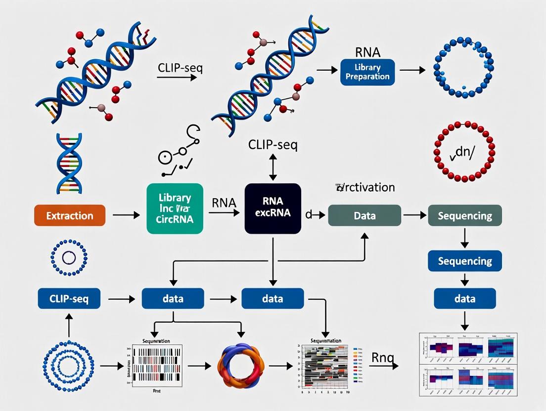
Visualization of Core Concepts
Title: Core CLIP-seq Experimental Workflow
Title: CLIP-seq Informs lncRNA/circRNA Function & Therapy
The Scientist's Toolkit: Essential Research Reagent Solutions
Table 2: Key Reagents and Materials for CLIP-seq Experiments
| Item | Function/Description | Critical Consideration for lncRNA/circRNA |
|---|---|---|
| UV Crosslinker | Delivers calibrated 254 nm (standard) or 365 nm (PAR-CLIP) UV irradiation. | Dose optimization is critical to preserve circRNA structure and protein binding. |
| 4-Thiouridine (4SU) | Photoactivatable nucleoside for PAR-CLIP; induces T-to-C transitions. | Enables single-nucleotide resolution mapping on specific transcripts. |
| RNase I | Endoribonuclease for partial digestion of RNA to fragments. | Digestion conditions must be optimized for structured lncRNAs/circRNAs. |
| High-Quality RBP Antibody | For specific immunoprecipitation. Must be CLIP-validated. | Key determinant of success; antibody must recognize the native, crosslinked RBP. |
| Pre-adenylated 3' Adapter | Modified DNA adapter for ligation to RNA 3' ends, prevents adapter dimerization. | Essential for efficient library construction from low-abundance RNA fragments. |
| T4 RNA Ligase 1 (truncated K227Q) | Specifically ligates pre-adenylated adapter to RNA 3' ends. | Reduces background ligation activity compared to wild-type ligase. |
| Proteinase K | Digests proteins after IP, releasing crosslinked RNA fragments. | Must be molecular biology grade, free of RNase activity. |
| RNase Inhibitors | Added to all lysis and reaction buffers to preserve RNA integrity. | Critical in early steps before stringent washes remove contaminants. |
| Magnetic Protein A/G Beads | Solid support for antibody capture during IP. | Provide low background and are compatible with stringent wash buffers. |
| Size-matched Input (SMI) Control Reagents | Identical library prep from a non-IP sample for eCLIP. | Crucial for normalizing sequencing biases and identifying authentic binding peaks. |
Why CLIP-seq is Indispensable for lncRNA and circRNA Functional Studies
The functional characterization of long non-coding RNAs (lncRNAs) and circular RNAs (circRNAs) presents a unique challenge, as their activities are largely mediated through dynamic interactions with RNA-binding proteins (RBPs) and other nucleic acids. Crosslinking and Immunoprecipitation followed by sequencing (CLIP-seq) has emerged as the cornerstone technology for mapping these interactions in vivo at nucleotide resolution. This whitepaper, framed within a broader thesis on advancing non-coding RNA biology, details the indispensable role of CLIP-seq in elucidating the mechanisms of lncRNAs and circRNAs, providing a technical guide for researchers and drug development professionals.
Unlike mRNAs, the primary functions of many lncRNAs and circRNAs—including transcriptional regulation, chromatin remodeling, sponge activity, and scaffold formation—are executed through direct, often transient, molecular interactions. Traditional knockdown/knockout and expression profiling cannot capture these critical binding events. CLIP-seq, by covalently capturing protein-RNA interactions via UV crosslinking, enables the precise identification of binding sites, distinguishing them from non-specific associations.
Core CLIP-seq Methodologies and Adaptations
Several CLIP-seq variants have been developed, each with specific advantages for studying lncRNA/circRNA complexes.
Key Experimental Protocols
2.1.1 HITS-CLIP (High-Throughput Sequencing CLIP)
- Crosslinking: Cells or tissues are irradiated with 254 nm UV-C light (typically 200-400 mJ/cm²) to form covalent bonds between RBPs and directly contacting RNAs.
- Cell Lysis & Partial RNase Digestion: Lysates are treated with limited RNase to fragment bound RNA, leaving a short "footprint" protected by the protein.
- Immunoprecipitation: The RBP-of-interest, with its crosslinked RNA fragments, is isolated using specific antibodies.
- RNA Adapter Ligation & Purification: After stringent washing, 3' RNA adapters are ligated to the purified RNA-protein complexes on beads. The complexes are separated by SDS-PAGE and transferred to a nitrocellulose membrane. A band corresponding to the RBP's molecular weight is excised.
- Proteinase K Digestion & cDNA Library Prep: Protein is digested, releasing the RNA fragments. A 5' RNA adapter is ligated, followed by reverse transcription, PCR amplification, and high-throughput sequencing.
2.1.2 PAR-CLIP (Photoactivatable-Ribonucleoside-Enhanced CLIP)
- Key Modification: Cells are cultured with nucleoside analogs (e.g., 4-thiouridine or 6-thioguanosine) incorporated into nascent RNA.
- Crosslinking: 365 nm UV light is used, which induces more efficient crosslinking specifically at analog sites and leads to thymidine-to-cytidine transitions in cDNA sequences, providing single-nucleotide resolution binding sites.
2.1.3 eCLIP (Enhanced CLIP)
- Key Improvement: Incorporates a size-matched input (SMInput) control that undergoes identical library preparation (including RNase digestion and adapter ligation) but without immunoprecipitation. This rigorously controls for sequencing biases and background noise.
Protocol Comparison Table
Table 1: Comparison of Major CLIP-seq Variants for lncRNA/circRNA Studies
| Method | Crosslinking Type | Key Feature | Resolution | Primary Application for lncRNA/circRNA |
|---|---|---|---|---|
| HITS-CLIP | UV-C (254 nm) | Standard method, robust. | 30-60 nt | Mapping RBP binding sites on abundant targets. |
| PAR-CLIP | UV-A (365 nm) with nucleoside analogs | T-to-C transitions pinpoint sites. | Single-nucleotide | Identifying precise interaction motifs and domains. |
| eCLIP | UV-C (254 nm) | Paired SMInput control reduces noise. | 30-60 nt | Sensitive detection in complex genomic regions. |
| iCLIP | UV-C (254 nm) | Captures cDNA truncations at crosslink sites. | Single-nucleotide | Studying structural interactions & binding topology. |
Application to lncRNA and circRNA Functional Discovery
Decoding lncRNA Mechanisms
CLIP-seq reveals how lncRNAs act as scaffolds, guides, or decoys. For example, CLIP for proteins like PRC2 (EZH2) or hnRNPs on lncRNAs like XIST or MALAT1 maps exact protein-binding domains, linking sequence to function in epigenetic silencing or mRNA processing.
Unraveling circRNA Protein Sponging
While miRNA sponging is proposed, many circRNAs function via protein interaction. CLIP-seq for an RBP (e.g., MBL, QKI) can identify circRNAs highly enriched in the IP versus input, confirming direct binding. Subsequent mechanistic studies (rescue experiments with binding-deficient mutants) validate functional importance.
Quantitative Data from Recent Studies
Table 2: Example CLIP-seq Findings in lncRNA/circRNA Biology (2022-2024)
| RBP Studied | Target RNA Class | Key Finding | Method | Validation |
|---|---|---|---|---|
| FUS | circRNAs (Neuronal) | Identified 120+ circRNAs bound by FUS in neurons; a subset co-aggregate in ALS models. | eCLIP | RIP-qPCR, imaging. |
| MATR3 | lncRNAs (Nuclear) | Mapped binding to 450+ lncRNAs; essential for NEAT1 paraspeckle integrity. | PAR-CLIP | CRISPR deletion, FISH. |
| IGF2BP2 | oncogenic circRNAs | Direct binding to circNDUFB2 and circCCDC66 promotes their stability in cancer. | iCLIP | Actinomycin D assays, mutational analysis. |
| EWS | LINP1 lncRNA | Interaction enhances non-homologous end joining DNA repair in breast cancer. | HITS-CLIP | Comet assay, IR sensitivity. |
The Scientist's Toolkit: Essential Research Reagent Solutions
Table 3: Key Reagents for CLIP-seq Experiments on lncRNAs and circRNAs
| Item | Function | Critical Consideration |
|---|---|---|
| UV Crosslinker | Covalently fixes protein-RNA interactions in vivo. | Calibrated energy output (mJ/cm²) is crucial for reproducibility. |
| RNase Inhibitors | Prevent non-specific RNA degradation during lysis and IP. | Must be added fresh to all lysis/wash buffers. |
| Magnetic Protein A/G Beads | Solid support for antibody-mediated IP. | Pre-clearing with beads reduces non-specific background. |
| High-Specificity Antibodies | Immunoprecipitate the target RBP. | Validation for IP (knockout/knockdown control) is mandatory. |
| PNK (Polynucleotide Kinase) | Facilitates adapter ligation in library prep. | Critical for efficient recovery of RNA fragments. |
| Reverse Transcriptase | Generates cDNA from crosslinked, fragmented RNA. | Must process RNA with protein adducts (high processivity). |
| Size Selection Kits | Isolate cDNA libraries of optimal length. | Essential for removing adapter dimers prior to sequencing. |
| rRNA Depletion Probes | Enrich for non-coding RNAs (including lnc/circRNA). | Poly-A selection is unsuitable for most lncRNAs & circRNAs. |
Integrated Experimental and Analytical Workflow
Diagram 1: End-to-end CLIP-seq workflow for lncRNA/circRNA studies.
Pathway Visualization: CLIP-seq Informs circRNA Sponge Function
Diagram 2: How CLIP-seq validates a circRNA protein sponge mechanism.
Within the thesis that precise molecular mapping is fundamental to understanding non-coding RNA function, CLIP-seq proves indispensable. It transforms lncRNAs and circRNAs from mere lists of sequences into dynamic interaction hubs. As protocols standardize and integrate with emerging techniques like single-cell sequencing and spatial transcriptomics, CLIP-seq will continue to be the definitive method for driving functional discovery and identifying novel, druggable RNA-protein interactions in disease.
Within the context of advancing the functional annotation of non-coding RNAs, CLIP-seq (Crosslinking and Immunoprecipitation followed by sequencing) stands as a cornerstone technique for mapping RNA-protein interactions in vivo. Its application diverges significantly when targeting linear long non-coding RNAs (lncRNAs) versus circular RNAs (circRNAs), the latter formed by back-splicing. This technical guide details these critical differences, framing them within a thesis focused on elucidating the distinct mechanistic roles of these RNA classes through protein interactome mapping.
Core Conceptual & Technical Divergences
The fundamental distinction lies in the RNA topology: linear lncRNAs have free ends, while circRNAs are covalently closed loops. This difference permeates every stage of CLIP-seq experimental design and data analysis.
Table 1: Key Strategic Differences in CLIP-seq Application
| Aspect | Linear lncRNA CLIP-seq | Back-Spliced circRNA CLIP-seq |
|---|---|---|
| Primary Target | Protein binding sites along a linear sequence. | Protein binding sites on a circular molecule; validation of circularity is paramount. |
| Pre-IP Enrichment | Often optional. May use cytoplasmic/nuclear fractionation. | Mandatory. RNase R treatment to degrade linear RNAs and enrich for circRNAs. |
| Library Construction | Standard protocols (e.g., iCLIP, eCLIP). | Must preserve back-splice junction reads. Use of random hexamers over poly(A) selection. |
| Read Alignment | Align to standard linear reference genome. | Requires BSJ-aware aligners (e.g., STAR, CIRI2, CircSplice) to detect non-colinear back-splice junctions. |
| Binding Site Analysis | Peaks identified across the gene body. | Peaks analyzed both within the circularized exons and specifically around the BSJ. |
| Validation | RT-qPCR with exon-spanning primers. Northern Blot. | BSJ-specific RT-qPCR. Divergent primer design. Northern Blot with RNase R control. |
| Key Challenge | Distinguishing specific signal from other overlapping transcripts. | Overcoming low abundance; confirming interactions are with the circRNA isoform, not its linear cognate. |
Detailed Experimental Protocols
Protocol A: circRNA-Focused CLIP-seq with RNase R Enrichment
- Crosslinking & Lysis: UV crosslink cells (254 nm). Lyse in stringent RIPA buffer with RNase inhibitors.
- RNase R Digestion: Treat a portion of lysate with RNase R (3 U/µg RNA, 37°C, 15 min) to digest linear RNAs. Keep an untreated control.
- Immunoprecipitation: Incubate lysate with antibody against target protein (e.g., anti-AGO2) and Protein A/G beads. Include IgG isotype control.
- RNA Processing: Wash stringently. Digest with Proteinase K. Recover RNA.
- Library Prep & Sequencing: Construct cDNA library using random primers (not oligo-dT). Sequence on a platform yielding >100bp reads to span back-splice junctions.
Protocol B: Validation via BSJ-specific RT-qPCR
- Primer Design: Design divergent primers that face away from each other, flanking the back-splice junction. Only cDNA from the circRNA will amplify.
- Reverse Transcription: Use random hexamers and circRNA-enriched RNA.
- qPCR: Perform SYBR Green qPCR with divergent primers. Normalize to a stable circRNA or use spike-in controls.
Data Analysis Workflow Visualization
Short Title: CLIP-seq Data Analysis Branch for lncRNA vs. circRNA
The Scientist's Toolkit: Essential Research Reagents & Materials
Table 2: Key Reagent Solutions for lncRNA/circRNA CLIP-seq
| Reagent / Material | Function | Application Note |
|---|---|---|
| RNase R (Epicentre) | 3'->5' Exoribonuclease that degrades linear RNAs but not circRNAs. | Critical for circRNA enrichment pre-IP. Validate digestion efficiency via gel. |
| UV Crosslinker (254 nm) | Creates covalent bonds between RNAs and directly interacting RBPs in vivo. | Standard for both; optimize energy dose (e.g., 150-400 mJ/cm²). |
| Anti-AGO2 Antibody | Immunoprecipitates Argonaute proteins for miRNA/RISC interaction studies. | Common for both, especially if studying sponging. |
| Divergent PCR Primers | Primers oriented away from each other, specific to the back-splice junction. | Gold standard for circRNA validation. Must flank the BSJ. |
| CircRNA-aware Aligner (STAR, CIRI2) | Aligns sequencing reads, detecting non-colinear back-splice junctions. | Mandatory software for circRNA CLIP-seq analysis. |
| RNase Inhibitor (Murine) | Prevents sample degradation during immunoprecipitation and RNA handling. | Use at high concentration in all lysis and wash buffers. |
| Control siRNA/shRNA | For knockdown of target RNA to confirm CLIP specificity. | Essential control to show loss of signal upon RNA depletion. |
| Proteinase K | Digests proteins after IP to recover crosslinked RNA fragments. | Standard in both protocols for RNA extraction post-IP. |
The systematic study of long non-coding RNAs (lncRNAs) and circular RNAs (circRNAs) demands precise mapping of their protein interaction partners and binding sites. Crosslinking and immunoprecipitation (CLIP) sequencing technologies are foundational for this mission, enabling transcriptome-wide profiling of RNA-protein interactions. This technical guide traces the evolution of CLIP methods, detailing their adaptations to overcome inherent limitations. Each advancement—from HITS-CLIP to eCLIP, iCLIP, and irCLIP—has incrementally enhanced resolution, specificity, and signal-to-noise ratio, directly empowering rigorous functional studies of lncRNA and circRNA mechanisms in development, disease, and as potential therapeutic targets.
The quantitative improvements across CLIP variants are summarized in the table below.
Table 1: Comparative Evolution of High-Throughput CLIP Methodologies
| Method | Key Innovation | Primary Advantage | Critical Limitation Addressed | Typical Input Material | Approximate Signal-to-Noise Improvement vs. Predecessor |
|---|---|---|---|---|---|
| HITS-CLIP (2009) | High-throughput sequencing of CLIP libraries. | First genome-wide, unbiased RBP binding maps. | Scalability of traditional CLIP. | ~1-10 million crosslinked cells | Baseline |
| PAR-CLIP (2010) | Incorporation of photoreactive nucleoside analogs (4-SU). | Induces T-to-C transitions for precise (<1-2 nt) binding site identification. | Ambiguity in crosslink site resolution. | 4-SU/6-SG treated cells | ~2-5 fold (via precise mutation calling) |
| iCLIP (2010) | Introduction of intermolecular cDNA truncation at crosslink sites. | Enables single-nucleotide resolution mapping and reveals truncated cDNAs. | Inefficient adapter ligation to RNA due to leftover peptide. | ~5-10 million cells | ~3-10 fold (reduced background) |
| eCLIP (2016) | Size-matched input controls and optimized ligation. | Dramatically lowers background, improves reproducibility and specificity. | Non-specific background and library complexity artifacts. | 1-10 million cells | ~10-1000 fold (via size-matched input normalization) |
| irCLIP (2017) | Inverted repeat adapter design for intramolecular ligation. | Extremely efficient ligation, high sensitivity with low input. | Low efficiency of intermolecular RNA adapter ligation. | As low as 100,000 cells | ~5-10 fold over iCLIP (higher library complexity) |
Detailed Experimental Protocols
This section outlines the critical, distinguishing steps for each advanced CLIP protocol.
iCLIP (Individual-nucleotide resolution CLIP)
Distinguishing Step: cDNA Truncation and Circularization
- UV Crosslinking & Immunoprecipitation: Cells are UV-C crosslinked (254 nm). Lysates are partially RNase-treated, and the RBP-RNA complex is immunoprecipitated.
- 3' Adapter Ligation: A 3' adapter is ligated to the RNA on beads.
- Reverse Transcription (RT): RT proceeds until it stalls at the crosslinked nucleotide, often resulting in cDNA truncation. A special RT primer contains a 5' adapter sequence and two cleavable groups.
- cDNA Circularization: The truncated cDNA is circularized using CircLigase, bringing the 5' and 3' ends together.
- Linearization & PCR: The circular cDNA is linearized by cleavage at the sites in the primer, and the final library is amplified by PCR for sequencing. The crosslink site is identified as the first nucleotide of the read (the truncation site).
eCLIP (Enhanced CLIP)
Distinguishing Step: Size-Matched Input (SMInput) Control
- Parallel Input Sample: Alongside the standard IP sample, a "size-matched input" control is prepared. An aliquot of pre-cleared lysate is incubated without antibody but is subjected to the same RNase digestion conditions.
- Library Construction: Both IP and SMInput samples undergo identical library prep: dephosphorylation, 3' adapter ligation, phosphorylation, and 5' adapter ligation.
- Gel Purification: Both samples are run on a SDS-PAGE gel, and a region corresponding to the RBP's size is excised. The SMInput is excised from the same gel region as the IP sample.
- Proteinase K Digestion & Purification: RNA is recovered, reverse transcribed, and PCR amplified.
- Bioinformatic Normalization: Sequencing reads from the IP are directly compared to those from the SMInput control to identify significantly enriched binding sites, drastically reducing false positives from abundant RNAs and RNA fragments.
irCLIP (Infrared-CLIP / improved CLIP)
Distinguishing Step: Inverted Repeat Adapter for Intramolecular Ligation
- Adapter Design: A single adapter containing an Illumina-compatible sequence flanked by two complementary inverted repeats is used.
- Ligation on Beads: After IP and washing, this single adapter is ligated to the RNA ends on the beads. The inverted repeats cause the adapter to self-hybridize, bringing its 5' and 3' ends into proximity.
- Intramolecular Ligation: This proximity enables highly efficient intramolecular ligation, circularizing the adapter-RNA molecule directly on the beads. This step bypasses the inefficient intermolecular ligation of a second adapter.
- Linearization & Amplification: The circular product is eluted and linearized via cleavage at a restriction site within the adapter. Subsequent RT-PCR generates the final sequencing library.
Visualizing CLIP Evolution: Workflows and Logic
Diagram 1: Evolutionary Relationships of CLIP Methods
Diagram 2: eCLIP Core: IP vs. Size-Matched Input Control
Diagram 3: irCLIP Key Innovation: Intramolecular Ligation
The Scientist's Toolkit: Essential Research Reagents
Table 2: Key Reagent Solutions for Modern CLIP-seq Experiments
| Reagent / Kit | Primary Function in CLIP | Critical Notes for lncRNA/circRNA Studies |
|---|---|---|
| UV Crosslinker (254 nm) | Induces covalent bonds between RBPs and directly contacting RNAs. | Critical for capturing transient interactions. Dose must be optimized to preserve lncRNA structure. |
| RNase I (or T1) | Partially digests RNA to leave short (~20-70 nt) protein-protected fragments. | Digestion condition is key; over-digestion can destroy structured lncRNA/circRNA binding sites. |
| Protein A/G Magnetic Beads | Solid support for antibody-based immunoprecipitation of RBP complexes. | Choice depends on antibody host species. Low RNA-binding beads are essential to reduce background. |
| High-Specificity Antibodies | Target the RBP of interest for IP. | Validated for CLIP/IP is mandatory. Poor antibodies are the leading cause of failure. |
| T4 PNK (Polynucleotide Kinase) | Phosphorylates 5' ends and dephosphorylates 3' ends for adapter ligation. | Essential step in most protocols to prepare RNA ends for ligation. |
| T4 RNA Ligase 1 & 2 (truncated) | Catalyzes 3' and 5' adapter ligation to RNA fragments. | The workhorse enzyme for library construction. Efficiency dictates library complexity. |
| CircLigase (for iCLIP) | Circularizes single-stranded DNA (cDNA). | Enables capture of cDNA truncation events marking the crosslink site. |
| Proteinase K | Digests proteins after gel purification to recover crosslinked RNA. | Must be molecular biology grade, RNase-free. |
| SPRI (Solid Phase Reversible Immobilization) Beads | Size-selective purification and cleanup of nucleic acids (cDNA, libraries). | Replaces traditional column-based kits for better size selection and recovery. |
| High-Fidelity PCR Mix | Amplifies final cDNA library for sequencing. | Low error rate is crucial to avoid false mutations. Minimal cycles to prevent duplicates. |
| Unique Molecular Identifiers (UMIs) | Short random nucleotide sequences added to adapters. | Allows bioinformatic removal of PCR duplicates, essential for accurate quantification of binding. |
Within the broader thesis of utilizing CLIP-seq to delineate the functional landscape of lncRNAs and circRNAs, a critical first step is the strategic selection of the RNA-binding protein (RBP) target for study. The choice of RBP dictates the biological question addressable, the experimental feasibility, and the translational potential of the findings. This guide provides a framework for making this pivotal decision, integrating current methodological and biological insights.
Biological & Functional Rationale
The justification for studying an RBP should be grounded in prior evidence linking it to non-coding RNA biology.
Table 1: Criteria for RBP Target Selection
| Criterion | Key Questions | Supporting Evidence Sources |
|---|---|---|
| Known Interaction | Is there literature or preliminary data (e.g., RIP, RNA-pulldown) linking the RBP to lncRNAs/circRNAs? | Ago2 with circCDR1as; HuR with MALAT1; RBFOX2 with circularizable exons. |
| Pathway Relevance | Does the RBP regulate processes central to your study (e.g., splicing, stability, translation)? | QKI in circRNA biogenesis; ADAR in A-to-I editing of circRNAs. |
| Disease Association | Are RBP mutations or dysregulated expressions linked to pathologies where lncRNAs/circRNAs are implicated? | FUS/TLS in ALS; LIN28 in cancer; EWSR1 in sarcomas. |
| Subcellular Localization | Is the RBP's localization congruent with the ncRNA's function (nuclear, cytoplasmic, specific organelles)? | IGF2BP family in cytoplasmic mRNA granules; SRSF1 in nuclear speckles. |
| Structural Motifs | Does the RBP have domains (e.g., RRM, KH, dsRBD) known to bind structural features of your ncRNA? | PKR binding to dsRNA regions in circRNAs. |
Technical Feasibility for CLIP-seq
Not all RBPs are equally amenable to CLIP-based studies. Practical considerations are paramount.
Table 2: Technical Considerations for CLIP-seq on Target RBP
| Consideration | High Feasibility | Lower Feasibility / Challenges |
|---|---|---|
| Antibody Availability | High-quality, validated commercial antibody for immunoprecipitation. | No antibody; antibody has poor IP efficiency or high background. |
| Crosslinking Efficiency | RBP binds directly to RNA (UV-C 254 nm crosslinking suitable). | RBP binds via large complexes or indirect (requires protein-protein crosslinkers like formaldehyde). |
| Expression Abundance | RBP is endogenously expressed at moderate-to-high levels. | RBP is lowly expressed, requiring overexpression which may alter biology. |
| CLIP Protocol Choice | eCLIP (enhanced CLIP) for robustness; iCLIP for single-nucleotide resolution. | PAR-CLIP for specific RBP classes using 4SU incorporation. |
Experimental Protocol: Standard eCLIP Workflow
Below is a detailed protocol for eCLIP, the current benchmark for in vivo RBP-RNA interaction mapping.
Protocol: Enhanced CLIP (eCLIP) for RBP-NcRNA Interaction Mapping
A. Cell Culture & Crosslinking
- Grow relevant cell lines (e.g., HEK293T, primary cells) to ~80% confluence.
- Wash cells once with cold PBS.
- UV-C Crosslinking: Irradiate cells in PBS with 254 nm UV light at 150-400 mJ/cm² (optimize per RBP).
- Harvest cells by scraping, pellet, and flash-freeze in liquid N₂.
B. Cell Lysis & Immunoprecipitation
- Lyse cell pellet in ice-cold lysis buffer (e.g., 50 mM Tris-HCl pH 7.4, 100 mM NaCl, 1% NP-40, 0.1% SDS, 0.5% sodium deoxycholate, protease/RNase inhibitors).
- Partial RNA digestion: Add RNase I to lysate (diluted 1:1000) and incubate 3 min at 37°C to generate RNA fragments.
- Pre-clear lysate with Protein A/G beads.
- Incubate pre-cleared lysate with antibody-conjugated beads overnight at 4°C. Include a size-matched input (SMInput) control.
C. Washing, Dephosphorylation & Ligation
- Wash beads stringently with high-salt wash buffers.
- 3' Dephosphorylation: Use T4 PNK (without ATP) to repair RNA 3' ends.
- 3' Ligation: Ligate a pre-adenylated DNA barcode adapter to RNA 3' ends using T4 RNA Ligase 1.
- Wash beads to remove excess adapter.
D. RNA-Protein Complex Transfer & Proteinase K Digestion
- Transfer complexes to a nitrocellulose membrane via a dot-blot apparatus.
- Proteinase K Digestion: Digest proteins on the membrane with Proteinase K to release crosslinked RNA fragments.
- Purify RNA via phenol-chloroform extraction and ethanol precipitation.
E. Reverse Transcription & cDNA Purification
- Reverse transcribe using a primer complementary to the 3' adapter.
- Run cDNA on a denaturing Bis-Tris NuPAGE gel.
- Excision of a size range (~70 kDa above RBP's molecular weight) to isolate RBP-bound RNA fragments.
- Extract and purify cDNA from gel slice.
F. Second Adapter Ligation & PCR Amplification
- 5' Ligation: Ligate a second DNA adapter to the cDNA 3' end (now representing the RNA 5' end).
- PCR amplify libraries using indexed primers.
- Sequence on an Illumina platform.
Data Interpretation & Pathway Analysis
Following CLIP-seq, identifying bound lncRNAs/circRNAs is the first step. The core analysis involves mapping reads, calling peaks, and annotating them to non-coding transcripts. Functional validation is critical. Key pathways often involved include:
Diagram 1: RBP Regulation of circRNA/lncRNA Function
The Scientist's Toolkit: Essential Research Reagents
Table 3: Key Reagent Solutions for RBP-ncRNA CLIP Studies
| Reagent / Material | Function & Critical Consideration |
|---|---|
| UV Crosslinker (254 nm) | Induces covalent bonds between RBP and directly bound RNA. Calibration of energy dose is crucial for efficiency vs. background. |
| Validated IP Antibody | Specific antibody for the target RBP. Must be validated for immunoprecipitation under denaturing conditions. |
| RNase I | Partially digests RNA to leave only RBP-protected fragments. Titration is essential for optimal fragment length. |
| Pre-adenylated 3' Adapter | Enables ligation to RNA 3' ends without ATP to prevent adapter concatemerization. Contains barcodes for multiplexing. |
| Proteinase K | Digests the RBP to release crosslinked RNA fragments for downstream library prep. Must be RNase-free. |
| Nitrogenous PAGE Gel System | For size selection of cDNA. Provides cleaner size separation than agarose gels, critical for removing adapter dimer. |
| CircRNA-specific Enrichment/Oligos | Poly(A)- selection depletes circRNAs. Use Ribo-depletion and/or RNase R treatment to enrich for circRNAs prior to library prep. |
| CLIP-seq Analysis Pipeline (e.g., CLIPper, PEAKachu) | Specialized software for peak calling from CLIP data, accounting for crosslinking-induced mutations and truncations. |
Diagram 2: Core CLIP-seq Experimental Workflow
Within the context of a thesis on CLIP-seq for functional studies of long non-coding RNAs (lncRNAs) and circular RNAs (circRNAs), experimental design is paramount. This guide delineates the core objectives, methodologies, and analytical frameworks distinguishing exploratory from hypothesis-driven Crosslinking and Immunoprecipitation (CLIP) approaches. Clarifying this distinction is critical for advancing from cataloging RNA-protein interactions to mechanistically defining their roles in gene regulation and disease.
CLIP-seq and its advanced variants (e.g., HITS-CLIP, PAR-CLIP, iCLIP, eCLIP) are indispensable for transcriptome-wide mapping of RNA-protein interactions. For lncRNAs and circRNAs—which often function through ribonucleoprotein complexes—CLIP provides direct evidence of physical binding. The research trajectory from discovery to mechanism mandates a clear strategic choice: exploratory profiling to generate novel interaction maps or hypothesis-driven experimentation to test a specific functional model.
Foundational Principles: Exploratory vs. Hypothesis-Driven Research
Table 1: Core Comparative Framework
| Aspect | Exploratory CLIP | Hypothesis-Driven CLIP |
|---|---|---|
| Primary Objective | Unbiased discovery of novel RNA-binding protein (RBP) binding sites, partners, or associated ncRNAs. | Test a specific model of RBP-ncRNA function (e.g., in a pathway, cellular process, or disease mechanism). |
| Starting Point | Often an RBP of interest with unknown RNA targets, or an ncRNA with unknown protein partners. | A prior observation (e.g., co-expression, genetic interaction, phenotypic correlation) suggesting a specific interaction/function. |
| Experimental Design | Comparative (e.g., WT vs. RBP knockdown, or IgG control IP). Focus on robustness and depth of coverage. | Perturbation-based (e.g., mutant RBP, mutant RNA, specific cellular stimulus). Includes precise positive/negative controls. |
| Analysis Emphasis | Comprehensive cataloging, de novo motif discovery, enrichment analysis for pathways/ontologies. | Differential binding analysis, precise mapping to functional genomic features, validation of mechanistic models. |
| Outcome | Generation of novel hypotheses and resource datasets. | Causal inference and mechanistic insight. |
Defining Objectives and Design Criteria
Objectives for Exploratory CLIP
- Census-taking: Identify the full repertoire of lncRNAs/circRNAs bound by a specific RBP (e.g., a splicing factor potentially binding to circRNAs).
- Characterization: Define the binding landscape—preferred sequence motifs, structural contexts, and genomic distribution (exonic, intronic, 3'UTR) on target ncRNAs.
- Hypothesis Generation: Correlate binding sites with ncRNA localization or putative functions to generate testable models.
Objectives for Hypothesis-Driven CLIP
- Mechanistic Testing: Determine if a specific protein-ncRNA interaction mediates a known function (e.g., does binding of HNRNPK to circFOXO3 promote its nuclear retention?).
- Regulation Analysis: Test if a cellular signal (e.g., DNA damage, differentiation cue) alters the binding affinity or landscape of an RBP for a set of ncRNAs.
- Pathway Integration: Validate if an RBP-ncRNA interaction is essential for the activity of a defined signaling pathway (e.g., p53 pathway).
Detailed Experimental Protocols
Universal CLIP-seq Core Protocol (Adaptable for Both Approaches)
Key Reagent Solutions:
- UV-C (254 nm) Crosslinker: Covalently links RBPs to RNA at zero-distance in vivo.
- RNase I (Partial Digestion): Trims unprotected RNA, leaving ~50-100 nt protein-bound footprints.
- Phosphatase and Polynucleotide Kinase: For controlling RNA ends during library prep.
- Proteinase K: Recovers crosslinked RNA fragments after IP.
- 3' Adaptor Ligation (Pre-IP in iCLIP): Minimizes bias from RNA degradation.
- High-Efficiency Reverse Transcription: Crucial for capturing crosslink-induced mutations/deletions.
- Illumina-Compatible Library Preparation: Includes size selection for ~50-200 nt fragments.
Workflow Diagram:
Title: Core CLIP-seq Experimental Workflow
Protocol Tailoring for Specific Objectives
Table 2: Tailored Methodological Variations
| Objective | Key Protocol Variation | Rationale & Notes |
|---|---|---|
| Exploratory: Broad Target ID | Use eCLIP or HITS-CLIP. Include size-matched input (SMI) control. | SMI controls for RNA abundance & background. Robust protocol for comprehensive maps. |
| Exploratory: circRNA-specific | RNase R treatment post-lysis to linear RNA. Use circRNA-optimized aligners (CIRCexplorer, CIRI2). | Enriches for circRNA-protein complexes. Requires careful validation to avoid artifacts. |
| Hypothesis: Binding Dynamics | PAR-CLIP (4-SU incorporation). Compare treated vs. untreated cells. | 4-SU causes T-to-C transitions, marking exact crosslink sites for high-resolution analysis. |
| Hypothesis: Functional Validation | CLIP-qPCR on specific candidates post-full CLIP-seq. Integrate with RBP/ncRNA knockout. | Provides rapid, quantitative validation of interactions before deep mechanistic studies. |
Analytical Pathways & Data Interpretation
Exploratory Analysis Pathway:
Title: Exploratory CLIP-seq Data Analysis Pipeline
Hypothesis-Driven Analysis Logic:
Title: Hypothesis-Driven CLIP Analysis & Validation Logic
The Scientist's Toolkit: Research Reagent Solutions
Table 3: Essential Reagents for CLIP-seq in ncRNA Studies
| Reagent / Material | Function & Rationale | Key Considerations |
|---|---|---|
| High-Affinity, Validated Antibodies | Immunoprecipitation of target RBP. | Specificity is critical. Knockout/knockdown validation recommended. |
| UV 254 nm Crosslinker | In vivo fixation of direct RNA-protein contacts. | Calibrate energy (e.g., 150-400 mJ/cm²) to optimize crosslinking vs. cell viability. |
| RNase I (Ambion) | Creates protein-protected RNA footprints. | Titration is essential; must be optimized per RBP. |
| [γ-32P] ATP or IRDye 800CW | For visualizing RNA-protein complexes on membranes. | Radioactive offers sensitivity; fluorescent is safer and facilitates size estimation. |
| Proteinase K | Releases crosslinked RNA fragments from the RBP. | Must be molecular biology grade, RNAse-free. |
| circRNA-enriched RNA Library Prep Kits | For downstream validation of circRNA targets. | Select kits with methods to avoid linear RNA amplification (e.g., RNase R treatment). |
| Crosslink-Induced Mutation Analysis Software (e.g., CIMS, CITS) | Pinpoints crosslink sites at single-nucleotide resolution. | Critical for PAR-CLIP and iCLIP data to define precise binding motifs. |
The strategic definition of objectives—either exploratory discovery or hypothesis-driven mechanism—fundamentally shapes every subsequent phase of a CLIP-seq experiment for lncRNA and circRNA research. Exploratory studies generate the essential atlases of interaction, while hypothesis-driven designs transform these observations into causal, mechanistic understanding. A clear alignment between the initial objective, experimental protocol, and analytical pathway is the cornerstone of robust, interpretable, and impactful research in functional ncRNA biology.
From Theory to Bench: A Step-by-Step CLIP-seq Protocol for Non-Coding RNAs
Cell/Tissue Preparation and Crosslinking Optimization for Native RNA-Protein Complexes
The study of long non-coding RNAs (lncRNAs) and circular RNAs (circRNAs) represents a frontier in understanding gene regulation and their roles in disease. A central thesis in this field posits that elucidating the precise in vivo RNA-protein interactome is critical for defining the molecular mechanisms of lncRNA and circRNA function. Crosslinking and immunoprecipitation followed by sequencing (CLIP-seq) is the cornerstone methodology for this endeavor. The fidelity of any CLIP-seq experiment, however, is fundamentally determined by the initial steps: the preservation of native RNA-protein interactions through optimal cell/tissue preparation and crosslinking. This guide provides an in-depth technical framework for these critical preparative steps, ensuring the capture of biologically relevant complexes for downstream CLIP-seq applications in functional genomics and drug target discovery.
Core Principles of Crosslinking for Native Complexes
Effective crosslinking must strike a balance between sufficient fixation to stabilize transient interactions and minimal perturbation to maintain complex native state and subsequent biochemical accessibility. Two primary modalities are employed:
- Ultraviolet (UV) Crosslinking (254 nm): Forms covalent bonds between RNA bases and amino acids in direct physical contact (primarily pyrimidines with aromatic/charged residues). It offers zero-length crosslinking with high spatial precision but limited penetration.
- Chemical Crosslinking (e.g., Formaldehyde/FA): Creates reversible protein-protein and protein-RNA bridges over longer distances (~2 Å), useful for stabilizing larger complexes but with lower resolution.
For mapping direct RNA-binding protein (RBP) binding sites, UV crosslinking is essential. For studying larger ribonucleoprotein (RNP) assemblies, a combination of UV and FA is often optimal.
Quantitative Comparison of Crosslinking Methods
Table 1: Quantitative Comparison of Crosslinking Modalities for CLIP-seq
| Parameter | UV-C (254 nm) | Formaldehyde (FA) | Combined (UV-C + FA) |
|---|---|---|---|
| Crosslink Type | Zero-length, covalent | Reversible, ~2Å spacer | Combined direct & proximal |
| Primary Target | RNA-protein (direct contact) | Protein-protein, Protein-RNA | Both direct and complex-stabilizing |
| Typical Energy/Dose | 150-400 mJ/cm² | 0.1-1% for 5-10 min | 250 mJ/cm² UV + 0.1% FA 5 min |
| Penetration Depth | Very shallow (<1 cell layer) | Good (whole tissue sections) | Limited by UV component |
| Crosslinking Reversal | Difficult (RNase digestion) | Reversible (heat, pH) | FA reversible, UV persistent |
| Optimal For | Direct RBP binding site mapping (eCLIP, iCLIP) | Stabilizing large RNP complexes | Studying lncRNA/circRNA protein complexes |
| Key Advantage | High resolution, no chemical handling | Stabilizes multi-component complexes | Captures direct binding within native context |
| Key Limitation | Poor tissue penetration, efficiency varies | Non-specific background, indirect links | More complex protocol optimization |
Table 2: Impact of Crosslinking Parameters on CLIP-seq Outcomes
| Optimized Parameter | Sub-optimal Condition | Effect on CLIP-seq Data Quality |
|---|---|---|
| UV Dose: 250 mJ/cm² | Too Low (<100 mJ/cm²) | Few crosslinks, low signal, high noise. |
| Too High (>500 mJ/cm²) | RNA degradation, epitope masking, poor IP efficiency. | |
| FA Concentration: 0.1% | Too High (>1%) | Excessive protein-protein crosslinking, inaccessible epitopes, high background. |
| Crosslinking Temperature: 4°C | Room Temp or 37°C | Increased non-physiological interactions, increased RNA degradation. |
| Lysis Buffer Stringency | Too Harsh (e.g., 1% SDS) | Complex disruption. |
| Too Mild (e.g., No detergent) | Incomplete lysis, high viscosity, non-specific binding. |
Detailed Experimental Protocols
Protocol 4.1: Optimized Preparation of Adherent Cells for lncRNA-CLIP
Objective: Harvest and crosslink adherent cells while preserving native RNA-protein complexes.
- Pre-cool: Place culture dish on an ice-cold metal block. Aspirate medium.
- Wash: Gently add 10 mL of ice-cold 1X PBS. Rock and aspirate.
- In-situ UV Crosslinking: Remove lid and irradiate cells in PBS with 254 nm UV light at 250 mJ/cm² in a calibrated UV crosslinker (e.g., Stratainker). Keep dish on ice during process.
- (Optional) FA Crosslinking: For combined crosslinking, add PBS containing 0.1% formaldehyde directly to dish after UV. Incubate for 5 min at room temperature with gentle rocking. Quench with 125 mM glycine for 5 min.
- Scraping: Aspirate liquid. Add 1 mL of ice-cold PBS. Use a cold cell scraper to dislodge cells. Transfer suspension to a pre-chilled microcentrifuge tube.
- Pellet: Centrifuge at 500 x g for 3 min at 4°C. Aspirate supernatant. Flash-freeze pellet in liquid nitrogen. Store at -80°C.
Protocol 4.2: Optimized Preparation of Murine Tissue for circRNA-CLIP
Objective: Crosslink and homogenize tissue to capture tissue-specific RNP complexes.
- Perfusion & Dissection: Perfuse mouse transcardially with ice-cold 1X PBS. Rapidly dissect tissue of interest.
- Sectioning: Slice tissue into <2 mm slices using a sterile blade on a chilled surface.
- UV Crosslinking: Place slices in a single layer in a Petri dish with ice-cold PBS. Irradiate with UV at 400 mJ/cm² (higher due to scattering).
- Dicing & FA Crosslinking (Optional): Dice slices into smaller pieces on dry ice. For combined crosslinking, transfer pieces to 0.1% formaldehyde in PBS for 10 min on a rotator at 4°C. Quench with 125 mM glycine.
- Wash & Homogenize: Wash pieces twice with ice-cold PBS. Homogenize in desired lysis buffer (e.g., IP Lysis Buffer + RNase inhibitors) using a Dounce homogenizer or gentle mechanical homogenizer on ice.
- Clarify & Freeze: Centrifuge homogenate at 12,000 x g for 10 min at 4°C. Aliquot supernatant (lysate) and snap-freeze in liquid N₂. Store at -80°C.
Protocol 4.3: Validation of Crosslinking Efficiency (Post-lysis QC)
Objective: Assess the success of RNA-protein crosslinking prior to immunoprecipitation.
- Prepare Lysate: Lysate crosslinked cells/tissue in strong RIPA buffer (1% SDS, 0.5% Na-deoxycholate) with protease/RNase inhibitors.
- Acid-Phenol:Chloroform Extraction: Mix 100 µL lysate with equal volume acid-phenol:chloroform (pH 4.5). Vortex vigorously, centrifuge.
- Phase Separation: The interphase (contains crosslinked RNA-protein complexes) will be significantly enlarged compared to a non-crosslinked control. Aqueous phase (free RNA) will be reduced.
- Quantification: Measure RNA concentration in the aqueous phase (Qiagen RNeasy). A >70% reduction in recoverable free RNA compared to non-crosslinked control indicates efficient UV crosslinking.
Visualizations
Diagram 1: Crosslinking Pathways for RNP Complex Stabilization
Diagram 2: Cell and Tissue Preparation Workflow for CLIP-seq
The Scientist's Toolkit: Key Reagents & Materials
Table 3: Essential Research Reagent Solutions for Crosslinking Optimization
| Item | Function & Rationale |
|---|---|
| Stratainker 2400 (or equivalent calibrated UV crosslinker) | Provides precise, reproducible 254 nm UV dosage critical for consistent RNA-protein crosslinking efficiency. |
| 37% Formaldehyde, Molecular Biology Grade | Source for fresh, low-polymerization formaldehyde for gentle chemical crosslinking; aliquot and store airtight. |
| RNase Inhibitor (e.g., Murine RNase Inhibitor, SUPERase•In) | Essential in all post-lysis buffers to prevent degradation of crosslinked RNA prior to capture. |
| Protease Inhibitor Cocktail (EDTA-free) | Preserves protein epitopes and complex integrity during lysis. EDTA-free is crucial for subsequent enzymatic steps. |
| Acid-Phenol:Chloroform, pH 4.5 | Used in QC assay to separate free RNA from crosslinked RNA-protein complexes via phase separation. |
| Glycine (2.5M stock) | Quenches formaldehyde crosslinking to prevent over-fixation and non-specific crosslinking during processing. |
| IP Lysis Buffer (e.g., 50mM Tris pH7.4, 150mM NaCl, 1% NP-40, 0.5% Na-deoxycholate) | Standard mild lysis buffer for CLIP; solubilizes membranes while preserving most protein-protein interactions. |
| Strong RIPA Lysis Buffer (with 1% SDS) | Used for validation QC and stringent washing; SDS helps disrupt non-covalent interactions for cleaner backgrounds. |
| Dounce Homogenizer (tight pestle) | For gentle mechanical disruption of crosslinked tissues, minimizing heat generation and complex shearing. |
| Dynabeads Protein A/G | Magnetic beads for efficient immunoprecipitation; consistent size and low non-specific binding are critical for CLIP. |
The functional characterization of long non-coding RNAs (lncRNAs) and circular RNAs (circRNAs) necessitates precise mapping of their interactions with RNA-binding proteins (RBPs). Crosslinking and immunoprecipitation followed by sequencing (CLIP-seq) is the cornerstone technique for this purpose. A critical, yet often under-optimized, step in CLIP-seq protocols is the partial digestion of RNA with Ribonuclease (RNase). This whitepaper details the principle and practice of RNase titration, establishing it as the fundamental determinant for achieving single-nucleotide resolution in defining protein-RNA binding sites, which is paramount for elucidating the mechanistic roles of lncRNAs and circRNAs in gene regulation and disease.
The Principle: From Protein Footprint to Nucleotide Resolution
Upon UV crosslinking, the RBP is covalently linked to its bound RNA segment, creating a physical barrier that protects a "footprint" of RNA from RNase digestion. The concentration of RNase dictates the extent of RNA digestion:
- Low RNase: Incomplete digestion yields long RNA fragments, obscuring the precise binding site.
- Excess RNase: Over-digestion risks damaging the protected footprint or eliminating the RNA fragment entirely.
- Optimal Titration: The ideal concentration trims unprotected RNA down to the immediate vicinity of the crosslink site, leaving a short fragment (often 20-50 nucleotides) that can be sequenced to identify the binding site at single-nucleotide precision.
Quantitative Data: RNase Concentrations and Outcomes Across CLIP Variants
The optimal RNase concentration varies significantly depending on the CLIP-seq variant, the specific RBP, and the cellular context. The following table summarizes typical ranges and outcomes.
Table 1: RNase Conditions in Major CLIP-seq Methodologies
| CLIP Variant | Typical RNase Type | Concentration Range | Target Fragment Size After Digestion | Key Outcome/Resolution |
|---|---|---|---|---|
| HITS-CLIP (Standard) | RNase I (non-specific) | 0.001 - 0.1 U/µL | 50 - 100 nt | Moderate resolution, identifies binding regions. |
| PAR-CLIP | RNase T1 (cleaves at G) | 0.05 - 0.5 U/µL | 20 - 40 nt | Higher resolution due to nucleotide-specific cleavage and T→C transitions. |
| iCLIP | RNase I (high dilution) | 0.0001 - 0.01 U/µL | 30 - 70 nt | Captures cDNA truncations at crosslink sites, enabling single-nucleotide mapping. |
| eCLIP | RNase I | 0.02 - 0.2 U/µL | 30 - 60 nt | Optimized for high signal-to-noise, reproducible peak calling. |
| circRNA-specific CLIP | RNase R (pre-treatment) + RNase I | RNase R: 1-5 U/µg; RNase I: as above | Varies | Depletes linear RNAs, enriching for circRNA-RBP complexes before standard digestion. |
Detailed Experimental Protocol: RNase Titration for iCLIP
The following protocol is adapted from recent optimized iCLIP methodologies for high-resolution mapping.
A. Reagents & Buffers
- RNase I Dilution Buffer: 10 mM Tris-HCl (pH 7.5), 50 mM NaCl, 0.1 mM EDTA, 50% Glycerol, 0.1% Triton X-100, 1 mM DTT.
- High-Salt Wash Buffer: 50 mM Tris-HCl (pH 7.5), 1 M NaCl, 1 mM EDTA, 1% NP-40, 0.1% SDS, 0.5% Sodium Deoxycholate.
- PNK Buffer: 50 mM Tris-HCl (pH 7.5), 10 mM MgCl₂, 0.5% NP-40.
B. Step-by-Step RNase Digestion & Titration Workflow
- Cell Lysis and Covalent Crosslinking: UV irradiate cells (254 nm, 150-400 mJ/cm²). Lyse in stringent RIPA buffer containing protease/RNase inhibitors.
- Partial RNase Digestion (Titration Core Step):
- Prepare a dilution series of RNase I in its dedicated dilution buffer (e.g., 1 U/µL, 0.1 U/µL, 0.01 U/µL, 0.001 U/µL).
- Aliquot equal volumes of clarified lysate (from step 1) into separate tubes.
- Add different volumes of each RNase dilution to the lysate aliquots to achieve a final concentration series (e.g., 0.01, 0.05, 0.1, 0.5 U/µL). Include a no-RNase control.
- Incubate at 37°C for 3-5 minutes. Immediately place on ice.
- Immunoprecipitation (IP): Add antibody-coupled magnetic beads to each titration point. Incubate at 4°C for 1-2 hours.
- Stringent Washes: Wash beads sequentially with: High-Salt Wash Buffer (twice), PNK Buffer (twice).
- 3' Dephosphorylation and 5' Phosphorylation: On-bead treatment with T4 PNK for dephosphorylation (removing 3' phosphates) and subsequent radiolabeling with [γ-³²P]ATP.
- RNA Isolation and Library Preparation: Run samples on a NuPAGE gel. Transfer to a membrane, expose, excise the region corresponding to the RBP-RNA complex, and extract RNA. Proceed with iCLIP cDNA library construction (reverse transcription with circularization, PCR amplification).
C. Validation of Titration
- Analyze the radiolabeled RNA on a denaturing gel after Step 5. The optimal condition shows a clear, defined smear centered around 30-70 nt. A high-molecular-weight smear indicates under-digestion; a weak or absent signal suggests over-digestion.
The Scientist's Toolkit: Essential Reagents for RNase Titration in CLIP
Table 2: Key Research Reagent Solutions
| Reagent/Category | Specific Example(s) | Function in RNase Titration/CLIP |
|---|---|---|
| RNase Enzyme | RNase I, RNase T1, RNase A, RNase R | Partially digests unprotected RNA to reveal protein-bound footprint. Choice defines specificity and resolution. |
| Crosslinker | UV-C light (254 nm) | Creates covalent bonds between RBP and bound RNA at zero-distance, freezing interactions. |
| Cell Lysis Buffer | RIPA Buffer (stringent) | Efficiently solubilizes crosslinked complexes while maintaining complex integrity and inhibiting endogenous RNases. |
| Magnetic Beads | Protein A/G or Epitope-Specific Beads | Immobilize antibodies for immunoprecipitation of the RBP-RNA complex. |
| Radiolabel | [γ-³²P]ATP | Allows sensitive visualization of size-distribution of immunoprecipitated RNA fragments on a membrane, critical for assessing digestion efficiency. |
| High-Fidelity Reverse Transcriptase | SuperScript IV, TGIRT | Essential for reading through UV-crosslinked nucleotides during cDNA synthesis, a key step in iCLIP/PAR-CLIP. |
| circRNA Enrichment Enzyme | RNase R | Digests linear RNAs with free ends, enriching for circular RNAs prior to CLIP protocol for circRNA-specific studies. |
Visualization: The CLIP-seq Workflow with RNase Titration Core
Title: CLIP-seq Workflow with RNase Titration Decision Point
Title: Principle of RNase Protection for Single-Nucleotide Mapping
Within the framework of CLIP-seq (Crosslinking and Immunoprecipitation followed by sequencing) for functional studies of lncRNAs and circRNAs, robust immunoprecipitation (IP) is the critical foundational step. The success of these experiments, which aim to map RNA-protein interactions with nucleotide resolution, hinges entirely on the specificity of the antibody and the efficiency of the bead capture. This guide provides an in-depth technical analysis of antibody validation strategies and bead selection to ensure high-quality, reproducible CLIP-seq data.
Part 1: Antibody Validation for CLIP-seq
The antibody must recognize its target antigen even after UV crosslinking, which can alter protein epitopes. Validation is therefore more stringent than for standard IP.
Key Validation Criteria & Methodologies
Crosslinking Compatibility Test:
- Protocol: Perform a standard IP and a CLIP-style IP (using UV-crosslinked lysate from cells expressing the target protein) in parallel. Compare the yield and specificity via western blot. A valid antibody will pull down the target from both lysates.
- Quantitative Measure: The ratio of target protein recovered from crosslinked vs. non-crosslinked lysate should be >70%.
Knockdown/Knockout Negative Control:
- Protocol: Transfert cells with siRNA targeting the protein of interest or use CRISPR-Cas9 generated knockout cell lines. Perform CLIP on wild-type and depleted cells. Specific signal should be abolished in the depleted sample.
- Essential for circRNA/lncRNA studies to confirm that observed RNA binding is not artifactual.
Immunofluorescence Colocalization: Confirm antibody specificity in situ before IP.
Comparison to Tag-based IP: For novel targets, compare results to an IP using a tagged (e.g., FLAG, GFP) version of the protein with a well-validated anti-tag antibody.
Table 1: Quantitative Metrics for Antibody Validation in CLIP-seq
| Validation Method | Optimal Result | Acceptable Threshold | Measurement Technique |
|---|---|---|---|
| Crosslinking Efficiency | >90% recovery | >70% recovery | Western blot densitometry |
| Signal-to-Noise Ratio | >10:1 | >5:1 | qPCR of known vs. negative control RNA target |
| Knockout Specificity | 100% signal loss | >95% signal loss | RNA-seq library complexity comparison |
| Inter-lot Consistency | CV < 10% | CV < 15% | Comparison of IP yield across lots |
Part 2: Bead Selection Matrix
The choice of bead determines capture efficiency, background, and compatibility with downstream RNA isolation.
Table 2: Bead Platform Comparison for CLIP-seq
| Bead Type | Binding Capacity (μg IgG/mg beads) | Binding Kinetics | Elution Condition | Pros for CLIP-seq | Cons for CLIP-seq |
|---|---|---|---|---|---|
| Protein A | >40 | Fast | Low pH (pH 2.0-3.0) | High capacity, robust for most IgG | Harsh elution can denature complexes; binds some IgM/IgA |
| Protein G | >35 | Fast | Low pH (pH 2.0-3.0) | Binds broader IgG range, incl. mouse IgG1 | Similar harsh elution as Protein A |
| Protein A/G | >40 | Fast | Low pH (pH 2.0-3.0) | Combined affinity of A & G | Harsh elution |
| Magnetic Sheep Anti-Mouse/Rabbit IgG | ~10-15 | Moderate | Mild, competitive (e.g., excess peptide) | Mild elution preserves RNA integrity; low non-specific binding | Lower binding capacity; species-specific |
| Streptavidin (for biotinylated antibodies) | Varies | Very Fast | Harsh (heat, denaturants) | Extremely tight binding for stringent washes | Elution incompatible with RNA recovery; used for pull-down, not elution |
Recommendation for CLIP-seq: Magnetic species-specific anti-IgG beads are often preferred. Their mild, competitive elution is superior for recovering intact RNA-protein complexes prior to RNA isolation and library prep.
Experimental Protocol: CLIP-seq Pre-clearing and IP
Materials: UV-crosslinked cell lysate, validated antibody, selected magnetic beads, stringent wash buffers (e.g., high-salt, mild detergent).
- Pre-clear Lysate: Incubate 500 μg of crosslinked lysate with 20 μL of bare magnetic beads (of the same type used for IP) for 30 min at 4°C. Discard beads.
- Antibody Coupling: Incubate 2-5 μg of validated antibody with 50 μL of washed magnetic beads in 500 μL IP buffer for 1 hour at RT.
- Immunoprecipitation: Add the pre-cleared lysate to the antibody-bead complex. Incubate with rotation for 2 hours at 4°C.
- Stringent Washes: Wash beads sequentially with:
- 2x with High-Salt Wash Buffer (1M NaCl, 0.1% SDS, 1% NP-40, 50mM Tris-HCl pH 7.5).
- 1x with Medium-Salt Wash Buffer (0.5M NaCl, 0.1% SDS, 1% NP-40, 50mM Tris-HCl pH 7.5).
- 2x with Low-Salt Wash Buffer (0.15M NaCl, 0.1% SDS, 1% NP-40, 50mM Tris-HCl pH 7.5).
- On-Bead RNase Treatment & Phosphatase/Kinase Treatment: (Standard iCLIP/eCLIP steps follow).
- Mild Elution: Elute RNA-protein complexes from the beads using a competitive peptide elution buffer (e.g., 0.5 mg/mL peptide corresponding to the antibody epitope) or a mild detergent solution for 30 min at 37°C.
- RNA Recovery: Extract RNA using Phenol:Chloroform:Isoamyl Alcohol and proceed to library construction.
Visualizing the Workflow
Title: CLIP-seq IP Workflow with Key Phases
Title: Antibody Validation Decision Tree for CLIP
The Scientist's Toolkit: Essential Reagents for CLIP-seq IP
Table 3: Key Research Reagent Solutions for CLIP-seq Immunoprecipitation
| Item | Function in CLIP-seq | Critical Consideration |
|---|---|---|
| Validated Primary Antibody | Specifically captures the RNA-binding protein (RBP) of interest, along with crosslinked RNA. | Must be validated for use with UV-crosslinked material (see Table 1). |
| Magnetic Protein A/G or Anti-IgG Beads | Solid-phase matrix for immobilizing the antibody and capturing the RBP-RNA complex. | Choice dictates elution strategy (see Table 2). Magnetic beads facilitate stringent washes. |
| RNase Inhibitor (e.g., RiboLock) | Protects uncrosslinked RNA from degradation during IP steps, reducing background. | Must be added fresh to all lysis and IP buffers. |
| Stringent Wash Buffers | Removes non-specifically bound proteins and RNA. High salt reduces ionic interactions. | Typical CLIP uses a graded series from 1M to 0.15M NaCl with mild detergents. |
| Competitive Elution Peptide | Gently displaces antibody-antigen complex from beads by competing for the binding site. | Preserves RNA integrity better than low-pH elution. Must be specific to the antibody. |
| Proteinase K Buffer | Used after IP to digest the protein component and release the crosslinked RNA fragment. | Essential step before RNA isolation for library construction. |
| RNA Clean-up Beads/Columns | Purifies the recovered small RNA fragments (typically 30-100 nt) after proteinase K treatment. | Must be efficient for small, possibly protein-adducted RNA fragments. |
The application of Crosslinking and Immunoprecipitation (CLIP-seq) to long non-coding RNAs (lncRNAs) and circular RNAs (circRNAs) is a cornerstone of modern functional RNA biology research. This technical guide focuses on the critical step of library preparation, specifically adapter ligation, and the unique considerations required for the successful capture of circRNAs. The overarching thesis posits that optimized CLIP-seq protocols are essential for mapping precise protein-RNA interaction sites on these non-coding species, which is fundamental for elucidating their roles in gene regulation, cellular pathways, and disease mechanisms—ultimately informing targeted drug development.
Core Principles of CLIP-seq Adapter Ligation
Following RNA-protein crosslinking, immunoprecipitation, and RNA fragmentation, the recovered RNA fragments must be converted into a sequencing library. Adapter ligation is the key step that introduces priming sites for reverse transcription and PCR amplification. Standard CLIP protocols (e.g., eCLIP, iCLIP) use a pre-adenylated 3' adapter to prevent adapter concatenation, which is ligated using a truncated T4 RNA Ligase 2 (RnI2). A 5' adapter is subsequently ligated after cDNA synthesis, often using T4 RNA Ligase 1.
CircRNA-Specific Challenges and Considerations
CircRNAs present unique technical hurdles for CLIP-seq:
- Back-Splice Junction (BSJ) Spanning: The defining feature of a circRNA is the back-splice junction (BSJ). CLIP reads must be long enough and the library preparation efficient enough to capture fragments spanning this junction for unambiguous identification.
- Ligation Bias: Standard adapter ligation efficiencies can vary with RNA substrate sequence and structure. CircRNAs may have unique local structures at or near the BSJ that affect ligation yield.
- RNase R Treatment: To enrich for circRNAs, samples are often treated with RNase R, a 3'→5' exonuclease that degrades linear RNAs but not circular RNAs. This treatment must be optimized post-immunoprecipitation to avoid disrupting the crosslinked ribonucleoprotein (RNP) complex.
Detailed Experimental Protocol for CLIP Library Prep with CircRNA Focus
Post-Immunoprecipitation Processing (Pre-Ligation)
Materials: Proteinase K, PNK (T4 Polynucleotide Kinase), RNase Inhibitor.
- Proteinase K Digestion: Elute the RNP complex in Proteinase K buffer. Incubate at 55°C for 30 minutes to digest proteins and release crosslinked RNA fragments.
- RNA Recovery: Phenol-chloroform extraction followed by ethanol precipitation. Use glycogen as a carrier.
- RNase R Treatment (Optional Enrichment): Resuspend RNA in supplied buffer. Add RNase R (20 U/µg estimated RNA) and incubate at 37°C for 15-30 minutes. Critical: A no-RNase R control sample is essential to assess enrichment efficiency and identify potential loss of certain circRNAs.
- 5' Phosphorylation and 3' Dephosphorylation: Treat with PNK to ensure all fragments have a 5'-phosphate (for later 5' adapter ligation) and a 3'-OH (for 3' adapter ligation). Incubate at 37°C for 30 minutes.
3' Adapter Ligation
Materials: Pre-adenylated 3' adapter, Truncated T4 RNA Ligase 2 (RnI2), PEG 8000.
- Set up the ligation reaction: RNA, pre-adenylated 3' adapter (1 µM final), 15% PEG 8000, 1X RnI2 buffer, RnI2 (10 U).
- Incubate at 16°C overnight (or 4°C for 24h for higher efficiency on structured substrates).
- Purify the ligation product via denaturing PAGE gel electrophoresis (10% urea gel). Excise the region corresponding to RNA fragments + adapter (~10-nt shift). Elute and precipitate.
Reverse Transcription and cDNA Cleanup
Materials: Reverse transcriptase (e.g., Superscript IV), custom RT primer complementary to the 3' adapter.
- Perform reverse transcription with a primer containing unique molecular identifiers (UMIs) and Illumina sequence.
- Clean up cDNA using Antartic Phosphatase and Exonuclease I treatment to degrade leftover primers and nucleotides, followed by phenol-chloroform extraction.
5' Adapter Ligation (on cDNA)
Materials: DNA oligonucleotide 5' adapter, T4 RNA Ligase 1, ATP.
- Set up the ligation reaction: cDNA, 5' DNA adapter (1 µM final), 1X T4 RNA Ligase buffer, T4 RNA Ligase 1 (10 U), ATP.
- Incubate at 22°C for 2 hours.
- Purify via PAGE gel electrophoresis as in step 4.2.
PCR Amplification and Final Purification
Materials: High-fidelity DNA polymerase, Illumina-compatible PCR primers.
- Perform limited-cycle PCR (12-18 cycles) to amplify the library.
- Perform a final PAGE or column-based purification to isolate the correct library size range (typically 150-300 bp insert + adapters).
- Validate library quality using a Bioanalyzer/Tapestation and quantify by qPCR.
Table 1: Comparison of Adapter Ligation Efficiency Metrics
| Parameter | Standard CLIP (mRNA/lncRNA) | circRNA-Optimized CLIP | Notes / Impact |
|---|---|---|---|
| 3' Adapter Ligation Time | 1-2 hours, 16°C | 12-24 hours, 4°C | Longer, colder incubation improves yield on structured circRNA BSJs. |
| RNase R Concentration | Not Applied | 10-20 U/µg RNA | Higher concentrations increase linear RNA depletion but may degrade some circRNA-protein complexes. |
| Optimal Insert Size Range | 70-80 nt | 100-150 nt | Longer reads improve probability of spanning the back-splice junction. |
| PCR Cycle Number | 12-15 cycles | 15-18 cycles | circRNA CLIP libraries often have lower starting material, requiring slightly more amplification. |
| BSJ Spanning Read % | N/A | 15-40% | Percentage of mapped reads that uniquely span the back-splice junction. Highly variable by target. |
Visualized Workflows and Pathways
Diagram Title: CLIP-seq Library Prep Workflow with CircRNA Focus
Diagram Title: CircRNA-Specific BSJ Capture in CLIP
The Scientist's Toolkit: Key Research Reagent Solutions
Table 2: Essential Materials for CLIP Library Preparation
| Item Category | Specific Product/Type | Function in Protocol |
|---|---|---|
| Crosslinker | UV-C Light (254 nm) | Creates covalent bonds between the protein of interest and bound RNA molecules in vivo or in situ. |
| Immunoprecipitation Beads | Protein A/G Magnetic Beads | Capture the antibody-protein-RNA complex. Magnetic separation facilitates washing. |
| 3' Adapter | Pre-adenylated DNA oligonucleotide (e.g., /5rApp/AGATCGGAAGAGCACACGTCTGAACTCCAGTCAC/3SpC3/) | Ligation substrate for truncated RnI2. Pre-adenylation prevents adapter multimerization. 3' C3 spacer blocks unwanted ligation. |
| 3' Ligation Enzyme | T4 RNA Ligase 2, Truncated K227Q (RnI2) | Catalyzes ligation of pre-adenylated adapter to the 3'-OH of RNA. Truncation eliminates adenylation activity, reducing background. |
| 5' Adapter | DNA oligonucleotide (e.g., 5' GUUCAGAGUUCUACAGUCCGACGAUC 3') | Ligated to the cDNA 3' end. Contains part of the Illumina sequencing primer site. |
| Reverse Transcriptase | High-temperature RT (e.g., Superscript IV) | Synthesizes cDNA from crosslinked, fragmented, and adapter-ligated RNA. High processivity and stability improve yield for structured RNAs. |
| RNase R | Recombinant RNase R (Epicentre) | Exonuclease that degrades linear RNAs with free 3' ends, enriching for circular RNAs in optional step. |
| Size Selection Medium | Denaturing Polyacrylamide Gel Electrophoresis (Urea-PAGE, 6-10%) | Critical purification step to isolate RNA-cDNA hybrids of correct size after each ligation, removing adapter dimers and unligated material. |
| PCR Polymerase | High-Fidelity DNA Polymerase (e.g., KAPA HiFi) | Amplifies the final library with low error rates and minimal bias during limited-cycle PCR. |
| Quantification | qPCR Library Quantification Kit (Illumina-compatible) | Accurately measures the concentration of amplifiable library fragments for precise pooling before sequencing. |
The functional characterization of long non-coding RNAs (lncRNAs) and circular RNAs (circRNAs) represents a frontier in regulatory biology. Within this thesis, employing UV crosslinking and immunoprecipitation followed by high-throughput sequencing (CLIP-seq) is paramount for mapping the precise binding sites of RNA-binding proteins (RBPs) to these non-coding transcripts. This technical whitepaper details the foundational bioinformatics pipeline—raw read processing, alignment, and peak calling—essential for converting raw sequencing data into robust, interpretable binding landscapes. The fidelity of this initial computational phase directly dictates the validity of downstream analyses on RBP-lncRNA/circRNA interactions, informing mechanisms relevant to development, disease, and therapeutic targeting.
Raw Read Processing
Raw CLIP-seq reads (FASTQ format) require meticulous quality control and preprocessing to remove artifacts and enhance signal-to-noise ratio.
Quality Assessment
Initial quality is assessed using FastQC. Key metrics include per-base sequence quality, adapter contamination, and nucleotide composition.
Preprocessing Steps
The following steps are executed sequentially, typically using tools like cutadapt, Fastp, or Trimmomatic.
- Adapter Trimming: Removal of 3' adapter sequences (e.g., Illumina TruSeq). CLIP-seq often uses 5' barcodes for PCR duplicate removal; these are also trimmed.
- Quality Trimming: Trimming of low-quality bases from read ends (e.g., Phred score <20).
- Length Filtering: Discarding reads shorter than a threshold (e.g., <18 nt) post-trimming.
- UMI/Barcode Handling: Extraction of Unique Molecular Identifiers (UMIs) from the read sequence for subsequent deduplication.
Table 1: Representative Preprocessing Parameters for CLIP-seq Data
| Step | Tool | Typical Parameters | Purpose |
|---|---|---|---|
| Adapter Trim | cutadapt | -a AGATCGGAAGAGC -q 20 –minimum-length 18 | Remove adapter, quality trim, filter short reads. |
| UMI Extraction | umi_tools extract | --bc-pattern=NNNNNNNN --log=processed.log | Extract 8nt UMI from read start and add to read name. |
| Quality Control (Post) | FastQC | N/A | Verify improvement in read quality after processing. |
Alignment to the Reference Genome
Processed reads are aligned to a reference genome and transcriptome to identify their genomic origin.
Alignment Strategy
A spliced aligner is mandatory due to the potential mapping of reads spanning exon-exon junctions of lncRNAs and circRNAs.
- Primary Aligner: STAR or HISAT2 for genome alignment.
- Consideration for circRNAs: Maps to back-splice junctions require a specialized aligner (e.g., STAR with chimeric alignment detection, BWA-MEM with CIRI2, or segemehl) or a separate step using a circRNA junction index.
Alignment Filtering
Post-alignment, stringent filtering is applied using SAMtools and custom scripts:
- Remove unmapped reads, non-primary alignments, and low mapping quality (MAPQ) reads.
- For CLIP-seq, uniquely mapping reads are often retained for peak calling, though multi-mappers can be rescued using probabilistic methods.
Table 2: Alignment Tools and Filtering Criteria
| Tool | Primary Use | Key Parameter for CLIP-seq | Rationale |
|---|---|---|---|
| STAR | Spliced Alignment | --outFilterMultimapNmax 10 --alignSJoverhangMin 5 | Allows detection of multi-mapping and chimeric (circRNA) reads. |
| HISAT2 | Spliced Alignment | --no-softclip --max-seeds 20 | Balances sensitivity and speed for known splice sites. |
| SAMtools | BAM Processing | view -q 10 -F 260 | Filters for MAPQ≥10 and removes unmapped/secondary alignments. |
| UMI-tools dedup | PCR Deduplication | --method=unique | Uses UMIs to collapse PCR duplicates, critical for CLIP. |
Experimental Protocol: Standard CLIP-seq Alignment Workflow
- Generate genome index for STAR:
STAR --runMode genomeGenerate --genomeDir /path/to/index --genomeFastaFiles hg38.fa --sjdbGTFfile gencode.v38.annotation.gtf. - Align reads:
STAR --genomeDir /path/to/index --readFilesIn processed.fastq --outFileNamePrefix sample1 --runThreadN 8. - Sort and index BAM file:
samtools sort -o sample1.sorted.bam sample1.Aligned.out.sam && samtools index sample1.sorted.bam. - Deduplicate using UMIs:
umi_tools dedup -I sample1.sorted.bam -S sample1.dedup.bam --method=unique. - Filter for uniquely mapping reads:
samtools view -q 10 -F 260 -b -o sample1.final.bam sample1.dedup.bam.
Peak Calling
Peak calling identifies genomic regions with a significant enrichment of aligned reads, corresponding to RBP binding sites.
CLIP-specific Peak Callers
Standard ChIP-seq peak callers (e.g., MACS2) are suboptimal due to CLIP-seq's shorter, narrower peaks and higher background noise. Dedicated tools are used:
- PURE-CLIP: Identifies binding sites by modeling crosslinking-induced mutations (CIMS) and read starts.
- CLIPper: Calls peaks from read start clusters, effective for various CLIP protocols.
- PARalyzer: Designed for PAR-CLIP, utilizes T-to-C conversions.
Input Control
While not always available, a matched input or IgG control sample is highly recommended to control for background noise and genomic artifacts. Tools like PEAKachu can use controls.
Table 3: Comparison of CLIP-seq Peak Calling Algorithms
| Tool | Core Algorithm | Key Feature | Best For |
|---|---|---|---|
| PURE-CLIP | Hidden Markov Model (HMM) | Uses nucleotide substitutions (mutations) and truncations. | eCLIP, iCLIP data with CIMS info. |
| CLIPper | Kernel Density Estimation | Identifies peaks from read start enrichment; control-optional. | Broad CLIP applications, including HITS-CLIP. |
| PARalyzer | Markov Clustering Algorithm | Specifically leverages T-to-C conversions for PAR-CLIP. | PAR-CLIP data exclusively. |
| PEAKachu | Random Forest Model | Can integrate multiple CLIP signals (starts, ends, mutations). | Diverse CLIP protocols, uses controls well. |
Experimental Protocol: Peak Calling with PURE-CLIP
- Input: Deduplicated, filtered BAM file (
sample1.final.bam) and reference genome (hg38.fa). - Run PURE-CLIP:
pureclip -i sample1.final.bam -bai sample1.final.bam.bai -g hg38.fa -ld -nt 8 -o sample1.bed -or sample1.crosslink_sites.bed. - Output:
sample1.bedcontains called peaks (genomic intervals).sample1.crosslink_sites.bedcontains single-nucleotide crosslink sites. - Post-filtering: Filter peaks based on significance score (e.g., -log10(p-value) > 3) and often overlap with gene annotations of interest (lncRNAs, circRNAs).
Visualizations
Title: CLIP-seq Bioinformatics Pipeline Workflow
Title: Pipeline Role in lncRNA/circRNA Thesis
The Scientist's Toolkit: Research Reagent Solutions
Table 4: Essential Reagents and Materials for CLIP-seq Wet-Lab & Analysis
| Item | Function in CLIP-seq Research | Example/Note |
|---|---|---|
| RNase Inhibitor | Prevents degradation of RNA-protein complexes during immunoprecipitation. | Murine RNase Inhibitor (New England Biolabs). |
| Proteinase K | Digests proteins after crosslinking, crucial for RNA recovery. | Molecular biology grade. |
| Anti-Flag/HA/Myc Beads | For immunoprecipitation of epitope-tagged RBPs. | Enables study of RBPs without specific antibodies. |
| T4 PNK Enzyme | Radiolabels RNA adapters for library visualization and repairs RNA ends. | Critical for 3' adapter ligation in iCLIP. |
| 5' App DNA/RNA Ligase | Ligates pre-adenylated adapters to RNA 3' ends, minimizing adapter dimer formation. | Truncated T4 RNA Ligase 2 (NEB). |
| UMI Adapters | Oligonucleotides containing random molecular barcodes to label individual RNA molecules. | Eliminates PCR duplicate bias. |
| Spliced Aligner Software | Maps reads across splice junctions to identify binding on lncRNA/circRNA. | STAR (open source). |
| CLIP-specific Peak Caller | Identifies significant binding sites from noisy CLIP data. | PURE-CLIP (open source). |
| CircRNA Detection Suite | Identifies and quantifies back-splice junctions. | CIRI2, CIRCexplorer2. |
Within a thesis focused on utilizing CLIP-seq to elucidate the functional mechanisms of lncRNAs and circRNAs, the annotation and prioritization of identified binding sites (peaks) is a critical step. This guide details a robust bioinformatics pipeline to transition from raw peak calls to a refined, biologically interpretable list of high-confidence, functionally relevant RNA regions.
Foundational Peak Annotation
The first step is to contextualize peaks within the genomic and transcriptomic landscape.
Key Annotation Databases & Sources:
- GENCODE / Ensembl: Provides comprehensive, up-to-date annotations for coding and non-coding transcripts, including lncRNAs and circRNAs.
- CircBase / circAtlas: Specialized databases for cataloged circRNAs, including back-splice junction coordinates.
- RBP binding site databases: (e.g., CLIPdb, POSTAR) for identifying overlaps with known protein-binding sites.
- Conservation scores: (e.g., PhastCons, PhyloP) to assess evolutionary constraint.
- Non-coding variant databases: (e.g., GWAS catalog, ncRNA-eQTLs) to link peaks to disease-associated SNPs.
Protocol 1.1: Genomic Feature Annotation withChIPseeker
Table 1: Common Genomic Feature Annotation Categories
| Feature | Description | Typical Biological Implication |
|---|---|---|
| Promoter | Region within -3kb to +3kb of a TSS. | Potential regulatory role in transcription. |
| 5' UTR | Untranslated region at the start of the mRNA. | May regulate translation initiation. |
| 3' UTR | Untranslated region at the end of the mRNA. | Hotspot for RBP binding affecting stability, localization, translation. |
| Exon | Protein-coding or retained sequence. | May affect splicing or code for domains in lncRNAs. |
| Intron | Sequence removed by splicing. | May contain regulatory elements, snoRNAs, or miRNA precursors. |
| Distal Intergenic | Region far from any annotated gene. | Possible enhancer RNA (eRNA) or novel transcript. |
Prioritization Strategies
Annotation generates a large list; prioritization filters for functional relevance.
Strategy 1: Cross-referencing with External Functional Evidence
Peaks overlapping functional genomic elements suggest importance.
Table 2: Functional Evidence for Prioritization
| Evidence Source | Data Type | Prioritization Metric | Tool for Integration |
|---|---|---|---|
| RBP Overlap | CLIP-seq peaks from public datasets. | Number/strength of overlapping peaks. | BEDTools intersect. |
| Sequence Conservation | PhyloP scores across species. | Average conservation score of the peak. | UCSC Genome Browser tools. |
| Genetic Variants | Disease-associated SNPs (GWAS). | Peak overlaps a significant trait/disease SNP. | SnpEff, GWAS catalog overlap. |
| Chromatin State | ChIP-seq (H3K27ac, H3K4me3). | Overlap with active enhancer/promoter marks. | ChIPseeker, HOMER. |
| Structure Prediction | RNA folding (e.g., MFE). | Low minimum free energy (stable structure). | RNAfold (ViennaRNA). |
Protocol 2.1: Integrative Prioritization Score Calculation
A simple weighted score can be implemented in Python or R.
The Scientist's Toolkit: Research Reagent Solutions
| Reagent / Resource | Function | Example Application |
|---|---|---|
| LOCKED NUCLEIC ACID (LNA) Gapmers | High-affinity, nuclease-resistant antisense oligonucleotides for knockdown. | Efficiently deplete specific lncRNA or circRNA isoforms for phenotype assay. |
| CRISPR-dCas9 Effector Fusions (e.g., dCas9-KRAB, dCas9-VPR) | Targeted transcriptional silencing or activation. | Modulate expression of the gene hosting the prioritized circRNA or lncRNA locus. |
| Biotinylated RNA Pulldown Probes | Isolate specific RNA sequences and their interacting partners. | Confirm direct binding of the RBP identified by CLIP-seq to the prioritized region. |
| circRNA-Specific qPCR Primers (Divergent) | Amplify back-splice junction unique to circRNA. | Validate circular RNA identity and measure expression after perturbations. |
| In Situ Hybridization (ISH) Probes (e.g., ViewRNA) | Visualize spatial expression of RNA in cells or tissues. | Determine subcellular localization of lncRNA/circRNA (e.g., nuclear vs. cytoplasmic). |
Visualization of the Integrated Pipeline
Diagram Title: Integrated CLIP-seq Peak Annotation and Prioritization Workflow
Diagram Title: Example Functional Hypothesis from a Prioritized RNA Region
1. Introduction and Thesis Context
Within a broader thesis investigating CLIP-seq for functional studies of lncRNAs and circRNAs, a critical step is moving from identifying RNA-binding protein (RBP) binding sites to understanding their functional consequences. Isolating CLIP-seq peaks provides a map of direct RNA-protein interactions, but it does not reveal whether these interactions regulate RNA stability, splicing, localization, or translation. Integrative analysis, correlating CLIP-seq data with RNA-seq data from RBP knockdown or knockout experiments, is the definitive methodology to establish post-transcriptional regulatory functions. This whitepaper details the technical framework for this integration, enabling researchers to pinpoint functional binding events among all identified peaks.
2. Core Data Types and Their Integration Logic
The analysis hinges on the systematic correlation of three primary data modalities:
- CLIP-seq: Identifies direct, in vivo binding sites of an RBP (e.g., eCLIP, iCLIP data). The primary output is a set of genomic coordinates (peaks).
- RNA-seq upon RBP Perturbation: Quantifies transcriptome-wide expression changes (differential expression) and/or isoform usage changes (differential splicing) when the RBP is depleted (knockdown/KO) or overexpressed.
- Annotation Databases: Genomic annotations (e.g., GENCODE) for mapping peaks to features (3'UTR, CDS, intron, etc.) and non-coding RNA catalogs for lncRNA and circRNA analysis.
The central hypothesis is that if an RBP binds to a transcript and regulates it, then perturbing that RBP should lead to an observable change in that transcript's abundance or isoform composition. Correlation is typically performed at the gene or transcript level.
Table 1: Core Data Inputs for Integrative Analysis
| Data Type | Key Metrics | Purpose in Integration |
|---|---|---|
| CLIP-seq Peaks | Peak coordinates, p-value, fold-enrichment, summit. | Identify direct binding targets of the RBP. |
| RNA-seq (RBP KD/KO) | Differential Expression: log2FoldChange, p-adj. Differential Splicing: ΔPercent Spliced In (ΔPSI), p-value. | Identify transcripts with functional responses to RBP loss. |
| Genomic Annotation | Gene/transcript coordinates, exon/intron boundaries, biotype (lncRNA, circRNA). | Map peaks to genomic features and classify target RNAs. |
3. Experimental Protocols
3.1. CLIP-seq Experimental Workflow (eCLIP Protocol Summary)
- In Vivo Crosslinking: Cells are irradiated with 254 nm UV-C light to covalently crosslink RBPs to bound RNA.
- Cell Lysis and Partial RNase Digestion: Lysates are treated with RNase I to fragment bound RNA, leaving ~50-70 nt footprints.
- Immunoprecipitation (IP): Target RBP is isolated using specific antibodies/protein tags.
- RNA Linker Ligation & Radioactive Labeling: A 3' RNA adapter is ligated to the crosslinked RNA fragments. The complex is labeled with P³² for visualization.
- SDS-PAGE and Transfer: Complexes are separated by size via gel electrophoresis and transferred to a nitrocellulose membrane.
- Membrane Excision and Proteinase K Digestion: The region corresponding to the RBP's molecular weight is excised, and proteins are digested to isolate crosslinked RNA fragments.
- RNA Extraction, Reverse Transcription, and cDNA Library Construction: RNA is extracted, reverse-transcribed, and a 5' adapter is ligated to the cDNA. The library is amplified via PCR and sequenced.
3.2. RBP Knockdown & RNA-seq Protocol Summary
- Perturbation: Perform siRNA, shRNA, or CRISPR-mediated knockout of the target RBP in biological replicates. Include matched negative control samples (scramble siRNA, wild-type cells).
- Validation: Confirm RBP depletion at mRNA and/or protein level (qRT-PCR, Western blot).
- RNA Extraction: Extract total RNA using TRIzol or column-based methods. For circRNA analysis, include an RNase R treatment step to degrade linear RNA and enrich circular transcripts.
- Library Preparation: Use stranded, ribosomal RNA-depleted kits to preserve strand information and capture non-polyadenylated lncRNAs and circRNAs.
- Sequencing & Alignment: Sequence on an appropriate platform (e.g., Illumina). Align reads to the reference genome (for linear transcripts) and a dedicated circRNA reference or use a circRNA-aware aligner (e.g., STAR with CIRI2, DCC).
4. Analytical Workflow for Correlation
The computational pipeline follows a structured path to integrate binding and functional data.
Diagram 1: Integrative Analysis Core Workflow.
Table 2: Correlation Logic and Interpretation
| CLIP-seq Binding | RNA-seq upon KD/KO | Potential Interpretation |
|---|---|---|
| Gene is bound | Gene is UP-regulated | RBP likely acts as a repressor/destabilizer of the target RNA. |
| Gene is bound | Gene is DOWN-regulated | RBP likely acts as an activator/stabilizer of the target RNA. |
| Gene is bound | Splicing change (ΔPSI) | RBP directly regulates alternative splicing at or near the binding site. |
| Gene is bound | No expression/splicing change | Binding may be non-functional, condition-specific, or function in a process not measured by RNA-seq (e.g., localization). |
5. Pathway Visualization: From Binding to Functional Consequence
The molecular pathways underlying the observed correlations can be modeled for key scenarios.
Diagram 2: RBP Role in RNA Stability Regulation.
6. The Scientist's Toolkit: Essential Research Reagents & Materials
Table 3: Key Research Reagent Solutions
| Reagent / Material | Function in Integrative Analysis |
|---|---|
| UV Crosslinker (254 nm) | Creates covalent bonds between RBP and RNA for CLIP-seq, capturing transient interactions. |
| RNase I (CLIP-grade) | Fragments RNA post-lysis to leave protein-protected footprints, defining binding resolution. |
| Magnetic Protein A/G Beads | Coupled with specific antibodies for immunoprecipitation of the RBP-RNA complex. |
| TRIzol Reagent | For simultaneous extraction of RNA, DNA, and proteins; used for total RNA isolation for RNA-seq. |
| RNase R | Exoribonuclease that degrades linear RNA but not circRNAs, used to enrich circular transcripts prior to RNA-seq library prep. |
| Stranded RNA-seq Library Prep Kit (rRNA depletion) | Prepares sequencing libraries that preserve strand information and capture non-polyadenylated RNAs (many lncRNAs, circRNAs). |
| siRNA or sgRNA targeting the RBP | Provides the specific perturbation (knockdown/knockout) required to assess functional consequences of RBP loss. |
| High-Affinity Anti-RBP Antibody | Critical for specific immunoprecipitation in CLIP-seq. Validation of knockdown efficiency via Western blot. |
Solving Common CLIP-seq Challenges: Optimization Tips for Robust lncRNA/circRNA Data
The precise mapping of RNA-protein interactions is fundamental to elucidating the functions of non-coding RNAs (ncRNAs), particularly long non-coding RNAs (lncRNAs) and circular RNAs (circRNAs). CLIP-seq (Crosslinking and Immunoprecipitation followed by sequencing) is the cornerstone technique for this purpose, enabling transcriptome-wide identification of protein binding sites at nucleotide resolution. However, its successful application to often low-abundance and structurally unique lncRNAs/circRNAs is critically dependent on two technical pillars: optimal crosslinking efficiency and a highly specific immunoprecipitation (IP) step. Failures in either domain directly lead to low yield and high background, obscuring genuine binding signals. This guide provides a systematic, technical framework for troubleshooting these core issues, framed within the broader thesis of achieving robust, reproducible CLIP-seq data for functional ncRNA studies.
Diagnosing and Optimizing Crosslinking Efficiency
Crosslinking creates covalent bonds between the target protein and its directly bound RNA, capturing transient interactions. Insufficient crosslinking leads to RNA loss during washing, while excessive crosslinking can mask epitopes, reduce IP efficiency, and introduce RNA fragmentation biases.
Key Variables & Quantitative Benchmarks:
| Variable | Optimal Range (Standard UV-C 254nm) | Impact of Sub-Optimal Condition | Recommended Test |
|---|---|---|---|
| UV Energy Dose | 0.15 - 0.4 J/cm² (150-400 mJ/cm²) | Low: Poor crosslinking yield. High: Epitope masking, RNA damage. | Dose-response with qPCR for a known RNA target. |
| Cell Density | 70-90% confluency (Adherent) / 5x10^6 - 1x10^7 cells/mL (Suspension) | Too high: Shadowing, uneven crosslinking. Too low: Low material yield. | Visual inspection pre-lysis. |
| Crosslinking Wavelength | UV-C (254 nm) for direct RNA-protein. UV-A (365 nm) with psoralen for in vivo distal interactions. | Incorrect choice: Failure to capture relevant interaction types. | Define interaction proximity (direct vs. indirect). |
| Post-CL Wash Rigor | Rapid, cold PBS washes. | Residual medium/scrum can scavenge UV photons. | Ensure complete medium removal. |
Experimental Protocol: Crosslinking Calibration using qPCR
- Materials: Cultured cells, PBS, UV crosslinker (254nm), qPCR reagents, primers for a known protein-bound RNA.
- Procedure:
- Prepare identical cell pellets (e.g., 1x10^6 cells per condition).
- In a thin-layer format, expose pellets to UV doses: 0, 50, 150, 300, 500 mJ/cm².
- Immediately lyse cells in strong denaturing lysis buffer (e.g., with Guanidine Isothiocyanate).
- Isolate total RNA, ensuring no DNase treatment at this stage to avoid losing DNA-RNA hybrids if relevant.
- Perform reverse transcription and qPCR for your target RNA and a control RNA not bound by the protein.
- Analysis: Plot % RNA recovery (compared to 0 mJ/cm² control) vs. UV dose. The optimal dose is at the plateau for the specific RNA target before overall yield declines.
Diagram Title: Workflow for Calibrating UV Crosslinking Dose
Immunoprecipitation Optimization for Specificity and Yield
The IP step must selectively enrich the crosslinked ribonucleoprotein (RNP) complex from a background of total cellular protein and RNA. Poor specificity is a major source of low signal-to-noise.
Critical IP Parameters & Troubleshooting Data:
| Parameter | Recommendation | Rationale & Troubleshooting Tip |
|---|---|---|
| Antibody Validation | Use CLIP-validated antibodies. Check for IP-grade specificity. | Non-specific antibodies are the primary failure point. Test by western blot after crosslinking. |
| Bead Selection | Protein A/G magnetic beads for antibodies. Streptavidin beads for biotinylated tools. | Ensure correct species/isotype matching. Pre-clear beads with lysate. |
| Lysis Buffer Stringency | High-salt RIPA (e.g., 150-300 mM NaCl) with RNase inhibitors and protease inhibitors. | Reduces non-specific background. Adjust salt to balance specificity and RNP integrity. |
| Wash Buffer Stringency | Graduated stringency: Start with high-salt RIPA, move to detergent-free (e.g., TBE) for final washes. | Removes non-covalently associated RNA. Monitor radioactivity or qC if using labeled RNA. |
| RNase Treatment (iCLIP/eCLIP) | Use optimized, titrated RNase I concentration (e.g., 0.001-0.1 U/μL) to leave short (~50-70 nt) protected fragments. | Over-digestion destroys the RNA epitope; under-digestion leads to long, messy reads. |
| Phosphatase/Kinase Treatment | Include for proteins where phosphorylation affects binding (critical for many RNA-binding proteins). | Increases access to RNA binding site and improves yield. |
Experimental Protocol: Pre-IP Antibody Validation for CLIP
- Materials: Crosslinked and non-crosslinked cell lysates, validated antibody for Western blot (WB), CLIP-grade antibody for IP, Protein A/G beads, wash buffers, elution buffer.
- Procedure:
- Split lysates (+/- crosslinking) into two aliquots.
- Aliquot A (Direct WB): Resolve by SDS-PAGE and perform WB for the target protein. Crosslinking should cause a slight gel shift due to crosslinked RNA.
- Aliquot B (IP-WB): Perform immunoprecipitation with your CLIP antibody under standard conditions. Elute the immunoprecipitate.
- Run the eluate alongside the direct lysates on SDS-PAGE. Perform WB for the target protein and for a common contaminant (e.g., GAPDH, Actin).
- Interpretation: A successful antibody shows a clean, specific band for the target in the IP eluate with minimal contaminant signal. The crosslinked IP sample should show the shifted band.
Diagram Title: Specific vs. Non-Specific Capture in CLIP IP
The Scientist's Toolkit: Key Research Reagent Solutions
| Item | Function in CLIP Optimization |
|---|---|
| UV Crosslinkers (254nm) | Calibrated energy output is critical for reproducible, efficient direct RNA-protein crosslinking. |
| RNase Inhibitors (e.g., RNasin, SUPERase•In) | Protect RNA from degradation during cell lysis and IP steps, preserving yield. |
| CLIP-Validated Antibodies | Antibodies proven to immunoprecipitate the target protein under the denaturing conditions of CLIP lysis buffers. |
| Magnetic Protein A/G Beads | Provide efficient, consistent capture of antibody complexes with minimal non-specific binding vs. agarose. |
| Recombinant RNase I | For iCLIP/eCLIP protocols; high-purity enzyme allows precise titration to generate ideal fragment lengths. |
| T4 PNK (Phosphatase-Kinase) | Critical for 5' end labeling in traditional CLIP and for managing RNA ends in modern protocols. |
| High-Fidelity Reverse Transcriptase (e.g., SuperScript IV) | Essential for reading through crosslink-induced reverse transcription stops and converting RNA to cDNA. |
| UMI (Unique Molecular Identifier) Adapters | Allow bioinformatic correction for PCR duplicates, crucial for accurate quantification of binding sites. |
Thesis Context: Within CLIP-seq (Crosslinking and Immunoprecipitation) studies aimed at defining the functional roles of lncRNAs and circRNAs, a paramount challenge is the high background noise from non-specific RNA recovery. This non-specific signal obscures genuine protein-RNA interactions, complicating data interpretation and validation. This guide details technical strategies to mitigate this issue, thereby enhancing the specificity and reliability of lncRNA/circRNA interaction maps critical for mechanistic insight and drug target identification.
Non-specific RNA recovery in CLIP-seq experiments arises from multiple sources:
- Non-specific Antibody Binding: Antibodies may bind to proteins other than the target, co-precipitating their associated RNAs.
- Non-specific RNA-Protein Interactions: RNA can adhere to beads, tubes, or non-cognate proteins during IP washes, especially in complexes with high RNA content like ribosomes.
- Inefficient Crosslinking: Low UV crosslinking efficiency fails to "freeze" genuine interactions, while excessive crosslinking can create non-specific RNA-protein adducts.
- Carrier RNA Contamination: Use of exogenous carrier RNA (e.g., yeast tRNA) to reduce non-specific binding can itself become a source of background if not properly controlled.
- Adapter Dimer Ligation: Excessive ligation of sequencing adapters without an intervening cDNA insert generates clusters that do not represent biological signals.
Quantitative impact of these factors is summarized in Table 1.
Table 1: Primary Sources of Non-Specific RNA Recovery and Their Estimated Impact
| Source of Noise | Typical Impact on Background (% of reads) | Key Influencing Factors |
|---|---|---|
| Non-specific Antibody Binding | 15-40% | Antibody specificity, affinity; stringency of wash buffers |
| Bead/Protein Non-specific RNA Adhesion | 10-30% | Bead type (e.g., protein A/G, magnetic); RNase inhibitor presence; salt concentration in washes |
| Inefficient UV Crosslinking | 5-25% | UV wavelength (254nm vs 365nm), dose, cell confluence |
| Adapter Dimer Contamination | 5-60% | Ligation efficiency, adapter concentration, size selection rigor |
| Genomic DNA Contamination | 1-10% | DNase I treatment efficiency, crosslinking specificity |
Core Experimental Strategies for Noise Reduction
Optimized Crosslinking and Lysis
Protocol: Controlled UV-C Crosslinking for CLIP
- Cell Preparation: Grow adherent cells to 70-80% confluence. Remove medium and wash once with ice-cold PBS.
- Crosslinking: Place culture dish on ice. Irradiate cells with 254 nm UV-C light at 150-400 mJ/cm² (optimize per cell type). For RNA-binding proteins (RBPs) with indirect RNA contacts, consider supplementing with chemical crosslinkers like EDC for protein-RNA complexes.
- Lysis: Immediately scrape cells into lysis buffer (e.g., 50 mM Tris-HCl pH 7.4, 100 mM NaCl, 1% NP-40, 0.1% SDS, 0.5% sodium deoxycholate, supplemented with RNase and protease inhibitors). Sonicate briefly to reduce viscosity (3 x 5 sec pulses at low power) without fragmenting RNA.
- Clarification: Centrifuge lysate at 20,000 x g for 10 min at 4°C. Transfer supernatant to a new tube.
Enhanced Specificity of Immunoprecipitation (IP)
Protocol: High-Stringency RNA-Protein Complex IP
- Pre-clearing: Incubate lysate with pre-washed bare magnetic beads (e.g., Dynabeads) for 30 min at 4°C. Discard beads.
- Antibody Coupling: Couple 2-5 µg of validated, high-specificity antibody to protein A/G magnetic beads in PBS for 1 hour at room temperature. Wash twice with PBS.
- IP: Incubate pre-cleared lysate with antibody-bound beads for 2 hours at 4°C with rotation.
- High-Stringency Washes: Perform sequential washes on a magnetic rack:
- Wash 1: High-salt wash buffer (50 mM Tris-HCl pH 7.4, 1 M NaCl, 1 mM EDTA, 1% NP-40, 0.1% SDS).
- Wash 2: Low-salt wash buffer (20 mM Tris-HCl pH 7.4, 10 mM MgCl₂, 0.2% Tween-20).
- Wash 3: PNK wash buffer (50 mM Tris-HCl pH 7.4, 20 mM EGTA, 0.5% NP-40).
- On-Bead RNase Treatment (Critical): Resuspend beads in 100 µL PNK buffer. Add 1 µL of diluted RNase I (e.g., 1:1000 dilution of Ambion RNase I) to partially digest exposed RNA not protected by the RBP. Incubate for 15 min at 22°C. Stop reaction with immediate wash.
Rigorous Adapter Ligation and Size Selection
Protocol: Minimizing Adapter Dimer Formation
- 3' Dephosphorylation and 5' Phosphorylation: Perform on-bead using T4 PNK.
- 3' Adapter Ligation: Use a pre-adenylated linker (e.g., TGGAATTCTCGGGTGCCAAGG) with a truncated T4 RNA Ligase 2 (K227Q) in the absence of ATP to suppress circularization and dimer formation. Incubate overnight at 16°C.
- RNA Isolation: Purify RNA-protein complexes from beads via proteinase K digestion and phenol-chloroform extraction.
- Denaturing PAGE Size Selection: Resuspend RNA in formamide loading dye, denature at 70°C, and load onto a pre-run 10% urea-PAGE gel. Run until bromophenol blue migrates ~2/3 down. Excise a gel slice corresponding to >70 nt (RBP-RNA complex + adapter) to exclude unligated adapter and dimers (~40-50 nt).
- Elution: Crush gel slice and elute RNA in 0.3 M NaCl overnight at 4°C. Precipitate with ethanol.
Advanced Methodological Variations
eCLIP (enhanced CLIP) and iCLIP (individual-nucleotide resolution CLIP) introduce critical improvements. iCLIP's use of a circularized cDNA library inherently eliminates reads from adapter dimers. eCLIP employs a paired-size selection and a matched input control (SMInput) for explicit background subtraction.
Diagram Title: Core CLIP-seq Workflow with Key Noise-Reduction Steps
The Scientist's Toolkit: Essential Research Reagent Solutions
Table 2: Key Reagents for Managing Background in CLIP-seq
| Reagent / Material | Function & Role in Noise Reduction | Example Product / Note |
|---|---|---|
| High-Specificity Antibody | Precipitates target RBP; primary determinant of specificity. Use monoclonal or validated polyclonal. | Sigma-Aldrich M2 FLAG antibody; Cell Signaling Technology validated antibodies. |
| Magnetic Protein A/G Beads | Solid support for IP; low non-specific RNA binding is critical. | Thermo Fisher Dynabeads Protein G. |
| RNase I (Ambion) | Partially digests RNA not protected by crosslinked RBP; defines binding footprint and reduces background. | Thermo Fisher AM2295. Dilution is key. |
| Pre-adenylated 3' Adapter | Enables ligation with truncated T4 Rnl2 without ATP, suppressing adapter dimer formation. | IDT, 5'-App/AGATCGGAAGAGCGGTTCAG-3ddC/-3'. |
| Truncated T4 RNA Ligase 2 | Ligates pre-adenylated adapter to RNA 3' end with high efficiency and low side-reactivity. | NEB, M0242S (T4 Rnl2(tr) K227Q). |
| T4 Polynucleotide Kinase (PNK) | Repairs RNA ends and radiolabels for visualization during size selection. | NEB, M0201S. |
| Urea-PAGE Gels | Denaturing gel matrix for precise size selection of RBP-RNA complexes, excluding adapter dimers. | Invitrogen, Novex WedgeWell 10% TBE-Urea Gels. |
| Proteinase K | Digests protein to recover crosslinked RNA after IP; must be RNase-free. | Thermo Fisher, AM2548. |
| RNase Inhibitor | Protects RNA during lysis and IP steps. Use a broad-spectrum inhibitor. | Protector RNase Inhibitor (Roche). |
| UV Crosslinker | Delivers calibrated 254 nm UV dose for consistent crosslinking efficiency. | Spectrolinker XL-1000 UV Crosslinker. |
Addressing PCR Duplicates and Amplification Bias in Library Prep
In the functional analysis of long non-coding RNAs (lncRNAs) and circular RNAs (circRNAs) via Cross-Linking and Immunoprecipitation sequencing (CLIP-seq), the integrity of library preparation is paramount. These techniques rely on capturing and sequencing RNA-protein interaction sites, often starting with limited material. PCR amplification is a necessary but problematic step, introducing two major artifacts: PCR duplicates (identical reads from a single original molecule) and amplification bias (uneven representation of sequences due to differential PCR efficiency). These artifacts can severely skew quantification, obscure true binding sites, and confound the identification of unique back-splice junctions critical for circRNA discovery. This guide details strategies to mitigate these issues, ensuring data accurately reflects the original RNA-protein interactome.
Understanding the Artifacts: Duplicates and Bias
PCR Duplicates: Arise when multiple sequencing reads originate from the same initial cDNA template. In CLIP-seq, they inflate the apparent read count at specific crosslink sites, leading to false confidence in peaks and inaccurate quantification of binding events.
Amplification Bias: Occurs due to sequence-specific differences in amplification efficiency (e.g., GC content, secondary structure). This can cause the under-representation of authentic lncRNA/circRNA fragments and distort the relative abundance of captured targets.
Table 1: Impact of Artifacts on CLIP-seq Data Interpretation
| Artifact Type | Primary Cause | Consequence for lncRNA/circRNA Studies |
|---|---|---|
| PCR Duplicates | Over-amplification of limited starting material; re-sequencing of the same cluster on flow cell. | False-positive peak calling; inaccurate quantification of protein-binding sites on lncRNAs. |
| Amplification Bias | Sequence-dependent polymerase efficiency; primer annealing variability. | Loss of true circRNA back-splice junction reads; skewed representation of lncRNA isoforms. |
Experimental Protocols for Mitigation
Unique Molecular Identifiers (UMIs)
UMIs are random nucleotide tags added to each original cDNA molecule before PCR amplification.
Detailed Protocol:
- During Reverse Transcription: Use primers containing a random UMI (e.g., 8-12N) and a defined anchor sequence.
- cDNA Purification: Clean up synthesized cDNA using bead-based purification (e.g., SPRIselect beads).
- Library Amplification: Amplify with standard library PCR primers.
- Bioinformatic Deduplication: After sequencing, group reads with identical genomic coordinates and identical UMIs. Count only one read per UMI group.
Limiting PCR Cycle Number
Empirically determining the minimum required PCR cycles reduces both artifacts.
Detailed Protocol:
- Test Amplification: Split a single library prep into multiple reactions.
- Cycle Gradient: Perform a qPCR or run parallel reactions with varying cycles (e.g., 8, 10, 12, 14).
- Assessment: Analyze products on a Bioanalyzer. Choose the lowest cycle number that yields sufficient library mass for sequencing (~15-20 nM).
- Application: Use this optimized cycle number for subsequent experimental libraries.
High-Fidelity Polymerase and Optimized Buffers
Using engineered polymerases reduces sequence-specific bias.
Detailed Protocol:
- Polymerase Selection: Use a high-fidelity, hot-start polymerase blend (e.g., KAPA HiFi, Q5).
- Buffer Optimization: If using a non-commercial kit, optimize MgCl₂ and dNTP concentrations.
- Cycle Conditions: Follow a three-step protocol (denaturation, annealing, extension) with shortened ramp times and a final hold at 4°C. Avoid 2-step protocols.
Duplex-Specific Nuclease (DSN) Normalization
Particularly relevant for circRNA studies where ribosomal RNA (rRNA) can dominate. DSN degrades abundant, double-stranded cDNA (from rRNA) while preserving single-stranded cDNA (from target RNAs).
Detailed Protocol:
- cDNA Generation: Perform first and second-strand synthesis.
- Hybridization: Denature and allow cDNA to reanneal. Abundant sequences reanneal faster.
- DSN Treatment: Add DSN enzyme to digest the double-stranded (common) cDNA.
- Amplification: Amplify the remaining, enriched target cDNA with limited PCR cycles.
Table 2: Comparative Summary of Mitigation Strategies
| Strategy | Key Mechanism | Advantages | Limitations | Best Suited For |
|---|---|---|---|---|
| UMIs | Tags original molecules for bioinformatic deduplication. | Gold standard for duplicate removal; enables accurate counting. | Adds complexity to library prep and analysis; requires longer read lengths. | All CLIP-seq applications, especially low-input. |
| Limited PCR Cycles | Reduces overall amplification. | Simple, cost-effective; reduces both duplicates and bias. | Risk of insufficient yield; requires careful titration. | Standard-input protocols. |
| High-Fidelity Polymerase | Improves uniform amplification across sequences. | Reduces bias directly; standard in modern kits. | May not fully eliminate bias or duplicates alone. | Used in combination with all methods. |
| DSN Normalization | Reduces high-abundance sequences pre-amplification. | Enriches for rare targets (e.g., circRNAs); reduces background. | Can be technically challenging; may lose some targets. | circRNA-focused or total RNA CLIP studies. |
The Scientist's Toolkit: Research Reagent Solutions
Table 3: Essential Materials for Mitigating Amplification Artifacts
| Item | Function | Example Product/Brand |
|---|---|---|
| UMI Adapter Kit | Provides primers with unique molecular identifiers for strand marking. | NEBNext Multiplex Oligos for Illumina (Unique Dual Index UMI Adapters) |
| High-Fidelity PCR Master Mix | Provides optimized buffer and enzyme for uniform, low-bias amplification. | KAPA HiFi HotStart ReadyMix, Q5 High-Fidelity DNA Polymerase |
| SPRIselect Beads | For size selection and clean-up, critical after UMI tagging and adapter ligation. | Beckman Coulter SPRIselect Reagent |
| Duplex-Specific Nuclease | Enzymatically normalizes cDNA libraries by degrading abundant dsDNA. | Evrogen DSN Enzyme |
| Bioanalyzer/TapeStation | For precise quantification and size distribution analysis of libraries pre-sequencing. | Agilent Bioanalyzer 2100, Agilent TapeStation |
| Dedicated UMI-Aware Pipeline | Software for accurate UMI collapsing and duplicate removal. | UMI-tools, Picard Tools UniqueMolecularIdentifier module |
Visualized Workflows and Pathways
Diagram 1: CLIP-seq Library Prep with UMI Integration
Diagram 2: Causes and Effects of Amplification Bias
Diagram 3: Integrated Solutions for Artifact Mitigation
Resolving Mapping Ambiguities for circRNAs and Repetitive Genomic Regions
1. Introduction
Within the broader thesis on CLIP-seq for lncRNA and circRNA functional studies, a paramount technical challenge is the accurate mapping of sequencing reads. This is particularly critical for circular RNAs (circRNAs), which are defined by back-splice junctions (BSJs), and for interactions occurring within repetitive genomic regions (e.g., Alu, LINE, SINE elements). Standard genomic alignment tools often discard or misassign reads spanning these features, leading to significant false negatives and ambiguous signal. This whitepaper provides an in-depth technical guide for resolving these mapping ambiguities to ensure robust and interpretable data in RNA-centric studies.
2. Core Challenges in Mapping
- circRNA-Specific Challenge: Reads spanning the BSJ are non-collinear with the reference genome. Standard aligners (e.g., BWA, STAR in default mode) will either fail to map these reads or map them incorrectly with large gaps or as multiple fragmented alignments.
- Repetitive Region Challenge: Reads originating from repetitive sequences have multiple, equally likely genomic locations (high multi-mapping count). Standard analysis pipelines either discard these reads or randomly assign them to one location, distorting the true biological signal and complicating the study of repetitive element-derived RNAs.
3. Computational Strategies and Tools
A two-pronged strategy is essential: 1) specialized detection of circRNAs, and 2) informed handling of multi-mapping reads.
Table 1: Comparison of Computational Tools for Ambiguity Resolution
| Tool Name | Primary Purpose | Key Algorithm/Strategy | Input | Output | Best For |
|---|---|---|---|---|---|
| CIRCexplorer2 | circRNA detection | Aligns RNA-seq reads to a reference using STAR, then parses chimeric or unmapped reads for BSJ discovery. | FASTQ, Genome Index | circRNA coordinates, BSJ reads | De novo circRNA identification from RNA-seq. |
| CIRI2 | circRNA detection | Uses maximum likelihood estimation based on paired-end mapping information (PEM) and GT-AG signals. | FASTQ, Genome Index | circRNA coordinates, BSJ reads | Accurate circRNA quantification from RNA-seq. |
| DCC | circRNA detection | Specifically designed for BSJ detection from unmapped reads post-STAR alignment. | SAM/BAM (unmapped) | circRNA coordinates, counts | Direct analysis of non-collinear reads. |
| Salmon | Transcript Quantification | Quasi-mapping + EM algorithm to proportionally assign multi-mapping reads to all possible transcripts of origin. | FASTQ, Transcriptome Decoy | Transcript Abundance | Quantifying expression in repetitive or multi-isoform contexts. |
STAR with --outFilterMultimapNmax |
Genome Alignment | Allows control over the number of allowed multi-mappings. Reads exceeding threshold are filtered out. | FASTQ, Genome Index | Aligned BAM | Reducing ambiguity by strict filtering (may lose data). |
| UMI-tools | Deduplication | Uses Unique Molecular Identifiers (UMIs) to collapse PCR duplicates, critical for accurate CLIP-seq quantification. | BAM (with UMIs) | Deduplicated BAM | All CLIP-seq variants (e.g., PAR-CLIP, iCLIP). |
4. Integrated Experimental Protocol for CLIP-seq on circRNAs
This protocol combines wet-lab and computational steps to specifically capture and accurately map protein interactions with circRNAs, even in repetitive regions.
A. Wet-Lab: Enhanced CLIP-seq with RNase R Treatment
- Crosslinking & Immunoprecipitation: Perform standard CLIP (e.g., iCLIP or eCLIP) on your cell line or tissue of interest using an antibody against the target RNA-binding protein (RBP).
- RNA Extraction: Recover RNA from the immunoprecipitated complexes.
- RNase R Digestion (Critical Step):
- Treat half of the extracted RNA with RNase R (3 U/µg RNA, 37°C for 30 min), which degrades linear RNA but not circRNAs.
- Keep the other half as a non-treated control.
- Library Preparation: Construct sequencing libraries from both RNase R-treated and untreated samples. Incorporate UMIs during adapter ligation or reverse transcription to enable precise deduplication.
- High-Throughput Sequencing: Sequence libraries (≥ 75 bp paired-end recommended).
B. Computational: Dedicated Analysis Workflow
- Preprocessing: Use
UMI-tools extractto extract UMIs from reads and add them to read headers. - Primary Alignment: Map reads to the reference genome using STAR (
--outSAMmultNmax 1 --outFilterMultimapNmax 20 --chimSegmentMin 15 --chimJunctionOverhangMin 15) to generate initial alignments and chimeric outputs. - circRNA Identification: Feed unmapped reads and/or chimeric alignments from the RNase R-treated sample into CIRCexplorer2 or CIRI2 to identify high-confidence BSJs.
- Custom Reference Creation: Create a custom reference that includes:
- The standard genome.
- A database of repetitive elements (from UCSC RepeatMasker).
- The identified circRNA sequences (constructed from flanking exons).
- Informed Re-alignment: Re-map all raw reads to this custom reference using a transcriptome-aware aligner/quantifier like Salmon in mapping-based mode. This allows proportional assignment of multi-mapping reads across repeats and precise mapping to BSJs.
- Peak Calling & Analysis: Perform peak calling (e.g., with CLIPper or PEAKachu) on the deduplicated, re-aligned BAM files to identify significant RBP binding sites on circRNAs and linear transcripts.
Diagram Title: Integrated Workflow for circRNA-CLIP Analysis
5. The Scientist's Toolkit: Key Reagent Solutions
Table 2: Essential Research Reagents and Materials
| Item | Function & Application in circRNA/Repetitive Region Studies |
|---|---|
| RNase R (Epicentre) | 3’->5’ exonuclease that robustly degrades linear RNAs but not circRNAs (due to lack of free ends). Critical for enriching circRNA populations prior to library prep. |
| CircLigase ssDNA Ligase | ATP-dependent ligase that circularizes single-stranded DNA/RNA. Used in some circRNA-enrichment protocols and for validating BSJs. |
| UMI-Adapters (IDT) | Adapters containing Unique Molecular Identifiers. Essential for accurate quantification in all NGS protocols, especially CLIP-seq, to remove PCR duplicate bias. |
| RBP-Specific Antibodies | High-quality, validated antibodies for immunoprecipitation of the RNA-binding protein of interest in CLIP experiments. |
| UV Crosslinker (254 nm) | For inducing covalent protein-RNA bonds in vivo (for CLIP-seq). Calibrated energy output is critical for reproducibility. |
| Ribo-Zero Gold Kit | Depletes ribosomal RNA from total RNA samples, improving sequencing depth for non-polyadenylated transcripts like many circRNAs and lncRNAs. |
| Random Hexamers with Anchor | Reverse transcription primers that ensure efficient cDNA synthesis from circRNAs and fragmented CLIP RNA, which lack poly-A tails. |
6. Conclusion
Accurately resolving mapping ambiguities is not a mere preprocessing step but a foundational requirement for credible research into circRNA function and RBP interactions within repetitive genomic landscapes. By integrating selective biochemical enrichment (e.g., RNase R), unique molecular identifiers, and sophisticated computational realignment strategies, researchers can transform ambiguous multi-mapping reads into high-confidence, biologically interpretable data. This rigorous approach is indispensable for advancing the thesis of understanding the specific regulatory roles played by circRNAs and repetitive element-associated RNAs in gene regulation and disease.
Within the expanding field of functional non-coding RNA research, CLIP-seq (Crosslinking and Immunoprecipitation followed by sequencing) has become indispensable for mapping the direct binding sites of RNA-binding proteins (RBPs) to their targets, including long non-coding RNAs (lncRNAs) and circular RNAs (circRNAs). The accurate interpretation of CLIP-seq data, however, is fraught with technical artifacts and background noise. This whitepaper, framed within the context of advancing a thesis on CLIP-seq for lncRNA and circRNA functional studies, details the essential control experiments—IgG, No-Crosslink, and RNase-Free—that are critical for rigorous data validation and biological insight.
The Imperative for Controls in CLIP-Seq
CLIP-seq experiments aim to capture transient, sequence-specific RBP-RNA interactions. The process involves in vivo crosslinking, fragmentation, immunoprecipitation (IP), and library preparation for high-throughput sequencing. Each step introduces potential biases:
- Non-specific antibody binding: Antibodies may bind to proteins other than the target RBP or to bead surfaces.
- Non-specific RNA capture: RNA fragments can bind to beads or antibody complexes without being specifically bound by the RBP.
- Background cellular RNA: Abundant RNAs can co-purify non-specifically.
- Post-lysis RNA-protein interactions: Interactions formed after cell lysis do not reflect in vivo states.
Without proper controls, these artifacts can be misinterpreted as genuine binding sites, leading to erroneous conclusions about lncRNA/circRNA regulation and function.
Detailed Analysis of Core Controls
IgG Control (Isotype Control)
This control assesses non-specific antibody and bead background.
Protocol:
- Process the biological sample identically to the experimental CLIP condition.
- During the IP step, use an equivalent concentration and species of a non-specific IgG (e.g., mouse IgG for a mouse monoclonal target antibody) instead of the specific anti-RBP antibody.
- Continue with identical adapter ligation, library preparation, and sequencing depth.
Interpretation: Genuine peaks in the experimental CLIP should be significantly enriched over the IgG control. This control is crucial for identifying regions of high non-specific background.
No-Crosslink Control
This control identifies RNA fragments that bind to the RBP or apparatus after cell lysis, which are not biologically relevant.
Protocol:
- Do not perform the UV crosslinking step (typically at 254 nm). Keep all other steps—cell lysis, IP with the specific antibody, washing, and library prep—identical.
- A variant is the "RNase-treated No-Crosslink" control, where RNA is fully digested after lysis to reveal protein/protein or antibody/bead interactions.
Interpretation: In a valid CLIP experiment, the No-Crosslink control should yield minimal reads. Significant peaks in the experimental data that are also present in this control likely represent post-lysis artifacts.
RNase-Free Control (or Size-Matched Input)
This control defines the background of RNA fragments present in the sample prior to IP. It is often replaced or complemented by a Size-Matched Input (SMInput) control.
Protocol:
- Take an aliquot of the crosslinked, fragmented lysate before the IP step.
- Deproteinize and purify the RNA.
- Size-Select the RNA to match the fragment size range (typically ~50-200 nt) isolated during the experimental CLIP library prep. This is critical, as total RNA is dominated by small RNAs (e.g., tRNAs) or large rRNAs that are not relevant to the CLIP profile.
- Process this size-matched RNA directly for library construction, bypassing the IP.
Interpretation: The SMInput control represents the "background RNA landscape." True binding sites should be enriched in the IP sample relative to this background. It is essential for normalizing CLIP-seq data for RNA abundance and accessibility.
Table 1: Expected Outcomes and Interpretations for CLIP-Seq Controls
| Control Type | Key Purpose | Typical Sequencing Depth (Relative to IP) | Expected Read Count | Interpretation of a Peak Shared with Experimental CLIP |
|---|---|---|---|---|
| Experimental CLIP | Identify true RBP binding sites | 1x (Reference) | High at binding sites | True Positive (if validated by controls) |
| IgG Control | Measure non-specific antibody/bead binding | 1x - 2x | Low, uniform background | Likely Technical Artifact (non-specific binding) |
| No-Crosslink Control | Detect post-lysis interactions | 0.5x - 1x | Very Low to None | Post-Lysis Artifact, not biologically relevant |
| RNase-Free / SMInput | Define RNA background & accessibility | 0.5x - 1x | Variable (reflects RNA abundance) | Requires statistical enrichment; may be Abundant RNA, not specific binding |
Table 2: Common Bioinformatics Tools for Control-Based Peak Calling
| Tool Name | Primary Method | Key Strength in Handling Controls |
|---|---|---|
| CLIPper | Peak calling based on enrichment over background | Designed explicitly for CLIP-seq; uses input controls effectively. |
| PURE-CLIP | Identifies binding sites from single-nucleotide mismatches | Models crosslinking-induced mutations; less reliant on controls but uses them for filtering. |
| PEAKachu | Machine learning classifier | Can train on multiple control datasets to distinguish true peaks. |
Experimental Workflow and Data Interpretation Logic
Title: CLIP-seq Experimental and Control Workflow
Title: Logic Flow for Validating CLIP-Seq Peaks Using Controls
The Scientist's Toolkit: Research Reagent Solutions
Table 3: Essential Reagents for Controlled CLIP-Seq Experiments
| Reagent / Kit | Function in CLIP-seq | Critical for Control? |
|---|---|---|
| UV Crosslinker (254 nm) | Covalently fixes in vivo RBP-RNA interactions. | No for No-Crosslink control; Yes for main IP. |
| Magnetic Protein A/G Beads | Solid support for antibody-based immunoprecipitation. | Yes (used in both IP and IgG control). |
| Specific Anti-RBP Antibody | High-affinity, validated antibody for target RBP. | No (not used in IgG or No-Crosslink controls). |
| Isotype Control IgG | Same species/isotype as specific antibody, no target specificity. | Yes, critical for IgG control. |
| RNase I (or A/T1 mix) | Fragments RNA to manageable sizes post-crosslinking. | Yes, but titration is critical for all conditions. |
| Phosphatase & Kinase Enzymes | Prepares RNA fragments for adapter ligation (dephosphorylation, re-phosphorylation). | Yes, for library prep of all samples. |
| Size Selection Beads (e.g., SPRI) | Isolates RNA/protein complexes or RNA in desired size range. | Yes, critical for SMInput control. |
| Crosslink-Reversal & Proteinase K | Releases RNA from RBP for downstream library prep. | Yes, for all IP and control samples. |
| SMARTer or NEXTflex Small RNA Library Kit | Constructs sequencing libraries from low-input, fragmented RNA. | Yes, for all final libraries. |
The path to credible mechanistic insights into lncRNA and circRNA biology via CLIP-seq is built on a foundation of rigorous negative controls. The IgG, No-Crosslink, and RNase-Free/SMInput controls are not optional but are fundamental components of the experimental design. They systematically deconvolute the signals of specific binding from the noise of technical artifacts and biological background. Integrating these controls with robust bioinformatic peak-calling strategies, as outlined in this guide, empowers researchers to generate high-confidence RBP binding maps, thereby solidifying the conclusions of any thesis aiming to elucidate the functional roles of non-coding RNAs in gene regulation and disease.
Best Practices for Reproducibility and Rigor in CLIP-seq Experiments
Within the broader thesis of using CLIP-seq to elucidate the functions of long non-coding RNAs (lncRNAs) and circular RNAs (circRNAs), establishing rigorous and reproducible experimental workflows is paramount. CLIP-seq (Crosslinking and Immunoprecipitation followed by sequencing) is the gold standard for mapping RNA-protein interactions in vivo. For lncRNAs and circRNAs, which often act as scaffolds, decoys, or guides for RNA-binding proteins (RBPs), identifying their protein interactomes is a critical first step in functional characterization. This guide details best practices to ensure data reliability, from experimental design through computational analysis, specifically contextualized for these challenging RNA species.
Foundational Principles: Controls and Experimental Design
A robust CLIP-seq experiment requires carefully planned controls to distinguish specific signal from background noise, which is especially crucial for often lowly expressed lncRNAs and circRNAs.
Essential Control Experiments
| Control Type | Purpose | Specific Consideration for lncRNA/circRNA |
|---|---|---|
| Input / Smashed Input | Controls for RNA abundance, sequence bias, and PCR amplification. | Critical for distinguishing bona fide binding from highly abundant linear isoforms or genomic DNA contamination for circRNAs. |
| IgG / Bead-Only | Controls for non-specific antibody and bead binding. | Essential when studying novel RBPs where antibody specificity may be less characterized. |
| RNase Titration | Defines optimal RNase concentration to generate protein-protected RNA footprints. | lncRNAs may have complex structures; careful titration is needed to avoid over-digestion. |
| Crosslinking Reversal (-UV) | Distinguishes UV-dependent crosslinking from background RNA co-purification. | Mandatory for any CLIP variant to confirm covalent interaction. |
| Biological Replicates | Accounts for biological variability and enables statistical testing. | Minimum of n=3 is recommended for robust identification of lower-affinity interactions. |
| Genetic Controls (e.g., RBP KO/K/D) | Validates specificity of interaction. | For circRNA studies, silencing the linear host gene may be necessary to isolate circRNA-specific signals. |
Key Quantitative Benchmarks
| Metric | Target Benchmark | Rationale |
|---|---|---|
| Library Complexity | > 50% of reads are non-duplicate (PCR deduplicated). | Indicates sufficient starting material and limited PCR bias. |
| Crosslink-induced Mutation Rate | ~5-10% of reads contain T>C mutations (for iCLIP/eCLIP). | Validates efficient protein-RNA crosslinking and correct pipeline application. |
| Peak Reproducibility (IDR) | Irreproducible Discovery Rate (IDR) < 0.05 between replicates. | Standard for ENCODE and ensures consistent peak calling. |
| Signal-to-Noise Ratio | FRiP (Fraction of Reads in Peaks) > 1-5% (varies by RBP). | Measures enrichment over background. Can be lower for specific lncRNA interactors. |
Detailed Experimental Protocol: A Rigorous iCLIP Workflow
The following protocol is adapted for studying an RBP's interaction with specific lncRNAs or circRNAs, integrating best practices from recent methodologies (e.g., iCLIP2, eCLIP).
Cell Culture and In Vivo Crosslinking
- Culture and Treatment: Grow relevant cells (e.g., HEK293, primary cells) under standardized conditions. Passage numbers and confluence should be documented.
- UV Crosslinking (254 nm): Wash cells with cold PBS. Irradiate once at 400 mJ/cm². Critical: Perform a crosslinking reversal control (-UV) in parallel.
- Cell Lysis: Scrape cells in strong lysis buffer (e.g., 50 mM Tris-HCl pH 7.4, 100 mM NaCl, 1% NP-40, 0.1% SDS, 0.5% sodium deoxycholate, protease/RNase inhibitors). Sonicate briefly to shear DNA and reduce viscosity.
- Partial RNase I Digestion: Add a titrated amount of RNase I (e.g., 0.01-0.1 U/µl) to the clarified lysate. Incubate 3 min at 37°C. Optimization is key: Test a range to yield ~50-100 nt protein-protected footprints. Quench immediately on ice.
Immunoprecipitation and RNA Processing
- Pre-clearing: Incubate lysate with protein A/G beads for 30 min at 4°C to reduce non-specific binding.
- Specific Immunoprecipitation: Incubate pre-cleared lysate with antibody-coupled beads for 2 hrs at 4°C. Use species-matched IgG beads for the control.
- Stringent Washes: Wash beads sequentially with high-salt buffer (e.g., 50 mM Tris-HCl, 1 M NaCl, 1% NP-40, 0.5% sodium deoxycholate), followed by medium-salt and low-salt buffers.
- 3' Dephosphorylation: On-bead, use T4 PNK (minus ATP) to repair 3' ends in preparation for adapter ligation.
- 3' Adapter Ligation: Ligate a pre-adenylated DNA adapter with a barcode sequence (for multiplexing) using T4 RNA Ligase 1.
- Radiolabeling (Optional QC): Use PNK and γ-³²P-ATP to label RNA 5' ends. Run a small sample on a NuPAGE gel, transfer to nitrocellulose, and expose to a phosphorimager. A clear shift above the IgG control indicates successful IP.
- Proteinase K Digestion: Elute RNA-protein complexes in Proteinase K buffer. Digest at 55°C for 30 min to reverse crosslinks and release RNA.
- RNA Isolation: Phenol-chloroform extract and ethanol precipitate the RNA.
Library Preparation and Sequencing
- Reverse Transcription: Use a primer complementary to the 3' adapter. For iCLIP, the reverse transcriptase will frequently stop at the crosslink site, introducing a mutation (T>C in cDNA).
- cDNA Purification: Run the cDNA on a denaturing urea-PAGE gel. Excision of the cDNA smear corresponding to the expected size range (e.g., 70-150 nt) removes excess adapter and primers.
- Circularization and PCR Amplification: Circularize the cDNA (for iCLIP) or ligate a 5' adapter (for eCLIP). Amplify with a low number of PCR cycles (10-15) using indexed primers to minimize duplicates. Use a polymerase suitable for GC-rich sequences.
- Sequencing: Perform paired-end 75-150 bp sequencing on an Illumina platform. Aim for 20-40 million reads per library after deduplication.
The Scientist's Toolkit: Research Reagent Solutions
| Item / Reagent | Function & Critical Feature | Example/Note |
|---|---|---|
| High-Affinity Antibody | Specific immunoprecipitation of the target RBP. Must be validated for CLIP (low non-specific binding). | Monoclonal or affinity-purified polyclonal. KO-validated preferred. |
| RNase I (Ultrapure) | Generates protein-protected RNA footprints. Requires titration for each cell type/RBP. | Thermo Fisher (EN0591) or equivalent. Avoid contaminating proteases/phosphatases. |
| Pre-adenylated 3' Adapter | Ligation to RNA 3' end without requiring ATP, preventing adapter concatemer formation. | IDT or Trilink, with a 5' rApp modification and a 3' dideoxy blocker. |
| T4 RNA Ligase 1 (High Conc.) | Efficiently ligates the pre-adenylated adapter to RNA. | NEB M0437M. Critical for low-input samples. |
| Proteinase K (Molecular Grade) | Digests the RBP and reverses crosslinks to release the bound RNA fragment. | Must be RNase-free (e.g., Roche, 3115887001). |
| Superscript IV Reverse Transcriptase | Generates cDNA from fragmented, crosslinked RNA with high processivity and low bias. | Thermo Fisher (18090050). Reduces template-switching artifacts. |
| KAPA HiFi HotStart ReadyMix | High-fidelity PCR amplification of libraries with minimal bias, crucial for maintaining complexity. | Roche (KK2602). Optimized for low-cycle amplification. |
| Magnetic Beads (Protein A/G) | Solid-phase support for antibody-based IP. Enable stringent washing. | Dynabeads (Thermo Fisher) or Sera-Mag beads. |
Data Analysis Workflow: From Reads to Rigorous Peaks
CLIP-seq Data Analysis Computational Pipeline
Special Considerations for lncRNA and circRNA Studies
Key Challenges in CLIP for lncRNA vs circRNA
Critical Analysis Steps
- For circRNAs: Reads must be aligned using splice-aware aligners (STAR, BWA) with a custom genome index containing back-splice junction sequences. Peaks must be called specifically on BSJ-containing reads to assign binding unambiguously to the circRNA.
- For lncRNAs: Annotation can be challenging. Use dedicated tools (e.g., ANNOgesic, ChiRA) that incorporate non-polyA transcriptomes (e.g., from TOTAL-seq). Consider using an oligo-dT independent CLIP protocol (like iCLAP) for nuclear lncRNAs.
Validation and Functional Integration
No CLIP-seq finding is complete without orthogonal validation, especially for novel lncRNA/circRNA interactions.
- Orthogonal Biochemical Validation: RNA Immunoprecipitation (RIP-qPCR) and Electrophoretic Mobility Shift Assays (EMSAs).
- Genetic Validation: CRISPRi knockdown of the lncRNA/circRNA or RBP, followed by assessment of reciprocal expression or localization changes.
- Functional Assays: Integrate CLIP-defined interactions with phenotypic screens (e.g., proliferation, differentiation) upon perturbation.
Reproducible and rigorous CLIP-seq is the cornerstone for building credible models of lncRNA and circRNA function through their protein interactions. By adhering to stringent controls, optimized protocols, and transparent bioinformatics, researchers can generate robust datasets that significantly advance our understanding of these enigmatic RNAs in health, disease, and as potential therapeutic targets.
Validating CLIP-seq Findings: Orthogonal Methods and Comparative Analysis
In the study of lncRNA and circRNA interactions via CLIP-seq (Crosslinking and Immunoprecipitation followed by sequencing), the initial high-throughput data represents a powerful but unvalidated hypothesis generator. CLIP-seq identifies RNA-protein binding sites genome-wide but is susceptible to artifacts from antibody specificity, crosslinking efficiency, and bioinformatic stringency. Therefore, orthogonal validation—using methodologically independent techniques—is paramount to confirm direct, specific, and functional interactions. This whitepaper details three essential orthogonal techniques: RIP-qPCR, Electrophoretic Mobility Shift Assay (EMSA), and RNA Pull-Down, providing a technical guide for their application within a CLIP-seq research thesis.
RIP-qPCR: Validating Enrichment in a Native Context
RNA Immunoprecipitation coupled with quantitative PCR (RIP-qPCR) serves as a critical follow-up to confirm that targets identified by CLIP-seq are genuinely enriched in immunoprecipitates under native (or mild crosslinking) conditions, bridging high-throughput discovery with quantitative assessment.
Protocol: Native RIP-qPCR
- Cell Lysis: Harvest cells and lyse in Polysome Lysis Buffer (PLB: 100 mM KCl, 5 mM MgCl2, 10 mM HEPES pH 7.0, 0.5% NP-40) supplemented with RNase inhibitors and protease inhibitors. Clear lysate by centrifugation.
- Pre-clearing: Incubate lysate with Protein A/G beads for 30 min at 4°C to reduce non-specific binding.
- Immunoprecipitation: Split lysate. Incubate one portion with target protein antibody (e.g., anti-AGO2 for miRNA studies) and the other with an isotype control IgG. Incubate overnight at 4°C.
- Bead Capture: Add Protein A/G beads, incubate 2 hours, and wash extensively with NT2 buffer (50 mM Tris pH 7.4, 150 mM NaCl, 1 mM MgCl2, 0.05% NP-40).
- RNA Extraction & DNase Treatment: Resuspend beads in Proteinase K buffer, digest proteins, and extract RNA with acid-phenol:chloroform. Treat with DNase I.
- Reverse Transcription & qPCR: Perform reverse transcription using random hexamers. Conduct qPCR using primers specific to the CLIP-seq-identified RNA region and control (non-enriched) regions. Calculate enrichment (% Input) or fold-change over IgG control.
Table 1: Representative RIP-qPCR Validation Data from a Hypothetical AGO2 CLIP-seq Study
| RNA Target | CLIP-seq Peak Signal (RPKM) | % Input (Anti-AGO2) | % Input (IgG Control) | Fold Enrichment (vs IgG) | p-value |
|---|---|---|---|---|---|
| circRNA-123 | 45.2 | 2.5% | 0.1% | 25.0 | <0.001 |
| lncRNA-X | 128.7 | 8.1% | 0.2% | 40.5 | <0.001 |
| mRNA-Control | N/A | 0.15% | 0.12% | 1.25 | 0.45 |
EMSA: Confirming Direct RNA-Protein Interaction
EMSA provides biophysical evidence of a direct interaction between a purified recombinant protein and an RNA probe, eliminating complexities of cellular context to prove direct binding.
Protocol: Non-Radioactive EMSA using Biotinylated RNA
- Probe Preparation: Synthesize a short RNA oligonucleotide (20-60 nt) encompassing the CLIP-seq peak sequence. Label 5' end with biotin. Anneal to form correct secondary structure.
- Protein Purification: Express and purify the recombinant protein of interest (e.g., GST-tagged RNA-binding domain).
- Binding Reaction: Combine labeled RNA probe (10-20 fmol) with purified protein (0-200 nM) in binding buffer (10 mM HEPES, 20 mM KCl, 1 mM MgCl2, 1 mM DTT, 5% glycerol, 50 ng/µL yeast tRNA, 0.1 U/µL RNase Inhibitor). Incubate 20-30 min at room temperature.
- Electrophoresis: Load samples onto a pre-run, native 4-6% polyacrylamide gel in 0.5X TBE at 4°C. Run at 100V until adequate separation.
- Transfer & Detection: Transfer to positively charged nylon membrane. Cross-link RNA to membrane. Detect biotinylated RNA using a chemiluminescent nucleic acid detection kit. A shifted band indicates a direct RNA-protein complex.
Table 2: EMSA Binding Affinity Analysis for Recombinant RBP with circRNA-123 Probe
| Protein Concentration (nM) | Free Probe Intensity (AU) | Bound Complex Intensity (AU) | % Probe Bound |
|---|---|---|---|
| 0 | 9500 | 150 | 1.6% |
| 25 | 7200 | 3100 | 30.1% |
| 50 | 4200 | 6100 | 59.2% |
| 100 | 1800 | 8500 | 82.5% |
| 200 | 800 | 9200 | 92.0% |
Calculated Kd (Apparent): ~32 nM
RNA Pull-Down: Isolating Protein Complexes with a Specific RNA
RNA Pull-Down (or RNA affinity purification) reverses the IP logic: it uses an in vitro transcribed, tagged RNA as bait to isolate interacting proteins from cell lysate, validating the interaction and identifying associated protein complexes.
Protocol: Biotinylated RNA Pull-Down
- Bait RNA Preparation: Clone the full-length or specific domain of the lncRNA/circRNA into a vector with T7 promoter. In vitro transcribe in the presence of biotin-UTP or biotin-CTP. Purify using spin columns. Validate integrity by denaturing gel.
- Streptavidin Bead Preparation: Pre-block streptavidin magnetic beads with BSA and yeast tRNA in binding buffer.
- RNA Capture: Immobilize biotinylated RNA (500 ng - 1 µg) on blocked beads. Use a sense-strand or mutated RNA as negative control.
- Incubation with Lysate: Incubate RNA-bound beads with pre-cleared nuclear or cytoplasmic cell lysate (500-1000 µg total protein) for 1-2 hours at 4°C.
- Washing & Elution: Wash beads stringently (e.g., with buffer containing 300-500 mM KCl). Elute bound proteins either by boiling in SDS sample buffer for western blot analysis of specific candidates (e.g., the CLIP-seq protein) or by on-bead trypsin digestion for mass spectrometry.
Table 3: RNA Pull-Down Western Blot Results for lncRNA-X Interactome
| Target Protein | lncRNA-X Pull-Down Signal | Sense Control Pull-Down Signal | Input Load | Validation Outcome |
|---|---|---|---|---|
| RBP-A (CLIP target) | Strong | Absent | 2% | Confirmed |
| RBP-B | Moderate | Absent | 2% | Novel Associate |
| Actin | Absent | Absent | 2% | Negative Control |
The Scientist's Toolkit: Key Research Reagent Solutions
| Item | Function & Rationale |
|---|---|
| RNase Inhibitor (e.g., Recombinant RNasin) | Crucial for all steps to prevent degradation of the target RNA, especially during cell lysis and IP washes. |
| Magna RIP or similar RIP Kit | Provides optimized, validated buffers and beads for robust, reproducible RIP assays. Includes essential negative control antibodies. |
| Biotin RNA Labeling Mix (e.g., Roche) | For efficient incorporation of biotinylated nucleotides during in vitro transcription for EMSA or Pull-Down probes. |
| Chemiluminescent Nucleic Acid Detection Module | Enables sensitive, non-radioactive detection of biotinylated RNA in EMSA gels post-transfer. |
| Streptavidin Magnetic Beads (e.g., Dynabeads) | High-binding capacity, low non-specific background beads for efficient RNA pull-down experiments. |
| Crosslinker (e.g., Formaldehyde or UV Light at 254nm) | For fixing RNA-protein interactions in vivo prior to RIP or CLIP protocols. Choice depends on application (protein-RNA or protein-protein crosslinking). |
| Protease & Phosphatase Inhibitor Cocktails | Essential additives to cell lysis buffers to preserve the post-translational state and integrity of RNA-binding proteins. |
Visualization of Experimental Workflows & Relationships
Title: Orthogonal Validation Strategy for CLIP-seq Data
Title: RIP-qPCR Experimental Protocol Steps
Title: EMSA Experimental Protocol Steps
Title: RNA Pull-Down Experimental Protocol Steps
Within the broader thesis on utilizing CLIP-seq to map RNA-protein interactions for functional studies of long non-coding RNAs (lncRNAs) and circular RNAs (circRNAs), a critical next step is experimental validation. This guide details the methodology of functional validation through mutagenesis, a cornerstone approach for definitively linking a specific RBP binding site, identified via CLIP-seq, to the biological activity of an lncRNA or circRNA. Moving from correlation to causation, this process is essential for both basic research and for identifying druggable targets in RNA-centric therapeutic development.
From CLIP-Seq Peak to Functional Hypothesis
CLIP-seq (Crosslinking and Immunoprecipitation followed by sequencing) provides a genome-wide map of protein-RNA interactions. When applied to an RBP of interest, it can reveal binding sites on specific lncRNAs or across circRNA junctions.
Quantitative Data from a Typical CLIP-seq Experiment on a Hypothetical RBP "X" Table 1: Example CLIP-seq peaks on candidate lncRNAs/circRNAs for functional follow-up.
| RNA ID | RNA Type | Genomic Locus | Peak Summit (Relative to Transcript) | Crosslink Count | P-value | Proposed Function |
|---|---|---|---|---|---|---|
| LINC-123 | lncRNA | chr5:55,100,230-55,105,400 | Exon 2 (+342 nt) | 1,245 | 1.2e-10 | Scaffold for transcription complex |
| hsacirc000776 | circRNA | chr12:6,543,210-6,548,990 (backsplice) | Junction-spanning | 892 | 3.5e-08 | miRNA sponge |
| MALAT1 | lncRNA | chr11:65,497,800-65,503,600 | 3'末端 (+6890 nt) | 5,678 | <1.0e-15 | Nuclear speckle localization |
A robust CLIP peak generates the hypothesis: "Disruption of RBP binding at this specific site will alter the function of the target lncRNA/circRNA, leading to a measurable phenotypic change." Mutagenesis is the tool to test this.
Core Experimental Protocol: Binding Site Mutagenesis & Functional Assay
This protocol outlines the steps for validating the function of an RBP binding site on a cytoplasmic circRNA acting as a miRNA sponge.
Detailed Methodology
Step 1: In Silico Design of Mutations
- Objective: Disrupt RBP binding while minimizing structural perturbations.
- Action: Analyze the CLIP-defined sequence motif. Create 5-10 nucleotide substitutions or deletions within the core motif. Use tools like RNAfold to ensure the overall secondary structure of the RNA (especially for circRNAs) is largely preserved. A "scrambled" control sequence with maintained GC% is often designed.
Step 2: Plasmid Construction for Expression
- Objective: Generate plasmids for expressing wild-type (WT) and mutant (MUT) circRNAs.
- Action: Clone the parental genomic region containing the exons and flanking introns necessary for backsplicing into a mammalian expression vector. Use site-directed mutagenesis (e.g., Q5 Site-Directed Mutagenesis Kit) to introduce the designed mutation into the RBP binding site within the exon. A negative control plasmid (e.g., empty vector) is also required.
Step 3: Cell-based Functional Assay
- Objective: Determine the functional consequence of disrupting RBP binding.
- Action:
- Transfection: Co-transfect HEK293T or relevant cell lines with: a) WT or MUT circRNA expression plasmids, and b) a luciferase reporter plasmid containing the miRNA target sequence in its 3'UTR.
- Principle: If the circRNA functions as a sponge for miRNA 'Y', successful sponging will de-repress the luciferase reporter. Disruption of RBP 'X' binding that is essential for circRNA stability, localization, or sponge function will reduce luciferase signal.
- Measurement: Harvest cells 48-72h post-transfection. Assay for luciferase activity (relative light units, RLU) and normalize to co-transfected Renilla luciferase or total protein.
Step 4: Validation of Binding Loss
- Objective: Confirm that the mutation specifically abrogates RBP binding.
- Action: Perform an RNA immunoprecipitation (RIP)-qPCR on cells expressing WT or MUT circRNAs. Immunoprecipitate RBP 'X' and quantify the associated circRNA levels by qPCR with junction-spanning primers. The MUT circRNA should show significantly reduced enrichment.
Step 5: Phenotypic Readout
- Objective: Link the molecular event to a cellular phenotype.
- Action: In a separate experiment, stably express WT or MUT circRNAs in a relevant cancer cell line. Perform assays for the hypothesized phenotype (e.g., cell proliferation via MTT, migration via transwell). The MUT should phenocopy the knockdown of either the circRNA or the RBP.
Pathway & Workflow Visualization
Diagram 1: Functional validation workflow from CLIP-seq to conclusion.
Diagram 2: Mechanism of RBP binding site disruption in a circRNA sponge.
The Scientist's Toolkit: Research Reagent Solutions
Table 2: Essential materials for RBP binding site mutagenesis and validation experiments.
| Reagent/Material | Function/Application | Example Product/Kit |
|---|---|---|
| CLIP-seq Validated Antibody | Immunoprecipitation of the RBP of interest for CLIP and validation RIP. | Anti-RBPX, high specificity for IP (e.g., from Abcam, CST). |
| Site-Directed Mutagenesis Kit | Introduction of specific nucleotide changes into plasmid DNA. | Q5 Site-Directed Mutagenesis Kit (NEB), QuikChange II (Agilent). |
| CircRNA Expression Vector | Plasmid backbone with flanking intronic sequences (e.g., ALU repeats) to promote efficient backsplicing. | pCD25-ciR (Addgene), pLC5 (Addgene). |
| Dual-Luciferase Reporter System | Quantitative measurement of miRNA activity or other post-transcriptional regulatory functions. | pmirGLO Vector (Promega), Dual-Glo Luciferase Assay. |
| Junction-Spanning qPCR Primers | Specific detection and quantification of circRNAs, avoiding linear isoforms. | Custom-designed primers across the backsplice junction. |
| Positive Control siRNA | Knockdown of RBP or target RNA as a positive control for phenotypic assays. | ON-TARGETplus siRNA (Horizon Discovery). |
| Cell Viability/Proliferation Assay | Measuring phenotypic outcomes after functional disruption. | CellTiter-Glo (Promega), MTT Assay Kit (Abcam). |
| Next-Gen Sequencing Library Prep Kit for eCLIP | For advanced validation or discovery of altered binding landscapes. | NEBNext Ultra II Directional RNA Library Prep Kit. |
Data Interpretation & Advancing the Thesis
Successful mutagenesis experiments yield quantitative data that must be rigorously analyzed.
Expected Results & Interpretation Table 3: Expected outcomes from a successful functional validation experiment.
| Assay | Wild-Type (WT) RNA | Binding Site Mutant (MUT) | Interpretation |
|---|---|---|---|
| RIP-qPCR for RBP Binding | High enrichment over IgG control (e.g., 10-fold). | Significant reduction in enrichment (e.g., <2-fold). | Mutation specifically disrupts RBP-RNA interaction in vivo. |
| Luciferase Sponge Assay | High RLU (derepression of reporter). | RLU significantly decreased toward empty vector control. | RBP binding is necessary for the miRNA sponge function of the RNA. |
| RNA Stability (Half-life) | Normal decay curve (e.g., t½ = 12h). | Accelerated decay (e.g., t½ = 4h). | RBP binding stabilizes the target RNA. |
| Phenotypic Assay (e.g., Invasion) | Increased invasion (if oncogenic). | Invasion reduced to control levels. | The RBP-RNA interaction drives the relevant cellular phenotype. |
This direct experimental linkage validates the CLIP-seq-derived hypothesis and moves the broader thesis from descriptive mapping to mechanistic insight. It identifies a specific, sequence-defined "functional module" within the lncRNA/circRNA. For drug development, this validated site becomes a potential target for small molecules or antisense oligonucleotides designed to disrupt this specific pathogenic interaction.
Within the expanding field of functional non-coding RNA research, particularly for long non-coding RNAs (lncRNAs) and circular RNAs (circRNAs), precise mapping of RNA-protein interactions is paramount. CLIP-seq (Crosslinking and Immunoprecipitation followed by sequencing) and its advanced variants are cornerstone technologies for achieving transcriptome-wide binding site resolution. This technical guide provides a comparative analysis of two leading, high-resolution methods: enhanced CLIP (eCLIP) and individual-nucleotide resolution CLIP (iCLIP). The discussion is framed within the context of a broader thesis on deploying these tools to elucidate the mechanistic roles of lncRNAs and circRNAs in gene regulation and their potential as therapeutic targets.
Core Methodological Principles and Key Innovations
Both iCLIP and eCLIP build upon the foundational CLIP protocol, which involves in vivo UV crosslinking to covalently link RNA-protein complexes, immunoprecipitation, and sequencing library preparation. Their key innovations address the inefficiencies and biases of earlier methods.
iCLIP introduces a critical step: during reverse transcription, the cDNA often truncates at the crosslinked nucleotide. iCLIP captures this event by circularizing the cDNA, placing the truncation site (the binding site) at the 5' end of the sequenced read, allowing for single-nucleotide resolution mapping.
eCLIP was developed primarily to drastically improve library complexity and scalability while reducing required input material. Its central innovation is the use of size-matched input (SMInput) control libraries, generated in parallel without immunoprecipitation, to control for background noise and technical artifacts introduced by RNase digestion and RNA fragment size selection. It also incorporates dual-indexed barcoding for higher throughput.
Quantitative Comparison of Performance Metrics
The following table summarizes the core quantitative differences, strengths, and weaknesses of each method based on recent literature and practical implementation.
Table 1: Comparative Summary of eCLIP vs. iCLIP for lncRNA/circRNA Studies
| Feature | iCLIP | eCLIP | Implication for lncRNA/circRNA Research |
|---|---|---|---|
| Resolution | Individual nucleotide (cDNA truncation). | ~20-60 nt (central position of read cluster). | iCLIP is superior for pinpointing exact protein binding sites on structured RNAs like circRNAs. |
| Input Requirement | High (often >10⁷ cells). | Low (~1-4 x 10⁶ cells). | eCLIP enables studies with scarce cell types or low-abundance RNP complexes. |
| Library Complexity | Moderate, can suffer from low yield. | High, due to optimized adapters and PCR. | eCLIP provides better signal-to-noise for genome-wide binding landscapes of ubiquitous RBPs. |
| Key Control | Often uses non-crosslinked or IgG controls. | Size-Matched Input (SMInput) control. | SMInput directly controls for RNA fragmentation bias, critical for accurate peak calling. |
| Throughput | Lower, manual intensive steps. | High, adaptable to 96-well format. | eCLIP is better suited for screening multiple RBPs or conditions in drug discovery pipelines. |
| Primary Strength | Unmatched precision in defining binding sites. | Robust, reproducible, high-signal, and scalable. | |
| Primary Weakness | Lower signal-to-noise, higher input, less scalable. | Lower nominal resolution. | |
| Optimal Use Case | Mechanistic studies requiring nucleotide-level detail (e.g., splicing factor on lariat, microRNA-binding site). | Large-scale profiling, clinical samples, or for RBPs with lower expression. |
Detailed Experimental Protocols
iCLIP Protocol (Key Steps)
- In Vivo Crosslinking & Lysis: Cells are irradiated with 254 nm UV-C light (typically 150-400 mJ/cm²) to form protein-RNA crosslinks. Cells are lysed in stringent RIPA buffer.
- Partial RNase Digestion: Lysates are treated with a high dilution of RNase I (e.g., 1:1000 to 1:10000) to fragment RNA to ~50-200 nt.
- Immunoprecipitation: The target RNA-binding protein (RBP) is precipitated using a specific antibody and Protein A/G beads.
- 3' Dephosphorylation & Adapter Ligation: RNA 3' ends are dephosphorylated, and a pre-adenylated 3' adapter is ligated using T4 RNA Ligase 1 (truncated).
- Radiolabeling & Transfer to Membrane: Complexes are radiolabeled with P³² at the RNA 5' end, separated by SDS-PAGE, and transferred to a nitrocellulose membrane.
- Proteinase K Digestion & RNA Isolation: A slice of membrane corresponding to the RBP's molecular weight is excised, treated with Proteinase K to digest the protein and release crosslinked RNA fragments, which are then purified.
- Reverse Transcription (Key Step): Reverse transcriptase is used, often stopping at the crosslinked nucleotide, creating truncated cDNA. A DNA adapter is ligated to the 3' end of the cDNA.
- cDNA Circularization & PCR: The single-stranded cDNA is circularized using Circligase. PCR with primers containing Illumina sequencing handles amplifies the library.
eCLIP Protocol (Key Innovations)
- Crosslinking, Digestion, IP: Similar to iCLIP for initial steps (UV crosslinking, stringent lysis, RNase I digestion, immunoprecipitation).
- On-Bead Enzymatic Steps: All subsequent steps (dephosphorylation, adapter ligation, phosphorylation) are performed on beads, minimizing sample loss.
- Size-Matched Input (SMInput) Control: In parallel, an aliquot of pre-cleared lysate is reserved. After RNase digestion, RNA is purified and size-selected to match the fragment distribution expected from the IP sample. This becomes the matched background control.
- Adapter Ligation & Reverse Transcription: A single-stranded RNA adapter is ligated to the 3' end. Reverse transcription uses a primer containing a random molecular barcode (Unique Molecular Identifier, UMI) and Illumina sequences.
- PCR Amplification: PCR is performed with dual-indexed primers to enable multiplexing. The inclusion of UMIs allows for computational removal of PCR duplicates.
Visualization of Workflows
iCLIP Experimental Workflow for Single-Nucleotide Resolution
eCLIP Workflow with Size-Matched Input Control
Decision Logic for CLIP Variant Selection
The Scientist's Toolkit: Key Research Reagent Solutions
Table 2: Essential Reagents for eCLIP and iCLIP Studies
| Item | Function | Key Consideration for lncRNA/circRNA |
|---|---|---|
| RNase I | Fragments RNA post-crosslinking to define binding footprints. | Concentration is critical; too harsh can destroy structured regions of circRNAs/lncRNAs. |
| High-Affinity Antibody | Specifically immunoprecipitates the target RBP. | Validation for IP is mandatory. Poor antibodies are the most common failure point. |
| Protein A/G Magnetic Beads | Solid support for antibody-antigen complex capture. | Improve washing efficiency and facilitate on-bead reactions (eCLIP). |
| T4 RNA Ligase 1 (truncated K227Q) | Ligates pre-adenylated 3' adapter to RNA without ATP. | Essential for minimizing adapter dimer formation. |
| CircLigase (for iCLIP) | Circularizes single-stranded cDNA to enable PCR of truncated fragments. | A proprietary enzyme critical to the iCLIP methodology. |
| UMI Adapters (for eCLIP) | Oligonucleotides containing random molecular barcodes. | Allows precise deduplication, improving quantitative accuracy of binding signals. |
| RNase Inhibitor | Protects RNA from degradation during all enzymatic steps. | Use a broad-spectrum inhibitor (e.g., recombinant) to maintain RNA integrity. |
| Proteinase K | Digests proteins to release crosslinked RNA fragments from beads/membrane. | Must be molecular biology grade, free of RNase activity. |
The choice between eCLIP and iCLIP is not one of superiority but of strategic alignment with research objectives. For the initial, broad profiling of an RBP's interaction with the entire landscape of lncRNAs and circRNAs, eCLIP offers a robust, reproducible, and efficient platform, especially when material is limited. Its SMInput control is invaluable for distinguishing true binding from abundant RNA species. Conversely, when the study progresses to mechanistic dissection—such as determining how an RBP binds to a specific structured region of a circRNA or precisely which nucleotide in an lncRNA is critical for a protein partner's recruitment—iCLIP provides the necessary nucleotide-resolution insight. Integrating data from both approaches can powerfully advance a thesis on the function and therapeutic potential of non-coding RNAs.
CLIP-seq (Crosslinking and Immunoprecipitation followed by sequencing) has become a cornerstone for identifying direct RNA-protein interactions, crucial for elucidating the functions of non-coding RNAs (ncRNAs) like lncRNAs and circRNAs. However, CLIP-seq has inherent limitations: it is inherently biased towards known, immunoprecipitated proteins and provides a one-protein, many-RNAs view. To achieve a holistic understanding of ncRNA function—particularly their roles as scaffolds for ribonucleoprotein (RNP) complexes or in chromatin regulation—complementary approaches are essential. This guide details two key methodologies, RAP-MS and ChIRP-MS, which invert the CLIP logic to provide a one-RNA, many-proteins view, enabling systematic mapping of the molecular partners and functional complexes associated with specific lncRNAs and circRNAs.
Table 1: Core Methodological Comparison
| Feature | CLIP-seq (e.g., eCLIP, iCLIP) | RAP-MS | ChIRP-MS / ChIRP-seq |
|---|---|---|---|
| Primary Objective | Identify RNA targets of a specific RNA-binding protein (RBP). | Identify proteins bound to a specific RNA molecule of interest. | Identify genomic DNA sites & associated proteins bound by a specific chromatin-associated RNA. |
| Starting Point | A known protein (antibody required). | A known RNA (sequence required). | A known chromatin-associated RNA (sequence required). |
| Key Output | RNA-binding landscape of an RBP. | Proteomic landscape of an RNP complex. | Genomic binding map & interactome of a chromatin RNA. |
| Crosslinking | UV-C (254 nm) for protein-RNA covalent bonds. | Formaldehyde (protein-protein & protein-RNA) or combined UV/Formaldehyde. | Formaldehyde (protein-DNA, protein-protein, protein-RNA). |
| Probe Design | Not applicable. | ~10 biotinylated antisense oligonucleotides tiling the target RNA. | Multiple (~10-20) biotinylated oligonucleotides tiling the target RNA. |
| Elution Method | Proteinase K digestion or competitive elution. | RNase H-mediated specific elution or high-temperature denaturation. | Heating in chelex buffer/SDS. |
| Typical Downstream Analysis | RNA-seq for bound RNA fragments. | Mass spectrometry (MS) for protein ID; RNA-seq for bound RNAs. | MS for protein ID (ChIRP-MS); DNA-seq for genomic loci (ChIRP-seq). |
| Best Suited For | Defining RBP roles in splicing, stability, translation. | Defining the protein complex of cytoplasmic or nuclear non-chromatin RNAs. | Defining chromatin occupancy and regulatory complexes of ncRNAs (e.g., Xist, NEAT1). |
Table 2: Representative Quantitative Data from Recent Studies
| Study (Year) | Target RNA | Method | Key Quantitative Finding | Significance for Functional Studies |
|---|---|---|---|---|
| Circulating CircNSUN2 (2021) | circNSUN2 | RAP-MS | Identified IGH2BP2 as a major interacting protein with >50-fold enrichment over control. | Explained mechanism of circNSUN2-mediated RNA stability and metastasis promotion. |
| lncRNA TINCR (2022) | TINCR | RAP-MS | Purified complex contained 18 high-confidence proteins including STAU1 and IGF2BP1. | Validated TINCR's role in stabilizing differentiation mRNAs via a specific RNP complex. |
| lncRNA Xist (2023) | Xist | ChIRP-MS | Mapped ~150 associated proteins across X chromosome territories, including SPEN and CIZ1. | Provided a dynamic protein occupancy map crucial for understanding X-chromosome inactivation. |
| Metastasis-linked CircRNA (2023) | circCCDC66 | ChIRP-MS | Identified HMGA2 as a chromatin-associated partner; >200 genomic binding peaks called. | Linked circRNA function to direct chromatin looping and transcriptional regulation. |
Detailed Experimental Protocols
Protocol 1: RAP-MS (RNA Antisense Purification coupled with Mass Spectrometry)
Objective: To isolate a specific endogenous RNA and its direct protein interactors for identification by MS.
- Cell Culture & Crosslinking: Grow ~2x10^7 cells. Crosslink with 1% formaldehyde for 10 min at room temperature (for protein-protein/RNA contacts) or UV-C (254 nm, 400 mJ/cm²) for protein-RNA contacts. Quench formaldehyde with 125 mM glycine.
- Cell Lysis: Lyse cells in a strong denaturing lysis buffer (e.g., containing 1% SDS, Protease inhibitors, RNase inhibitors) to disrupt non-crosslinked interactions. Sonicate to shear DNA and reduce viscosity.
- Hybridization & Capture: Incubate lysate with a pool of 5'-biotinylated DNA oligonucleotides (typically 20-25nt, tiling the target RNA with ~50nt overlap) for 2 hours at 55°C in hybridization buffer (e.g., 25% formamide, 5x SSC, 0.1% SDS). Add pre-washed streptavidin magnetic beads and incubate for 30 min.
- Stringent Washes: Wash beads sequentially with: a) Low Salt Wash Buffer (2x SSC, 0.1% SDS), b) High Salt Wash Buffer (0.5x SSC, 0.1% SDS), c) 1x SSC. All washes at 55°C.
- Specific Elution (RNase H Method): Resuspend beads in RNase H elution buffer. Add RNase H and incubate at 37°C for 1 hour. This enzymatically cleaves the DNA:RNA hybrid, releasing the target RNP complex. Alternative: Non-specific elution in Laemmli buffer at 95°C.
- Protein Digestion & MS Prep: Treat eluate with Proteinase K to reverse crosslinks. Precipitate proteins. Digest with Trypsin/Lys-C. Desalt peptides using C18 stage tips.
- Mass Spectrometry & Analysis: Analyze peptides by LC-MS/MS (e.g., Q-Exactive HF). Identify proteins using search engines (MaxQuant, Proteome Discoverer). Compare against a negative control (scrambled oligonucleotides or irrelevant RNA capture) to define significantly enriched proteins.
Protocol 2: ChIRP-MS (Chromatin Isolation by RNA Purification coupled with Mass Spectrometry)
Objective: To map genomic binding sites and associated protein complexes of a chromatin-associated RNA.
- Double Crosslinking: For ~5x10^7 cells, first crosslink with 3 mM DSG (disuccinimidyl glutarate) for 45 min (protein-protein), then with 1% formaldehyde for 30 min (protein-DNA/RNA). Quench with glycine.
- Chromatin Preparation: Lyse cells in Farnham Lysis Buffer. Sonicate chromatin to an average fragment size of 200-500 bp. Clarify by centrifugation.
- Hybridization: Divide sonicated chromatin into two equal pools for "odd" and "even" probe sets. Incubate each with the respective biotinylated oligonucleotide set (designed against non-overlapping regions of the target RNA) in hybridization buffer overnight at 37°C.
- Streptavidin Capture: Add excess streptavidin magnetic beads and incubate for 2 hours.
- Washes: Perform 5x stringent washes with 2x SSC/0.1% SDS at 37°C, followed by 5x washes with 0.1x SSC/0.1% SDS at 37°C.
- Split for Downstream Analysis:
- For ChIRP-MS (Proteins): Elute proteins from an aliquot of beads by heating in 1x LDS buffer with 10 mM DTT at 95°C for 30 min. Process for MS as in RAP-MS Step 7.
- For ChIRP-seq (DNA): Elute DNA from the remaining beads in Chelex buffer with Proteinase K at 50°C for 1 hr, then 95°C for 1 hr. Purify DNA for library preparation and sequencing.
- Bioinformatics: For ChIRP-seq, map reads to the genome, call peaks enriched over a control (lacZ probes). Use "odd+even" overlap for high-confidence loci. For ChIRP-MS, analyze as in RAP-MS.
Visualizations of Workflows and Pathways
Diagram 1: CLIP vs. RAP vs. ChIRP Conceptual Workflow
Diagram 2: RAP-MS Detailed Protocol Schematic
The Scientist's Toolkit: Essential Research Reagent Solutions
Table 3: Key Reagents and Kits for RAP-MS/ChIRP Studies
| Reagent / Kit Name | Category | Function & Critical Note |
|---|---|---|
| Formaldehyde (37%) | Crosslinker | Fixes protein-protein and protein-nucleic acid interactions in situ. Critical for ChIRP and optional for RAP-MS. |
| DSS or DSG (Disuccinimidyl Substrate/Glutarate) | Crosslinker | Amine-reactive, cell-permeable protein-protein crosslinker. Used for primary fixation in ChIRP to stabilize large chromatin complexes. |
| Ultrapure Streptavidin Magnetic Beads | Capture Matrix | High binding capacity for biotinylated oligos. Low non-specific binding is crucial for clean backgrounds. |
| Biotinylated DNA Oligonucleotides | Capture Probes | Designed as antisense "tiles" against target RNA. HPLC purification is mandatory. "Odd/Even" split in ChIRP controls for specificity. |
| RNase H | Elution Enzyme | Enables specific, gentle elution in RAP-MS by cleaving the DNA:RNA hybrid. Preserves protein-protein interactions within the RNP. |
| Proteinase K | Digestion Enzyme | Reverses formaldehyde crosslinks after capture, essential for recovering proteins and DNA for downstream analysis. |
| Phase Lock Gel Tubes | Laboratory Supplies | Used during phenol-chloroform extraction in ChIRP DNA recovery to maximize yield and prevent interface carryover. |
| Murine RNase Inhibitor | Enzyme Inhibitor | Added to all lysis and hybridization buffers to protect the target RNA and its interactions from degradation. |
| Mass Spectrometry-Grade Trypsin/Lys-C | Protease | For on-bead or in-solution digestion of eluted proteins into peptides for LC-MS/MS analysis. |
| ChIRP-seq Kit / RAP-MS Protocol-Specific Buffers | Commercial Kits | Some vendors offer optimized, validated buffer systems and protocols, improving reproducibility for novice users. |
By integrating CLIP-seq with RAP-MS and ChIRP-MS, researchers can move from identifying RNA binders of a protein to defining the comprehensive protein interactome and genomic engagement of an RNA. This multi-methodological approach is indispensable for deconvoluting the multifaceted mechanisms of lncRNAs and circRNAs in development, disease, and as potential therapeutic targets.
Benchmarking CLIP-seq Data in Public Repositories (CLIPdb, ENCODE)
This whitepaper serves as a critical technical resource for a broader thesis investigating the functions of long non-coding RNAs (lncRNAs) and circular RNAs (circRNAs) through CLIP-seq (Crosslinking and Immunoprecipitation coupled with sequencing). A fundamental step in such research is the rigorous benchmarking of publicly available CLIP-seq datasets to establish quality standards, define analytical pipelines, and enable comparative meta-analyses. Repositories like CLIPdb and ENCODE provide vast resources, but their utility depends on systematic evaluation of data quality, uniformity, and annotation. This guide provides a framework for that essential benchmarking process.
Diagram Title: Data Sources for CLIP-seq Benchmarking
Table 1: Core Features of CLIPdb vs. ENCODE for CLIP-seq
| Feature | CLIPdb | ENCODE |
|---|---|---|
| Primary Focus | Comprehensive RBP-RNA interactions, multiple CLIP variants. | Consortium-generated, standardized data for functional genomics. |
| Data Curation | Manually curated from literature & direct submissions. | Rigorous, uniform pipeline from data generation to processing. |
| Number of RBPs/Samples (approx.) | ~200 RBPs, ~1,500 samples (as of latest search). | ~150 RBPs, ~1,200 eCLIP experiments (as of latest search). |
| Standardized Pipeline | Provides unified peak-calling (PIPE-CLIP) on processed data. | Mandatory, uniform processing (CLC, IDR). |
| Metadata Richness | Experimental details (antibody, cell line, condition). | Extensive, controlled vocabulary (biosample, target validation). |
| Integration | Links to other ncRNA databases. | Integrated with ChIP-seq, RNA-seq, chromatin data. |
| Best For | Method comparison, novel RBP discovery, meta-analysis across studies. | Cross-assay analysis, quality-controlled reference sets, genome browser visualization. |
Key Benchmarking Metrics and Quantitative Comparison
Benchmarking requires assessment across multiple dimensions.
Table 2: Essential Metrics for CLIP-seq Dataset Benchmarking
| Metric Category | Specific Metric | Ideal Benchmark | Tool/Method for Assessment |
|---|---|---|---|
| Sequencing Quality | Read Depth (Million reads) | >20M for eCLIP | FastQC, MultiQC |
| PCR Bottleneck Coefficient (PBC) | >0.9 | ENCODE ChIP-seq guidelines | |
| Mapping Rate (%) | >70% (genome) | STAR, HISAT2 | |
| Experimental Quality | Crosslink-induced Mutation Rate | Method-dependent (e.g., high for PAR-CLIP) | CLIP Toolkits (e.g., CLIPper) |
| Unique Read Fraction | High (low duplicates) | Picard MarkDuplicates | |
| Signal-to-Noise Ratio | High (enrichment over size-matched input) | IDR (Irreproducible Discovery Rate) | |
| Peak Calling Reproducibility | IDR Score (for replicates) | < 0.05 (high-confidence set) | ENCODE IDR pipeline |
| Peak Number Consistency | Consistent between replicates | Bedtools, correlation analysis | |
| Biological Relevance | Motif Enrichment (e.g., for known RBP) | Significant (p < 1e-5) | HOMER, MEME Suite |
| Gene Ontology of Target Genes | Relevant to RBP function | GREAT, clusterProfiler | |
| Comparison to Known Binding Sites | High overlap (e.g., from literature) | LiftOver, Bedtools intersect |
Detailed Experimental & Computational Protocols for Benchmarking
Protocol: Unified Processing Pipeline for Cross-Repository Comparison
Objective: Reprocess raw FASTQ files from different repositories using a single, standardized pipeline to enable fair comparison.
Data Retrieval:
- From ENCODE: Use the official portal (
encodeproject.org) orwgetwith JSON-based file accession lists. - From CLIPdb/GEO: Download SRA files using
prefetch(SRA Toolkit) and convert to FASTQ usingfasterq-dump.
- From ENCODE: Use the official portal (
Quality Control & Trimming:
- Run
FastQCon all files. - Use
cutadaptto remove adapters. For CLIPdb data, carefully review original publication for adapter sequences. - Trim low-quality bases with
Trimmomatic.
- Run
Alignment:
- Align to the human (hg38) or mouse (mm10) genome using
STARwith parameters optimized for short, potentially mutated reads:--outFilterMultimapNmax 1 --alignEndsType EndToEnd --outSAMtype BAM SortedByCoordinate.
- Align to the human (hg38) or mouse (mm10) genome using
Duplicate Marking:
- Use
Picard MarkDuplicateswithREMOVE_DUPLICATES=falseto mark but not remove duplicates, as some are biologically valid in CLIP.
- Use
Peak Calling:
- Use a unified peak caller. For eCLIP data, the
ENCODE eCLIP pipeline(available on GitHub) is recommended. For other types,CLIPperorPIPE-CLIPcan be used. - Critical: Process the matched input (SMInput) sample identically and use it for background subtraction.
- Use a unified peak caller. For eCLIP data, the
Reproducibility Assessment:
- For datasets with replicates, use the
ENCODE IDR pipelineto generate high-confidence peak sets.
- For datasets with replicates, use the
Protocol: Validation of lncRNA/circRNA Binding Sites
Objective: To specifically benchmark RBP binding on lncRNA/circRNA loci from public CLIP-seq data.
Custom Annotation:
- Create a custom GTF file merging standard annotations (GENCODE) with high-confidence lncRNA (e.g., from LNCipedia) and circRNA (e.g., from circBase) coordinates.
Peak Annotation:
- Annotate unified peaks (from 4.1) to genomic features using
annotatePeaks.pl(HOMER) with the custom GTF.
- Annotate unified peaks (from 4.1) to genomic features using
Locus-specific Analysis:
- Extract read coverage at specific lncRNA (e.g., XIST, NEAT1) or circRNA loci using
bedtools coverageordeepTools. - Visualize with
Integrative Genomics Viewer (IGV)to inspect crosslink mutation sites (mismatches) as evidence of direct binding.
- Extract read coverage at specific lncRNA (e.g., XIST, NEAT1) or circRNA loci using
Motif Discovery on ncRNA Targets:
- Extract sequences from peaks annotated to lncRNA/circRNA.
- Run de novo motif discovery using
MEMEorHOMERto identify binding motifs, comparing them to known RBP motifs (from CISBP-RNA).
Diagram Title: CLIP-seq Benchmarking Workflow for ncRNA Studies
The Scientist's Toolkit: Research Reagent Solutions
Table 3: Essential Tools and Reagents for CLIP-seq Benchmarking Analysis
| Item | Category | Function in Benchmarking |
|---|---|---|
| ENCODE eCLIP Pipeline | Software | Standardized workflow for processing eCLIP data; essential for fair comparison. |
| IDR Pipeline | Software | Assesses reproducibility between replicates; key quality metric. |
| Bedtools | Software | Computes overlaps, coverage, and intersections between genomic intervals (peaks, gene loci). |
| Integrative Genomics Viewer (IGV) | Software | Visualizes read alignments and peaks for manual inspection of binding on specific lnc/circRNAs. |
| HOMER Suite | Software | Performs peak annotation, de novo motif discovery, and functional enrichment analysis. |
| Custom ncRNA Annotation GTF | Data File | Reference file merging canonical and ncRNA annotations to accurately assign peaks. |
| CISBP-RNA Database | Data Resource | Repository of known RBP motifs; used for validating motif enrichment in benchmarked peaks. |
| High-Performance Computing (HPC) Cluster | Infrastructure | Necessary for reprocessing large volumes of sequencing data from public repositories. |
| R/Bioconductor (e.g., ggplot2, ChIPseeker) | Software | For generating standardized quality plots and comparative visualizations. |
The advent of Crosslinking and Immunoprecipitation coupled with high-throughput sequencing (CLIP-seq) has revolutionized our ability to map RNA-protein interactions in vivo. Within the broader thesis of functional studies for long non-coding RNAs (lncRNAs) and circular RNAs (circRNAs), CLIP-seq transitions research from mere cataloging of binding sites to mechanistic dissection of regulatory function. This progression is critical for unlocking the therapeutic potential of these RNA classes, which are implicated in cancers, neurological disorders, and metabolic diseases. This whitepaper serves as a technical guide for interpreting CLIP-derived data to establish causality and mechanism.
Core CLIP-seq Methodologies for lncRNA/circRNA Studies
Experimental Protocol: Enhanced CLIP (eCLIP)
Objective: To map precise protein-binding sites on lncRNAs and circRNAs with high specificity and reduced background. Key Steps:
- In Vivo Crosslinking: Cells are irradiated with 254 nm UV-C light (150-400 mJ/cm²) to create covalent bonds between RNAs and directly interacting proteins.
- Cell Lysis and Partial RNase Digestion: Lysed cells are treated with RNase I (e.g., 0.5 U/µl) to fragment bound RNAs, leaving a short "footprint" protected by the protein.
- Immunoprecipitation (IP): A specific antibody against the RNA-binding protein (RBP) of interest is used to purify RNA-protein complexes. A size-matched input (SMInput) control is processed in parallel.
- Adapter Ligation and Library Preparation: After dephosphorylation and 3' adapter ligation on-bead, complexes are radiolabeled, separated by SDS-PAGE, and transferred to a nitrocellulose membrane. A region corresponding to the RBP's molecular weight is excised. Protein is digested with Proteinase K, and recovered RNA is used to construct a sequencing library.
- Bioinformatic Analysis: Reads are aligned to the genome/transcriptome. Peak-calling algorithms (e.g., CLIPper, PEAKachu) identify significant binding sites, which are then annotated to genomic features (e.g., circRNA junctions, lncRNA exons).
Experimental Protocol: Circular RNA CLIP (circCLIP)
Objective: To specifically identify RBPs bound to circRNAs, distinguishing signals from linear mRNA isoforms. Key Steps:
- RNase R Treatment: Post-crosslinking and lysis, samples are treated with RNase R (3 U/µg RNA, 37°C, 30 min) to degrade linear RNAs, enriching for circRNA-protein complexes.
- Standard CLIP Procedure: Follow eCLIP steps 2-5 for immunoprecipitation and library prep.
- Back-Splice Junction (BSJ)-Centric Analysis: Sequencing reads are aligned using tools like STAR or BWA, with specific detection of reads spanning BSJs as definitive evidence of circRNA binding.
Quantitative Data from Key Studies
Table 1: Summary of CLIP-seq Studies on lncRNA/circRNA-RBP Interactions
| RBP | RNA Class | Target(s) | # of Binding Sites | Key Functional Mechanism | Therapeutic Implication | Study (Year) |
|---|---|---|---|---|---|---|
| RBFOX2 | circRNA | circHIPK3, others | ~9,500 on circRNAs | Sponging; regulates splicing | Cancer progression (e.g., glioma) | (2022) |
| FUS | lncRNA | NEAT1, MALAT1 | ~3,200 on lncRNAs | Paraspeckle assembly; transcriptional regulation | Amyotrophic Lateral Sclerosis (ALS) | (2023) |
| QKI | circRNA | Numerous circRNAs | Promotes >600 circRNA biogenesis | Binds to flanking introns to promote back-splicing | Cardiovascular disease | (2021) |
| EIF4A3 | circRNA | circPABPN1 | Specific BSJ binding | Blocks RBP binding, affecting translation | Muscle wasting disorders | (2020) |
| HNRNPK | lncRNA | LINC00263 | 1,450 peaks | Chromatin looping; epigenetic regulation | Lung adenocarcinoma | (2023) |
Table 2: Comparison of CLIP-seq Variants for Non-Coding RNA Applications
| Method | Key Feature | Advantage for lncRNA/circRNA | Primary Challenge |
|---|---|---|---|
| HITS-CLIP | Uses RNA-protein complex size selection via gel. | Broad applicability, well-established. | Lower resolution, higher background. |
| PAR-CLIP | Uses 4-Thiouridine incorporation, causes T-to-C transitions. | Single-nucleotide resolution. | Requires metabolic labeling, not suitable for all cell/tissue types. |
| eCLIP | Incorporates size-matched input control. | Dramatically reduced background, high reproducibility. | More complex protocol. |
| iCLIP | Captures cDNA truncations at crosslink sites. | Nucleotide-resolution mapping of crosslink sites. | Lower library complexity, technical sensitivity. |
| circCLIP | Includes RNase R enrichment step. | Highly specific for circRNA interactions. | May miss RBPs that bind both linear and circular isoforms. |
From Binding Sites to Mechanistic Models
Interpreting peaks involves multi-modal data integration:
- Peak Annotation: Determine if binding occurs in a conserved domain of the lncRNA, near the circRNA BSJ, or in a specific functional sequence motif.
- Motif Analysis: Discover de novo sequence motifs within peaks using MEME-ChIP or HOMER. Compare to known RBP motifs.
- Integration with Functional Genomics: Correlate binding sites with:
- Epigenetic Data (ChIP-seq): Does RBP binding co-localize with specific histone marks?
- Transcriptional Data (RNA-seq): Does knockdown of the RBP or lncRNA/circRNA alter gene expression?
- Structural Data (SHAPE-MaP): Does binding occur in a structured region?
- Validation & Mechanism: Use RIP-qPCR, MS2-tagging, and genetic deletion to validate interactions. Employ techniques like CHART (Capture Hybridization Analysis of RNA Targets) for lncRNAs to link binding to chromatin effects.
Pathway to Therapeutic Potential
The mechanistic insights reveal druggable nodes:
- Target Identification: A circRNA sponging a tumor-suppressive miRNA in cancer.
- Modality Selection: Antisense oligonucleotides (ASOs) to degrade the circRNA, or small molecules to disrupt a critical RBP-circRNA interaction.
- Biomarker Development: Detecting specific RBP-bound circRNA isoforms in liquid biopsies.
The Scientist's Toolkit
Table 3: Essential Research Reagent Solutions
| Reagent/Material | Function | Example/Product Note |
|---|---|---|
| UV Crosslinker | Creates covalent RNA-protein bonds in vivo. | Spectrolinker (254 nm), must calibrate energy output. |
| RNase I | Fragments RNA to leave protein-protected footprints. | Thermo Fisher, used at optimized concentrations to avoid over-digestion. |
| Magnetic Protein A/G Beads | For immunoprecipitation of RNA-protein complexes. | Pre-clearing beads is critical to reduce non-specific background. |
| RNase R | Degrades linear RNAs for circRNA enrichment in circCLIP. | Epicentre, quality control for absence of circRNA degradation is vital. |
| 3' RNA Adapter (Pre-adenylated) | Ligated to RNA 3' ends during library prep; prevents adapter dimer formation. | IDT, pre-adenylated form is essential as there is no ATP in reaction. |
| High-Fidelity Reverse Transcriptase | Generates cDNA from crosslinked, fragmented, and adapter-ligated RNA. | Superscript IV, must handle modified nucleotides at crosslink sites. |
| RBP-Specific Antibody | Core reagent for specific IP; must be validated for CLIP. | Use antibodies with high affinity and specificity (e.g., validated by KO/WB). |
| PCR Clean-Up/Size Selection Kit | To purify final libraries and select optimal insert size. | SPRIselect beads allow for fine-tuned size selection. |
Visualization of Workflows and Mechanisms
Title: CLIP-seq Experimental Workflow
Title: From CLIP Data to Therapeutic Insight
Conclusion
CLIP-seq has emerged as a powerful and essential framework for moving beyond the mere identification of lncRNAs and circRNAs to deciphering their molecular functions through RNA-protein interactions. As outlined, a successful study requires a solid grasp of foundational principles, meticulous execution of optimized protocols, proactive troubleshooting, and rigorous validation with orthogonal methods. The continuous evolution of CLIP methodologies and bioinformatic tools is enhancing resolution, sensitivity, and throughput. For biomedical and clinical research, the integration of robust CLIP-seq data with functional assays is crucial for elucidating disease mechanisms involving non-coding RNAs, ultimately paving the way for the development of novel RNA-targeted diagnostics and therapeutics. Future directions will likely involve single-cell CLIP applications, in vivo mapping in complex tissues, and high-throughput screening approaches to systematically chart the functional interaction landscape of the non-coding transcriptome.