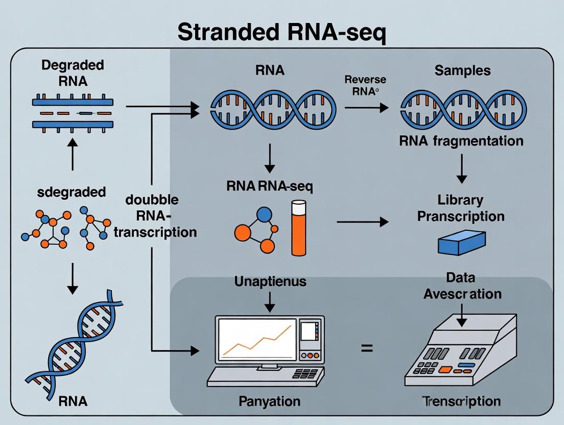
Unlocking Transcriptomic Insights from Challenging Samples: A Comprehensive Guide to Stranded RNA-Seq for Degraded RNA
This article provides researchers and drug development professionals with a complete framework for successfully applying stranded RNA sequencing to degraded RNA samples.
Unlocking Transcriptomic Insights from Challenging Samples: A Comprehensive Guide to Stranded RNA-Seq for Degraded RNA
Abstract
This article provides researchers and drug development professionals with a complete framework for successfully applying stranded RNA sequencing to degraded RNA samples. It begins by explaining the critical challenges posed by RNA degradation, such as 3' bias and reduced alignment efficiency, and establishes why strand-specificity is non-negotiable for accurate data from compromised material [citation:1][citation:9]. The guide then details and compares modern library preparation methodologies, including dUTP-based and ligation-based protocols, highlighting kits optimized for low-input and degraded samples [citation:2][citation:6][citation:10]. A dedicated troubleshooting section offers solutions for common issues like low library complexity and high ribosomal content, with an emphasis on protocol optimization and QC metrics [citation:1][citation:3]. Finally, the article outlines robust validation strategies, from analytical benchmarks using reference samples to comparative performance analyses against orthogonal methods, ensuring data reliability for downstream biomedical and clinical research [citation:4][citation:6].
Why RNA Integrity Matters: The Science of Degradation and the Critical Need for Strandedness
Technical Support Center
Troubleshooting Guides & FAQs
Q1: What are the primary causes of RNA degradation in my samples, and how can I prevent it during extraction?
A: The main causes are Ribonuclease (RNase) contamination, improper sample handling/storage, and physical shearing. To prevent it:
- Always use RNase-free consumables, reagents, and dedicated workspace.
- Use guanidinium thiocyanate-based lysis buffers to immediately inhibit RNases during homogenization.
- Keep samples on ice, flash-freeze tissue in liquid nitrogen, and store at -80°C.
- Limit freeze-thaw cycles of RNA stocks.
Q2: My Bioanalyzer/Tapestation shows a low RIN (e.g., RIN < 7). Should I proceed with traditional Poly-A selection for my stranded RNA-seq experiment?
A: No, proceeding with standard Poly-A selection is not recommended for degraded RNA. Low RIN indicates significant 18S/28S ribosomal peak breakdown and 5' bias. Poly-A selection relies on intact 3' poly-A tails, which are often compromised in degradation, leading to massive loss of transcript coverage and severe 3' bias. For degraded samples (RIN < 7), use rRNA depletion protocols instead, as they do not depend on the 3' mRNA integrity.
Q3: How does RNA degradation quantitatively impact yield and quality metrics in a standard Poly-A selected library prep?
A: Degradation severely impacts key metrics, as summarized below:
Table 1: Impact of RNA Integrity on Poly-A Selection Workflow
| Metric | High Integrity RNA (RIN 9-10) | Degraded RNA (RIN 4-6) | Observed Outcome for Degraded Samples |
|---|---|---|---|
| Library Yield | High (nM) | Very Low to Failed | Insufficient material for sequencing |
| % mRNA Mapped | >70% | Drastically Reduced | High rRNA contamination |
| Transcript Coverage | Uniform 5' to 3' | Strong 3' Bias | Loss of 5' transcript information |
| Gene Detection | Maximized | Significantly Reduced | Missing key differential expression data |
Q4: What is a robust experimental protocol for stranded RNA-seq of degraded FFPE or low-quality RNA samples?
A: Follow this detailed protocol for rRNA depletion-based library prep:
Protocol: Stranded Total RNA-seq for Degraded Samples
- RNA QC: Quantify RNA using a fluorometric method (e.g., Qubit). Assess degradation level with the RNA Integrity Number Equivalent (RINe) on the Fragment Analyzer or Tapestation. Do not rely on RIN from the Bioanalyzer for highly degraded samples; use DV200 (% of fragments >200nt) instead.
- rRNA Depletion: Use a probe-based ribosomal RNA depletion kit (e.g., Illumina Ribo-Zero Plus). Use 10-100 ng of total RNA as input. Perform the hybridization and removal steps strictly per manufacturer's instructions.
- Library Construction: Use a strand-specific library prep kit designed for low-input and degraded RNA (e.g., Illumina Stranded Total RNA Prep). Key steps:
- Fragmentation: Omit enzymatic fragmentation if input RNA is already sheared (DV200 < 30%). Use the "no fragmentation" protocol option.
- cDNA Synthesis: Perform first and second-strand synthesis.
- Adapter Ligation: Use dual-indexed adapters for multiplexing.
- PCR Amplification: Use a low number of PCR cycles (8-12) to minimize duplicates and bias.
- Library QC: Validate library size distribution (peak ~200-300bp) on a Fragment Analyzer and quantify by qPCR.
- Sequencing: Pool libraries and sequence on an appropriate platform (e.g., NovaSeq). Aim for 40-60 million paired-end reads per sample.
The Scientist's Toolkit
Table 2: Key Research Reagent Solutions for Degraded RNA-seq
| Item | Function | Example Product/Brand |
|---|---|---|
| RNase Inhibitors | Inactivate contaminating RNases during extraction and handling. | Recombinant RNase Inhibitor |
| Guanidinium-Based Lysis Buffer | Denatures proteins and RNases immediately upon cell/tissue disruption. | TRIzol, QIAzol |
| Fluorometric RNA Assay | Accurately quantifies RNA concentration independent of degradation. | Qubit RNA HS Assay |
| Fragment Analyzer/Capillary Electrophoresis | Assesses RNA degradation profile and calculates DV200 metric. | Agilent Fragment Analyzer, TapeStation |
| Ribosomal RNA Depletion Kit | Removes cytoplasmic and mitochondrial rRNA without poly-A selection. | Illumina Ribo-Zero Plus, NEBNext rRNA Depletion |
| Stranded Total RNA Library Prep Kit | Constructs sequencing libraries from rRNA-depleted RNA, preserving strand info. | Illumina Stranded Total RNA Prep, NuGEN Universal Plus |
| Dual-Indexed Adapters | Allows high-level multiplexing of low-yield degraded samples. | IDT for Illumina UD Indexes |
Visualizations
Title: Library Prep Decision Flow for RNA Integrity
Title: Impact of Degradation on RNA-seq Outcomes
Troubleshooting Guides & FAQs
Q1: Our stranded RNA-seq data from degraded clinical samples shows extreme 3' bias. How can we confirm this and what wet-lab steps can mitigate it in the next run?
A: Confirmation involves bioinformatic QC. Use tools like Picard CollectRnaSeqMetrics or Qualimap to calculate the 5'->3' coverage uniformity. A median 3' bias metric > 80% indicates severe bias. For mitigation:
- Protocol: Use a ribosomal RNA depletion kit (Ribo-zero Gold) instead of poly-A selection, as fragmentation leaves no intact poly-A tails.
- Protocol: Optimize fragmentation time/temperature after cDNA synthesis (post-fragmentation) to avoid physical breakage of already-short RNA.
- Reagent: Use random hexamer primers with template-switching technology (e.g., SMARTer kits) to generate full-length cDNA from fragments.
Q2: Alignment rates to the reference genome are unexpectedly low (<70%) for our FFPE-derived stranded RNA-seq libraries. What are the primary causes and solutions? A: Low alignment often stems from excessive adapter content or sequence artifacts from degradation.
- Diagnosis: Run FastQC and check for high adapter content per sequence and elevated "N" counts.
- Solution 1 (Bioinformatics): Perform aggressive adapter trimming with tools like
cutadaptorTrimmomatic, allowing minimal overlap (e.g., 1-base). Consider trimming low-quality 3' ends. - Solution 2 (Wet-lab): Use PCR library amplification kits designed for damaged DNA/RNA (e.g., those containing uracil-DNA glycosylase (UDG) to combat cytosine deamination artifacts common in FFPE samples). Reduce PCR cycles to minimize duplicate reads and chimeras.
Q3: Even after normalization, our gene expression counts from degraded samples are skewed towards longer genes. How do we account for this analytically? A: This is a known artifact. Standard normalization (e.g., TPM, FPKM) assumes uniform transcript coverage, which is violated. Employ bias-aware methods.
- Protocol: Use length-aware normalization tools like
salmonorkallistoin alignment-free mode, which model the fragment length distribution. - Protocol: Apply a correction factor such as "Transcript Integrity Number (TIN)" score as a covariate in differential expression analysis (e.g., in
DESeq2orlimma).
Q4: For stranded RNA-seq on degraded RNA, what is the optimal workflow to choose from sample QC to analysis? A: Follow this modified workflow designed for integrity-challenged samples.
Workflow for Stranded RNA-seq with Degraded Samples
Table 1: Impact of RNA Integrity Number (RIN) on Key Metrics in Stranded RNA-seq
| RIN Score Range | Typical DV200 | Median 3' Bias | Expected Alignment Rate | Recommended Protocol |
|---|---|---|---|---|
| 8-10 (High Integrity) | >90% | Low (< 60%) | >90% | Standard poly-A selection |
| 5-7 (Moderate Degradation) | 70-90% | Moderate (60-75%) | 80-90% | Consider rRNA depletion |
| 3-4 (Highly Degraded) | 40-70% | High (75-90%) | 70-85% | Mandatory rRNA depletion, UMIs |
| <2 (Severely Degraded) | <30% | Very High (>90%) | <70% | Specialized kits required; expect high noise |
Table 2: Comparison of Common rRNA Depletion Kits for Degraded RNA
| Kit Name | Principle | Optimal Input (Degraded) | Handles 5' Fragments? | Key Advantage for Low-Quality RNA |
|---|---|---|---|---|
| Ribo-zero Gold (Illumina) | Solution-phase hybridization | 10-100 ng | Yes | Proven robustness for FFPE; broad organism range. |
| NEBNext rRNA Depletion | Probe-based depletion (RNase H) | 1-100 ng | Moderate | Effective with very low inputs. |
| QIAseq FastSelect | Fast hybridization/bead removal | 10-100 ng | Yes | Rapid protocol (≤30 min), reduces handling loss. |
Experimental Protocols
Protocol 1: DV200 Assessment as an Alternative to RIN for Degraded RNA
- Run sample on an Agilent TapeStation or Bioanalyzer using the RNA ScreenTape assay.
- In the associated software, set the lower marker to 200 nucleotides.
- The software calculates the percentage of RNA fragments >200 nucleotides (DV200).
- Interpretation: For mammalian RNA-seq, a DV200 > 30% is often the minimum for proceeding with rRNA depletion-based library prep.
Protocol 2: Stranded RNA-seq Library Prep with UMI for Degraded RNA (Core Steps)
- rRNA Depletion: Use 10-50 ng of total RNA (DV200 > 30%) with the Ribo-zero Gold rRNA Removal Kit. Purify with RNA Clean XP beads.
- First-Strand cDNA Synthesis: Use random hexamer primers and SMARTER template-switching reverse transcriptase to generate full-length cDNA from fragments. This step incorporates a universal adapter sequence.
- Second-Strand Synthesis & Amplification: Perform limited-cycle PCR with indexing primers. Use a polymerase mix suitable for GC-rich sequences.
- UMI Incorporation & Clean-up: Use commercially available adapters containing Unique Molecular Identifiers (UMIs). Perform a double-sided bead clean-up (0.6x then 1.0x ratios) to remove adapter dimers.
- Library QC: Quantify by qPCR (e.g., Kapa Biosystems kit) and profile fragment size on a TapeStation D1000/High Sensitivity screen.
Signaling Pathway & Bias Mechanism
Mechanism of 3' Bias from Poly-A Selection on Degraded RNA
The Scientist's Toolkit: Research Reagent Solutions
| Item | Function & Relevance to Degraded RNA-seq |
|---|---|
| Agilent Bioanalyzer/TapeStation | Provides RNA Integrity Number (RIN) and, critically, the DV200 metric for assessing degraded samples. Essential for pre-library QC. |
| Ribo-zero Gold rRNA Removal Kit | Removes cytoplasmic and mitochondrial rRNA via hybridization, crucial for samples where poly-A tails are absent due to fragmentation. |
| SMARTer Stranded Total RNA-Seq Kit | Uses template-switching technology to generate full-length cDNA from fragmented RNA, mitigating 3' bias. Incorporates strand specificity. |
| Unique Molecular Index (UMI) Adapters | Molecular barcodes that tag each original molecule, allowing bioinformatic correction of PCR duplicates and noise—critical for low-input, amplified libraries. |
| RNA Clean XP/AMPure XP Beads | Solid-phase reversible immobilization (SPRI) beads for size selection and clean-up. Critical for removing adapter dimers and selecting optimal insert sizes. |
| Kapa Library Quantification Kit | qPCR-based assay for accurate quantification of amplifiable library fragments, ensuring balanced pooling for sequencing. |
Troubleshooting & FAQ Guide
This support center addresses common issues encountered during stranded RNA-seq library preparation and analysis, particularly for degraded RNA samples, within the thesis context of optimizing transcriptome resolution in challenging samples.
Frequently Asked Questions (FAQs)
Q1: My RNA samples are partially degraded (e.g., from FFPE or challenging tissues). Can I still use stranded RNA-seq, and will it provide an advantage over non-stranded methods? A: Yes, stranded RNA-seq is particularly beneficial for degraded samples. While fragmentation reduces read length, the strand information remains crucial. It prevents misassignment of reads from overlapping transcripts on opposite strands, which is a common source of false-positive gene expression and fusion calls in degraded samples. Use a protocol specifically optimized for degraded RNA (see Protocol 1 below).
Q2: I am observing a high rate of "unstranded" or "reverse" reads in my final alignment. What are the primary causes? A: This is a common issue. Primary causes include:
- Incomplete dUTP incorporation or inefficient UDG digestion: This is the core of the most common stranded protocol. Ensure fresh reagents and correct incubation times.
- RNA degradation: Overly fragmented RNA can lead to template-switching during reverse transcription or amplification, scrambling strand origin.
- Over-amplification: Excessive PCR cycles can promote the synthesis of "second-strand" artifacts that lose strand information.
- Contamination with genomic DNA: gDNA will produce reads aligning to both strands. Always include a rigorous DNase I treatment step.
Q3: How can I validate that my library is truly stranded before sequencing? A: Perform a pilot sequencing run or spike-in controls. Use a synthetic RNA spike-in mix with known strand-of-origin (e.g., from External RNA Controls Consortium (ERCC) or other providers). Align the spike-in reads and calculate the strand specificity percentage. A well-prepared library should achieve >95% strand specificity.
Q4: For antisense expression analysis, what alignment and quantification tools are recommended?
A: Use a spliced aligner that is strand-aware (e.g., STAR, HISAT2) with the --outSAMstrandField parameter correctly set. For quantification, use featureCounts (-s 1 or -s 2 for reverse-stranded protocols) or Salmon in selective alignment mode with --libType set appropriately. Always use a comprehensive annotation file (GTF) that includes antisense features.
Q5: My coverage across transcripts is uneven. Is this related to the stranded protocol? A: Not directly. Uneven coverage (" coverage bias") is often due to:
- Sequence-specific bias during reverse transcription or PCR. Use random hexamers and limit PCR cycles.
- RNA secondary structure. Fragmentation by chemical hydrolysis (e.g., magnesium-based) can reduce this bias compared to enzymatic fragmentation for degraded samples.
- GC content. Protocols using bead-based cleanup can under-represent very high or low GC fragments. Consider column-based cleanups for consistent recovery.
Detailed Experimental Protocols
Protocol 1: Stranded RNA-seq Library Prep for Degraded RNA (e.g., FFPE) using dUTP Method
- Principle: Incorporation of dUTP during second-strand synthesis, followed by Uracil-DNA Glycosylase (UDG) digestion, prevents amplification of the second strand, preserving strand information.
- Key Citations: ,
- Steps:
- RNA Input: Use 10-100 ng of total RNA. For highly degraded samples, higher input may be necessary.
- DNase I Treatment: Treat with RNase-free DNase I (15 min, 25°C). Purify using RNA clean-up beads.
- Fragmentation: For already fragmented RNA (RIN < 4), fragmentation may be omitted. If needed, use mild chemical fragmentation (Mg²⁺, 94°C, 5-7 min) to avoid over-fragmentation.
- First-Strand Synthesis: Use random hexamers and reverse transcriptase.
- Second-Strand Synthesis: Use DNA Polymerase I and a dNTP mix containing dUTP instead of dTTP.
- End Repair, A-tailing, and Adapter Ligation: Perform standard steps.
- UDG Treatment: Incubate with UDG (15-30 min, 37°C) to digest the dUTP-marked second strand.
- Library Amplification: Use a high-fidelity polymerase for 10-12 PCR cycles only. The UDG-treated second strand cannot serve as a template, ensuring only the first strand is amplified.
- Clean-up & QC: Purify with magnetic beads. Assess size distribution (~200-500 bp smear expected) and concentration via Bioanalyzer/TapeStation and qPCR.
Protocol 2: Strand Specificity Validation using Synthetic Spike-ins
- Steps:
- Spike-in Addition: Add a known amount of a stranded RNA spike-in control (e.g., 0.1-1% of total RNA mass) before library preparation.
- Library Prep & Sequencing: Proceed with Protocol 1. Sequence to a shallow depth (~1-2 million reads).
- Analysis: Align reads to a combined reference (your genome + spike-in sequences). For reads aligning uniquely to spike-ins, calculate:
- Strand Specificity = (Reads aligning to correct strand) / (All aligned spike-in reads) * 100%.
Table 1: Comparison of Stranded vs. Non-Stranded RNA-seq for Degraded Samples
| Metric | Non-Stranded | Stranded (dUTP) | Advantage |
|---|---|---|---|
| Antisense Detection | Impossible (ambiguous) | Accurate | Enables study of antisense regulation. |
| Overlap Resolution | Poor (reads pooled) | High (reads assigned to correct gene) | Reduces false positives in differential expression. |
| Fusion Gene Detection | High false positive rate from read-through transcripts | Greatly improved specificity | Critical for clinical biomarker discovery. |
| Data Utility | Limited to well-annotated sense genes | Maximized; full transcriptome complexity | Essential for novel transcript discovery. |
| Required RNA Integrity | High (RIN > 7) | Tolerant (RIN 2-7) | Enables work with archival (FFPE) samples. |
Table 2: Troubleshooting Common Issues in Stranded Library Prep
| Problem | Potential Cause | Solution |
|---|---|---|
| Low Strand Specificity (<90%) | Inefficient UDG digestion, gDNA contamination | Use fresh UDG, optimize incubation, add DNase I step. |
| Low Library Yield | Over-degraded input RNA, insufficient PCR cycles | Increase RNA input (up to 200 ng), add 1-2 PCR cycles (do not exceed 15). |
| High Duplication Rate | Very low input, over-amplification | Increase input, use unique molecular identifiers (UMIs), reduce PCR cycles. |
| Short Library Fragment Size | Excessive RNA fragmentation | Reduce fragmentation time/heat or omit step for degraded samples. |
| No Library | Failed reverse transcription or PCR | Include positive control RNA, check reagent integrity. |
Visualizations
Diagram 1: dUTP Stranded RNA-seq Workflow
Diagram 2: Stranded vs. Non-Stranded Read Assignment
The Scientist's Toolkit: Essential Research Reagents & Materials
| Item | Function in Stranded RNA-seq for Degraded RNA |
|---|---|
| RNase Inhibitor (e.g., Recombinant RNasin) | Critical for protecting already-fragile RNA samples from further degradation during library prep. |
| DNase I (RNase-free) | Essential for removing genomic DNA, a major source of background and strand ambiguity. |
| Magnetic Beads (RNA Clean-up & Size Selection) | For efficient purification and size selection of libraries; crucial for removing adaptor dimers. |
| dUTP Mix (dATP, dCTP, dGTP, dUTP) | The key reagent in the dUTP method. dUTP incorporation marks the second strand for enzymatic removal. |
| Uracil-DNA Glycosylase (UDG) | Enzyme that excises uracil bases, fragmenting the dUTP-marked second strand to prevent its amplification. |
| Stranded RNA Spike-in Controls (e.g., ERCC Mix) | Synthetic RNAs of known sequence and strand used to quantitatively assess library strand specificity. |
| High-Fidelity DNA Polymerase | For limited-cycle PCR amplification to minimize duplicates and sequence bias. |
| RNA Integrity Assay (Bioanalyzer/TapeStation) | To assess the degree of degradation in input RNA (even if RIN is low) and final library size profile. |
| Strand-Specific Alignment Software (e.g., STAR) | Bioinformatics tool that uses the --outSAMstrandField parameter to correctly interpret stranded library data. |
Troubleshooting Guides & FAQs
Q1: How do I objectively determine if my RNA sample is too degraded for standard RNA-seq? A: RNA Integrity Number (RIN) from a Bioanalyzer or TapeStation is the standard metric. While a RIN of ≥8 is ideal for standard protocols, stranded RNA-seq can often yield usable data from samples with RIN values as low as 2-5. However, degradation is contextual. Key quantitative thresholds are summarized below:
Table 1: RNA Degradation Assessment and Protocol Suitability
| Metric | High-Quality RNA | Moderately Degraded RNA | Severely Degraded RNA | Recommended Protocol |
|---|---|---|---|---|
| RIN Value | 8 - 10 | 5 - 7.9 | 2 - 4.9 | Stranded, rRNA-depleted |
| DV200 (% >200nt) | ≥70% | 30% - 69% | <30% | Ultra-low input/stranded |
| 285:185 rRNA Ratio | ≥1.5 (Mammalian) | ~1 | <1 | Not reliable for degraded samples |
| Primary Concern | Full-length transcript coverage | 3' bias, lower gene detection | Extreme 3' bias, high duplication | Stranded protocol is critical for strand info amid 3' bias. |
Q2: Why do stranded protocols perform better with degraded samples compared to non-stranded (e.g., TruSeq)? A: Standard non-stranded kits often rely on poly-A enrichment, which fails when the 5' end of transcripts is degraded. Stranded protocols typically use rRNA depletion (Ribo-Zero) or total RNA approaches, capturing fragmented RNA regardless of poly-A tail integrity. More importantly, they preserve strand orientation, which is crucial for accurate gene quantification and identifying antisense transcription when you can only sequence from the 3' fragment.
Q3: During library prep from a low-RIN sample, my final yield is extremely low. What are the critical steps to optimize? A: Low yield is common. Focus on:
- Input RNA: Use the maximum input volume/amount your kit allows.
- RNA Isolation: Use a bead-based or column-based kit designed for fragmented RNA recovery.
- rRNA Depletion: For severely degraded samples, consider probe-based depletion (e.g., Ribo-Zero Gold) over poly-A selection.
- Adapter Ligation: Use kits optimized for low-input/degraded samples, often involving template-switching or single-stranded ligation with truncated adapters.
- PCR Amplification: Increase PCR cycles cautiously (e.g., 13-17 cycles), but be aware of increased duplication rates and bias. Use a polymerase designed for complex templates.
Q4: My sequencing data from a degraded sample shows high duplicate rates and 3' bias. Is this normal, and can I still use the data? A: Yes, this is expected. Stranded protocols provide a lifeline by allowing you to interpret this biased data correctly.
- High Duplication: Due to limited starting RNA diversity, identical cDNA fragments are repeatedly sequenced. In-silico duplicate removal is standard but interpret results with caution.
- 3' Bias: Reads will cluster towards the 3' ends of transcripts. Use quantification tools (e.g., Salmon, kallisto) that are "alignment-free" or explicitly model 3' bias. Crucially, stranded information prevents misassignment of overlapping genes on opposite strands, which is common at 3' ends.
Protocol: Stranded Total RNA-seq Library Prep from Degraded/Fragmented RNA
- RNA QC: Quantify using fluorometry (Qubit RNA HS Assay). Assess integrity using Agilent TapeStation with High Sensitivity RNA tapes (calculate DV200).
- rRNA Depletion: Use 10-100 ng total RNA (or maximum volume). Perform Ribo-Zero Gold (Human/Mouse/Rat) or similar probe-based rRNA depletion. Clean up with magnetic beads (e.g., RNAClean XP).
- Fragmentation & cDNA Synthesis: Chemical or enzymatic fragmentation may be omitted if RNA is already fragmented. Perform first-strand cDNA synthesis using random hexamers and reverse transcriptase. Add Actinomycin D to suppress spurious DNA-dependent synthesis. Immediately perform second-strand synthesis using dUTP instead of dTTP to label the second strand.
- Double-stranded cDNA Cleanup: Purify cDNA using magnetic beads.
- Library Construction: Perform end-repair, A-tailing, and adapter ligation using truncated or standard adapters compatible with your sequencer.
- Strand Selection: Treat with Uracil-Specific Excision Reagent (USER) enzyme to digest the dUTP-labeled second strand, ensuring only the first strand is amplified.
- Library Amplification: Perform limited-cycle PCR (e.g., 12-15 cycles) with indexed primers. Clean up final library with magnetic beads.
- QC & Sequencing: Validate library size distribution on TapeStation (D1000/High Sensitivity D1000 tape) and quantify by qPCR. Sequence on Illumina platform with paired-end reads (≥2x75bp).
The Scientist's Toolkit: Research Reagent Solutions
Table 2: Essential Reagents for Stranded RNA-seq of Degraded Samples
| Reagent / Kit | Function in Degraded RNA Context | Example Product(s) |
|---|---|---|
| Magnetic Bead Cleanup | Efficient recovery of short RNA/cDNA fragments; scalable. | RNAClean XP, AMPure XP Beads |
| Ribosomal RNA Depletion Kit | Captures non-poly-A transcripts; essential for fragmented RNA. | Illumina Ribo-Zero Plus, QIAseq FastSelect |
| Stranded RNA Library Prep Kit | Incorporates dUTP marking and strand degradation for strand specificity. | Illumina TruSeq Stranded Total RNA, NEBNext Ultra II Directional |
| UV-inactivated RNase Inhibitor | Protects already vulnerable RNA fragments during reaction setup. | RiboSafe RNase Inhibitor |
| Reverse Transcriptase for FFPE | Engineered for better processivity on damaged/fragmented templates. | Maxima H Minus, SuperScript IV |
| High-Sensitivity DNA Assay | Accurate quantification of dilute, low-mass final libraries. | Qubit dsDNA HS Assay |
| Size/Degradation Analyzer | Provides DV200 metric, more informative than RIN for low-RIN samples. | Agilent TapeStation, Bioanalyzer |
Visualizations
Title: Protocol Choice Impact on Degraded RNA Success
Title: Stranded Library Prep Workflow for Degraded RNA
Navigating the Toolkit: Optimized Stranded RNA-Seq Protocols for Degraded and Low-Input Samples
Technical Support Center: Troubleshooting Guide & FAQs
Context: This support center is designed to assist researchers implementing key protocols for stranded RNA-seq library preparation, particularly when working with degraded RNA samples (e.g., from FFPE, ancient tissues, or challenging clinical biopsies). The focus is on troubleshooting the two dominant methods for strand preservation.
Frequently Asked Questions (FAQs)
Q1: For degraded RNA samples, which protocol is generally more robust: dUTP second-strand marking or direct RNA ligation? A: Based on current literature, the dUTP second-strand marking protocol is often considered more robust for significantly degraded samples. This is because the critical strand-informative step (dUTP incorporation) occurs early during cDNA synthesis, before extensive RNA fragmentation. Direct RNA ligation requires intact 3' ends of RNA fragments for adapter ligation, which can be compromised in degraded samples, leading to lower library complexity and yield.
Q2: We observe low library yields with the dUTP method. What are the most likely causes? A: Low yields in dUTP-based protocols commonly stem from:
- Inefficient dUTP incorporation: Verify the ratio of dUTP to dTTP in the second-strand synthesis mix.
- Incomplete digestion of the dUTP-marked second strand: Ensure the Uracil-Specific Excision Reagent (USER) enzyme or equivalent is fresh and active. Check incubation time and temperature.
- Over-fragmentation of input RNA: For already degraded samples, additional fragmentation may be unnecessary and counterproductive.
Q3: In direct RNA ligation, we see high adapter-dimer formation. How can this be mitigated? A: High adapter-dimer formation is a key challenge. Mitigation strategies include:
- Using truncated adapters that lack the promoter sequence until after ligation.
- Implementing strict size selection (e.g., with dual SPRI beads) before the amplification step to physically remove adapter-dimers.
- Optimizing adapter concentration and using thermostable ligases that favor substrate-specific ligation.
Q4: Our strandedness fidelity is poor (>95%) in FFPE samples using a dUTP protocol. What should we check? A: Strandedness fidelity loss often points to:
- Residual RNA template or second-strand cDNA carryover: Confirm the efficiency of the RNA degradation step (e.g., RNase H, alkaline hydrolysis) following first-strand synthesis.
- Inadequate dUTP incorporation: If the second strand is not sufficiently marked, it will not be efficiently excised, leading to anti-sense reads.
- PCR over-amplification: Excessive PCR cycles can lead to the amplification of contaminating strands. Use the minimum cycles necessary.
Troubleshooting Guide: Common Issues & Solutions
| Problem | Protocol | Potential Cause | Recommended Solution |
|---|---|---|---|
| Low Library Complexity | Direct RNA Ligation | Degraded RNA lacking 3'-OH groups for ligation. | Pre-repair RNA termini using PNK. Consider switching to dUTP method. |
| High Duplicate Reads | Both | Starting input RNA is too low. | Increase input RNA if possible. Use unique molecular identifiers (UMIs). |
| Sequence Bias | Direct RNA Ligation | Sequence preference of RNA ligase. | Use a ligase with reduced bias (e.g., CircLigase II). Optimize ligation buffer. |
| Incomplete 2nd Strand Removal | dUTP Marking | Inactive USER enzyme or short incubation. | Freshly aliquot USER enzyme. Extend incubation time to 30-45 min. |
| Low Final Library Yield | dUTP Marking | Over-digestion by USER enzyme damaging adapter sequences. | Precisely follow incubation times. Titrate USER enzyme amount. |
Detailed Experimental Protocols
Protocol 1: dUTP Second-Strand Marking for Stranded RNA-seq (Adapted from citation:2) Principle: Strand information is encoded by incorporating dUTP during second-strand cDNA synthesis, followed by enzymatic excision of this strand prior to PCR.
- RNA Fragmentation: Fragment 10-100 ng of total RNA (degraded or intact) to ~200-300 nt using divalent cations at 94°C for 5-8 min.
- First-Strand cDNA Synthesis: Use random hexamers and reverse transcriptase (e.g., SuperScript IV) to synthesize cDNA. Degrade RNA template with RNase H.
- Second-Strand Synthesis (Marking): Synthesize the second strand using DNA Polymerase I, RNase H, and a dNTP mix where dTTP is partially replaced by dUTP (e.g., 200 µM dUTP, 50 µM dTTP).
- dUTP-Strand Excision: Treat double-stranded cDNA with USER Enzyme (Uracil-Specific Excision Reagent), which cleaves at uracil bases, rendering the second strand non-amplifiable.
- Library Construction: Ligate blunt-ended adapters to the remaining first strand. Perform a limited-cycle PCR with Illumina primers to generate the final sequencing library.
Protocol 2: Direct RNA Ligation for Stranded RNA-seq (Adapted from citation:9) Principle: Strand-specificity is achieved by ligating adapters directly to the RNA molecule before any cDNA synthesis steps.
- RNA Repair & Dephosphorylation: Treat fragmented RNA with T4 PNK (optional, for samples with damaged ends). Dephosphorylate 3' ends using a phosphatase to prevent circularization.
- 3' Adapter Ligation: Ligate a pre-adenylated, blocked 3' adapter specifically to the RNA 3'-OH using a truncated T4 RNA Ligase 2 (Rnl2).
- 5' Adapter Ligation: Phosphorylate the RNA 5' ends using T4 PNK. Ligate a 5' adapter using T4 RNA Ligase 1 (Rnl1).
- Reverse Transcription: Prime from the 3' adapter sequence and perform first-strand cDNA synthesis.
- PCR Amplification: Amplify the cDNA library using primers complementary to the adapter sequences.
| Parameter | dUTP Second-Strand Marking | Direct RNA Ligation |
|---|---|---|
| Key Strand Information Step | During 2nd-strand cDNA synthesis (dUTP incorporation). | At the start (adapter ligation to RNA). |
| Optimal RNA Integrity (RIN) | More tolerant of low RIN (degraded samples). | Requires higher RIN for optimal efficiency. |
| Handling Time (Pre-PCR) | Longer (more enzymatic steps). | Shorter (fewer steps before RT). |
| Adapter-Dimer Formation | Less common (adapters ligated to cDNA). | More common (adapters ligated to RNA). |
| Sequence Bias | Lower (cDNA synthesis is relatively unbiased). | Higher (RNA ligases have sequence bias). |
| Typical Strandedness Fidelity | >99% (when optimized). | ~95-99% (can be impacted by RNA damage). |
| Recommended for | FFPE, clinical, degraded samples. | High-quality RNA, small RNAs. |
Visualizations
Title: dUTP Strand Marking Workflow
Title: Direct RNA Ligation Workflow
Title: Protocol Selection Logic
The Scientist's Toolkit: Research Reagent Solutions
| Reagent / Kit | Protocol | Function & Rationale |
|---|---|---|
| SuperScript IV Reverse Transcriptase | dUTP, Ligation | High processivity and yield for cDNA synthesis from degraded RNA. |
| USER II Enzyme (NEB) | dUTP Marking | Critical for excising the dUTP-marked second cDNA strand. |
| Truncated T4 RNA Ligase 2 (Rnl2) | Direct Ligation | Specifically ligates pre-adenylated 3' adapters to RNA, minimizing side reactions. |
| Pre-adenylated 3' Adapters | Direct Ligation | Prevents adapter concatemerization; only requires ligase for a single nick. |
| RNase H | dUTP Marking | Degrades RNA template after first-strand synthesis to prevent spurious second-strand initiation. |
| T4 Polynucleotide Kinase (PNK) | Direct Ligation | Repairs/phosphorylates 5' ends of RNA fragments to enable 5' adapter ligation. |
| Dual-indexed UMI Adapters | Both | Enables accurate PCR duplicate removal and sample multiplexing, crucial for low-input degraded samples. |
| SPRIselect Beads | Both | For precise size selection and purification, essential for removing adapter dimers and selecting optimal insert sizes. |
Troubleshooting Guides & FAQs
Q1: Why is ribosomal depletion recommended over poly-A selection for degraded or fragmented RNA samples? A: Poly-A selection relies on intact poly-A tails, which are often degraded in low-quality RNA (e.g., from FFPE or challenging tissues). Ribosomal depletion (Ribo-Zero, RiboGone) removes abundant ribosomal RNA (rRNA) sequences independent of the 3' poly-A tail, preserving the representation of both coding and non-coding RNA, including degraded transcripts. This results in superior library complexity and data quality for degraded samples.
Q2: My post-depletion RNA yield is very low. What could be the cause and how can I mitigate this? A: Low yield is common with highly degraded samples, as fragmentation reduces RNA mass. Ensure accurate quantification using fluorometric methods (e.g., Qubit) over spectrophotometry. Use the entire input amount recommended for the depletion kit (often 100ng-1µg). If yield remains low, proceed directly to library prep, as the critical metric is not total yield but the successful removal of rRNA and capture of informative fragments.
Q3: I observe residual rRNA reads in my sequencing data after ribosomal depletion. What is an acceptable threshold and how can I reduce it further? A: Residual rRNA is typical but should be minimized. For mammalian samples, a well-optimized depletion should achieve <5-10% rRNA reads. High residuals can stem from:
- Over-input or Under-input of RNA: Follow the kit's input range strictly.
- Incomplete magnetic bead handling: Ensure beads are thoroughly resuspended and not overheated during washing.
- Sample Degradation: Heavily degraded RNA may have fragments matching rRNA probes poorly. Consider using a kit specifically optimized for degraded RNA (e.g., RiboGone).
Q4: How does ribosomal depletion impact the success of stranded RNA-seq libraries from degraded samples? A: Ribosomal depletion is compatible with stranded library prep protocols. It preserves strand information by removing rRNA before the reverse transcription step that encodes strandality. For degraded samples, using a depletion method followed by a random-primed, stranded library prep is the optimal workflow to capture full transcriptome information without 3' bias.
Q5: Can I use ribosomal depletion for very low-input samples (e.g., < 100 ng total RNA)? A: Yes, but it requires caution. Many kits have low-input protocols. Key steps include:
- Using rigorous RNase-free techniques and glycogen/linear acrylamide carriers during precipitation.
- Performing fewer purification steps to minimize loss.
- Using a library amplification kit designed for low input. Verify performance with a Bioanalyzer/TapeStation.
Table 1: Comparison of RNA-Seq Methods for Degraded Samples
| Method | Optimal RNA Integrity Number (RIN) | % Usable Reads (Non-rRNA) | 3' Bias | Detection of Non-Coding RNA |
|---|---|---|---|---|
| Poly-A Selection | ≥ 7 (High Quality) | High (if intact) | Low (if intact) | No |
| Ribo-Zero/RiboGone (Ribosomal Depletion) | 2 - 10 (Degraded to Intact) | 90-95%+ | Low (Random Priming) | Yes |
| Total RNA (No Depletion) | Any | <10% (High rRNA) | Variable | Yes |
Table 2: Troubleshooting Common Ribosomal Depletion Issues
| Problem | Potential Cause | Recommended Solution |
|---|---|---|
| High rRNA Reads | RNA over/under input, degraded RNA, poor bead handling | Re-optimize input amount, use degradation-optimized kit, perfect bead technique. |
| Low Library Complexity | Extreme RNA degradation, insufficient PCR cycles | Use random hexamers, adjust PCR cycle number, start with higher RNA input if possible. |
| Failed QC Post-Depletion | RNA degraded below kit threshold, enzymatic failure | Check RNA fragment size distribution; use positive control RNA to test kit reagents. |
Experimental Protocol: Stranded RNA-seq with RiboGone for Degraded RNA
Detailed Methodology (Adapted from citation 3 & 10):
RNA Quality Assessment:
- Quantify degraded RNA using a fluorescence assay (Qubit RNA HS Assay).
- Assess fragmentation profile using a Bioanalyzer RNA 6000 Pico kit or TapeStation. Do not rely on RIN alone.
Ribosomal RNA Depletion with RiboGone:
- Use 100 ng - 1 µg of total RNA as input. For low input (<100 ng), follow the manufacturer's low-input protocol.
- Combine RNA, hybridization buffer, and RiboGone rRNA probes in a PCR tube.
- Denature at 95°C for 2 minutes, then hybridize at 68°C for 10 minutes to allow probes to bind rRNA sequences.
- Add RNase H and incubate at 37°C for 30 minutes to specifically cleave rRNA:probe hybrids.
- Purify the rRNA-depleted RNA using the provided spin columns or bead-based cleanup. Elute in a small volume (e.g., 10 µL).
Stranded RNA-seq Library Construction:
- Use the purified, rRNA-depleted RNA for first-strand cDNA synthesis with random hexamer primers and dUTP incorporation (strand-marking).
- Synthesize second-strand cDNA.
- Perform end-repair, A-tailing, and adapter ligation using dual-indexed adapters.
- Treat with Uracil-Specific Excision Reagent (USER) enzyme to degrade the second strand (containing dUTP), ensuring strand specificity.
- Amplify the library with 10-15 cycles of PCR.
- Clean up libraries using double-sided SPRI bead selection (e.g., 0.8x and 1.0x ratios) to remove adapter dimers and select optimal insert size.
Library QC and Sequencing:
- Quantify library yield via Qubit dsDNA HS Assay.
- Assess library size distribution using a Bioanalyzer High Sensitivity DNA kit.
- Pool libraries at equimolar ratios and sequence on an Illumina platform (e.g., NovaSeq) with paired-end reads (e.g., 2x150 bp).
Visualizations
Title: Workflow Decision Tree for Degraded RNA-Seq
Title: Ribosomal Depletion Mechanism for Degraded RNA
The Scientist's Toolkit: Research Reagent Solutions
Table 3: Essential Materials for Ribosomal Depletion of Degraded RNA
| Reagent / Kit | Function & Role in the Workflow |
|---|---|
| RiboGone (Mammalian) | A ribosomal depletion kit optimized for a broad range of RNA integrity, using probe hybridization and RNase H cleavage to remove rRNA. Ideal for degraded samples. |
| Ribo-Zero Plus rRNA Depletion Kit | Removes cytoplasmic and mitochondrial rRNA via probe hybridization and magnetic bead capture. Robust for various sample types. |
| Qubit RNA HS Assay Kit | Fluorescence-based quantification critical for accurately measuring degraded RNA, which spectrophotometers may misquantify. |
| Agilent RNA 6000 Pico Kit | Microfluidics-based analysis to visualize the fragment size distribution of degraded RNA pre- and post-depletion. |
| NEBNext Ultra II Directional RNA Library Prep Kit | A common stranded library preparation protocol compatible with rRNA-depleted RNA, using dUTP marking for strand specificity. |
| RNAClean XP / AMPure XP Beads | Solid-phase reversible immobilization (SPRI) magnetic beads for size selection and cleanup during library preparation. |
| USER Enzyme (Uracil-Specific Excision Reagent) | Critical enzyme in stranded protocols that digests the second strand containing dUTP, preserving the directionality of the original RNA. |
| Dual Indexed UMI Adapters | Adapters containing unique molecular identifiers (UMIs) to correct for PCR duplicates and unique dual indexes to reduce index hopping in multiplexed sequencing. |
Troubleshooting Guides & FAQs
Q1: My RNA integrity number (RIN) is low (<3). Which library prep kit is most tolerant for successful stranded RNA-seq? A1: For severely degraded RNA (e.g., FFPE, necrotic samples), Takara Bio's SMARTer Stranded Total RNA-Seq Kit v2 (Pico Input Mammalian) is specifically optimized for inputs as low as 1-125 pg, even from degraded samples, by utilizing SMART (Switching Mechanism at 5' End of RNA Template) technology for whole transcriptome amplification. Illumina's Stranded Total RNA Prep with Ribo-Zero Plus also performs well with moderate degradation (RIN >2) due to its efficient ribosomal RNA depletion. IDT's xGen Stranded RNA-Seq Kit is robust for inputs down to 1-100 ng but may show reduced library complexity with extreme degradation.
Q2: I am getting high adapter-dimer formation during library prep from low-input samples. How can I mitigate this? A2: High adapter-dimer is common with low/ degraded RNA. Solutions are kit-dependent:
- Illumina: Use double-sided SPRI bead cleanups at recommended ratios rigorously. For the Ligation-Based workflow, ensure the RNA is fragmented to the optimal size range to minimize blunt-end ligation of adapters.
- Takara Bio: Use the included size selector magnetic beads precisely. Do not over-dry beads during cleanup steps.
- IDT: Optimize the PCR cycle number to prevent over-amplification of small fragments. Use a high-fidelity, hot-start polymerase.
Q3: My library yield is insufficient for sequencing after using a pico-input protocol. What are the critical steps? A3: Ensure:
- Input Quantification: Use a fluorescence-based assay (Qubit, Picogreen) over absorbance (Nanodrop) for accuracy.
- Reverse Transcription Efficiency: Use fresh reverse transcriptase and dNTPs. Avoid repeated freeze-thaws of master mix components.
- PCR Amplification: Use the minimum recommended PCR cycles to maintain complexity. Validate PCR primer premix integrity.
- Bead Cleanup: Do not exceed binding time; elute in low-EDTA TE buffer or nuclease-free water pre-warmed to 55°C.
Q4: How do I handle ribosomal RNA (rRNA) depletion in degraded samples where poly-A selection fails? A4: Both Illumina (Ribo-Zero Plus) and Takara Bio (rRNA depletion beads) offer probe-based ribosomal depletion that is independent of RNA integrity and suitable for degraded samples. IDT's kit is compatible with multiple external depletion modules. For highly degraded human/mouse/rat samples, consider using targeted RNA-seq panels that bypass the need for global rRNA depletion.
Experimental Protocols for Degraded RNA-Seq
Protocol 1: Library Preparation from FFPE-Derived RNA (RIN 1.5-3.0) using Takara Bio SMARTer Pico Kit
- RNA Isolation: Extract using a method optimized for FFPE (e.g., with proteinase K digestion and DNase I treatment). Quantify by Qubit RNA HS Assay.
- First-Strand cDNA Synthesis: Combine 1-125 pg of RNA with 3' SMART CDS Primer IIA and heat denature. Add SMARTer V4 Oligonucleotide and SMARTScribe Reverse Transcriptase. Incubate at 42°C for 90 min, then 70°C for 10 min.
- PCR Amplification: Add SeqAmp DNA Polymerase and ISPCR Primer (containing partial adapter sequences). Cycle: 98°C 1 min; [98°C 10 sec, 65°C 30 sec, 68°C 3 min] for 12-18 cycles; 68°C 5 min.
- Size Selection & Cleanup: Use included MagSi-GS beads per manufacturer's protocol to remove fragments <200 bp.
- Library Indexing (Illumina adaptor addition): Perform a second, limited-cycle (4-8 cycles) PCR using Indexing Primers containing full i5 and i7 adapters.
Protocol 2: Stranded Library Prep from Low-Quality Total RNA using Illumina Stranded Total RNA Prep, Ligation
- rRNA Depletion: Use Ribo-Zero Plus to hybridize and remove cytoplasmic and mitochondrial rRNA from 10-100 ng total RNA.
- RNA Fragmentation & Priming: Fragment the RNA and prime for first-strand synthesis using Elute, Prime, Fragment Mix (8 min, 94°C).
- First-Strand Synthesis: Synthesize cDNA using Reverse Transcriptase and Activating Reaction Buffer.
- Second-Strand Synthesis: Incorporate dUTP to preserve strand information using Second Strand Marking Master Mix.
- Adapter Ligation: A-tailing followed by ligation of Illumina Stranded RNA UD Indexes.
- Clean-up & PCR Amplification: Perform bead cleanups. Use PCR Master Mix and PCR Primer Cocktail (5-15 cycles) to amplify libraries. Treat with Uracil-Specific Excision Reagent (USER) to degrade dUTP-containing second strand before sequencing.
Table 1: Kit Comparison for Challenging RNA Inputs
| Feature | Illumina Stranded Total RNA Prep, Ligation | Takara Bio SMARTer Stranded Total RNA-Seq Kit v2 (Pico) | IDT xGen Stranded RNA-Seq Kit |
|---|---|---|---|
| Minimum Input (Degraded) | 10 ng (RIN >2) | 1 pg (Highly degraded compatible) | 1 ng |
| Recommended Input Range | 10-1000 ng | 1 pg - 10 ng | 1-1000 ng |
| rRNA Depletion Method | Ribo-Zero Plus (probe-based) | Proprietary bead-based removal | Compatible with multiple external modules |
| Strandedness Method | dUTP, Second Strand Degradation | SMART technology (template switching) | dUTP, Enzymatic Fragmentation |
| Workflow Duration | ~6.5 hours | ~6 hours | ~5.5 hours |
| Optimal For | Moderately degraded samples, standard inputs | Extremely low input & highly degraded samples (FFPE, single-cell) | Flexible inputs, DNA/RNA dual-mode workflows |
Table 2: Troubleshooting Common Issues
| Problem | Likely Cause | Illumina Solution | Takara Bio Solution | IDT Solution |
|---|---|---|---|---|
| Low Library Yield | Over-fragmentation, bead loss | Check FFPE fragmentation time; incubate beads at RT | Ensure no bead carryover; elute in warm buffer | Verify PCR enzyme activity; increase input if possible |
| High Adapter Dimer | Low RNA input, over-amplification | Use Sample Purification Beads at 0.9X ratio | Use Size Selector Beads precisely | Optimize PCR cycles; use bead double cleanup |
| Low Complexity Libraries | RNA degradation, over-cycling | Reduce PCR cycles; use unique dual indexes | Use minimum recommended PCR cycles | Use starting RNA with highest possible RIN |
| rRNA Residual High | Depletion probe inefficiency | Ensure Ribo-Zero Plus probe is fresh, hybridize at 80°C | Ensure depletion beads are thoroughly resuspended | Validate external depletion module protocol |
Visualization
Stranded RNA-seq Workflows for Degraded RNA
SMART Technology for Degraded RNA
The Scientist's Toolkit: Research Reagent Solutions
| Item | Function in Degraded RNA-Seq |
|---|---|
| Qubit RNA HS Assay | Fluorometric quantification critical for accurate measurement of low-concentration, impure RNA where UV absorbance fails. |
| Agilent RNA 6000 Pico Kit | Microfluidics-based analysis to assess RNA integrity (RIN/equivalent) and quantify trace amounts of RNA from precious samples. |
| RNase Inhibitor (e.g., RNasin) | Essential in all pre-amplification steps to prevent further degradation of already compromised RNA templates. |
| AMPure XP / SPRI Beads | Magnetic beads for size-selective cleanup and purification of libraries, crucial for removing adapter dimers and short fragments. |
| Unique Dual Indexes (UDIs) | PCR indexing primers with unique i5 and i7 combinations to enable sample multiplexing and accurate demultiplexing, reducing index hopping effects. |
| Proteinase K | For effective de-crosslinking and RNA extraction from formalin-fixed, paraffin-embedded (FFPE) tissue sections. |
| USER Enzyme (Uracil-Specific Excision Reagent) | Used in dUTP-based stranded kits to enzymatically degrade the second strand, preserving strand-of-origin information. |
| SMARTScribe Reverse Transcriptase | A proprietary enzyme mix with high processivity and template-switching activity, enabling full-length cDNA synthesis from fragmented RNA. |
FAQs & Troubleshooting
Q1: During library prep for degraded RNA-seq, my Bioanalyzer/Fragment Analyzer trace shows a very broad smear or a peak below 100bp. What does this indicate and how should I proceed? A: This is expected when input RNA is highly degraded (e.g., RIN < 3). The broad smear represents the heterogeneous fragment sizes of your starting material. Proceed with protocol normalization by input mass (e.g., 100ng total RNA) rather than by volume. Ensure you are using a stranded, rRNA-depletion protocol designed for low-input/degraded samples. Do not use poly-A selection. The final library size distribution will be improved post-capture and amplification.
Q2: My long-read (PacBio or ONT) sequencing run on damaged RNA samples has very low yield. What are the primary culprits? A: The issue is often upstream of sequencing. First, verify the integrity and quantity of your cDNA post-amplification. For damaged RNA, the cDNA synthesis step is critical. Use a reverse transcriptase known for high processivity and the ability to handle modified/base-damaged templates. Ensure you are not over-amplifying the cDNA, as this can lead to chimeric artifacts and size bias. Refer to the "Degraded RNA to cDNA" workflow below.
Q3: When performing degradome sequencing to identify cleavage sites, I get a high background of random fragments. How can I improve signal-to-noise? A: This typically stems from non-specific RNA fragmentation or adapter ligation biases. Key solutions:
- Use 5'-adenylated adapters with a thermostable 5' AppDNA/RNA ligase to minimize adapter dimer formation and favor ligation to genuine 5'-monophosphate ends left by endonucleolytic cleavage.
- Implement size selection stringently post-ligation (e.g., double-sided SPRI bead cleanup) to exclude very small (<15nt) fragments which are often technical noise.
- Include biological replicates and use analytical pipelines (like PAREsnip2) that statistically compare treated vs. control samples to identify significant cleavage events.
Experimental Protocols
Protocol 1: Stranded Total RNA-Seq Library Prep for Degraded Samples (Adapted from )
- Input: 10-100ng of total RNA (RIN 2-7).
- rRNA Depletion: Use probe-based ribosomal RNA depletion kits (e.g., Ribo-Zero Plus). Do not use poly-A selection.
- Fragmentation: If RNA is not already sufficiently fragmented (RIN >5), use controlled metal-ion catalyzed fragmentation (94°C, 6-8 minutes in Mg²⁺ buffer). For RIN <5, fragmentation may be omitted.
- cDNA Synthesis: Perform first-strand synthesis using random hexamers and a reverse transcriptase with high fidelity and template-switching capability.
- Strandedness Preservation: Use dUTP incorporation during second-strand synthesis, followed by UDG digestion prior to PCR to ensure strand specificity.
- Adapter Ligation: Use T4 RNA Ligase 1 for single-indexed, unique dual indexing adapters to reduce index hopping.
- PCR Amplification: Use low-cycle (8-12 cycles), high-fidelity PCR. Excess cycles can exacerbate bias in degraded samples.
Protocol 2: cDNA Preparation for Long-Read Sequencing of Damaged RNA (Adapted from )
- RNA Pretreatment: Treat RNA with T4 Polynucleotide Kinase (PNK) to repair damaged 5' ends and ensure a 5'-phosphate. This is critical for subsequent adapter ligation in ONT workflows.
- Reverse Transcription: Use a group II intron-derived reverse transcriptase (e.g., TGIRT) or other engineered enzymes with high thermostability and strand-displacement activity. This improves cDNA yield from damaged, structured, or modified RNA templates.
- cDNA Amplification (If Required): For PacBio HiFi, perform Large-Insert PCR (10-12 cycles) with long-fragment polymerases. For ONT, consider PCR-free direct RNA or cDNA ligation approaches to avoid amplification bias.
- Size Selection: Perform a gentle, large-fragment size selection (e.g., BluePippin or calibrated SPRI beads) to remove very short cDNAs and retain molecules >500bp for meaningful long-read data.
Data Summary Tables
Table 1: Comparison of RNA-Seq Approaches for Degraded Samples
| Method | Optimal RNA Integrity (RIN) | Key Feature | Primary Application | Potential Pitfall |
|---|---|---|---|---|
| Standard Stranded mRNA-seq | >7.0 | Poly-A selection | Gene expression, splicing | Complete failure on degraded RNA |
| Stranded Total RNA-seq (rRNA-) | 2.0 - 10.0 | Ribosomal depletion | Degradome, viral, bacterial RNA | Requires more input; cost of depletion |
| 3'-Tagged Sequencing (e.g., 3'SEQ) | 1.0 - 3.0 | 3' poly-A capture only | High-throughput profiling of degraded FFPE | Loss of isoform and splicing data |
| Single-Cell RNA-seq | N/A (uses poly-T) | Cell-specific barcoding | Profiling individual cells from mixed/low-quality tissue | High technical noise, high cost |
Table 2: Long-Read Platform Suitability for Damaged RNA Analysis
| Platform | Read Type | Sample Prep Relevance for Damaged RNA | Main Advantage for Degraded Samples | Main Challenge |
|---|---|---|---|---|
| Pacific Biosciences (HiFi) | Circular Consensus Sequencing (cDNA) | High-fidelity PCR of full-length cDNA | High single-read accuracy (>99.9%) | Polymerase may stall at damage sites, causing truncation. |
| Oxford Nanopore (ONT) | Direct RNA / cDNA | Direct sequencing of RNA or single-step cDNA | Can sequence modified bases; no PCR required for Direct RNA | Higher per-read error rate (~5%); basecalling from damaged templates is complex. |
The Scientist's Toolkit: Research Reagent Solutions
| Item | Function in Degraded RNA Workflows |
|---|---|
| Ribo-Zero Plus / RiboCop | Chemically removes ribosomal RNA without poly-A bias, crucial for fragmented samples. |
| TGIRT-III Reverse Transcriptase | Thermostable group II intron RT, excels at synthesizing cDNA from damaged, structured, or modified RNA. |
| T4 Polynucleotide Kinase (PNK) | Repairs 5' ends by phosphorylating RNA, essential for efficient adapter ligation in degradome/ONT protocols. |
| 5' AppDNA/RNA Ligase | Ligates 5'-adenylated adapters specifically to 5'-monophosphate RNA ends, reducing background in degradome seq. |
| RNase Inhibitor (e.g., RNasin, SUPERase-In) | Protects already-fragile RNA from further degradation during sample prep. |
| SPRIselect Beads | For precise, double-sided size selection to remove adapter dimers and select optimal cDNA fragment sizes. |
Visualizations
Degraded RNA to Long-Read cDNA Workflow
Degradome Seq vs Standard RNA-seq Logic
Troubleshooting Guides and FAQs
Q1: Our RNA extracted from FFPE tissue has low RNA Integrity Number (RIN) values (<2.0). Can we still proceed with stranded RNA-seq, and what are the key library preparation adjustments? A1: Yes, stranded RNA-seq is feasible with low RIN RNA. Key adjustments include:
- Use of rRNA depletion over poly-A selection: Poly-A selection fails for degraded RNA. Use ribo-depletion kits designed for low-quality RNA (e.g., Illumina Ribo-Zero Plus).
- Reduced RNA fragmentation time or omission: FFPE RNA is already fragmented. Omit or drastically reduce enzymatic fragmentation steps in the kit protocol.
- Increased input RNA: Input 100-200 ng of total RNA, even if outside the kit's standard range, to compensate for loss during library prep.
- Increased PCR cycles: Increase library amplification cycles by 4-6 to generate sufficient yield from low-input, damaged templates.
Q2: We observe high duplication rates (>50%) in our stranded RNA-seq data from archived samples. What are the primary causes and solutions? A2: High duplication rates typically stem from low input material and amplification bias.
- Cause: Starting with very low amounts of fragmented RNA leads to stochastic capture of fragments, which are then amplified, creating duplicate reads.
- Solutions:
- Increase input RNA if possible.
- Use unique molecular identifiers (UMIs) in your library prep kit. UMIs tag each original molecule before amplification, allowing bioinformatic removal of PCR duplicates.
- Bioinformatic filtering: Use tools like
picard MarkDuplicatesorUMI-tools(if UMIs were used) to accurately flag and handle duplicates.
Q3: How do we accurately assess the quality of severely degraded RNA from FFPE samples when traditional bioanalyzer profiles are unreliable? A3: Rely on metrics other than RIN for FFPE RNA.
- DV200: The percentage of RNA fragments >200 nucleotides is a more robust metric for FFPE RNA. Aim for DV200 > 30% for successful library prep (see Table 1).
- qPCR-based QC: Use a multiplexed qPCR assay targeting amplicons of different lengths (e.g., short (100 bp) vs. long (300 bp)) to assess degradation level and amplifiability.
- Fluorometric quantitation: Use Qubit or PicoGreen for accurate concentration, as UV absorbance can be inflated by contaminants.
Q4: What are the critical steps to minimize contamination and RNA degradation during the microdissection of archived samples for RNA-seq? A4:
- Microtome Decontamination: Wipe the microtome blade and stage with RNase decontamination solution (e.g., RNaseZap) between samples. Use fresh, disposable blades when possible.
- Slide Preparation: Use RNase-free glass slides. Keep slides on dry ice or in a -20°C freezer until immediately before microdissection.
- Lysis Buffer: Collect dissected tissue directly into a lysis buffer containing strong chaotropic salts (e.g., guanidine thiocyanate) to immediately inactivate RNases.
- Time & Temperature: Minimize dissection time. Perform dissection in a cold environment if possible.
Table 1: Quality Metrics and Their Implications for Stranded RNA-seq Success
| Metric | Ideal Value (Fresh Frozen) | Acceptable Value (FFPE/Degraded) | Measurement Tool | Implication for Library Prep |
|---|---|---|---|---|
| RIN | 8.0 - 10.0 | Often < 2.0; Not reliable | Bioanalyzer/TapeStation | Not predictive for FFPE. Use DV200. |
| DV200 | > 90% | > 30% (minimum) | Bioanalyzer/TapeStation | Primary QC metric. Predicts library yield. |
| Concentration | > 50 ng/μL | > 10 ng/μL | Qubit/Fluorometer | Determines required input volume. |
| 260/280 Ratio | 1.8 - 2.0 | 1.6 - 2.0 (may be lower) | Spectrophotometer | Indicates protein/phenol contamination. |
Table 2: Comparison of Library Prep Strategies for Degraded RNA
| Strategy | Principle | Best For | Key Advantage | Key Limitation |
|---|---|---|---|---|
| Poly-A Selection | Enriches mRNA via poly-T oligos | High-quality RNA (RIN > 7) | Clean data, low ribosomal RNA | Fails completely on degraded RNA. |
| Ribo-depletion | Removes ribosomal RNA via hybridization | All RNA qualities, especially degraded | Works on fragmented RNA, retains non-polyA transcripts | More complex protocol, higher cost. |
| SMART-based (e.g., NuGEN) | Template-switching at 3' end | Extremely low input & degraded RNA | Works with very low input (1-10 ng), good for 3' bias | Strong 3' bias, not full-transcript. |
Experimental Protocols
Protocol 1: DV200 Assessment using TapeStation Analysis
- Prepare FFPE RNA sample and RNA ScreenTape reagents as per manufacturer instructions.
- Load 1 μL of sample onto the designated well of the RNA ScreenTape ladder plate.
- Run the TapeStation using the "RNA" algorithm.
- In the analysis software, view the electrophoretogram. The software automatically calculates the DV200 value (percentage of the area under the curve above the 200 nucleotide marker).
- Proceed to library preparation if DV200 ≥ 30%.
Protocol 2: Stranded Total RNA-seq Library Prep with Ribo-depletion for FFPE Samples (Based on Illumina TruSeq Stranded Total RNA)
- RNA QC: Quantify RNA using Qubit RNA HS Assay. Check DV200 via TapeStation.
- Ribosomal RNA Depletion: Use Ribo-Zero Plus to deplete rRNA from 50-100 ng total RNA (input scalable). Incubate probes with RNA, capture with magnetic beads, and retain supernatant containing enriched non-rRNA.
- RNA Fragmentation & Elution: Omit the standard fragmentation step. Simply elute the rRNA-depleted RNA from the beads.
- First Strand cDNA Synthesis: Synthesize cDNA using random hexamers. Actinomycin D is included to prevent spurious DNA-dependent synthesis.
- Second Strand Synthesis: Using dUTP instead of dTTP creates strand marking. This step generates double-stranded cDNA.
- End Repair, A-tailing, and Adapter Ligation: Prepare cDNA ends for ligation to indexed adapters.
- Clean-up and Size Selection: Perform two rounds of SPRI bead clean-up to select for library fragments typically ~200-300 bp.
- Library Amplification: Perform 13-15 cycles of PCR (increased from standard 10) using primers that anneal to the adapters. The dUTP-marked second strand is not amplified, preserving strand information.
- Final Library QC: Quantify by Qubit dsDNA HS Assay. Check size distribution (~280 bp peak) on TapeStation D1000/High Sensitivity D1000 ScreenTape.
Visualizations
Title: Stranded RNA-seq Workflow for FFPE/Degraded RNA
Title: Decision Tree for Degraded Sample RNA-seq Feasibility
The Scientist's Toolkit: Research Reagent Solutions
| Item | Function in Degraded RNA-seq | Example Product(s) |
|---|---|---|
| Ribo-depletion Kit | Removes abundant ribosomal RNA without requiring intact poly-A tails, crucial for fragmented RNA. | Illumina Ribo-Zero Plus, QIAseq FastSelect – RNA Remove |
| RNA Extraction Kit (FFPE-optimized) | Maximizes yield and removes cross-links from formalin fixation. Includes robust proteinase K digestion. | Qiagen RNeasy FFPE Kit, Promega Maxwell RSC RNA FFPE Kit |
| Fluorometric Quantitation Assay | Accurately measures concentration of fragmented RNA, unaffected by contaminants that skew A260. | Qubit RNA HS Assay, Invitrogen Picogreen |
| DV200 Assay | Provides the key quality metric for degraded RNA, assessing the fraction of fragments >200 nt. | Agilent RNA ScreenTape Analysis, Fragment Analyzer |
| Library Prep Kit with UMIs | Incorporates Unique Molecular Identifiers to accurately correct for PCR duplication bias from low input. | Illumina Stranded Total RNA Prep with UD Indexes, SMARTer Stranded Total RNA-Seq Kit v3 |
| High-Sensitivity DNA Assay | Accurately quantifies the final, picogram-level cDNA libraries prior to sequencing. | Qubit dsDNA HS Assay, Kapa Library Quant Kit |
| RNase Decontaminant | Critical for eliminating RNases from work surfaces and equipment during microdissection and RNA handling. | RNaseZap Wipes/Spray |
From Failed Libraries to High-Quality Data: Troubleshooting and Optimizing Your Degraded RNA Workflow
Troubleshooting Guides & FAQs
Q1: My RNA sample has a RIN value of 5.2. Should I proceed with stranded RNA-seq, and what will the impact be?
A: A RIN (RNA Integrity Number) of 5.2 indicates significant degradation. Proceeding is possible but requires caution.
- Impact: You will observe a severe 3' bias in coverage, reduced library complexity, and potential loss of 5' end information. This compromises differential expression analysis for long transcripts and isoform detection.
- Recommendation: For a thesis focused on degraded samples, proceed but:
- Use a 3'-biased stranded mRNA-seq protocol (e.g., QuantSeq REV).
- Increase sequencing depth by 30-50% to compensate for low complexity.
- Include an External RNA Controls Consortium (ERCC) spike-in mix to quantify technical bias.
- Compare results explicitly to a high-integrity (RIN > 8) control sample in your analysis.
Q2: The Bioanalyzer electrophoretogram shows a broad smear from ~200-1000 nucleotides but no distinct 18S or 28S ribosomal peaks. How should I interpret this profile?
A: This profile is a classic "degradation smear." It indicates non-specific, random fragmentation of the RNA population.
- Interpretation: The absence of ribosomal peaks confirms severe degradation. The broad smear means the fragment size distribution is unpredictable.
- Actionable Step: Quantify the area under the curve (AUC) for the region below 300 nucleotides versus the total AUC. A high percentage (>40%) suggests the sample may be unsuitable for standard protocols.
Q3: Can I "salvage" a low-RIN sample for my degraded RNA-seq thesis project, and what protocol modifications are critical?
A: Yes, salvage is often the goal in degraded sample research. Key modifications are mandatory.
| Protocol Step | Standard Protocol (RIN > 8) | Modified for Degraded RNA (RIN 3-6) |
|---|---|---|
| RNA Input | 100-1000 ng total RNA | Increase to 200-500 ng to counter lost mass. |
| Poly-A Selection | Standard oligo-dT purification | AVOID. Use rRNA depletion (Ribo-zero) or skip selection entirely. |
| Fragmentation | Chemical (Mg2+, heat) or enzymatic | OMIT. RNA is already fragmented. |
| Adapter Ligation | Standard T4 RNA ligase | Use high-efficiency, low-bias ligases (e.g., T4 Rn12, Circligase). |
| PCR Amplification | 10-15 cycles | Minimize cycles (8-12) to limit duplicate reads and bias. |
Q4: Are there specific Bioanalyzer metrics beyond RIN that are more informative for degraded samples?
A: Absolutely. RIN is less discriminative at low values. Focus on these Agilent 2100 Expert software metrics:
| Metric | Definition | Relevance for Degraded Samples |
|---|---|---|
| DV200 | Percentage of RNA fragments > 200 nucleotides. | More robust metric for FFPE/degraded samples. A DV200 > 30% is often the threshold for proceeding. |
| RNA Area | Total signal in the RNA region. | Helps confirm quantification and detects contaminants. |
| Peak Ratio | Ratio of 28S to 18S peak heights (fast region). | Becomes irrelevant when peaks disappear; monitor the shift of the main peak to lower nucleotides. |
Detailed Protocol: Stranded RNA-seq Library Prep from Degraded RNA (RIN 3-6)
Principle: Bypass poly-A selection and RNA fragmentation to build libraries from short, randomly fragmented RNA.
Materials (Research Reagent Solutions):
- Ribonuclease Inhibitor: Prevents further RNA degradation during reaction setup.
- rRNA Depletion Kit (e.g., Ribo-zero Gold): Removes ribosomal RNA without relying on intact poly-A tails.
- RNA Cleanup Beads (SPRI): For size selection and cleanup between steps; adjust bead-to-sample ratio to retain small fragments.
- Stranded RNA-seq Kit with Random Priming: Kits specifically designed for fragmented RNA (e.g., Illumina TruSeq Stranded Total RNA, NEB Ultra II Directional).
- High-Fidelity DNA Polymerase: For limited-cycle PCR to minimize amplification bias.
- ERCC RNA Spike-In Mix (1:100 dilution): Added at lysis to monitor technical performance.
Workflow:
- QC: Run 1 µL of RNA on an Agilent Bioanalyzer RNA Nano chip. Record RIN and, crucially, DV200.
- rRNA Depletion: Starting with 50-200 ng total RNA, perform ribosomal RNA depletion according to kit instructions. Do not use poly-A selection.
- RNA Fragmentation: OMIT THIS STEP.
- First-Strand cDNA Synthesis: Using random hexamers (not oligo-dT), synthesize cDNA. Include dUTP for strand marking.
- Second-Strand Synthesis & End Repair: Generate double-stranded cDNA and create blunt ends.
- Adapter Ligation: Ligate indexed, strand-specific adapters to cDNA ends. Use a high-concentration ligase for short fragments.
- Size Selection: Perform double-sided SPRI bead cleanup. Example: For fragments ~150-300 bp, use a 0.6X bead ratio (keep supernatant) followed by a 1.0X ratio (keep beads).
- Library Amplification: Perform 8-12 cycles of PCR with unique dual index primers.
- Final QC: Analyze 1 µL of library on an Agilent Bioanalyzer High Sensitivity DNA chip to confirm library size (~250-350 bp).
The Scientist's Toolkit: Key Reagents for Degraded RNA-seq
| Item | Function in Degraded RNA Context |
|---|---|
| Agilent Bioanalyzer 2100 / TapeStation | Provides essential quantitative metrics (RIN, DV200) and visual profile for sample triage. |
| Ribonuclease Inhibitor (e.g., Recombinant RNasin) | Critical for halting ongoing degradation during sample handling and reaction setup. |
| rRNA Depletion Kit (e.g., Ribo-zero Gold) | Enriches for mRNA without requiring an intact poly-A tail, unlike standard poly-A selection. |
| Stranded Library Prep Kit with Random Primers | Designed to start synthesis from random sites on fragmented RNA, not the 3' poly-A tail. |
| ERCC ExFold RNA Spike-In Mixes | Added at known concentrations to diagnose technical bias (3' bias, amplification bias) in the final data. |
| Solid Phase Reversible Immobilization (SPRI) Beads | Used for precise size selection to retain short cDNA fragments and remove adapter dimer. |
| High-Sensitivity DNA Assay Kit (Bioanalyzer/TapeStation) | For final library QC, confirming correct size distribution and absence of primer dimer. |
Troubleshooting Guides & FAQs
Issue 1: Low Library Yield
Q1: Why is my final library yield low after stranded RNA-seq prep with degraded samples (e.g., FFPE)? A: Low yield is common with degraded RNA due to reduced numbers of intact, amplifiable molecules. Primary causes include:
- Input RNA Quality: Excessive fragmentation (DV200 < 30%) leaves too few fragments with proper priming sites.
- Bead-Based Cleanup Losses: Overly aggressive size selection or inaccurate bead-to-sample ratio washes away target fragments.
- Incomplete cDNA Synthesis: Reverse transcriptase inhibition by sample contaminants or suboptimal reaction conditions.
- Poor PCR Amplification: Too few PCR cycles for low-input samples, or polymerase inhibition.
Q2: How can I troubleshoot and improve yield? A: Follow this systematic protocol:
- Quantify Input Integrity: Use fragment analyzer (e.g., Agilent TapeStation) to determine DV200. For heavily degraded samples, increase input mass within kit limits (see Table 1).
- Optimize Bead Cleanups: Precisely calibrate bead:sample ratios. For post-cDNA cleanup, a 1:1 ratio is often better for retaining small fragments. Elute in warmer, nuclease-free water (e.g., 55°C).
- Verify Enzymatic Steps: Include a spike-in RNA control (e.g., ERCC RNA Spike-In Mix) to distinguish between enzymatic failure and poor input quality. Perform a test qPCR after cDNA synthesis to assess amplification potential.
- Adjust PCR Amplification: Increase PCR cycles incrementally (e.g., from 11 to 15 cycles), but monitor for increased duplicate reads and adapter dimer formation. Use a high-fidelity, bead-friendly polymerase.
Issue 2: High Adapter Dimer Content
Q3: My final library shows a prominent peak ~120-130bp. How do I reduce adapter-dimer contamination? A: A peak at ~120bp indicates ligation of adapters to each other without an insert. This wastes sequencing capacity.
- Cause 1: Inefficient purification of fragmented/primed RNA before adapter ligation, leaving free primers that compete with insert.
- Cause 2: Excessive adapter concentration or overly long ligation incubation.
- Cause 3: Inadequate post-ligation cleanup.
Q4: What is the recommended protocol for adapter dimer removal? A: Implement a dual-size selection strategy.
- Post-Ligation Cleanup: Use a double-sided bead cleanup. First, add beads at a ratio that binds fragments larger than your target (e.g., 0.5X beads to supernatant to remove large fragments). Discard these beads. Then, add more beads to the supernatant to a final ratio that binds your target library fragments (e.g., an additional 0.5X for a total of 1.0X). This "clears" both large species and small adapter dimers from the supernatant before the final binding step.
- Validate: Always check the library profile on a high-sensitivity DNA assay (e.g., Agilent Bioanalyzer High Sensitivity DNA chip) before and after cleanup.
Issue 3: Poor Strand Specificity
Q5: My stranded RNA-seq data shows loss of strand information (>10% anti-sense reads from sense transcripts). What went wrong? A: Strand specificity is compromised when the second strand is synthesized and subsequently amplified. Key failure points are:
- dUTP Incorporation Failure: In the standard dUTP second-strand marking method, if dUTP is not efficiently incorporated, the UDG enzyme cannot digest the second strand.
- UDG/Heat Inactivation: Incomplete UDG digestion or residual enzyme activity during PCR can lead to amplification of both strands.
- RNA Template Degradation: Physical nicking of the first-strand cDNA-RNA hybrid before UDG treatment can allow polymerase to use the cDNA as a template.
Q6: Provide a detailed protocol to validate and ensure high strand specificity. A: Validation Protocol for Stranded Prep:
- Spike-in Control: Use a strand-specific RNA spike-in (e.g., from Sequins or custom constructs) at the beginning of the prep.
- dUTP Incorporation Check: Run a sample of the second-strand synthesis product on an agarose gel. A successful reaction should show a clear size shift from first-strand cDNA to a longer double-stranded product.
- UDG Treatment Optimization: Ensure fresh UDG is used. Perform the UDG incubation at the recommended temperature (typically 37°C) for the full duration. Include a heat inactivation step (e.g., 95°C for 2 min) post-digestion.
- Bioinformatic Verification: Map sequencing reads to a known stranded transcriptome and calculate the "strand specificity percentage" using tools like
RSeQCorPicard CollectRnaSeqMetrics. Aim for >95%.
Table 1: Recommended Input Adjustments for Degraded RNA Samples
| RNA Degradation Level (DV200) | Recommended Input Mass | Suggested PCR Cycles | Expected Yield Recovery |
|---|---|---|---|
| High (>70%) | 10-100 ng | 10-12 | 100% (Baseline) |
| Moderate (30-70%) | 50-200 ng | 12-15 | 50-80% |
| Low (<30%) | 200-1000 ng | 15-17 | 10-40% |
Table 2: Troubleshooting Summary for Common Issues
| Issue | Primary Cause | Diagnostic Check | Solution |
|---|---|---|---|
| Low Yield | Overly degraded input | Measure DV200; run cDNA qPCR | Increase input mass; optimize bead ratios |
| High Adapters | Inefficient cleanup | Bioanalyzer peak at ~120bp | Implement double-sided bead cleanup |
| Poor Specificity | Failed dUTP/UDG steps | Analyze spike-in alignment; check dUTP inc. | Fresh enzyme lots; optimize UDG treatment |
Experimental Protocol: Stranded RNA-seq for Degraded Samples
Protocol: Strand-Specific Library Preparation from Low-Quality/FFPE RNA
- RNA Assessment: Quantify RNA using fluorometry (Qubit). Assess integrity on a Fragment Analyzer using the DV200 metric.
- RNA Fragmentation & Priming: For degraded samples (DV200<50), reduce or omit chemical fragmentation step. Proceed directly to random primer-based first-strand synthesis.
- First-Strand cDNA Synthesis: Use reverse transcriptase with high processivity and tolerance to common inhibitors (e.g., Maxima H-). Include actinomycin D to suppress spurious second-strand synthesis during this step.
- Second-Strand Synthesis: Use dUTP mix (dATP, dCTP, dGTP, dUTP) with DNA Polymerase I and RNase H to create the marked second strand.
- Double-Sided Size Selection: Before adapter ligation, use bead cleanup to remove very small fragments. After adapter ligation, perform the double-sided bead cleanup (as described in FAQ A4) to remove dimer.
- Strand Degradation & Amplification: Treat with USER Enzyme (UDG + Endonuclease VIII) to digest dUTP-marked second strand. Immediately follow with index PCR using a hot-start, bead-compatible polymerase. Cycle number determined by Table 1.
- Final Purification & QC: Perform a final 1X bead cleanup. Quantify by qPCR (for accurate molarity) and analyze size distribution on a Bioanalyzer.
Diagrams
The Scientist's Toolkit: Research Reagent Solutions
| Reagent/Material | Function in Degraded RNA Stranded-seq | Key Consideration |
|---|---|---|
| DV200 Reagent (Fragment Analyzer) | Accurately assesses proportion of RNA fragments >200nt, critical for input mass decisions. | More informative than RIN for FFPE/degraded samples. |
| Solid Phase Reversible Immobilization (SPRI) Beads | Size selection and purification. Crucial for adapter dimer removal. | Calibrate bead lot ratios for consistent recovery of small fragments. |
| dNTP/dUTP Mix | Provides dUTP for specific labeling of second-strand cDNA. | Ensure fresh stock; improper balance reduces yield. |
| USER Enzyme (UDG + Endo VIII) | Enzymatically degrades the dUTP-marked second strand, preserving strand info. | Must be fully inactivated before PCR to prevent degradation of library. |
| RNA Spike-in Controls (e.g., ERCC, Sequins) | Distinguish technical failures from biological variation; verify strand specificity. | Add at very first step (RNA denaturation) for accurate diagnostics. |
| Reverse Transcriptase with Actinomycin D | Synthesizes first-strand cDNA; Actinomycin D inhibits DNA-dependent synthesis, improving strand specificity. | Essential for preventing "self-priming" of cDNA. |
| Bead-Compatible Hot-Start PCR Master Mix | Amplifies library post-UDG treatment. Must work in bead-containing buffers. | Reduces non-specific amplification and adapter dimer formation during PCR. |
| High-Sensitivity DNA Assay Chips (Bioanalyzer) | Visualizes library size distribution and detects adapter dimers (~120bp peak). | Critical QC step before sequencing. |
Technical Support Center: Troubleshooting & FAQs
Thesis Context: This support content is framed within a broader thesis investigating robust stranded RNA-seq protocols for formalin-fixed, paraffin-embedded (FFPE) and other degraded RNA samples, focusing on the critical balance between input mass, PCR cycles, and final library quality.
Frequently Asked Questions (FAQs)
Q1: My RNA Integrity Number (RIN) is very low (<3). How much input RNA should I use, and should I increase the PCR cycles to compensate? A: For severely degraded RNA (RIN <3), increasing input mass is generally preferred over increasing PCR cycles. Start with 100-200 ng of total RNA if possible. Excessive PCR cycles (>12-14) will significantly amplify background noise and duplicate reads, reducing library complexity. Consider using a specialized low-input or degraded RNA library prep kit designed for sub-optimal samples.
Q2: My final library yield is sufficient, but my sequencing data shows extremely high PCR duplicate rates (>80%). What went wrong, and how can I fix it? A: High duplicate rates directly indicate loss of library complexity due to over-amplification or insufficient starting material. This is a critical issue for downstream analysis.
- Primary Cause: Too many PCR cycles during library amplification.
- Solution: Re-optimize by titrating PCR cycles. Use a qPCR-based library quantification method to determine the minimum number of cycles required to reach your target yield. For degraded samples, a slight increase in input coupled with a reduction of 2-4 PCR cycles often resolves this.
Q3: I followed the standard protocol with low-quality RNA, but my library yield is too low for sequencing. Should I just add more PCR cycles? A: Adding cycles is a common but problematic fix. First, investigate the cause of low yield:
- Check RNA quantity post-fragmentation: Degraded RNA may over-fragment, leading to loss during size selection.
- Verify reverse transcription efficiency: Use kits with robust reverse transcriptases for damaged RNA.
- Titrate PCR cycles systematically: Perform a test amplification with a range of cycles (e.g., 10, 12, 14, 16) on aliquots of your library prep. Use the table below as a guide to find the optimal balance.
Q4: How do I objectively decide on the optimal PCR cycle number for my sample type? A: Perform a cycle titration experiment and measure key QC metrics. The optimal cycle is the minimum required to achieve adequate yield while preserving complexity.
Table 1: Impact of PCR Cycle Titration on Library Metrics from Low-Quality RNA (RIN=2.5)
| Input RNA (ng) | PCR Cycles | Library Yield (nM) | % Duplicate Reads | % Reads Aligned | CV of Gene Coverage* |
|---|---|---|---|---|---|
| 50 | 12 | 3.2 | 65% | 78% | 0.42 |
| 50 | 15 | 10.5 | 88% | 75% | 0.51 |
| 100 | 10 | 4.1 | 45% | 82% | 0.38 |
| 100 | 12 | 8.7 | 58% | 80% | 0.39 |
| 100 | 14 | 18.2 | 82% | 77% | 0.47 |
*Coefficient of Variation (lower is better, indicates more uniform coverage).
Experimental Protocols
Protocol 1: PCR Cycle Titration for Optimizing Amplification of Low-Quality RNA Libraries Purpose: To empirically determine the minimum number of PCR cycles required for sufficient library yield while minimizing duplication rates.
- Library Preparation: Prepare a single stranded RNA-seq library from your low-quality RNA sample (e.g., 100 ng input) using your chosen kit, following the protocol through adapter ligation and cDNA synthesis.
- Aliquoting: After cDNA purification and prior to the PCR amplification step, split the product into 4-5 equal aliquots.
- Amplification: Amplify each aliquot with a different number of PCR cycles (e.g., 8, 10, 12, 14). Use a high-fidelity polymerase.
- Purification: Purify each reaction separately using SPRi beads.
- Quantification & QC: Quantify each library by qPCR. Analyze each on a Bioanalyzer or TapeStation for size distribution.
- Sequencing & Analysis: Pool libraries at equimolar ratios and sequence shallowly (e.g., 5-10M reads). Analyze for duplicate read percentage, alignment rate, and coverage uniformity.
Protocol 2: Assessing Library Complexity via qPCR (Pre-Sequencing QC) Purpose: Estimate potential library complexity to predict over-amplification before expensive sequencing.
- Dilute Library: Dilute your final purified library to a working concentration (e.g., 1:1000 in TE buffer).
- Prepare qPCR Reaction: Use a library quantification kit based on universal primer sites (e.g., P5/P7). Set up reactions in triplicate for your library and a standard curve of known concentration.
- Run qPCR: Perform the qPCR run according to kit instructions.
- Calculate Molarity: Determine the molar concentration from the standard curve.
- Interpret: Compare this post-PCR molarity to the pre-PCR mass of cDNA. A yield that seems disproportionately high for the input and cycle number suggests amplification of a limited number of original molecules, indicating high duplication risk.
Mandatory Visualizations
Diagram Title: PCR Cycle Titration Workflow for Degraded RNA
Diagram Title: Balancing Input Mass vs. PCR Cycles
The Scientist's Toolkit: Research Reagent Solutions
Table 2: Essential Materials for Stranded RNA-seq with Low-Quality Input
| Reagent / Solution | Function in Context of Degraded RNA | Key Consideration |
|---|---|---|
| Robust Reverse Transcriptase | Converts fragmented RNA into cDNA; critical for damaged templates with nicks or breaks. | Choose enzymes engineered for high processivity and tolerance to common RNA damage (e.g., from FFPE). |
| Ribonuclease Inhibitors | Protects already-fragile RNA from further degradation during library preparation steps. | Use a potent, non-ionic inhibitor. Add fresh to all reaction mixes prior to RNA addition. |
| Dual-Size Selection SPRI Beads | Recovers cDNA/library fragments in a target size range, removing very short adapter-dimer and very long fragments. | For degraded RNA, adjust ratios to select for a broader lower range (e.g., 150-500 bp) to recover short fragments. |
| High-Fidelity PCR Polymerase | Amplifies the final library with low error rates and minimal bias during the limited cycle amplification. | Essential for maintaining sequence accuracy. Do not substitute with standard Taq. |
| PCR Additives (e.g., Betaine, DMSO) | Can help ameliorate amplification bias from GC-rich regions or secondary structures in damaged cDNA. | May require optimization. Betaine is often used at 1M final concentration. |
| qPCR-Based Library Quant Kit | Accurately measures amplifiable library concentration for pooling, avoiding over-cycling of under-represented libraries. | More accurate than fluorometry for molarity, crucial for calculating duplication risk. |
| Degraded RNA/FFPE Library Prep Kit | Integrated solutions optimized for low-input, fragmented RNA, often incorporating protocols for RNA repair. | Typically includes specialized buffers and enzymes for the end-repair and ligation of damaged ends. |
Technical Support & Troubleshooting Center
FAQ & Troubleshooting Guide
Q1: Our input RNA from FFPE or stressed tissues has a DV200 of 40-60%. The standard fragmentation protocol yields very short fragments and poor library complexity. What modifications are recommended?
A1: For degraded samples, replace standard chemical fragmentation (e.g., divalent cation hydrolysis) with controlled, physical shearing or optimized enzymatic fragmentation.
- Protocol Modification: Use a targeted sonication protocol (e.g., Covaris focused-ultrasonication) for 60-90 seconds to achieve a tighter size distribution around 200-300bp. Alternatively, use a kit specifically designed for degraded RNA, such as the NEBNext Ultra II FS Kit, which employs a fragmentation buffer optimized for lower-quality RNA. The standard 94°C for 5-8 minutes should be reduced to 3-4 minutes to prevent over-fragmentation.
- Data: See Table 1 for comparative yields.
Q2: Clean-up steps with standard 1.8x SPRI bead ratios appear to lose a significant portion of our already limited suboptimal RNA. How can we improve recovery?
A2: Modify bead-based clean-up ratios and incorporate carrier agents.
- Protocol Modification: For post-fragmentation and cDNA clean-up, use a reduced SPRI bead ratio (e.g., 1.2x to 1.5x) to retain smaller fragments. Always perform clean-ups at room temperature. Incorporate linear polyacrylamide (LPA) or glycogen (1-2 µL of 5 mg/mL) as an inert carrier during ethanol precipitations to maximize pellet recovery.
- Critical Note: If using LPA, ensure it is compatible with downstream enzymatic steps. Re-quantify after clean-up with a fluorescence assay sensitive to ssDNA (e.g., Qubit dsDNA HS Assay).
Q3: rRNA depletion efficiency on degraded samples is consistently lower (~70%) compared to intact RNA (>90%). How can we optimize this key step?
A3: Optimize hybridization conditions and consider probe design compatibility with degradation.
- Protocol Modification: For kits using hybridization probes (e.g., Ribo-Zero), increase the hybridization time from 10 minutes to 15-20 minutes. Slightly reduce the hybridization temperature by 2-4°C to accommodate potential fragment ends and improve probe binding to partially degraded rRNA. For probe-based methods, verify that the kit targets multiple regions of the rRNA molecule to capture fragments.
- Alternative: Consider using a combination of rRNA depletion and poly-A enrichment if there is suspicion of mRNA truncation, though this will bias against non-polyadenylated transcripts.
Q4: Our final stranded RNA-seq libraries from degraded samples show a loss of strand specificity. What could be causing this and how do we fix it?
A4: This is often due to RNA breakage exposing internal 5' phosphates that can be ligated by adapters, bypassing the strand-marking enzyme.
- Protocol Modification: Implement a repair step using a phosphatase (e.g., Antarctic Phosphatase) before the first strand synthesis to remove these exposed 5' phosphates from internal nicks. This prevents rogue adapter ligation that compromises strand information. Use a stranded library prep kit specifically validated for degraded samples, which often includes this step.
Q5: What are the key QC checkpoints for a suboptimal RNA workflow, and what thresholds should we aim for?
A5:
- Input QC: DV200 > 30% is typically the minimum for proceeding with rRNA depletion. Below this, consider total RNA-seq.
- Post-Depletion: Use a Bioanalyzer/Fragment Analyzer to visually confirm rRNA peak reduction. A successful depletion should show a flat trace between the 5S and 28S regions.
- Post-Library: Check for a clean, monodisperse peak ~50-100bp larger than your target insert size. A broad or smeared profile indicates inconsistent fragmentation or excessive degradation.
Table 1: Quantitative Optimization Outcomes for Degraded RNA (DV200 40-60%)
| Optimization Step | Standard Protocol Yield/Result | Modified Protocol Yield/Result | Key Change |
|---|---|---|---|
| Fragmentation | Library peak at ~150bp; Low complexity | Library peak at ~220bp; Higher complexity | Reduced time (4min) or use of focused ultrasonication |
| SPRI Clean-up | 30-40% recovery of fragments <200bp | 60-75% recovery of fragments <200bp | Bead ratio reduced to 1.3x; Added LPA carrier |
| rRNA Depletion | ~70-80% efficiency | ~85-90% efficiency | Increased hybridization time to 20min; Temp reduced by 3°C |
| Final Library Yield | 8-12 nM from 100ng input | 18-25 nM from 100ng input | Combination of all above modifications |
Experimental Protocol: Optimized Stranded RNA-seq for Degraded Samples
Based on [citation:5,10]
1. RNA QC and Pre-Processing.
- Quantify RNA using Qubit RNA HS Assay.
- Assess integrity using TapeStation/Fragment Analyzer. Record DV200.
- For RNA with DV200 between 30-70%, proceed with this protocol.
2. Phosphatase Treatment (Critical for Strandedness).
- Combine: 100ng RNA, 1µL Antarctic Phosphatase (5U/µL), 2µL 10x Reaction Buffer, Nuclease-free H2O to 20µL.
- Incubate at 37°C for 15 minutes. Purify immediately using 1.2x SPRI beads.
3. Modified rRNA Depletion.
- Use a commercial depletion kit (e.g., RiboCop, Ribo-Zero Plus).
- Modify manufacturer's instructions: Increase hybridization time to 20 minutes. Reduce hybridization temperature by 3°C from standard.
- Purify depleted RNA using 1.5x SPRI beads.
4. Optimized Fragmentation & First-Strand Synthesis.
- If using chemical fragmentation: Use 3x diluted fragmentation buffer or reduce time to 3.5-4 minutes at 94°C.
- If using enzymatic fragmentation: Follow kit instructions for "low input/degraded" samples.
- Immediately proceed to first-strand synthesis using random hexamers (superior to oligo-dT for degraded samples) and strand-marking dUTP incorporation.
5. Modified Clean-ups Throughout.
- For all subsequent SPRI clean-ups after cDNA synthesis and adapter ligation, use a 1.5x bead ratio. Incubate at room temperature for all steps.
- Elute in 17-22 µL of nuclease-free water or low-EDTA TE buffer.
6. Library Amplification & Final Clean-up.
- Perform PCR with 8-12 cycles. Use a polymerase mix suitable for amplifying GC-rich and complex templates.
- Perform final library clean-up with a 1.2x SPRI ratio to remove primer dimers and select for the target insert size range.
- QC on Fragment Analyzer; quantify by qPCR.
Workflow Visualization
Title: Optimized Stranded RNA-seq Workflow for Degraded RNA
Title: Troubleshooting Logic for Suboptimal RNA-seq
The Scientist's Toolkit: Key Research Reagent Solutions
| Reagent / Material | Function in Optimized Workflow | Key Consideration for Degraded Samples |
|---|---|---|
| Antarctic Phosphatase (NEB) | Removes 5' phosphates from internal RNA breaks. | Critical for maintaining strand specificity in degraded samples by preventing false adapter ligation. |
| Ribo-Zero Plus / RiboCop rRNA Depletion Kit | Removes cytoplasmic and mitochondrial rRNA. | Choose kits with broad probe coverage. Modify hybridization time/temp as per protocol. |
| NEBNext Ultra II FS Reagent | Enzymatic fragmentation & first-strand synthesis module. | Designed for fragmented RNA; offers more controlled fragmentation than chemical methods. |
| SPRIselect Beads (Beckman Coulter) | Size-selective magnetic clean-up of nucleic acids. | Ratios are crucial. Use 1.2x-1.5x for retention of short fragments; 1.8x for stringent size selection. |
| Linear Polyacrylamide (LPA) Carrier | Inert polymer that co-precipitates with nucleic acids. | Improves recovery of low-concentration samples during ethanol precipitation steps. Do not use in bead clean-ups. |
| Random Hexamer Primers | Initiate first-strand cDNA synthesis. | Superior to oligo-dT for degraded RNA where poly-A tails may be truncated. |
| Dual-Indexed UMI Adapters | Allow sample multiplexing and PCR duplicate removal. | UMIs are highly recommended for degraded samples to accurately assess library complexity and remove PCR biases. |
| Qubit dsDNA HS / HS RNA Assays | Fluorescence-based quantitation. | More accurate than A260 for dilute, low-concentration samples typical after multiple clean-ups. |
Troubleshooting Guides & FAQs
Q1: My stranded RNA-seq data from degraded FFPE samples has very low alignment rates to the reference genome (<30%). What are the primary trimming strategies to apply? A: Low alignment rates in degraded libraries are often due to adapter contamination and poor base quality at read ends. Implement a two-step trimming protocol:
- Aggressive Adapter Trimming: Use tools like
cutadaptorTrim Galore!in paired-end mode, specifying a low minimum overlap (e.g.,-O 3) to detect short adapter remnants. For single-indexed libraries, provide both the adapter and its reverse complement. - Quality/Read-Length Trimming: Follow adapter removal with quality trimming. Use
Trimmomaticwith settings:LEADING:20 TRAILING:20 SLIDINGWINDOW:4:15 MINLEN:30. This removes low-quality bases and discards very short fragments that cannot be mapped uniquely. After trimming, realign to a transcriptome-aware reference to improve rates.
Q2: How do I choose between a fixed-length trim versus sliding-window quality trimming for fragmented RNA? A: The choice depends on the degradation profile. Use the following table to decide:
| Trimming Method | Best For Degradation Type | Tool Example & Typical Parameters | Key Consideration |
|---|---|---|---|
| Fixed-length trim | Uniform degradation, or when a specific read length is required for downstream analysis. | fastx_trimmer -l 70 |
Risky; may discard good data if degradation is uneven. |
| Sliding-window | Recommended for most degraded samples. Non-uniform degradation, random fragmentation. | Trimmomatic SLIDINGWINDOW:4:15 |
Dynamically trims where quality drops, preserving more informative sequence. |
| Quality-based end trim | General purpose, good first pass. | Trimmomatic LEADING:20 TRAILING:20 |
Less aggressive than sliding-window. Often combined with it. |
Q3: Should I filter reads based on complexity (e.g., removal of low-complexity or poly-A/T tails) for degraded RNA-seq data? A: Yes, but with caution. Degraded RNA often has artifactual enrichment of short, low-complexity sequences (e.g., oligo-dT remnants from library prep).
- Protocol: Use
prinseq-lite(-lc_method dust -lc_threshold 7) orfastp(--low_complexity_filter) to filter low-complexity reads. To trim poly-A/T tails, usecutadaptwith a very short anchor:-a "A{10}" -a "T{10}" --minimum-length=25. - Warning: Overly aggressive complexity filtering can remove genuine reads from low-diversity genomic regions. Always compare pre- and post-filtering alignment statistics.
Q4: What is a key quality metric shift to expect when pre-processing successful vs. degraded RNA-seq libraries? A: The distribution of read lengths post-trimming is the key differentiator. Successful libraries from high-quality RNA will show a tight size distribution around the intended insert size. Degraded libraries will show a broad, shifted distribution toward shorter lengths.
| Library Quality | Pre-Trim Mean Read Length | Post-Trim Mean Read Length | Post-Trim Length Distribution |
|---|---|---|---|
| High-Quality RNA | ~150 bp (paired-end) | ~145 bp | Narrow peak near original insert size. |
| Degraded RNA (FFPE) | ~150 bp | 50-100 bp | Broad, right-skewed distribution with many short fragments. |
Q5: How can I verify that my trimming and filtering parameters are not introducing bias in strandedness information for degraded samples?
A: Strandedness must be preserved. Post-trimming, use RSeQC (infer_experiment.py) on a subset of reads that align to known strand-specific features.
- Method: Run the tool on both the raw and trimmed BAM files. The calculated "Fraction of reads failed to determine" should not increase, and the strand-specific ratios (e.g.,
"1++,1--,2+-,2-+") should remain consistent (within ~2%). A significant shift indicates the trimming is disproportionately removing reads from one strand, likely due to sequence-specific bias in adapter or quality filtering settings.
Experimental Protocols
Protocol 1: Comprehensive Pre-Processing Workflow for Degraded Stranded RNA-Seq Data
Objective: To uniformly process raw FASTQ files from degraded RNA (e.g., FFPE, low-input) to produce clean, alignable reads while preserving strand information.
- Initial QC: Run
FastQCon raw FASTQ files to assess per-base quality, adapter content, and sequence duplication levels. - Multi-Step Trimming with
fastp:- Command:
fastp -i in.R1.fq.gz -I in.R2.fq.gz -o out.R1.fq.gz -O out.R2.fq.gz --detect_adapter_for_pe --trim_poly_g --trim_poly_x --length_required 25 --low_complexity_filter --complexity_threshold 30 -j report.json -h report.html - Rationale:
fastpperforms adapter trimming, poly-G/X trimming (common in Nextera kits), low-complexity filtering, and length filtering in one step, preserving read pairing.
- Command:
- Post-Trim QC: Run
FastQCagain on the trimmed files and compare reports, focusing on the "Per base sequence quality" and "Adapter content" modules. - Alignment & Strandedness Verification: Align with a splice-aware aligner (e.g.,
STARorHISAT2) using appropriate stranded library options (--outSAMstrandField intronMotifforSTAR). Validate strand specificity usingRSeQCas described in FAQ #5.
Protocol 2: Salvaging Data from Severely Degraded Libraries via Ultra-Stringent Filtering
Objective: To extract biologically meaningful reads from libraries with extreme fragmentation and high contamination, often at the cost of depth.
- Aggressive Adapter/Contaminant Removal: Use
Kraken2against a database of common contaminants (phiX, E. coli, etc.) to identify and remove contaminant reads. - Strict Trimming: Use
Trimmomaticwith parameters:ILLUMINACLIP:adapters.fa:2:30:10:2:keepBothReads LEADING:25 TRAILING:25 SLIDINGWINDOW:4:20 MINLEN:45. The highMINLENensures only longer, more reliable fragments proceed. - Deduplication: Use
picard MarkDuplicateswithREMOVE_SEQUENCING_DUPLICATES=true. This is critical for degraded samples where PCR duplicates can dominate. - Transcript-Abundance Focus: Consider aligning directly to the transcriptome using
Salmonin selective alignment mode (--validateMappings). This method is more robust to the mismatches and indels common in damaged templates.
Visualizations
Workflow for Pre-Processing Degraded RNA-Seq Data
Decision Tree for Troubleshooting Poor Alignment
The Scientist's Toolkit: Research Reagent & Software Solutions
| Item | Category | Function in Degraded RNA-Seq Pre-Processing |
|---|---|---|
| Trim Galore! | Software | Wrapper for Cutadapt and FastQC. Automates adapter/quality trimming and provides easy reporting. Ideal for initial pipeline setup. |
| fastp | Software | All-in-one pre-processing tool. Performs adaptive adapter trimming, quality filtering, poly-X trimming, and low-complexity filtering with ultra-fast speed. |
| RSeQC | Software | Suite for evaluating RNA-seq data quality. Critical for verifying strandedness preservation post-trimming. |
| rRNA Depletion Probes | Wet-lab Reagent | Targeted removal of ribosomal RNA before sequencing. More effective than poly-A selection for degraded RNA, increasing informative reads. |
| UMI Adapters | Wet-lab Reagent | Unique Molecular Identifiers (UMIs) incorporated during library prep enable precise removal of PCR duplicates, salvaging quantitative accuracy in low-input/degraded samples. |
| High-Fidelity PCR Enzyme | Wet-lab Reagent | Used during library amplification. Minimizes PCR errors and biases, which are more impactful when amplifying damaged, low-abundance templates. |
| STAR Aligner | Software | Spliced Transcripts Alignment to a Reference. Allows alignment of reads across splice junctions, which is crucial even for degraded RNA, using genome and annotation. |
| Salmon | Software | Alignment-free, transcript-level quantifier. Robust for degraded data as it can work with shorter, accurately trimmed reads and model sample-specific bias. |
Ensuring Confidence: Analytical Validation, Benchmarking, and Comparative Analysis of Stranded Data
Technical Support Center
Troubleshooting Guides & FAQs
Q1: Our RNA-seq data from degraded FFPE samples shows high variability in gene counts between replicates. How can we determine if this is technical noise or biologically relevant? A: This is a common challenge. First, check the correlation of ERCC spike-in counts across your replicates. The ERCC mix contains known, non-biological RNAs at fixed ratios. High technical variability will manifest as poor correlation (Pearson R² < 0.95) of log-transformed ERCC counts between samples. If ERCCs correlate well, the variability is likely biological. Implement the following protocol:
- Spike-in Addition: Add 1 µl of ERCC RNA Spike-In Mix (1:100 dilution from stock) to 1000 ng of your fragmented total RNA before library prep.
- Data Analysis: After sequencing, map reads to a combined reference (your genome + ERCC sequences). Isolate ERCC counts.
- Validation Plot: Generate a scatter plot of log10(ERCC counts) for one replicate vs. another. Calculate the R² value. A low R² indicates your library prep or sequencing introduced unacceptable technical noise, and the experiment should be repeated with closer attention to input RNA quality and prep consistency.
Q2: When using Universal Human Reference RNA (UHRR) with degraded RNA workflows, what specific metrics should we compare to validate performance? A: UHRR serves as a process control. For degraded workflow validation, compare the following metrics from a fresh UHRR library vs. a library from artificially or naturally degraded UHRR:
- Mapping Statistics: Percentage of reads mapping to exonic, intronic, and intergenic regions.
- 3‘ Bias: Calculate the normalized coverage across gene bodies from 5’ to 3‘ end. Degraded samples will show a sharp drop in 3’ coverage.
- Detection Rate: Number of genes detected above a specific threshold (e.g., TPM > 1). Expect a reduction in degraded samples.
- Differential Expression (False Positive Rate): Perform a "differential expression" analysis between technical replicates of fresh UHRR and degraded UHRR. Few genes should be called differentially expressed in this controlled comparison. A high number indicates workflow instability.
Protocol: Artificially Degrading UHRR for Workflow Validation
- Fragmentation: Take 1 µg of UHRR (e.g., from Thermo Fisher Scientific). Use 1x RNA Fragmentation Buffer (e.g., from NEB) and incubate at 94°C for 5-15 minutes to achieve a fragment distribution mimicking your FFPE/extraction protocol.
- Clean-up: Purify RNA using RNA Clean & Concentrator-5 columns (Zymo Research).
- Quality Assessment: Analyze on a Bioanalyzer using the RNA 6000 Pico Kit. Aim for a peak distribution between 50-200 nucleotides.
- Parallel Processing: Use this degraded UHRR and intact UHRR (control) as inputs into your stranded RNA-seq library preparation protocol for degraded samples (e.g., with rRNA depletion and random hexamers).
Q3: How do we use ERCC data to normalize gene counts in severely degraded samples where global scaling methods (like TPM) may fail? A: ERCC-based normalization corrects for technical variation in capture efficiency and amplification bias. Use the following table to choose and apply a method:
| Normalization Method | Use Case for Degraded RNA | Brief Protocol | Expected Outcome |
|---|---|---|---|
| ERCC Spike-in Based Size Factors (DESeq2) | Samples with widely differing degradation levels and RNA integrity. | 1. Count reads mapping to ERCC transcripts. 2. In R, use DESeq2::estimateSizeFactorsForMatrix() on the ERCC count matrix. 3. Apply these size factors to the entire gene count matrix. |
Removes technical bias introduced during library prep, allowing comparison between samples of different quality. |
| RUVg (Remove Unwanted Variation using controls) | Batch effects or unknown covariates dominating the signal in degraded sample studies. | 1. Use the ERCC counts or a set of housekeeping genes as negative controls. 2. Use the RUVSeq package in R (RUVg function) to estimate and remove factors of variation. |
A cleaned expression matrix where technical artifacts from degradation and processing are reduced. |
Q4: In stranded RNA-seq for degraded RNA, we observe low strand specificity. How can we troubleshoot this? A: Low strand specificity is often due to adapter-dimer contamination or inefficient second-strand synthesis/removal. Follow this guide:
- Check Library QC: Run the final library on a Bioanalyzer (High Sensitivity DNA kit). A prominent peak at ~120-130bp indicates adapter-dimer carryover, which will disproportionately affect strandedness metrics. Re-optimize bead-based clean-up ratios.
- Verify Protocol Steps: Ensure the use of dUTP for second-strand marking in your stranded protocol. Confirm that the UDG (Uracil-DNA Glycosylase) treatment step is functioning correctly by including a positive control (intact UHRR).
- Calculate Metric: Use a tool like
infer_experiment.pyfrom RSeQC against a set of known strand-specific genes (e.g., from GENCODE). Aim for >85% strandedness.
The Scientist's Toolkit: Key Research Reagent Solutions
| Item | Vendor Examples (Catalog #) | Function in Degraded RNA-seq Workflow |
|---|---|---|
| ERCC RNA Spike-In Mix | Thermo Fisher (4456740) | Exogenous controls for absolute quantification, sensitivity limits, and normalization across samples with variable integrity. |
| Universal Human Reference RNA (UHRR) | Agilent (740000) / Thermo Fisher (QPCR0101) | Process control for benchmarking workflow performance, identifying biases, and establishing detection baselines. |
| RNA Clean & Concentrator Kits | Zymo Research (R1013/R1015) | Efficient recovery of short, fragmented RNA after enzymatic reactions or fragmentation steps. |
| Stranded Total RNA Prep with Ribo-Zero Plus | Illumina (20040525) / NuGEN | Depletes ribosomal RNA from fragmented total RNA, preserving strand information and enriching for mRNA and lncRNAs. |
| RNA Fragmentation Reagents | Thermo Fisher (AM8740) / NEB | Artificially degrade reference RNA to mimic FFPE/extraction degradation for validation studies. |
| High Sensitivity DNA/RNA Analysis Kits | Agilent (5067-4626 / 5067-1513) | Accurately assess fragment size distribution and concentration of input RNA and final libraries. |
| Single-Indexed UDI Adapters | Illumina (20040553) | Reduce index hopping errors, critical for multiplexing precious degraded samples. |
Experimental Workflow & Logical Relationships
Title: Degraded RNA-seq Validation Workflow with ERCC & UHRR
Title: Normalization Method Decision Tree for Degraded RNA
Troubleshooting Guides & FAQs
Q1: During stranded RNA-seq of degraded samples, my strand specificity score is lower than expected (<90%). What could be the cause and how can I fix it? A: Low strand specificity in degraded RNA-seq often results from RNA fragmentation or improper library prep. For degraded samples, the inherent breaks can expose RNA ends, leading to spurious second-strand synthesis. To fix this: 1) Optimize fragmentation: Use controlled, mild chemical fragmentation (e.g., Magnesium-based) instead of excessive heat, to create cleaner ends. 2) Verify RNase H digestion: In Ribo-Zero/rRNA depletion protocols, ensure complete removal of RNA-DNA hybrids. 3) Use fresh dUTP: Degraded dUTP in strand-marking kits leads to incomplete second-strand incorporation. 4) Increase PCR cycles judiciously: Over-amplification can dilute strand signal; use just enough to obtain sufficient library yield (10-12 cycles for degraded samples).
Q2: My library from a degraded FFPE sample shows low complexity (high PCR duplication rate). How can I improve this? A: Low complexity stems from limited unique starting molecules. Solutions: 1) Input more RNA: Use the maximum input your protocol allows (e.g., 100ng for FFPE). 2) Employ unique molecular identifiers (UMIs): Incorporate UMIs during reverse transcription to bioinformatically collapse PCR duplicates. 3) Reduce cleanup steps: Each purification loses material; switch to bead-based cleanups with lower elution volumes (e.g., 15µL). 4) Optimize reverse transcription: Use a template-switching protocol with high-efficiency enzymes (e.g., Maxima H Minus) to maximize cDNA yield from fragments.
Q3: Coverage across gene bodies is highly uneven, particularly 3' biased with my degraded RNA. Is this inevitable, and how can uniformity be measured? A: 3' bias is common but manageable. It's measured via the Coverage Uniformity metric (e.g., 5' to 3' coverage ratio). To improve: 1) Fragmentation after cDNA synthesis: Use dsDNA fragmentation (e.g., Covaris) post-cDNA synthesis to create uniform fragments regardless of RNA integrity. 2) Random priming: Supplement oligo-dT priming with a fraction of random hexamers during RT. 3) Protocol choice: Use single-stranded library prep kits (e.g., Swift Biosciences) which are less sensitive to RNA fragmentation.
Q4: I am detecting fewer genes than expected from my degraded sample. Which steps in the workflow are most critical for gene detection sensitivity? A: Gene detection relies on capturing low-abundance, often fragmented, transcripts. Critical steps: 1) rRNA Depletion: Use probe-based depletion (Ribo-Zero) over poly-A selection for degraded RNA, as it does not require intact 3' poly-A tails. 2) Library Amplification: Use a low-cycle, high-fidelity PCR polymerase to prevent skewing of low-abundance molecules. 3) Sequencing Depth: Increase depth to 50-100 million reads per sample to compensate for fragments spread across many transcripts.
Table 1: Expected KPIs for Stranded RNA-seq on Degraded RNA Samples
| Metric | Optimal Target (Intact RNA) | Acceptable Range (Degraded RNA, RIN > 5) | Acceptable Range (Highly Degraded, e.g., FFPE) | Primary Influencing Factor |
|---|---|---|---|---|
| Strand Specificity | ≥ 95% | 85% - 94% | 75% - 89% | Library prep chemistry, dUTP integrity |
| Library Complexity (% Unique Reads) | ≥ 70% | 50% - 70% | 20% - 50% | Input RNA mass & quality, PCR duplication |
| Coverage Uniformity (5'/3' Ratio) | 0.9 - 1.1 | 0.6 - 0.9 | 0.3 - 0.6 | RNA integrity, priming/fragmentation method |
| Gene Detection (% of Known Transcriptome) | ≥ 80% | 60% - 80% | 40% - 70% | rRNA depletion efficiency, sequencing depth |
Table 2: Comparison of Key Protocol Choices for Degraded Samples
| Protocol Step | Standard Approach (Intact RNA) | Recommended Adaptation for Degraded RNA | Impact on Key Metrics |
|---|---|---|---|
| rRNA Removal | Poly-A Selection | Probe-based Depletion (Ribo-Zero/Globin-Zero) | Gene Detection ↑, Strand Specificity ↑ |
| Fragmentation | RNA Fragmentation (94°C, Mg2+) | Post-cDNA Synthesis dsDNA Fragmentation | Coverage Uniformity ↑↑ |
| Second-Strand Marking | dUTP Incorporation | dUTP Incorporation with UMI Adapters | Complexity ↑ (with bioinformatics), Strand Specificity ↑ |
| PCR Cycles | 10-12 cycles | 12-15 cycles (with UMI correction) | Yield ↑, Complexity ↓ (without UMIs) |
Experimental Protocols
Protocol 1: Assessing Strand Specificity Using ERCC Spike-In Controls
- Spike-In Addition: Add 1 µL of ERCC ExFold RNA Spike-In Mix (Thermo Fisher) to 100ng of total RNA sample before library preparation.
- Library Prep: Proceed with your chosen stranded RNA-seq kit (e.g., Illumina Stranded Total RNA Prep with Ribo-Zero Plus).
- Sequencing & Alignment: Sequence the library to a minimum depth of 5M reads. Align reads to a combined reference genome + ERCC sequence using a splice-aware aligner (e.g., STAR) with strand-specific parameters (
--outSAMstrandField intronMotif). - Calculation: Using aligned reads assigned to ERCC transcripts, compute:
Strand Specificity (%) = [ (Sense Reads) / (Sense Reads + Anti-sense Reads) ] * 100for each known sense-direction spike-in transcript. Report the median value across all spike-ins.
Protocol 2: Calculating Library Complexity via Non-Redundant Fraction (NRF)
- Deduplication: After alignment and before any other filtering, use Picard's
MarkDuplicatestool on the BAM file to identify potential PCR duplicates based on alignment coordinates. - Calculation:
NRF = (Number of unique genomic positions with at least one read) / (Total number of reads)A higher NRF indicates higher complexity. For degraded samples, perform this calculation after UMI-based deduplication if UMIs were used.
Protocol 3: Measuring 5'-3' Coverage Uniformity
- Gene Body Coverage: Use
geneBody_coverage.pyfrom the RSeQC package. Input the aligned BAM file and a BED file of gene annotations. - Normalization: The script divides each transcript into 100 bins, calculates read coverage in each bin, and normalizes across all transcripts.
- Visualization & Ratio: Plot normalized coverage from 5' (bin 1) to 3' (bin 100). Calculate a uniformity ratio:
5'-3' Ratio = (Mean coverage in first 20% of gene body) / (Mean coverage in last 20% of gene body). A ratio of 1 indicates perfect uniformity; <1 indicates 3' bias.
Visualizations
Strand Specificity Assessment Workflow
Degraded RNA-seq Library Complexity Factors
The Scientist's Toolkit: Research Reagent Solutions
Table 3: Essential Reagents for Stranded RNA-seq on Degraded Samples
| Item | Function & Rationale for Degraded RNA | Example Product |
|---|---|---|
| Probe-Based rRNA Depletion Kit | Removes ribosomal RNA without reliance on intact poly-A tails, preserving fragmented mRNA. | Illumina Ribo-Zero Plus, QIAseq FastSelect |
| High-Efficiency Reverse Transcriptase | Maximizes first-strand cDNA yield from fragmented, often modified (FFPE) RNA templates. | Maxima H Minus, SuperScript IV |
| UMI Adapter Kit | Incorporates Unique Molecular Identifiers to bioinformatically distinguish true biological duplicates from PCR duplicates. | Illumina Stranded Total RNA UMI, SMARTer smRNA-Seq Kit |
| Dual-Size Selection Beads | Enables precise selection of small cDNA fragments (e.g., 150-300bp) typical of degraded samples, removing adapter dimers. | SPRISelect (Beckman Coulter), AMPure XP |
| ERCC ExFold Spike-In Mix | Provides absolute, strand-specific controls for quantifying sensitivity, specificity, and dynamic range. | Thermo Fisher Scientific 4456739 |
| dsDNA Fragmentase | Enables fragmentation after cDNA synthesis, generating uniform library inserts independent of RNA fragment size. | Covaris sonicator, NEBNext dsDNA Fragmentase |
Troubleshooting Guides & FAQs
Q1: We observe very low library yields when starting with FFPE or other degraded RNA samples. Which kit components or steps are most critical for success? A1: Low yields are commonly linked to RNA input quantification and fragmentation. Degraded RNA (RIN < 3) is often overestimated by bioanalyzer. Use a fluorescence-based assay (e.g., Qubit RNA HS) for accurate quantitation. For kits that require chemical fragmentation, optimize fragmentation time based on your sample's degradation level using a test sample. Ensure RNA purification beads are fully resuspended and that ethanol is thoroughly removed in wash steps.
Q2: Our stranded RNA-seq data from degraded samples shows high duplication rates and poor gene body coverage. What could be the cause? A2: High duplication often stems from insufficient starting material leading to over-amplification. Use the maximum recommended input volume of RNA and consider performing more PCR cycles if yield is low. Poor gene body coverage, particularly 3' bias, is inherent to degraded samples. To mitigate, use a kit specifically optimized for degraded RNA that employs random priming during cDNA synthesis, rather than oligo-dT priming.
Q3: We are getting high rRNA reads in our stranded libraries from degraded RNA. How can we reduce this? A3: Standard poly-A selection is ineffective for degraded RNA. You must use a kit that incorporates a solution-based rRNA depletion method (e.g., RiboZero/Gloria, RNase H). Ensure the depletion probes are designed for your species. Post-library hybridization incubations must be performed at the correct temperature and duration. For FFPE samples, consider using a probe set designed for degraded rRNA fragments.
Q4: The strand specificity of our libraries is lower than advertised when using degraded inputs. How can we verify and improve this? A4: Strand specificity can be compromised if RNA is overly fragmented or if dUTP incorporation during second-strand synthesis is incomplete. Verify strand specificity by calculating the ratio of reads aligning to the sense vs. antisense strands of known strand-specific regions (e.g., mitochondrial genes, specific lncRNAs). To improve, ensure the dUTP concentration is correct and that the enzymatic steps for second-strand digestion are performed at the optimal temperature without deviation.
Data Presentation
Table 1: Performance Metrics of Stranded RNA-seq Kits with Degraded RNA (RIN 2-4)
| Kit Name | Avg. % Mapping Rate | Avg. % rRNA Reads | % Strand Specificity | Avg. 3' Bias (β) | Required Input (ng) |
|---|---|---|---|---|---|
| Kit A (citation:6) | 85.2% | 5.1% | 99.3% | 0.78 | 10-100 |
| Kit B (citation:6) | 79.8% | 12.7% | 97.1% | 0.82 | 10-100 |
| Kit C (citation:10) | 91.5% | 2.3% | 99.8% | 0.69 | 1-10 |
| Kit D (citation:10) | 82.4% | 8.5% | 99.5% | 0.85 | 50-200 |
Table 2: Detection Sensitivity Across Gene Types (citation:10)
| Kit Name | Protein-Coding Genes | Long Non-Coding RNAs (lncRNAs) | Fusion Transcripts |
|---|---|---|---|
| Kit A | 15,234 | 1,203 | 12 |
| Kit B | 14,987 | 1,101 | 9 |
| Kit C | 16,005 | 1,450 | 18 |
| Kit D | 14,560 | 980 | 8 |
Experimental Protocols
Protocol 1: Benchmarking Workflow for Degraded RNA Kits (Adapted from citation:6)
- Sample Preparation: Obtain a single source of degraded RNA (e.g., FFPE-extracted, heat-degraded cell line RNA) with a RIN of 2-3. Aliquot into identical portions.
- Library Construction: Construct sequencing libraries from each aliquot using the different stranded RNA-seq kits according to their low input or degraded RNA protocols. Do not deviate. Use the same PCR cycle number for all kits post-amplification.
- Library Quantification & Pooling: Quantify final libraries by qPCR. Pool equimolar amounts of each library.
- Sequencing: Sequence the pooled library on an Illumina HiSeq/NovaSeq platform (2x150 bp) to a minimum depth of 40 million read pairs per sample.
- Bioinformatic Analysis: Process all data through a uniform pipeline (e.g., FastQC -> Trim Galore! -> STAR alignment -> featureCounts). Calculate mapping rate, rRNA %, gene body coverage, and strand specificity using dedicated tools like RSeQC.
Protocol 2: Strand Specificity Verification Assay (Adapted from citation:10)
- Spike-in Control Addition: To each RNA sample prior to library prep, add a defined amount of strand-specific RNA spike-in mix (e.g., ERCC ExFold RNA Spike-in Mixes, adding both sense and antisense transcripts).
- Library Prep & Sequencing: Proceed with standard library preparation and sequencing.
- Analysis: After alignment, calculate the ratio of reads aligning to the sense versus antisense strands of the spike-in controls. A true stranded library should show >99% alignment to the correct sense strand for each control.
Visualizations
Diagram 1: Core Workflow of Stranded RNA-seq Kits
Diagram 2: Key Factor Impact on Degraded RNA-seq Success
The Scientist's Toolkit: Research Reagent Solutions
| Item | Function in Degraded RNA-seq |
|---|---|
| Fluorometric RNA Assay (e.g., Qubit RNA HS) | Accurately quantifies RNA concentration without overestimating due to degradation fragments, unlike absorbance (A260) or bioanalyzer. |
| Ribonuclease Inhibitors | Essential to prevent further RNA degradation during library preparation, especially during lengthy hybridization steps. |
| RNA Spike-in Controls (Stranded) | Added to the sample pre-prep to monitor and quantitatively assess strand specificity, library complexity, and detection sensitivity. |
| Magnetic Beads (SPRI) | Used for size selection and clean-up throughout the protocol. Critical for removing adaptor dimers and selecting the optimal insert size range. |
| dUTP Nucleotide | Incorporated during second-strand cDNA synthesis. Subsequent digestion with UDG ensures strand information is retained, a core principle of most stranded kits. |
| Target-Specific rRNA Depletion Probes | Probes designed to hybridize to fragmented rRNA (common in FFPE/degraded samples) are crucial for effective ribosomal RNA removal in the absence of intact poly-A tails. |
Technical Support Center: Troubleshooting & FAQs
Context: This support center is framed within a thesis research project focused on stranded RNA-seq for degraded RNA samples. The goal is to validate novel or differential expression findings using orthogonal methods.
Frequently Asked Questions (FAQs)
Q1: When validating stranded RNA-seq data from degraded samples, which orthogonal method (qPCR, Microarray, or DNA-Seq) is most suitable, and why? A1: The choice depends on your target and RNA quality. For degraded samples:
- qPCR: Best for validating a small number of high-priority targets. It is highly sensitive and can handle highly fragmented RNA. Design amplicons <100 bp to match the fragment length distribution from your degraded RNA-seq library.
- Microarray: Useful for genome-wide expression correlation if a reference exists for your organism. Less sensitive to degradation than standard RNA-seq but requires sufficient RNA integrity (RIN > 5 typically).
- DNA-Seq (from the same sample): Not for expression validation. Used to verify the presence of genomic variants (SNPs, mutations) discovered in RNA-seq data, ensuring they are not mapping artifacts.
Q2: My correlation coefficient between RNA-seq and qPCR fold-change values is low (<0.8). What are the primary technical causes? A2: Low correlation often stems from:
- Amplicon Design: qPCR amplicon length or location does not match the region effectively sequenced in the fragmented RNA-seq library.
- Normalization: Using different reference genes for qPCR than were used for RNA-seq normalization. Validate your reference genes for the degraded sample condition.
- qPCR Efficiency: Primer pairs with low or variable amplification efficiency skew quantification.
- RNA-seq Mapping Bias: For degraded samples, 3' bias in stranded protocols can be severe. If qPCR targets the 5' end, correlation will be poor.
Q3: How do I design qPCR assays for validation when my RNA-seq data comes from severely degraded (FFPE) samples? A3:
- Analyze your RNA-seq data: Plot the read distribution across your gene of interest. Identify the region where reads consistently map.
- Design Short Amplicons: Target amplicons of 60-80 bp within the region of consistent coverage.
- Use Intron-Spanning Probes: If possible, design assays that span a prominent exon-exon junction confirmed in your stranded data to preclude genomic DNA amplification.
- Validate Efficiency: Mandatory. Run a standard curve for each assay to confirm efficiency between 90-110%.
Q4: What are the key considerations for correlating degraded RNA-seq data with microarray data from the same sample? A4:
- Probe Alignment: Re-annotate microarray probe sets to the current genome build. Ensure probes target exonic regions covered by your RNA-seq reads.
- Background Correction: Use robust multi-array average (RMA) or similar algorithms that perform well with noisy data.
- Strandedness Awareness: Microarrays do not detect strand orientation. Correlate with sense strand counts from your stranded RNA-seq data only.
- 3' Bias Management: Since both degraded RNA-seq and microarrays (especially 3' IVT arrays) have 3' bias, correlation may be higher but is confined to the 3' end of transcripts.
Troubleshooting Guides
Issue: Poor correlation between RNA-seq and microarray data for low-abundance transcripts.
- Potential Cause: Low signal-to-noise ratio in microarray data for genes near detection limit.
- Solution: Filter both datasets to include only genes expressed above a reliable threshold (e.g., FPKM > 1 in RNA-seq, detected in >20% of arrays). Recalculate correlation.
Issue: Suspected genomic DNA contamination causing false-positive validation in qPCR.
- Potential Cause: Inefficient DNase treatment during RNA extraction from degraded samples.
- Solution:
- Include a no-reverse transcription (No-RT) control for each qPCR assay.
- Treat RNA sample with a rigorous DNase I digest, followed by heat inactivation.
- Design primers spanning large introns where possible, so any gDNA product is much larger than the cDNA product.
Issue: Discrepancy in variant calls between RNA-seq and DNA-seq from the same sample.
- Potential Cause: RNA editing, splicing variants, or allele-specific expression.
- Solution: Visually inspect the alignment of RNA-seq and DNA-seq reads at the locus using a genome browser (e.g., IGV). Check for known RNA editing sites in databases.
Experimental Protocols
Protocol 1: Targeted qPCR Validation for Degraded RNA-seq Hits Objective: Validate the differential expression of 10 candidate genes from stranded RNA-seq of FFPE samples. Materials: See "Research Reagent Solutions" table. Steps:
- Candidate Selection: Select genes with significant p-values and fold-changes from RNA-seq analysis.
- Amplicon Design: Using the RNA-seq BAM file, visualize read coverage per gene in IGV. Design TaqMan assays or SYBR Green primers (amplicon 60-80 bp) within the region of highest, most consistent coverage.
- cDNA Synthesis: Using 100-500 ng of the same total RNA used for sequencing, perform reverse transcription with random hexamers and a high-efficiency reverse transcriptase (e.g., SuperScript IV).
- qPCR Setup: Run reactions in technical triplicates. Include a serial dilution standard curve for each assay and No-RT controls.
- Data Analysis: Calculate relative quantification (ΔΔCq) using validated reference genes. Correlate log2(fold-change) from qPCR with log2(fold-change) from RNA-seq.
Protocol 2: Genome-Wide Correlation with Microarray Objective: Assess global technical correlation between stranded RNA-seq (FFPE) and microarray platforms. Steps:
- Platform Selection: Choose a modern microarray platform (e.g., Clarion D or similar) with 3' bias-compatible design.
- RNA Processing: Split the same FFPE RNA aliquot. Use one part for library prep (following stranded RNA-seq protocol for degraded RNA). Use the other for microarray processing, following the manufacturer's protocol for low-input or FFPE RNA, including an amplification step.
- Data Processing: Generate gene-level expression values from RNA-seq (e.g., RSEM). Process microarray CEL files with appropriate normalization (e.g., SST-RMA for FFPE).
- Gene Matching: Map both datasets to a common gene identifier (e.g., Ensembl ID). Retain only genes detected in both platforms.
- Correlation Analysis: Perform scatterplot and Pearson/Spearman correlation on log2-transformed expression values.
Data Presentation
Table 1: Comparison of Orthogonal Validation Methods for Degraded RNA-seq
| Method | Throughput | Optimal Input | Cost Per Sample | Key Strength for Degraded RNA | Primary Limitation | Typical Correlation (R) with RNA-seq* |
|---|---|---|---|---|---|---|
| qPCR | Low (1-100 targets) | 10 ng - 100 ng | Low | Sensitivity; works on very short fragments | Targeted; not genome-wide | 0.85 - 0.95 (for well-designed assays) |
| Microarray | High (Genome-wide) | 100 ng - 1 µg | Medium | Broad coverage; established for FFPE | 3' bias; lower dynamic range | 0.70 - 0.88 (for expressed genes) |
| DNA-Seq | High (Genome-wide) | 50 ng - 1 µg | High | Confirms variants are genomic | Does not measure expression | N/A (for expression) |
*Correlation range is illustrative and depends heavily on sample quality, platform, and analysis parameters.
The Scientist's Toolkit: Research Reagent Solutions
Table 2: Essential Reagents for Orthogonal Validation Experiments
| Item | Function | Example Product (Research Use Only) |
|---|---|---|
| High-Efficiency RT Kit | Converts fragmented RNA into cDNA with high yield and fidelity, critical for degraded samples. | SuperScript IV VILO Master Mix |
| qPCR Assay Design Software | Designs short amplicons targeting specific exonic regions identified in RNA-seq data. | Primer-BLAST (NCBI), ThermoFisher Custom TaqMan Assay Design Tool |
| Microarray for FFPE RNA | Array platform optimized for the 3'-biased, fragmented nature of archival RNA. | Affymetrix Clarion D Pico, Illumina RNA Access |
| Whole Genome Amplification Kit | Amplifies low-input gDNA from the same sample for DNA-seq validation of variants. | REPLI-g Single Cell Kit |
| DNase I, RNase-free | Removes genomic DNA contamination from RNA preps to prevent false positives in qPCR. | Qiagen RNase-Free DNase Set |
| Strand-Specific RNA-seq Kit | The primary method generating data for validation. Kits designed for low-input/degraded RNA. | Illumina Stranded Total RNA Prep with Ribo-Zero Plus, NuGEN Ovation FFPE RNA-Seq System |
Visualizations
Diagram Title: Orthogonal Validation Workflow for Degraded RNA-seq
Diagram Title: qPCR Assay Design Strategy Based on RNA-seq Coverage
Technical Support Center: Troubleshooting & FAQs
Q1: Our RNA Integrity Number (RIN) from degraded FFPE tumor samples is consistently below 2.0. Is stranded RNA-seq still feasible, and what are the critical pre-sequencing checkpoints?
A: Yes, stranded RNA-seq is feasible with RIN < 2.0, but requires specific validation. The critical checkpoint is the DV200 value (percentage of RNA fragments > 200 nucleotides). A DV200 > 30% is generally required for successful library construction. Prioritize samples with DV200 > 30% even if RIN is low. Use a fluorometric assay (e.g., Qubit RNA HS) for accurate quantitation over spectrophotometry, as the latter is unreliable with degradation. Implement a pre-library PCR step using a limited cycle number (e.g., 10-12 cycles) to assess amplifiability before proceeding to full library prep.
Q2: We observe high duplication rates (>60%) and low library complexity in our stranded RNA-seq data from degraded samples. What are the primary causes and solutions?
A: High duplication rates stem from limited input material and RNA fragmentation. Solutions are multi-faceted:
- Input Material: Use the maximum recommended input RNA (e.g., 100ng) even if it requires pooling from multiple sections.
- Library Prep Kit: Employ kits specifically designed for degraded/low-input RNA that utilize random hexamers for cDNA synthesis and incorporate unique molecular identifiers (UMIs). UMIs are critical for post-sequencing deduplication and accurate quantification.
- Protocol Adjustments: Reduce cDNA fragmentation time or omit it entirely, as the RNA is already fragmented. Optimize PCR cycle number to just reach the required library yield to avoid over-amplification.
Table 1: Impact of Key Parameters on Library Complexity from Degraded RNA
| Parameter | Typical Range for Intact RNA | Recommended Range for Degraded RNA (RIN<2) | Rationale |
|---|---|---|---|
| RNA Input | 10-100 ng | 50-100 ng (max possible) | Counteracts loss of amplifiable templates |
| DV200 Threshold | Not typically used | >30% | Key metric for degraded RNA; predicts success |
| Fragmentation | Chemical/Enzymatic, 3-15 min | Omitted or <3 min | RNA is pre-fragmented |
| PCR Cycles | 10-15 | 12-15 (with UMI) | Balances yield and duplication |
| UMI Incorporation | Optional | Mandatory | Enables bioinformatic deduplication |
Q3: What is the optimal bioinformatic pipeline for analyzing degraded tumor RNA-seq data, especially for fusion gene detection?
A: The pipeline must be robust to short, degraded fragments. Key steps:
- Preprocessing: Use a trimmer (e.g.,
cutadapt) to remove adapters and low-quality bases. UMI-aware deduplication (e.g.,umitools) is essential before alignment. - Alignment: Use a splice-aware aligner (e.g.,
STAR) with parameters adjusted for shorter read lengths (--alignSJoverhangMinreduced to 5-8). - Fusion Detection: Use multiple specialized callers in parallel, as performance varies. We recommend a consensus approach using at least two of:
STAR-Fusion,Arriba, andFusionCatcher. Filter results against a curated panel of normal samples to remove artifacts.
Experimental Protocol: Validation of Fusion Detection Accuracy in Degraded RNA
- Objective: To establish the sensitivity and specificity of a fusion detection pipeline for degraded tumor RNA.
- Materials: RNA from FFPE tumor blocks with known fusion status (via orthogonal clinical assay), stranded RNA-seq kit with UMI, DV200-qualified samples.
- Method:
- Extract total RNA and measure DV200.
- Prepare sequencing libraries using a UMI-equipped, degradation-tolerant kit, following the "no fragmentation" protocol.
- Sequence to a minimum depth of 50 million paired-end reads (2x75bp or 2x100bp).
- Process data through the bioinformatic pipeline described in A3.
- Compare called fusions to the known orthogonal results.
- Analysis: Calculate sensitivity (True Positives / All Known Positives) and positive predictive value, PPV (True Positives / All Called Positives). A validated pipeline for diagnostic use must achieve >95% sensitivity and PPV for priority fusions.
Diagram Title: Bioinformatic Pipeline for Fusion Detection in Degraded RNA
Q4: How do we clinically validate a stranded RNA-seq assay for degraded tumors to meet regulatory standards for diagnostics?
A: Validation must follow a fit-for-purpose framework assessing:
- Accuracy/Concordance: Compare results (gene expression, fusions) to an approved orthogonal method (e.g., FISH, RT-PCR) on a representative sample set (≥50 positive, ≥50 negative cases). Target >95% positive/negative agreement.
- Precision: Repeatability (same operator, day, instrument) and reproducibility (different operators, days, instruments) tests. Target CV <15% for expression, 100% concordance for fusion calls.
- Analytical Sensitivity (Limit of Detection): Determine the minimum input RNA/DV200 and mutant allele frequency reliably detected via dilution series.
- Robustness: Deliberately introduce variations (e.g., incubation time +/- 10%, different reagent lots) and demonstrate assay performance remains within spec.
- Reportable Range & Reference Range: Establish the dynamic range of quantification and define thresholds for positive calls.
Table 2: Minimum Recommended Sample Sizes for Clinical Validation Studies
| Validation Parameter | Sample Type | Minimum Number of Samples | Acceptable Performance Threshold |
|---|---|---|---|
| Accuracy (Fusion Detection) | Known Positive FFPE Cases | 50 | >95% Sensitivity |
| Accuracy (Fusion Detection) | Known Negative FFPE Cases | 50 | >95% Specificity |
| Repeatability | 3 Degraded RNA Levels (High/Med/Low DV200) | 5 replicates each | 100% Fusion Concordance; CV<15% Expression |
| Reproducibility | 3 Degraded RNA Levels | 3 replicates x 3 operators/lots | 100% Fusion Concordance; CV<20% Expression |
Diagram Title: Components of a Diagnostic Assay Clinical Validation Framework
The Scientist's Toolkit: Research Reagent Solutions
| Item | Function in Degraded RNA-Seq | Example/Note |
|---|---|---|
| FFPE RNA Extraction Kit | Optimized to recover short, cross-linked RNA fragments from formalin-fixed tissue. | Kits with proteinase K digestion and high-temperature incubation. |
| DV200 Assay | Measures the percentage of RNA fragments >200nt on a Bioanalyzer/TapeStation; critical QC for degraded samples. | Agilent Bioanalyzer 2100 with RNA 6000 Nano/Pico Kit. |
| Stranded RNA-Seq Kit with UMIs | Library construction that preserves strand info and incorporates Unique Molecular Identifiers for accurate deduplication. | Illumina Stranded Total RNA Prep with UMIs, Takara SMARTer Stranded. |
| RNA Spike-In Controls | Exogenous RNA added at known concentrations to monitor technical performance, especially for low-input samples. | ERCC (External RNA Controls Consortium) ExFold RNA Spike-In Mix. |
| RNA Preservation Solution | Stabilizes RNA in fresh tissues immediately post-collection to prevent degradation prior to fixation. | RNAlater or similar. |
| Nuclease-Free Water & Tubes | Essential to prevent trace RNase activity from further degrading already compromised samples. | Certified nuclease-free consumables. |
Conclusion
Successfully employing stranded RNA-seq on degraded RNA samples is a multifaceted but surmountable challenge that unlocks invaluable transcriptomic data from otherwise unusable clinical and archival specimens. As outlined, the process begins with a foundational understanding of degradation artifacts and the unambiguous necessity of strand-specificity for accurate biological interpretation. Researchers must then strategically select from a growing methodological toolkit—favoring ribosomal depletion over poly-A selection and choosing protocols validated for low-input, compromised material. Rigorous troubleshooting and optimization at both the wet-lab and computational stages are paramount to transform challenging inputs into high-complexity libraries. Finally, establishing robust, multi-faceted validation frameworks is essential to ensure data reliability for downstream discovery or clinical application. Future directions point toward the continued refinement of single-protocol integrated DNA/RNA assays [citation:4], the adoption of long-read sequencing to better characterize isoforms from damaged RNA [citation:7], and the development of universal analytical standards, ultimately solidifying the role of stranded RNA-seq as a cornerstone technology in precision medicine and biomarker discovery from real-world, imperfect samples.