Whole Transcriptome Profiling: A Comprehensive Guide to Methods, Applications, and Clinical Translation
This article provides a comprehensive introduction to whole transcriptome profiling, a powerful approach for analyzing the complete set of RNA transcripts in a biological sample.
Whole Transcriptome Profiling: A Comprehensive Guide to Methods, Applications, and Clinical Translation
Abstract
This article provides a comprehensive introduction to whole transcriptome profiling, a powerful approach for analyzing the complete set of RNA transcripts in a biological sample. Tailored for researchers, scientists, and drug development professionals, it covers foundational concepts, key methodological approaches including RNA-Seq and single-cell analysis, and their diverse applications in drug discovery, biomarker identification, and precision medicine. The content also addresses critical troubleshooting and optimization strategies for robust experimental design and explores the comparative advantages of transcriptomic data over other omics layers, such as proteomics, for validating biological function and guiding clinical decision-making.
Decoding the Transcriptome: Foundations and Discovery Power
Whole transcriptome profiling represents a comprehensive approach to understanding gene expression by capturing and quantifying the entire RNA content within a biological sample. Unlike targeted methods that focus only on specific RNA types, this technique provides a complete landscape of the transcriptome, encompassing all coding messenger RNAs (mRNAs) and a diverse array of non-coding RNAs (ncRNAs) [1] [2]. Every human cell arises from the same genetic information, yet only a fraction of genes is expressed in any given cell at any given time. This carefully controlled pattern of gene expression differentiates cell types—such as liver cells from muscle cells—and distinguishes healthy from diseased states [1]. Consequently, understanding these expression patterns can reveal molecular pathways underlying disease susceptibility, drug response, and fundamental biological processes.
The transcriptome consists of multiple RNA classes: protein-coding mRNAs, which serve as blueprints for protein synthesis; and various non-coding RNAs, including long non-coding RNAs (lncRNAs), circular RNAs (circRNAs), and microRNAs (miRNAs) that perform crucial regulatory functions [2] [3]. Technological advances, particularly high-throughput DNA sequencing platforms, have provided powerful methods for both mapping and quantifying these complete transcriptomes. RNA-Sequencing (RNA-Seq) has emerged as an innovative approach that offers significant qualitative and quantitative improvements over previous methods like microarrays, enabling detection of genes with low expression, sense and antisense transcripts, RNA edits, and novel isoforms—all at base-pair resolution [1]. This comprehensive profiling bridges the gap between genomics and phenotype, providing a powerful tool germane to precision medicine and therapeutic development.
Technological Basis of Whole Transcriptome Analysis
Core Methodology and Workflow
The fundamental workflow of whole transcriptome sequencing begins with RNA isolation from biological samples, followed by removal of highly abundant ribosomal RNA (rRNA), which can account for as much as 98% of the total RNA content [2]. This rRNA depletion step is crucial for optimizing sequencing reads covering RNAs of actual interest. Unlike mRNA sequencing that uses poly-A selection to target only polyadenylated transcripts, whole transcriptome sequencing prepares libraries from the entire RNA population after ribosomal depletion [4] [2]. The remaining RNA undergoes reverse transcription into complementary DNA (cDNA), which is fragmented, adapter-ligated, and sequenced using high-throughput platforms such as Illumina [1] [2].
Following sequencing, millions of short reads are computationally mapped to a reference genome or transcriptome, revealing a comprehensive transcriptional map [1]. This alignment process is particularly challenging for reads spanning splice junctions and those that may be assigned to multiple genomic regions. Advanced bioinformatics tools use gene annotation to achieve proper placement of spliced reads and handle ambiguous mappings [1]. Overlapping reads mapped to particular exons are clustered into gene or isoform levels for quantification. The resulting data enables characterization of gene expression levels that can be applied to investigate distinct features of transcriptome diversity, including alternative splicing events, novel isoforms, and allele-specific expression [1].
Table 1: Comparison of Transcriptome Profiling Methods
| Feature | Whole Transcriptome Sequencing | 3' mRNA-Seq | Microarrays |
|---|---|---|---|
| Principle | High-throughput sequencing | High-throughput sequencing of 3' ends | Hybridization |
| Transcript Coverage | All RNA species (coding & non-coding) | Only polyadenylated mRNA | Pre-defined sequences only |
| Ability to Distinguish Isoforms | Yes | Limited | Limited |
| Dynamic Range | >8,000-fold | Limited by 3' end diversity | Few 100-fold |
| Required RNA Input | Low (nanograms) | Low (nanograms) | High (micrograms) |
| Novel Transcript Discovery | Yes | No | No |
| Typical Read Depth | High (>25 million reads) | Lower (1-5 million reads) | Not applicable |
Comparison with Other Transcriptomic Approaches
When compared to other transcriptomic techniques, whole transcriptome sequencing offers several distinct advantages. Microarrays, which previously served as the most cost-effective and reliable method for high-throughput gene expression profiling, require a priori knowledge of sequences to be investigated, limiting discovery of novel exons, transcripts, and genes [1]. Additionally, hybridization-based methods used in microarrays can limit the dynamic range of gene expression quantification, casting doubt on measurements of transcripts with either very high or low abundance [1].
The distinction between whole transcriptome sequencing and 3' mRNA-Seq is equally important. While 3' mRNA-Seq provides a cost-effective approach for gene expression quantification by sequencing only the 3' ends of transcripts, it cannot detect non-coding RNAs (as most lack poly-A tails) or provide comprehensive information about alternative splicing and isoform-level expression [4]. Whole transcriptome sequencing, in contrast, offers a complete view of transcriptome complexity, making it indispensable for studies requiring discovery of novel transcripts, fusion genes, or comprehensive isoform characterization [4].
Key Applications in Research and Drug Development
Characterizing Transcriptome Diversity and Regulation
Whole transcriptome profiling enables researchers to investigate multiple dimensions of transcriptional regulation that are inaccessible with targeted approaches. One of the most powerful applications is the analysis of alternative splicing, a process that joins exons in different combinations to produce distinct mRNA isoforms from the same gene, dramatically expanding proteomic diversity [1] [5]. Up to 95% of multi-exon human genes undergo alternative splicing, which plays a key role in shaping biological complexity and is exceptionally susceptible to hereditary and somatic mutations associated with a broad range of diseases [1] [5]. RNA-Seq enables exploration of transcriptome structure with nucleotide-level resolution, allowing annotation of new exon-intron structures and detection of relative isoform abundance without relying on prior knowledge of transcriptome structure [1].
The technology also facilitates investigation of gene expression regulation by identifying expression quantitative trait loci (eQTLs)—genetic polymorphisms associated with variation in gene expression levels [1]. Most single-nucleotide polymorphisms identified through genome-wide association studies reside in non-coding or intergenic regions, suggesting that many causal variants influence phenotypes by impacting gene expression rather than protein structure [1]. Whole transcriptome profiling at single-nucleotide resolution enables detection of allele-specific expression (ASE), where one allele is expressed more highly than the other, signaling the presence of genetic or epigenetic determinants that influence transcriptional activity [1]. These regulatory mechanisms provide crucial insights into the molecular basis of disease susceptibility and potential variability in drug response.
Advancing Pharmacogenomics and Precision Medicine
In pharmacogenomics, whole transcriptome profiling reveals how gene expression patterns influence variable drug response, complementing genetic approaches that focus primarily on DNA sequence variations [1]. Gene expression represents the most immediate phenotype that can be associated with cellular conditions such as drug exposure or disease state [1]. Regulatory variants that govern gene expression are key mediators of overall phenotypic diversity and frequently represent causal mutations in pharmacogenomics [1].
By comparing transcriptomes across different conditions—such as drug-treated versus untreated cells, or diseased versus healthy tissues—researchers can identify candidate genes accounting for drug response variability [1]. This approach is particularly valuable for understanding drug mechanisms of action, identifying biomarkers of drug response, and discovering novel therapeutic targets. The comprehensive nature of whole transcriptome analysis ensures that important regulatory mechanisms involving non-coding RNAs or alternative isoforms are not overlooked, providing a more complete understanding of the molecular networks governing drug efficacy and toxicity.
Enabling Spatial Transcriptomics and Clinical Applications
Recent technological advances have expanded whole transcriptome profiling to include spatial context within tissues. Emerging spatial profiling technologies enable high-plex molecular profiling while preserving the spatial and morphological relationships between cells [6]. For example, Digital Spatial Profiling with Whole Transcriptome Atlas assays allows quantification of entire transcriptomes in user-defined regions of interest within tissue sections [6]. This spatial dimension is crucial for understanding tissue organization, development, and pathophysiology, particularly in complex tissues like tumors where the microenvironment significantly influences gene expression patterns.
In clinical settings, whole transcriptome profiling has been successfully applied to formalin-fixed paraffin-embedded (FFPE) samples—the most common preservation method for pathological specimens [7]. Despite challenges with RNA degradation in FFPE material, studies have demonstrated that ribosomal RNA depletion methods yield transcriptome data with median correlations of 0.95 compared to fresh-frozen samples, supporting the clinical utility of FFPE-derived RNA [7]. This compatibility with archival clinical samples enables large-scale retrospective studies and facilitates the integration of transcriptomic data into clinical decision-making.
Table 2: Key Research Applications of Whole Transcriptome Profiling
| Application Domain | Specific Uses | Relevance |
|---|---|---|
| Basic Research | Transcript discovery, isoform characterization, allele-specific expression | Elucidates fundamental biological mechanisms |
| Disease Mechanisms | Pathway analysis in diseased vs. normal tissues, biomarker discovery | Identifies molecular pathways underlying disease |
| Pharmacogenomics | Drug mechanism of action, toxicity prediction, response biomarkers | Guides personalized therapeutic approaches |
| Spatial Transcriptomics | Tumor heterogeneity, developmental biology, tissue organization | Preserves morphological context of gene expression |
| Agricultural Biology | Trait development, pigment formation, stress response [8] [3] | Improves breeding strategies and crop quality |
| Clinical Diagnostics | Cancer subtyping, fusion detection, expression signatures | Informs diagnosis, prognosis, and treatment selection |
Experimental Design and Methodological Considerations
Research Reagent Solutions and Experimental Workflow
Successful whole transcriptome profiling requires careful selection of research reagents and methodological approaches at each step of the experimental workflow. The following components are essential for generating high-quality transcriptome data:
- RNA Isolation Reagents: Specialized reagents that maintain RNA integrity while effectively removing contaminants. For tissues rich in RNases or complex matrices, additional stabilization or purification steps may be necessary.
- Ribosomal Depletion Kits: Commercial kits designed to remove abundant ribosomal RNA (rRNA) which otherwise dominates sequencing libraries. These employ probes targeting species-specific rRNA sequences followed by magnetic bead-based removal.
- Library Preparation Kits: Strand-specific library preparation kits that convert RNA to cDNA while preserving information about the original transcriptional strand. These typically include fragmentation, reverse transcription, adapter ligation, and PCR amplification components.
- Quality Control Assays: Bioanalyzer or TapeStation reagents that assess RNA integrity (RIN) and library quality before sequencing, crucial for predicting sequencing success.
- Sequencing Reagents: Flow cells, polymerases, and nucleotides specific to the sequencing platform (e.g., Illumina, Ion Torrent) that enable high-throughput sequencing of prepared libraries.
The experimental workflow encompasses sample collection, RNA extraction, quality control, ribosomal depletion, library preparation, sequencing, and bioinformatic analysis. Each step requires optimization based on sample type and research objectives. For challenging samples such as FFPE tissues, specialized extraction protocols incorporating micro-homogenization or increased digestion times may be necessary to recover sufficient quality RNA [7].
Analytical Framework for Whole Transcriptome Data
The analytical workflow for whole transcriptome data involves multiple computational steps that transform raw sequencing reads into biological insights. After base calling and quality assessment, reads are aligned to a reference genome or transcriptome using splice-aware aligners that can handle reads spanning exon-exon junctions [1]. Following alignment, reads are assigned to genomic features (genes, exons, transcripts) and counted. Normalization methods account for technical variables such as transcript length and sequencing depth, with Reads/Fragments Per Kilobase per Million (R/FPKM) representing a commonly used normalized expression measure [1].
Downstream analyses include differential expression testing to identify genes or transcripts that vary between conditions, alternative splicing analysis to detect isoform ratio changes, and co-expression network analysis to identify functionally related gene modules. For studies integrating genetic data, expression quantitative trait locus (eQTL) mapping identifies genetic variants associated with expression variation, while allele-specific expression analysis detects imbalances in allelic expression that may indicate functional regulatory variants [1] [5]. Functional interpretation typically involves gene set enrichment analysis to identify biological pathways, processes, or functions that are overrepresented among differentially expressed genes.
Whole transcriptome profiling represents a transformative approach for comprehensively characterizing transcriptional landscapes, enabling discoveries across diverse fields from basic biology to clinical research. By capturing both coding and non-coding RNA species, this methodology provides unprecedented insights into the complexity of gene regulation, including alternative splicing, allele-specific expression, and spatial organization of transcription. As technologies continue to advance—particularly in sensitivity, spatial resolution, and compatibility with challenging sample types—whole transcriptome profiling will play an increasingly central role in elucidating molecular mechanisms of disease, identifying therapeutic targets, and advancing personalized medicine. For researchers and drug development professionals, mastery of this powerful approach is essential for remaining at the forefront of genomic science and translational innovation.
The journey from a raw genome sequence to clinically actionable biomarkers represents a cornerstone of modern precision medicine. This process integrates genome annotation, which identifies functional elements within a DNA sequence, with network biology, which maps the complex interactions between these elements, to ultimately enable biomarker discovery for diagnosing diseases, predicting treatment responses, and developing new therapeutics [9]. Within the context of whole transcriptome profiling, this pipeline transforms massive, complex sequencing data into a coherent understanding of biological systems and their dysregulation in disease states. The transcriptome serves as a dynamic intermediary, reflecting the interplay between the static genome and the functional proteome, making it exceptionally valuable for identifying signatures of health and disease [1] [10]. This technical guide details the key objectives, methodologies, and experimental protocols that underpin this critical analytical pathway, providing a framework for researchers and drug development professionals to navigate from fundamental genomic sequence to clinically relevant insights.
Foundational Stage: High-Quality Genome Annotation
Genome annotation is the foundational process of identifying the location and function of genetic elements within a genome sequence. The quality of this initial stage is paramount, as errors here propagate through all subsequent analyses [11].
Key Objectives in Genome Annotation
- Structural Annotation: Identify the precise physical boundaries of genes, including exons, introns, and untranslated regions (UTRs).
- Functional Annotation: Assign biological roles to predicted genes, such as enzymatic functions or involvement in specific pathways, using databases like Gene Ontology (GO) and KEGG [12].
- Evidence Integration: Combine multiple types of supporting data, such as RNA-Seq reads, protein alignments, and ab initio predictions, to generate consensus, high-confidence gene models [13].
- Completeness Assessment: Evaluate the annotation using metrics like BUSCO to ensure a core set of evolutionarily conserved genes is present and complete [14] [11].
Experimental and Computational Methodologies
A robust annotation pipeline strategically integrates various types of evidence to overcome the limitations of any single method.
Table 1: Core Components of a Genome Annotation Pipeline
| Pipeline Stage | Key Tools & Technologies | Primary Function | Considerations |
|---|---|---|---|
| Data Preprocessing | FastQC, Trimmomatic | Assess and improve raw sequencing data quality. | Critical for reducing artifacts and mis-assemblies. |
| Evidence Alignment | STAR [14], Minimap2 [14], StringTie [11] | Align RNA-Seq and long-read transcriptome data to the genome. | Provides direct evidence of transcribed regions and splice sites. |
| Gene Prediction | AUGUSTUS [11], BRAKER [11], MAKER2 [11] | Predict gene models using aligned evidence and/or ab initio algorithms. | Combining evidence-based and ab initio approaches yields the best results. |
| Functional Annotation | BLAST, InterProScan [14] [12], Diamond [14] | Assign functional terms based on sequence homology and domain architecture. | Relies on curated databases, which can be incomplete for non-model organisms. |
| Validation & QC | BUSCO [14] [11], GeneValidator [11] | Benchmark annotation completeness and identify problematic models. | Essential for estimating the reliability of the final annotation. |
For non-model organisms or those with limited genomic resources, a modular pipeline that combines de novo and reference-based assembly, as demonstrated in the SmedAnno pipeline for Schmidtea mediterranea, can reveal thousands of novel genes and improve existing models [13]. Furthermore, the NCBI Eukaryotic Genome Annotation Pipeline (EGAP) exemplifies continuous improvement, with recent versions incorporating advancements such as:
- Automated computation of maximum intron length for alignment tools [14].
- Assignment of Gene Ontology terms via InterProScan [14].
- Generation of normalized gene expression counts from RNA-Seq data [14].
- Enhanced handling of long-read technologies from PacBio and Oxford Nanopore [14].
Figure 1: A typical genome annotation workflow, illustrating the progression from raw sequence data to a validated, functionally annotated genome.
Bridging Stage: Network Biology and Pathway Analysis
Network biology provides the conceptual framework to move from a static list of annotated genes to a dynamic understanding of their functional interactions. It views cellular processes as interconnected webs, where perturbations in one node can ripple through the entire system [15].
Key Objectives in Network Biology
- Contextualize Gene Function: Understand genes not in isolation, but within their functional modules and pathways.
- Identify Key Regulators: Pinpoint hub genes that occupy central positions in networks and are often critical for network stability and function.
- Uncover Mechanistic Insights: Generate hypotheses about the underlying mechanisms of disease or drug response by analyzing perturbed networks.
- Integrate Multi-Omics Data: Overlay transcriptomic, proteomic, and metabolomic data onto interaction networks to build a more comprehensive model of biology.
Methodologies for Network Construction and Analysis
Network-based models leverage protein-protein interaction (PPI) data and curated pathway databases to analyze high-throughput transcriptomic data.
The PathNetDRP Framework: A novel approach for biomarker discovery exemplifies the power of network biology. It integrates PPI networks, biological pathways, and gene expression data from transcriptomic studies to predict response to immune checkpoint inhibitors (ICIs) [15]. Its methodology involves:
- Candidate Gene Prioritization: Application of the PageRank algorithm on a PPI network, initialized with known ICI target genes, to identify biologically relevant candidate genes associated with drug response [15].
- Pathway Identification: Statistical enrichment analysis (e.g., hypergeometric test) of the prioritized genes against pathway databases to identify ICI-response-related pathways [15].
- Gene Scoring: Calculation of a "PathNetGene" score by applying PageRank to pathway-specific subnetworks, quantifying the contribution of each gene within its biological context [15].
This framework demonstrates that network-based biomarkers can achieve superior predictive performance (AUC of 0.940 in validation studies) compared to models relying solely on differential gene expression [15].
Figure 2: The PathNetDRP framework integrates transcriptome data with network biology to identify functionally relevant biomarkers.
Application Stage: Biomarker Discovery in Drug Development
The ultimate application of the annotation-to-network pipeline is the discovery and validation of biomarkers. In drug discovery and development, biomarkers are used to understand disease mechanisms, identify drug targets, predict patient response, and assess toxicity [16] [9] [17].
Key Objectives in Biomarker Discovery
- Target Identification: Pinpoint genes or pathways whose activity is crucial to a disease, representing potential points of therapeutic intervention [16].
- Stratification Biomarkers: Identify molecular signatures that can segment patient populations into likely responders and non-responders to a specific therapy.
- Pharmacodynamic Biomarkers: Measure the biological effects of a drug on its target, providing early evidence of mechanism of action.
- Resistance Biomarkers: Uncover mechanisms, such as alternative splicing or expression of drug efflux pumps, that lead to treatment failure [16].
Transcriptome-Driven Methodologies for Biomarker Discovery
Whole transcriptome sequencing serves as a primary tool for biomarker discovery by providing an unbiased view of all coding and non-coding RNAs in a sample [10].
Table 2: Applications of Transcriptome Profiling in Biomarker Discovery and Drug Development
| Application Area | Methodology | Output | Case Study Example |
|---|---|---|---|
| mRNA Profiling | Bulk RNA-Seq of diseased vs. normal tissue. | Differentially expressed genes (DEGs) as candidate biomarkers. | Identifying oncogene-driven transcriptome profiles for cancer therapy targets [16]. |
| Alternative Splicing Analysis | Junction-spanning RNA-Seq read analysis. | Detection of disease-specific splice variants as biomarkers. | Revealing tissue-specific splicing factors and regulatory elements [1]. |
| Drug Repurposing | Transcriptome profiling of primary disease specimens treated with existing drugs. | Identification of novel therapeutic indications. | Screening in Acute Myeloid Leukemia (AML) revealed efficacy of Mubritinib, a breast cancer drug [16]. |
| Pharmacogenomics | Correlation of transcriptome profiles with drug response data. | Expression Quantitative Trait Loci (eQTLs) and gene signatures for drug response. | Optimizing drug dosages to maximize efficacy and minimize side effects [1] [16]. |
| Single-Cell Profiling | scRNA-Seq of tumor microenvironments. | Identification of cell-type-specific biomarkers and drug targets. | DeepGeneX model reduced 26,000 genes to six key genes in macrophage populations [15] [9]. |
Overcoming Challenges with Time-Resolved Transcriptomics: A significant challenge in drug discovery is distinguishing the primary, direct effects of a drug from secondary, indirect effects on the transcriptome. Time-resolved RNA-Seq addresses this by profiling RNA abundances at multiple time points after drug treatment. Techniques like SLAMseq enable the investigation of RNA kinetics, allowing researchers to resolve complex regulatory networks and more accurately identify direct drug targets [16].
The Scientist's Toolkit: Essential Reagents and Solutions
Successful execution of the pipeline from genome annotation to biomarker discovery relies on a suite of well-established reagents, software tools, and databases.
Table 3: Essential Research Reagents and Solutions for Transcriptome-Based Discovery
| Category / Item | Function | Example Use Case |
|---|---|---|
| rRNA Depletion Kits | Removes abundant ribosomal RNA from total RNA samples. | Enriches for coding and non-coding RNA of interest in whole transcriptome sequencing [10]. |
| Strand-Specific cDNA Library Prep Kits | Preserves the original orientation of RNA transcripts during cDNA synthesis. | Allows accurate determination of transcription from sense vs. antisense strands. |
| Single-Cell RNA Barcoding Reagents | Tags cDNA from individual cells with unique molecular identifiers (UMIs). | Enables multiplexing and tracing of transcripts back to their cell of origin in scRNA-Seq [9]. |
| Automated Sample Prep Systems | Standardizes and scales up RNA library preparation for transcriptomics. | Enables high-throughput processing of hundreds of samples for large cohort studies [17]. |
| Reference Transcriptomes | Curated sets of known transcripts for an organism (e.g., RefSeq). | Serves as a reference for RNA-Seq read alignment and expression quantification [14]. |
| Pathway Analysis Software | Tools for statistical enrichment analysis of gene lists. | Identifies biological pathways significantly enriched in a set of differentially expressed genes. |
| Interaction Network Databases | Databases of known protein-protein and genetic interactions (e.g., STRING). | Provides the scaffold for constructing functional biological networks for analysis [15]. |
The integrated pathway from high-quality genome annotation through context-aware network biology to functionally validated biomarker discovery creates a powerful engine for scientific and clinical advancement. As technologies evolve—including the incorporation of long-read sequencing for more accurate annotation, the application of artificial intelligence for network analysis, and the rise of microsampling for decentralized biomarker profiling—this pipeline will only increase in its resolution, efficiency, and translational impact [14] [17]. For researchers and drug developers, mastering the key objectives, methodologies, and tools outlined in this guide is essential for harnessing the full potential of whole transcriptome data to drive the next generation of personalized medicine.
The comprehensive analysis of the transcriptome, the complete set of RNA transcripts within a cell, is fundamental to understanding functional genomics, cellular responses, and the molecular mechanisms underlying disease and drug response [1]. The evolution of technologies for profiling this transcriptome, from the early expressed sequence tag (EST) sequencing to contemporary high-throughput next-generation sequencing (NGS), represents a paradigm shift in biological research [18] [1]. This progression has been driven by the need to move beyond static genomic information to a dynamic view of gene expression, which reflects the immediate phenotype of a cell and is influenced by genetic variation, cellular conditions, and environmental factors [1]. Framed within the context of whole transcriptome profiling, this technical guide details the core methodologies, their experimental protocols, and their transformative impact on biomedical research and drug development.
Historical Foundations: EST and Sanger Sequencing
The journey into transcriptome analysis began with Expressed Sequence Tag (EST) sequencing, a methodology reliant on the Sanger sequencing platform. ESTs are short, single-pass sequence reads (typically <500 base pairs) derived from the 5' or 3' ends of complementary DNA (cDNA) clones [18].
Core Methodology of EST Sequencing
The experimental workflow for generating ESTs involved several key steps [18]:
- cDNA Library Construction: mRNA was isolated from a biological sample and reverse-transcribed into a library of cDNA clones.
- Clone Picking and Sequencing: Individual cDNA clones were picked and subjected to Sanger sequencing, which used dideoxynucleotides (ddNTPs) as chain-terminating inhibitors [18].
- Fragment Separation and Reading: The resulting DNA fragments, originally separated by size using gel electrophoresis and detected with radioactive labels, were later automated using capillary electrophoresis and fluorescent dyes [18].
- Sequence Compilation: A computer program interpreted the fluorescent traces and compiled the sequence, enabling the identification of new genes by matching EST sequences to existing databases [18].
Limitations and Legacy
EST sequencing was a groundbreaking tool for gene discovery, famously contributing to the identification of genes linked to human diseases like Huntington's [18]. However, its limitations were significant: it was relatively low-throughput, costly, and time-consuming [18]. The National Center for Biotechnology Information (NCBI) maintains an EST database that continues to serve as a historical gene discovery tool [18].
Table 1: Comparison of Sequencing Eras
| Feature | Sanger/EST Sequencing | Next-Generation Sequencing |
|---|---|---|
| Throughput | Low (a few hundred base pairs in days) [18] | Very High (millions/billions of reads in a run) [18] |
| Read Length | Long (<1 kilobase) [18] | Short (initially 30-500 bp) to Long (>10 kb) [1] [18] |
| Cost (Human Genome) | ~$1 billion [18] | ~$100,000 (circa 2005) and falling [18] |
| Key Technology | Chain-terminating ddNTPs [18] | Massive parallel sequencing [18] |
| Primary Application | Gene discovery, individual gene sequencing [18] | Whole genomes, transcriptomes, epigenomics [18] |
The Next-Generation Sequencing Revolution
Next-generation sequencing (NGS), or high-throughput sequencing, transformed genomics by enabling the massive parallel sequencing of DNA fragments, drastically reducing both cost and time [18]. A key technological advance was the development of reversible dye terminator technology, which allowed for the addition of a single nucleotide at a time during DNA synthesis, followed by fluorescence imaging and chemical cleavage of the terminator to enable the next cycle of incorporation [18]. This core principle is shared by several major NGS platforms.
Key NGS Platform Technologies
The following dot script outlines the core decision points and workflows for establishing an NGS-based transcriptome profiling project.
- 454/Roche GS FLX Titanium: This was the first commercially available NGS platform (2005) and was based on pyrosequencing [18]. This technique detects nucleotide incorporation in real-time by measuring the light generated when a released pyrophosphate (PPi) is converted to ATP, which then drives a luciferase reaction [18]. DNA fragments were amplified on beads via emulsion PCR, and the platform was capable of longer reads than some competitors, completing a human genome in about two months in 2005 [18].
- Illumina (Solexa) Sequencing: Illumina platforms became the dominant technology, utilizing a reversible dye terminator method [18]. Library preparation uses bridge amplification on a flow cell, where DNA fragments bend over and hybridize to complementary oligonucleotides to form clusters [18]. The sequencing-by-synthesis process involves cyclical nucleotide addition, fluorescence imaging, and terminator cleavage, generating vast amounts of short-read data [18].
- SOLiD Sequencing: The Supported Oligonucleotide Ligation and Detection (SOLiD) system employed a different biochemistry based on sequential ligation of fluorescently labeled di-base probes [18]. This method sequenced DNA two nucleotides at a time, which provided inherent error-checking capability [19].
- Ion Torrent: This platform also used emulsion PCR but adopted a semiconductor-based approach, detecting the pH change that occurs when a nucleotide is incorporated into a growing DNA strand, rather than using optics [18].
NGS in Whole Transcriptome Profiling
The application of NGS to RNA, known as RNA-Seq, has emerged as the premier method for transcriptome analysis, superseding microarrays [1].
RNA-Seq Methodology and Workflow
A standard RNA-Seq workflow involves the following key experimental steps [1] [10]:
- RNA Extraction and Quality Control: Total RNA is isolated from the sample. RNA Integrity Number (RIN) is a critical quality metric.
- Library Preparation: The population of input RNA (total RNA or fractionated) is converted into a sequencing library.
- Target Enrichment (if applicable): Two primary strategies are used:
- Poly(A) Enrichment: Captures messenger RNAs (mRNAs) with poly-A tails, focusing on protein-coding genes [20].
- rRNA Depletion: Removes abundant ribosomal RNA (rRNA), allowing for sequencing of the remaining RNA, including both coding and non-coding species (the basis of Whole Transcriptome Sequencing) [10] [20].
- High-Throughput Sequencing: The library is amplified and sequenced on an NGS platform, generating millions of short reads.
- Bioinformatic Analysis: Reads are computationally mapped to a reference genome or transcriptome. Overlapping reads are clustered, and gene expression is quantified using normalized metrics like FPKM (Fragments Per Kilobase of exon model per Million mapped reads) [1]. Subsequent analyses identify differentially expressed genes, alternative splicing events, and other features.
Table 2: Comparison of RNA-Seq Methodologies
| Parameter | mRNA Sequencing (mRNA-Seq) | Whole Transcriptome Sequencing (WTS) |
|---|---|---|
| Principle | Poly(A) enrichment of mRNA [20] | rRNA depletion from total RNA [10] [20] |
| Transcripts Captured | Primarily poly-adenylated mRNA [20] | All RNA species: coding mRNA and non-coding RNA (lncRNA, circRNA, miRNA) [10] [20] |
| Required RNA Input | Low (nanograms) [20] | Higher (≥ 500ng total RNA) [21] [20] |
| Sequencing Depth | Lower (25-50 million reads/sample) [20] | Higher (100-200 million reads/sample) [20] |
| Ideal For | Differential expression of known protein-coding genes [22] | Discovery of novel transcripts, non-coding RNA, full splice variants [22] [10] |
| Cost | Generally lower [20] | Generally higher [20] |
Advantages of RNA-Seq over Microarrays
RNA-Seq provides a significant qualitative and quantitative improvement over earlier hybridization-based microarray technologies [1].
Table 3: RNA-Seq vs. Microarrays
| Feature | Microarrays | RNA-Seq |
|---|---|---|
| Principle | Hybridization [1] | High-throughput sequencing [1] |
| Background Noise | High [1] | Low [1] |
| Dynamic Range | Few 100-fold [1] | >8,000-fold [1] |
| Reliance on Genomic Sequence | Yes (requires pre-designed probes) [1] | Not necessarily [1] |
| Ability to Distinguish Isoforms | Limited [1] | Yes [1] |
| Ability to Detect Novel Transcripts | No [1] | Yes [1] |
Advanced Applications and Cutting-Edge Innovations
The resolution of NGS has enabled sophisticated applications that are integral to modern drug development and biomedical research.
Key Research Applications
- mRNA Expression Profiling: The comparison of transcriptomes across conditions (e.g., diseased vs. normal) to identify differentially expressed genes key to disease mechanisms or drug response [1].
- Alternative Splicing Analysis: RNA-Seq allows for the exploration of transcriptome structure and the detection of different patterns of splice junctions with high accuracy, revealing a previously unprecedented diversity of splice variants linked to disease [1].
- Allele-Specific Expression (ASE): The single-nucleotide resolution of RNA-Seq enables investigators to detect imbalances in the expression of two alleles in heterozygous individuals, signaling the presence of genetic or epigenetic regulatory elements [1].
- Biomarker Discovery: Comprehensive transcriptome profiling is invaluable for identifying RNA signatures for cancer classification, treatment response prediction, and neurodegenerative disease characterization [21].
Emerging Technologies and Recent Advances
The field of transcriptomics continues to evolve rapidly with several groundbreaking technologies:
- Long-Read Sequencing (Roche SBX & Nanopore): Newer technologies address the short-read limitation of early NGS by producing read lengths >10 kb, simplifying genome assembly and improving the detection of structural variations [18]. Roche's recently unveiled Sequencing by Expansion (SBX) technology, for instance, combines speed, flexibility, and longer reads, and was used to set a GUINNESS WORLD RECORD for the fastest DNA sequencing technique (from sample to variant call in under four hours) [23].
- Direct RNA Sequencing (Nanopore): Oxford Nanopore Technologies (ONT) enables direct sequencing of native RNA molecules without cDNA conversion. This allows for the simultaneous detection of transcript isoforms and epitranscriptomic modifications (e.g., m6A), capturing a more complete picture of the RNA molecule [24].
- Spatial Transcriptomics: Technologies like Bruker's CosMx Whole Transcriptome Assay allow for highly detailed, subcellular imaging of nearly the entire human protein-coding transcriptome within intact tissue samples (FFPE), preserving the crucial spatial context of gene expression [19].
- Multi-Omics Integration: A major trend is the convergence of sequencing data types. For example, Roche's SBX technology is being applied to combine rapid sequencing with the analysis of methylation maps (epigenomics) and gene expression (transcriptomics) to redefine the interpretation of disease biology [23].
The Scientist's Toolkit: Essential Reagents and Materials
Successful execution of a whole transcriptome study requires careful selection of reagents and materials. The following table details key components.
Table 4: Key Research Reagent Solutions for Whole Transcriptome Sequencing
| Reagent/Material | Function | Considerations |
|---|---|---|
| rRNA Depletion Kits | Selective removal of ribosomal RNA (rRNA) from total RNA to enrich for coding and non-coding RNAs of interest. [10] [20] | Critical for WTS. Efficiency directly impacts sequencing sensitivity and cost. |
| Strand-Specific Library Prep Kits | Preserves the original orientation of the RNA transcript during cDNA library construction, allowing determination of which DNA strand was transcribed. [20] | Essential for accurately annotating overlapping genes and non-coding RNAs. |
| Unique Molecular Identifiers (UMIs) | Short, random nucleotide sequences ligated to each RNA molecule before amplification, enabling accurate digital quantification and removal of PCR duplicates. [21] | Dramatically improves quantification accuracy, especially for low-abundance transcripts. |
| Methylation Mapping Kits (e.g., TAPS) | High-fidelity methods for identifying and analyzing DNA methylation, an key epigenetic modification, which can be combined with sequencing. [23] | Enables integrative multi-omics analysis of genetics and epigenetics. |
| Spatial Barcoding Oligonucleotides | Barcoded probes used in spatial transcriptomics to hybridize to RNA targets in situ, linking transcript identity to spatial coordinates in a tissue section. [19] | Required for any spatial transcriptomics workflow to preserve location data. |
| High-Fidelity DNA Polymerase | Enzyme used during library amplification for accurate replication of cDNA fragments with minimal introduction of errors. | Ensures high sequencing data fidelity and reduces artifacts. |
The evolution from EST sequencing to modern NGS platforms has fundamentally transformed our capacity to interrogate the transcriptome. This journey, marked by orders-of-magnitude improvements in throughput, cost, and resolution, has made comprehensive whole transcriptome profiling an accessible and powerful tool for researchers and drug developers. The ability to dynamically profile not just, not only coding genes but also the vast realm of non-coding RNAs and splice variants, provides an immediate and deep phenotype that is bridging the gap between genomics and clinical outcomes. As technologies like long-read sequencing, direct RNA analysis, and spatial transcriptomics continue to mature and integrate, they promise to further refine our understanding of biology and accelerate the pace of discovery in precision medicine.
The transcriptome represents the complete set of RNA transcripts, including multiple RNA species, produced by the genome in a specific cell or tissue at a given time. This dynamic entity extends far beyond messenger RNA (mRNA) to encompass a diverse array of non-coding RNAs (ncRNAs) that play crucial regulatory roles, fundamentally shifting our understanding of gene regulation, cellular plasticity, and disease pathogenesis [25]. While every human cell contains the same genetic information, the carefully controlled pattern of gene expression differentiates cell types and states, making transcriptome analysis the most immediate phenotype that can be associated with cellular conditions [1].
High-throughput sequencing technologies have revolutionized our ability to characterize transcriptome diversity, moving from hybridization-based microarrays to comprehensive RNA sequencing (RNA-Seq) that enables both transcript discovery and quantification in a single assay [1] [9]. These advances have revealed that less than 2% of the human genome encodes proteins, while the vast majority is transcribed into ncRNAs that play diverse and crucial roles in cellular function [26]. This guide provides an in-depth technical examination of the core components of the transcriptome, their functional mechanisms, and the experimental frameworks for their study.
Core Components of the Transcriptome
Messenger RNA (mRNA)
Messenger RNA (mRNA) serves as the crucial intermediary that carries genetic information from DNA in the nucleus to the ribosomes in the cytoplasm, where it directs protein synthesis. These protein-coding RNAs represent one of the most extensively studied transcriptome components, with their expression levels reflecting the combined influence of genetic factors, cellular conditions, and environmental influences [1].
A critical layer of mRNA complexity arises from alternative splicing, where exons are joined in different combinations to produce distinct mRNA isoforms from the same gene. Recent advances in sequencing technologies have revealed that up to 95% of multi-exon genes undergo alternative splicing in humans, dramatically expanding proteomic diversity beyond the ~20,000 protein-coding genes [5]. Additional mechanisms generating mRNA diversity include alternative transcription start sites and alternative polyadenylation sites, all contributing to the remarkable complexity of the protein-coding transcriptome [5].
Table 1: Key Characteristics of Messenger RNA (mRNA)
| Property | Description | Functional Significance |
|---|---|---|
| Coding Capacity | Contains open reading frame (ORF) for protein translation | Directs synthesis of proteins essential for cellular structure and function |
| Structural Features | 5' cap, 5' UTR, coding region, 3' UTR, poly-A tail | Facilitates nuclear export, translation efficiency, and stability regulation |
| Isoform Diversity | Generated via alternative splicing, start sites, polyadenylation | Expands proteomic diversity from limited gene set; enables tissue-specific functions |
| Regulation | Subject to transcriptional and post-transcriptional control | Allows dynamic response to cellular signals and environmental changes |
| Abundance | Varies from few to thousands of copies per cell | Enables precise control of protein expression levels |
Long Non-Coding RNA (lncRNA)
Long non-coding RNAs (lncRNAs) are defined as RNA transcripts longer than 200 nucleotides that lack significant protein-coding potential. Once considered transcriptional "noise," lncRNAs are now recognized as crucial regulators of gene expression at multiple levels [25]. The field has moved beyond simplistic uniform descriptions, recognizing lncRNAs as diverse ribonucleoprotein scaffolds with defined subcellular localizations, modular secondary structures, and dosage-sensitive activities that often function at low abundance to achieve molecular specificity [25].
Mechanistically, lncRNAs employ several functional paradigms:
- Chromatin modification: LncRNAs recruit and scaffold epigenetic modifiers to specific genomic loci, enabling targeted histone modification and DNA methylation [25].
- Transcriptional regulation: They act as guides, decoys, or scaffolds to modulate transcription factor activity and RNA polymerase II recruitment [25].
- Nuclear organization: Certain lncRNAs contribute to the formation and maintenance of nuclear subdomains and paraspeckles.
- Post-transcriptional processing: They influence splicing, stability, and translation of other RNA transcripts.
Table 2: Functional Mechanisms of Long Non-Coding RNAs
| Mechanism | Molecular Function | Biological Example |
|---|---|---|
| Scaffolding | Assembly of ribonucleoprotein complexes | X-chromosome inactivation by Xist lncRNA |
| Guide | Directing ribonucleoprotein complexes to specific genomic loci | Epigenetic regulation by HOTAIR |
| Decoy | Sequestration of transcription factors or miRNAs | PANDA lncRNA sequesters transcription factors |
| Enhancer | Facilitating enhancer-promoter interactions | eRNA-mediated chromatin looping |
| Signaling | Molecular sensors of cellular signaling pathways | LncRNAs responding to DNA damage |
Circular RNA (circRNA)
Circular RNAs (circRNAs) represent a unique class of covalently closed RNA molecules generated through a non-canonical splicing event known as back-splicing, where a downstream splice donor site joins an upstream splice acceptor site [26]. This circular conformation provides exceptional stability compared to linear RNAs due to resistance to exonuclease-mediated degradation. Initially discovered as viral RNAs or splicing byproducts, circRNAs gained significant attention with the advancement of high-throughput sequencing and specialized computational pipelines [26].
The functional repertoire of circRNAs has expanded considerably beyond their original characterization as miRNA sponges:
- miRNA sponging: Some circRNAs contain multiple binding sites for specific microRNAs, sequestering them and preventing their interaction with target mRNAs [25] [27].
- Protein binding: CircRNAs can function as protein scaffolds or decoys, modulating protein function, localization, and stability [25] [26].
- Translation capacity: Contrary to initial classification as non-coding, some circRNAs can be translated into proteins or micropeptides via cap-independent mechanisms, particularly those containing internal ribosome entry sites (IRES) or N6-methyladenosine (m6A) modifications [25] [27].
- mRNA regulators: Emerging evidence demonstrates direct circRNA-mRNA interactions that influence mRNA stability and translation, representing a novel layer of post-transcriptional regulation [26].
Diagram: Multifunctional Roles of circRNAs in Gene Regulation. circRNAs employ diverse mechanisms including miRNA sponging, protein scaffolding, direct mRNA regulation, and translation into functional peptides.
Additional Transcriptome Components
Beyond these major categories, the transcriptome includes several other specialized RNA classes:
- MicroRNAs (miRNAs): Short (~22 nt) non-coding RNAs that regulate gene expression through post-transcriptional silencing by binding to target mRNAs, leading to translational repression or degradation [25] [16].
- Transfer RNAs (tRNAs): Adaptor molecules that deliver specific amino acids to the ribosome during protein translation.
- Ribosomal RNAs (rRNAs): Structural and catalytic components of the ribosome, representing the most abundant RNA species in most cells.
- Enhancer RNAs (eRNAs): Short, unstable non-coding RNAs transcribed from enhancer regions that contribute to enhancer function [28].
Table 3: Quantitative Comparison of Major Transcriptome Components
| RNA Class | Size Range | Cellular Abundance | Stability | Key Functions |
|---|---|---|---|---|
| mRNA | 0.5-10+ kb | Highly variable | Moderate (hours-days) | Protein coding |
| lncRNA | 0.2-100+ kb | Generally low | Variable | Chromatin regulation, scaffolding |
| circRNA | 100-4000 nt | Variable, often tissue-specific | High (days+) | miRNA sponging, translation, scaffolds |
| miRNA | 20-25 nt | Variable | Moderate | Post-transcriptional repression |
| eRNA | 0.1-9 kb | Very low | Low (minutes) | Enhancer function |
Experimental Approaches for Transcriptome Analysis
Whole Transcriptome Profiling Technologies
The evolution of transcriptomic technologies has progressively enhanced our ability to characterize RNA populations with increasing resolution and comprehensiveness:
- Gene Expression Microarrays: Hybridization-based technology that enables parallel quantification of predefined transcripts using fluorescently labeled probes [1] [9]. While limited to detecting known sequences, microarrays offer high throughput, rapid analysis, and established validation pipelines [9].
- RNA Sequencing (RNA-Seq): A transformative next-generation sequencing approach that enables comprehensive transcriptome characterization without prerequisite sequence knowledge [1]. RNA-Seq provides a significantly broader dynamic range (>8,000-fold) compared to microarrays (few 100-fold), enables distinction of alternative isoforms, and permits detection of novel transcripts, all at single-base resolution [1].
- Single-Cell RNA Sequencing (scRNA-seq): Represents a revolutionary advancement that enables transcriptome profiling at individual cell resolution, revealing cellular heterogeneity inaccessible to bulk tissue analysis [29]. This approach includes both whole transcriptome and targeted gene expression methods, each with distinct advantages [29].
- Nascent Transcript Sequencing: Specialized methods like PRO-seq (Precision Run-On Sequencing) and its recent advancement rPRO-seq (rapid PRO-seq) map transcriptionally engaged RNA polymerase with nucleotide resolution, capturing unstable and low-abundance nascent transcripts that conventional RNA-Seq misses [28].
Diagram: Experimental Workflow for Transcriptome Profiling Technologies. Multiple approaches enable transcriptome characterization at different resolutions, from bulk tissue analysis to single-cell and nascent transcript mapping.
Specialized Methodologies for RNA-RNA Interaction Mapping
Understanding the functional networks within the transcriptome requires technologies that capture the complex interactions between different RNA species:
- Protein-Centric Methods: Approaches including CLASH, MARIO, RIC-seq, AGO-CLIP, hiCLIP, and PIP-seq immunoprecipitate crosslinked ribonucleoprotein complexes to identify RNA-RNA interactions mediated by specific RNA-binding proteins [26].
- RNA-Centric Strategies: Techniques such as PARIS, LIGR-seq, and SPLASH utilize psoralen crosslinking to stabilize native RNA-RNA interactions directly, followed by proximity ligation and sequencing to map base-paired regions [26].
- Functional Validation: Following identification of potential interactions, researchers employ antisense oligonucleotides (e.g., LNA-modified) to disrupt specific RNA-RNA pairs and assess functional consequences [26].
The Scientist's Toolkit: Essential Research Reagents
Table 4: Key Research Reagent Solutions for Transcriptome Analysis
| Reagent/Category | Function | Application Examples |
|---|---|---|
| Poly-A Selection Beads | Enrichment of polyadenylated transcripts | mRNA sequencing, library preparation |
| RNase Inhibitors | Protection against RNA degradation | Sample processing, cDNA synthesis |
| Reverse Transcriptase | cDNA synthesis from RNA templates | RNA-Seq library construction, RT-qPCR |
| Crosslinking Reagents | Stabilization of molecular interactions | CLIP-based methods, RNA-protein crosslinking |
| Barcoded Adapters | Sample multiplexing & identification | High-throughput sequencing |
| Antisense Oligonucleotides | Targeted RNA perturbation | Functional validation (e.g., LNA GapmeRs) |
| rPRO-seq Components | Nascent transcript profiling | P-3' App-DNA adapters, dimer-blocking oligos |
Applications in Drug Discovery and Development
Transcriptome analysis has become integral throughout the drug development pipeline, from initial target discovery to clinical application:
- Target Identification and Validation: RNA-Seq helps uncover genes and pathways playing important roles in disease by detecting differentially expressed transcripts and revealing new molecular mechanisms [16]. Single-cell whole transcriptome sequencing enables de novo cell type identification and uncovering of novel disease pathways in heterogeneous tissues [29].
- Biomarker Discovery: Transcriptome profiling identifies expression signatures correlating with disease presence, progression, or therapeutic response [9] [16]. Circular RNAs are particularly promising biomarkers due to their stability and frequent dysregulation in pathological conditions like cancer [27].
- Mechanism of Action Elucidation: Targeted gene expression panels provide highly sensitive readouts of drug activity on intended pathways while simultaneously screening for potential off-target effects [29]. Time-resolved RNA-Seq approaches like SLAMseq enable distinction between primary (direct) and secondary (indirect) drug effects by observing RNA kinetics [16].
- Overcoming Drug Resistance: RNA-Seq identifies genes and regulatory networks associated with treatment failure, enabling development of combination strategies to circumvent resistance mechanisms [25] [16]. For example, restoring tumor-suppressive miR-142-3p can overcome tyrosine-kinase-inhibitor resistance in hepatocellular carcinoma by coordinating multiple nodes in resistance pathways [25].
The transcriptome represents a dynamic and complex network of coding and non-coding RNA molecules that collectively orchestrate cellular function. The core components—mRNA, lncRNA, circRNA, and other regulatory RNAs—interconnect through multilayered regulatory systems that rewire cells in development, stress, and pathology [25]. Rapidly advancing technologies for transcriptome mapping continue to refine our understanding of these components, revealing an increasingly sophisticated regulatory landscape.
The field is progressing toward precision engineering of RNA biology, integrating single-cell and spatial transcriptomics with targeted RNA-protein crosslinking to sharpen functional maps of ncRNA activity [25]. As these technologies mature and therapeutic applications advance, transcriptome analysis will continue to drive innovations in disease mechanism understanding, biomarker development, and targeted therapeutic interventions across the spectrum of human disease.
From Lab to Insight: Methodologies and Real-World Applications
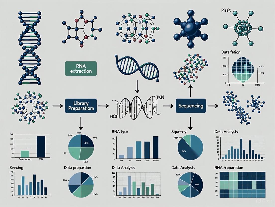
Whole transcriptome profiling via RNA Sequencing (RNA-Seq) has revolutionized the study of gene expression, enabling researchers to capture a snapshot of cellular processes by identifying and quantifying RNA transcripts present in a biological sample at a specific time [30] [31]. This comprehensive approach provides invaluable insights into changes in the transcriptome in response to environmental stimuli, disease states, or therapeutic interventions, allowing for the detection of mRNA splicing variants, single nucleotide polymorphisms, and novel transcriptional events [30]. Unlike microarrays, which require a known template and are notoriously unreliable for detecting low and very high abundance RNAs, RNA-Seq offers an unbiased platform for transcriptome-wide discovery [30]. The core of this technology involves converting RNA into complementary DNA (cDNA) through reverse transcription, followed by high-throughput sequencing of the resulting cDNA library [30] [31]. This technical guide details the standard workflow from RNA isolation to cDNA library preparation, providing researchers, scientists, and drug development professionals with the foundational protocols essential for robust whole transcriptome analysis.
RNA Isolation and Quality Control
RNA Extraction and Integrity Preservation
The success of any RNA-Seq experiment is critically dependent on the quality and integrity of the starting RNA material. Maintaining RNA integrity requires special precautions during extraction, processing, storage, and experimental use [32]. Best practices to prevent RNA degradation include wearing gloves, pipetting with aerosol-barrier tips, using nuclease-free labware and reagents, and thorough decontamination of work areas [32] [30]. Optimal purification methods must also remove common inhibitors that interfere with the activity of reverse transcriptases, including both endogenous compounds from biological sample material and inhibitory carryover compounds from RNA isolation reagents, such as salts, metal ions, ethanol, and phenol [32].
RNA should be extracted from tissues using established methods (e.g., trizol-based extraction), with special consideration for the source materials (e.g., blood, tissues, cells, plants) and experimental goals [32] [30]. For cell cultures, most cells should be in the same stage of growth, and harvesting should occur quickly with minimal osmotic or temperature shock. Flash freezing and grinding the resulting powder in liquid nitrogen is a preferred method to achieve minimally damaged nucleic acids [30]. Once purified, RNA should be stored at –80°C with minimal freeze-thaw cycles to preserve stability [32].
RNA Quality Assessment and Genomic DNA Removal
After RNA extraction, checking RNA integrity is critical before proceeding with library preparation. The RNA Integrity Number (RIN) determined by the RIN algorithm provides a standardized measure of RNA quality, ranging from 10 (intact) to 1 (completely degraded) [30]. Samples with RIN values below 7 should generally not be used for RNA-Seq, as there is little point in working with degraded RNA [30].
A crucial step in sample preparation is the removal of trace genomic DNA (gDNA) that may be co-purified with RNA, as contaminating gDNA can interfere with reverse transcription and lead to false positives, higher background, or lower detection sensitivity in downstream applications like RT-qPCR [32]. The traditional method involves adding DNase I to preparations of isolated RNA; however, DNase I must be thoroughly removed prior to cDNA synthesis since any residual enzyme would degrade single-stranded DNA and compromise results [32]. As an alternative, double-strand-specific DNases (e.g., Invitrogen ezDNase Enzyme) offer advantages by eliminating contaminating gDNA without affecting RNA or single-stranded DNAs. These thermolabile enzymes enable simpler protocols with inactivation at relatively mild temperatures (e.g., 55°C) without the RNA loss or damage associated with DNase I inactivation methods [32].
Table 1: RNA Quality Assessment Metrics
| Parameter | Optimal Value/Range | Importance |
|---|---|---|
| RNA Integrity Number (RIN) | ≥7 [30] | Indicates overall RNA degradation level; critical for library complexity |
| 260/280 Ratio | ~2.0 | Assesses protein contamination |
| 260/230 Ratio | >2.0 | Detects contaminants like salts, carbohydrates |
| Genomic DNA Contamination | Not detectable | Prevents false positives and background noise in sequencing [32] |
| Total Quantity | Varies by protocol (e.g., ≥200 ng for SHERRY [33]) | Ensures sufficient material for library preparation |
RNA Selection and cDNA Synthesis
RNA Selection and Enrichment Strategies
Following quality control, the total RNA often requires selection or enrichment of specific RNA types depending on the research objectives. A key consideration is the removal of ribosomal RNA (rRNA), which constitutes approximately 90% of total RNA and would otherwise drown out the signal from other RNA species [30]. The simplest approach is to use commercial rRNA removal kits such as the NEBNext rRNA Depletion Kit or Ribo-Zero rRNA Removal Kit [30].
Further RNA selection depends on the specific goals of the study:
- Mature mRNA Isolation: For studies focusing on protein-coding genes or splice variants, mature mRNA can be isolated using the polyA tails that bind to poly(T) oligomers attached to beads [30].
- Small RNA Enrichment: For investigation of small RNAs such as miRNA, size selection through gel filtration or affinity chromatography is typically employed [30].
- Targeted RNA Capture: Specific transcripts of interest can be enriched through hybridization with tailored probes [30].
The choice of tissue or cell type is also critical, as the expression of relevant genes must be detectable in the chosen material. For instance, in neurodevelopmental disorders, peripheral blood mononuclear cells (PBMCs) express up to 80% of genes in intellectual disability and epilepsy panels, making them a suitable and minimally invasive source [34].
Reverse Transcription and cDNA Synthesis
The synthesis of cDNA from an RNA template through reverse transcription is a crucial first step in many molecular biology protocols, serving as the foundation for downstream applications [32]. This process creates complementary DNA (cDNA) that can then be used as template in a variety of RNA studies [32].
Reverse Transcriptase Selection: Most reverse transcriptases used in molecular biology are derived from the pol gene of avian myeloblastosis virus (AMV) or Moloney murine leukemia virus (MMLV) [32]. The AMV reverse transcriptase possesses strong RNase H activity that degrades RNA in RNA:cDNA hybrids, resulting in shorter cDNA fragments (<5 kb) [32]. MMLV reverse transcriptase became a popular alternative due to its monomeric structure, which allowed for simpler cloning and modifications. Although MMLV is less thermostable than AMV reverse transcriptase, it is capable of synthesizing longer cDNA (<7 kb) at a higher efficiency due to its lower RNase H activity [32]. Engineered MMLV reverse transcriptases (e.g., Invitrogen SuperScript IV Reverse Transcriptase) feature even lower RNase H activity (RNaseH–), higher thermostability (up to 55°C), and enhanced processivity, resulting in increased cDNA length and yield, higher sensitivity, improved resistance to inhibitors, and faster reaction times [32].
Table 2: Comparison of Reverse Transcriptase Enzymes
| Attribute | AMV Reverse Transcriptase | MMLV Reverse Transcriptase | Engineered MMLV Reverse Transcriptase |
|---|---|---|---|
| RNase H Activity | High | Medium | Low [32] |
| Reaction Temperature | 42°C | 37°C | 55°C [32] |
| Reaction Time | 60 minutes | 60 minutes | 10 minutes [32] |
| Target Length | ≤5 kb | ≤7 kb | ≤14 kb [32] |
| Relative Yield (with challenging RNA) | Medium | Low | High [32] |
Reaction Components: A complete reverse transcription reaction includes several key components beyond the enzyme and RNA template: buffer (to maintain favorable pH and ionic strength), dNTPs (generally at 0.5–1 mM each, preferably at equimolar concentrations), DTT (a reducing agent for optimal enzyme activity), RNase inhibitor (to prevent RNA degradation by RNases), nuclease-free water, and primers [32].
Primer Selection: The choice of primer depends on the experimental aims:
- Oligo(dT) Primers: Anneal to the polyA tail of mRNA, enriching for protein-coding transcripts.
- Random Hexamers: Prime throughout the transcriptome, providing broader coverage including non-polyadenylated RNAs.
- Gene-Specific Primers: Target particular transcripts of interest, offering high sensitivity for specific targets.
Reaction Conditions: Reverse transcription reactions typically involve three main steps: primer annealing, DNA polymerization, and enzyme deactivation [32]. The temperature and duration of these steps vary by primer choice, target RNA, and reverse transcriptase used. For RNA with high GC content or secondary structures, an optional denaturation step can be performed by heating the RNA-primer mix at 65°C for 5 minutes followed by chilling on ice for 1 minute [32]. If using random hexamers, incubating the reverse transcription reaction at room temperature (~25°C) for 10 minutes helps anneal and extend the primers [32]. DNA polymerization is a critical step where reaction temperature and duration vary depending on the reverse transcriptase used. Using a thermostable reverse transcriptase allows for higher reaction temperatures (e.g., 50°C), which helps denature RNA with secondary structures without impacting enzyme activity, resulting in increased cDNA yield, length, and representation [32].
cDNA Library Preparation for Sequencing
Standard and Advanced Library Preparation Methods
Once cDNA is synthesized, it must be prepared into a sequencing library compatible with high-throughput platforms. The exact procedure varies depending on the platform and specific research requirements, but generally involves fragmenting the cDNA, adding platform-specific adapters, and performing quality control before sequencing [30].
Traditional library preparation methods involve several steps: cDNA fragmentation, end-repair, adapter ligation, and size selection. However, newer, more efficient protocols have been developed. For example, the SHERRY (sequencing hetero RNA-DNA-hybrid) protocol profiles polyadenylated RNAs by direct tagmentation of RNA/DNA hybrids and offers a robust and economical method for gene expression quantification, particularly suitable for low-input samples (e.g., 200 ng of total RNA) [33]. This method streamlines the process by combining tagmentation and library generation steps, reducing hands-on time and potential sample loss.
Nonsense-Mediated Decay (NMD) Considerations
In certain applications, particularly in clinical diagnostics for rare disorders, it is important to consider the effects of Nonsense-Mediated Decay (NMD), a cellular surveillance mechanism that eliminates transcripts containing premature termination codons [34]. When investigating genetic variants expected to introduce premature stop codons, NMD can mask the underlying molecular event by degrading the mutant transcript before it can be detected.
To address this challenge, researchers can use NMD inhibitors such as cycloheximide (CHX) during cell culture prior to RNA extraction [34]. Treatment with CHX has been shown to successfully inhibit NMD, allowing for the detection of transcripts that would otherwise be degraded [34]. The effectiveness of NMD inhibition can be monitored using internal controls such as the NMD-sensitive SRSF2 transcript, which shows increased expression upon successful NMD inhibition [34].
RNA-Seq Experimental Workflow from Sample to Sequence
The Scientist's Toolkit: Essential Reagents and Materials
Table 3: Key Research Reagent Solutions for RNA-Seq Library Preparation
| Reagent/Material | Function/Purpose | Examples/Notes |
|---|---|---|
| RNase Inhibitors | Prevents RNA degradation during extraction and processing; critical for maintaining RNA integrity [32]. | Included in reaction buffers or added separately to prevent degradation by environmental RNases. |
| DNase Reagents | Removes contaminating genomic DNA to prevent false positives and background noise [32]. | Traditional DNase I or thermolabile double-strand-specific DNases (e.g., Invitrogen ezDNase Enzyme) [32]. |
| rRNA Depletion Kits | Removes abundant ribosomal RNA (90% of total RNA) to enrich for other RNA types [30]. | NEBNext rRNA Depletion Kit, Ribo-Zero rRNA Removal Kit [30]. |
| Reverse Transcriptases | Synthesizes complementary DNA (cDNA) from RNA template [32]. | AMV RT, MMLV RT, or engineered MMLV RT (e.g., SuperScript IV) with improved properties [32]. |
| NMD Inhibitors | Inhibits nonsense-mediated decay to detect transcripts with premature termination codons [34]. | Cycloheximide (CHX) treatment of cells before RNA extraction [34]. |
| Library Prep Kits | Prepares cDNA for high-throughput sequencing through fragmentation, adapter ligation. | Standard Illumina kits or specialized protocols like SHERRY for low-input RNA [33]. |
| Quality Control Assays | Assesses RNA integrity and quantity before library preparation. | RIN analysis, fluorometric quantification, capillary electrophoresis. |
The standard RNA-Seq workflow from RNA isolation to cDNA library preparation represents a sophisticated yet accessible methodology that forms the foundation of modern transcriptomics. By adhering to rigorous quality control measures during RNA extraction, selecting appropriate reverse transcription and library preparation strategies, and understanding the functional roles of key reagents, researchers can generate high-quality cDNA libraries suitable for comprehensive whole transcriptome profiling. This technical foundation enables the investigation of complex biological questions in basic research and drug development, from identifying novel biomarkers to understanding mechanisms of disease pathogenesis. As RNA-Seq technologies continue to evolve, with innovations in low-input methods and streamlined protocols, the core principles outlined in this guide will remain essential for generating robust, reproducible transcriptome data.
Whole transcriptome profiling aims to generate a comprehensive picture of gene expression. However, a significant technical hurdle exists: in total RNA extracts, ribosomal RNA (rRNA) constitutes 70–90% of all RNA content, while messenger RNA (mRNA) represents only a small fraction (approximately 1–5%) [35] [36]. Sequencing total RNA without pre-treatment is therefore highly inefficient, as the majority of sequencing reads and resources are consumed by abundant, often non-target rRNA species.
To overcome this, two primary strategies are employed: mRNA enrichment via poly(A) selection and rRNA depletion. The choice between these methods is a foundational decision that directly impacts data quality, experimental cost, and the biological scope of a whole transcriptome study. This guide provides an in-depth technical comparison to inform this critical choice.
Core Methodologies and Mechanisms
The two strategies operate on fundamentally different principles to enhance the signal-to-noise ratio in RNA-Seq data.
mRNA Enrichment via Poly(A) Selection
This method uses oligo(dT) probes attached to magnetic beads to selectively bind the poly(A) tails of mature, protein-coding mRNAs. After hybridization, non-polyadenylated RNA is washed away, and the purified mRNA is eluted from the beads [36]. This process is highly effective for enriching mature mRNA, which typically makes up only about 5% of total RNA [37].
rRNA Depletion
rRNA depletion uses species-specific probes that are complementary to rRNA sequences. These probes hybridize to the rRNA in a total RNA sample. The probe-rRNA complexes are then removed, typically through magnetic separation (if the probes are biotinylated) or enzymatic digestion (e.g., using RNase H). This leaves behind a diverse pool of RNA, including both polyadenylated and non-polyadenylated species [38] [36].
Quantitative Performance Comparison
The choice between enrichment and depletion has profound and quantifiable impacts on sequencing efficiency and output. The following table summarizes key performance metrics derived from comparative studies [37].
Table 1: Performance Comparison Between Poly(A) Enrichment and rRNA Depletion
| Feature | Poly(A) Enrichment | rRNA Depletion |
|---|---|---|
| Usable exonic reads (blood) | 71% | 22% |
| Usable exonic reads (colon) | 70% | 46% |
| Extra reads needed for same exonic coverage | — | +220% (blood), +50% (colon) |
| Transcript types captured | Mature, coding mRNAs | Coding + non-coding RNAs (lncRNAs, snoRNAs, pre-mRNA) |
| 3'–5' coverage uniformity | Pronounced 3' bias | More uniform coverage |
| Performance with degraded RNA (FFPE) | Poor; strong 3' bias, low yield | Robust; does not rely on intact poly(A) tails |
| Sequencing cost per usable read | Lower | Higher (requires greater depth) |
Efficiency and Cost Implications
The data in Table 1 highlights a critical trade-off. Poly(A) enrichment is vastly more efficient for sequencing mRNA, yielding a high percentage of exonic reads. One study found that to achieve similar exonic coverage, rRNA depletion required 220% more reads from blood and 50% more from colon tissue compared to poly(A) selection [37]. This directly translates to higher sequencing costs for rRNA depletion when the goal is standard mRNA expression analysis.
Transcriptomic Breadth
While less efficient for mRNA, rRNA depletion provides a much broader view of the transcriptome. It captures both polyadenylated and non-polyadenylated transcripts, including long non-coding RNAs (lncRNAs), circular RNAs, and pre-mRNA [21] [37]. This makes it indispensable for comprehensive transcriptome annotation and studies focused on non-coding RNA biology.
Experimental Protocols and Optimization
Optimized Protocol for mRNA Enrichment with Oligo(dT) Beads
Following manufacturer protocols for mRNA enrichment can yield suboptimal results, with rRNA sometimes still constituting up to 50% of the output [35]. An optimized protocol for S. cerevisiae, which can be adapted for other eukaryotes, involves:
- Input and Bead Ratio: Use 5-75 μg of high-quality total RNA (RIN > 8). A critical parameter is the beads-to-RNA ratio. Increasing the ratio of Oligo(dT)25 Magnetic Beads to RNA to 50:1 or 125:1 significantly reduces residual rRNA content to about 20% [35].
- Two-Round Enrichment: For maximum purity, a two-round enrichment process is highly effective.
- First Round: Perform an initial enrichment with a standard beads-to-RNA ratio (e.g., 13.3:1).
- Second Round: Use all eluted RNA from the first round as input for a second enrichment with a high beads-to-RNA ratio (e.g., 90:1). This strategy can reduce the rRNA content in the final sample to less than 10% [35].
- Quality Control: Assess enrichment efficiency using capillary electrophoresis (e.g., TapeStation) or Bioanalyzer to quantify the reduction in 18S and 28S rRNA peaks [35].
rRNA Depletion Methodologies and Considerations
rRNA depletion methods can be broadly categorized, with performance differences noted in comparative studies:
- Probe Hybridization & Capture: This method uses biotinylated DNA or LNA probes that hybridize to rRNA. The complexes are removed with streptavidin-coated magnetic beads. Kits employing this method (e.g., riboPOOLs) have shown high efficiency, comparable to the discontinued but highly effective RiboZero kit [38].
- Enzymatic Depletion (RNase H): This method uses DNA probes hybridized to rRNA, followed by digestion of the RNA-DNA hybrids by the RNase H enzyme. While fast and streamlined, studies indicate that this method can cause partial mRNA degradation, leading to 3' bias in subsequent sequencing data, meaning coverage is skewed toward the 3' end of transcripts [39]. It is therefore more suitable for total RNA sequencing applications where full-length transcript integrity is less critical.
Table 2: Research Reagent Solutions for RNA Selection
| Reagent / Kit | Type | Key Function | Considerations |
|---|---|---|---|
| Oligo(dT)25 Magnetic Beads | mRNA Enrichment | Selects polyadenylated RNA via magnetic separation. | Requires optimization of bead-to-RNA ratio; cost-effective for bulk reagents [35]. |
| RiboMinus Transcriptome Isolation Kit | rRNA Depletion | Depletes rRNA using pan-prokaryotic or eukaryotic-specific probes. | May not target 5S rRNA; efficiency varies [35] [38]. |
| riboPOOLs (rRNA Depletion) | rRNA Depletion | Uses DNA probes for specific rRNA depletion via magnetic capture. | Highly efficient; species-specific versions available; good RiboZero replacement [38]. |
| NEBNext Globin & rRNA Depletion Kit | rRNA Depletion | Enzymatic (RNase H) removal of rRNA and globin mRNA. | Can introduce 3' bias; faster, single-tube workflow [39]. |
| Duplex-Specific Nuclease (DSN) | Normalization/Depletion | Normalizes cDNA populations by digesting abundant double-stranded cDNA. | Unspecific depletion; can remove any highly abundant transcript, not just rRNA [36]. |
Strategic Selection Guide
The decision between mRNA enrichment and rRNA depletion is dictated by the experimental goals, sample type, and organism.
Table 3: Decision Matrix for Method Selection
| Scenario / Goal | Recommended Method | Rationale |
|---|---|---|
| Standard mRNA expression (eukaryotes, high-quality RNA) | Poly(A) Enrichment | Highest efficiency and lowest cost for profiling protein-coding genes [37]. |
| Total RNA sequencing (non-coding RNA, bacterial RNA) | rRNA Depletion | Captures the full diversity of RNA species, essential for prokaryotes and non-coding RNA studies [36] [37]. |
| Degraded samples (FFPE, RIN < 7) | rRNA Depletion | Does not rely on intact 3' poly(A) tails, providing more representative coverage [4] [37]. |
| Splicing, isoform, or fusion analysis | rRNA Depletion | Provides more uniform 5'->3' coverage across transcripts, enabling accurate isoform resolution [37]. |
| High-throughput or cost-sensitive projects | Poly(A) Enrichment | Lower sequencing depth requirements drastically reduce overall cost per sample [37]. |
Special Considerations for Challenging Samples
- Whole Blood: Globin mRNA is as abundant as rRNA in blood-derived RNA, comprising up to 80% of transcripts. For optimal results, use a probe-based hybridization method to deplete both globin mRNA and rRNA, as it demonstrates superior performance over enzymatic depletion, yielding more junction reads and minimal 3' bias [39].
- Single-Cell RNA-Seq: Most single-cell technologies (e.g., 10x Genomics) inherently use poly(A) enrichment during the reverse transcription step, making it the default and most efficient choice for profiling mRNA in individual cells [29].
Within the framework of whole transcriptome research, the choice between mRNA enrichment and rRNA depletion is a fundamental strategic decision. There is no universally superior technique; each serves a distinct purpose.
- For focused, cost-effective analysis of protein-coding gene expression in eukaryotes with high-quality RNA, poly(A) enrichment is the unequivocal choice. Its high efficiency and lower sequencing requirements make it ideal for large-scale gene expression profiling.
- For comprehensive transcriptome discovery, studies involving non-coding RNA, prokaryotes, or degraded samples, rRNA depletion is the necessary approach. Its ability to capture a wider array of RNA species ensures that no critical biological insights are overlooked.
By aligning the technical strengths of each method with specific research objectives and sample characteristics, scientists can design robust, efficient, and informative whole transcriptome studies that effectively advance our understanding of gene expression and regulation.
Single-cell RNA sequencing (scRNA-seq) has revolutionized genomic investigations by enabling the exploration of gene expression heterogeneity at the individual cell level, providing unprecedented resolution for studying complex biological systems [40]. This technology systematically profiles the expression levels of mRNA transcripts for each gene at single-cell resolution, allowing researchers to uncover cellular diversity and heterogeneity that would be overlooked in bulk-cell RNA sequencing [40] [41]. Since its initial demonstration on a 4-cell blastomere stage in 2009 and the development of the first multiplexed method in 2014, scRNA-seq has become a pivotal tool for investigating cellular heterogeneity, identifying rare cell types, mapping developmental pathways, and exploring tumor diversity [41]. The ability to profile individual cells has transformed our understanding of biological processes, from early embryo development to disease mechanisms, making it possible to discern how different cells behave at single-cell levels and providing new insights into highly organized organs or tissues [42] [41].
The fundamental advantage of scRNA-seq lies in its capacity to reveal the unique expression characteristics of individual cells, capturing cellular states and transitions that are masked in population-averaged measurements [40] [43]. Whereas bulk RNA sequencing analyzes the transcriptome of a group of cells or tissues, providing an average gene activity level within the sample, scRNA-seq captures the distinct gene expression patterns of each cell, enabling a more comprehensive understanding of cellular function and organization [41]. This technology has become increasingly preferred for addressing crucial biological inquiries related to cell heterogeneity, particularly in cases involving limited cell numbers or complex cellular ecosystems like tumor microenvironments [41].
Technical Foundations of scRNA-seq
Core Experimental Workflows
The scRNA-seq workflow encompasses multiple specialized steps, from sample preparation to sequencing. The initial stage involves extracting viable individual cells from the tissue under investigation, which can be challenging for complex tissues or frozen samples [41]. Novel methodologies like isolating individual nuclei for RNA-seq (snRNA-seq) have been developed for conditions where tissue dissociation is difficult or when samples are frozen [41]. Another innovative approach uses "split-pooling" scRNA-seq techniques that apply combinatorial indexing (cell barcodes) to single cells, offering distinct advantages including the ability to handle large sample sizes (up to millions of cells) and greater efficiency in parallel processing of multiple samples without expensive microfluidic devices [41].
Following cell isolation, individual cells undergo lysis to facilitate RNA capture. Poly[T]-primers are frequently employed to selectively analyze polyadenylated mRNA molecules while minimizing ribosomal RNA capture [41]. After converting RNA to complementary DNA (cDNA), the resulting molecules undergo amplification by either polymerase chain reaction (PCR) or in vitro transcription (IVT) methods [41]. To mitigate amplification biases, Unique Molecular Identifiers (UMIs) are used to label each individual mRNA molecule during reverse transcription, enhancing the quantitative aspect of scRNA-seq and improving data interpretation accuracy by eliminating biases from PCR amplification [41].
Protocol Variations and Methodologies
Different scRNA-seq technologies have emerged with distinct characteristics and applications. These protocols vary significantly in multiple aspects, including cell isolation methods, reverse transcription approaches, amplification techniques, and transcript coverage [41]. A key distinction lies in transcript coverage: some techniques generate full-length (or nearly full-length) transcript sequencing data (e.g., Smart-Seq2, MATQ-Seq, Fluidigm C1), while others capture and sequence only the 3' or 5' ends of transcripts (e.g., Drop-Seq, inDrop, 10x Genomics) [41].
Each approach offers unique advantages and limitations. Full-length scRNA-seq methods excel in tasks like isoform usage analysis, allelic expression detection, and identifying RNA editing due to comprehensive transcript coverage [41]. They also outperform 3' end sequencing methods in detecting specific lowly expressed genes or transcripts [41]. In contrast, droplet-based techniques like Drop-Seq, InDrop, and 10x Genomics Chromium enable higher throughput of cells and lower sequencing cost per cell, making them particularly valuable for detecting diverse cell subpopulations within complex tissues or tumor samples [41].
Recent methodological advances continue to expand scRNA-seq capabilities. RamDA-seq, for instance, represents the first full-length total RNA-sequencing method for single cells, showing high sensitivity to non-poly(A) RNA and near-complete full-length transcript coverage [43]. This method enables researchers to reveal dynamically regulated non-poly(A) transcripts, profile recursive splicing in >300-kb introns, and detect enhancer RNAs and their cell type-specific activity in single cells [43].
Table 1: Comparison of Major scRNA-seq Protocol Categories
| Protocol Type | Key Examples | Transcript Coverage | Amplification Method | Throughput | Primary Applications |
|---|---|---|---|---|---|
| Full-length | Smart-Seq2, MATQ-Seq, Fluidigm C1 | Full-length or nearly full-length | PCR | Lower | Isoform analysis, allele-specific expression, rare transcript detection |
| 3'/5' Counting | Drop-Seq, inDrop, 10x Genomics, Seq-Well | 3' or 5' ends only | PCR or IVT | High | Large-scale cell typing, population heterogeneity, atlas construction |
| Total RNA | RamDA-seq, SUPeR-seq | Full-length with non-poly(A) RNA | Specialized (e.g., RT-RamDA) | Variable | Non-poly(A) transcript detection, enhancer RNA analysis, recursive splicing |
Computational Analysis Framework
Core Analytical Workflow
The analysis of scRNA-seq data presents unique computational challenges due to its high-dimensional, sparse, and noisy nature [41]. A standardized analytical workflow has emerged to transform raw sequencing data into biological insights. The process begins with quality control to identify and remove low-quality cells, multiplets, and empty droplets [44] [41]. This is followed by normalization to account for technical variations, feature selection to identify highly variable genes, and dimensionality reduction to visualize and explore the high-dimensional data in two or three dimensions [44].
Clustering analysis represents a fundamental step where cells are grouped into populations based on similarity of gene expression patterns [45]. This step relies on graph-based clustering methods like the Louvain and Leiden algorithms, which balance speed and efficiency [46]. Downstream analyses include differential expression testing to identify marker genes, cell type annotation using known markers or reference datasets, trajectory inference to reconstruct developmental processes, and cell-cell communication analysis to study signaling networks [44] [40].
Table 2: Key Steps in scRNA-seq Computational Analysis
| Analysis Step | Purpose | Common Tools/Methods | Key Considerations |
|---|---|---|---|
| Quality Control | Filter low-quality cells and artifacts | Scater, Scanpy, Seurat | Thresholds based on counts, genes, mitochondrial percentage [44] |
| Normalization | Remove technical biases | scran, SCnorm, Linnorm | Account for library size differences, zero inflation [44] |
| Feature Selection | Identify biologically relevant genes | Seurat, Scanpy | Focus on highly variable genes for downstream analysis [45] |
| Dimensionality Reduction | Visualize and explore data structure | PCA, UMAP, t-SNE | UMAP preferred for preserving global structure [47] [44] |
| Clustering | Identify cell populations | Leiden, Louvain algorithms | Resolution parameter controls granularity [46] [40] |
| Cell Annotation | Assign biological identity to clusters | Marker genes, SingleR, CellTypist | Combines manual and automated approaches [40] |
| Downstream Analysis | Extract biological insights | Differential expression, trajectory inference, cell-cell communication | Depends on biological question [44] [40] |
Advanced Computational Methods
Recent computational advances have addressed specific challenges in scRNA-seq analysis. The stochastic nature of clustering algorithms leads to variability in results across different runs, compromising reliability [46]. To address this, methods like single-cell Inconsistency Clustering Estimator (scICE) evaluate clustering consistency using the inconsistency coefficient (IC), achieving up to 30-fold improvement in speed compared to conventional consensus clustering-based methods [46]. This approach helps researchers identify stable clustering results and avoid false interpretations based on stochastic clustering variations.
Ensemble clustering algorithms represent another advancement for addressing methodological bias in clustering analyses. The scEVE algorithm integrates multiple clustering methods (monocle3, Seurat, densityCut, and SHARP) to identify robust clusters while quantifying their uncertainty [45]. Instead of minimizing differences between input clustering results, scEVE describes these differences to identify clusters robust to methodological variations and prevent over-clustering [45].
Deep learning approaches have also transformed scRNA-seq analysis. Graph neural networks (GNNs) show particular promise for leveraging the inherent graph structure of single-cell data [42]. Methods like scE2EGAE learn cell-to-cell graphs during model training through differentiable edge sampling, enhancing denoising performance and downstream analysis compared to fixed-graph approaches [42]. Similarly, variational autoencoders as implemented in scvi-tools provide superior batch correction, imputation, and annotation through probabilistic modeling of gene expression [48].
Essential Research Tools and Reagents
Experimental Reagents and Platforms
The scRNA-seq experimental workflow relies on specialized reagents and platforms that have been optimized for single-cell analysis. Cell isolation represents a critical first step, with various methodologies available including fluorescence-activated cell sorting (FACS), microfluidic isolation, and microdroplet-based approaches [41]. Commercial platforms like 10x Genomics Chromium, BD Rhapsody, and Parse Biosciences offer integrated solutions that combine cell isolation, barcoding, and library preparation in standardized workflows [48] [41].
Unique Molecular Identifiers (UMIs) have become essential reagents for quantitative scRNA-seq, enabling accurate counting of individual mRNA molecules by correcting for amplification biases [41]. These barcodes are incorporated during reverse transcription and allow distinction between biological variation and technical artifacts. For full-length transcript protocols, template-switching oligonucleotides facilitate cDNA amplification, while for 3' counting methods, barcoded beads capture polyadenylated transcripts in nanoliter-scale reactions [41].
Recent protocol advancements have also introduced specialized reagents for emerging applications. For example, RamDA-seq uses not-so-random primers (NSRs) designed to avoid synthesizing cDNA from rRNA sequences, thereby reducing ribosomal contamination while maintaining sensitivity to non-poly(A) transcripts [43]. Similarly, multiome approaches combine RNA measurement with other modalities like ATAC-seq for chromatin accessibility, requiring specialized reagents that preserve multiple molecular species from the same cell [49].
Computational Tools and Ecosystems
The computational analysis of scRNA-seq data relies on sophisticated software ecosystems that have evolved to handle the scale and complexity of single-cell datasets. Two dominant platforms have emerged: Seurat for R users and Scanpy for Python users [48]. Seurat remains the R standard for versatility and integration, with expanded capabilities for spatial transcriptomics, multiome data, and protein expression via CITE-seq [48]. Scanpy dominates large-scale scRNA-seq analysis, especially for datasets exceeding millions of cells, with architecture optimized for memory use and seamless integration with the broader scverse ecosystem [48].
For preprocessing raw sequencing data, Cell Ranger remains the gold standard for 10x Genomics platforms, reliably transforming raw FASTQ files into gene-barcode count matrices using the STAR aligner [48]. Specialized tools have also been developed to address specific analytical challenges: Harmony efficiently corrects batch effects across datasets; CellBender uses deep learning to clean ambient RNA noise; Velocyto enables RNA velocity analysis to infer cellular dynamics; and Monocle 3 advances pseudotime and trajectory inference [48].
Integrated platforms like OmniCellX provide user-friendly browser-based interfaces that simplify and streamline scRNA-seq data analysis while addressing key challenges in accessibility, scalability, and usability [40]. These platforms combine a comprehensive suite of analytical tools with intuitive interfaces, making sophisticated analyses accessible to researchers without advanced computational expertise [40].
Table 3: Essential Computational Tools for scRNA-seq Analysis
| Tool Category | Representative Tools | Primary Function | Key Features |
|---|---|---|---|
| Comprehensive Platforms | Seurat, Scanpy, OmniCellX | End-to-end analysis | Modular workflows, extensive documentation, multiple visualization options [48] [40] |
| Preprocessing & QC | Cell Ranger, scater, CellBender | Data processing & quality control | FASTQ to count matrix, doublet detection, ambient RNA removal [48] |
| Batch Correction | Harmony, scVI, ComBat | Data integration | Remove technical variation while preserving biology [48] |
| Clustering & Annotation | Leiden algorithm, SingleR, CellTypist | Cell type identification | Multiple resolution parameters, reference-based annotation [46] [40] |
| Trajectory Inference | Monocle 3, PAGA, Slingshot | Reconstruction of dynamic processes | Pseudotime ordering, branch point detection [48] |
| Specialized Analysis | Velocyto, CellPhoneDB, Squidpy | RNA velocity, cell-cell communication, spatial analysis | Predictive modeling, interaction databases, spatial neighborhoods [48] |
Applications in Biomedical Research
Fundamental Biological Insights
scRNA-seq has dramatically expanded our understanding of cellular heterogeneity in both normal development and disease states. In developmental biology, it has enabled the reconstruction of lineage trajectories and the identification of novel progenitor states during embryogenesis, organ formation, and tissue regeneration [41]. The technology has proven particularly valuable for characterizing rare cell populations that play critical roles in developmental processes but are difficult to detect with bulk approaches [43] [41].
In cancer research, scRNA-seq has transformed our understanding of tumor microenvironments by simultaneously profiling malignant cells, immune infiltrates, stromal cells, and vascular components [41]. This comprehensive cellular census has revealed previously unappreciated heterogeneity within tumors, identified resistance mechanisms to therapy, and uncovered new therapeutic targets [41]. The ability to map cellular ecosystems within tumors has positioned scRNA-seq as a cornerstone technology for advancing cancer immunotherapy and personalized treatment approaches.
Neurology has particularly benefited from scRNA-seq applications, given the extraordinary cellular diversity of the nervous system. Studies of human and mouse brains have identified numerous neuronal and glial subtypes, revealing unexpected complexity and regional specialization [46] [41]. These cellular atlases provide foundational resources for understanding brain function and dysfunction, with important implications for neurodegenerative diseases, psychiatric disorders, and neural repair.
Translational and Clinical Applications
The resolution provided by scRNA-seq has enabled numerous translational applications with direct clinical relevance. In drug discovery, scRNA-seq enables comprehensive characterization of drug responses at cellular resolution, identifying responsive and resistant subpopulations and revealing mechanisms of action [41]. This information guides target selection, candidate optimization, and patient stratification strategies [41].
Biomarker discovery represents another major application area, where scRNA-seq identifies cell type-specific expression signatures associated with disease progression, treatment response, or clinical outcomes [41]. The technology's sensitivity for detecting rare cell populations makes it particularly valuable for identifying minimal residual disease in cancer or rare pathogenic cells in autoimmune conditions [41].
As scRNA-seq technologies continue to advance, they are being integrated into clinical trial designs to provide mechanistic insights and pharmacodynamic biomarkers [41]. The ongoing development of scalable, robust, and standardized workflows will likely accelerate the translation of scRNA-seq from basic research to clinical applications in diagnostics, therapeutic monitoring, and personalized treatment strategies [40] [41].
Future Perspectives and Challenges
The scRNA-seq field continues to evolve rapidly, with several emerging trends shaping its future trajectory. Multi-omic integration represents a major frontier, with technologies that simultaneously profile RNA alongside other molecular features such as chromatin accessibility (ATAC-seq), surface proteins, DNA methylation, or spatial position [49] [48]. These integrated approaches provide complementary views of cellular states and regulatory mechanisms, enabling more comprehensive characterization of biological systems.
Computational methods are advancing to address the growing scale and complexity of single-cell data. Machine learning approaches, particularly graph neural networks and generative models, show promise for enhancing data denoising, imputation, and interpretation [42]. As datasets grow to millions of cells, efficient algorithms and data structures will be essential for manageable computation and storage [48] [40].
Spatial transcriptomics represents another rapidly advancing area that complements dissociated scRNA-seq by preserving architectural context [47] [48]. Methods like 10x Visium, MERFISH, and Slide-seq map gene expression within tissue sections, enabling researchers to relate cellular heterogeneity to tissue organization and cell-cell interactions [48]. Computational tools like Squidpy have emerged to analyze these spatial datasets, constructing neighborhood graphs and identifying spatially restricted patterns [48].
Despite remarkable progress, scRNA-seq still faces important challenges. Technical noise, batch effects, and sparsity continue to complicate data interpretation, particularly for rare cell types and subtle biological variations [42] [41]. Analytical standardization remains elusive, with hundreds of available tools and workflows creating reproducibility challenges [44] [41]. As the technology becomes more widely adopted, developing robust benchmarks, best practices, and user-friendly platforms will be essential for maximizing its biological impact and clinical utility [40] [41].
The ongoing innovation in both experimental protocols and computational methods ensures that scRNA-seq will continue to be a transformative technology across biological and biomedical research. By enabling the systematic characterization of cellular heterogeneity at unprecedented resolution, scRNA-seq provides a powerful lens for studying development, physiology, and disease, ultimately advancing our fundamental understanding of life processes and accelerating the development of novel therapeutics.
The transition from bulk to single-cell resolution has fundamentally transformed transcriptomic research, enabling scientists to dissect cellular heterogeneity with unprecedented detail. Whole transcriptome profiling aims to capture the complete set of RNA transcripts within a biological sample, providing a snapshot of cellular activity and gene regulation. For researchers and drug development professionals, selecting the appropriate profiling strategy—bulk RNA sequencing (bulk RNA-seq), single-cell RNA sequencing (scRNA-seq), or targeted gene expression profiling—represents a critical decision point that directly impacts data quality, interpretability, and research outcomes. Each approach offers distinct advantages and limitations, making them suited to different phases of the research pipeline, from initial discovery to clinical validation.
Bulk RNA-seq provides a population-averaged gene expression profile, blending signals from all cells within a sample and offering a broad overview of transcriptional activity [50] [51]. In contrast, scRNA-seq isolates and sequences RNA from individual cells, revealing the cellular diversity and rare cell populations that are masked in bulk analyses [52] [53]. Targeted profiling occupies a middle ground, focusing sequencing resources on a predefined set of genes to achieve superior sensitivity and quantitative accuracy for specific research questions [29]. Understanding the technical considerations, applications, and practical implications of each method is essential for designing efficient and informative transcriptomic studies that advance our understanding of biological systems and accelerate therapeutic development.
Core Technologies and Methodological Principles
Bulk RNA Sequencing: Population-Averaged Profiling
Bulk RNA sequencing is a next-generation sequencing (NGS)-based method that measures the whole transcriptome across a population of cells, providing an averaged gene expression profile for the entire sample [50]. The methodology involves digesting the biological sample to extract RNA, which may be total RNA or enriched for mRNA through ribosomal RNA depletion. This RNA is then converted to complementary DNA (cDNA), followed by library preparation steps to create a sequencing-ready gene expression library [50]. After sequencing, data analysis reveals gene expression levels across the tissue sample, representing the average expression levels for individual genes across all cells that compose the sample [50].
The primary advantage of bulk RNA-seq lies in its ability to provide a holistic view of the average gene expression profile, making it particularly valuable for differential gene expression analysis between different experimental conditions, such as disease versus healthy states, treated versus control groups, or across developmental stages [50]. This approach enables the identification of distinct genes that are upregulated or downregulated under these conditions and supports applications like discovering RNA-based biomarkers and molecular signatures for diagnosis, prognosis, or disease stratification [50]. Additionally, bulk RNA-seq remains the preferred method for large cohort studies, biobank projects, and establishing baseline transcriptomic profiles for new or understudied organisms or tissues [50].
Single-Cell RNA Sequencing: Resolution at the Cellular Level
Single-cell RNA sequencing represents a paradigm shift in transcriptomics, enabling researchers to study gene expression at the resolution of individual cells rather than population averages [52]. The scRNA-seq workflow begins with generating viable single-cell suspensions from whole samples through enzymatic or mechanical dissociation, cell sorting, or other cell isolation techniques [50]. This is followed by cell counting and quality control steps to ensure appropriate concentration of viable cells free from clumps and debris [50]. In platforms like the 10x Genomics Chromium system, single cells are isolated into individual micro-reaction vessels (Gel Beads-in-emulsion, or GEMs) where cell-specific barcodes are added to RNA transcripts, ensuring that analytes from each cell can be traced back to their origin [50].
This technology has proven invaluable for characterizing heterogeneous cell populations, including novel cell types, cell states, and rare cell types that would otherwise be overlooked in bulk analyses [50] [52]. It enables researchers to determine what cell types or states are present in a tissue, their proportional representation, and gene expression differences between similar cell types or subpopulations [50]. Furthermore, scRNA-seq allows reconstruction of developmental hierarchies and lineage relationships by tracking how cellular heterogeneity evolves over time during development or disease progression [50]. The ability to profile how individual cells respond to stimuli or perturbations makes it particularly powerful for identifying specific cells or cell states that drive disease biology or treatment resistance [50].
Targeted Gene Expression Profiling: Focused Sequencing for Specific Applications
Targeted gene expression profiling represents a strategic approach that focuses sequencing resources on a pre-defined set of genes, ranging from a few dozen to several thousand, to achieve specific research objectives [29]. Unlike the unbiased nature of whole transcriptome methods, targeted profiling requires prior knowledge of the genes of interest, making it ideal for validation studies, interrogating specific biological pathways, or developing robust quantitative assays for translational research [29]. There are two primary techniques for target enrichment: hybridization capture and amplicon-based enrichment [54].
Hybridization capture utilizes synthesized oligonucleotide probes complementary to the genetic sequences of interest [54]. In solution-based methods, these biotinylated probes are added to the genetic material in solution to hybridize with target regions, followed by capture using magnetic streptavidin beads to isolate the desired sequences [54]. Array-based capture attaches probes directly to a solid surface, where target regions hybridize and unbound material is washed away [54]. Amplicon-based enrichment, exemplified by technologies like Ion AmpliSeq, uses carefully designed PCR primers to flank targets and specifically amplify regions of interest [54]. This approach offers advantages in targeting difficult genomic regions, including homologous sequences like pseudogenes and paralogs, hypervariable regions such as T-cell receptors, and low-complexity regions with di- and tri-nucleotide repeats [54].
Technical Comparison and Experimental Considerations
Methodological Comparison and Key Characteristics
The selection between bulk, single-cell, and targeted profiling approaches requires careful consideration of their technical specifications, applications, and limitations. Each method offers distinct advantages for particular research scenarios, with significant implications for experimental design, data quality, and interpretation.
Table 1: Comparative Analysis of Transcriptome Profiling Methods
| Characteristic | Bulk RNA-seq | Single-Cell RNA-seq | Targeted Profiling |
|---|---|---|---|
| Resolution | Population average | Individual cells | Individual cells (pre-defined genes) |
| Gene Coverage | Comprehensive, whole transcriptome | Comprehensive, whole transcriptome | Focused on pre-selected gene panels |
| Sensitivity to Rare Cell Types | Low, signals diluted | High, can identify rare populations | High for targeted genes in rare cells |
| Technical Complexity | Moderate | High | Moderate to High |
| Cost per Sample | Low | High | Moderate |
| Data Output Volume | Moderate | Very High | Low to Moderate |
| Primary Applications | Differential expression, biomarker discovery, population studies | Cell atlas construction, heterogeneity analysis, developmental trajectories | Validation studies, clinical assays, pathway-focused research |
| Key Limitations | Masks cellular heterogeneity | High cost, technical noise, data complexity | Limited to pre-defined genes, discovery blind spots |
Analytical Considerations and Normalization Challenges
The analysis of transcriptomic data presents distinct challenges for each profiling method. For bulk RNA-seq, analytical approaches typically focus on identifying differentially expressed genes between conditions using statistical methods that account for biological variability and technical noise [51]. However, a significant limitation arises in heterogeneous tissues, where expression changes in rare cell populations may be diluted or completely masked by dominant cell types [51].
Single-cell RNA-seq data analysis involves specialized computational approaches to manage the high dimensionality, technical variability, and sparsity inherent in these datasets [55]. A critical consideration often overlooked in scRNA-seq analysis is the variation in transcriptome size across different cell types [55]. Transcriptome size refers to the total number of mRNA molecules within each cell, which can vary significantly—often by multiple folds—across different cell types [55]. Standard normalization approaches like Counts Per 10,000 (CP10K) operate on the assumption that transcriptome size is constant across all cells, which eliminates technology-derived effects but also removes genuine biological variation in transcriptome size [55]. This can create substantial problems when comparing different cell types, including obstacles in identifying authentic differentially expressed genes [55]. Advanced methods like ReDeconv's CLTS (Count based on Linearized Transcriptome Size) approach aim to preserve these biological variations while still accounting for technical artifacts [55].
Targeted profiling analyses are generally more streamlined due to the focused nature of the data [29]. With sequencing resources concentrated on a smaller number of genes, the resulting datasets are less sparse, simplifying differential expression analysis and quantification [29]. However, targeted approaches require careful validation of gene panels to ensure they capture the biological processes of interest, and they are inherently limited by their inability to detect expression changes in genes not included in the panel [29].
Experimental Design and Workflow Selection
Decision Framework for Method Selection
Choosing the most appropriate transcriptomic profiling method requires careful consideration of research objectives, sample characteristics, and practical constraints. The following diagram illustrates a systematic approach to method selection based on key experimental factors:
This decision framework emphasizes that research questions focused on cellular heterogeneity, rare cell populations, or developmental trajectories are best addressed with scRNA-seq [50] [53]. For studies examining overall transcriptional changes between conditions in well-characterized systems or requiring large sample sizes, bulk RNA-seq remains the most practical choice [50] [51]. Targeted approaches excel when resources are limited, specific pathways are of interest, or when transitioning from discovery to validation phases in drug development [29].
Integrated Approaches and Sequential Applications
In many research scenarios, particularly in therapeutic development, a sequential approach that leverages multiple methods provides the most comprehensive insights [29]. A common strategy begins with scRNA-seq for unbiased discovery in a limited set of samples to identify novel cell types, states, and potential therapeutic targets [29]. Following target identification, researchers can employ targeted profiling to validate findings across larger patient cohorts in a cost-effective manner [29]. This integrated approach maximizes the strengths of each technology while mitigating their individual limitations.
For example, in a study on B-cell acute lymphoblastic leukemia (B-ALL), researchers leveraged both bulk and single-cell RNA-seq to identify developmental states driving resistance and sensitivity to asparaginase, a common chemotherapeutic agent [50]. Similarly, in atrial fibrillation research, an integrated analysis of bulk and single-nucleus RNA sequencing revealed lactate metabolism-related signatures and T cell alterations that would have been challenging to identify using either approach alone [56]. These integrated workflows demonstrate how combining methods at different research stages can yield insights inaccessible to any single approach.
Application Contexts and Case Studies
Disease Research and Biomarker Discovery
In biomedical research, each profiling method finds distinct applications across the disease research continuum. Bulk RNA-seq has been instrumental in identifying molecular signatures associated with disease states, treatment responses, and clinical outcomes [51]. For instance, in atrial fibrillation studies, bulk transcriptomic analyses have revealed modifications in T cell-mediated immunity and lactate metabolism pathways, providing insights into disease mechanisms beyond electrophysiological abnormalities [56].
Single-cell RNA-seq has revolutionized our understanding of cellular heterogeneity in diseases like cancer, where it has enabled the identification of rare cell populations, including cancer stem cells, drug-resistant clones, and metastatic clones that drive disease progression and treatment failure [53]. The technology has proven particularly valuable for characterizing complex tissues such as neural tissues and the immune system, where cellular diversity is extensive and functionally significant [53].
Targeted profiling bridges the gap between discovery research and clinical application, providing the robust, reproducible, and cost-effective assays required for translational medicine [29]. Once candidate biomarkers are identified through discovery-phase scRNA-seq or bulk analyses, targeted panels enable validation across large patient cohorts for clinical trial enrollment or companion diagnostic development [29]. This approach is particularly valuable for monitoring therapeutic response and pharmacodynamics, allowing researchers to track specific gene expression changes following treatment without the noise and expense of whole transcriptome profiling [29].
Protocol Implementation and Technical Considerations
Successful implementation of transcriptomic profiling requires careful attention to experimental protocols and technical considerations. The following section outlines key methodological details for each approach, drawing from established research protocols.
Table 2: Experimental Protocols and Reagent Solutions
| Method | Key Protocol Steps | Essential Reagents/Technologies | Function |
|---|---|---|---|
| Bulk RNA-seq | 1. Total RNA extraction2. RNA quality assessment3. Library preparation (mRNA enrichment or rRNA depletion)4. Sequencing5. Bioinformatic analysis | Poly(A) selection beadsrRNA depletion kitsReverse transcriptaseNGS library prep kits | mRNA enrichmentrRNA removalcDNA synthesisLibrary construction |
| Single-Cell RNA-seq | 1. Tissue dissociation2. Single-cell suspension3. Cell viability assessment4. Partitioning (e.g., GEM generation)5. Barcoding and library prep6. Sequencing7. Computational analysis | Enzymatic dissociation kitsCell viability dyes10x Chromium controllerGel Beads with barcodesSingle-cell 3' reagent kits | Tissue dissociationViability assessmentSingle-cell partitioningCell-specific barcoding |
| Targeted Profiling | 1. Panel design/selection2. Target enrichment (hybridization or amplicon)3. Library preparation4. Sequencing5. Targeted analysis | Hybridization capture probesPCR primers for amplicon panelsIon AmpliSeq designerBarcoded adapters | Sequence-specific enrichmentTargeted amplificationCustom panel designSample multiplexing |
For bulk RNA-seq, the GSE79768 dataset analysis on atrial fibrillation exemplifies standard methodology: RNA extraction from atrial tissue samples, library preparation, sequencing on platforms like Illumina, followed by differential expression analysis using tools like limma with thresholds of |log2FC| >1 and FDR-adjusted p < 0.05 [56]. Functional annotation typically involves Gene Ontology (GO) and KEGG pathway enrichment analyses using clusterProfiler [56].
Single-cell protocols, as demonstrated in the atrial fibrillation study GSE255612, involve single-nucleus RNA sequencing data processing using Seurat, normalization via SCTransform, clustering through Principal Component Analysis and t-SNE, and cell type annotation using manual curation based on marker genes [56]. Intercellular communication analysis may employ tools like CellChat with default ligand-receptor pairs to infer signaling networks between cell populations [56].
Targeted approaches using amplicon-based enrichment, such as Ion AmpliSeq technology, enable highly multiplexed PCR (up to 24,000 primer pairs in a single reaction) followed by primer digestion, barcoded adapter ligation, and library purification [54]. This approach is particularly valuable for limited samples, with demonstrated success using as little as 1 ng of input DNA or RNA, including challenging samples like FFPE tissue or circulating nucleic acids [54].
Future Directions and Emerging Technologies
The field of transcriptome profiling continues to evolve with emerging technologies that address current limitations and expand analytical capabilities. Spatial transcriptomics represents a pivotal advancement that preserves the spatial context of RNA transcripts within tissue architecture, addressing a key limitation of standard scRNA-seq that requires tissue dissociation [52]. This technology facilitates the identification of RNA molecules in their original spatial context within tissue sections at near-single-cell resolution, providing valuable insights for neurology, embryology, cancer research, and immunology [52].
Computational innovations are also enhancing data analysis and interpretation. Methods like ReDeconv incorporate transcriptome size variation into scRNA-seq normalization and bulk deconvolution, correcting for scaling effects that can misidentify differentially expressed genes [55]. By maintaining transcriptome size variation through approaches like Count based on Linearized Transcriptome Size (CLTS) normalization, these tools improve the accuracy of both single-cell analyses and bulk deconvolution [55].
Adaptive sampling technologies represent another frontier, particularly in targeted sequencing applications. This approach enables real-time selection of DNA or RNA molecules for sequencing based on initial reads, allowing dynamic enrichment of targets without predefined panels [57]. In cancer research, adaptive sampling has demonstrated potential for rapid intraoperative diagnosis, with workflows like ROBIN achieving CNS tumor classification in as little as two hours [57]. Similar approaches are being applied to characterize antimicrobial resistance genes, sequence low-abundance pathogens directly from patient samples, and complete challenging regions of genomes [57].
As these technologies mature, integration across multiple omics layers and analytical approaches will further enhance our ability to comprehensively characterize biological systems. The strategic selection and combination of bulk, single-cell, and targeted profiling methods will remain essential for advancing both basic research and therapeutic development across diverse applications.
Whole transcriptome profiling represents a transformative technology in modern drug development, enabling an unbiased, system-wide analysis of gene expression. By capturing the entire spectrum of RNA transcripts within a biological sample, this approach provides comprehensive insights into cellular states and responses to therapeutic interventions. Within the context of drug development, whole transcriptome sequencing serves as a foundational tool for three critical processes: target identification (Target ID), mechanism of action (MoA) elucidation, and patient stratification. This technical guide examines the applications, methodologies, and experimental protocols that leverage whole transcriptome profiling to accelerate and de-risk the drug development pipeline.
The power of whole transcriptome analysis lies in its discovery-oriented nature, which requires no prior knowledge of specific genes, making it indispensable for early-stage research and the identification of novel therapeutic targets [29]. Unlike targeted approaches that focus on predefined gene sets, whole transcriptome profiling captures all mRNA transcripts, enabling researchers to construct comprehensive cellular maps and identify previously unknown disease pathways [29]. As the field advances, integration of artificial intelligence and machine learning with transcriptomic data has further enhanced our ability to deconvolute complex drug responses and identify clinically relevant biomarkers [58].
Target Identification (Target ID)
Fundamental Approaches and Workflows
Target identification involves pinpointing specific genes, proteins, or pathways that can be therapeutically modulated to treat a disease. Whole transcriptome sequencing excels in this initial discovery phase by comparing gene expression profiles between diseased and healthy tissues at a system-wide level, revealing dysregulated pathways and novel therapeutic targets [29].
The standard workflow begins with sample preparation from relevant biological sources (e.g., diseased tissue, cell models), followed by RNA extraction, library preparation, and sequencing. The resulting data undergoes a comprehensive bioinformatic analysis pipeline to identify differentially expressed genes (DEGs) and dysregulated pathways. A key advantage of this approach is its ability to identify not only individual gene targets but entire functional networks and pathways that drive disease pathology.
Table 1: Transcriptomic Approaches for Target Identification
| Approach | Key Features | Primary Applications | Considerations |
|---|---|---|---|
| Whole Transcriptome Profiling | Unbiased discovery of all RNA transcripts; no prior gene knowledge required [29] | De novo target discovery; comprehensive pathway analysis; cellular atlas creation | Higher cost per sample; computational complexity; gene dropout potential [29] |
| Targeted Gene Expression Profiling | Focuses on predefined gene sets; superior sensitivity for specific targets [29] | Target validation; pathway-focused screening; clinical assay development | Limited to known genes; blind to novel targets [29] |
| Single-Cell RNA Sequencing | Resolves cellular heterogeneity; identifies rare cell populations [29] | Tumor microenvironment mapping; immune cell profiling; developmental biology | Increased technical complexity; sparser data matrices |
Experimental Protocol for Target Identification
Sample Preparation and RNA Extraction
- Obtain biological samples (tissues, primary cells, or cell lines) from both disease and control conditions
- Preserve samples immediately in RNAlater or similar stabilization reagents to prevent RNA degradation
- Extract total RNA using column-based or magnetic bead methods, assessing quality via RNA Integrity Number (RIN) with a minimum threshold of 8.0 for sequencing
- For single-cell analyses, immediately proceed to cell dissociation and sorting into appropriate preservation buffers
Library Preparation and Sequencing
- Select mRNA using poly-A selection or ribosomal RNA depletion kits
- Convert mRNA to cDNA using reverse transcriptase with unique molecular identifiers (UMIs) to correct for amplification bias
- Prepare sequencing libraries using platform-specific kits (Illumina, PacBio, or Oxford Nanopore)
- Perform quality control on libraries using fragment analyzers or Bioanalyzers before sequencing
- Sequence on appropriate platforms (Illumina NovaSeq for high-throughput, PacBio for isoform resolution)
Bioinformatic Analysis Pipeline
- Process raw FASTQ files through quality control (FastQC) and adapter trimming (Trimmomatic) [59]
- Align reads to reference genome using splice-aware aligners (HISAT2, STAR) [59]
- Quantify gene expression using featureCounts or similar tools [59]
- Identify differentially expressed genes using statistical packages (DESeq2, edgeR)
- Perform pathway enrichment analysis (GSEA, GO enrichment, KEGG mapping)
Mechanism of Action (MoA) Elucidation
Chemo-Transcriptomic Profiling and AI Integration
Mechanism of action elucidation involves determining how a therapeutic compound produces its pharmacological effects at the molecular level. Whole transcriptome profiling enables MoA deconvolution by capturing comprehensive gene expression changes in response to drug treatment, creating distinctive "chemo-transcriptomic fingerprints" that are characteristic of specific molecular mechanisms [60] [61].
Machine learning algorithms have emerged as powerful tools for stratifying compounds with similar MoAs based on these transcriptomic signatures. In antimalarial drug discovery, ML models achieved 76.6% classification accuracy in grouping compounds by MoA using only a limited set of 50 biomarker genes [60] [61]. The GPAR (Genetic Profile-Activity Relationship) AI platform further demonstrates how deep learning can model MOAs from large-scale gene-expression profiles, outperforming traditional Gene Set Enrichment Analysis (GSEA) in prediction accuracy [62].
Advanced Computational Approaches
The DeepTarget computational tool represents a significant advancement in MoA elucidation by integrating large-scale drug and genetic knockdown viability screens with omics data to predict a drug's mechanisms driving cancer cell killing [63]. Unlike structure-based methods limited to predicting direct binding interactions, DeepTarget captures both direct and indirect, context-dependent mechanisms by leveraging the principle that CRISPR-Cas9 knockout of a drug's target gene can mimic the drug's effects across diverse cancer cell lines [63].
DeepTarget employs a three-tiered analytical approach:
- Primary Target Prediction: Identifies main protein targets using Drug-KO Similarity (DKS) scores that quantify how closely gene knockout viability patterns match drug treatment effects
- Context-Specific Secondary Targets: Discovers alternative mechanisms that contribute to efficacy when primary targets are absent or ineffective
- Mutation-Specific Targeting: Determines whether drugs preferentially target wild-type or mutant protein forms, enabling better patient stratification
When benchmarked across eight gold-standard datasets of high-confidence cancer drug-target pairs, DeepTarget achieved a mean AUC of 0.73, significantly outperforming structure-based methods like RosettaFold All-Atom (AUC 0.58) and Chai-1 (AUC 0.53) [63].
Experimental Protocol for MoA Studies
Compound Treatment and Sample Collection
- Culture appropriate cell models (primary cells or established cell lines) under standardized conditions
- Treat with test compounds across a concentration range (typically IC50, IC70, IC90) and multiple time points (e.g., 6h, 24h, 48h)
- Include appropriate controls (vehicle-treated and untreated cells)
- Collect cells at each time point using trypsinization or direct lysis in RNA stabilization buffers
- Perform viability assays in parallel to correlate transcriptomic changes with phenotypic effects
Transcriptomic Profiling and Data Analysis
- Extract RNA following the same protocols as for target identification
- Process samples in batches with appropriate randomization to minimize batch effects
- Sequence with sufficient depth (typically 30-50 million reads per sample for bulk RNA-seq)
- Generate differential expression profiles comparing compound-treated vs. control samples
- Apply machine learning classifiers (random forest, neural networks) to identify MoA-specific signatures
Validation Experiments
- Select top candidate pathways from transcriptomic analysis for functional validation
- Use CRISPRi/CRISPRa to modulate candidate target genes and test for compound resistance/sensitization
- Perform high-content imaging to correlate transcriptomic signatures with morphological phenotypes
- Validate findings in orthogonal assays (proteomics, metabolomics) to confirm mechanistic insights
Patient Stratification
Biomarker Discovery and Validation
Patient stratification involves identifying biological markers that predict therapeutic response, enabling targeted treatment of patient subgroups most likely to benefit from a specific therapy. Whole transcriptome profiling facilitates the discovery of novel biomarker signatures by comprehensively characterizing gene expression patterns associated with treatment outcomes across diverse patient populations.
While whole transcriptome approaches excel in initial biomarker discovery, targeted gene expression panels often serve as the translation bridge to clinical applications. Once candidate biomarkers are identified through comprehensive profiling, focused panels provide the robustness, reproducibility, and cost-effectiveness required for clinical application [29]. These panels can be rigorously validated and deployed to screen thousands of patients for clinical trial enrollment or companion diagnostic development.
Table 2: Transcriptomic Approaches for Patient Stratification
| Parameter | Whole Transcriptome Discovery | Targeted Validation |
|---|---|---|
| Primary Goal | Unbiased identification of novel biomarker signatures [29] | Clinical validation and deployment of specific biomarkers [29] |
| Throughput | Lower due to cost and complexity [29] | Higher, enabling large patient cohorts [29] |
| Sensitivity | Lower for individual genes due to sequencing breadth [29] | Higher for targeted genes due to read depth [29] |
| Clinical Utility | Foundational for novel biomarker discovery | Essential for companion diagnostic development |
| Cost Considerations | Higher per-sample sequencing costs | More cost-effective for large-scale screening |
Implementation Framework
The implementation of transcriptomic-based patient stratification follows a structured workflow:
- Discovery Cohort Analysis: Profile pre-treatment samples from well-characterized patient cohorts using whole transcriptome sequencing to identify gene expression signatures correlated with treatment response
- Signature Refinement: Apply machine learning approaches to refine multi-gene classifiers that optimally separate responders from non-responders
- Assay Development: Convert whole transcriptome signatures into targeted panels (nanostring, ampliseq) for clinical validation
- Clinical Validation: Prospectively validate the biomarker panel in independent patient cohorts to establish clinical utility
- Regulatory Approval: Develop analytically validated tests for implementation as companion diagnostics
The Scientist's Toolkit: Research Reagent Solutions
Table 3: Essential Research Reagents and Platforms for Transcriptomic Applications
| Reagent/Platform | Function | Application Context |
|---|---|---|
| Parse Biosciences Evercode Whole Transcriptome | Split-pool combinatorial barcoding for single-cell RNA-seq without specialized equipment [64] | Target ID in heterogeneous tissues; MoA studies at single-cell resolution |
| 10x Genomics Chromium | Microfluidic platform for single-cell library preparation | Cellular atlas generation; tumor microenvironment characterization |
| LINCS L1000 Assay | High-throughput gene expression profiling of 978 landmark genes [62] | Large-scale compound screening; chemo-transcriptomic fingerprinting |
| Trimmomatic | Quality control and adapter trimming of raw sequencing reads [59] | Essential preprocessing step in RNA-seq analysis pipeline |
| HISAT2 | Splice-aware alignment of RNA-seq reads to reference genome [59] | Transcript quantification and differential expression analysis |
| featureCounts | Assignment of sequence reads to genomic features [59] | Gene-level quantification from aligned RNA-seq data |
| DESeq2 | Statistical analysis of differential gene expression [59] | Identification of significantly regulated genes and pathways |
Integrated Data Analysis and Visualization
Effective analysis of transcriptomic data requires a sophisticated bioinformatics pipeline that transforms raw sequencing data into biologically interpretable results. The standard workflow begins with quality assessment of raw FASTQ files using tools like FastQC, followed by adapter trimming and quality filtering using programs such as Trimmomatic [59]. Processed reads are then aligned to a reference genome using splice-aware aligners like HISAT2, and gene-level counts are generated using featureCounts or similar quantification tools [59].
Downstream statistical analysis typically employs R-based packages such as DESeq2 for identification of differentially expressed genes, followed by pathway enrichment analysis using tools like Gene Set Enrichment Analysis (GSEA) [59]. Visualization of results incorporates multiple approaches including heatmaps for pattern recognition, volcano plots for visualizing significance versus magnitude of expression changes, and pathway mapping tools for biological interpretation.
For MoA classification studies, machine learning frameworks implemented in Python or R are essential for building predictive models from chemo-transcriptomic profiles. These typically employ feature selection algorithms to identify the most informative genes, followed by classifier training using methods such as random forests, support vector machines, or neural networks [60] [62].
The field of whole transcriptome profiling in drug development continues to evolve rapidly, with several emerging trends shaping its future trajectory. Single-cell sequencing technologies are overcoming previous limitations, with new methods like Parse Biosciences' FFPE-compatible barcoding enabling whole transcriptome analysis from archived tissue samples at single-cell resolution [64]. This breakthrough expands access to translational and clinical research by leveraging vast repositories of archived samples.
Artificial intelligence and machine learning are increasingly integrated throughout the drug development pipeline, from target identification to clinical translation [58]. The most effective models combine chemical structure, protein context, and cellular state information while treating missing data as a norm rather than an exception [58]. Future advancements will likely focus on multimodal and multi-scale integration, combining transcriptomic data with proteomic, metabolomic, and clinical information to generate more comprehensive models of drug action.
The maturation of omics-anchored pharmacology represents a third layer in computational drug design, complementing traditional physics-driven and data-centric approaches [58]. In this framework, transcriptomic, proteomic, and interactome signals ground mechanism-of-action inference, drug repurposing, and patient stratification, enabling more reliable and translatable therapeutic innovations.
As these technologies continue to advance, whole transcriptome profiling will remain an indispensable tool in the drug developer's arsenal, providing unprecedented insights into biological systems and therapeutic interventions. By embracing integrated approaches that combine comprehensive transcriptomic profiling with advanced computational analytics, researchers can accelerate the development of safer, more effective, and precisely targeted therapies.
Oncogenic gene fusions and splice variants represent a critical class of genomic alterations driving tumorigenesis across a broad spectrum of cancers. These hybrid genes form when previously separate genes become juxtaposed through DNA rearrangements such as reciprocal translocations, insertions, deletions, tandem duplications, inversions, or chromothripsis [65]. The resulting chimeric proteins often function as potent oncogenic drivers, leading to constitutive activation of key signaling pathways that promote cancer cell proliferation, survival, and metastasis [65]. Notably, cancers driven by gene fusion products tend to respond exceptionally well to matched targeted therapies when available, making their detection crucial for optimal treatment selection [65].
The clinical importance of these variants is underscored by their role as defining features in specific cancer types. The BCR-ABL fusion is found in almost all cases of chronic myeloid leukemia (CML), while ETS family gene fusions occur in approximately 50% of prostate cancers [65]. Fusions affecting the NTRK gene are present in >80% of cases of infantile congenital fibrosarcoma, secretory breast carcinoma, and mammary-analog secretory carcinoma of the salivary gland [65]. Beyond these prevalence hotspots, oncogenic fusions also occur at lower frequencies across a wide range of common cancers, including non-small cell lung cancer (NSCLC), colorectal cancer, pancreatic cancer, and breast cancer [65] [66].
Similarly, alternative splicing variants play crucial roles in cancer initiation and progression. Splice variants can serve as novel cancer biomarkers, with specific alternative splicing events significantly associated with patient survival outcomes in various malignancies [67]. These variants can perturb cellular functions by rewiring protein-protein interactions, potentially leading to gains or losses of functionally important protein domains [67]. The clinical detection and characterization of both gene fusions and splice variants have therefore become essential components of comprehensive cancer diagnostic workflows.
Detection Technologies and Methodological Approaches
Sequencing-Based Detection Platforms
Next-generation sequencing (NGS) technologies have revolutionized the detection of gene fusions and splice variants in clinical oncology. Both DNA-based and RNA-based sequencing approaches offer distinct advantages for comprehensive genomic profiling.
DNA-based NGS assays interrogate the genome for structural variants that may lead to gene fusions. While these panels can detect various alteration types including single-nucleotide variants, insertions/deletions, and copy-number variants, their ability to detect fusions is limited by breakpoint location, particularly when they occur in large intronic regions [66]. Enhanced DNA panels address this limitation by using both exonic and select intronic probes for improved fusion detection in a targeted set of genes [66].
RNA-based NGS directly sequences the transcriptome, providing evidence of expressed fusion transcripts and alternatively spliced variants. Whole transcriptome sequencing (WTS) enables global, unbiased detection of known and novel fusions across any expressed gene, without prior knowledge of fusion partners [68]. Targeted RNA sequencing panels focus on genes with known clinical significance in specific malignancies, offering deeper coverage for enhanced sensitivity [69]. Multiple studies have demonstrated that RNA-seq significantly outperforms DNA-seq for fusion detection, with one pan-cancer analysis showing that combined RNA and DNA sequencing increased the detection of driver gene fusions by 21% compared to DNA sequencing alone [66].
Emerging long-read transcriptome sequencing technologies (PacBio and Oxford Nanopore) produce reads typically exceeding 1 kb in length, allowing most transcript sequences to be covered by a single read. This approach avoids the need for complex transcriptome assembly and provides special advantages for analyzing genomic regions with complex structures [70]. Tools like GFvoter have been developed specifically for fusion detection in long-read data, demonstrating superior performance in balancing precision and recall compared to existing methods [70].
Non-Sequencing Detection Methods
Traditional molecular techniques continue to play important roles in fusion and splice variant detection, particularly in resource-limited settings.
Fluorescence in situ hybridization (FISH) allows visual localization of specific DNA sequences within chromosomes and is considered a gold standard for detecting known fusion events [65] [68]. However, FISH requires prior knowledge of the genes involved and cannot identify novel fusion partners [68].
Reverse transcription PCR (RT-PCR) amplifies specific RNA sequences and can detect known fusion transcripts with high sensitivity [34] [68]. While effective for targeted detection, RT-PCR requires customized assays for each target and may miss novel fusions or complex structural rearrangements [68].
Immunohistochemistry (IHC) detects aberrant protein expression patterns that may result from fusion events or splice variants [65]. Although IHC is widely available and cost-effective, it provides indirect evidence of genomic alterations and may lack specificity compared to molecular methods.
Table 1: Comparison of Major Detection Technologies
| Method | Key Advantages | Key Limitations | Best Applications |
|---|---|---|---|
| DNA-based NGS | Detects multiple variant types; identifies genomic breakpoints | May miss fusions with intronic breakpoints; doesn't confirm expression | Comprehensive genomic profiling; panel-based testing |
| RNA-based NGS (WTS) | Unbiased detection; confirms expression; identifies novel fusions | Requires high-quality RNA; more complex bioinformatics | Discovery research; complex cases; novel fusion detection |
| RNA-based NGS (Targeted) | High sensitivity for known targets; focused analysis | Limited to predefined genes; may miss novel fusions | Routine clinical testing; validated biomarker detection |
| Long-read RNA-seq | Captures full-length transcripts; resolves complex isoforms | Higher error rates; lower throughput | Complex splicing analysis; isoform characterization |
| FISH | Gold standard for known fusions; visual confirmation | Limited multiplexing; cannot find novel partners | Confirmation of specific known fusions |
| RT-PCR | High sensitivity; quantitative potential | Targeted approach only; primer design critical | Monitoring minimal residual disease; validating specific fusions |
Bioinformatics Tools for Variant Detection
The analysis of NGS data for fusion and splice variant detection requires sophisticated bioinformatics tools to distinguish true positive events from technical artifacts.
For fusion detection in short-read RNA-seq data, ensemble methods that integrate multiple detection algorithms (e.g., STAR-Fusion and Mojo) with robust filtering strategies have demonstrated high accuracy [66]. In long-read transcriptome data, tools like GFvoter employ a multivoting strategy that combines multiple aligners and fusion callers, achieving superior performance with an average F1 score of 0.569 across experimental datasets [70].
For splice variant analysis, specialized tools have been developed to address the challenges of identifying clinically relevant mis-splicing events amidst abundant transcriptional noise. SpliceChaser improves identification of clinically relevant atypical splicing by analyzing read length diversity within flanking sequences of mapped reads around splice junctions [69]. BreakChaser processes soft-clipped sequences and alignment anomalies to enhance detection of targeted deletion breakpoints associated with atypical splice isoforms [69]. Together, these tools achieved a positive percentage agreement of 98% and a positive predictive value of 91% for detecting clinically relevant splice-altering variants in chronic myeloid leukemia [69].
For splicing outcome prediction, the NEEP (null empirically estimated p-values) method provides a statistically robust approach for identifying splice variants significantly associated with patient survival, enabling high-throughput survival analysis at the splice variant level without distribution assumptions [67].
Experimental Protocols and Workflow Specifications
Sample Preparation and Quality Control
Robust detection of gene fusions and splice variants begins with appropriate sample acquisition and RNA quality assessment. Formalin-fixed paraffin-embedded (FFPE) tumor samples represent the most common specimen type in clinical practice, though their RNA quality can be variable.
Sample Requirements: For optimal WTS results, samples should contain at least 20% tumor content, with a minimum of 10 sections of a 5 × 5 mm² tissue piece [68]. Both primary and metastatic site biopsies are suitable, with comparable success rates reported [71].
RNA Extraction and QC: Total RNA is typically extracted using commercial kits (e.g., RNeasy FFPE Kit). RNA quality is assessed using multiple metrics including DV200 (percentage of RNA fragments >200 nucleotides), with a threshold of ≥30% recommended for reliable fusion detection [68]. Additional quantification methods include NanoDrop, Qubit fluorometry, and Agilent Bioanalyzer profiling [68].
Library Preparation: For WTS, ribosomal RNA is depleted using specific kits (e.g., NEBNext rRNA Depletion Kit), followed by cDNA synthesis and library preparation with compatible kits (e.g., NEBNext Ultra II Directional RNA Library Prep Kit) [68]. For samples with DV200 ≤50%, the fragmentation step is typically omitted to preserve already degraded RNA [68].
Sequencing Parameters: Sequencing is performed to generate approximately 25 gigabases of data per sample, consisting of 100 bp paired-end reads, achieving an average of 80 million mapped reads for optimal sensitivity [68].
Whole Transcriptome Sequencing Protocol for Fusion Detection
The following detailed protocol outlines the complete workflow for WTS-based fusion detection:
RNA Quality Assessment:
- Determine RNA concentration using Qubit fluorometer
- Assess RNA integrity using Agilent Bioanalyzer
- Calculate DV200 value; proceed if ≥30%
- Minimum input: 100 ng total RNA [68]
Library Preparation:
- Perform ribosomal RNA depletion using hybridization-based probes
- For high-quality RNA (DV200>50%), fragment RNA to 200-300 bp
- Synthesize cDNA using random hexamer priming
- Add sequencing adapters with unique dual indexes
- Amplify library with 10-12 PCR cycles [68]
Sequencing:
- Pool libraries at appropriate molar ratios
- Sequence on compatible platform (e.g., Gene+ seq 2000)
- Generate 100 bp paired-end reads
- Target 80-100 million read pairs per sample [68]
Bioinformatic Analysis:
- Perform quality control (FastQC)
- Align reads to reference genome (STAR, Minimap2)
- Quantify gene expression (Kallisto, featureCounts)
- Detect fusions using ensemble callers (STAR-Fusion, Mojo)
- Apply filters based on supporting read count and expression
- Annotate fusions with clinical relevance [68] [66]
Figure 1: Whole Transcriptome Sequencing Workflow for Fusion Detection
Targeted RNA Sequencing for Splice Variant Detection
For focused analysis of splice variants in specific genes, targeted RNA sequencing offers enhanced sensitivity:
Capture Panel Design: Design biotinylated probes to target exons of interest, plus flanking intronic regions (50-100 bp) to capture splice junctions [69]. Panels typically include 130-500 genes associated with specific malignancies.
Hybridization Capture: Hybridize sequencing libraries with biotinylated probes, then capture with streptavidin beads. Wash under stringent conditions to remove non-specific binding [69].
NMD Inhibition: For detecting transcripts subject to nonsense-mediated decay (NMD), treat cells with cycloheximide (CHX, 100 µg/mL for 4-5 hours) prior to RNA extraction [34]. Use SRSF2 transcript expression as an internal control for NMD inhibition efficacy.
Data Analysis: Use specialized tools (SpliceChaser, BreakChaser) to identify aberrant splicing patterns. Filter out inconsequential splice events using metrics including read support, junctional diversity, and expression levels [69].
Clinical Validation and Performance Characteristics
Analytical Validation of Detection Assays
Rigorous validation is essential for implementing clinical tests for fusion and splice variant detection. Performance characteristics should be established according to regulatory guidelines.
For the Tempus xR RNA-seq assay, the limit of blank (LOB) was determined using 24 fusion-negative samples across multiple cancer types, establishing a threshold of ≥4 total supporting reads required to call a positive fusion [66]. Accuracy was evaluated against an orthogonal method (FusionPlex Solid Tumor Panel), demonstrating a positive percent agreement of 98.2% (95% CI: 94.97%-99.40%) and negative percent agreement of 99.993% (95% CI: 99.96%-≥99.99%) across 290 samples [66].
For WTS assays, validation studies have demonstrated high sensitivity and specificity. One study successfully identified 62 out of 63 known gene fusions, achieving a sensitivity of 98.4%, with 100% specificity as no fusions were detected in 21 fusion-negative samples [68]. The assay showed good repeatability and reproducibility in replicates, except for the TPM3::NTRK1 fusion which was expressed below the detection threshold [68].
Table 2: Performance Characteristics of RNA-Seq Detection Methods
| Performance Metric | Whole Transcriptome Sequencing | Targeted RNA Sequencing | Long-Read RNA Sequencing |
|---|---|---|---|
| Sensitivity | 98.4% for known fusions [68] | >95% for targeted genes [69] | Variable by tool (40-80%) [70] |
| Specificity | 100% in validation studies [68] | 91-98% after filtering [69] | Higher precision with GFvoter (58.6%) [70] |
| Repeatability | Good in technical replicates [68] | High for high-expression targets [69] | Moderate; depends on coverage [70] |
| Reportable Range | All expressed genes (unbiased) | Predefined gene panels (targeted) | All expressed genes with long isoforms |
| Key Limitations | Requires high RNA quality and input | Limited to designed targets | Higher error rates; lower throughput |
Quality Control Metrics and Thresholds
Establishing appropriate QC metrics is crucial for reliable clinical implementation:
Sample QC: DV200 ≥30% indicates minimally degraded RNA suitable for fusion detection [68]. For FFPE samples stored at 4°C, RNA quality remains relatively stable for up to one year [68].
Sequencing QC: Minimum of 80 million mapped reads for WTS; minimum of 40 copies/ng input RNA for optimal sensitivity [68].
Fusion Calling QC: Minimum of 4 supporting reads for fusion detection; filtering based on expression levels (TPM ≥1) and junctional support [68] [66].
Splice Variant QC: For targeted panels, metrics include read depth (>500x), junctional diversity, and filtering against background splicing noise [69].
Clinical Significance and Therapeutic Implications
Actionable Gene Fusions Across Cancer Types
Gene fusions represent important therapeutic targets across multiple cancer types, with several matched targeted therapies approved by regulatory agencies.
In NSCLC, actionable fusions are found in ALK (5%), ROS1 (2%), RET (1%), and NTRK (0.1%) genes [68]. MET exon 14 skipping occurs in approximately 4% of lung adenocarcinomas and up to 22% of lung sarcomatoid carcinomas [68]. Beyond lung cancer, fusions affecting these genes occur across diverse malignancies, with a pan-cancer study finding that 29% of detected fusions occurred outside of FDA-approved indications, highlighting opportunities for therapeutic expansion [66].
The tumor-agnostic approval of TRK inhibitors (larotrectinib, entrectinib) for NTRK fusion-positive cancers regardless of histology represents a paradigm shift in precision oncology [65] [72]. Similarly, RET inhibitors are approved across tumor types harboring RET fusions [66]. This approach recognizes that driver fusions can be effectively targeted regardless of their tissue of origin.
Table 3: Clinically Actionable Gene Fusions in Oncology
| Gene Fusion | Primary Cancer Types | Prevalence | Approved Therapies |
|---|---|---|---|
| BCR-ABL1 | Chronic Myeloid Leukemia | >95% [65] | Imatinib, dasatinib, nilotinib |
| ALK Fusions | NSCLC, Lymphoma | 5% in NSCLC [68] | Crizotinib, alectinib, lorlatinib |
| ROS1 Fusions | NSCLC | 2% in NSCLC [68] | Crizotinib, entrectinib |
| RET Fusions | Multiple, pan-cancer | 1% in NSCLC [68] | Selpercatinib, pralsetinib |
| NTRK Fusions | Multiple, pan-cancer | 0.1-80% by type [65] | Larotrectinib, entrectinib |
| NRG1 Fusions | Multiple | <1% in common cancers [65] | Afatinib (investigational) |
| FGFR Fusions | Cholangiocarcinoma, Bladder | 10-15% in specific types [65] | Erdafitinib, pemigatinib |
Splice Variants as Diagnostic and Prognostic Biomarkers
Splice variants play important roles in cancer diagnosis, prognosis, and treatment response prediction. Specific splice variants can serve as diagnostic biomarkers to distinguish various cancer types [67]. For example, the SS18::SSX fusion gene is a characteristic marker of synovial sarcoma, while COL1A1::PDGFB is specific to dermatofibrosarcoma protuberans [68].
In lung adenocarcinoma, computational methods have identified splice variants significantly associated with patient survival, with several implicated in DNA repair through homologous recombination [67]. For instance, increased expression of the RAD51C-202 splice variant is associated with lower patient survival and loses ability to bind to key DNA repair proteins including XRCC3 and HELQ [67].
Splice variants can also mediate resistance to targeted therapies. In chronic myeloid leukemia, specific splice variants of BCR-ABL1 can cause resistance to tyrosine kinase inhibitors, necessitating specialized detection methods for optimal treatment selection [69].
Therapeutic Decision-Making Based on Fusion Status
The detection of gene fusions and splice variants directly impacts therapeutic decision-making in multiple clinical scenarios:
First-Line Treatment Selection: For NSCLC patients with ALK, ROS1, or RET fusions, first-line treatment with matched targeted therapy is standard of care, producing superior outcomes compared to chemotherapy [65] [68].
Tumor-Agnostic Therapy: For patients with NTRK fusions, TRK inhibitors are recommended regardless of cancer type, with response rates exceeding 75% in clinical trials [65] [72].
Clinical Trial Eligibility: Many investigational therapies require documentation of specific fusions or splice variants for enrollment. Emerging fusion drivers with targets in drug development were found in an additional 218 patients in one pan-cancer study, with combined RNA and DNA sequencing increasing detection of these variants by 127% [66].
Figure 2: Therapeutic Decision Pathway Based on Fusion Status
The Scientist's Toolkit: Essential Research Reagents and Materials
Successful detection and characterization of gene fusions and splice variants requires specific reagents and materials optimized for various experimental workflows.
Table 4: Essential Research Reagents for Fusion and Splice Variant Detection
| Reagent/Material | Function | Example Products | Application Notes |
|---|---|---|---|
| RNA Extraction Kits | Isolation of high-quality RNA from various sample types | RNeasy FFPE Kit, miRNeasy Mini Kit | Critical for FFPE samples; maintain cold chain [68] |
| rRNA Depletion Kits | Removal of ribosomal RNA to enrich for mRNA | NEBNext rRNA Depletion Kit, Ribo-Zero | Essential for whole transcriptome sequencing [68] |
| Library Prep Kits | Preparation of sequencing libraries | NEBNext Ultra II Directional RNA Library Prep Kit, TruSeq Stranded mRNA | Directional libraries preserve strand information [68] |
| Hybridization Capture Panels | Target enrichment for focused sequencing | Tempus xR, FusionPlex Panels | Custom designs available for specific cancer types [66] [69] |
| NMD Inhibitors | Block nonsense-mediated decay to detect unstable transcripts | Cycloheximide (CHX), Puromycin (PUR) | CHX generally more effective; use SRSF2 as control [34] |
| QC Instruments | Assessment of RNA and library quality | Agilent Bioanalyzer, Qubit Fluorometer | DV200 critical for FFPE samples; minimum 30% recommended [68] |
| Bioinformatics Tools | Detection and annotation of variants | GFvoter, SpliceChaser, STAR-Fusion | Ensemble approaches improve accuracy [69] [70] |
The detection of gene fusions and splice variants has evolved from a specialized research application to an essential component of comprehensive cancer diagnosis and treatment selection. RNA-based sequencing technologies, particularly whole transcriptome and targeted RNA sequencing, have demonstrated superior performance for detecting these alterations compared to DNA-based methods alone. The integration of both DNA and RNA sequencing in clinical workflows increases the detection of clinically actionable fusions by over 20%, potentially expanding the population of patients eligible for matched targeted therapies [66].
Future developments in this field will likely focus on several key areas. Long-read transcriptome sequencing technologies show promise for resolving complex splicing patterns and fusion events that challenge short-read technologies [70]. Computational methods continue to evolve, with tools like GFvoter, SpliceChaser, and BreakChaser demonstrating improved accuracy through sophisticated filtering strategies and ensemble approaches [69] [70]. The expanding list of actionable fusions and splice variants will drive development of even more comprehensive profiling approaches, potentially incorporating single-cell analyses to resolve tumor heterogeneity.
As the therapeutic landscape continues to evolve with an increasing number of tumor-agnostic treatment approvals, comprehensive molecular profiling including RNA sequencing will become increasingly central to oncology practice. The continued refinement of detection technologies and analytical methods will further enhance our ability to match patients with optimal targeted therapies, ultimately improving outcomes across diverse cancer types.
Ensuring Success: Critical Challenges and Optimization Strategies
The Critical Role of RNA Quality and Integrity (RIN)
In whole transcriptome profiling research, the quality of the starting RNA material is a fundamental determinant of experimental success. The RNA Integrity Number (RIN) has emerged as the gold standard for quantitatively assessing RNA quality, providing researchers with a critical metric to evaluate sample suitability for downstream applications [73]. Unlike DNA, RNA is a highly sensitive nucleic acid that can be easily degraded by ubiquitous RNase enzymes, heat, contaminated chemicals, and inadequate buffer conditions [73]. This degradation can profoundly compromise results from sophisticated and expensive downstream analyses such as RNA sequencing (RNA-Seq), microarrays, and quantitative PCR [74].
The introduction of RIN has revolutionized RNA quality control by replacing subjective assessments with an automated, reproducible scoring system ranging from 1 (completely degraded) to 10 (perfectly intact) [73] [74]. For whole transcriptome studies, which aim to capture a comprehensive view of all RNA species, including coding, non-coding, and small RNAs, the requirement for high-quality RNA is particularly stringent [4]. This guide provides researchers and drug development professionals with in-depth technical knowledge regarding RIN assessment, interpretation, and its critical relationship with whole transcriptome profiling outcomes.
Understanding RNA Integrity Number (RIN)
The Principle and Calculation of RIN
The RIN algorithm was developed by Agilent Technologies based on a method that combines microfluidic capillary electrophoresis with Bayesian adaptive learning methods [73] [74]. This approach analyzes the entire electrophoretic trace of an RNA sample, going beyond the traditional 28S:18S ribosomal RNA ratio, which has been shown to be an inconsistent measure of RNA integrity [74].
The calculation incorporates features from multiple regions of the electropherogram [74]:
- Total RNA ratio: Covers 79% of the entropy of categorical values
- 28S region characteristics: Peak height and area ratio
- Fast region analysis: Comparison of 18S and 28S area to fast region area, linear regression end points
- Overall profile metrics: Relationship between mean and median values across the trace
This comprehensive analysis provides a robust, user-independent assessment of RNA integrity that can be standardized across laboratories and platforms [74].
Interpreting RIN Values for Research Applications
RIN values provide a standardized scale for evaluating RNA quality, but different downstream applications have varying integrity requirements. The following table summarizes the general interpretation of RIN scores and their suitability for common transcriptomic applications:
Table 1: Interpretation of RNA Integrity Number (RIN) Values and Application Suitability
| RIN Range | RNA Integrity Level | Suitable Applications | Whole Transcriptome Suitability |
|---|---|---|---|
| 9-10 | Excellent | All applications, including sensitive RNA-Seq and single-cell analyses | Excellent |
| 8-9 | Good | RNA-Seq, microarrays, most NGS applications | Good to Excellent |
| 7-8 | Moderate | Gene arrays, some RNA-Seq protocols | Acceptable with potential bias |
| 5-6 | Partially Degraded | RT-qPCR, targeted analyses | Not recommended |
| 1-4 | Highly Degraded | Limited utility, may yield misleading results | Unsuitable |
For whole transcriptome sequencing, which provides a global view of all RNA types including coding and non-coding RNA, and enables detection of alternative splicing, novel isoforms, and fusion genes, RIN scores >8.0 are generally considered essential for generating high-quality data [73] [4]. Studies comparing whole transcriptome sequencing with 3' mRNA-Seq have demonstrated that the former is more sensitive to RNA quality, as it requires integrity throughout the entire transcript length [4].
RIN Assessment Methodologies
Experimental Workflow for RNA Quality Control
The process of assessing RNA integrity follows a standardized workflow that ensures consistent and reproducible results. The diagram below illustrates the key steps from sample preparation to final RIN assessment:
Essential Reagents and Research Tools
Successful RIN assessment requires specific reagents and instrumentation designed to preserve RNA integrity and enable accurate measurement. The following table details key components of the RNA quality control toolkit:
Table 2: Essential Research Reagent Solutions for RNA Quality Assessment
| Tool/Reagent | Primary Function | Technical Considerations |
|---|---|---|
| Agilent 2100 Bioanalyzer | Microfluidic capillary electrophoresis system | Uses laser-induced fluorescence detection; requires specific RNA chips [74] |
| RNA 6000 Nano/Pico LabChip Kits | Microfluidic chips for RNA separation | Separates RNA by molecular weight; minimal sample consumption [74] |
| RNase Inhibitors | Prevent RNA degradation during extraction | Critical for maintaining native RNA state; should be used throughout processing [73] |
| RNA Stabilization Reagents | Preserve RNA integrity in tissue/samples | Particularly important for clinical samples or difficult tissues [75] |
| Fluorescent RNA Dyes | Intercalating dyes for detection | Ethidium bromide alternatives with higher sensitivity [73] |
| RNA Extraction Kits | Isolate high-quality RNA from samples | Protocol effectiveness varies by tissue type; should effectively inactivate RNases [73] [76] |
Factors Influencing RNA Integrity and RIN Scores
Technical and Biological Variables
Multiple factors throughout the experimental workflow can impact RNA quality and consequently RIN scores. Understanding these variables is essential for optimizing RNA integrity:
RNA Extraction Protocols: Methods that effectively inactivate RNases yield higher RIN scores. Tissue-specific optimization may be necessary, as some tissues are enriched in RNases or present challenging processing conditions [73].
Sample Handling and Storage: Proper stabilization after elution and appropriate storage conditions (-80°C) are critical. Multiple freeze-thaw cycles can significantly degrade RNA and reduce RIN values [73].
Tissue Processing Methods: Studies comparing different tissue preparation methods, such as fixed and stained sections for laser microdissection, have shown that optimized protocols can maintain RNA quality comparable to native tissue [75].
Sample Concentration: According to Agilent, RNA concentrations greater than 50 ng/μL typically produce uniform RIN scores, while concentrations below 25 ng/μL are not recommended for reliable RIN assessment due to potential inconsistencies [73].
Biological Source Variations: Different tissues and organisms exhibit varying RNA stability profiles. Research on diverse plant species has demonstrated that RIN assessment can be reliably applied across a wide spectrum of biological materials with proper methodological adaptation [76].
Impact of Sample History on RNA Integrity
Long-term storage conditions significantly affect RNA integrity, as demonstrated in seed preservation research. Studies on diverse endangered plant species have shown that properly genebanked seeds (stored at low humidity and -18°C) maintained high RIN values even after 16-41 years of storage, highlighting the importance of controlled preservation conditions [76].
RIN Requirements for Whole Transcriptome Profiling
Application-Specific Integrity Thresholds
Whole transcriptome profiling encompasses several technological approaches, each with specific RNA quality requirements. The relationship between RIN values and methodological suitability is detailed in the following diagram:
For whole transcriptome sequencing (WTS), which aims to capture the entire breadth of the transcriptome, including alternative splicing events, novel isoforms, and fusion genes, the requirement for high-quality RNA is particularly critical [4]. WTS utilizes random primers during cDNA synthesis, distributing sequencing reads across the entire transcript. This approach demands RNA integrity throughout the transcript length to avoid 3' bias and ensure uniform coverage [4].
Alternative Approaches for Suboptimal RNA Samples
When working with samples that have suboptimal RIN values but are scientifically valuable (e.g., clinical specimens), researchers can consider alternative approaches:
3' mRNA Sequencing: This method is more tolerant of partially degraded RNA, as it focuses sequencing resources on the 3' end of transcripts [4]. While it sacrifices the ability to detect splice variants and full-length transcript information, it can provide reliable gene expression quantification from samples with RIN values as low as 5-6 [4].
Targeted Gene Expression Profiling: For focused research questions, targeted approaches that sequence a predefined set of genes can achieve superior sensitivity with lower-quality input RNA, as all sequencing reads are directed to specific targets of interest [29].
Experimental Adjustment: In cases where RIN values are borderline (7-8), increasing sequencing depth and replication can sometimes mitigate the effects of partial degradation, though this increases project costs [73].
RNA Integrity Number assessment represents a critical quality control checkpoint in whole transcriptome profiling research. The rigorous standardization provided by RIN scoring enables researchers to make informed decisions about sample utility, potentially saving considerable time and resources by preventing the use of compromised RNA in costly downstream applications. As transcriptomic technologies continue to evolve, with emerging approaches including real-time sequencing and enhanced single-cell methods [77] [49], the fundamental importance of RNA quality remains constant. By integrating systematic RIN assessment into experimental workflows, researchers can ensure the reliability, reproducibility, and biological validity of their whole transcriptome profiling data, ultimately advancing drug development and basic biological understanding.
Whole transcriptome sequencing (WTS) provides a comprehensive view of all RNA types within a cell, enabling researchers to investigate coding and non-coding RNAs, alternative splicing, novel isoforms, and fusion genes [4]. However, the transformative potential of this technology can only be fully exploited with meticulous experimental planning that accounts for numerous technical biases introduced during library preparation and amplification [4] [78]. These biases significantly impact downstream analyses, potentially compromising biological interpretations and conclusions, particularly in drug development contexts where accurate transcriptome quantification is essential for identifying therapeutic targets and biomarkers [79].
The fundamental challenge in RNA sequencing lies in converting a population of RNA molecules into a sequencing-ready library while faithfully preserving relative abundance information. Each step—from RNA extraction and reverse transcription to adapter ligation and PCR amplification—introduces specific technical artifacts that can distort the true biological signal [80] [81]. Understanding these biases is particularly crucial for clinical and translational research settings where sample quality may be severely compromised, such as with formalin-fixed, paraffin-embedded (FFPE) specimens [82] [83]. This technical guide provides a comprehensive framework for identifying, understanding, and mitigating biases in library preparation and amplification to enhance the robustness of whole transcriptome profiling research.
Library Preparation Methodologies: A Comparative Analysis
Whole Transcriptome versus 3' mRNA-Seq Approaches
The choice between whole transcriptome and 3' mRNA-Seq approaches represents a fundamental strategic decision in experimental design, with significant implications for bias profiles and analytical outcomes [4]. Whole transcriptome sequencing employs random priming during cDNA synthesis, distributing reads across entire transcripts, but requires effective ribosomal RNA depletion either through poly(A) selection or specific rRNA removal [4]. This method demands higher sequencing depth to provide sufficient coverage across transcripts but delivers comprehensive information including alternative splicing, isoform expression, and structural variations [4].
In contrast, 3' mRNA-Seq utilizes oligo(dT) priming that localizes sequencing reads to the 3' ends of polyadenylated RNAs, streamlining library preparation and enabling accurate gene expression quantification with lower sequencing depth (typically 1-5 million reads/sample) [4]. This approach generates one fragment per transcript, simplifying data analysis through direct read counting without normalization for transcript length [4]. However, its limitation to 3' regions makes it unsuitable for investigating transcript structure, isoform discrimination, or non-polyadenylated RNAs [4].
Table 1: Comparison of Whole Transcriptome and 3' mRNA-Seq Approaches
| Parameter | Whole Transcriptome Sequencing | 3' mRNA-Seq |
|---|---|---|
| Priming Method | Random primers | Oligo(dT) primers |
| Read Distribution | Across entire transcript | Localized to 3' end |
| Sequencing Depth | Higher (varies by application) | Lower (1-5 M reads/sample) |
| rRNA Removal | Poly(A) selection or rRNA depletion | In-prep poly(A) selection via priming |
| Data Analysis | Complex, requires normalization | Simplified, direct read counting |
| Isoform Resolution | Yes | No |
| Cost per Sample | Higher | Lower |
| Ideal Application | Transcript discovery, splicing analysis | Gene expression quantification, large-scale studies |
Performance Across Sample Types and Commercial Kits
Library preparation performance varies significantly across sample types, particularly with challenging specimens like FFPE tissues where RNA is often fragmented and degraded [82] [83]. A 2022 study comparing two Illumina whole transcriptome kits (TruSeq Stranded Total RNA with Ribo-Zero Gold and TruSeq RNA Access) using human cancer FFPE specimens found that the capture-based RNA Access method yielded over 80% exonic reads across quality samples, indicating higher exome selectivity compared to the random priming of the Stranded Total kit [82]. Both kits demonstrated high cross-vendor concordance, with Spearman correlations of 0.87 and 0.89 respectively, though library concentration correlated better with inter-vendor consistency than RNA quantity [82].
A 2025 comparative analysis of stranded RNA-seq library preparation kits (TaKaRa SMARTer Stranded Total RNA-Seq Kit v2 and Illumina Stranded Total RNA Prep Ligation with Ribo-Zero Plus) revealed that despite differences in RNA input requirements (20-fold less for TaKaRa), both kits generated highly similar gene expression profiles with a 91.7% concordance in differentially expressed genes [83]. The Illumina kit showed better alignment performance with higher percentages of uniquely mapped reads, while the TaKaRa kit exhibited increased ribosomal RNA content (17.45% vs. 0.1%) and duplication rates (28.48% vs. 10.73%) [83]. Nevertheless, pathway analysis demonstrated consistent biological interpretations regardless of the kit used [83].
A 2022 comparison of three library preparation methods (TruSeq, SMARTer, and TeloPrime) demonstrated that TruSeq detected approximately twice as many splicing events as SMARTer and three times as many as TeloPrime [81]. While expression patterns between TruSeq and SMARTer strongly correlated (R=0.883-0.906), TeloPrime detected fewer genes and showed lower correlation (R=0.660-0.760) [81]. The study also found that SMARTer and TeloPrime methods underestimated expression of longer transcripts, while TeloPrime provided superior coverage at transcription start sites [81].
Diagram 1: Library Preparation Decision Framework for Whole Transcriptome Studies. This workflow outlines key decision points for selecting appropriate library preparation methods based on research objectives and sample characteristics.
Technical Biases in Library Preparation and Amplification
Sequence-Dependent Biases
GC content bias represents one of the most significant technical artifacts in RNA sequencing, where fragments with extreme GC compositions (either very low or very high) are under-represented in final libraries [80]. This bias stems from multiple sources including differential fragmentation efficiency, reverse transcription kinetics, and PCR amplification efficiency across varying GC contents [80] [84]. Genes with GC content below 40% or above 60% show systematically biased expression estimates, potentially leading to false conclusions in differential expression analysis [80].
Random hexamer priming bias occurs during reverse transcription when hexamer primers anneal non-randomly to RNA templates based on local sequence composition [80]. This results in uneven coverage along transcripts, with specific nucleotide motifs at priming sites leading to consistently higher or lower representation of certain transcripts [80] [84]. These biases are particularly problematic for isoform quantification and alternative splicing analysis, where coverage uniformity is essential for accurate interpretation [81].
PCR amplification biases emerge during library amplification, where differences in fragment amplification efficiency lead to over- or under-representation of specific sequences [85] [84]. Factors influencing PCR bias include template length (shorter fragments amplify more efficiently), GC content (fragments with extremely high or low GC content amplify less efficiently), and sequence complexity (low-complexity regions may show reduced amplification) [85]. Duplication rates serve as a key metric for assessing PCR bias, with optimal levels below 20% [83].
Selection-Based Biases
rRNA depletion efficiency varies significantly among commercial kits, with performance differences leading to substantial variations in library complexity and useful sequencing yield [83]. Incomplete rRNA removal results in wasted sequencing capacity on uninformative ribosomal reads, reducing coverage on target transcripts [4] [83]. The 2025 FFPE study demonstrated striking differences in residual rRNA content between kits (17.45% vs. 0.1%), highlighting the importance of kit selection for samples with limited RNA [83].
Poly(A) selection bias affects the representation of non-polyadenylated RNAs and truncated transcripts, potentially excluding important RNA classes such as histone mRNAs, some non-coding RNAs, and partially degraded transcripts from analysis [4]. This bias is particularly relevant when studying non-coding RNAs or working with degraded samples where the poly(A) tail may be compromised [4] [78].
Transcript length bias manifests differently across library preparation methods. In whole transcriptome approaches, longer transcripts generate more fragments, leading to higher counts independent of actual abundance unless proper normalization (e.g., RPKM/FPKM) is applied [80] [84]. Conversely, 3' mRNA-Seq methods eliminate length bias by generating one fragment per transcript, but may miss important regulatory events occurring in other transcript regions [4].
Table 2: Common Technical Biases in Library Preparation and Amplification
| Bias Type | Primary Causes | Impact on Data | Detection Metrics |
|---|---|---|---|
| GC Content Bias | Differential fragmentation, amplification efficiency | Under-representation of low/high GC transcripts | Correlation between expression and GC content |
| Hexamer Priming Bias | Non-random primer annealing | Uneven coverage along transcripts | Nucleotide-specific coverage patterns |
| PCR Amplification Bias | Differential amplification efficiency | Over-representation of efficiently amplified fragments | Duplication rates, fragment size distribution |
| rRNA Depletion Bias | Variable removal efficiency | Reduced library complexity, wasted sequencing | Percentage of rRNA reads |
| Poly(A) Selection Bias | Exclusion of non-polyadenylated RNAs | Loss of specific RNA classes | Absence of known non-polyA transcripts |
| Transcript Length Bias | More fragments from longer transcripts | Over-estimation of long transcript expression | Correlation between counts and transcript length |
Mitigation Strategies and Best Practices
Experimental Design Considerations
Replication strategy profoundly impacts the ability to distinguish technical artifacts from biological signals. Biological replicates (different samples from the same condition) are essential for measuring biologically relevant variation and provide greater power for detecting differential expression compared to technical replicates or increased sequencing depth [84]. For most studies, a minimum of five biological replicates per condition provides substantially better power than fewer replicates with higher depth [84].
Sequencing depth optimization requires balancing cost with analytical requirements. While deeper sequencing increases detection sensitivity for low-abundance transcripts, diminishing returns occur beyond certain thresholds [84]. For standard differential expression analysis in mammalian transcriptomes, 20-30 million reads per sample often provides sufficient coverage, though applications like isoform discovery or novel transcript identification may require greater depth [4] [78]. Importantly, power analyses demonstrate that sequencing depth can often be reduced to 15% of typical levels without substantial impacts on false positive or true positive rates when adequate biological replication is implemented [84].
RNA quality assessment using appropriate metrics is crucial for predicting library preparation success. For FFPE and other compromised samples, DV200 (percentage of RNA fragments >200 nucleotides) provides a more relevant quality measure than traditional RNA Integrity Number (RIN) [83]. Samples with DV200 values below 30% are generally considered too degraded for reliable whole transcriptome analysis, though 3' mRNA-Seq may still yield usable data [83].
Computational Correction Methods
Generalized additive models (GAMs) effectively correct multiple sources of bias simultaneously by modeling read counts as a function of sequence features such as GC content, transcript length, and dinucleotide frequencies [80]. This approach reduces systematic biases in gene-level expression estimates and improves agreement with gold-standard measurements like quantitative PCR [80].
Normalization strategies must be carefully selected based on library preparation method. For whole transcriptome data, methods accounting for both library size and transcript length (e.g., RPKM, FPKM, TPM) are essential, while 3' mRNA-Seq data can utilize count-based normalization without length adjustment [4] [80]. Cross-sample normalization methods like TMM (trimmed mean of M-values) or median ratio normalization help address composition biases between samples [84].
Coverage uniformity assessment identifies persistent biases along transcript bodies, with 5'-3' coverage profiles revealing capture efficiencies and degradation patterns [81]. Tools like Picard CollectRnaSeqMetrics provide quantitative measures of coverage uniformity, while visual inspection of gene body coverage helps identify method-specific biases [81].
The Scientist's Toolkit: Essential Reagents and Methodologies
Table 3: Key Research Reagent Solutions for Library Preparation and Bias Mitigation
| Reagent/Method | Function | Bias Considerations |
|---|---|---|
| Ribonuclease Inhibitors | Prevent RNA degradation during processing | Critical for maintaining RNA integrity, especially in low-input protocols |
| Template-Switching Reverse Transcriptases | Improve full-length cDNA synthesis | Reduces 5' bias, enhances coverage uniformity |
| UMI (Unique Molecular Identifiers) | Distinguish biological from PCR duplicates | Enables accurate quantification despite amplification bias |
| Ribosomal Depletion Kits | Remove abundant rRNA sequences | Efficiency varies; critical for non-polyA targeted approaches |
| Poly(A) Selection Beads | Enrich for polyadenylated transcripts | Excludes non-polyA RNAs; optimized buffers reduce 3' bias |
| Fragmentation Enzymes | Controlled RNA or DNA fragmentation | More uniform than mechanical shearing; size selection critical |
| Low-Bias Polymerase Kits | Amplify library with minimal sequence preference | Reduces GC bias; essential for complex transcriptomes |
| Methylated Adapters | Prevent adapter-dimer formation | Reduces wasted sequencing on non-informative fragments |
| ERCC RNA Spike-In Controls | Monitor technical performance | Quantifies sensitivity, accuracy, and dynamic range |
| GC-Rich Enhancers | Improve amplification of difficult templates | DMSO, ethylene glycol mitigate GC bias in PCR |
Navigating technical biases in library preparation and amplification requires a multifaceted approach combining thoughtful experimental design, appropriate method selection, and computational correction. The expanding toolkit for whole transcriptome analysis offers researchers multiple pathways to address specific biological questions, but simultaneously demands careful consideration of the bias profiles associated with each method [4] [78]. As RNA sequencing applications continue evolving toward single-cell resolution, spatial transcriptomics, and multi-omics integration, understanding and controlling technical variability becomes increasingly critical for generating biologically meaningful data [79].
Future methodological developments will likely focus on minimizing amplification requirements through more efficient library construction, enhancing the accuracy of unique molecular identifiers for absolute quantification, and improving compatibility with degraded clinical samples [82] [83]. For researchers in drug development and clinical translation, where sample material is often limited and quality variable, selecting robust library preparation methods validated for specific sample types remains paramount for generating reliable, actionable transcriptomic data [82] [83]. By systematically addressing technical biases through the strategies outlined in this guide, researchers can maximize the biological insights gained from whole transcriptome profiling studies while maintaining confidence in their analytical conclusions.
Diagram 2: Comprehensive Bias Mitigation Workflow. This diagram illustrates the relationship between major bias sources in library preparation and corresponding mitigation strategies, culminating in quality assessment checkpoints for ensuring data reliability.
Addressing the Single-Cell 'Dropout' Problem and Computational Challenges
A critical challenge in single-cell RNA sequencing (scRNA-seq) is the "dropout" phenomenon, where a gene that is actually expressed in a cell is not detected during sequencing due to technical noise, limited sequencing depth, or low mRNA capture efficiency [86]. This results in scRNA-seq data being highly sparse, with excessive zero counts that can mask true biological signals and complicate the analysis of cellular heterogeneity [86] [87]. As whole transcriptome profiling advances to reveal cellular diversity at unprecedented resolution, addressing these dropouts and the associated computational burdens has become a prerequisite for obtaining biologically meaningful insights, particularly in applications such as drug discovery and developmental biology [88] [89].
Understanding the Nature and Impact of Dropouts
Technical vs. Biological Zeros
In scRNA-seq data, zero counts can represent two distinct scenarios:
- Technical zeros (Dropouts): Caused by technical limitations in the sequencing process. An expressed gene fails to be detected in a cell where it is actually present.
- Biological zeros: Represent the genuine absence of expression of a gene in a particular cell.
Distinguishing between these two types of zeros is crucial for accurate downstream analysis, as they carry different biological meanings [90].
Impact on Downstream Analysis
The prevalence of dropouts significantly affects key analytical processes in scRNA-seq studies:
- Cell Type Identification: Dropouts can obscure meaningful transcriptional differences between cell types, leading to misclassification or failure to identify rare populations [86] [87].
- Trajectory Inference: Methods that reconstruct developmental pathways rely on continuous gene expression patterns, which can be disrupted by dropout events [87].
- Differential Expression Analysis: Statistical power is reduced when true expression differences are masked by technical zeros [89].
Table 1: Characterizing the Single-Cell Dropout Problem
| Aspect | Description | Impact on Analysis |
|---|---|---|
| Primary Cause | Technical noise, limited sequencing depth, stochastic mRNA capture [86] | Zero-inflated data distribution requiring specialized statistical approaches |
| Typical Sparsity | Up to 97.41% zeros in PBMC datasets (2700 cells, 32,738 genes) [86] | Challenges in distinguishing true biological signals from technical artifacts |
| Data Structure | Zero-inflated, high-dimensional matrices [91] | Necessitates specialized normalization and dimensionality reduction techniques |
| Variable Effect | Affects lowly expressed genes more severely [86] | Biases in identifying highly variable genes and marker genes |
Computational Frameworks and Scalable Solutions
Distributed Computing for Large-Scale Data
The emergence of "big single-cell data science" addresses the computational challenges posed by datasets containing millions of cells. The scSPARKL framework leverages Apache Spark to enable efficient analysis of single-cell transcriptomic data through distributed computing [91]. This approach provides:
- Unlimited scalability through parallel processing across multiple machines
- Fault tolerance via Resilient Distributed Datasets (RDDs)
- Memory efficiency by bringing data directly to RAM for processing
- Modular algorithms for data reshaping, preprocessing, normalization, dimensionality reduction, and clustering [91]
Foundational Tools for scRNA-seq Analysis
Table 2: Essential Computational Tools for scRNA-seq Analysis
| Tool Name | Primary Function | Key Features | Applicability to Dropout Challenge |
|---|---|---|---|
| Scanpy [48] | Large-scale scRNA-seq analysis | Python-based, optimized memory use, integrates with scVI-tools | Preprocessing, clustering, visualization of sparse data |
| Seurat [48] | Versatile scRNA-seq analysis | R-based, data integration, spatial transcriptomics support | Dimensionality reduction, batch correction, integration |
| scvi-tools [48] | Deep generative modeling | Variational autoencoders, probabilistic framework | Explicitly models count distributions and technical noise |
| Cell Ranger [48] | 10x Genomics data preprocessing | STAR aligner, generates count matrices | Foundation for quality control pre-imputation |
| Harmony [48] | Batch effect correction | Scalable, preserves biological variation | Addresses technical variation without amplifying dropouts |
Strategic Approaches to the Dropout Problem
Imputation Methods: Technical Solutions
Imputation represents the most direct approach to addressing dropouts by estimating values for technical zeros. Current methods fall into three main categories:
Smoothing-Based Approaches These methods impute data by averaging gene expression information from similar cells or genes:
- DrImpute: Performs multiple clustering iterations and imputes based on averaged expression of similar cells [87]
- MAGIC: Uses a Markov matrix defining intercellular distances for imputation [87]
- scTsI: A two-stage algorithm that first uses K-nearest neighbors (KNN) of cells and genes, then constrains imputation with bulk RNA-seq data via ridge regression [87]
Model-Based Approaches These methods employ probabilistic models to distinguish technical zeros from biological zeros:
- scImpute: Uses a two-component mixture model to calculate missing probabilities and LASSO for imputation [87]
- SAVER: Employs a Bayesian approach to estimate true expression levels borrowing information between genes [87]
Reconstruction-Based Approaches These methods identify potential representations of cells in latent space and reconstruct expression matrices:
- ALRA: Achieves singular value decomposition and obtains low-rank approximation of gene expression [87]
- SCRABBLE: Uses alternating direction method of multipliers algorithm with bulk RNA-seq constraints [87]
- DCA: Applies deep count autoencoder network for capturing nonlinear correlations [87]
The Alternative Paradigm: Leveraging Dropout Patterns
Contrary to imputation approaches, some methodologies propose leveraging dropout patterns as useful biological signals rather than treating them as noise to be eliminated. The co-occurrence clustering algorithm [86]:
- Binarizes the scRNA-seq count matrix (non-zero → 1)
- Computes co-occurrence measures between gene pairs
- Constructs a gene-gene graph filtered and adjusted by Jaccard index
- Partitions the graph into gene clusters/pathways using community detection
- Computes pathway activities for each cell as percentage of detected genes
- Builds a cell-cell graph based on pathway activity representation
- Applies community detection to identify cell clusters
This approach has demonstrated effectiveness in identifying major cell types in PBMC datasets, suggesting that binary dropout patterns can be as informative as quantitative expression of highly variable genes for cell type identification [86].
Diagram 1: Co-occurrence clustering workflow for leveraging dropout patterns.
Targeted and Advanced Imputation Frameworks
Recent advances in imputation focus on precision and biological relevance:
SmartImpute: Targeted Imputation This framework addresses limitations of conventional imputation methods through:
- Marker gene focus: Imputation concentrated on biologically informative predefined marker genes
- Multi-task GAIN architecture: Modified generative adversarial imputation network with a discriminator that preserves true biological zeros
- Customizable gene panels: GPT-based function for selecting relevant marker genes
- Scalability: Successful application to datasets with over one million cells [90]
Single-Cell Foundation Models (scFMs) Inspired by large language models, scFMs represent a paradigm shift:
- Transformer architectures treat cells as "sentences" and genes as "words"
- Self-supervised pretraining on massive single-cell corpora (e.g., CZ CELLxGENE with >100 million cells)
- Multi-modal integration capacity (scATAC-seq, spatial transcriptomics, proteomics)
- Tokenization strategies that address the non-sequential nature of gene expression data [92]
Experimental Protocols for Dropout Mitigation
Protocol 1: Two-Stage Imputation with scTsI
Purpose: To impute missing values while preserving high expression values and leveraging bulk RNA-seq constraints [87].
Materials:
- scRNA-seq count matrix (genes × cells)
- Optional: Bulk RNA-seq data from similar tissue
Procedure:
- First Stage - Neighborhood Imputation:
- For each zero value at position (i, j), identify k₁ nearest neighbor cells of cell j and k₂ nearest neighbor genes of gene i
- Compute initial imputed value as:
X̂ᵢⱼ = (1/(k₁ + k₂)) × (ΣXᵢᵤ + ΣXᵥⱼ)where u ∈ neighbor cells, v ∈ neighbor genes
Second Stage - Bulk Data Constrained Adjustment:
- Transform expression matrix into vector and separate zero/non-zero values
- Adjust initially imputed values using ridge regression with bulk RNA-seq constraint:
min‖X - X̂‖² + λ‖(1/n)Xa - d‖²where d is bulk expression vector
Validation:
- Assess preservation of high expression values
- Evaluate clustering performance and cell-type separation
- Compare with ground truth if available
Protocol 2: Co-occurrence Clustering Using Dropout Patterns
Purpose: To identify cell populations based on binary dropout patterns without imputation [86].
Materials:
- scRNA-seq count matrix
- Computational environment with community detection algorithms (e.g., Louvain)
Procedure:
- Data Binarization:
- Convert count matrix to binary format: 0 for zero counts, 1 for non-zero counts
Gene Pathway Identification:
- Compute pairwise gene co-occurrence measures across all cells
- Construct weighted gene-gene graph filtered by Jaccard index
- Apply community detection to partition graph into gene clusters/pathways
Pathway Activity Calculation:
- For each gene pathway, compute percentage of detected genes in each cell
- Use these percentages as low-dimensional representation of cells
Cell Cluster Identification:
- Build cell-cell graph using Euclidean distances in pathway activity space
- Filter cell-cell graph using Jaccard index
- Apply community detection to identify cell clusters
- Merge clusters that don't show differential pathway activities based on thresholds (SNR > 1.5, mean difference > 0.5, mean ratio > 2)
Hierarchical Refinement:
- Iteratively apply the process to each identified cell cluster
- Terminate when no further subdivisions meet differential activity criteria
Diagram 2: Decision framework for selecting appropriate dropout handling strategies.
Applications in Drug Discovery and Development
The resolution of dropout problems enables more reliable application of scRNA-seq in pharmaceutical research:
Target Identification and Validation
- Cell type-specific expression: Identification of drug targets with specific expression in disease-relevant cell types predicts clinical trial success [88]
- CRISPR perturbation screening: Combining scRNA-seq with CRISPR enables large-scale mapping of regulatory elements and gene interactions [88]
Drug Repurposing for Immuno-Oncology
Computational drug repurposing tools leveraging scRNA-seq data:
- scDrug: Predicts tumor cell-specific cytotoxicity by analyzing single-cell profiles
- scDrugPrio: Prioritizes drugs by reversing gene signatures associated with ICI non-responsiveness across diverse tumor microenvironment cell types [89]
Biomarker Identification and Patient Stratification
- Accurate biomarker definition: scRNA-seq enables identification of biomarkers in specific cell populations, overcoming limitations of bulk transcriptomics
- Precise patient stratification: Cellular heterogeneity analysis allows for more precise classification of patient subgroups for targeted therapies [88]
Research Reagent Solutions
Table 3: Essential Research Reagents and Platforms for scRNA-seq Studies
| Reagent/Platform | Function | Application in Dropout Mitigation |
|---|---|---|
| Parse Biosciences Evercode v3 [88] | Combinatorial barcoding for scRNA-seq | Enables massive scaling (10M cells, 1000+ samples) for robust rare cell detection |
| 10x Genomics Chromium [48] | Droplet-based single-cell partitioning | Standardized workflow compatibility with Cell Ranger and imputation tools |
| CellBender [48] | Deep learning for ambient RNA removal | Reduces technical noise confounding dropout identification |
| CZ CELLxGENE [92] | Unified access to annotated single-cell data | Provides pretraining corpora for foundation models and benchmarking datasets |
| Apache Spark [91] | Distributed analytical engine for big data | Enables scalable processing of million-cell datasets beyond RAM limitations |
Addressing the single-cell dropout problem requires a multifaceted approach tailored to specific research objectives. While imputation methods like scTsI and SmartImpute offer precise value estimation for missing data, alternative strategies like co-occurrence clustering demonstrate that dropout patterns themselves can be valuable biological signals. The emergence of single-cell foundation models and distributed computing frameworks represents the next frontier in scalable, accurate whole transcriptome analysis. As drug discovery increasingly relies on single-cell insights, resolving these computational challenges becomes essential for identifying novel therapeutic targets, repurposing existing drugs, and developing personalized treatment strategies based on comprehensive understanding of cellular heterogeneity.
Best Practices for Experimental Design and Reproducibility
The scientific community currently faces a significant reproducibility crisis, with studies indicating that over 70% of researchers cannot reproduce their peers' experiments, and approximately 60% cannot replicate their own findings [93]. In transcriptomics research, this challenge is particularly acute, as false positive claims of differentially expressed genes (DEGs) remain a substantial concern [94]. Recent analyses of single-cell RNA-sequencing (scRNA-seq) studies reveal that a large fraction of genes identified as differentially expressed in individual datasets fail to reproduce in other datasets, especially in complex neurodegenerative diseases [94].
The financial implications are staggering—irreproducible preclinical research wastes approximately $28 billion annually in the United States alone [95]. For drug development pipelines, the failure to replicate findings before initiating further research can result in delays of 3 months to 2 years and costs exceeding $500,000 per study [93]. Within transcriptomics, these challenges are compounded by technical variations across platforms, analytical methodologies, and biological complexities [96] [94]. This whitepaper outlines comprehensive best practices to enhance experimental design and reproducibility specifically within whole transcriptome profiling research, providing a framework for generating robust, verifiable scientific findings.
Core Principles of Reproducible Research
Defining Reproducibility and Replicability
A precise understanding of verification terminology is fundamental to improving research quality. Reproducibility refers to the ability to obtain consistent results when reanalyzing the same data with the same methods, while Replicability (or repeatability) involves confirming findings through independent replication of the experiment [97]. A third concept, Robustness, refers to the consistency of conclusions when different methods are applied to the same data or when the same methods are applied to different datasets [97].
In transcriptomics research, these distinctions manifest clearly: reproducibility ensures that the same bioinformatic pipeline applied to the same dataset yields identical DEG lists; replicability confirms that the same experimental protocol applied to new biological samples produces consistent expression patterns; and robustness validates that key findings persist across different analytical approaches or sequencing platforms [96] [94].
The Open Science Framework
The emerging 2025 Open Science requirements emphasize complete transparency throughout the research lifecycle [95]. This framework mandates that researchers provide full methodological details, share raw and processed data, make analysis code available, and preregister experimental designs. These practices collectively address the primary drivers of the reproducibility crisis: incomplete methodological reporting, analytical flexibility, and publication bias [95] [97].
Implementation of open science principles in transcriptomics includes pre-registering analytical protocols before data collection, depositing sequencing data in public repositories like NCBI's GEO or SRA, and providing full computational code for analysis [95] [98]. Journals and funding agencies increasingly mandate these practices, with platforms like Zenodo and Figshare facilitating data sharing, and platforms like GitHub enabling code distribution [95].
Experimental Design Considerations
Sample Size and Power Considerations
Inadequate sample sizing remains a predominant cause of irreproducible findings. Proper power analysis must precede data collection to ensure sufficient statistical power to detect biologically relevant effects [98] [94]. Recent evaluations of scRNA-seq studies indicate that studies with larger sample sizes (>150 cases and controls) yield significantly more reproducible DEGs [94].
Table 1: Sample Size Guidelines for Transcriptomics Studies
| Experiment Type | Minimum Sample Size | Biological Replicates | Technical Replicates | Key References |
|---|---|---|---|---|
| Bulk RNA-seq (Animal studies) | ≥5 independent individuals/group (non-inbred strains: ≥8) | 3-5 independent experiments | Optional for sequencing; 3 for qPCR validation | [98] |
| scRNA-seq (Human tissue) | >150 cases/controls for robust DEG detection | Multiple donors; avoid cells from single donor | Platform-specific quality controls | [94] |
| Microbial transcriptomics | ≥3 independent culture batches | 3 biological replicates | 3 technical replicates per batch | [98] |
Control Systems and Reference Standards
Appropriate control systems are essential for distinguishing technical artifacts from biological signals. Well-designed experiments incorporate multiple control types, including negative controls (e.g., sterile media, empty vectors) and positive controls (e.g., reference RNA standards, known housekeeping genes) [96] [98].
For transcriptomics studies, the use of external RNA control consortium (ERCC) spike-ins enables normalization across platforms and protocols [96]. Reference RNA standards facilitate cross-platform standardization and allow researchers to assess technical performance across sequencing runs [96]. International reference materials, such as standardized RNA samples from defined cell lines, provide benchmarks for method validation and inter-laboratory comparisons [96].
Platform and Protocol Selection
The choice of sequencing platform and library preparation method significantly impacts transcriptional profiling results. Each technology presents distinct advantages and limitations for specific applications [77] [96].
Table 2: Platform Comparison for Transcriptome Profiling
| Platform/Technology | Optimal Applications | Read Characteristics | Reproducibility Considerations | Cost Efficiency |
|---|---|---|---|---|
| Illumina short-read | Differential expression quantification, large sample numbers | High accuracy (Q30+), 50-300 bp reads | High intra-platform concordance for expression measures | Moderate to high depending on scale |
| Oxford Nanopore | Real-time analysis, full-length transcript identification, isoform detection | Long reads, lower per-base accuracy, real-time sequencing | Enables adaptive sampling; rapid quality control | Cost-effective through early termination [77] |
| Single-cell RNA-seq | Cellular heterogeneity, rare cell populations, developmental trajectories | 3' or 5' enriched, UMI-based for quantification | Cell type annotation consistency critical [94] | High per-cell cost, requires specialized analysis |
| Ribo-depletion vs. PolyA-selection | Degraded samples (FFPE), non-polyadenylated RNAs | Broader transcript coverage including non-coding RNAs | Enables analysis of degraded samples [96] | Protocol-dependent |
Methodological Implementation
Standardized Experimental Workflows
Implementation of consistent, documented procedures across all experimental stages reduces technical variability. The following workflow diagram outlines key decision points in transcriptomic experimental design:
Quality Control and Validation Procedures
Rigorous quality control measures must be implemented throughout the experimental process. For transcriptomics studies, this includes both wet-lab and computational QC checkpoints [77] [96].
Pre-sequencing QC: Assess RNA integrity (RIN > 8 for bulk sequencing), quantify samples accurately, and verify absence of contaminants. For degraded samples (e.g., FFPE), ribosomal RNA depletion rather than polyA selection improves data quality [96].
Real-time QC during sequencing: Technologies like Nanopore sequencing enable real-time quality assessment, allowing researchers to monitor sequencing quality, assess sample/condition variability, and determine the number of identified genes per condition as sequencing progresses [77]. Tools like NanopoReaTA can identify differentially expressed genes as early as one hour post-sequencing initiation, enabling rapid decisions about continuing or terminating runs [77].
Post-sequencing QC: Evaluate base quality values, mapping rates, duplicate rates, genomic coverage, and batch effects. Platform-specific considerations include monitoring quality value distribution across read positions, with particular attention to the first 1-16 bases where reverse transcriptase priming bias commonly occurs [96].
The Scientist's Toolkit: Essential Research Reagents and Solutions
Table 3: Key Research Reagent Solutions for Transcriptomics
| Reagent Category | Specific Examples | Function & Importance | Quality Control Requirements |
|---|---|---|---|
| Reference RNAs | ERCC spike-ins, Standard RNA samples (e.g., MAQC samples) | Normalization across platforms, technical performance assessment | Quantified aliquots, stability monitoring |
| Cell Line Standards | Certified cell lines (e.g., ATCC with STR profiling) | Experimental reproducibility, cross-site comparisons | Regular authentication, contamination screening |
| Library Prep Kits | PolyA-selection, Ribo-depletion, Single-cell kits | Transcript capture, library construction | Lot-to-lot validation, protocol adherence |
| Bioinformatic Tools | Alignment software (STAR, HISAT2), DEG methods (DESeq2, edgeR) | Data processing, differential expression analysis | Version control, parameter documentation |
| Reference Genomes | GENCODE, Ensembl, UCSC annotations | Read alignment, transcript quantification | Consistent version usage, annotation updates |
Data Management and Analytical Transparency
Comprehensive Documentation Practices
Complete methodological documentation is essential for experimental reproducibility. This includes detailed records of sample provenance, experimental conditions, instrument parameters, and analytical procedures [95] [98]. Specific requirements for transcriptomics studies include:
- Sample metadata: Collection details, processing history, storage conditions
- Instrument parameters: Sequencing platform, software versions, flow cell/lot information
- Library preparation: Protocol versions, batch information, quality control metrics
- Computational methods: Software versions, parameters, reference genome builds
Electronic laboratory notebooks (ELNs) provide superior solutions for maintaining these records compared to paper notebooks or scattered digital files, offering improved searchability, data integration, and audit trails [93]. Platforms like E-WorkBook Cloud create centralized repositories for experimental information, facilitating protocol standardization and data traceability [93].
Statistical and Bioinformatics Rigor
Appropriate statistical application is crucial for generating reliable transcriptomic data. Common pitfalls include inadequate multiple testing correction, inappropriate normalization, and treating technical replicates as biological replicates [94].
For differential expression analysis, pseudo-bulk approaches that aggregate signals within individuals before group comparisons better control false positive rates in single-cell studies than methods treating individual cells as replicates [94]. For cross-study comparisons, non-parametric meta-analysis methods like SumRank—based on reproducibility of relative differential expression ranks across datasets—can identify DEGs with improved predictive power compared to standard inverse variance weighted p-value aggregation methods [94].
Robust analytical pipelines incorporate version control for all software tools, containerization for computational environment reproducibility, and explicit documentation of all parameters and thresholds. Transparent reporting includes effect sizes alongside p-values, clear descriptions of outlier handling procedures, and comprehensive disclosure of all analytical decisions made during the investigation [97] [94].
Validation and Verification Strategies
Cross-Platform and Cross-Methodological Validation
The 2014 ABRF-NGS study demonstrated that while high inter-platform concordance exists for gene expression measures across deep-count sequencing platforms, efficiency and cost for splice junction and variant detection vary considerably [96]. These findings highlight the importance of technical validation through:
- Orthogonal validation: Using different methodologies (e.g., qRT-PCR, nanostring) to confirm key findings
- Cross-platform replication: Verifying results across different sequencing technologies
- Methodological consistency: Assessing robustness to different analytical approaches
For whole transcriptome studies, validation of at least 30% of differentially expressed genes via qPCR has been recommended, with particular attention to genes with fold-changes below 2.0 [98]. For single-cell studies, cross-dataset validation using independent cohorts provides essential verification of reported findings [94].
Meta-Analytic Approaches for Robust Findings
As transcriptomic datasets proliferate, meta-analytic approaches become increasingly essential for distinguishing robust biological signals from study-specific artifacts [94]. The SumRank method exemplifies this approach, prioritizing genes that show consistent differential expression patterns across multiple independent datasets rather than relying on significance thresholds within individual studies [94].
Implementation of meta-analytic thinking at the study design phase includes planning for future integration by using consistent annotation systems, reporting standards, and data formats. For ongoing research programs, prospective meta-analysis designs—where multiple teams coordinate to address similar questions using harmonized methods—provide particularly powerful approaches for generating definitive findings [94].
Enhancing reproducibility in transcriptomics research requires systematic attention to experimental design, methodological transparency, analytical rigor, and data sharing. The practices outlined in this whitepaper provide a comprehensive framework for generating reliable, verifiable research findings that can accelerate scientific discovery and therapeutic development.
By adopting these standards—including appropriate sample sizing, comprehensive controls, detailed documentation, independent validation, and open data sharing—researchers can substantially improve the robustness and utility of their transcriptomic studies. As the field evolves, continued attention to reproducibility fundamentals will remain essential for translating transcriptional profiling insights into meaningful biological understanding and clinical applications.
Whole transcriptome profiling represents a cornerstone of modern functional genomics, providing a comprehensive view of the complete set of RNA transcripts within a biological sample at a given moment. This approach has revolutionized our understanding of gene expression dynamics, cellular responses, and regulatory mechanisms in both health and disease. The field has witnessed significant technological evolution, transitioning from microarray-based technologies to the widespread adoption of high-throughput RNA sequencing (RNA-seq), which enables the study of novel transcripts with higher resolution, broader detection range, and reduced technical variability compared to earlier methods [99]. Within the context of a broader thesis on transcriptome research, managing the substantial data complexity generated by these technologies has become paramount, necessitating sophisticated bioinformatic workflows and computational strategies to transform raw sequencing data into biologically meaningful insights.
The fundamental goal of transcriptome analysis is to explore, monitor, and quantify the complete set of coding and non-coding RNAs within a given cell under specific conditions [100]. This investigation is crucial for understanding functional genome elements and their roles in cellular function, development, and disease pathogenesis [100]. As the power and accessibility of sequencing technologies have grown, so too have the challenges associated with processing, analyzing, and interpreting the vast datasets generated, making robust bioinformatic pipelines essential for researchers across biological and medical disciplines.
Core Bioinformatics Workflow for Transcriptome Analysis
The analysis of whole transcriptome data typically follows a multi-step workflow, with each stage employing specialized tools and algorithms to ensure data quality and analytical accuracy. This process transforms raw sequencing reads into interpretable biological information through a series of computational transformations.
Raw Data Processing and Quality Control
The initial stage involves assessing the quality of raw sequencing data and preparing it for subsequent analysis. Quality control tools like FastQC evaluate read quality scores, nucleotide composition, and potential contaminants [99]. Trimming algorithms such as Trimmomatic, Cutadapt, or BBDuk are then employed to remove adapter sequences, low-quality nucleotides, and reads below a minimum length threshold (typically >50 bp) [99]. This quality trimming is crucial for improving mapping rates and ensuring the reliability of downstream analyses, though it must be applied judiciously to avoid introducing unpredictable changes in gene expression measurements [99].
Read Alignment and Quantification
Following quality control, processed reads are aligned to a reference genome or transcriptome using specialized mapping tools. The selection of aligner depends on the experimental design and organism characteristics. Common aligners for RNA-seq data include STAR, HISAT2, and minimap2 (particularly for long-read sequencing) [100]. After alignment, reads are assigned to specific genes or transcripts in a process known as counting or quantification, utilizing gene transfer format (GTF) files containing gene model information [101]. Tools such as featureCounts, HTSeq, or the Salmon pseudoaligner are frequently employed for this quantification step, generating raw count data that forms the basis for subsequent expression analyses [100] [101] [99].
Normalization and Differential Expression
Raw read counts are influenced by factors such as transcript length and total sequencing depth, making normalization essential for cross-sample comparisons [101]. The choice of normalization method depends on the experimental design and the specific questions being addressed. Common approaches include RPKM (reads per kilobase of exon model per million reads), FPKM (fragments per kilobase of exon model per million reads mapped), and TPM (transcripts per million) [101] [102]. For differential expression analysis, statistical methods implemented in tools like DESeq2 and edgeR are widely used to identify genes exhibiting significant expression changes between experimental conditions [100] [99]. These tools employ robust statistical models that account for biological variability and technical noise to generate reliable lists of differentially expressed genes.
Table 1: Key Bioinformatics Tools for Transcriptome Analysis
| Analysis Step | Tool Options | Primary Function | Considerations |
|---|---|---|---|
| Quality Control | FastQC, Trimmomatic, Cutadapt | Assess read quality, remove adapters, trim low-quality bases | Aggressive trimming can affect gene expression measurements [99] |
| Alignment | STAR, HISAT2, minimap2 | Map reads to reference genome/transcriptome | Choice depends on sequencing technology and reference quality |
| Quantification | featureCounts, HTSeq, Salmon | Generate raw counts for genes/transcripts | Pseudoalignment offers speed advantages for certain designs [99] |
| Differential Expression | DESeq2, edgeR, DEXSeq | Identify statistically significant expression changes | Different statistical models underlying each approach [100] [99] |
| Visualization | Seurat, Heatmaps, PCA plots | Explore data structure, present results | Dimensionality reduction crucial for high-dimensional data [103] |
Experimental Design Considerations
Effective management of data complexity begins with thoughtful experimental design that anticipates analytical requirements and potential sources of variation. Several critical factors must be considered when planning transcriptome profiling experiments.
Biological replication is essential for distinguishing technical artifacts from true biological effects, with most statistical frameworks for differential expression requiring multiple replicates per condition to reliably estimate variability [99]. Sequencing depth represents another crucial consideration, as it directly impacts the ability to detect low-abundance transcripts and perform specialized analyses such as isoform quantification or splicing analysis [100]. For standard differential expression studies, 20-30 million reads per sample often suffices, while isoform-level analyses may require significantly greater depth [100].
The choice between bulk RNA-seq and single-cell approaches represents a fundamental design decision with profound implications for data complexity and analytical requirements. Bulk RNA-seq provides a population-average view of gene expression, while single-cell RNA-seq (scRNA-seq) enables the resolution of cellular heterogeneity and identification of rare cell populations [103]. Each approach demands specialized computational methods, with scRNA-seq requiring additional steps for cell quality control, normalization to account for variable RNA content, and dimensionality reduction for visualization and clustering [103].
Table 2: Sequencing Technologies for Transcriptome Profiling
| Technology | Key Features | Advantages | Limitations |
|---|---|---|---|
| Short-read Sequencing (Illumina) | High accuracy, high throughput | Well-established analysis pipelines, lower cost per base | Limited resolution of complex isoforms, PCR amplification bias [100] |
| Long-read Sequencing (Nanopore) | Real-time sequencing, native RNA detection | Full-length transcript resolution, no PCR bias, direct RNA sequencing | Higher error rate, larger data storage requirements [100] |
| Single-cell RNA-seq | Cell-level resolution, identifies heterogeneity | Reveals cellular diversity, identifies rare populations | Technical noise, high cost per cell, complex data analysis [103] |
| 3' RNA-seq (QuantSeq) | Focused on 3' end, reduced complexity | Cost-effective for large sample numbers, simplified analysis | Limited transcript-level information, biased toward 3' end [104] |
Advanced Analytical Frameworks
Single-Cell and Multi-Omic Integration
As transcriptome profiling advances beyond bulk analysis, computational workflows must adapt to address the unique challenges of single-cell and multi-omic data. The scRNA-seq analysis pipeline incorporates specialized steps for quality control, including filtering cells based on detected gene counts, total reads, and mitochondrial content [103]. Nonlinear dimensionality reduction techniques such as t-SNE and UMAP are then employed to visualize high-dimensional data in two or three dimensions, enabling the identification of cellular subpopulations through clustering algorithms [103].
The integration of scRNA-seq with other data modalities, such as scATAC-seq for chromatin accessibility, represents a powerful approach for comprehensive regulatory profiling [103]. Multi-omic integration requires specialized computational methods to reconcile different data types while preserving biological signals, with tools like Seurat providing frameworks for cross-modal data integration and joint analysis [103]. These approaches facilitate the annotation of cell types following subpopulation discovery and enable the construction of regulatory networks linking chromatin accessibility to gene expression patterns.
Real-Time Transcriptomic Analysis
Emerging technologies are enabling real-time transcriptomic analysis, particularly with Oxford Nanopore sequencing, which provides immediate access to data as it is generated [100]. This approach allows researchers to monitor sequencing quality and conduct preliminary analyses while sequencing is ongoing, potentially reducing costs by enabling early termination once data quality thresholds are met [100]. Tools like NanopoReaTA facilitate real-time differential expression analysis, with studies demonstrating the detection of differentially expressed genes as early as one hour post-sequencing initiation [100].
Real-time analytical frameworks incorporate multiple quality control layers that address both experimental and sequencing metrics, assessing sample variability and gene detection rates throughout the sequencing process [100]. This paradigm shift from retrospective to concurrent analysis holds particular promise for clinical applications where rapid turnaround is critical, potentially enabling diagnostic applications that leverage transcriptomic signatures for disease classification or treatment response prediction.
Workflow Implementation and Reproducibility
Containerization and Pipeline Management
Ensuring reproducibility and facilitating collaboration represent significant challenges in transcriptome bioinformatics. Containerization approaches using Docker or Singularity provide powerful solutions by encapsulating complex software dependencies into portable, isolated environments [103]. Packages such as docker4seq and rCASC have been developed specifically to simplify the deployment of computationally demanding next-generation sequencing applications through Docker containers [103]. This approach offers multiple advantages, including simplified software installation, pipeline organization, and reproducible research through the sharing of container images across research teams [103].
Effective workflow management systems, such as Nextflow or Snakemake, further enhance reproducibility by providing frameworks for defining, executing, and sharing multi-step analytical pipelines. These systems support version control, checkpointing, and scalable execution across computing environments from local servers to high-performance computing clusters, addressing the diverse computational requirements of different transcriptomic analyses.
Validation and Benchmarking
Rigorous validation of bioinformatic workflows is essential for ensuring reliable results. Benchmarking studies have systematically evaluated alternative methodological pipelines for RNA-seq analysis, comparing combinations of trimming algorithms, aligners, counting methods, and normalization approaches [99]. These investigations typically assess performance metrics such as precision, accuracy, and false discovery rates using validated reference datasets or orthogonal validation methods like qRT-PCR [99].
The selection of appropriate validation genes is critical for meaningful benchmarking. Housekeeping gene sets comprising constitutively expressed genes across diverse tissues and conditions provide valuable references for assessing technical performance [99]. Additionally, spike-in controls of known concentrations can help monitor technical variability and facilitate cross-platform comparisons. For differential expression analysis, qRT-PCR validation of selected genes remains a gold standard, though careful normalization strategies are required to account for potential biases introduced by experimental treatments [99].
Visualization and Data Interpretation
Effective visualization is indispensable for interpreting high-dimensional transcriptomic data and communicating findings. Different visualization techniques serve distinct analytical purposes throughout the analytical workflow.
Dimensionality reduction methods, including Principal Component Analysis (PCA) and nonlinear techniques like t-SNE and UMAP, enable the visualization of global sample relationships and the identification of batch effects or outliers [103]. Heatmaps facilitate the visualization of expression patterns across genes and samples, often in conjunction with clustering algorithms that group genes with similar expression profiles [103]. Co-expression network analysis, implemented in tools like WGCNA (Weighted Gene Co-expression Network Analysis), identifies modules of coordinately expressed genes that may represent functional pathways or regulatory units [102].
For single-cell data, visualization techniques must effectively represent cellular heterogeneity and subpopulation structure. Dimensionality reduction methods are particularly valuable for exploring the continuum of cellular states in development or disease progression [103]. Interactive visualization platforms enable researchers to dynamically explore scRNA-seq datasets, testing hypotheses about marker gene expression and cell type identity in an iterative manner.
Diagram 1: Comprehensive Transcriptome Analysis Workflow. This diagram illustrates the multi-stage computational pipeline for whole transcriptome data analysis, from raw data processing to biological interpretation.
Essential Research Reagents and Computational Tools
Successful transcriptome profiling requires both wet-lab reagents and computational resources carefully selected to match experimental goals. The following table details key components of the transcriptomics research toolkit.
Table 3: Research Reagent Solutions for Transcriptome Profiling
| Category | Specific Examples | Function | Application Notes |
|---|---|---|---|
| Library Preparation Kits | TruSeq Stranded mRNA, QuantSeq 3' mRNA-Seq | Convert RNA to sequencing-ready libraries | Strandedness preserves transcript orientation; 3' kits reduce complexity [104] [99] |
| RNA Stabilization Reagents | DNA/RNA Shield, RNAlater | Preserve RNA integrity post-collection | Critical for field sampling or clinical settings [104] |
| Quality Assessment | Bioanalyzer, TapeStation, Qubit | Assess RNA quality and quantity | RIN (RNA Integrity Number) >8 recommended for optimal results [99] |
| Reference Annotations | GENCODE, RefSeq, Ensembl | Provide gene models for quantification | Version control critical for reproducibility [101] |
| Computational Environments | Docker4seq, rCASC, Jupyter | Containerized analysis environments | Ensure reproducibility and simplify software management [103] |
| Specialized Analysis Packages | Seurat, NanopoReaTA, DESeq2 | Perform specific analytical tasks | Seurat for single-cell; NanopoReaTA for real-time Nanopore [103] [100] |
Future Directions and Emerging Challenges
The field of transcriptome bioinformatics continues to evolve rapidly, presenting both opportunities and challenges for managing data complexity. Several emerging trends are likely to shape future developments in computational workflows.
The integration of multi-omic datasets represents a frontier in transcriptome analysis, requiring novel computational approaches to reconcile data from genomics, epigenomics, proteomics, and metabolomics. Multi-view learning and tensor-based methods show promise for identifying coherent biological signals across data modalities while accounting for technical differences in measurement technologies and scales. Similarly, the rise of spatial transcriptomics technologies adds a geographical dimension to gene expression data, necessitating computational methods that can integrate spatial localization with expression patterns.
Machine learning and deep learning approaches are increasingly being applied to transcriptomic data for tasks ranging from cell type identification to clinical outcome prediction. These methods can capture complex nonlinear relationships in high-dimensional data but require careful validation and interpretation to ensure biological relevance rather than technical artifact detection. As these models become more complex, developing explainable AI approaches that provide biological insights beyond black-box predictions will be essential.
The scaling of analytical workflows to accommodate ever-larger datasets presents ongoing computational challenges. Single-cell atlases encompassing millions of cells and population-scale transcriptomic studies require efficient algorithms and distributed computing strategies. Cloud-based solutions and optimized file formats are helping address these challenges, but computational efficiency remains a active area of methodological development.
Finally, the translation of transcriptomic findings into clinical applications demands specialized computational approaches that ensure robustness, reproducibility, and regulatory compliance. Standardized analytical protocols, rigorous validation frameworks, and transparent reporting standards will be essential as transcriptomic technologies move toward diagnostic implementation.
Diagram 2: Ecosystem of Transcriptome Data Complexity. This diagram illustrates the interrelationships between sequencing technologies, computational approaches, and application domains in managing transcriptome data complexity.
Beyond Transcription: Validation and Multi-Omic Integration
Why RNA is Superior to DNA for Detecting Expressed Gene Fusions
Gene fusions represent a critical class of genomic alterations in cancer, serving as diagnostic biomarkers and therapeutic targets. While both DNA and RNA sequencing methodologies can detect these rearrangements, significant technical and biological factors confer substantial advantages to RNA-based approaches. This technical guide examines the inherent superiority of RNA sequencing for identifying expressed gene fusions, detailing the molecular basis, performance metrics, and methodological considerations. Within the broader context of whole transcriptome profiling, RNA sequencing emerges as the definitive approach for comprehensive fusion detection, enabling more accurate cancer diagnostics and personalized treatment strategies.
Gene fusions are hybrid genes formed through chromosomal rearrangements such as translocations, deletions, inversions, or duplications, leading to the juxtaposition of previously independent genes [105]. These chimeric genes can produce oncogenic proteins with constitutive activity that drive tumorigenesis in numerous malignancies, including non-small cell lung cancer (NSCLC), hematological neoplasms, and gliomas [105] [106] [107]. The detection of these fusion events has direct clinical implications, as many, such as ALK, ROS1, RET, and NTRK fusions, serve as biomarkers for targeted therapies with tyrosine kinase inhibitors [105].
The functional consequence of a genomic rearrangement—the expressed fusion transcript—is the critical determinant of oncogenic potential. DNA-level analysis identifies structural variants, but cannot confirm whether these rearrangements produce stable, translated transcripts [105]. RNA-based analysis directly addresses this by sequencing the transcriptome, providing definitive evidence of expressed gene fusions and their specific isoform structures, which is essential for both diagnostic accuracy and therapeutic decision-making [108].
Technical Limitations of DNA-Based Fusion Detection
DNA-level approaches for fusion detection, including whole genome sequencing (WGS) and targeted panels, face several inherent limitations that reduce their sensitivity and specificity for identifying functionally relevant gene fusions.
Challenges with Genomic Architecture
The structure of eukaryotic genes presents substantial obstacles for DNA-based fusion detection:
- Large Intronic Regions: Breakpoints often occur within large introns, which can span thousands of base pairs. Targeted DNA panels frequently fail to comprehensively cover these extensive non-coding regions [108].
- Repetitive Sequences: Intronic regions are rich in repetitive elements that complicate unique alignment of short sequencing reads, leading to mapping ambiguities and false-negative results [108].
- Breakpoint Heterogeneity: Fusion breakpoints vary considerably between patients and cancer types, requiring extensive sequencing coverage that is often impractical with targeted DNA approaches [106].
Limitations in Predicting Functional Expression
A fundamental limitation of DNA-based approaches is their inability to distinguish between expressed fusion transcripts and non-productive rearrangements:
- Transcriptionally Silent Rearrangements: DNA sequencing can identify structural variants that do not result in stable transcripts due to nonsense-mediated decay, improper splicing, or epigenetic silencing [105].
- Complex Post-Transcriptional Modifications: DNA analysis cannot detect alternative splicing patterns or RNA editing events that may affect the final protein product [108].
Table 1: Key Limitations of DNA-Based versus RNA-Based Fusion Detection
| Parameter | DNA-Based Approaches | RNA-Based Approaches |
|---|---|---|
| Breakpoint Resolution | Challenged by large introns and repetitive sequences [108] | Focuses on expressed exonic regions; avoids intronic complexity [105] |
| Expression Confirmation | Cannot distinguish expressed from non-expressed fusions [105] | Directly detects expressed fusion transcripts [105] [108] |
| Fusion Isoform Detection | Limited to genomic breakpoint identification | Identifies all expressed isoforms and splicing variants [107] [108] |
| Novel Partner Discovery | Restricted by panel design or alignment challenges in WGS [107] | Capable of discovering novel partners without prior knowledge [107] |
| Technical Complexity for Complex Rearrangements | Struggles with multiple translocation events [106] | Long-read technologies can span entire fusion transcripts [106] [107] |
Fundamental Advantages of RNA-Based Approaches
RNA sequencing technologies directly address the limitations of DNA-based methods by focusing on the expressed transcriptome, providing functional validation of gene fusions and their specific structures.
Direct Interrogation of Expressed Transcripts
RNA sequencing captures the functional products of genomic rearrangements, offering several decisive advantages:
- Confirmation of Functional Expression: By sequencing cDNA generated from mRNA, RNA-Seq directly demonstrates that a genomic rearrangement has produced a stable transcript, eliminating false positives from non-productive rearrangements [105] [108].
- Exon-Level Resolution: RNA-Seq focuses on exonic regions, effectively bypassing the challenge of large introns that complicate DNA-based analysis [105]. This enables precise identification of fusion junctions within mature transcripts.
- Isoform Characterization: Many gene fusions undergo alternative splicing, producing multiple transcript variants with potentially different clinical implications. RNA-Seq can identify and quantify these specific isoforms [108].
Detection of Novel and Complex Fusions
RNA-based approaches, particularly whole transcriptome sequencing, offer unparalleled capability for discovering previously uncharacterized gene fusions:
- Partner-Agnostic Detection: Unlike targeted DNA panels that require prior knowledge of potential partners, RNA-Seq can identify fusions involving any gene, making it ideal for discovering novel rearrangements [107].
- Comprehensive Transcriptome Coverage: Whole transcriptome sequencing provides an unbiased survey of all expressed fusions without being limited to predefined gene sets [107].
Performance Metrics and Clinical Validation
Recent studies directly comparing DNA and RNA sequencing approaches demonstrate the superior performance of RNA-based methods for fusion detection in clinical samples.
Sensitivity and Specificity in Clinical Cohorts
Evidence from multiple cancer types confirms the enhanced detection rates of RNA-based approaches:
- Identification of Therapeutically Relevant Fusions: In myeloid neoplasms, targeted nanopore RNA sequencing successfully identified tyrosine kinase fusions in 18 of 20 cases, including complex rearrangements involving ABL1, PDGFRA, and PDGFRB genes [106].
- Detection of Cryptic Rearrangements: In acute myeloid leukemia, RNA-Seq complemented standard diagnostics and identified recurring NRIP1-MIR99AHG rearrangements that would likely be missed by DNA-based methods [109].
- Superior Performance in Panel-Negative Cases: A 2025 study analyzing glioma samples with negative short-read fusion panel results identified 20 candidate novel fusions using whole-transcriptome long-read sequencing, all of which were experimentally validated [107].
Table 2: Performance Comparison of DNA vs RNA Sequencing for Fusion Detection
| Study | Cancer Type | DNA-Based Detection Rate | RNA-Based Detection Rate | Key Findings |
|---|---|---|---|---|
| Rybacki et al., 2025 [107] | Glioma | 0/24 (targeted panel) | 20/24 novel fusions | Long-read RNA-Seq identified novel fusions in panel-negative cases |
| Leukemia Study, 2025 [106] | Myeloid neoplasms | N/A | 18/20 known TK fusions | Nanopore RNA sequencing detected known and novel TK fusions |
| NSCLC Review [105] | Lung cancer | Variable (challenged by introns) | Higher accuracy on tumor tissue | RNA more accurate than DNA panels on tumor tissue |
Advancements with Long-Read RNA Sequencing
Emerging long-read sequencing technologies further enhance the advantages of RNA-based fusion detection:
- Complete Transcript Characterization: Long-read technologies (Oxford Nanopore, PacBio) can sequence entire fusion transcripts in single reads, eliminating the need for complex assembly and providing unambiguous isoform information [106] [107].
- Rapid Turnaround Times: Nanopore sequencing enables real-time data analysis, with one study reporting results in <72 hours from DNA to result, significantly faster than traditional approaches [106].
- Adaptive Sampling Approaches: Computational enrichment methods allow targeted sequencing of regions of interest while maintaining the ability to detect novel fusions [106].
Methodological Framework for RNA-Based Fusion Detection
Implementing robust RNA sequencing workflows requires careful consideration of experimental design, library preparation, and bioinformatic analysis.
Experimental Design and Sample Preparation
Proper sample handling is critical for successful RNA-based fusion detection:
- RNA Quality Assessment: RNA integrity number (RIN) or similar metrics should be evaluated, with higher quality samples (RIN >7) preferred for fusion detection [22].
- rRNA Depletion vs. Poly-A Selection: Ribosomal RNA depletion preserves non-coding and partially degraded transcripts, making it preferable for fusion detection in degraded samples (e.g., FFPE), while poly-A selection enriches for mature mRNA [22].
- Strand-Specific Library Preparation: Maintaining strand information during library preparation improves fusion detection accuracy by resolving overlapping transcripts [110].
Bioinformatics Pipelines for Fusion Detection
Specialized computational tools are required to identify fusion events from RNA-Seq data:
- Alignment-Based Tools: STAR-Fusion, Arriba, and JAFFAL align reads to reference genomes and identify discordant mappings indicative of fusion events [107].
- De Novo Assembly Approaches: Tools like StringTie and Cufflinks reconstruct transcripts without reference bias, enabling novel fusion discovery [59].
- Validation and Filtering: Implement stringent filtering to remove artifacts, including requiring minimum read support, strand consistency, and exclusion of fusions involving housekeeping genes or mitochondrial DNA [107].
The Scientist's Toolkit: Essential Reagents and Tools
Table 3: Essential Research Reagents and Tools for RNA-Based Fusion Detection
| Category | Specific Tools/Reagents | Function/Purpose |
|---|---|---|
| Library Prep Kits | Ligation Sequencing Kit (Oxford Nanopore) [106] | Preparation of sequencing libraries from RNA |
| RNA Extraction | Various kits for different sample types (FFPE, blood, cells) [111] | High-quality RNA isolation preserving integrity |
| rRNA Depletion | Biotinylated probes or DNA probes with RNase H [22] | Removal of abundant ribosomal RNA |
| Quality Control | FastQC [59] | Assessment of read quality before analysis |
| Alignment Tools | HISAT2, STAR, Minimap2 [59] [106] | Mapping reads to reference genome |
| Fusion Callers | JAFFAL, LongGF, FusionSeeker [107] | Specific detection of fusion events |
| Validation | RT-PCR, Sanger sequencing [106] | Experimental confirmation of predicted fusions |
Integration in Drug Discovery and Development
The application of RNA-based fusion detection extends throughout the drug development pipeline, from target identification to patient stratification.
- Target Identification: Unbiased whole transcriptome sequencing enables discovery of novel therapeutic targets in rare cancers or underrepresented populations [111].
- Biomarker Development: RNA-Seq facilitates development of companion diagnostics for targeted therapies, such as NTRK and RET inhibitors [105] [111].
- Clinical Trial Enrollment: Comprehensive fusion testing enables more precise patient stratification for clinical trials of targeted agents [111].
RNA sequencing represents the superior approach for detecting expressed gene fusions due to its direct interrogation of functionally relevant transcripts, ability to resolve complex isoforms, and capacity for novel fusion discovery. While DNA-based methods retain value for identifying genomic rearrangements, the critical functional information provided by RNA-Seq makes it indispensable for both basic cancer research and clinical diagnostics. As sequencing technologies continue to advance, particularly with the maturation of long-read platforms, RNA-based fusion detection will play an increasingly central role in precision oncology, enabling more accurate diagnosis and personalized therapeutic interventions.
In genomic research, the transcriptome and proteome represent sequential layers of cellular information. The transcriptome constitutes the complete set of RNA transcripts, including messenger RNA (mRNA), ribosomal RNA (rRNA), transfer RNA (tRNA), and various non-coding RNAs, produced under specific conditions [112]. The proteome refers to the entire complement of proteins, including their modifications and interactions, expressed by a cell, tissue, or organism at a given time [113]. While the central dogma of molecular biology outlines a straightforward flow of information from DNA to RNA to protein, the actual relationship between transcript abundance and protein expression is complex and non-linear due to regulatory mechanisms at transcriptional, post-transcriptional, translational, and post-translational levels [114] [115].
Whole transcriptome profiling provides a powerful approach for discovering novel RNA species and quantifying gene expression patterns, but it cannot fully capture the functional state of a biological system, which is largely mediated by proteins [114] [112] [115]. Integrated proteotranscriptomic analysis has emerged as a crucial methodology for uncovering novel disease characteristics that remain invisible when examining either dataset alone [114] [115] [116]. This technical guide explores the relationship between transcriptomic and proteomic data, detailing methodologies, analytical frameworks, and practical applications for researchers and drug development professionals engaged in comprehensive molecular profiling.
Fundamental Differences and Technological Platforms
Molecular and Technical Distinctions
Transcriptome and proteome analyses target different molecular entities with distinct biochemical properties and functional implications. Table 1 summarizes the key characteristics and technological approaches for profiling each.
Table 1: Fundamental Characteristics of Transcriptome and Proteome Analysis
| Characteristic | Transcriptome | Proteome |
|---|---|---|
| Molecular Entity | RNA transcripts | Proteins and peptides |
| Primary Function | Information transfer, regulation | Biological execution, structural support, catalysis |
| Dynamic Range | ~5-6 orders of magnitude [112] | >10 orders of magnitude in biological samples [113] |
| Common Profiling Technologies | Microarrays, RNA-Seq, single-cell RNA-Seq [117] | Mass spectrometry (LC-MS/MS), gel electrophoresis [113] [118] |
| Typical Sample Preparation | Poly(A) selection, rRNA depletion, fragmentation [112] | Cell lysis, fractionation, digestion to peptides, desalting [118] |
| Key Quantification Metrics | FPKM (Fragments Per Kilobase Million), TPM (Transcripts Per Million) [117] | Spectral counts, label-free quantification, TMT (Tandem Mass Tag) [114] |
| Information Content | Sequence abundance, alternative splicing, fusion genes, novel isoforms [119] | Sequence coverage, post-translational modifications, protein-protein interactions [118] |
Technology Selection Guide
Choosing appropriate profiling technologies depends heavily on research goals and sample characteristics. For transcriptomics, the decision between whole transcriptome and 3' mRNA sequencing is particularly crucial:
- Choose Whole Transcriptome Sequencing when investigating alternative splicing, novel isoforms, fusion genes, or non-coding RNAs, or when working with samples where the poly(A) tail is absent [4].
- Choose 3' mRNA Sequencing for accurate, cost-effective gene expression quantification, high-throughput screening of many samples, or when working with degraded samples like FFPE tissues [4].
For proteomics, the selection of mass spectrometry approaches depends on the required throughput and sample complexity. MALDI-MS enables higher throughput (e.g., 96 samples per hour) but requires extensive offline sample preparation, while LC-MS/MS provides superior sensitivity for complex mixtures with minimal sample preparation but lower throughput [118].
Experimental Design and Methodological Considerations
Transcriptome Profiling Workflows
RNA sequencing has become the method of choice for comprehensive transcriptome analysis due to its high sensitivity, broad dynamic range, and ability to detect both known and novel features without predesigned probes [119]. A successful RNA-Seq experiment requires careful planning and execution at each step.
Sample Collection and RNA Extraction: RNA integrity is paramount for reliable transcriptome data. The RNA Integrity Number (RIN) should be at least 6 for most samples, though formalin-fixed, paraffin-embedded (FFPE) tissues may have acceptable RIN values as low as 2, with DV200 values (percentage of RNA fragments >200 nucleotides) above 70% being critical for these samples [117].
RNA Selection: The choice between poly(A) selection and rRNA depletion depends on the research goals. Poly(A) selection using oligo-dT beads or priming effectively enriches for mRNA and many long non-coding RNAs, simplifying the transcriptome but potentially introducing 3' bias [112]. rRNA depletion is essential for analyzing non-polyadenylated RNAs (e.g., bacterial mRNAs, histone transcripts) or degraded samples, using methods such as probe-directed degradation or sequence-specific probes [112].
Library Preparation and Sequencing: RNA fragmentation (chemical or enzymatic) followed by reverse transcription creates cDNA libraries compatible with sequencing platforms. The choice between whole transcriptome and 3' mRNA-Seq approaches significantly impacts the information content and required sequencing depth [4].
Proteome Profiling Workflows
Mass spectrometry-based proteomics faces unique challenges due to the extensive dynamic range of protein concentrations in biological samples, often exceeding 10 orders of magnitude [113]. Successful proteomic analysis requires careful sample preparation to manage this complexity.
Sample Preparation and Complexity Reduction: Cell lysis must be performed with appropriate detergents and protease inhibitors to prevent protein degradation [118]. Due to the immense dynamic range of protein concentrations, depletion of highly abundant proteins (e.g., albumin and immunoglobulins from blood samples) or enrichment of subcellular fractions may be necessary to detect low-abundance proteins [113] [118]. These strategies improve the detection of less abundant proteins but risk co-depleting bound proteins or complexes [118].
Protein Processing and Digestion: Proteins are typically denatured with chaotropic agents (urea or thiourea), followed by reduction of disulfide bonds with TCEP or DTT, and alkylation of cysteine residues with iodoacetamide to prevent reformation of disulfide bonds [118]. Proteolytic digestion (usually with trypsin) cleaves proteins into peptides that are more easily separated by liquid chromatography and analyzed by MS [118].
Mass Spectrometry Analysis: Liquid chromatography coupled to tandem mass spectrometry (LC-MS/MS) separates peptides and fragments them to generate spectra for protein identification [114] [120]. Both untargeted and targeted approaches can be employed, with the latter providing higher sensitivity for specific proteins of interest [120].
Quality Control Considerations
Rigorous quality control is essential for both transcriptomic and proteomic studies. For transcriptomics, RIN values and DV200 metrics ensure RNA integrity [117]. For proteomics, controlling for variations in protein extraction, digestion efficiency, and instrument performance is critical [120]. Experimental designs should include appropriate biological replicates, randomization, and blinding to minimize technical artifacts and biases [120].
Concordance and Discordance Between Transcriptome and Proteome
Global Patterns of Correlation
Integrated proteotranscriptomic analyses across multiple biological systems have revealed both concordance and discordance between mRNA and protein levels. In breast cancer, a global increase in protein-mRNA concordance was observed in tumors compared to adjacent non-cancerous tissues, with highly correlated protein-gene pairs enriched in protein processing and metabolic pathways [114] [115]. This increased concordance was associated with aggressive disease subtypes (basal-like/triple-negative tumors) and decreased patient survival [114].
Several factors contribute to the generally imperfect correlation between transcript and protein levels:
- Different Turnover Rates: Proteins generally have longer half-lives than mRNAs, creating a temporal disconnect between transcript and protein abundance [114].
- Translation Efficiency: Regulatory elements in untranslated regions (UTRs), miRNA targeting, and codon usage bias affect translation rates independently of transcript abundance [114].
- Post-translational Modifications: Proteins undergo extensive modifications (phosphorylation, glycosylation, etc.) that affect function without altering transcript levels [118].
- Technical Limitations: The dynamic range and sensitivity limitations of current technologies for both transcriptomics and proteomics affect quantification accuracy [113].
Biological Insights from Discordant Cases
Discordant cases where transcript and protein levels show poor correlation often reveal important biological insights. In the breast cancer study, proteins rather than mRNAs were more commonly upregulated in tumors, potentially related to shortening of the 3' untranslated region of mRNAs [114] [115]. The proteome, but not the transcriptome, revealed activation of infection-related signaling pathways in basal-like and triple-negative tumors [114].
In a study of ergosterone's antitumor effects in H22 tumor-bearing mice, combined transcriptome and proteome analysis identified three critical genes/proteins (Lars2, Sirpα, and Hcls1) as key regulators that would not have been identified using either approach alone [116].
Table 2 summarizes key findings from integrated proteotranscriptomic studies.
Table 2: Key Findings from Integrated Proteotranscriptomic Studies
| Biological System | Transcriptome-Specific Findings | Proteome-Specific Findings | Integrated Insights |
|---|---|---|---|
| Breast Cancer [114] [115] | Subtype classification, expression signatures | Activation of infection-related pathways in basal-like/triple-negative tumors | Increased protein-mRNA concordance associated with aggressive disease and poor survival |
| Ergosterone Treatment in H22 Tumor-Bearing Mice [116] | 472 differentially expressed genes | 658 differentially expressed proteins | Identification of Lars2, Sirpα, and Hcls1 as key antitumor regulators |
| Osteoarthritis [117] | Dysregulated pathways in cartilage, bone, and synovium | Not assessed in cited study | Molecular endotypes for patient stratification and biomarker identification |
The Scientist's Toolkit: Essential Research Reagents and Materials
Successful integrated proteotranscriptomic analysis requires specialized reagents and materials throughout the workflow. Table 3 outlines key solutions for various stages of experimental analysis.
Table 3: Essential Research Reagents and Materials for Proteotranscriptomic Analysis
| Application Stage | Reagent/Material | Function | Examples/Specifications |
|---|---|---|---|
| RNA Extraction & QC | RNA Stabilization Reagents | Preserve RNA integrity during sample collection | RNAlater, PAXgene Tissue systems |
| RNA Extraction Kits | Isolate high-quality RNA from various sample types | Column-based or magnetic bead systems | |
| Bioanalyzer/RIN Algorithm | Assess RNA integrity | RIN ≥6 for standard samples, DV200 ≥70 for FFPE | |
| Transcriptomics | Poly(A) Selection Beads | Enrich for polyadenylated transcripts | Oligo-dT magnetic beads |
| rRNA Depletion Kits | Remove abundant ribosomal RNA | Probe-based hybridization methods | |
| Library Prep Kits | Prepare sequencing libraries | Illumina Stranded mRNA Prep, QuantSeq 3' mRNA-Seq | |
| Protein Extraction | Lysis Buffers | Disrupt cells and solubilize proteins | RIPA buffer with detergents (SDS, Triton) |
| Protease Inhibitors | Prevent protein degradation during extraction | Cocktails targeting serine, cysteine, metalloproteases | |
| Subcellular Fractionation Kits | Isolate organelle-specific proteins | Mitochondrial, nuclear, membrane protein kits | |
| Proteomics | Protein Depletion Kits | Remove highly abundant proteins | Immunoaffinity columns for serum albumin, IgG |
| Protein Assays | Quantify protein concentration | BCA, Bradford assays | |
| Digestion Enzymes | Cleave proteins into peptides | Trypsin, Lys-C, Glu-C with high specificity | |
| Mass Spectrometry Standards | Calibrate instruments and quantify proteins | Isobaric tags (TMT), labeled reference peptides | |
| Integrated Analysis | Bioinformatics Tools | Analyze and correlate multi-omics data | Proteome Discoverer, DESeq2, Omics Playground |
Applications in Disease Research and Drug Development
Integrated transcriptome and proteome analyses have proven particularly valuable in disease research and therapeutic development, enabling deeper understanding of pathophysiology and identification of novel biomarkers and drug targets.
In osteoarthritis research, transcriptomics has revealed molecular pathways dysregulated in various joint tissues, including those involved in cartilage degradation, matrix and bone remodeling, neurogenic pain, inflammation, apoptosis, and angiogenesis [117]. This knowledge directly facilitates patient stratification and identification of candidate therapeutic targets and biomarkers for monitoring disease progression [117].
In cancer research, proteotranscriptomic integration has identified clinically relevant subgroups with different survival outcomes. The co-segregation of protein expression profiles with Myc activation signature in breast cancer separated tumors into two subgroups with different survival outcomes [114] [115]. Similarly, in the ergosterone antitumor mechanism study, integrated analysis revealed key regulators that could drive future development of anticancer agents [116].
For biomarker discovery, proteomics offers direct measurement of potential circulating biomarkers, but requires careful experimental design, appropriate statistical power, and rigorous validation [120]. Combined with transcriptomic insights into regulatory pathways, this approach can identify robust biomarker signatures with clinical utility for diagnosis, prognosis, and treatment response prediction [117] [120].
Transcriptome and proteome analyses provide complementary rather than redundant insights into biological systems. While transcriptomics excels at cataloging potential molecular players and identifying novel RNA species, proteomics directly characterizes the functional effectors of cellular processes. The integration of these approaches reveals regulatory relationships and disease mechanisms that remain invisible to either method alone.
The global increase in protein-mRNA concordance observed in aggressive breast cancer subtypes highlights the biological significance of coordinated transcript and protein expression [114] [115]. Similarly, the identification of key regulators in ergosterone's antitumor mechanism through combined analysis demonstrates the power of integrated approaches for understanding drug actions [116].
As technologies advance, making both transcriptomic and proteomic profiling more accessible and comprehensive, their integration will become increasingly standard in biological research and drug development. This multi-layered molecular perspective provides a more complete understanding of biological systems and disease processes, ultimately accelerating the development of novel therapeutics and biomarkers for precision medicine.
Leveraging Proteomics for Functional Validation of Transcriptomic Findings
Whole transcriptome profiling provides a comprehensive snapshot of cellular activity by revealing the full set of RNA transcripts present in a biological sample. However, mRNA abundance alone presents an incomplete picture of functional biology, as transcripts undergo complex post-transcriptional regulation that ultimately determines protein synthesis and degradation. Proteomics, the large-scale study of proteins, their structures, and functions, serves as a critical bridge connecting genomic information to biological function. The integration of transcriptomic and proteomic data addresses a fundamental need in systems biology: to move beyond correlation and establish functional validation of transcriptional findings through direct measurement of the effector molecules—proteins—that execute cellular processes [121].
This technical guide outlines rigorous experimental and computational frameworks for leveraging proteomics to validate transcriptomic discoveries, with particular emphasis on methodological considerations essential for researchers conducting whole transcriptome profiling studies. By implementing the standardized protocols and integrative analyses described herein, scientists can significantly enhance the biological relevance and translational potential of their transcriptomic research.
Fundamental Principles of Proteomic-Transcriptomic Integration
The Biological Disconnect Between mRNA and Protein Abundance
Several biological factors contribute to the frequently observed discordance between mRNA transcript levels and their corresponding protein products:
- Translation Rate Variability: The efficiency with which mRNAs are translated into proteins varies significantly between transcripts due to differences in codon usage, secondary structure, and regulatory element interactions.
- Post-translational Modifications: Proteins undergo extensive modifications (phosphorylation, glycosylation, ubiquitination) that dramatically affect their function, stability, and localization without altering transcriptional rates.
- Protein Turnover Dynamics: Different proteins exhibit vastly different half-lives, ranging from minutes to days, creating temporal disparities between transcript appearance and functional protein presence.
Validation Paradigms in Multi-Omics Research
Proteomics validates transcriptomic findings through several complementary approaches:
- Directional Concordance Analysis: Determining whether proteins corresponding to differentially expressed transcripts show expression changes in the same direction.
- Pathway Enrichment Validation: Confirming that biological pathways identified as significant in transcriptomic analyses also show protein-level perturbations.
- Network-Level Integration: Constructing unified molecular networks where transcript-protein pairs form nodes whose relationships illuminate regulatory hierarchies.
Experimental Design for Correlative Studies
Sample Preparation Considerations
Matched Samples: Proteomic and transcriptomic analyses should ideally be performed on aliquots of the same biological sample extract to minimize biological variability. When this is impossible, samples should be collected, processed, and preserved using parallel protocols from biologically matched sources [122].
Temporal Considerations: Given the temporal delay between transcription and translation, carefully consider timing relationships in time-series experiments. Capture the appropriate proteomic window based on protein half-lives relevant to your biological system.
Quantitative Proteomics Method Selection
The selection of appropriate proteomic quantification methods depends on research goals, sample type, and available resources. The table below summarizes the primary approaches:
Table 1: Quantitative Proteomics Methodologies for Validation Studies
| Method | Principle | Throughput | Proteome Coverage | Best Application Context |
|---|---|---|---|---|
| Label-Free Quantification | Compares peptide signal intensities across runs | High | Moderate to High | Discovery-phase studies with many samples [123] |
| Isobaric Labeling (TMT, ITRAQ) | Multiplexes samples using isotope-encoded tags | Medium | High | Controlled comparison of multiple conditions [123] |
| Data-Independent Acquisition (DIA) | Fragments all ions within predetermined m/z windows | Medium | Very High | Studies requiring high reproducibility [124] |
| Selected/Multiple Reaction Monitoring (SRM/MRM) | Targets specific peptides with optimized transitions | Very High | Low | Targeted validation of specific candidates [123] |
Replication and Statistical Power
Adequate biological replication is crucial for meaningful correlation studies. Generally, proteomics requires more replicates than transcriptomics due to higher technical variability. For robust correlation analysis, aim for a minimum of 6-8 biological replicates per condition, with higher numbers (12+) providing greater power to detect moderate correlations.
Proteomic Workflows for Validation Studies
Standardized LC-MS/MS Proteomics Protocol
The following workflow outlines a standardized protocol for liquid chromatography-tandem mass spectrometry (LC-MS/MS) proteomic analysis, optimized to ensure validity of results in accordance with ISO/IEC 17025:2017 guidelines [122]:
Sample Preparation Phase:
- Protein Extraction: Use appropriate lysis buffers compatible with both proteomic and transcriptomic analyses when processing matched samples.
- Reduction and Alkylation: Reduce disulfide bonds with dithiothreitol (5-10mM, 30-60°C, 30min) and alkylate with iodoacetamide (10-25mM, room temperature, 30min in darkness).
- Digestion: Perform overnight tryptic digestion at 30-37°C using sequencing-grade modified trypsin at 1:20-1:50 enzyme-to-protein ratio.
- Desalting: Purify peptides using C18 solid-phase extraction cartridges or StageTips.
Liquid Chromatography Separation:
- Column Selection: Use reversed-phase C18 columns (25-50cm length, 1.5-2μm particle size) for optimal peptide separation.
- Gradient Optimization: Implement 60-180min linear gradients from 2-95% acetonitrile in 0.1% formic acid, adjusted based on sample complexity.
- Quality Control: Analyze a standardized quality control sample (e.g., digested protein standard) at the beginning of each batch to monitor system performance [122].
Mass Spectrometry Analysis:
- Ion Source Conditions: Electrospray ionization voltage 1.8-2.5kV, capillary temperature 250-320°C.
- Data Acquisition:
- DDA Mode: Include top 20-30 most intense precursors for fragmentation per cycle.
- DIA Mode: Use variable windows covering 400-1000 m/z range.
- Mass Resolution: Set to ≥60,000 for MS1 and ≥15,000 for MS2 on Orbitrap instruments.
Mass Spectrometer Calibration and Quality Control:
- Perform mass calibration using appropriate calibration solutions before each measurement series [122].
- Monitor key performance parameters: retention time stability (<0.5% deviation), peak width consistency, mass accuracy (<5ppm error), and intensity stability.
- Include quality control checks for chromatographic conditions using simple calibration mixtures with low levels of peptides, such as those from enzymatic hydrolysis of a single protein [122].
Workflow for Proteomic Validation
Bioinformatics and Data Analysis Pipeline
Protein Identification and Quantification:
- Database Searching: Use search engines (MaxQuant, Spectronaut, DIA-NN) against appropriate protein sequence databases.
- False Discovery Control: Apply 1% FDR thresholds at both peptide and protein levels.
- Normalization: Apply robust normalization methods (e.g., quantile, LOESS) to correct technical variation.
Quality Assessment Metrics:
- Monitor protein/peptide identification counts across replicates
- Assess coefficient of variation (CV) in quality control samples (<20% for technical replicates)
- Evaluate missing value patterns (random vs. systematic)
Data Integration Methodologies
Statistical Correlation Frameworks
Effective integration requires specialized statistical approaches that account for the unique characteristics of proteomic and transcriptomic data:
Multi-Level Concordance Analysis:
- Individual Gene Level: Calculate Pearson/Spearman correlations between matched transcript-protein pairs. Expect typical correlations of 0.4-0.7 in mammalian systems.
- Pathway Level: Use gene set enrichment analysis (GSEA) to test whether pathways enriched in transcriptomic data show coordinated protein-level changes.
- Network Level: Construct bipartite networks where transcript-protein connections are weighted by correlation strength.
Handling Technical Challenges:
- Missing Data: Use appropriate imputation methods (e.g., minimum value, k-nearest neighbors) for missing protein measurements, which commonly affect low-abundance proteins.
- Batch Effects: Implement ComBat or remove unwanted variation (RUV) methods to correct for technical artifacts across platforms.
- Multiple Testing: Apply false discovery rate control (Benjamini-Hochberg) across all correlation tests.
Table 2: Data Integration Strategies and Applications
| Integration Approach | Methodology | Data Requirements | Key Output |
|---|---|---|---|
| Pairwise Correlation | Spearman/Pearson correlation for matched features | Matched transcript-protein pairs | Correlation coefficients for individual genes |
| Multivariate Modeling | Partial least squares regression, canonical correlation | Full paired datasets | Latent factors connecting both data types |
| Cluster-Based Integration | Joint clustering (multi-omics factor analysis) | Any dataset structure | Molecular subtypes defined by both layers |
| Pathway Enrichment Mapping | Over-representation analysis, GSEA | Prior knowledge databases | Validated functional pathways |
Functional Validation Workflow
Data Integration for Validation
The Scientist's Toolkit: Essential Research Reagents and Materials
Table 3: Essential Research Reagents for Proteomic-Transcriptomic Integration Studies
| Reagent/Material | Function | Application Notes |
|---|---|---|
| Sequencing-Grade Modified Trypsin | Proteolytic digestion of proteins into peptides | Essential for bottom-up proteomics; ensures specific cleavage and minimal autolysis [123] |
| Isobaric Labeling Reagents (TMT, iTRAQ) | Multiplexed sample labeling for relative quantification | Enables simultaneous analysis of multiple conditions; critical for experimental designs with limited material [123] |
| Stable Isotope-Labeled Standards | Absolute quantification reference | Synthesized heavy peptides (AQUA) enable precise measurement of specific target proteins [123] |
| C18 Solid-Phase Extraction Cartridges | Peptide desalting and cleanup | Removes detergents, salts, and other interferents prior to LC-MS analysis |
| Quality Control Reference Digests | System performance monitoring | Simple protein digests (e.g., single protein) used to validate LC-MS system stability [122] |
| High-Purity Solvents and Additives | Mobile phase preparation | LC-MS grade acetonitrile, water, and formic acid essential for optimal chromatography and ionization |
| Mass Calibration Solutions | Instrument mass accuracy calibration | Ensures precise m/z measurements; required before each measurement series [122] |
Advanced Applications and Case Studies
Disease Mechanism Elucidation
In oncology research, integrated proteogenomic approaches have successfully validated novel therapeutic targets initially identified through transcriptomic profiling. For example, in IDH-mutant gliomas, researchers identified a gene signature (KRT19, RUNX3, and SCRT) associated with early recurrence through transcriptomics, then validated corresponding protein-level dysregulation, ultimately integrating these molecular signatures with imaging features for improved patient stratification [125].
Plant Biology and Stress Response
A comprehensive study of tomato plants under salt stress demonstrated how proteomics validates and extends transcriptomic findings. Researchers observed that carbon nanomaterial exposure restored expression of 358 proteins affected by salt stress at the proteome level, while transcriptomics showed corresponding changes. Integrated analysis identified 86 upregulated and 58 downregulated features showing the same expression trend at both omics levels, confirming activation of MAPK and inositol signaling pathways in stress response [121].
Emerging Technologies and Future Directions
Single-Cell Multi-Omics
Recent advances in single-cell proteomics using data-independent acquisition (DIA) mass spectrometry now enable protein measurement at single-cell resolution. Benchmarking studies show that tools like DIA-NN and Spectronaut can quantify 3,000+ proteins from single mammalian cells, opening possibilities for direct transcript-protein correlation at the cellular level without the averaging effects of bulk analysis [124].
Computational Integration Tools
New computational frameworks specifically designed for multi-omics integration are emerging, including:
- Multi-Omics Factor Analysis (MOFA): Identifies principal sources of variation across multiple data types.
- Integrative Subtyping Algorithms: Uncover disease subtypes based on coordinated molecular patterns across transcriptomic and proteomic dimensions.
- Dynamic Bayesian Networks: Infer causal relationships between transcript and protein levels.
Proteomic validation represents an essential step in translating transcriptomic discoveries into biologically meaningful mechanistic insights. By implementing the rigorous experimental designs, standardized protocols, and integrative analytical frameworks outlined in this guide, researchers can significantly enhance the reliability and impact of their functional genomics research. The converging advances in mass spectrometry sensitivity, computational tools, and multi-omics integration methodologies promise to further strengthen our ability to connect transcriptional regulation to functional proteomic outcomes, ultimately accelerating the translation of genomic discoveries into therapeutic applications.
The completion of the human genome project marked a transformative moment in biological science, paving the way for the era of omics technologies. While whole transcriptome profiling initially emerged as a revolutionary tool for quantifying gene expression, it soon became apparent that a comprehensive understanding of cellular machinery requires more than just RNA-level measurements. The central dogma of biology once suggested a straightforward relationship between mRNA transcripts and their corresponding proteins, but extensive research has revealed this correlation to be surprisingly weak due to complex post-transcriptional regulation, varying half-lives of molecules, and intricate translational control mechanisms [126]. This realization has driven the development of multi-omic platforms that simultaneously capture transcriptomic and proteomic data from the same biological sample, providing unprecedented insights into the complex regulatory networks governing cellular behavior, particularly in areas such as drug development, cancer research, and developmental biology.
Technological Foundations: From Sequential to Simultaneous Measurement
Evolution of Omics Technologies
The journey toward simultaneous multi-omics began with separate technological streams for nucleic acid and protein analysis. Next-generation sequencing (NGS) platforms, notably those employing sequencing by synthesis (SBS) chemistry, enabled highly parallel, ultra-high-throughput sequencing of entire transcriptomes [127]. Unlike microarray technologies, which are limited by background noise and signal saturation, RNA-Seq provides a broad dynamic range for expression profiling, enables detection of novel RNA variants and splice sites, and does not require a priori knowledge of sequence targets [1] [127]. Concurrently, advances in mass spectrometry (MS)-based proteomics, including improved liquid chromatography (LC) separation, tandem mass spectrometry (MS/MS) fragmentation techniques, and isobaric labeling methods like tandem mass tags (TMT), dramatically enhanced our ability to identify and quantify thousands of proteins from minimal sample inputs [128] [126].
The Integration Challenge and Technical Hurdles
Initial attempts to correlate transcriptome and proteome data faced significant challenges. Studies consistently demonstrated poor correlation between mRNA and protein expression from the same cells under similar conditions, attributable to factors including differing molecular half-lives, translational efficiency influenced by codon bias and Shine-Dalgarno sequences, ribosome density, and post-translational modifications [126]. Furthermore, technical limitations included the destructive nature of many measurement techniques, which made joint measurement from a single cell impossible, and the fundamental differences in data structure and normalization between sequencing and proteomic datasets [126]. These challenges highlighted the need for truly integrated platforms rather than retrospective data integration.
Advanced Platforms for Simultaneous Profiling
Single-Cell Simultaneous Transcriptome and Proteome (scSTAP) Analysis
The scSTAP workflow represents a significant technological breakthrough, enabling simultaneous transcriptome and proteome analysis of individual cells by integrating microfluidics, high-throughput sequencing, and mass spectrometry technology [129]. This platform employs a specialized microfluidic device to partition single-cell lysates for parallel analysis, achieving remarkable quantification depths of approximately 19,948 genes and 2,663 protein groups from individual mouse oocytes [129]. Applied to studying meiotic maturation stages in oocytes, this approach has identified 30 transcript-protein pairs as specific maturational signatures, providing unprecedented insights into the relationship between transcription and translation during cellular differentiation [129].
Table 1: Performance Metrics of scSTAP Platform in Single Mouse Oocytes
| Parameter | Transcriptome Coverage | Proteome Coverage |
|---|---|---|
| Quantification Depth | 19,948 genes | 2,663 protein groups |
| Application | Oocyte meiotic maturation | Oocyte meiotic maturation |
| Key Finding | 30 transcript-protein maturational signatures | 30 transcript-protein maturational signatures |
| Technology Integration | Microfluidics + High-throughput sequencing | Microfluidics + Mass spectrometry |
Nanodroplet Splitting for Linked Multimodal Investigations (nanoSPLITS)
The nanoSPLITS platform employs an innovative nanodroplet splitting approach to divide single-cell lysates into two separate nanoliter droplets for parallel RNA sequencing and mass spectrometry-based proteomics [130]. This technology builds upon the nanoPOTS platform to minimize sample loss through extreme miniaturization, achieving average identifications of 5,848 genes and 2,934 proteins from single cells [130]. The platform utilizes an image-based single-cell isolation system to sort individual cells into lysis buffer, followed by a precise droplet splitting procedure that maintains precise splitting ratios (approximately 46-47% between chips) while ensuring compatibility with both proteomic and transcriptomic workflows through optimized lysis buffers containing n-dodecyl-β-D-maltoside (DDM) to reduce non-specific protein binding [130].
Complementary Multi-Omic Integration Platforms
Beyond truly simultaneous capture platforms, significant advances have been made in parallel measurement technologies and computational integration methods. CITE-seq and REAP-seq enable coupled measurement of transcriptome and cell surface protein expression by using oligonucleotide-labeled antibodies, allowing immunophenotyping alongside gene expression analysis [131]. Similarly, the 10X Multiome platform enables simultaneous profiling of the transcriptome and epigenome from the same single cell by capturing RNA and accessible chromatin in a single nucleus [131]. For spatial context, platforms like 10X Visium, MERFISH, and CODEX provide spatially resolved transcriptomic and proteomic data within complex tissues, revealing cellular organization and interactions in microenvironmental contexts such as tumor microenvironments or lymphoid organs [131].
Experimental Protocols and Methodological Details
Sample Preparation and Library Construction
The success of simultaneous transcriptome and proteome profiling hinges on optimized sample preparation that preserves both molecular types while enabling efficient downstream processing. For nanoSPLITS, the critical steps include:
- Cell Lysis Optimization: Utilizing a hypotonic buffer (10 mM Tris, pH 8) with 0.1% DDM to reduce non-specific protein adsorption while maintaining RNA integrity without traditional RNase inhibitors, which can suppress proteomic identifications [130].
- Single-Cell Isolation and Lysis: Employing an image-based cell sorting system to deposit individual cells in 200 nL lysis buffer droplets constrained by hydrophobic/hydrophilic patterning on a microchip, followed by a single freeze-thaw cycle for complete lysis [130].
- Droplet Splitting Procedure: Precisely aligning "donor" and "acceptor" chips and performing controlled merging and separation cycles to split lysates with consistent ratios, favoring proteomic analysis on the donor chip which retains approximately 75% of proteins [130].
- Parallel Processing Pathways: The acceptor chip proceeds to Smart-seq2 protocol for full-length transcript amplification and library preparation, while the donor chip undergoes trypsin digestion in nanoliter volumes followed by LC-MS/MS analysis using ion-mobility-enhanced data acquisition methods [130].
Data Analysis and Integration Workflow
The computational pipeline for integrated multi-omics analysis involves several critical stages, as exemplified by the protocol for analysis of RNA-sequencing and proteome data [132]:
- Data Preprocessing: RNA-seq count data is normalized using methods like transcripts per million (TPM), while proteomic data from peptide spectral matches (PSM) is processed through search engines and normalized using variance-stabilizing normalization (VSN) methods [132].
- Quality Control and Batch Effect Correction: Implementation of principal component analysis (PCA) to identify technical artifacts and application of ComBat or other batch correction algorithms to account for platform-specific variations, particularly when integrating data from multiple TMT mass spectrometry runs [132].
- Clustering and Subgroup Identification: Utilization of non-negative matrix factorization (NMF) or similar algorithms to identify molecular subgroups across transcriptomic and proteomic dimensions, followed by differential expression analysis using packages like edgeR or limma to identify significantly altered genes and proteins between conditions [132].
- Cross-Omic Integration and Correlation Analysis: Pairwise correlation analysis between RNA and protein expression levels, followed by pathway enrichment analysis to identify biological processes exhibiting coordinated or discordant regulation across molecular tiers [132].
Table 2: Key Research Reagent Solutions for Simultaneous Multi-Omic Profiling
| Reagent/Category | Specific Examples | Function in Workflow |
|---|---|---|
| Cell Lysis Buffers | 0.1% n-dodecyl-β-D-maltoside (DDM) in 10 mM Tris (pH 8) | Compatible lysis for both RNA and protein recovery, reduces surface adsorption |
| Proteomic Enzymes | Trypsin (sequencing grade) | Protein digestion into measurable peptides for mass spectrometry |
| RNA Amplification Kits | Smart-seq2 reagents | Full-length cDNA amplification from small RNA inputs |
| Mass Spectrometry Labels | Tandem Mass Tags (TMT) | Multiplexed quantitative proteomics across samples |
| Microfluidic Platforms | nanoPOTS chips, nanoSPLITS droplet arrays | Miniaturized reaction environments to minimize sample loss |
| Library Preparation | Illumina sequencing kits | Preparation of sequencing-ready libraries from cDNA |
| Alignment & Quantification | STAR aligner, featureCounts, MaxQuant | Read alignment and molecular quantification from raw data |
Applications and Biological Insights
Cell Cycle and Developmental Biology
Application of simultaneous transcriptome-proteome profiling to oocyte meiosis has revealed intricate temporal dynamics between mRNA and protein expression during cellular maturation. The identification of 30 specific transcript-protein pairs as maturational signatures provides a refined regulatory map of this critical developmental process, highlighting key nodes where transcriptional and translational control intersects [129]. Similarly, studies of cyclin-dependent kinase 1 (CDK1) inhibited cells using nanoSPLITS have quantified phospho-signaling events alongside global protein and mRNA measurements, offering systems-level insights into cell cycle regulation beyond what single-omics approaches could reveal [130].
Cancer Research and Precision Oncology
In pancreatic neuroendocrine neoplasms, integrated analysis of paired transcriptome and proteome data has identified biologically distinct molecular subgroups with differential therapeutic vulnerabilities [132]. This approach has proven particularly valuable for biomarker discovery, where combined RNA-protein signatures provide more robust classification than either modality alone. The ability to map transcriptomic data to existing single-cell RNA sequencing reference databases enables efficient identification of unknown cell types and their corresponding protein markers in complex tissues like human pancreatic islets, facilitating the discovery of novel cell-type-specific surface markers for targeted therapies [130].
Immunology and Vaccine Development
Multi-omic immunoprofiling has dramatically advanced our understanding of immune responses to vaccines and immunotherapies. Studies leveraging CITE-seq data have identified pre-vaccination NF-κB and IRF-7 transcriptional programs that predict antibody responses to 13 different vaccines, revealing immune endotypes (high, mixed, and low responders) that broadly predict vaccine effectiveness across individuals [131]. The integration of T-cell receptor (TCR) and B-cell receptor (BCR) sequencing with transcriptomic data further enables tracing of clonal expansion and differentiation in response to antigen stimulation, providing critical insights for rational vaccine design [131].
Analytical Frameworks and Computational Tools
Data Integration Methodologies
The complex nature of multi-omic data has necessitated development of sophisticated computational approaches, which generally fall into eight main categories: correlation-based approaches, concatenation-based integration, multivariate statistical methods, network-based integration, kernel-based methods, similarity-based integration, model-based approaches, and pathway-based integration [126]. Each method offers distinct advantages for specific biological questions and data structures. For instance, network-based approaches using tools like Cytoscape enable visualization and analysis of complex molecular interactions, revealing emergent properties that might be missed in single-dimensional analyses [133].
Machine Learning for Multi-Omic Integration
Recent advances in machine learning have dramatically enhanced our ability to extract biological insights from multi-omic data. Techniques including multi-view learning, multi-kernel learning, deep neural networks, and multi-task learning can effectively handle the high-dimensionality, noise, and heterogeneity inherent in combined transcriptomic and proteomic datasets [131]. These approaches are particularly valuable for identifying molecular patterns predictive of disease outcomes or treatment responses, enabling development of robust biomarkers that leverage complementary information from both molecular tiers.
Future Directions and Concluding Perspectives
The field of simultaneous transcriptome and proteome profiling is rapidly evolving toward increased sensitivity, throughput, and spatial resolution. Emerging technologies are pushing detection limits to enable comprehensive multi-omic profiling from even rarer cell types and subcellular compartments. The integration of spatial transcriptomics and spatial proteomics will provide crucial context by preserving tissue architecture while measuring multiple molecular layers. Computational methods will continue to advance toward more sophisticated multi-view machine learning approaches that can automatically learn shared and unique patterns across omics layers without relying on simplistic correlation measures.
In conclusion, the rise of multi-omic platforms for simultaneous transcriptome and proteome profiling represents a paradigm shift in biological investigation, moving beyond the limitations of single-dimensional analyses toward a more holistic understanding of cellular systems. These technologies are positioned to become central tools in both basic research and translational applications, ultimately accelerating the development of novel diagnostics and therapeutics across a spectrum of human diseases. As these platforms continue to mature and become more accessible, they will undoubtedly uncover new layers of complexity in the regulatory networks connecting genes, transcripts, and proteins, further illuminating the intricate machinery of life.
Comparative Analysis with Microarrays and Targeted RNA Sequencing
Whole transcriptome profiling represents a cornerstone of modern molecular biology, providing critical insights into the dynamic landscape of gene expression that bridges genomics and phenotype. Within the framework of introductory research in this field, two powerful technologies have emerged as primary tools for comprehensive transcriptome analysis: microarrays and RNA sequencing (RNA-Seq). For researchers and drug development professionals, understanding the technical capabilities, limitations, and appropriate applications of each platform is essential for designing effective studies and accurately interpreting results. Microarrays, a well-established hybridization-based technology, have enabled genome-wide expression profiling for decades, while RNA-Seq leverages next-generation sequencing to offer unprecedented depth and discovery power [134] [1]. This comparative analysis examines the fundamental principles, performance characteristics, and practical implementation of both platforms within the context of whole transcriptome research, with particular emphasis on their evolving roles in drug discovery and development pipelines where identifying subtle transcriptomic changes can determine therapeutic success.
Microarray Technology: Hybridization-Based Profiling
Gene expression microarrays operate on the principle of complementary hybridization between predefined labeled probes immobilized on a solid surface and target cDNA sequences derived from sample RNA [134]. The foundational workflow begins with researchers immobilizing known gene templates onto a vector to establish a platform for subsequent analysis. RNA extraction from both test and control samples is followed by reverse transcription into complementary DNA (cDNA), with distinct fluorescent dyes (typically red for test and green for reference) facilitating sample discrimination [134]. The critical hybridization step involves incubating the labeled cDNAs with the microarray chip, allowing sequence-specific binding to their corresponding probes. After elution of unbound sequences, each locus is examined using laser excitation, with emitted fluorescent signals captured and quantified to determine relative mRNA abundance at each genomic site [134]. The resulting signal patterns reveal expression dynamics: balanced expression appears yellow, while upregulated genes in treatment groups appear as deeper red shades [134]. This technology requires prior sequence knowledge for probe design, inherently limiting its capacity for novel transcript discovery but providing a robust, standardized approach for profiling known transcripts.
RNA Sequencing: A Sequencing-Based Approach
RNA sequencing represents a paradigm shift in transcriptome analysis, utilizing high-throughput sequencing of cDNA molecules to directly determine RNA sequence and abundance [119]. The core methodology involves converting extracted RNA into a library of fragmented cDNA fragments, with platform-specific adaptors ligated to fragment ends [1]. These prepared libraries undergo massive parallel sequencing, generating millions of short reads that are computationally mapped to reference genomes or assembled de novo [1] [135]. Unlike microarray technology, RNA-Seq requires no prior sequence knowledge, enabling simultaneous discovery and quantification of transcripts [119] [1]. This fundamental difference in principle provides RNA-Seq with significant advantages, including a broader dynamic range, superior sensitivity for low-abundance transcripts, and the ability to identify novel genes, splice variants, gene fusions, and nucleotide polymorphisms [136] [119]. The direct sequencing approach generates discrete, digital read counts rather than analog fluorescence intensity measurements, resulting in more precise and accurate quantification across an extremely wide expression range [136].
Technical Comparison: Performance Metrics and Capabilities
Quantitative Performance Characteristics
Direct comparison of key performance metrics reveals significant differences between microarray and RNA-Seq technologies that directly impact their suitability for various research applications. The dynamic range of RNA-Seq exceeds 10⁵, substantially wider than the approximately 10³ range typical of microarrays, enabling RNA-Seq to quantify both highly expressed and rare transcripts within a single experiment [136] [134]. This expanded range avoids the signal saturation issues that affect microarrays at high expression levels and background limitations at low expression levels [136]. RNA-Seq also demonstrates superior sensitivity and specificity, detecting a higher percentage of differentially expressed genes, particularly those with low expression [136]. Studies have confirmed that RNA-Seq exhibits higher correlation with gold-standard validation methods like quantitative PCR compared to microarray data [137]. Additionally, while microarrays require relatively large RNA input amounts (typically micrograms), RNA-Seq protocols can generate comprehensive libraries from nanogram quantities, enabling analysis of limited clinical samples [1].
Table 1: Comparative Performance Metrics of Microarrays and RNA-Seq
| Performance Characteristic | Microarrays | RNA Sequencing |
|---|---|---|
| Principle of Detection | Hybridization with predefined probes | Direct sequencing of cDNA |
| Dynamic Range | ~10³ [136] | >10⁵ [136] |
| Required RNA Input | High (μg level) [1] | Low (ng level) [1] |
| Background Noise | High [1] | Low [1] |
| Ability to Detect Novel Features | Limited to pre-designed probes [1] | Comprehensive discovery of novel transcripts, isoforms, fusions [136] [119] |
| Resolution | >100 bp [1] | Single-base [1] |
| Dependence on Genomic Sequence | Required [1] | Not required [1] |
| Quantification Precision | Analog fluorescence intensity [136] | Digital read counts [136] |
Analytical Capabilities and Applications
Beyond basic quantification, RNA-Seq provides substantially enhanced analytical capabilities for complex transcriptome characterization. While microarrays struggle to distinguish between transcript isoforms due to probe design limitations, RNA-Seq can precisely identify alternative splicing events, alternative transcription start and end sites, and allele-specific expression through examination of splice junctions and nucleotide-level resolution [1]. This capability is particularly valuable for understanding biological complexity and disease mechanisms, as alternative splicing significantly contributes to proteomic diversity and functional specialization [1]. RNA-Seq additionally enables comprehensive analysis of non-coding RNA species, including miRNAs, lncRNAs, and circRNAs, when combined with appropriate library preparation methods [135]. The technology's ability to detect novel gene fusions—important drivers in cancer malignancy—without prior knowledge of fusion partners represents another significant advantage for both basic research and clinical applications [16] [138]. Microarrays, in contrast, are generally limited to profiling known, annotated transcripts and cannot identify structural variants or sequence variations outside predetermined probe regions.
Table 2: Analytical Capabilities for Transcriptome Feature Detection
| Transcriptome Feature | Microarray Capability | RNA-Seq Capability |
|---|---|---|
| Known Gene Expression | Excellent for predefined targets [134] | Excellent for all known genes [119] |
| Novel Gene Discovery | Not possible [1] | Comprehensive detection [119] [1] |
| Alternative Splicing/Isoforms | Limited resolution [1] | Base-pair resolution [1] |
| Gene Fusions | Not detectable [136] | Sensitive detection of known and novel fusions [136] [119] |
| Single Nucleotide Variants | Not detectable [136] | Detection possible [136] [119] |
| Non-Coding RNA Analysis | Limited to predefined probes | Comprehensive with appropriate protocols [135] |
| Allele-Specific Expression | Limited [1] | Precise quantification [1] |
Experimental Design and Methodological Protocols
Sample Preparation and Quality Assessment
Rigorous sample preparation and quality assessment represent critical first steps for both microarray and RNA-Seq experiments, directly impacting data quality and experimental success. For both technologies, RNA integrity is paramount, with RNA Integrity Number (RIN) values ≥7.0 generally recommended, particularly for RNA-Seq applications [135]. Formalin-fixed, paraffin-embedded (FFPE) tissues, common in clinical research, can be challenging due to RNA fragmentation but remain compatible with both platforms using specialized protocols [119]. Microarray protocols typically require higher RNA input amounts (e.g., 30ng for amplification in one documented protocol [137]), while RNA-Seq can produce quality libraries from as little as 10ng of total RNA, enabling analysis of precious biopsy samples [119]. For RNA-Seq, mRNA enrichment represents a key methodological decision point: poly-A selection specifically captures coding transcripts, while ribosomal RNA depletion retains both coding and non-coding RNA species, enabling comprehensive whole transcriptome analysis [139] [119]. Experimental replication remains crucial for both technologies, with biological replicates (samples from different individuals or batches) providing greater power than technical replicates for identifying biologically significant differences.
Protocol Workflows: From Sample to Data
The microarray workflow encompasses RNA isolation, reverse transcription into cDNA with fluorescent labeling, hybridization to array chips, laser scanning, and fluorescence quantification [134]. The hybridization step typically occurs over 12-20 hours at optimized temperatures to ensure specific binding [137]. After stringent washing to remove non-specifically bound cDNA, arrays are scanned using confocal laser scanners that detect fluorescence intensity at each probe location, with gridding and image analysis performed using specialized software like Agilent Feature Extraction [137]. Data preprocessing includes background subtraction, log2 transformation for normal distribution, and normalization approaches like quantile normalization to adjust for technical variation [137].
RNA-Seq workflows involve RNA extraction, cDNA synthesis, library preparation with platform-specific adaptors, sequencing, and computational analysis [1]. Library preparation methods vary significantly based on research goals: stranded mRNA protocols preserve strand orientation information, total RNA workflows maintain both coding and non-coding transcripts, and targeted RNA approaches enrich for specific gene panels [119]. Critical parameters include read length (typically 50-300bp) and sequencing depth, with the ENCODE consortium recommending minimum 25 million reads per sample for standard mRNA expression analysis [1]. After sequencing, reads undergo quality control, alignment to reference genomes, and gene-level or transcript-level quantification using normalized metrics like FPKM (Fragments Per Kilobase of exon per Million mapped reads) or TPM (Transcripts Per Million) [1]. Differential expression analysis then identifies statistically significant changes between experimental conditions.
Applications in Drug Discovery and Development
Transcriptomics in the Pharmaceutical Pipeline
RNA profiling technologies have become indispensable throughout the drug discovery and development process, from initial target identification to clinical trial optimization. In early discovery phases, both microarrays and RNA-Seq enable mapping molecular disease mechanisms by comparing transcriptome profiles of healthy and diseased tissues [16] [138]. RNA-Seq's superior discovery power provides particular advantage for identifying novel drug targets, including previously uncharacterized genes, pathogenic splice variants, and expression quantitative trait loci (eQTLs) that correlate with disease susceptibility [1] [138]. During preclinical development, transcriptome profiling aids mode-of-action studies, toxicity assessment, and compound optimization by revealing genome-wide expression changes in response to drug candidates [16]. In clinical phases, these technologies contribute to biomarker development for patient stratification, drug response prediction, and pharmacogenomic profiling to optimize therapeutic efficacy while minimizing adverse effects [16] [138]. The growing adoption of RNA-Seq in pharmaceutical contexts is evidenced by shifting grant funding allocations, with NIH grants increasingly favoring RNA-Seq over microarray-based approaches [136].
Specialized Applications of RNA-Seq in Pharmaceutical Research
Beyond conventional expression profiling, RNA-Seq enables several specialized applications with particular relevance to drug development. Single-cell RNA sequencing (scRNA-seq) resolves cellular heterogeneity within tissues and tumors, identifying rare cell populations that may drive disease progression or treatment resistance [140]. In cancer research, scRNA-seq has revealed distinct tumor cell states and ecosystems in diffuse large B cell lymphoma, breast cancer, and other malignancies, providing insights for developing targeted therapies [140]. Time-resolved RNA-Seq methodologies, such as SLAMseq, enable differentiation between primary (direct) and secondary (indirect) drug effects by monitoring transcriptional kinetics following treatment [16]. This temporal dimension helps resolve complex regulatory networks and identifies upstream regulators as potential therapeutic targets. RNA-Seq also plays a crucial role in drug repurposing efforts by revealing novel therapeutic applications for existing compounds through comprehensive transcriptome profiling of drug responses across different disease contexts [16]. Additionally, RNA-Seq facilitates biomarker discovery for patient stratification, with applications in identifying predictive signatures for checkpoint immunotherapy response in melanoma and detecting minimal residual disease in hematological malignancies [140].
Research Reagent Solutions and Experimental Tools
Essential Materials for Transcriptome Profiling
Successful transcriptome profiling requires careful selection of reagents and platforms optimized for specific research goals and sample types. For microarray workflows, key components include specific microarray chips (e.g., Agilent Human 8×60K microarrays), amplification kits (e.g., WTA2 kit), fluorescent labeling kits (e.g., Kreatech ULS), and specialized scanning equipment with associated feature extraction software [137]. RNA-Seq workflows involve more diverse options, including library preparation kits tailored to different RNA species (Illumina Stranded mRNA Prep, TruSeq RNA Exome), ribosomal depletion kits for total RNA analysis, targeted RNA panels for focused experiments (TruSight RNA Pan-Cancer Panel), and platform-specific sequencing instruments (Illumina NextSeq/HiSeq, PacBio SMRT, Nanopore) [119] [135]. Quality control reagents, including Agilent Bioanalyzer kits for RNA integrity assessment and library quantification solutions, are essential for both platforms to ensure data reliability [137] [119].
Table 3: Essential Research Reagents and Platforms for Transcriptome Analysis
| Reagent Category | Specific Examples | Function and Application |
|---|---|---|
| Microarray Platforms | Agilent Human 8×60K microarrays [137] | Predefined probe sets for gene expression profiling |
| RNA-Seq Library Prep | Illumina Stranded mRNA Prep [119] | Library construction with strand specificity |
| Targeted RNA Panels | TruSight RNA Pan-Cancer Panel [138] | Focused analysis of cancer-relevant transcripts |
| RNA Quality Assessment | Agilent Bioanalyzer [137] [135] | RNA Integrity Number (RIN) calculation |
| Sequencing Platforms | Illumina NextSeq/HiSeq [135], PacBio SMRT [135] | High-throughput sequencing with different read lengths |
| Data Analysis Tools | Partek Flow [119], R/Bioconductor packages [137] | Bioinformatics analysis and visualization |
The comparative analysis of microarray and RNA-Seq technologies reveals a rapidly evolving landscape in whole transcriptome profiling. While microarrays remain a cost-effective solution for focused expression analysis of known genes in well-characterized systems, RNA-Seq provides unequivocal advantages for discovery-oriented research, characterization of transcriptome complexity, and applications requiring maximum sensitivity and dynamic range [136] [1]. The pharmaceutical industry increasingly leverages RNA-Seq throughout the drug development pipeline, from target identification and validation to biomarker discovery and pharmacogenomics [16] [138]. Emerging methodologies including single-cell RNA sequencing, spatial transcriptomics, and time-resolved kinetic profiling further expand the experimental possibilities, enabling unprecedented resolution of transcriptional dynamics in health and disease [16] [140]. As sequencing costs continue to decline and analytical methods mature, RNA-Seq is positioned to become the dominant technology for comprehensive transcriptome analysis, though microarrays will likely retain utility for large-scale, targeted applications where their lower cost and analytical simplicity provide practical advantages. For researchers embarking on whole transcriptome studies, the choice between platforms should be guided by specific experimental goals, sample characteristics, and analytical requirements rather than technological preference alone.
Conclusion
Whole transcriptome profiling has fundamentally transformed our ability to capture the dynamic complexity of gene expression, providing unparalleled insights into cellular function, disease mechanisms, and therapeutic opportunities. By mastering its foundational principles, methodological nuances, and optimization strategies, researchers can reliably generate robust data. The integration of transcriptomic data with other omics layers, particularly proteomics, strengthens functional predictions and accelerates the translation of discoveries into clinical applications. Future directions will be shaped by advancements in single-cell and spatial transcriptomics, AI-driven data analysis, and the continued development of multi-omic technologies, further solidifying its role as a cornerstone of precision medicine and biomedical research.
References
- 1. Whole Transcriptome Profiling: An RNA‐Seq Primer and ... [https://pmc.ncbi.nlm.nih.gov/articles/PMC5866981/]
- 2. Whole Transcriptome Sequencing [https://eurofinsgenomics.eu/en/eurofins-genomics/material-and-methods/whole-transcriptome-sequencing/]
- 3. Whole-transcriptome analysis reveals the profiles and roles of ... [https://bmcgenomics.biomedcentral.com/articles/10.1186/s12864-025-11264-y]
- 4. Whole Transcriptome vs 3' mRNA-Seq: How to Choose ... [https://www.lexogen.com/blog/choosing-between-whole-transcriptome-total-rna-seq-and-3-mrna-seq/]
- 5. Integrating whole genome and transcriptome sequencing ... [https://www.nature.com/articles/s41467-025-64336-8]
- 6. Spatially resolved whole transcriptome profiling in human and ... [https://pmc.ncbi.nlm.nih.gov/articles/PMC9712633/]
- 7. Large scale, robust, and accurate whole transcriptome ... [https://www.nature.com/articles/s41598-020-74483-1]
- 8. Transcriptome and Metabolome Profiling Unveil Pigment ... [https://pmc.ncbi.nlm.nih.gov/articles/PMC10049672/]
- 9. High-Throughput Transcriptome Profiling in Drug and ... [https://www.frontiersin.org/journals/genetics/articles/10.3389/fgene.2020.00019/full]
- 10. Overview of Whole Transcriptome Sequencing [https://rna.cd-genomics.com/resource/whole-transcriptome-sequencing-brief-introduction-workflow-advantages-and-applications.html]
- 11. Tips for improving genome annotation quality [https://www.maxapress.com/article/id/67e36fa6fa6c5866a7a3e810]
- 12. Bioinformatics Pipeline For Genome Annotation [https://www.meegle.com/en_us/topics/bioinformatics-pipeline/bioinformatics-pipeline-for-genome-annotation]
- 13. A case study on Schmidtea mediterranea [https://www.sciencedirect.com/science/article/pii/S088875432500120X]
- 14. Software release notes for the NCBI Eukaryotic Genome ... - NIH [https://www.ncbi.nlm.nih.gov/refseq/annotation_euk/release_notes]
- 15. PathNetDRP: a novel biomarker discovery framework ... [https://bmcbioinformatics.biomedcentral.com/articles/10.1186/s12859-025-06125-0]
- 16. RNA Sequencing in Drug Discovery and Development [https://www.lexogen.com/blog/rna-sequencing-in-drug-discovery-and-development/]
- 17. Advances in Biomarker Discovery and Analysis [https://www.technologynetworks.com/proteomics/articles/advances-in-biomarker-discovery-and-analysis-398108]
- 18. The evolution of next-generation sequencing technologies [https://pmc.ncbi.nlm.nih.gov/articles/PMC10246072/]
- 19. Introducing Whole Transcriptome Subcellular Spatial Imaging [https://www.rna-seqblog.com/introducing-whole-transcriptome-subcellular-spatial-imaging-reimagine-spatial-biology/]
- 20. mRNA Sequencing vs Total RNA Sequencing [https://www.cd-genomics.com/mrna-sequencing-vs-total-rna-sequencing.html]
- 21. Total RNA Sequencing Breakthroughs Transform ... [https://broadclinicallabs.org/total-rna-sequencing-breakthroughs-transform-research-accessibility/]
- 22. RNAseq and Whole Transcriptome Sequencing [https://blog.championsoncology.com/blog/rnaseq-vs-wts]
- 23. Roche presents major advances for its sequencing by ... [https://www.roche.com/media/releases/med-cor-2025-10-16]
- 24. Advances in nanopore direct RNA sequencing and its ... [https://www.sciencedirect.com/science/article/abs/pii/S073497502500196X]
- 25. Special Issue “Regulation by Non-Coding RNAs 2025” - PMC [https://pmc.ncbi.nlm.nih.gov/articles/PMC12652732/]
- 26. Direct circRNA-mRNA Binding Controls mRNA Fate [https://www.mdpi.com/2311-553X/11/4/53]
- 27. Decoding the functional network of circular RNAs encoding ... [https://www.sciencedirect.com/science/article/pii/S2090123225007003]
- 28. Enhancing transcriptome mapping with rapid PRO-seq ... [https://www.sciencedirect.com/science/article/abs/pii/S1097276525005799]
- 29. Whole Transcriptome vs. Targeted Gene Expression Profiling [https://missionbio.com/company/blog/comparison-whole-transcriptome-vs-targeted-gene-expression-profiling/]
- 30. RNAseq Library Preparation: From Cells to cDNA [https://bitesizebio.com/42973/rnaseq-library-preparation-from-cells-to-cdna/]
- 31. The hitchhikers' guide to RNA sequencing and functional ... [https://pmc.ncbi.nlm.nih.gov/articles/PMC9851315/]
- 32. Five Steps to Optimal cDNA Synthesis [https://www.thermofisher.com/us/en/home/life-science/pcr/reverse-transcription/5steps-cDNA.html]
- 33. Protocol for RNA-seq library preparation from low-volume ... [https://pubmed.ncbi.nlm.nih.gov/41187055/]
- 34. Cracking rare disorders: a new minimally invasive RNA- ... [https://www.nature.com/articles/s41525-025-00502-7]
- 35. A comparative analysis of mRNA enrichment strategies ... [https://www.nature.com/articles/s41598-025-02082-z]
- 36. RNA Pretreatment: Enrichment or Depletion? | RNA Lexicon [https://www.lexogen.com/blog/rna-lexicon-rna-pretreatment-enrichment-or-depletion/]
- 37. Poly(A) Enrichment vs. rRNA Depletion Strategy [https://rna.cd-genomics.com/resource/choose-polya-vs-rrna-depletion.html]
- 38. Comparison of rRNA depletion methods for efficient ... [https://pmc.ncbi.nlm.nih.gov/articles/PMC8986838/]
- 39. Comparative evaluation for the globin gene depletion ... [https://bmcgenomics.biomedcentral.com/articles/10.1186/s12864-020-07304-4]
- 40. OmniCellX: A Versatile and Comprehensive Browser ... [https://pmc.ncbi.nlm.nih.gov/articles/PMC12562230/]
- 41. Navigating single-cell RNA-sequencing: protocols, tools ... [https://genomicsinform.biomedcentral.com/articles/10.1186/s44342-025-00044-5]
- 42. scE2EGAE: enhancing single-cell RNA-Seq data analysis ... [https://biologydirect.biomedcentral.com/articles/10.1186/s13062-025-00616-z]
- 43. Single-cell full-length total RNA sequencing uncovers ... [https://pmc.ncbi.nlm.nih.gov/articles/PMC5809388/]
- 44. Current best practices in single‐cell RNA‐seq analysis [https://pmc.ncbi.nlm.nih.gov/articles/PMC6582955/]
- 45. scEVE: a single-cell RNA-seq ensemble clustering ... [https://pmc.ncbi.nlm.nih.gov/articles/PMC12147100/]
- 46. scICE: enhancing clustering reliability and efficiency of ... [https://www.nature.com/articles/s41467-025-60702-8]
- 47. spatially aware color palette optimization for single-cell and ... [https://pmc.ncbi.nlm.nih.gov/articles/PMC9272793/]
- 48. Top 10 Bioinformatics Tools for scRNA-seq in 2025 [https://neovarsity.org/blogs/top-bioinformatics-tools-for-scrna-seq]
- 49. New Single cell Methods Guide 2025 [https://www.rna-seqblog.com/new-single-cell-methods-guide-2025/]
- 50. Bulk vs. single cell RNA-seq: Experimental differences and ... [https://www.10xgenomics.com/blog/bulk-vs-single-cell-RNA-seq-experimental-differences-and-advantages-of-single-cell-resolution]
- 51. Bioinformatics perspectives on transcriptomics [https://www.rna-seqblog.com/bioinformatics-perspectives-on-transcriptomics-a-comprehensive-review-of-bulk-and-single-cell-rna-sequencing-analyses/]
- 52. Advancements in single-cell RNA sequencing and spatial ... [https://www.frontierspartnerships.org/journals/acta-biochimica-polonica/articles/10.3389/abp.2025.13922/full]
- 53. Top Benefits of Using the Technique of Single Cell RNA-Seq [https://www.rna-seqblog.com/top-benefits-of-using-the-technique-of-single-cell-rna-seq/]
- 54. Targeted Sequencing Approaches for NGS [https://www.thermofisher.com/br/en/home/life-science/sequencing/sequencing-education/next-generation-sequencing-basics/targeted-sequencing-approaches.html]
- 55. size matters for Transcriptome - single ... | Nature Communications cell [https://www.nature.com/articles/s41467-025-56623-1?error=cookies_not_supported]
- 56. Frontiers | Integrated single - cell and bulk analysis... transcriptome [https://www.frontiersin.org/journals/cell-and-developmental-biology/articles/10.3389/fcell.2025.1644702/full]
- 57. Guided by data: adaptive sampling for targeted sequencing [https://nanoporetech.com/blog/guided-by-data-adaptive-sampling-for-targeted-sequencing]
- 58. Editorial: Advancing drug discovery with AI [https://pmc.ncbi.nlm.nih.gov/articles/PMC12592929/]
- 59. A Guide to Basic RNA Sequencing Data Processing and ... [https://pmc.ncbi.nlm.nih.gov/articles/PMC12067304/]
- 60. Machine Learning Uses Chemo-Transcriptomic Profiles to ... [https://pmc.ncbi.nlm.nih.gov/articles/PMC8277430/]
- 61. Machine Learning Uses Chemo-Transcriptomic Profiles to ... [https://pubmed.ncbi.nlm.nih.gov/34268139/]
- 62. Modeling drug mechanism of action with large scale gene ... [https://bmcbioinformatics.biomedcentral.com/articles/10.1186/s12859-020-03915-6]
- 63. DeepTarget predicts anti-cancer mechanisms of action ... [https://www.nature.com/articles/s41698-025-01111-4]
- 64. Parse Biosciences Announces FFPE-compatible ... [https://www.parsebiosciences.com/news/parse-biosciences-announces-ffpe-compatible-barcoding-technology-for-whole-transcriptome-single-cell-analysis/]
- 65. Oncogenic gene fusions in cancer: from biology to therapy [https://www.nature.com/articles/s41392-025-02161-7]
- 66. Molecular Characterization of Oncogenic Gene Fusions in a ... [https://pmc.ncbi.nlm.nih.gov/articles/PMC12589939/]
- 67. Uncovering and characterizing splice variants associated with ... [https://pmc.ncbi.nlm.nih.gov/articles/PMC6834284/]
- 68. Development and application of a whole transcriptome ... [https://pmc.ncbi.nlm.nih.gov/articles/PMC12063385/]
- 69. Enhanced detection of splice altering variants in ... [https://www.sciencedirect.com/science/article/abs/pii/S1525157825002387]
- 70. Gene fusion detection in long-read transcriptome sequencing ... [https://bmcgenomics.biomedcentral.com/articles/10.1186/s12864-025-11866-6]
- 71. Whole genome and transcriptome profiling in advanced ... [https://www.nature.com/articles/s41467-025-60808-z]
- 72. Innovative Therapies for NTRK Gene Fusion Testing [https://www.accc-cancer.org/home/learn/precision-medicine/cancer-diagnostics/biomarkers/emerging-biomarkers-innovative-therapies-for-ntrk-gene-fusion-testing]
- 73. What is the RNA Integrity Number (RIN)? [https://www.cd-genomics.com/longseq/resource-rna-integrity-number.html]
- 74. The RIN: an RNA integrity number for assigning integrity ... [https://pmc.ncbi.nlm.nih.gov/articles/PMC1413964/]
- 75. RNA Quality after Different Tissue Sample Preparation [https://www.leica-microsystems.com/science-lab/life-science/rna-quality-after-different-tissue-sample-preparation/]
- 76. Assessing the RNA integrity in dry seeds collected from ... [https://www.frontiersin.org/journals/plant-science/articles/10.3389/fpls.2025.1585631/full]
- 77. Real-time transcriptomic profiling in distinct experimental ... [https://elifesciences.org/reviewed-preprints/98768v2]
- 78. Experimental and Data Analysis Planning for RNA-Seq [https://www.lexogen.com/blog/rna-lexicon-experimental-and-data-analysis-planning-for-rna-sequencing/]
- 79. Technical and Biological Biases in Bulk Transcriptomic Data ... [https://pmc.ncbi.nlm.nih.gov/articles/PMC11660120/]
- 80. Bias detection and correction in RNA-Sequencing data [https://bmcbioinformatics.biomedcentral.com/articles/10.1186/1471-2105-12-290]
- 81. A comparison of mRNA sequencing (RNA-Seq) library ... [https://bmcgenomics.biomedcentral.com/articles/10.1186/s12864-022-08543-3]
- 82. Comparison of Two Illumina Whole Transcriptome RNA ... [https://pubmed.ncbi.nlm.nih.gov/35138205/]
- 83. Comparative analysis of library preparation approaches for ... [https://www.nature.com/articles/s41598-025-12992-7]
- 84. Efficient experimental design and analysis strategies for the ... [https://bmcgenomics.biomedcentral.com/articles/10.1186/1471-2164-13-484]
- 85. Four Tips for Optimizing Your PCR Amplification [https://www.genscript.com/synthetic-biology-news/four-tips-for-optimizing-your-pcr-amplification.html]
- 86. Embracing the dropouts in single-cell RNA-seq analysis [https://www.nature.com/articles/s41467-020-14976-9]
- 87. scTsI: an effective two-stage imputation method for single- ... [https://pmc.ncbi.nlm.nih.gov/articles/PMC12204820/]
- 88. Single Cell Sequencing and The Future of Drug Discovery [https://www.parsebiosciences.com/blog/single-cell-sequencing-and-the-future-of-drug-discovery/]
- 89. Harnessing Single-Cell RNA-Seq for Computational Drug ... [https://www.mdpi.com/1424-8247/18/11/1769]
- 90. SmartImpute is a targeted imputation framework for single- ... [https://www.sciencedirect.com/science/article/pii/S2667237525001584]
- 91. Enabling scalable single-cell transcriptomic analysis ... [https://www.nature.com/articles/s41598-025-12897-5]
- 92. Single-cell foundation models: bringing artificial ... [https://pmc.ncbi.nlm.nih.gov/articles/PMC12586647/]
- 93. 提高实验室的可重复性 [https://www.idbs.com/zh/2019/06/%E6%8F%90%E9%AB%98%E5%AE%9E%E9%AA%8C%E5%AE%A4%E7%9A%84%E5%86%8D%E7%8E%B0%E6%80%A7/]
- 94. Improving reproducibility of differentially expressed genes ... [https://www.nature.com/articles/s41467-025-62579-z]
- 95. 实验可重现性指南:符合2025年开放科学要求的研究实践手册 [https://editverse.com/%E5%AE%9E%E9%AA%8C%E5%8F%AF%E9%87%8D%E7%8E%B0%E6%80%A7%E6%8C%87%E5%8D%97%EF%BC%9A%E7%AC%A6%E5%90%882025%E5%B9%B4%E5%BC%80%E6%94%BE%E7%A7%91%E5%AD%A6%E8%A6%81%E6%B1%82%E7%9A%84%E7%A0%94%E7%A9%B6/]
- 96. Multi-platform and cross-methodological reproducibility of ... [https://pmc.ncbi.nlm.nih.gov/articles/PMC4167418/]
- 97. 重复研究:提高科学严谨性和可靠性 [https://mindthegraph.com/blog/zh/%E9%87%8D%E5%A4%8D%E7%A0%94%E7%A9%B6/]
- 98. 微生物学通报杂志投稿实验设计规范 - 瀚海学术 [https://www.hhxueshu.com/h-nd-812.html]
- 99. Systematic comparison and assessment of RNA-seq ... [https://www.nature.com/articles/s41598-020-76881-x]
- 100. Real-time transcriptomic profiling in distinct experimental ... [https://elifesciences.org/reviewed-preprints/98768]
- 101. Quantification | Functional genomics II [https://www.ebi.ac.uk/training/online/courses/functional-genomics-ii-common-technologies-and-data-analysis-methods/rna-sequencing/performing-a-rna-seq-experiment/data-analysis/quantification/]
- 102. Transcriptome profiling and co-expression network ... [https://www.nature.com/articles/s41597-025-05533-4]
- 103. Whole transcriptome data analysis [https://www.embl.org/about/info/course-and-conference-office/events/dat26-01/]
- 104. Transcriptome profiling dataset of different developmental ... [https://pmc.ncbi.nlm.nih.gov/articles/PMC9270202/]
- 105. Next Generation Sequencing for Gene Fusion Analysis in Lung Cancer: A Literature Review - PMC [https://www.ncbi.nlm.nih.gov/pmc/articles/PMC7460167/]
- 106. Long read nanopore DNA sequencing with adaptive ... [https://www.nature.com/articles/s41375-025-02801-5]
- 107. Combining panel-based and whole-transcriptome-based ... [https://pmc.ncbi.nlm.nih.gov/articles/PMC12461587/]
- 108. Differences Between RNA Sequencing and DNA Sequencing [https://rna.cd-genomics.com/resource/differences-rna-seq-dna-seq.html]
- 109. Fusion gene detection by RNA-sequencing complements diagnostics of acute myeloid leukemia and identifies recurring NRIP1-MIR99AHG rearrangements | Haematologica [https://haematologica.org/article/view/haematol.2021.278436]
- 110. RNA-Seq: a revolutionary tool for transcriptomics - PMC [https://pmc.ncbi.nlm.nih.gov/articles/PMC2949280/]
- 111. RNA-Seq Experimental Design Guide for Drug Discovery [https://www.lexogen.com/blog/planning-for-success-a-strategic-design-guide-for-rna-seq-experiments-in-drug-discovery/]
- 112. RNA-Seq methods for transcriptome analysis - PMC [https://pmc.ncbi.nlm.nih.gov/articles/PMC5717752/]
- 113. Modeling Experimental Design for Proteomics - PMC [https://pmc.ncbi.nlm.nih.gov/articles/PMC3745767/]
- 114. Integrated proteotranscriptomics of breast cancer reveals globally... [https://pubmed.ncbi.nlm.nih.gov/30501643/]
- 115. Integrated proteotranscriptomics of breast cancer reveals globally... [https://genomemedicine.biomedcentral.com/articles/10.1186/s13073-018-0602-x]
- 116. Comparative transcriptome and proteome analysis ... [https://www.sciencedirect.com/science/article/abs/pii/S0014299923003424]
- 117. Applications of transcriptomics in support of drug development ... [https://pmc.ncbi.nlm.nih.gov/articles/PMC9718143/]
- 118. Protein Sample Preparation for Mass Spectrometry [https://www.thermofisher.com/us/en/home/life-science/protein-biology/protein-biology-learning-center/protein-biology-resource-library/pierce-protein-methods/sample-preparation-mass-spectrometry.html]
- 119. RNA Sequencing | RNA-Seq methods & workflows [https://www.illumina.com/techniques/sequencing/rna-sequencing.html]
- 120. Tutorial: best practices and considerations for mass ... [https://www.nature.com/articles/s41596-021-00566-6]
- 121. Integration of transcriptomics and proteomics data for ... [https://pubs.rsc.org/en/content/articlehtml/2025/en/d5en00327j]
- 122. A Standardized Protocol for Assuring the Validity ... [https://pmc.ncbi.nlm.nih.gov/articles/PMC10093877/]
- 123. Quantitative Proteomics | Thermo Fisher Scientific - US [https://www.thermofisher.com/us/en/home/life-science/protein-biology/protein-biology-learning-center/protein-biology-resource-library/pierce-protein-methods/quantitative-proteomics.html]
- 124. Benchmarking informatics workflows for data-independent ... [https://www.nature.com/articles/s41467-025-65174-4]
- 125. A strategy for multimodal integration of transcriptomics, ... [https://pubmed.ncbi.nlm.nih.gov/40214613/]
- 126. Integrated Analysis of Transcriptomic and Proteomic Data - PMC [https://pmc.ncbi.nlm.nih.gov/articles/PMC3637682/]
- 127. Next-Generation Sequencing (NGS) | Explore the technology [https://www.illumina.com/science/technology/next-generation-sequencing.html]
- 128. Proteomic Profiling: Methods and Protocols [https://link.springer.com/book/10.1007/978-1-0716-1186-9]
- 129. Simultaneous deep transcriptome and proteome profiling ... [https://www.sciencedirect.com/science/article/pii/S2211124723014675]
- 130. Parallel measurement of transcriptomes and proteomes ... [https://www.nature.com/articles/s41467-024-54099-z]
- 131. Multi-omic immunoprofiling and integration using machine ... [https://pmc.ncbi.nlm.nih.gov/articles/PMC10730168/]
- 132. Protocol for analysis of RNA-sequencing and proteome ... [https://pmc.ncbi.nlm.nih.gov/articles/PMC9133752/]
- 133. Analysis and integration of transcriptome and proteome data [https://www.embl.org/about/info/course-and-conference-office/events/pro24-01/]
- 134. Microarray vs. RNA Sequencing [https://www.cd-genomics.com/resource-microarray-vs-rna-sequencing.html]
- 135. Whole Transcriptome Sequencing [https://rna.cd-genomics.com/whole-transcriptome-sequencing.html]
- 136. RNA-Seq vs Microarrays | Compare technologies [https://www.illumina.com/science/technology/next-generation-sequencing/beginners/advantages/rna-seq-vs-arrays.html]
- 137. Advantages of RNA-seq Compared to RNA Microarrays for ... [https://pmc.ncbi.nlm.nih.gov/articles/PMC5787041/]
- 138. Drug Response RNA Biomarker Discovery and Profiling [https://www.illumina.com/areas-of-interest/genomics-in-drug-development/ngs-for-drug-development/rna-biomarker-discovery-profiling.html]
- 139. Whole Transcriptome Analysis for Gene Expression [https://www.thermofisher.com/us/en/home/life-science/dna-rna-purification-analysis/rna-extraction/rna-applications/transcriptome-analysis.html]
- 140. Applications of single-cell RNA sequencing in drug ... [https://www.nature.com/articles/s41573-023-00688-4]